Embed Size (px)
Citation preview
igg antibodies to BP180in a subset of oral lichen planus patients
JJA Buijsrogge, C Hagel†, U Duske†, A Kromminga†, A Vissink‡, AJ Kloosterhuis,
JE van der Wal*, MF Jonkman and HH Pas
Center for Blistering Diseases, Department of Dermatology, ‡Department of Oral and Maxillofacial Surgery and *Department of Pathology,
University Medical Center Groningen, University of Groningen,
Groningen, the Netherlands†Institute for Immunology, Clinical Pathology, and Molecular Medicine,
Hamburg, Germany
Published in J Dermatol sci 2007; 47(3):256-8
4

69
IgG antibodies to BP180 in a subset of oral lichen planus patients
Lichen planus is a chronic inflammatory disease of skin, and oral and genital mucous membranes.
The histopathology of oral lichen planus (OLP) is quite uniform consisting of a prominent
inflammatory band-like lymphocytic infiltrate, normal maturation of the epithelium, a saw-
toothed appearance of rete ridges, civatte or colloid bodies, hyperkeratosis, and liquefactive
degeneration of basal epithelial cells. Direct immunofluorescence microscopy (DIF) shows
shaggy deposits of fibrin and fibrinogen along the epidermal basal membrane zone (BMZ). The
band-like T-cell infiltrate that concentrates underneath the basal keratinocytes suggests that
the pathogenesis takes place around the BMZ. In the infiltrate, especially the cells underneath
the BMZ, the CD8+ T-cells dominate, with CD4+ T-cells somewhat lower in the lamina propria. A
Th1 immune response to promote CD8+ cytotoxic T-cell activity was suggested in OLP. Antigen
presentations to both CD8+ and CD4+ T cells can generate CD8+ cytotoxic T-cell activity.1
In our laboratory sera from patients suffering from inflammatory lesions of the oral mucosa
are routineously screened for pemphigus or pemphigoid antibodies. We then noticed that
some patients with proven OLP had IgG autoantibodies to BP180 at low titer. Consequently,
we analysed the prevalence of these autoantibodies in a larger series of OLP patients. From
each patient one oral biopsy was collected for histopathological analysis and one for DIF. Also
a serum sample was taken for immunoblot and ELISA analyses. From 2002 to 2004 we included
47 consecutive patients with OLP (proven by clinical presentation and histopathology).2,3 DIF
of the mucosal biopsies showed in all cases shaggy deposits of fibrin and fibrinogen (1+ to 3+)
along the epidermal BMZ, but no IgA, IgG or IgM, neither along the BMZ nor intraepidermal, in
line with the diagnosis OLP.4
For immunoblotting we used cellular extracts of cultured normal keratinocytes and of cultured
BP180-deficient keratinocytes from a patient with generalized atrophic benign epidermolysis
bullosa, baculovirus produced recombinant full-length-BP180 and bacterial recombinant
NC16A substrate.
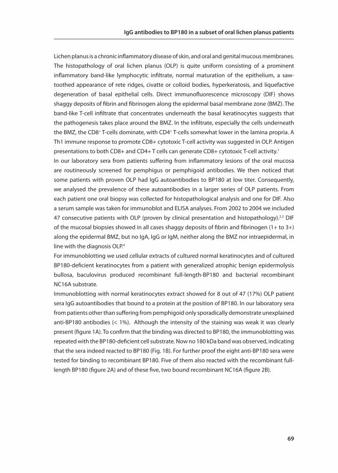
Immunoblotting with normal keratinocytes extract showed for 8 out of 47 (17%) OLP patient
sera IgG autoantibodies that bound to a protein at the position of BP180. In our laboratory sera
from patients other than suffering from pemphigoid only sporadically demonstrate unexplained
anti-BP180 antibodies (< 1%). Although the intensity of the staining was weak it was clearly
present (figure 1A). To confirm that the binding was directed to BP180, the immunoblotting was
repeated with the BP180-deficient cell substrate. Now no 180 kDa band was observed, indicating
that the sera indeed reacted to BP180 (Fig. 1B). For further proof the eight anti-BP180 sera were
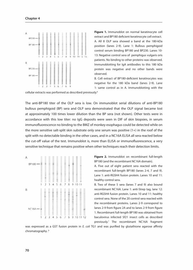
tested for binding to recombinant BP180. Five of them also reacted with the recombinant full-
length BP180 (figure 2A) and of these five, two bound recombinant NC16A (figure 2B).
70
Chapter 4
figure 1. Immunoblot on normal keratinocyte cell extract and BP180 deficient keratinocyte cell extract. A. All 8 OLP sera showed a band at the 180-kDa position (lanes 2-9). Lane 1: Bullous pemphigoid control serum binding BP180 and BP230. Lanes 10-13: Negative control sera of pemphigus vulgaris oris patients. No binding to other proteins was observed. Immunoblotting for IgA antibodies to this 180 kDa protein was negative and no other bands were observed. B. Cell extract of BP180-deficient keratinocytes was negative for the 180 kDa band (lanes 2-9). Lane 1: same control as in A. Immunoblotting with the
cellular extracts was performed as described previously.6
The anti-BP180 titer of the OLP sera is low. On immunoblot serial dilutions of anti-BP180
bullous pemphigoid (BP) sera and OLP sera demonstrated that the OLP signal became lost
at approximately 100 times lower dilution than the BP sera (not shown). Other tests were in
accordance with this low titer: no IgG deposits were seen in DIF of skin biopsies, in serum
immunofluorescence no binding to the BMZ of monkey esophagus could be detected while on
the more sensitive salt-split skin substrate only one serum was positive (1+) in the roof of the
split with no detectable binding in the other cases, and in a NC16A ELISA all sera reacted below
the cut-off value of the test. Immunoblot is, more than ELISA or immunofluorescence, a very
sensitive technique that remains positive when other techniques reach their detection limits.
figure 2. Immunoblot on recombinant full-length BP180 (and the recombinant NC16A domain). A. Five out of eight patient sera reacted with the recombinant full-length BP180 (lanes 2-4, 7 and 9). Lane 1: anti-RGSH4 fusion protein. Lanes 10 and 11: healthy control sera. B. Two of these 5 sera (lanes 7 and 9) also bound recombinant NC16A. Lane 1: anti-Strep tag, lane 12: anti-RGSH4 fusion protein. Lanes 10 and 11: healthy control sera. None of the 20 control sera reacted with the recombinant proteins. Lanes 2-9 correspond to lanes 2-9 from figure 2A and to lanes 2-9 from figure 1. Recombinant full-length BP180 was obtained from baculovirus infected Sf21 insect cells as described previously.7 The recombinant NC16A fragment
was expressed as a GST fusion protein in E. coli TG1 and was purified by glutathione agarose affinity chromatography .8

71
IgG antibodies to BP180 in a subset of oral lichen planus patients
Five of the eight sera were positive for the recombinant full-length BP180 from insect cells.
That not all eight patients reacted with recombinant BP180 may be due to incorrect folding
or incomplete glycolysation of the recombinant protein. Two of the five sera that reacted with
recombinant full-length BP180 also bound the recombinant NC16A domain. While the NC16A
domain is immunodominant in BP, in mucous membrane pemphigoid (MMP) also the carboxy-
terminal domain is targeted. Similarly, in OLP other epitopes than NC16A may be targeted, as we
could only demonstrate immunoblot binding to the NC16A for two of the eight cases.
BP180 is a transmembrane hemidesmosomal molecule, with an elongated ectodomain long
enough to overspan the lamina lucida. It is the most prominent autoimmune target in the
blistering diseases bullous pemphigoid, pemphigoid gestationis, linear IgA dermatosis, mucous
membrane pemphigoid and in lichen planus pemphigoides. What aspects make BP180 to such
a dominant autoantigen is unknown.
The current hypothesis on OLP pathogenesis is that cytotoxic CD8+ T cells, assisted by CD4+
T-cells, destroy basal keratinocytes through triggering apoptosis. It is currently unknown
whether activation of both CD8+ T-cells and CD4+ T-cells occurs through the same antigen or
through two different antigens.1 It is also unknown if the antigen is a self-protein making OLP
to a true autoimmune disease, although OLP appears to have many features that support such
vision. The development of antibodies might be a secondary, humoral, response to an antigen,
in this case BP180, which becomes exposed in the chronic inflammatory process caused by the
primary cellular response. Such a process is known as epitope spreading. Vice versa, humoral
T-cell activity may start the pathogenesis before cytotoxic CD8+ T cells get the advantage.
Recently, Cooper et al. described BMZ-binding IgG antibodies in 61% of patients with clinical
erosive lichen planus of the vulva.5 This IgG was chiefly directed to BP180. In serum laboratory
diagnosis such observations would normally coin the diagnosis of mucous membrane
pemphigoid. Biopsies did not demonstrate BMZ-deposits of IgG. Their biopsies, however, had
been taken from lesional skin, and thus were at risk for becoming negative for potential Ig
deposits as these can be destroyed in the inflammatory process. Also the fibrinogen deposits,
characteristic to lichen planus lesions, were absent in all their biopsies. All our eight patients had
the shaggy deposits of fibrin/fibrinogen along the BMZ typically encountered with lichenoid
infiltrates. Nevertheless it is striking that they, as we, found BP180 autoantibodies in patients
with lichen planus histology.
This study showed that low circulating anti-BP180 IgG titers of unknown function exist in a
minority (17%) of OLP patients. The presence of anti-BP180 antibodies in a subgroup of OLP
patients might be considered a connecting element to the IgG mediated pemphigoid diseases
lichen planus pemphigoides and mucous membrane pemphigoid. If BP180 is also an OLP T-cell
antigen remains elusive.

72
Chapter 4
ACknowLeDgements
This study was supported by grant from J.P. Naterfonds.

73
IgG antibodies to BP180 in a subset of oral lichen planus patients
referenCes
1. Lodi, G., Scully, C., Carrozzo, M., Griffiths, M., Sugerman, P. B., and Thongprasom, K.: Current
controversies in oral lichen planus: report of an international consensus meeting. Part 1.
Viral infections and etiopathogenesis. Oral Surg Oral Med Oral Pathol Oral Radiol Endod
100: 40 51,2005.
2. van der Meij, E. H. and der Waal van, I: Lack of clinicopathologic correlation in the
diagnosis of oral lichen planus based on the presently available diagnostic criteria and
suggestions for modifications. J Oral Pathol Med 32.9: 507 512,2003.
3. Epstein, J. B., Wan, L. S., Gorsky, M., and Zhang, L.: Oral lichen planus: progress in
understanding its malignant potential and the implications for clinical management.
Oral Surg Oral Med Oral Pathol Oral Radiol Endod 96: 32 37,2003.
4. Daniels, T. E. and Quadra-White, C.: Direct immunofluorescence in oral mucosal disease:
a diagnostic analysis of 130 cases. Oral Surg Oral Med Oral Pathol 51: 38 47,1981.
5. Cooper, S. M., Dean, D., Allen, J., Kirtschig, G., and Wojnarowska, F.: Erosive lichen planus
of the vulva: weak circulating basement membrane zone antibodies are present. Clin
Exp Dermatol 30: 551 556,2005.
6. Pas, H. H., Kloosterhuis, G. J., Heeres, K., van der Meer, J. B., and Jonkman, M. F.: Bullous
pemphigoid and linear IgA dermatosis sera recognize a similar 120-kDa keratinocyte
collagenous glycoprotein with antigenic cross-reactivity to BP180. J Invest Dermatol
108: 423 429,1997.
7. Kromminga, A., Scheckenbach, C., Georgi, M., Hagel, C., Arndt, R., Christophers, E., Brocker,
E. B., and Zillikens, D.: Patients with bullous pemphigoid and linear IgA disease show a
dual IgA and IgG autoimmune response to BP180. J Autoimmun 15: 293 300,2000.
8. Zillikens, D., Mascaro, J. M., Rose, P. A., Liu, Z., Ewing, S. M., Caux, F., Hoffmann, R. G., Diaz,
L. A., and Giudice, G. J.: A highly sensitive enzyme-linked immunosorbent assay for the
detection of circulating anti-BP180 autoantibodies in patients with bullous pemphigoid.
J Invest Dermatol 109: 679 683,1997.