Embed Size (px)
Citation preview
D I S T I N C T F U N C T I O N S O F M O N O C L O N A L IgG A N T I B O D Y
D E P E N D O N A N T I G E N - S I T E S P E C I F I C I T I E S
B~¢ WOLFGANG SCHALCH,* J. KEITH WRIGHT, L. SCOTT RODKEY,:[: AND DIETMAR G. BRAUN
From the Basel Institute for Immunology, and the Biocenter of the University of Basel, Postfach, CH-4005 Basel 5, Switzerland
Standard immune responses are the sum of many clonal responses, cross-reactive and specific. Any study of clonal expression as well as analysis of its products requires the separation of clones. There are several approaches to this goal (1-3). The one we used was to generate immune responses of restricted heterogeneity where a limited number ofclonotypes determine the response (4, 5). The prerequisites for the induction of such responses are homogeneity of the immunogen and the genetic background of the animals immunized (I, 4-9). Bacterial cell walls carrying regularly spaced polysaccharide moieties on their surface meet the demand for homogeneity of the antigen (1). Certain breeds of rabbits and inbred mouse strains fulfill the genetic requirements (4-9). Considering both of these conditions antibody responses of predictable clonal patterns can be elicited (10).
Although most of the early work emphasized the restricted heterogeneity of anti- polysaccharide antibody responses (1, 4-9) we report here in detail that clonal IgG antibody patterns in response to polysaccharide determinants are rather complex. Despite this complexity it is possible to isolate, by a combination of methods, anti- streptococcal group A-variant antibodies in single-band purity (11). In using these antibodies, distinct functions of monoclonal IgG antibody could be ascribed to fine specificities for different determinants on a linear polysaccharide chain.
Mater ia l and Me thods
Rabbit Hyperimmune Antisera and Antigen Preparations. The production ofhyperimmune,antisera in selectively bred rabbits to the streptococcal group A-variant polysaccharide (Av-CHO) 1 and the identification and quanti tat ion of predominant clonotypes has been described previously (5).
The A v - C H O was purified from K43 streptococcal cell walls with >98% purity (12, 13). Aliquots were labeled for radioimmunoassays with 12sI or lalI (5). For coating sheep erythrocytes, (SRBC) the O-stearoyl-ester of Av-CHO was made (14).
* Recipient of EMBO Long Term Fellowship. :~ Recipient of a National Institutes of Health Research Career Development award 1-K04-AI00217.
Contribution 79-127-J, Division of Biology, Kansas Agricultural Experiment Station, Manhattan, KS. Present address: Division of Biology, Kansas State University, Manhattan, KS 66506.
1 Abbreviations used in this paper." Aggl, agglutination; Av-CHO, streptococcal group A-variant polysaccha- ride; Av-CHO-SRBC, sheep erythrocytes coated with the O-stearoyl-ester of Av-CHO; CD, circular dichroism; CdL, complement-dependent lysis; DNP-Av-CHO, 2,4-dinitrophenyl-hydrazone of Av-CHO; DTT, dithio-threitol; 12SI-Av-CHO, talI-Av-CHO, Av-CHO labeled in its tyraminated form with 1251 or lalI; IEF, isoelectric focusing; KD, dissociation constant; PBS, 0.01 M sodium potassium phosphate; pIEF, preparative isoelectric focusing; Ppt, precipitation.
J. Exp. MED. © The Rockefeller University Press • 0022-1007/79/04/0923/15 $1.00 923 Volume 149 April 1979 923-937
Dow
nloaded from http://rupress.org/jem
/article-pdf/149/4/923/1089913/923.pdf by guest on 07 February 2022
924 ANTIBODY FINE SPECIFICITY AND FUNCTION
Preparation of 2,4 Dinitrophenylhydrazone of the Av-CHO. The method of Lloyd and Doherty was used for the preparation of this derivative (15). From 55 mg of starting material, 25 mg of 2,4-dinitrophenyl-hydrazone of Av-CHO (DNP-Av-CHO) were obtained.
Circular Dichroism Spectroscopy. Circular dichroism (CD) spectra of DNP-Av-CHO antibody complexes were measured in a Cary 60 instrument (Monrovia, Calif,) rebuilt for CD measure- ments and equipped with a Jobin-Yon modulator (Instruments SA, Longjumeau, France), using 1-cm quartz cells thermostated at 20°C. Otherwise, conditions were those described previously (16).
Affinity Chromatography. Av-CHO affinity columns were prepared by using epoxy-activated Sepharose 6B, Pharmacia Fine Chemicals, Uppsala, Sweden (17). Bound antibody was eluted by 0.15 N NaCI in 0.1 M glycine-HCl, pH 3.0.
Analytical lsoelectric Focusing. Analytical isoelectric focusing (IEF) was performed in 5% polyacrylamide gels, pH 5-10, (18). Patterns of antibodies were visualized both by staining with bromophenol blue and by autoradiography using the 131I-labeled Av-CHO (19).
Preparative Electrophoresis and Preparative IEF. Antibody was purified from whole immune sera first by preparative agarose block electrophoresis (4), and then by one or more runs of pIEF in horizontal layers (11).
Papain Digestion. Purified antibodies were digested by 2-fl-mercaptoethanol activated pa- pain (Sigma Chemical Co., St. Louis, Mo.), and Fab monomers were isolated (20).
Affinity Measurements. Binding of the Av-CHO by antibody from whole sera or purified fractions was determined by the quenching of antibody tryptophan fluorescence (21).
Equilibrium binding studies of x25I-labeled Av-CHO to antibody were performed with plastic mierocells of 200 #1 capacity (22). Glucose (5 mM in phosphate-buffered saline [PBS]) was added to eliminate weak unspecific binding of the Av-CHO to the dialysis tubing which had an average exclusion limit of 17,000 dahons. Under this condition equilibrium was complete after 32 h at 25°C. The specific radioactivity of hapten and antigen in these experiments varied from 11-45 cpm/nmol. Samples were counted in a Packard Gamma spectrometer (model 5130) (Packard Instrument Co., Inc., Downers Grove, Ill.). Dissociation constants (KD) were obtained for the stoichiometry of ligand binding from replots of the data according to Scatchard (23).
Ultracentrifugation Analysis. Sedimentation experiments were performed in a Beckman model E analytical ultracentrifuge (Beckman Instruments, Inc., Fullerton, Calif.). The weight-average molecular weights were calculated employing a value of 0.73 cm3/g for the partial specific volume of antibody and antigen-antibody complexes (24).
Immunochemical Methods. Quantitative precipitin and inhibition analyses as well as binding of the laSI-Av-CHO by antibody was performed as described (6, 9). Inhibition studies of precipitation and binding analyses were conducted by addition of increasing amounts of L- rhamnose or a synthesized probe of L-rhamnosyl a-1 ---, 2-L-rhamnose (25). Double-diffusion analyses in gels were performed according to established methods (26).
Agglutination of O-stearoyl-ester Av-CHO-coated SRBC (Av-CHO-SRBC) was performed in microtiter plates (14). Complement-dependent lysis (CdL) was determined qualitatively by the spot assay (27) and quantitatively by reacting whole inactivated (56°C 30 min.) SRBC absorbed rabbit anti-Av-CHO antisera or purified antibody fractions in the presence of 0.1 ml 1:10 PBS diluted complement solution (guinea pig complement, Behring-Werke AG, Marburg/ Lahn, West Germany). Av-CHO-SRBC and SRBC (OD, 540 nm = 0.4) were used in final test volumes of 1.1 ml at 37°C for 60 min. Lysis was expressed as a percent against water-lysed Av- CHO-SRBC and SRBC controls in supernates. These experiments were also performed in the presence of 10 mM dithio-threitol (DTT), Clelands reagent, Calbiochem for two low affinity antibody fractions of single-band purity (28).
Competition experiments between lytic and nonlytic antibodies for Av-CHO sites were performed at 10, 50, and 75% lysis in the CdL assay using increasing amounts (up to 10-fold molar excess) of nonlytic Av-CHO specific antibody.
Binding of Av-CHO specific antibodies to Av-CHO-SRBC was determined qualitatively and quantitatively. Two preparations of Av-CHO specific antibody fractions of essentially single-band purity were used, the high affinity fraction K 151-748II and the low affinity fraction K127-760II. Qualitatively, binding was determined by incubating 10 fig of antibody with 100
Dow
nloaded from http://rupress.org/jem
/article-pdf/149/4/923/1089913/923.pdf by guest on 07 February 2022
SCHALCH, WRIGHT, RODKEY, AND BRAUN 925
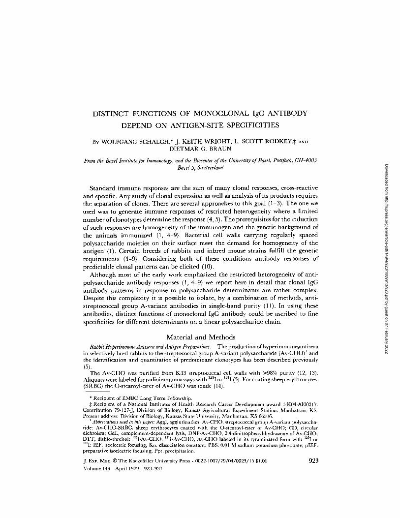
Fro. 1. Analytical isoelectric focusing (A and B) of 10/~1 of 16 rabbit hyperimmune anti-A-variant carbohydrate (Av-CHO) antisera. A. Pattern develo'pment with bromophenolblue. The heavy band at pH 5.9 corresponds to rabbit serum albumin. Bands cathodal of the albumin represent mainly IgG antibody. B. Pattern development by binding of 131I-Av-CHO and subsequent autoradiography. Antibody responses of restricted heterogeneity (samples No. 7, 10, 12, 14, and 16), clear-cut from the pattern developed for protein concentration, are lost on the autoradiogram as a result of the overwhelming background noise of heterogeneity.
pl of 10% Av-CHO-SRBC and 100 #1 of 10% SRBC as control, respectively, followed by three rapid washes in cold PBS, and subsequent treatment with a fluoresceine-conjugated goat anti- rabbit IgG immunoglobulin fraction.
Quantitatively, binding of antibody was determined as follows: using the chloramine T method (29), K151-748II was labeled with 125I (sp act 52 #Ci/mg) and K127-760II with 1311 (sp act 110 pCi/mg). To a constant number of isotope counts (corresponding to 128 and 113 ng of antibody per test, respectively), increasing amounts of Av-CHO-SRBC and SRBC were added, in a constant final volume of 50 #1 PBS containing 0.1% bovine serum albumin. Av-CHO- SRBC and SRBC treated in this fashion for 12 h at 4°C were either washed three times in PBS or layered on top o fa 1-cm high fetal calf serum solution in conical polypropylene tubes. These were centrifuged quickly (5 min) to the bottom of the tube, then snap frozen in dry ice and the tips of the tubes cut off. The tips and supernates were counted (30).
R e s u l t s
Clonal Restriction of Anti-Av-CHO Antibodies is a Quantitative Trait. High resolut ion analysis of 40 rabb i t h y p e r i m m u n e a n t i - A v - C H O ant isera by IEF revealed s imple spec t ro type pa t te rns (two to three b a n d pat terns) when s ta ined with b romopheno l b lue (Fig. 1 A), conf i rming the ra ther restr ic ted heterogenei ty evidenced by microzone e lec t rophoret ic analysis (samples 7, 10-12, and 16). When , however, pa t t e rns were visual ized by b ind ing o f I~II-Av-CHO (Fig. 1 B) accord ing to Keck et al. (18) very complex spec t ro type pa t te rns emerged with up to 75 bands , cor responding to 20-30 clonotypes (31). T h e d o m i n a t i n g clonotypes were no longer ident i f ied as such but
Dow
nloaded from http://rupress.org/jem
/article-pdf/149/4/923/1089913/923.pdf by guest on 07 February 2022
926 ANTIBODY FINE SPECIFICITY AND FUNCTION
TABLE I
Concentration* and Functional Properties of Anti-Av-CHO Antibody (Ab) in Rabbit Hyperimmune hera
Rabbit No. Low affinity (pH 7.0) Ab High affinity (pH 3.0) Ab
mg/ml Percent Ppt. Aggl. CdL mg/ml Percent Ppt. Aggl. CdL
1. K6-137 28.1 93.4 - - - 2.0 6.6 + + + 2. K33-380 22.9 90.6 - - - 2.4 9.4 + + + 3. K34-413 13.2 94.3 - - - 0.8 5.7 + + + 4. K45-426 36.1 97.3 - - - 1.0 2.7 + + + 5. K49-501 30.2 97.1 - - - 0.9 2.9 + + + 6. K81-543 32.2 92.0 - - - 2.8 8.0 + + + 7. K81-545 24,8 89.5 - - - 2.9 10.5 + + + 8. K116-700 29.3 84.9 - - - 5.2 15.l + + + 9. Kl33-730 37.1 97.6 - - - 0.9 2.4 + + +
10. K133-732 15.4 92.8 - - - 1.2 7.2 + + + 11. K151-748 31.8 97.2 - - - 0.9 2.8 + + + 12. K128-755 49.6 99.0 - - - 0.5 1.0 + + + 13. K127-760 51.7 99.2 - - - 0.4 0.8 + + + 14. K155-772 11.7 90.7 - - - 1.2 9.3 + + + 15. K155-776 13.0 89.0 - - - 1.6 11.0 + + +
* Concentrations of Av-CHO-specific antibody determined by the Farr assay (9) in pH 7.0 and pit 3.0 antibody fractions from Av-CHO-epoxy-Sepharose affinity columns. Ppt, precipitating activity with the hot formamide extracted Av-CHO at concentrations of 50 and 100 ~g/ml, determined by the Lancefield precipitation test. Aggl., agglutination of Av-CHO-SRBC.
r e p l a c e d b y o t h e r b a n d s (Fig. 1). F u r t h e r , t he p a t t e r n a p p e a r a n c e o f d o m i n a n t
c l o n o t y p e s was o f t e n c o m p l i c a t e d b y m i n o r b a n d s b e t w e e n or a l m o s t o v e r l a p p i n g
w i t h these . S t r o n g a n t i g e n b i n d i n g in pos i t i ons w i t h n o or ve ry l i t t l e p r o t e i n by
s t a i n i n g s u g g e s t e d s i g n i f i c a n t d i f f e rences in b i n d i n g a f f in i ty for A v - C H O .
Distribution of Affinities in Av-CHO-Specific Antisera. A g a r o s e b l o c k - p u r i f i e d I g G f rom
r a b b i t a n t i - A v - C H O a n t i s e r a y i e lded b i p h a s i c S c a t c h a r d p lo t s b y a n t i b o d y f luores-
cence q u e n c h i n g ana lys i s w i t h u p p e r a n d lower KD l imi t s o f 10 -6 a n d 0.9 × 10 .9 M ,
respec t ive ly , s u g g e s t i n g t h e p r e s e n c e of m a i n l y two d i s t i n c t l y d i f f e r en t a n t i b o d y
p o p u l a t i o n s . T o g e n e r a l i z e th i s f i n d i n g , 15 r a b b i t a n t i - A v - C H O a n t i s e r a were sepa-
r a t e d b y A v - C H O S e p h a r o s e - a f f i n i t y c h r o m a t o g r a p h y i n t o a p H 7.0 a n d a p H 3.0
f rac t ion . T h e c o n c e n t r a t i o n o f A v - C H O b i n d i n g a n t i b o d y o f b o t h f r a c t i o n s was
e s t i m a t e d b y a m o d i f i e d F a r r t e c h n i q u e (9), a n d it was c o m p a r e d to t he c o n c e n t r a t i o n
o f A v - C H O - s p e c i f i c a n t i b o d y in t h e w h o l e a n t i s e r a ( T a b l e I). T h i s c o m p a r i s o n
r e v e a l e d 8 5 - 9 9 % of t h e A v - C H O b i n d i n g a n t i b o d y to be c o n t a i n e d in t h e p H 7.0
f r a c t i o n a l o n g w i t h t h e d o m i n a n t s p e c t r o t y p e b a n d s . B o t h t h e p H 7.0 a n d t h e p H 3.0
f r ac t i ons r e v e a l e d l i n e a r b i n d i n g curves , i n d i c a t i n g h o m o g e n e i t y o f b i n d i n g ( T a b l e
II).
S u b s e q u e n t m e a s u r e m e n t s o f b i n d i n g a f f in i t i es we re p e r f o r m e d w i t h low a f f in i ty
a n t i b o d y f r ac t i ons o f s i n g l e - b a n d p u r i t y . T h e s e S c a t c h a r d p lo t s a t t e s t e d f u n c t i o n a l
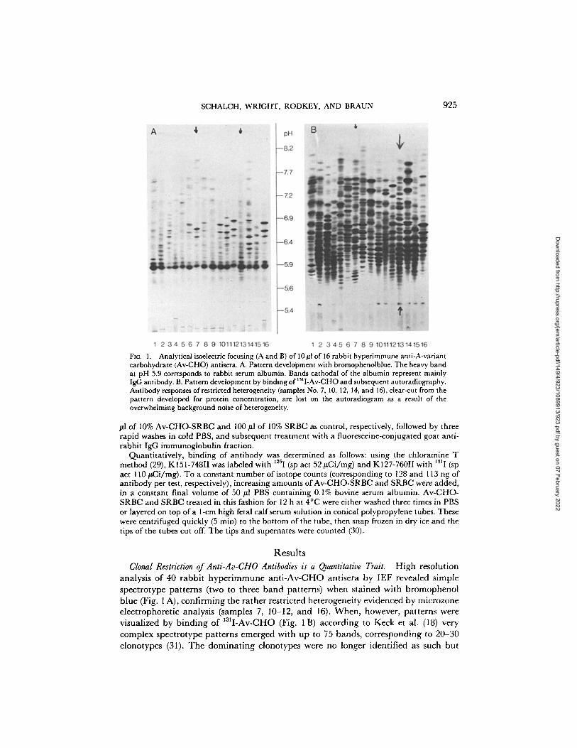
h o m o g e n e i t y Fig. 2 A - C .
A n t i b o d i e s e l u t i n g f r o m A v - C H O - e p o x y - S e p h a r o s e c o l u m n s a t p H 3.0 s h o w e d
a f f in i t i es t w o to t h r e e o rde r s o f m a g n i t u d e h i g h e r (KD < 10 - s M ) t h a n t h e low a f f in i ty
a n t i b o d i e s (Fig. 2 D) , t h e i r d i s s o c i a t i o n c o n s t a n t s a re s u m m a r i z e d in T a b l e II.
Fine Specificities of Anti-Av-CHO Antibodies. T h e A v - C H O is a l i n e a r h o m o p o l y m e r
o f L - r h a m n o s e w i t h a l t e r n a t i n g a l ~ 2 a n d a l ~ 3 g lycos id ic b o n d s (32). T h e
Dow
nloaded from http://rupress.org/jem
/article-pdf/149/4/923/1089913/923.pdf by guest on 07 February 2022
SCHALCH, WRIGHT, RODKEY, AND BRAUN 927
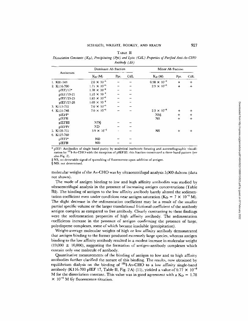
TABLE II
Dissociation Constants (KD), Precipitating (Ppt) and Lytic (CdL) Properties of Pur~ed Anti-Av-CHO Antibody (Ab)
Antiserum Dominant Ab fraction Minor Ab fraction
KD (M) Ppt. CdL Ko (M) Ppt. CdL
1. K81-543 2. Kl16-700
pIEF/17* pIEF/19-21 pIEF/23-25 pIEF/27-28
3. K113-732 4. K151-748
pIEFI* pIEFII pIEFIII pIEFIV
5. K128-755 6. K127-760
pIEFI* pIEFII
2.0 X 10 -~ 1.71 X 10 -6 1.78 X 10 -6 1.53 X 10 -6 1.83 X 10 -6 1.69 X 10 .6 7.0 x 10 -5 7.0 x 10 -6
ND§ ND
3.9 X 10 -5
ND ND
0.98 x 10 -9 + .4. 2.9 x 10 -s + +
2.3 X 10 .9 + +
NS~: + + NS + +
NS + +
* pIEF: Antibodies of single band purity by analytical isoelectric focusing and autoradiographic visuali- zation by 1311-Av-CHO with the exception of pIEFIII; this fraction constituted a three-band pattern (see also Fig. 2).
:~ NS, no detectable signal of quenching of fluorescence upon addition of antigen. § ND, not determined.
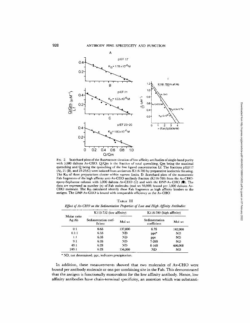
molecular weight of the Av-CHO was by ultracentrifugal analysis 5,000 daltons (data not shown).
The mode of antigen binding to low and high affinity antibodies was studied by ultracentrifugal analysis in the presence of increasing antigen concentrations (Table III). The binding of antigen to the low affinity antibody barely altered the sedimen- tation coefficient even under conditions near antigen saturation (KD ---- 7 × i0 -~ M). The slight decrease in the sedimentation coefficient may be a result of the smaller partial specific volume or the larger translational frictional coefficient of the antibody antigen complex as compared to free antibody. Clearly contrasting to these findings were the sedimentation properties of high affinity antibody. The sedimentation coefficients increase in the presence of antigen confirming the presence of large, polydisperse complexes, some of which became insoluble (precipitation).
Weight-average molecular weights of high or low affinity antibody demonstrated that antigen binding to the former produced extremely large species, whereas antigen binding to the low affinity antibody resulted in a modest increase in molecular weight (19,000 + 10,000), suggesting the formation of antigen-antibody complexes which contain only one molecule of antibody.
Quantitative measurements of the binding of antigen to low and to high affinity antibodies further clarified the nature of this binding. The results, now obtained by equilibrium dialysis on the binding of 125I-Av-CHO to a low affinity single-band antibody (K116-700 pIEF 17, Table II, Fig. 2A) (11), yielded a value of 0.77 × 10 -6 M for the dissociation constant. This value was in good agreement with a KD = 1.78 X 10 -6 M I~y fluorescence titration.
Dow
nloaded from http://rupress.org/jem
/article-pdf/149/4/923/1089913/923.pdf by guest on 07 February 2022
928 ANTIBODY FINE SPECIFICITY AND FUNCTION
A
' 7 plEF 17 0 . 4 j ~ ~,~o"4"- • K D" 1.78 x 10⁻⁶M
o~-I "%-. .~. t , . . . . . . ,".-.-.,.
B
Era plEF21 o.q ::I. oL KD= 1.53x10.6 M ~ 0.8'
' • --~ 0.4" n i
C 0.2"
o.J~_ °'~ ~3-~ ~ - . - . . . ~ ~o- 1.83x,o-°~
o2-1 ""--,..,>... ] , . . . . . ", :--.>, 0 0.2 0.4 0.6 0.8 1.0
Q/Qm
D
1.2- , K116-700hi aff.Ab
1'0" ~ ~ o ~
°Xt(o.2.9,,~o-~M KO" 3xlO*SXX
1 2 3 4 n (Fab/5,0OOM W)
FXG. 2. Scatchard plots of the fluorescence titration of low affinity antibodies of single-band purity with 5,000 daltons Av-CHO. Q/Qm is the fraction of total quenching, Qm being the maximal quenching and Q being the quenching of the free ligand concentration Lf. The fractions pIEFI 7 (A), 21 (B), and 23-25(C) were isolated from antiserum K116-700 by preparative isoelectric focusing. The KD of these preparations cluster within narrow limits. D. Scatchard plots of the monomeric Fab fragments of the high affinity anti-Av-CHO antibody fraction (K116-700) from the Av-CHO- epoxy-Sepharose column with 5,000 daltons Av-CHO (O) and with the DNP-Av-CHO (0). The data are expressed as number (n) of Fab molecules (mol wt 50,000) bound per 5,000 daltons Av- CHO molecule. The Ko calculated identify these Fab fragments as high affinity binders to the antigen. The DNP-Av-CHO is bound with comparable efficiency as the Av-CHO.
TABLE III
Effect of Av-CHO on the Sedimentation Properties of Low and High Affinity Antibodies
Molar ratio Ag:Ab
K113-732 (low affinity) K116-700 (high affinity)
Sedimentation coef- Sedimentation Mol wt Mol wt ficient coefficient
0:1 6.6S 137,000 6.7S 142,000 0 . 1 : 1 6.5S ND ppt* ND
1 : 1 6.3S ND ppt ND 9:1 6.3S ND 7-28S ND
49:1 6.2S ND 8-16S 400,000 249:1 6.2S 156,000 ND ND
* ND, not determined; ppt, indicates precipitation.
I n a d d i t i o n , t he se m e a s u r e m e n t s s h o w e d t h a t two m o l e c u l e s o f A v - C H O were
b o u n d p e r a n t i b o d y m o l e c u l e o r o n e p e r c o m b i n i n g s i te in t h e F a b . T h i s d e m o n s t r a t e d
t h a t t h e a n t i g e n is f u n c t i o n a l l y m o n o v a l e n t for t h e low a f f in i ty a n t i b o d y . H e n c e , low
a f f i n i t y a n t i b o d i e s h a v e c h a i n - t e r m i n a l spec i f ic i ty , a n a s s e r t i o n w h i c h was s u b s t a n t i -
Dow
nloaded from http://rupress.org/jem
/article-pdf/149/4/923/1089913/923.pdf by guest on 07 February 2022
SCHALCH, WRIGHT, RODKEY, AND BRAUN 929
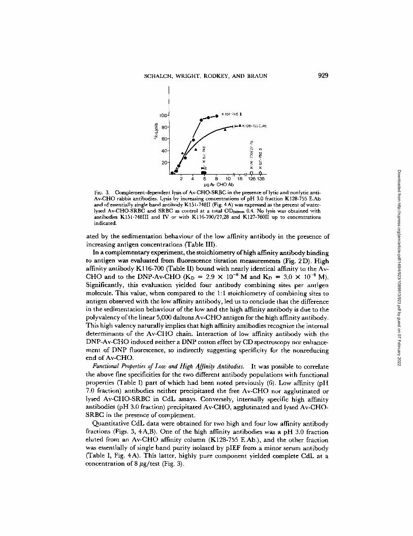
i K 151-748 100 0~.0...0 ~9
40- ~
20- ~
, ~ , , t ; ,n ~,
tag Av- CHO Ab
FI~. 3. Complement-dependent lysis of Av-CHO-SRBC in the presence of lytic and nonlytic anti- Av-CHO rabbit antibodies. Lysis by increasing concentrations of pH 3.0 fraction K128-755 E.Ab and of essentially single band antibody K 151-748II (Fig. 4 A) was expressed as the percent of water- lysed Av-CHO-SRBC and SRBC as control at a total OD~om~ 0.4. No lysis was obtained with antibodies K151-748III and IV or with K116-700/27,28 and K127-760II up to concentrations indicated.
ated by the sedimentation behaviour of the low affinity antibody in the presence of increasing antigen concentrations (Table III).
In a complementary experiment, the stoichiometry of high affinity antibody binding to antigen was evaluated from fluorescence titration measurements (Fig. 2 D). High affinity antibody K116-700 (Table II) bound with nearly identical affinity to the Av- CHO and to the DNP-Av-CHO (KD = 2.9 × 10 - s M a n d K D ---- 3.0 X 10 -sM). Significantly, this evaluation yielded four antibody combining sites per antigen molecule. This value, when compared to the 1:1 stoichiometry of combining sites to antigen observed with the low affinity antibody, led us to conclude that the difference in the sedimentation behaviour of the low and the high affinity antibody is due to the polyvalency of the linear 5,000 daltons Av-CHO antigen for the high affinity antibody. This high valency naturally implies that high affinity antibodies recognize the internal determinants of the Av-CHO chain. Interaction of low affinity antibody with the DNP-Av-CHO induced neither a DNP cotton effect by CD spectroscopy nor enhance- ment of DNP fluorescence, so indirectly suggesting specificity for the nonreducing end of Av-CHO.
Functional Properties of Low and High Affinity Antibodies. It was possible to correlate the above fine specificities for the two different antibody populations with functional properties (Table I) part of which had been noted previously (6). Low affinity (pH 7.0 fraction) antibodies neither precipitated the free Av-CHO nor agglutinated or lysed Av-CHO-SRBC in CdL assays. Conversely, internally specific high affinity antibodies (pH 3.0 fraction) precipitated Av-CHO, agglutinated and lysed Av-CHO- SRBC in the presence of complement.
Quantitative CdL data were obtained for two high and four low affinity antibody fractions (Figs. 3, 4A,B). One of the high affinity antibodies was a pH 3.0 fraction eluted from an Av-CHO affinity column (K128-755 E.Ab.), and the other fraction was essentially of single band purity isolated by pIEF from a minor serum antibody (Table I, Fig. 4A). This latter, highly pure component yielded complete CdL at a concentration of 8/tg/test (Fig. 3).
Dow
nloaded from http://rupress.org/jem
/article-pdf/149/4/923/1089913/923.pdf by guest on 07 February 2022
930 ANTIBODY FINE SPECIFICITY AND FUNCTION
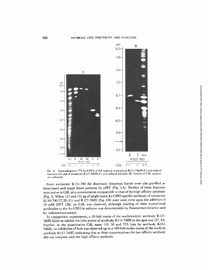
131 Flu. 4. Autoradiograms ( I-Av-CHO) of IEF analysis of antiserum KI57-748(W.S.) and isolated fractions (A) and of antiserurn K127-760('¢(.S.) and isolated fractions (B). Results of CdL analyses are indicated.
From antiserum K151-748 the dominant clonotype bands were also purified as three-band and single band patterns by pIEF (Fig. 4A). Neither of these fractions were active in CdL at a concentration comparable to that of the high affinity antibody (Fig. 3). When 125 and 135 ~g of single-band Av-CHO-specific antibody of antiserum K116-700/27,28 (11) and K127-760II (Fig. 4B) were used, even upon the addition of 10 m M D T T (28), no CdL was observed, although binding of these monoclonal antibodies to the Av-CHO in solution was demonstrable by fluorescence titration and
by radioimmunoassays. In competition experiments, a 20-fold excess of the nonhemolytic antibody K127-
760II failed to inhibit the lyric action of antibody K151-748II in the spot test (27, 33). Further, in the quantitative CdL assay (10, 50 and 75% lysis by antibody K151- 748II), no inhibition of lysis was observed up to a 100-fold molar excess of the nonlytic antibody K127-760II, indicating that at these concentrations the low affinity antibody did not compete with the high affinity antibody.
Dow
nloaded from http://rupress.org/jem
/article-pdf/149/4/923/1089913/923.pdf by guest on 07 February 2022
SCHALCH, WRIGHT, RODKEY, AND BRAUN 931
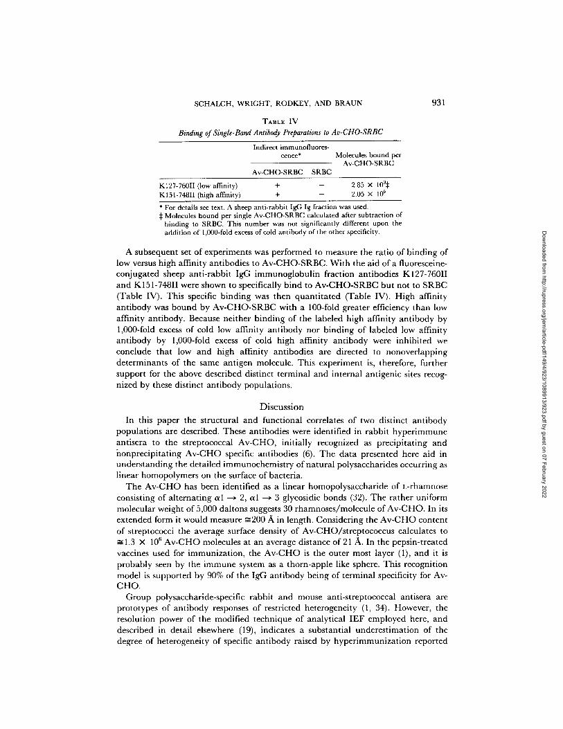
TABLE IV Binding of Single-Band Antibody Preparations to Av-CHO-SRBC
Indirect immunofluores- cence* Molecules bound per
Av-CHO-SRBC Av-CHO-SRBC SRBC
K127-760II (low affinity) + - 2.85 × 10a:~ K151-748II (high affinity) + - 2.05 × 10 s
* For details see text. A sheep anti-rabbit IgG Ig fraction was used. :~ Molecules bound per single Av-CHO-SRBC calculated after subtraction of
binding to SRBC. This number was not significantly different upon the addition of 1,000-fold excess of cold antibody of the other specificity.
A subsequent set of experiments was performed to measure the ratio of binding of low versus high affinity antibodies to Av-CHO-SRBC. With the aid of a fluoresceine- conjugated sheep anti-rabbit IgG immunoglobulin fraction antibodies K127-760II and K151-748II were shown to specifically bind to Av-CHO-SRBC but not to SRBC (Table IV). This specific binding was then quantitated (Table IV). High affinity antibody was bound by Av-CHO-SRBC with a 100-fold greater efficiency than low affinity antibody. Because neither binding of the labeled high affinity antibody by 1,000-fold excess of cold low affinity antibody nor binding of labeled low affinity antibody by 1,000-fold excess of cold high affinity antibody were inhibited we conclude that low and high affinity antibodies are directed to nonoverlapping determinants of the same antigen molecule. This experiment is, therefore, further support for the above described distinct terminal and internal antigenic sites recog- nized by these distinct antibody populations.
Discussion
In this paper the structural and functional correlates of two distinct antibody populations are described. These antibodies were identified in rabbit hyperimmune antisera to the streptococcal Av-CHO, initially recognized as precipitating and honprecipitating Av-CHO specific antibodies (6). The data presented here aid in understanding the detailed immunochemistry of natural polysaccharides occurring as linear homopolymers on the surface of bacteria.
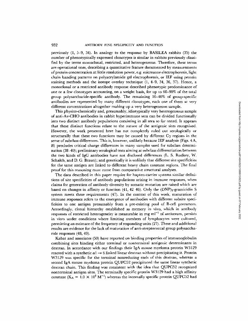
The Av-CHO has been identified as a linear homopolysaccharide of L-rhamnose consisting of alternating a l --o 9, a l --o 3 glycosidic bonds (32). The rather uniform molecular weight of 5,000 daltons suggests 30 rharnnoses/molecule of Av-CHO. In its extended form it would measure ~200 A in length. Considering the Av-CHO content of streptococci the average surface density of Av-CHO/streptococcus calculates to
1.3 × 10⁶ Av-CHO molecules at an average distance of 21 A. In the pepsin-treated vaccines used for immunization, the Av-CHO is the outer most layer (1), and it is probably seen by the immune system as a thorn-apple like sphere. This recognition model is supported by 90% of the IgG antibody being of terminal specificity for Av- CHO.
Group polysaccharide-specific rabbit and mouse anti-streptococcal antisera are prototypes of antibody responses of restricted heterogeneity (1, 3,t-). However, the resolution power of the modified technique of analytical IEF employed here, and described in detail elsewhere (19), indicates a substantial underestimation of the degree of heterogeneity of specific antibody raised by hyperimmunization reported
Dow
nloaded from http://rupress.org/jem
/article-pdf/149/4/923/1089913/923.pdf by guest on 07 February 2022
932 ANTIBODY FINE SPECIFICITY AND FUNCTION
previously (1, 5-9, 34). In analogy to the response by BASILEA rabbits (35) the number of phenotypically expressed clonotypes is similar in rabbits previously classi- fied by the terms monoclonal, restricted, and heterogeneous. Therefore, these terms are operational ones, describing a quantitative feature documented by measurements of protein concentration at little resolution power, e.g. microzone electrophoresis, light chain banding patterns on polyacrylamide gel electrophoresis, or IEF using protein staining methods and the isotope overlay technique (1, 4-9, 34, 36, 37). Hence, a monoclonal or a restricted antibody response described phenotypic predominance of one or a few clonotypes accounting, on a weight basis, for up to 60-90% of the total group polysaccharide-specific antibody. The remaining 10-40% of group-specific antibodies are represented by many different clonotypes, each one of them at very different concentrations altogether making up a very heterogeneous sample.
This physico-chemically and, presumably, idiotypically very heterogeneous sample of anti-Av-CHO antibodies in rabbit hyperimmune sera can be divided functionally into two distinct antibody populations coexisting in all sera so far tested. It appears that these distinct functions relate to the nature of the antigenic sites recognized. However, the work presented here has not completely ruled out serologically or structurally that these two functions may be caused by different Cy regions in the sense of subclass differences. This is, however, unlikely because IEF analysis (Figs. 4 A, B) precludes critical charge differences in many samples used for subclass determi- nation (38-40); preliminary serological tests aiming at subclass differentiation between the two kinds of IgG antibodies have not disclosed differences (L. S. Rodkey, W. Schalch, and D. G. Braun); and genetically it is unlikely that different site specificities for the same antigen are linked to different heavy chain constant regions. The final proof for this reasoning must come from comparative structural analyses.
The data described in this paper require for hapten-carrier systems similar defini- tions of site specificities of antibody populations arising in immune responses, when claims for generation of antibody diversity by somatic mutation are raised which are based on changes in affinity or function (41, 42-46). Only the (DNP)2-gramicidin S system meets these requirements (47). In the context of this work, maturation of immune responses refers to the emergence of antibodies with different subsite speci- ficities to one antigen presumably from a pre-existing pool of B-cell precursors. Accordingly, clonal hierarchy established as memory in vivo, which in antibody responses of restricted heterogeneity is measurable in mg ml -~ of antiserum, persists in vitro under conditions where limiting numbers of lymphocytes were cultured, permitting an estimate of the frequency of responding units (27). These and additional results are evidence for the lack of maturation of anti-streptococcal group polysaccha- ride responses (48, 49).
Kabat and associates (50) have reported on binding properties of immunoglobulin combining sites binding either terminal or nonterminal antigenic determinants in dextran. In accordance with our findings their IgA mouse myeloma protein W3129 reacted with a synthetic a I ---* 6 linked linear dextran without precipitating it. Protein W3129 was specific for the terminal nonreducing ends of this dextran, whereas a second IgA mouse myeloma protein QUPC52 precipitated the same linear synthetic dextran chain. This finding was consistent with the idea that QUPC52 recognized nonterminal antigen sites. The terminally specific protein W3129 had a high affinity constant (KA = 1.0 × 10⁵ M -1) whereas the internally specific protein QUPC52 had
Dow
nloaded from http://rupress.org/jem
/article-pdf/149/4/923/1089913/923.pdf by guest on 07 February 2022
SCHALGH, WRIGHT, RODKEY, AND BRAUN 933
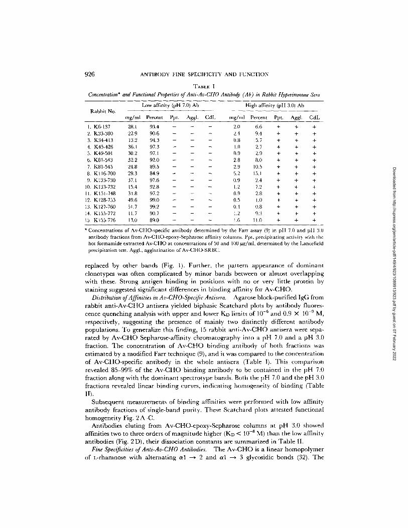
Cell wall I I
[L-Rh Q1.2- L. Rh nl,3-]15
FIc. 5. Model of the group A-variant streptoe~cal cell used in vaccines (to the right). Given are the calculated figures for Av-CHO molecules and surface area. To the left is schematically depicted an enlarged area of the Av-CHO projections, along with the terminal and internal determinants as they may be recognized by their respective antibodies depicted in analogous dimensions. Av-CHO 1% ~ 1.3 X 10 6 molecules/cell (5.3 ~m ~)
a low association constant (KA ---- 8.4 × 10⁸ M-l). In marked contrast to this situation are the internally specific anti-Av-CHO antibodies. The remarkable high affinity for anti-polysaccharide antibodies of KD < I0 -s M may be explained by hydrophobic interactions established via the C6 methyl groups. Chemically these methyl groups are the notable differences in comparing the Av-CHO and the dextran structures. The association constants of the two terminally specific antibodies W3129 and the low affinity anti-Av-CHO antibodies are very close, indicating similar kinds of interactions.
Immunization with cell surface-associated antigenic determinants (in this case of bacteria) may generally elicit a mixture of antibody molecules with very different, nonoverlapping site specificities for linear polysaccharide antigens. The Av-CHO system cogently suggests a critical role of simple mechanistic principles in antigen recognition (schematically shown in Fig. 5), where the most abundantly presented antigenic determinants are the nonreducing ends of single rhamnosyl chains. As a result, antibody of this specificity accounts entirely for dominant clonotypes. Con- versely, the high affinity fraction of antibodies is not preferentially selected, as would be predicted by the maturation argument, and the mechanistic model of antigen presentation (Fig. 5) easily explains this failure. These data as well as those obtained with nonprecipitating equine antibodies of high affinity to the p-azophenyl-fl-lactoside (Lac) haptenic group coupled to protein carrier molecules rule out univalence and low affinity as explanations for functional differences (51). Because mild reduction by 10 mM DTT did not convert the low affinity anti-Av-CHO antibodies into lytic ones, these antibodies do not correspond structurally to the incomplete anti-Rh antibodies (28). Depending on the functional properties of anti-Av-CHO antibodies residing in the determinant specificity for this linear poly-rhamnosyl moiety, the associative model of complement activation and not the allosterie model appears to account for the phenomenon (52).
Dow
nloaded from http://rupress.org/jem
/article-pdf/149/4/923/1089913/923.pdf by guest on 07 February 2022
934 ANTIBODY FINE SPECIFICITY AND FUNCTION
A final comment relates to the high degree of purity of the terminally and internally specific anti-Av-CHO antibodies used in this study. Although this is the highest degree of purity achieved for elicited antibodies functional criteria must additionally be considered. Single band patterns or triplets of bands upon IEF may still contain antibodies of two distinct functional properties9 This caution is critical because analytical IEF is often considered to be the most stringent physico-chemical method to prove structural homogeneity of antibodies.
S u m m a r y
Intraveneous hyperimmunization of selectively bred rabbits with streptococcal group A-variant vaccines elicits antibody responses of restricted heterogeneity at high antibody levels. All antisera contain two functionally distinct antibody populations, which can be isolated in single-band purity upon analytical isoelectric focusing. Typical examples of these two kinds of single-band antibodies were investigated in great detail for several parameters by a variety of methods. 85-99% of the streptococcal group A-variant polysaccharide (Av-CHO)-specific antibody in the antisera does not precipitate the isolated 5,000 daltons poly-L-rhamnose antigen, neither agglutinates nor lyses in the presence of complement Av-CHO-coated sheep erythrocytes (SRBC), binds the radio-labeled Av-CHO with an association constant in the range of 10 a- 106
M -a, and is of terminal specificity (nonreducing end) for the linear Av-CHO. In contrast, the minor fraction of Av-CHO-specific antibody (1-15%) does precipitate the linear Av-CHO, both agglutinates and lyses Av-CHO-coated SRBC in the presence of complement, has an affinity range of 10s-109 M -x, and is of internal specificity for the Av-CHO. The antigenic determinants of the Av-CHO for the antibodies are nonoverlapping, only one Fab of the low affinity antibody can be bound whereas four Fab of the high affinity antibody are accommodated. Hence, the determinant specificity explains the functional differences observed, for there is no indication of subclass differences. A mechanistic model of the A-variant carbohydrate presentation on the vaccine appears to account best for the unbalanced levels of low and high affinity antibody.
Received for publication 8 November 1978.
References 1. Krause, R. M. 1970. T he search for ant ibodies with molecular uniformity. Adv. Immunol. 12:
1. 2. Askonas, B. A., A. R. Williamson, and B. E. G. Wright. 1970. Selection of a single antibody-
forming cell clone and its propagation in syngeneic mice. Proc. NatL Acad. Sci. U. S. A. 67: 1398.
3. KShler, G., and C. Milstein. 1976. Derivation of specific antigen-producing tissue culture and tumor lines by cell fusion. Eur. j . Immunol. 6:511.
4. Braun, D. G., and R. M, Krause. 1968. The individual antigenic specificity of antibodies to streptococcal carbohydrates. J. Exp. Med. 128"969.
5. Braun, D. G., E. Kjems, and M. Cramer. 1973. A rabbit family of restricted high responders to the streptococcal group A-variant polysaccharide. Selective breeding narrows the isoe-
z Rodkey, L. S., W. Schalch, and D. G. Braun. 1979. Lytic and non-lytic activity associated with clonally distinct IgG antibodies. Immunochernistry. In press.
Dow
nloaded from http://rupress.org/jem
/article-pdf/149/4/923/1089913/923.pdf by guest on 07 February 2022
SCHALCH, WRIGHT, RODKEY, AND BRAUN 935
lectric focusing spectra of dominant clones. J. Exp. Med. 138:645. 6. Braun, D. G., K. Eichmann, and R. M. Krause. 1969. Rabbit antibodies to streptococcal
carbohydrates. Influence of primary and secondary immunization and of possible genetic factors on the antibody response.J. Exp. Med. 129:809.
7. Eichmann, K., D. G. Braun, and R. M. Krause. 1971. Influence of genetic factors on the magnitude and the heterogeneity of the immune response in the rabbit.J. Exp. Med. 134: 48.
8. Eichmann, K. 1973. Idiotype expression and the inheritance of mouse antibody clones. J. Exp. Med. 137:603.
9. Cramer, M., and D. G. Braun. 1974. Genetics of restricted antibodies to streptococcal group polysaccharides in mice. I. Strain differences of isoelectric focusing spectra of group A hyperimmune antisera.J. Exp. Med. 139:1513.
10. Braun, D. G., H. Huser, and W. F. Riesen. 1976. Variability patterns ofanti-polysaccharide antibodies. In The Generation of Antibody Diversity. A New Look. A. J. Cunningham, editor, Academic Press, Inc., New York. 31.
11. Schalch, W., and D. G. Braun. 1979. Isolation of monoclonal antibody by preparative isoelectric focusing in horizontal layers of Sephadex G-75. In Research Methods in Immu- nology, I. Lefkovits and B. Pernis, editors, Academic Press, Inc. New York. In press.
12. Krause, R. M., and M. McCarty. 1962. Studies on the chemical structure of the strepto- coccal cell wall. II. The composition of group C cell walls and chemical basis for serological specificity of the carbohydrate moiety.J. Exp. Med. 115:49.
13. Dische, Z., and L. B. Shettles. 1948. A specific color reaction of methylpentoses and a spectrophotometric micromethod for their determination.J. Biol. Chem. 175:595.
14. Read, S. E., and D. G. Braun. 1974. In vitro antibody response of primed rabbit peripheral blood lymphocytes to group A-variant streptococcal polysaccharide. Eur. J. Immunol. 4:422.
15. Lloyd, E. A. and D. G. Doherty. 1952. 2,4-Dinitrophenyl-hydrazones of some hexoses and pentoses.J. Am. Chem. Soc. 74:4214.
16. Jaton, J.-C., H. Huser, Y. Blatt, and I. Pecht. 1975. Circular dichroism and fluorescence studies of homogeneous antibodies to type III pneumococcal polysaccharide. Biochemistry. 14:5308.
17. Vretblad, P. 1976. Purification of lectins by biospecific affinity chromatography. Biochim. Biophys. Acta. 434:169.
18. Keck, K., A. L. Grossberg, and D. Pressman. 1973. Specific characterization of isoelectro- focused immunoglobulins in polyacrylamide gel by reaction with 125I-labelled protein antigens or antibodies. Eur. J. Immunol. 3:99.
19. Braun, D. G., K. Hild, and A. Ziegler. 1979. Resolution of immunoglobnlin patterns by analytical isoelectric focusing. In Research Methods in Immunology, I. Lefkovits and B. Pernis, editors. Academic Press, Inc., New York. In press.
20. Porter, R. R. 1959. The hydrolysis of rabbit y-globulin and antibodies with crystalline papain. Biochem. J. 73:119.
21. Wright, J. K., W. Schaleh, L. S. Rodkey, and D. G. Braun. 1978. High affinity anti- carbohydrate antibodies identified in anti-A-variant streptococcal antisera. FEBS (Fed. Eur. Biochem. Soc.) Lett. 93:317.
22, Eisen, H. N. 1971. Equilibrium dialysis. V. Microtechnique. In Methods in Immunology and Immunochemistry. C. A. Williams and M. W. Chase, editors. Academic Press, Inc., New York. 3:393.
23. Scatchard, G. 1949. The attraction of proteins for small molecules and ions. Ann. N. E Acad. Sci. 51:660.
24. Tanford, C. 1961. Physical Chemistry of Macromolecules. John Wiley and Sons, Inc., New York. 381.
25. Schalch, W., W. Hochstrasser, and D. G. Braun. 1978. Koenigs-Knorr synthesis of part of
Dow
nloaded from http://rupress.org/jem
/article-pdf/149/4/923/1089913/923.pdf by guest on 07 February 2022
936 ANTIBODY FINE SPECIFICITY AND FUNCTION
the immunodeterminant group in a streptococcal polysaccharide: 2-0-a-L-rhamnopryano- syl-L-rhamnop.y.ranose. Tetrahedron Lett. 43:4153.
26. Ouchterlony, O. 1962. Diffusion-in-gel methods for immunological analysis. II. Progr. Allergy. 6:30.
27. Braun, D. G., J. Quintfins, A. L. Luzzati, I. Lefkovits, and S. E. Read. 1976. Antibody response of rabbit blood lymphocytes in vitro. Kinetics, clone size, and clonotype analysis in response to streptococcal group polysaccharide antigens.J. Exp. Med. 143:360.
28. Romans, D. G., C. Tilley, M. C. Crooleston, R. E. Falk, and K. J. Dorrington. 1977. Conversion of incomplete antibodies to direct agglutinins by mild reduction: evidence for segmental flexibility within the Fe fragment of immunoglobulin G. Proc. Natl. Acad. Sci. U. S. A. 74"2531.
29. Greenwood, F. C., W. M. Hunter, and J. S. Glover. 1963. The preparation of x31I-labeled human growth hormone of high specific radioactivity. Biochem. J. 89:114.
30. Heusser, C. H., C. L. Anderson, and H. M. Grey. 1977. Receptors for IgG: subclass specificity of receptors on different mouse cell types and the definition of two distinct receptors on a macrophage cell line.J. Exp. Med. 145:1316.
31. Awdeh, Z., A. R. Williamson, and B. A. Askonas. 1970. One cell-one immunoglobulin. Origin of limited heterogeneity of myeloma proteins. Biochem. J. 116:241.
32. Coligan, J. E., W. C. Schnute, and T. J. Kindt. 1975. Immunoehemical and chemical studies on streptococcal group-specific carbohydrates. J. Immunol. 114:1654.
33. Lefkovits, I. 1972. Induction of antibody-forming cell clones in microcultures. Eur. J. Immunol. 2:360.
34. Braun, D. G., and J.-C. Jaton. 1974. Homogeneous antibodies: induction and value as probe for the antibody problem. Curr. Top. Microbiol. Immunol. 66:29.
35. Weiss, S., A. S. Kelus, and D. G. Braun. 1977. Antibody response to the streptococcal group A-variant polysaccharide in BASILEA rabbits lacking ~-polypeptide chains. J. Exp. Med. 146:1195.
36. Eiehmann, K., H. Lackland, L. Hood, and R. M. Krause. 1970. Induction of rabbit antibody with molecular uniformity after immunization with group C streptococci.J. Exp. Med. 13h207.
37. Haber, E. 1971. Homogeneous elicited antibodies: Induction, characterization, isolation and structure. Ann. N.Y. Acad. Sci. 190:283.
38. Herzenberg, L. A., J. O. MeDevitt, and L. A. Herzenberg. 1968. Genetics of antibodies. Annu. Rev. Genet. 2:209.
39. Nussenzweig, V., and B. Benacerraf. 1967. Synthesis, structure and specificity of 7S guinea pig immunoglobulins. Nobel Syrup. 3:233.
40. Natvig, J. B., and H. G. Kunkel. 1973. Human immunoglobulins: Classes, subclasses, genetic variants, and idiotypes. Adv. Immunol. 16:1.
41. M~ikel~i, O., and K. Karjalainen. 1977. Inheritance of antibody specificity. IV. Control of related molecular species by one VH gene. Cold Spring Harbor Symp. Quant. Biol. 4h735.
42. Ju, S.-T., F. L. Owen, and A. Nisonoff. 1977. Structure and immunosuppression of a cross- reactive idiotype associated with anti-p-azophenylarsonate antibodies of strain A mice. Cold Spring Harbor Symp. Quant. Biol. 4h699.
43. Tite, J. P., and J. H. L. Playfair. 1978. Generation of immunological memory in tolerant mice. Immunology. 34:1097.
44. Klinman, N. R., and J. L. Press. 1975. The characterization of the B-cell repertoire specific for the 2,4-dinitrophenyl and 2,4,6-trinitrophenyl determinants in neonatal BALB/c mice.
J. Exp. Med. 141:1133. 45. Goidl, E. A., and G. W. Siskind. 1974. Ontogeny of B-lymphocyte function. I. Restricted
heterogeneity of the antibody response of B-lymphocytes from neonatal and foetal mice. J. Exp. Med. 140:1285.
Dow
nloaded from http://rupress.org/jem
/article-pdf/149/4/923/1089913/923.pdf by guest on 07 February 2022
SCHALCH, WRIGHT, RODKEY, AND BRAUN 937
46. Cunningham, A.J. 1976. Implications of the finding that antibody diversity develops after antigenic stimulation. In The Generation of Antibody'Diversity. A New Look. Academic Press, Inc., New York. 89.
47. Montgomery, P. C., J. H. Rockey, R. L. Kahn, and C. A. Skandera. 1975. Molecular restriction of anti-DNP antibodies induced by (DNP)2-gramicidin S.J. Immunol. 115:904.
48. Briles, D. E., and J. M. Davie. 1975. Clonal dominance. I. Restricted nature of the IgM antibody response to group A streptococcal carbohydrate in mice.J. Exp. Med. 141:1291.
49. Cramer, M., and D. G. Braun. 1975. Immunological memory: stable IgG patterns determine in vivo responsiveness at the clonal level. Stand. J. Immunol. 4:63.
50. Cisar, J., E. A. Kabat, M. M. Dorner, and J. Liao. 1975. Binding properties of immuno- globulin combining sites specific for terminal or nonterminal antigenic determinants in dextran. J. Exp. Med. 142:435.
51. Klinman, N. R., J. H. Rockey, and F. Karush. 1964. Valence and affinity of equine nonprecipitating antibody to a haptenic group. Science (Wash. D.C.). 146:401.
52. Metzger, H. 1974. Effect of antigen binding on the properties of antibody. Adv. Immunol. 18: 169.
Dow
nloaded from http://rupress.org/jem
/article-pdf/149/4/923/1089913/923.pdf by guest on 07 February 2022


