Embed Size (px)
Citation preview
Generation and characterization of a monoclonal
antibody that cross-reacts with the envelope protein
from the four dengue virus serotypes
MOIS�ES LE�ON-JU�AREZ,1 JULIO GARC�IA-CORDERO,1 LEOPOLDO SANTOS-ARGUMEDO,1
H�ECTOR ROMERO-RAM�IREZ,1 JAZM�IN GARC�IA-MACHORRO,1 JOS�E BUSTOS-ARRIAGA,1
BENITO GUTI�ERREZ-CASTA ~NEDA,2 NICOL�AS V. SEP �ULVEDA,1 GABRIELA MELLADO-S�ANCHEZ3 and LETICIA CEDILLO-BARR�ON1
1Department of Molecular Biomedicine, CINVESTAV, M�exico City, M�exico D.F.; 2Laboratory ofImmunology Morphophisiology, Unit of the Superior Studies, Faculty IZTACALA, UNAM; and 3Instituto
de Investigaciones Medico-Biologicas Universidad Veracruzana, Tlalnepantla, M�exico
Le�on-Ju�arez M, Garc�ıa-Cordero J, Santos-Argumedo L, Romero-Ram�ırez H, Garc�ıa-Machorro J,Bustos-Arriaga J, Guti�errez-Casta~neda B, Sep�ulveda NV, Mellado-S�anchez G, Cedillo-Barr�on L.Generation and characterization of a monoclonal antibody that cross-reacts with the envelopeprotein from the four dengue virus serotypes. APMIS 2013.
Dengue viruses (DENVs; serotypes 1–4) are members of the flavivirus family. The envelope protein (E) ofDENV has been defined as the principal antigenic target in terms of protection and diagnosis. Antibodiesthat can reliably detect the E surface glycoprotein are necessary for describing and mapping new DENVepitopes as well as for developing more reliable and inexpensive diagnostic assays. In this study, wedescribe the production and characterization of a monoclonal antibody (mAb) against a recombinantDENV-2 E protein that recognizes a sequential antigen in both native and recombinant form located indomain II of the E protein of all four DENV serotypes. We confirmed that this mAb, C21, recognizes asequence located in the fusion peptide. In addition, C21 does not have neutralizing activity against DENV-2 in an in vitro system. Furthermore, the C21 mAb is an ideal candidate for the development of researchreagents for studying DENV biology because it cross-reacts with the four dengue serotypes.
Key words: Dengue virus; monoclonal antibody; dengue viruse-envelope protein.
Leticia Cedillo-Barr�on, Department of Molecular Biomedicine, CINVESTAV, Av. IPN # 2508 Col., SanPedro Zacatenco, 07360 M�exico City, M�exico D.F. e-mail: [email protected]
The four dengue viruses (DENVs 1–4) belongto the Flaviviridae family. The DENV genomeencodes a polypeptide precursor that is proteo-lytically cleaved into the structural proteins,capsid (C), premembrane (prM) and envelope(E) protein, and the non-structural proteins,NS1, NS2A, NS2B, NS3, NS4A, NS4B, andNS5 (1, 2).
The flavivirus E glycoprotein dimmer is themost exposed antigen of the Flaviviridae fam-ily and induces protective immunity. Thisprotein is critical for binding to cellularreceptors and endosomal membrane fusion.Based on the crystallography studies, the Eprotein folds itself into three distinct struc-tural and functional domains (3). Domain I(DI) is organized into an eight-stranded, cen-tral b-barrel structure. This domain containsboth virus-specific and cross-reactive epitopes,which are predominately non-neutralizing. In
Received 31 October 2011. Accepted 27 November2012
1
APMIS © 2013 The Authors
APMIS © 2013 APMIS
DOI 10.1111/apm.12044
contrast, domain II (DII) contains a con-served ‘fusion loop’, which actively partici-pates in structural rearrangements that occurat low pH when the virus fuses with an en-dosomal membrane. In addition, DII has sev-eral overlapping immunodominant epitopesthat stimulate neutralizing antibodies. Theseepitopes are clustered at the tip of DII, inthe hinge region between DI and DII.Finally, Domain III (DIII) can fold indepen-dently into an immunoglobulin-like module.This domain also contains the host cell recep-tor-recognition site, which elicits the produc-tion of virus-specific, highly protectiveneutralizing antibodies that are conformation-dependent (4–6). Because this protein isinvolved in the immune response, severalgroups have generated monoclonal antibodies(mAbs) against DENV. Modis et al. (7)developed a mAb that efficiently binds to pH5.0-treated virus particles and defined a com-mon sequence of amino acid residues at posi-tions 98–110 that are characteristic of theflavivirus group. This amino acid sequence isthe defined DII viral fusion sequence. Anadditional antibody has been recentlyreported, and it also has variable neutralizingactivity. Den et al. (2011) described a mono-clonal antibody, 2A10G6, directed at the con-served fusion loop, which also possessesneutralizing activity (8). Strong evidence indi-cates that antibodies directed to DII of the Eprotein can affect fusion of the virus and theendosomal compartment by generatingchanges in structural integrity (9). Moreover,cumulative evidence has shown that a naturalinfection with DENV or any other flavivirusinduces cross- reactive antibodies directedtoward the fusion peptide in DII of the Eprotein. These antibodies have either weak orno neutralizing activity (10–12). Furthermore,an analysis of memory B cells from DENV-infected individuals identified potently neu-tralizing, serotype-specific IgGs that bind tounknown epitopes on either DI or DII in theloop or adjacent to it (13).The identification of antibodies with neutral-
izing or non-neutralizing activity is crucial forthe study of virus-host cell interactions, protec-tion, and pathogenesis of hemorrhagic denguefever. Monoclonal antibodies directed againstfusion peptide regions have been characterized,
but currently, there is still not enough infor-mation about this region. Thus, it is necessaryto continue characterizing the epitopes locatedin this region. In this study, we characterized amouse mAb, C21, which was developedagainst the DENV-2 E recombinant protein.Previous mAbs were generated against confor-mational fusion loop and non-fusion residuesin the adjacent region; in this work, wedescribe a mAb that recognizes a highly con-served linear sequence close to the fusion pep-tide located in DII. No neutralizing activitywas observed in vitro, and no significant pro-tection against the homologous virus wasobserved in vivo. Due to its cross-reactivitywith the four serotypes, C21 is an ideal candi-date for the development of research reagentsfor investigating DENV biology. Furthermore,dengue-specific mAbs that recognize antigeni-cally different serotypes are particularly impor-tant for reliable dengue antigen-captureenzyme-linked immunosorbent assays (ELI-SAs) for clinical diagnosis.
MATERIALS AND METHODS
Preparation of viral stocks and cells
All four DENV serotypes were used in this study.The DENV-2 clinical isolate was described previ-ously (14); DENV-1 (Hawaii M340), DENV-3(M93110), and DENV-4 (H241) were also used inthis study. Mosquito C6/36 cells were grown in min-imal essential medium (MEM) supplemented with10% fetal bovine serum (FBS) (Gibco, Carlsbad,CA, USA) at 34 °C. Baby hamster kidney (BHK-21) cells were cultured at 37 °C in the presence of5% CO2 in MEM supplemented with 10% FBS,1 IU/mL penicillin, 1 lg/mL streptomycin and2.4 ng/mL amphotericin B. Each virus stock wasprepared by infecting the C6/36 cell monolayerwhen it reached 75–85% confluence in 75 cm2 tissueculture flasks. The infected monolayers were homo-genized and diluted in 40% polyethylene glycolsolution in 2 M NaCl (Sigma-Aldrich, St. Louis,MO, USA) and incubated at 4 °C overnight. Thesuspension was centrifuged at 20 000 g for 1 h. Theviruses were resuspended in 1/15 of the total volumein a glycine buffer (50 mM tris, 200 mM glycine,100 mM NaCl and 1 mM EDTA) and 1/30 of thetotal volume in FBS. The viruses were homoge-nized, aliquoted, and frozen at �70 °C until use.The viral stocks were titrated using a standard pla-que-forming assay technique with BHK-21 cells, asdescribed previously (15).
2 © 2013 The Authors APMIS © 2013 APMIS
LE�ON-JU�AREZ et al.
Preparation of DENV-2 E proteins
Cultures of Escherichia coli strain BL21 were trans-formed with the recombinant plasmid, pGEX/E,containing the envelope protein sequence fromDENV-2, described previously by Mellado-Sanchezet al. (2010). Protein expression was induced by theaddition of 1 mM isopropyl thiogalactoside (IPTG)to the transformed bacteria. The inclusion bodieswere prepared, and the GST-E fusion protein wasthen purified from a 10% preparative SDS-PAGEgel, as described previously (16). In addition, thefollowing two DENV-2 recombinant proteins (Gar-cıa Machorro J, Lopez Gonzalez M, Barrios RojasO, Fernandez Pomares , Sandoval Montes C, San-tos Argumedo L., Gutierrez Castaneda B, CedilloBarron L., Submitted paper) were constructed:(i) prMEII*, a protein containing aa 1–55 fromDENV-2 prM and aa L65 to G112 from Ell* DII;and (ii) EII*EIII, a protein containing the EII*described above fused to aa 295–394 located inDIII. The DENV-2 (GenBank accession:AF038403) constructs were used as templates (17).The primers coding for prMEII* and EII*EIII areshown in Table 1. The primers were used to obtainPCR products that were cloned in a shuttle vectorcontaining a metallothionein-inducible expressioncassette for Drosophila cells. The PCR productswere digested and then inserted into the BglII andNotI sites of the pMT/BiP/V5-HisA plasmid (Invi-trogen, Carlsbad, CA, USA). The DENV-Esequences were placed in-frame with a BiP secre-tory sequence. In the expression vector, theprMEII* and EII*EIII sequences are followed bythe V5 epitope and six histidine residues at theirC-termini. Drosophila S2 cells (Invitrogen) were sta-bly transfected, and CuSO4 (Sigma-Aldrich) wasadded at a final concentration of 750 lM to inducesynthesis and secretion of recombinant solubleDENV-prMEII* and DENV-EII*EIII proteins.The cell culture supernatants were passed through0.2 lm filters. The protein samples were concen-trated using 10 000-MWCO Millipore columns(Viva sciences, Hannover, Germany) and were thendialyzed in PBS.
Production of mAbs specific for E protein
Four 6–8-week-old BALB/c mice (H�2d) wereobtained from the breeding facilities at the CINVE-STAV (Centro de Investigation y de Estudios Avan-zados Del Instituto Politecnico Nacional). Allanimals were housed and handled in accordancewith institutional guidelines. The mice were immu-nized with four doses of 50 lg of recombinantGST-E protein. These immunizations were adminis-tered subcutaneously with incomplete Freund’sadjuvant. A final protein boost was administeredintravenously 3 days before the splenocytes wereharvested. The splenocytes were fused at a ratio of10:1 with the mouse plasmacytoma cell line, Ag8,using polyethylene glycol 1500 (Sigma-Aldrich), asdescribed by Kohler (18). The fused cells were resus-pended in 40 mL of selection medium consisting ofRPMI 1640 medium with 20% FBS, 100 U/mLpenicillin, 100 lg/mL streptomycin, 100 lM hypo-xanthine, 16 lM thymidine and 400 nM aminop-terin. The cell suspension (100 lL) was dispensedinto 96-well plates and incubated at 37 °C in a 5%CO2 atmosphere. After 12 h, an additional 100 lLof selection medium was added to each well. After24 h, half the medium from each well was removed,and fresh selection medium was added. Every 2 or3 days, the medium was replaced with fresh selec-tion medium. After 10 days, aminopterin was omit-ted from the medium. Between the 12th and 14thdays, the supernatants from the 96-well plates werescreened by ELISA.
Monoclonal antibody screening and isotype
determination
Specific mAb-secreting hybridomas were initiallyidentified by ELISA using 96-well polyvinyl plates(Nunc, Roskilde, Denmark) previously coated witheither purified recombinant GST-E (6 lg/mL) orGST protein only as a control. The wells wereblocked with 0.05% Tween-20 and 3% skim milkin PBS. The supernatants were then incubated at4 °C overnight. The plates were washed threetimes with PBS containing 0.1% Tween-20(PBS-T) and were incubated with HRP-conjugatedanti- mouse IgG (Zymed Laboratories Inc., SanFrancisco, CA, USA). The plates were washedagain and then incubated with the peroxidasesubstrate, o-phenylenediamine dihydrochloride incitrate buffer, pH 5.6. The reaction was stoppedwith 2 N H2SO4, and the plates were read at490 nm using a microplate reader (Sunrise Tecan,Salzburg, Austria). The selected clones were subcl-oned using the limiting dilution technique. ThemAb isotypes were determined using a Monoclo-nal Antibody Isotyping Kit II [Mouse MonoABID kit (HRP); Invitrogene] according to the manu-facturer’s instructions.
Table 1. Primers used in this study
Primer Sequence
prMF 5′-GGGGTACCTACCATGTTCCATTTAACCACA
prMR 5′-ATCACGTACMGIGTCCTTTTGAATTCCCG
EII*EIIIF 5′-CCAGGTACCAAACCATGGCAAAAMCAAA
EII*EIIIR 5′-GCTATGT TCACATGCGGGAATTCC
© 2013 The Authors APMIS © 2013 APMIS 3
A MONOCLONAL ANTIBODY AGAINST THE DENV-E PROTEIN
Western blot analyses of Vero cells infected with
DENV (1–4)
Vero cells were infected with DENV-1, DENV-2,DENV-3 or DENV-4 or were mock-infected. After72 h, the cells were washed and lysed with RIPAbuffer (100 mM Tris–HCl pH 8.3, 2% Triton X-100, 150 mM NaCl, 0.6 M KCl, 5 mM EDTA, 1%aprotinin, 3 mM PMSF, 1 lg/mL leupeptin, and5 lg/mL soybean trypsin inhibitor). The cell lysateswere analyzed by SDS-PAGE and transferred tonitrocellulose membranes; the membranes were thenincubated with the C21 mAb, followed by HRPconjugate (Invitrogen).
To identify the epitope recognized by C21, addi-tional Western blot analyses were performed withpurified proteins, including prMEII*, EII*EIII, andGST-E, and DENV-infected cells. For these analyses,the membrane was first blotted with the HRP-V5antibody (Invitrogen), and then the membrane wasstripped (19) and re-blotted with C21. Next, the mem-brane was washed three times with PBS-T and thenincubated with anti-mouse HRP-conjugated antibod-ies for 1 h. Finally, the HRP substrate was added.
Immunofluorescence staining of infected cells
The mAbs were used to identify the native E proteinin DENV-infected cells. The cells were seeded onglass coverslips at 6 9 104 cells/coverslip (Bellco,Vineland, NJ, USA). After 48 h, the culture med-ium was removed, and monolayers were infectedwith active DENV-2 or UV-inactivated virus, bothat five PFU per cell. The cells were then incubatedat 37 °C and were analyzed by immunofluorescencemicroscopy at different times. Briefly, the cells werefixed with 4% para-formaldehyde (Sigma-Aldrich)in PBS for 20 min at room temperature. The cellswere then permeabilized with 0.1% Triton X-100 inPBS and blocked with 10% normal goat serum. Thecell monolayer was incubated for 60 min with C21(anti-E protein), followed by incubation with a fluo-rochrome-conjugated secondary antibody, mouseIgG H+L (Caltag; Invitrogen). An irrelevant isotypeantibody that matched the monoclonal antibodywas used as a negative control. Finally, the nucleiwere labeled with 1 lg/mL of 4′6-diamidino-2-phenylindole dihydrochloride (DAPI) in PBS for10 min, and the slides were mounted with Vecta-shield (Vector, Burlingame, CA, USA). Images werecaptured using two different confocal microscopes(a Leica SP2 and an OLYMPUS FVX).
Purification and concentration of monoclonal
antibodies
The C21 anti-E mAb was purified from cell super-natants using an ultra-filtration system (Stirred
Ultrafiltration Cell model 8010; Millipore, Billeri-ca, MA, USA) and a membrane with a cutoff of30 kDa (GM30; Millipore), which was first washedwith 0.1 M sodium hydroxide for 30 min and thenconcentrated to 10 mL; next, the sample was affin-ity purified through a column of protein G withan FPLC system (General Electric, Buckingham-shire, UK). The mAb was concentrated and dia-lyzed in PBS with the Centricon system with acutoff of 10 kDa and finally the mAb concentra-tion was determined, and then its activity andspecificity were measured by ELISA and Westernblot.
Competition assay
ELISA plates were coated overnight at 4 °C with6 lg/mL of the GST-E protein, and the C21 mAbwas used at 0.2 lg, as previously described. Forthe competition experiments, a panel of four over-lapping peptides of 15 residues was designed tocover 45 residues from the E region of DII pro-tein P1: NTTTDSRCPTQGEPS (aa N66-S80), P2:QGEPSLNEEQDKRFV (aa Q77-V92), P3: DKRFVCKHSMVDRGW (aaD87-W102), and P4: VDRGWGNGCGLFGKG (aaL97-G112) and two addi-tional control peptides, a scrambled peptide fromthe P4 sequence named P5 (WLGMCNVGRGDGG) and a peptide that included the regionshared between peptide 3 and 4 named P6(VCKHSMVDRGWG (described in the Resultssection). Three experiments were performed withthe four peptides, a scrambled P4 peptide (P5)and an additional peptide that included the regionshared between peptide 3 and 4 (P6). All the pep-tides were synthesized by GenScript Corporation(Piscataway, NJ, USA). For each peptide, we con-sidered the charge of the peptide to determinewhether water or an organic solvent should beadded. All the stock peptides were prepared at a1 mg concentration to minimize the potential forprecipitation. For the competition experiments,duplicate dilutions of the synthetic peptides, P1,P2, P3, P4, P5, and P6, at final concentrations of0.25, 0.5, and 1 lg/mL were used. As controls,the C21 mAb incubated with the GST-E proteinor prMEII* were used as negative controls so thatC21 was inhibited from binding to the GST-Epresent on the plate. Next, 100 lL of each solu-tion was added to the wells and incubated atroom temperature for 1–3 h. The wells werewashed four times with PBS-T. Subsequently,100 lL of HRP-conjugated anti-mouse IgG inblocking buffer was added to each well, and thenthe HRP substrate was added. The peroxidasereaction was stopped after 20 min by the additionof 50 lL 0.5 M H2SO4. The OD values were mea-sured at 490 nm (20).
4 © 2013 The Authors APMIS © 2013 APMIS
LE�ON-JU�AREZ et al.
Neutralization test
The neutralizing antibodies in mice were titrated asdescribed previously (21). Twofold serial dilutionsof test sera were mixed with active DENV-2. Briefly,BHK-21 cells were plated at a density of 1.5 9 105
per well in 24-well plates. Next, 30 or 70 PFUs ofDENV-2 were added to each well. The DENV-2was previously mixed with different amounts of theC21 mAb diluted in 2% FBS in Hanks’ medium.The plates were incubated for 2 h at 37 °C with a5% CO2 atmosphere, and 500 lL of overlay med-ium was then added to each well. After further incu-bation for 4–5 days under the same conditions, thesupernatant was discarded, and the cells werewashed by immersion in water. The cells were thenstained with Naphthol blue black dye. The plateswere extensively washed with water and air- dried.Viral titers were estimated using the 50% plaquereduction neutralization test (PRNT50). The controlfor the basal viral titer was established by addingnormal mouse serum to the virus mixture, and posi-tive controls for anti-DENV were performed in eachexperiment. The results are expressed as describedelsewhere (22).
RESULTS
Generation and identification of mAbs against the
DENV-2 E protein
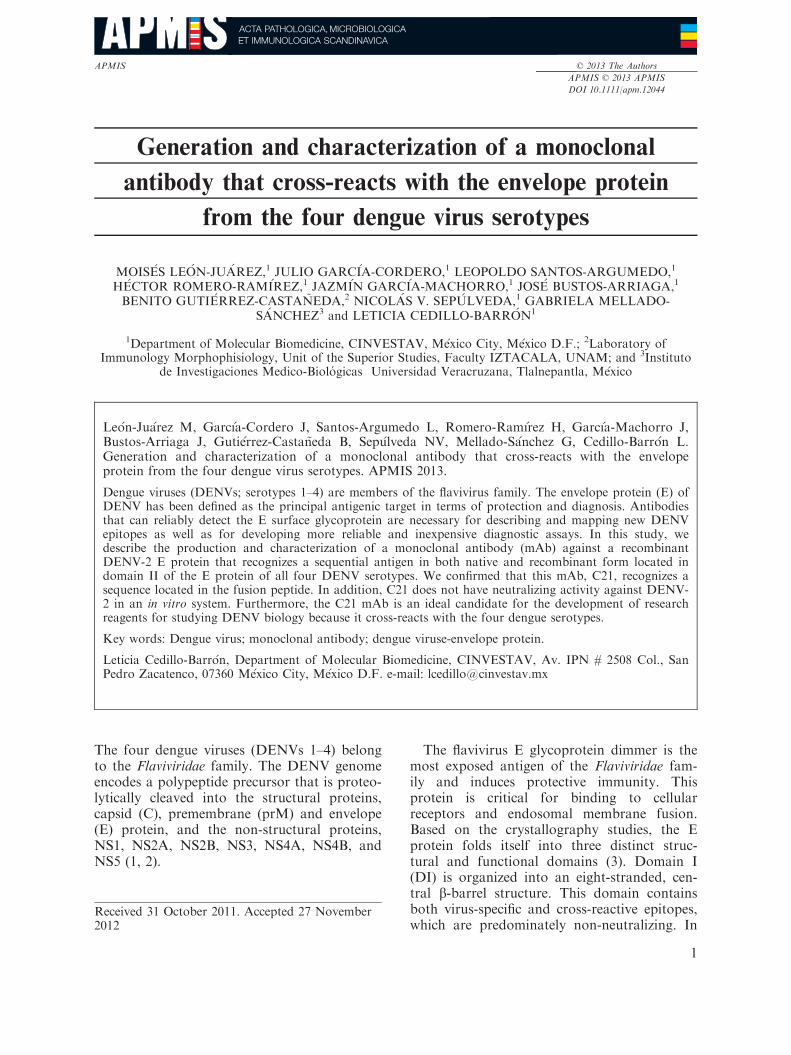
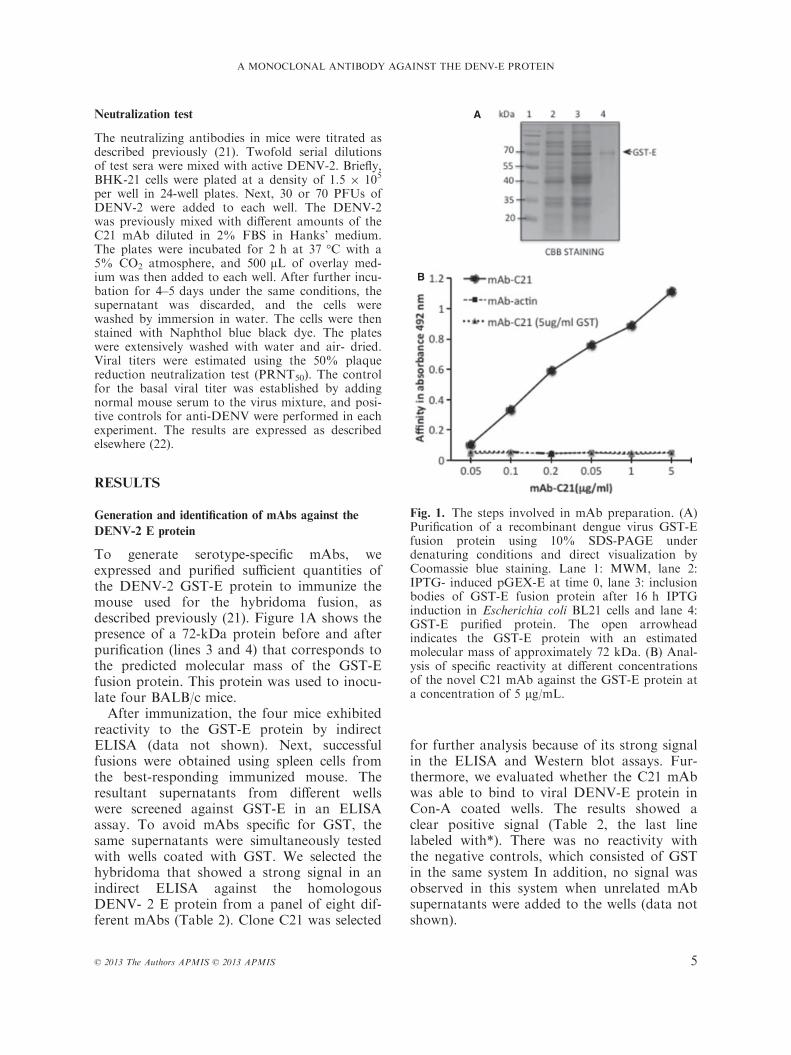
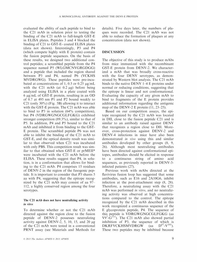
To generate serotype-specific mAbs, weexpressed and purified sufficient quantities ofthe DENV-2 GST-E protein to immunize themouse used for the hybridoma fusion, asdescribed previously (21). Figure 1A shows thepresence of a 72-kDa protein before and afterpurification (lines 3 and 4) that corresponds tothe predicted molecular mass of the GST-Efusion protein. This protein was used to inocu-late four BALB/c mice.After immunization, the four mice exhibited
reactivity to the GST-E protein by indirectELISA (data not shown). Next, successfulfusions were obtained using spleen cells fromthe best-responding immunized mouse. Theresultant supernatants from different wellswere screened against GST-E in an ELISAassay. To avoid mAbs specific for GST, thesame supernatants were simultaneously testedwith wells coated with GST. We selected thehybridoma that showed a strong signal in anindirect ELISA against the homologousDENV- 2 E protein from a panel of eight dif-ferent mAbs (Table 2). Clone C21 was selected
for further analysis because of its strong signalin the ELISA and Western blot assays. Fur-thermore, we evaluated whether the C21 mAbwas able to bind to viral DENV-E protein inCon-A coated wells. The results showed aclear positive signal (Table 2, the last linelabeled with*). There was no reactivity withthe negative controls, which consisted of GSTin the same system In addition, no signal wasobserved in this system when unrelated mAbsupernatants were added to the wells (data notshown).
A
B
Fig. 1. The steps involved in mAb preparation. (A)Purification of a recombinant dengue virus GST-Efusion protein using 10% SDS-PAGE underdenaturing conditions and direct visualization byCoomassie blue staining. Lane 1: MWM, lane 2:IPTG- induced pGEX-E at time 0, lane 3: inclusionbodies of GST-E fusion protein after 16 h IPTGinduction in Escherichia coli BL21 cells and lane 4:GST-E purified protein. The open arrowheadindicates the GST-E protein with an estimatedmolecular mass of approximately 72 kDa. (B) Anal-ysis of specific reactivity at different concentrationsof the novel C21 mAb against the GST-E protein ata concentration of 5 lg/mL.
© 2013 The Authors APMIS © 2013 APMIS 5
A MONOCLONAL ANTIBODY AGAINST THE DENV-E PROTEIN
The C21 hybridoma was determined tobelong to the IgG2b subtype with a kappalight chain. Then, the specific anti-E C21 mAbwas concentrated and purified, and titer wasdetermined. ELISA binding assays wereperformed to quantify the binding affinity ofC21. A total of 6 lg/mL of recombinant Eprotein was used per well for the ELISAs. Apositive signal was observed with 0.05 lg ofthe mAb, and the signal steadily increaseduntil the concentration of the mAb was 1 lg(Fig. 1B). An unrelated monoclonal antibody
(anti-actin) incubated with the GST-E antigenwas used as a negative control. In addition, anegative control for specificity was performedby coating the plates with GST protein, andwe found that the C21 mAb did not recognizeGST at any concentration.
Reactivity of anti-E mAb with native or denatured
DENV envelope glycoprotein
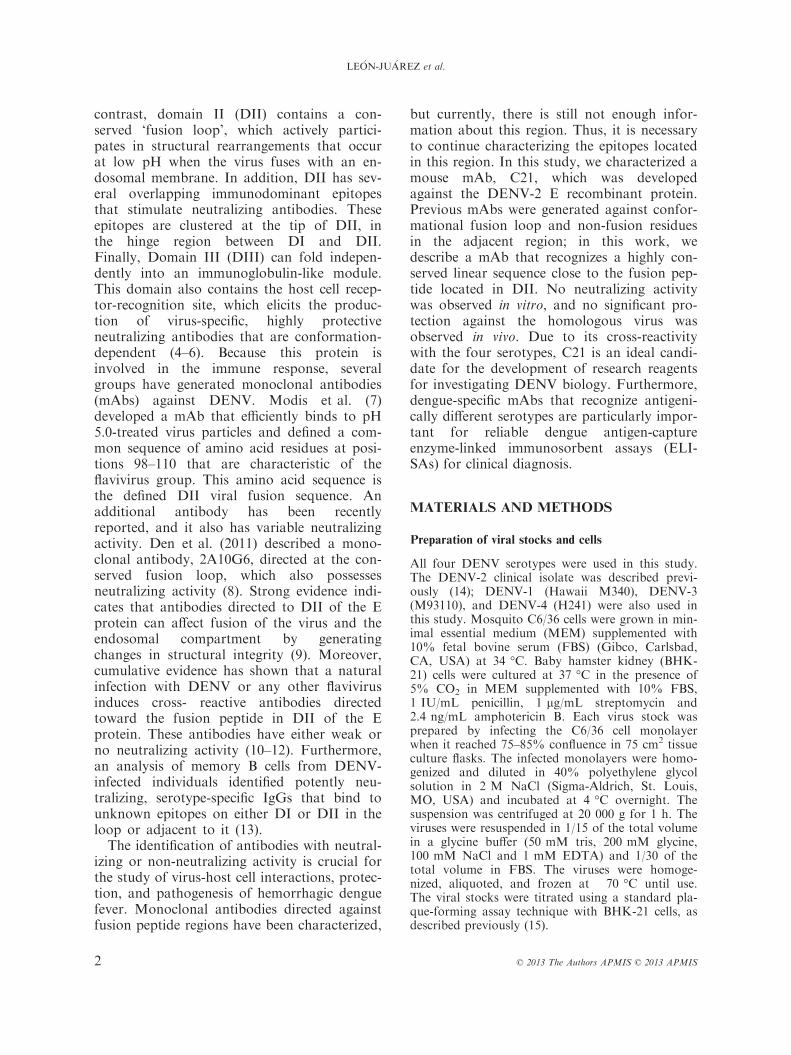
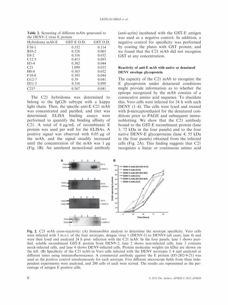
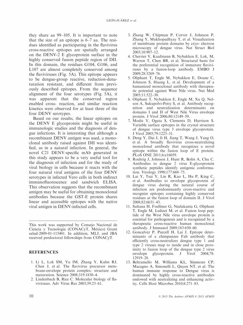
The capacity of the C21 mAb to recognize theE glycoprotein under denatured conditionsmight provide information as to whether theepitope recognized by the mAb consists of aconsecutive amino acid sequence. To elucidatethis, Vero cells were infected for 24 h with eachDENV (1–4). The cells were lysed and treatedwith b-mercaptoethanol for the denatured con-ditions prior to PAGE and subsequent immu-noblotting. We show that the C21 antibodybound to the GST-E recombinant protein (lane1; 72 kDa in the four panels) and to the fournative DENV-E glycoproteins (lane 4; 55 kDain the four panels) obtained from the infectedcells (Fig. 2A). This finding suggests that C21recognizes a linear or continuous amino acid
A B
Fig. 2. C21 mAb cross-reactivity: (A) Immunoblot analysis to determine the serotype specificity. Vero cellswere infected with 5 m.o.i. of the four serotypes, dengue virus 1 (DENV-1) to DENV4 (all cases, lane 4) andwere then lysed and analyzed 24 h post- infection with the C21 mAb. In the four panels, lane 1 shows puri-fied, soluble recombinant GST-E protein from DENV-2, lane 2 shows non-infected cells, lane 3 containsmock-infected cells, and lane 4 shows DENV-infected cells. Protein molecular weights (in kDa) are shown onthe left. (B) Specificity of the C21 mAb in Vero cells infected with the DENV serotypes 2–4 and analyzed atdifferent times using immunofluorescence. A commercial antibody against the E protein (D3-2H2-9-21) wasused as the positive control simultaneously for each serotype. Five different microscope fields from three inde-pendent experiments were analyzed, and 200 cells of each were scored. The results are represented as the per-centage of antigen E positive cells.
Table 2. Screening of different mAbs generated tothe DENV-2 virus E protein
Hybridoma mAb-E GST-E O.D. GST O.D.
F10-1 0.332 0.114B10-2 0.328 0.065E8-2 0.316 0.032C12-5 0.415 0.093H5-4 0.302 0.044C21 1.099 0.035H6-8 0.363 0.032F10-8 0.393 0.044G12-7 0.39 0.041D11-5 0.316 0.098
C21* 0.567 0.041
6 © 2013 The Authors APMIS © 2013 APMIS
LE�ON-JU�AREZ et al.
sequence. In contrast, no signal was observedin the uninfected cells or mock-infected cells(lanes 2 and 3 in the four panels).Furthermore, the specificity of this mAb was
evaluated in an experiment on the kinetics ofinfection with 2 m.o.i. (Multiplicity of infec-tion) of three DENV serotypes (DENV 2, 3,and 4). The results showed that the stainingwas similar among all of the serotypes tested,and compared with a commercial mAb, no sig-nificant differences were observed (Fig. 2B).In general, specific mAbs have utility as diag-
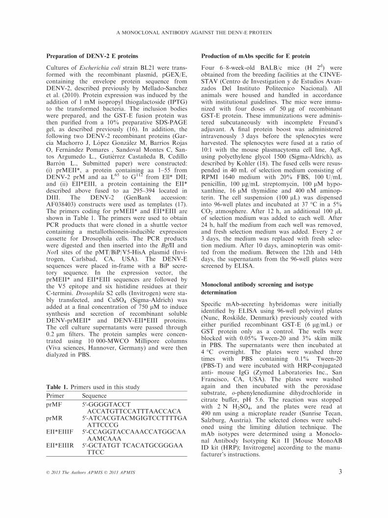
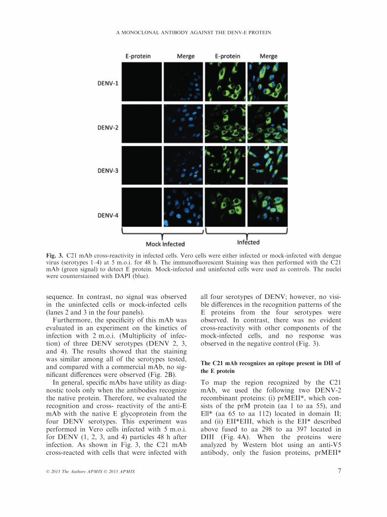
nostic tools only when the antibodies recognizethe native protein. Therefore, we evaluated therecognition and cross- reactivity of the anti-EmAb with the native E glycoprotein from thefour DENV serotypes. This experiment wasperformed in Vero cells infected with 5 m.o.i.for DENV (1, 2, 3, and 4) particles 48 h afterinfection. As shown in Fig. 3, the C21 mAbcross-reacted with cells that were infected with
all four serotypes of DENV; however, no visi-ble differences in the recognition patterns of theE proteins from the four serotypes wereobserved. In contrast, there was no evidentcross-reactivity with other components of themock-infected cells, and no response wasobserved in the negative control (Fig. 3).
The C21 mAb recognizes an epitope present in DII of
the E protein
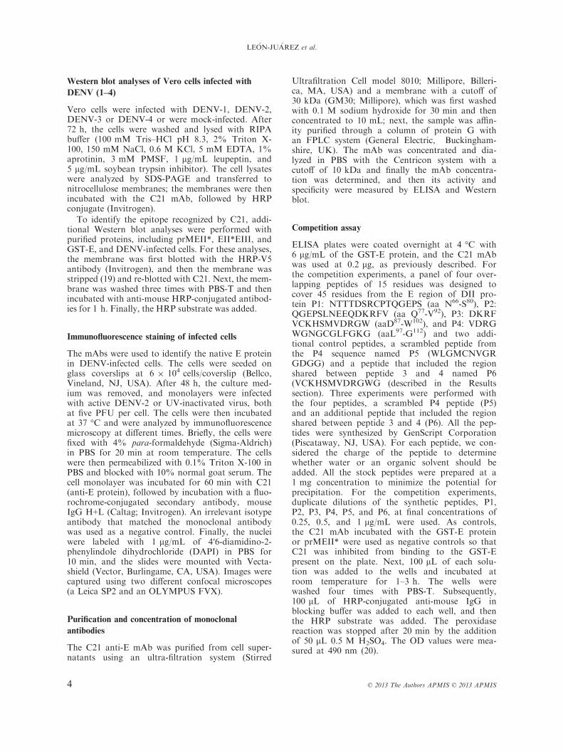
To map the region recognized by the C21mAb, we used the following two DENV-2recombinant proteins: (i) prMEII*, which con-sists of the prM protein (aa 1 to aa 55), andEll* (aa 65 to aa 112) located in domain II;and (ii) EII*EIII, which is the EII* describedabove fused to aa 298 to aa 397 located inDIII (Fig. 4A). When the proteins wereanalyzed by Western blot using an anti-V5antibody, only the fusion proteins, prMEII*
Fig. 3. C21 mAb cross-reactivity in infected cells. Vero cells were either infected or mock-infected with denguevirus (serotypes 1–4) at 5 m.o.i. for 48 h. The immunofluorescent Staining was then performed with the C21mAb (green signal) to detect E protein. Mock-infected and uninfected cells were used as controls. The nucleiwere counterstained with DAPI (blue).
© 2013 The Authors APMIS © 2013 APMIS 7
A MONOCLONAL ANTIBODY AGAINST THE DENV-E PROTEIN
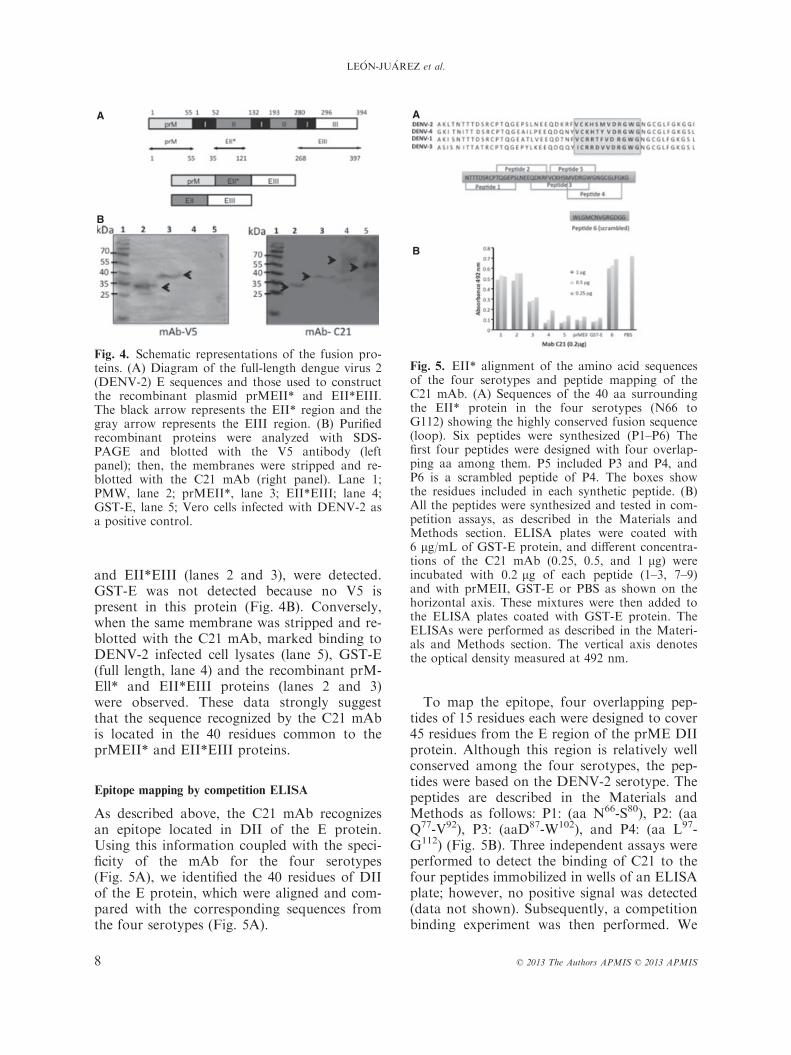
and EII*EIII (lanes 2 and 3), were detected.GST-E was not detected because no V5 ispresent in this protein (Fig. 4B). Conversely,when the same membrane was stripped and re-blotted with the C21 mAb, marked binding toDENV-2 infected cell lysates (lane 5), GST-E(full length, lane 4) and the recombinant prM-Ell* and EII*EIII proteins (lanes 2 and 3)were observed. These data strongly suggestthat the sequence recognized by the C21 mAbis located in the 40 residues common to theprMEII* and EII*EIII proteins.
Epitope mapping by competition ELISA
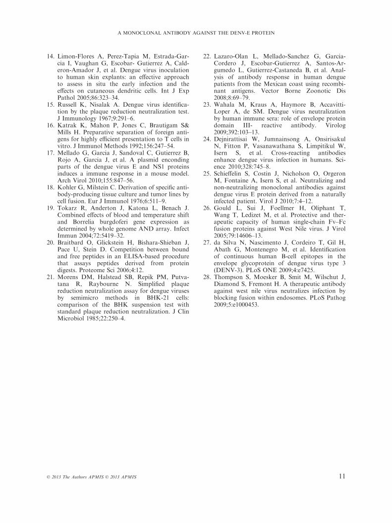
As described above, the C21 mAb recognizesan epitope located in DII of the E protein.Using this information coupled with the speci-ficity of the mAb for the four serotypes(Fig. 5A), we identified the 40 residues of DIIof the E protein, which were aligned and com-pared with the corresponding sequences fromthe four serotypes (Fig. 5A).
To map the epitope, four overlapping pep-tides of 15 residues each were designed to cover45 residues from the E region of the prME DIIprotein. Although this region is relatively wellconserved among the four serotypes, the pep-tides were based on the DENV-2 serotype. Thepeptides are described in the Materials andMethods as follows: P1: (aa N66-S80), P2: (aaQ77-V92), P3: (aaD87-W102), and P4: (aa L97-G112) (Fig. 5B). Three independent assays wereperformed to detect the binding of C21 to thefour peptides immobilized in wells of an ELISAplate; however, no positive signal was detected(data not shown). Subsequently, a competitionbinding experiment was then performed. We
A
B
Fig. 4. Schematic representations of the fusion pro-teins. (A) Diagram of the full-length dengue virus 2(DENV-2) E sequences and those used to constructthe recombinant plasmid prMEII* and EII*EIII.The black arrow represents the EII* region and thegray arrow represents the EIII region. (B) Purifiedrecombinant proteins were analyzed with SDS-PAGE and blotted with the V5 antibody (leftpanel); then, the membranes were stripped and re-blotted with the C21 mAb (right panel). Lane 1;PMW, lane 2; prMEII*, lane 3; EII*EIII; lane 4;GST-E, lane 5; Vero cells infected with DENV-2 asa positive control.
A
B
Fig. 5. EII* alignment of the amino acid sequencesof the four serotypes and peptide mapping of theC21 mAb. (A) Sequences of the 40 aa surroundingthe EII* protein in the four serotypes (N66 toG112) showing the highly conserved fusion sequence(loop). Six peptides were synthesized (P1–P6) Thefirst four peptides were designed with four overlap-ping aa among them. P5 included P3 and P4, andP6 is a scrambled peptide of P4. The boxes showthe residues included in each synthetic peptide. (B)All the peptides were synthesized and tested in com-petition assays, as described in the Materials andMethods section. ELISA plates were coated with6 lg/mL of GST-E protein, and different concentra-tions of the C21 mAb (0.25, 0.5, and 1 lg) wereincubated with 0.2 lg of each peptide (1–3, 7–9)and with prMEII, GST-E or PBS as shown on thehorizontal axis. These mixtures were then added tothe ELISA plates coated with GST-E protein. TheELISAs were performed as described in the Materi-als and Methods section. The vertical axis denotesthe optical density measured at 492 nm.
8 © 2013 The Authors APMIS © 2013 APMIS
LE�ON-JU�AREZ et al.
evaluated the ability of each peptide to bind tothe C21 mAb in solution prior to testing thebinding of the C21 mAb to full-length GST-Ein ELISA plates. Peptides 3 and 4 blocked thebinding of C21 to GST-E- coated ELISA plates(data not shown). Interestingly, P3 and P4(which compete highly with E protein) containthe fusion peptide sequences. On the basis ofthese results, we designed two additional con-trol peptides; a scrambled peptide from the P4sequence named P5 (WLGMCNVGRGDGG)and a peptide that included the region sharedbetween P3 and P4, named P6 (VCKHSMVDRGWG). These peptides were pre-incu-bated at concentrations of 1, 0.5 or 0.25 lg/mLwith the C21 mAb (at 0.2 lg) before beinganalyzed using ELISA in a plate coated with6 lg/mL of GST-E protein, resulting in an ODof 0.7 at 495 nm P1 and P2 bound weakly toC21 (only 30%) (Fig. 5B) allowing it to interactwith the GST-E protein. The C21 mAb was ableto bind to P3 in solution (64% competition),but P4 (VDRGWGNGCGLFGKG) exhibitedstronger competition (89.3%), similar to that ofP5. In addition, P4 and P5 bound to the C21mAb and inhibited it from binding to the GST-E protein. The scrambled peptide P6 was notable to inhibit the binding of the C21 mAb toGST-E, and the optical density result was simi-lar to that observed when C21 was incubatedwith only PBS. This competition result was sim-ilar to that obtained when GST-E or prMEII*were incubated with the C21 mAb before theELISA. These results suggest that P4, in solu-tion, is in a conformation that allows for bind-ing to the C21 mAb. P4 comprises 15 residuesof DENV-2 in the region of the fusogenic pep-tide. It is important to consider that P3 shares 5aa with P4, suggesting that the epitope recog-nized by the C21 mAb may consist of aa 97–112, a highly conserved region among the fourserotypes.
The C21 mAb does not have neutralizing activity
in vitro
To determine whether or not the C21 mAbdirected against the region close to the fusionpeptide of DENV-2 possesses neutralizingactivity against DENV-2, 5, 10, 15, and 20 lgof the C21 mAb were tested in a conventionalPRNT assay (see Materials and Methods for
details). Five days later, the numbers of pla-ques were recorded. The C21 mAb was notable to reduce the formation of plaques at anyconcentration (data not shown).
DISCUSSION
The objective of this study is to produce mAbsfrom mice immunized with the recombinantGST-E protein from DENV-2. We character-ized a mAb that was broadly cross-reactivewith the four DENV serotypes, as demon-strated by Western blot analysis. The C21 mAbbinds to the native DENV 1–4 E proteins undernormal or reducing conditions, suggesting thatthe epitope is linear and not conformational.Evaluating the capacity of any given mAb tobind to fragments of the E protein providesadditional information regarding the antigenicmap of the DENV-2 E protein (11, 23–25).Based on our competition assays, the epi-
tope recognized by the C21 mAb was locatedin DII, close to the fusion peptide C21 and issimilar to an antibody raised against DENVthat recognizes a region close to DII. More-over, cross-protection against DENV-2 andDENV-4 infections in mice have also beendemonstrated in vivo using this and similarantibodies developed by other groups (8, 9,26). Although most neutralizing antibodieshave been directed against conformational epi-topes, antibodies should be elicited in responseto a continuous string of amino acidsequences, as previously reported in DENV-3-infected patients (27).Previous work with mAbs directed at the
flavivirus fusion loop has suggested that someantibodies, such as E16 and 2A10G6, inhibitinfection at the post-attachment step (8, 28).Therefore, a neutralizing assay with the C21mAb was performed in vitro, and no neutraliz-ing activity was observed at high concentra-tions compared to the control. The epitoperecognized by the C21 mAb described in thiswork recognized a continuous sequence of theE glycoprotein peptide, P4. The sequence ofthis peptide is VDRGWGNGCGLFGKG (aaV97-G111). The C21 mAb also showed partialinhibition of P3, the sequence of which isDKRFVCKHSMVDRGW (aa D87-V101).These two peptides may be inhibited because
© 2013 The Authors APMIS © 2013 APMIS 9
A MONOCLONAL ANTIBODY AGAINST THE DENV-E PROTEIN
they share aa 99–105. It is important to notethat the size of an epitope is 6–7 aa. The resi-dues identified as participating in the flaviviruscross-reactive epitopes are spatially arrangedon the DENV-2 E glycoprotein surface in thehighly conserved fusion peptide region of DII.In this domain, the residues G104, G106, andL107 are almost completely conserved amongthe flaviviruses (Fig. 5A). This epitope appearsto be dengue-group reactive, reduction-dena-turation resistant, and different from previ-ously described epitopes. From the sequencealignment of the four serotypes (Fig. 5A), itwas apparent that the conserved regionenabled cross- reaction, and similar reactionkinetics were observed for at least three of thefour DENV serotypes.Based on our results, the linear epitopes on
the DENV E glycoprotein might be useful inimmunologic studies and the diagnosis of den-gue infections. It is interesting that although arecombinant DENV antigen was used, a mono-clonal antibody raised against DII was identi-fied, as in a natural infection. In general, thenovel C21 DENV-specific mAb generated inthis study appears to be a very useful tool forthe diagnosis of infection and for the study ofviral biology in cells because it reacts with thefour natural viral antigens of the four DENVserotypes in infected Vero cells in both indirectimmunofluorescence and sandwich ELISA.This observation suggests that the recombinantantigen may be useful for obtaining monoclonalantibodies because the GST-E protein shareslinear and accessible epitopes with the nativeviral antigen in DENV-infected cells.
This work was supported by Consejo Nacional deCiencia y Tecnolog�ıa (CONACyT, M�exico) Grantsalud-2009-01-115401. In addition, MLJ, and JBAreceived predoctoral fellowships from CONACyT.
REFERENCES
1. Li L, Lok SM, Yu IM, Zhang Y, Kuhn RJ,Chen J, et al. The flavivirus precursor mem-brane-envelope protein complex: structure andmaturation. Science 2008;319:1830–4.
2. Lindenbach B, Rice C. Molecular biology of fla-viviruses. Adv Virus Res 2003;59:23–61.
3. Zhang W, Chipman P, Corver J, Johnson P,Zhang Y, Mukhopadhyay T, et al. Visualizationof membrane protein domains by cryo- electronmicroscopy of dengue virus. Nat Struct Biol2003;10:907–12.
4. Cherrier V, Kaufmann B, Nybakken E, Lok, M,Warren T, Chen BR, et al. Structural basis forthe preferential recognition of immature flavivi-ruses by a fusion-loop antibody. EMBO J2009;28:3269–76.
5. Oliphant T, Engle M, Nybakken E, Doane C,Johnson S, Huang L, et al. Development of ahumanized monoclonal antibody with therapeu-tic potential against West Nile virus. Nat Med2005;11:522–30.
6. Oliphant T, Nybakken E, Engle M, Xu Q, Nel-son A, Sukupolvi-Petty S, et al. Antibody recog-nition and neutralization determinants ondomains I and II of West Nile Virus envelopeprotein. J Virol 2006;80:12149–59.
7. Modis Y, Ogata S, Clements D, Harrison S.Variable surface epitopes in the crystal structureof dengue virus type 3 envelope glycoprotein.J Virol 2005;79:1223–31.
8. Deng Y, Dai J, Ji H, Jiang T, Wang J, Yang O,et al. A broadly flavivirus cross-neutralizingmonoclonal antibody that recognizes a novelepitope within the fusion loop of E protein.PLoS ONE 2011;6:e16059.
9. Roehrig J, Johnson J, Hunt R, Bolin A, Chu C.Antibodies to dengue 2 virus E-glycoproteinsynthetic peptides identify antigenic conforma-tion. Virology 1990;177:668–75.
10. Lai Y, Tsai Y, Lin R, Kao L, Hu P, King C,et al. Antibodies to envelope glycoprotein ofdengue virus during the natural course ofinfection are predominantly cross-reactive andrecognize epitopes containing highly conservedresidues at the fusion loop of domain II. J Virol2008;82:6631–43.
11. Sultana H, Foellmer G, Neelakanta G, OliphantT, Engle M, Ledizet M, et al. Fusion loop pep-tide of the West Nile virus envelope protein isessential for pathogenesis and is recognized by atherapeutic cross-reactive human monoclonalantibody. J Immunol 2009;183:650–60.
12. Goncalvez P, Purcell H, Lai J. Epitope deter-minants of a chimpanzee Fab antibody thatefficiently cross-neutralizes dengue type 1 andtype 2 viruses map to inside and in close prox-imity to fusion loop of the dengue type 2 virusenvelope glycoprotein. J Virol 2004;78:12919–28.
13. Beltramello M, Williams KL, Simmons CP,Macagno A, Simonelli L, Quyen NT, et al. Thehuman immune response to Dengue virus isdominated by highly cross-reactive antibodiesendowed with neutralizing and enhancing activ-ity. Cells Host Microbes 2010;8:271–83.
10 © 2013 The Authors APMIS © 2013 APMIS
LE�ON-JU�AREZ et al.
14. Limon-Flores A, Perez-Tapia M, Estrada-Gar-cia I, Vaughan G, Escobar- Gutierrez A, Cald-eron-Amador J, et al. Dengue virus inoculationto human skin explants: an effective approachto assess in situ the early infection and theeffects on cutaneous dendritic cells. Int J ExpPathol 2005;86:323–34.
15. Russell K, Nisalak A. Dengue virus identifica-tion by the plaque reduction neutralization test.J Immunology 1967;9:291–6.
16. Katrak K, Mahon P, Jones C, Brautigam S&Mills H. Preparative separation of foreign anti-gens for highly efficient presentation to T cells invitro. J Immunol Methods 1992;156:247–54.
17. Mellado G, Garcia J, Sandoval C, Gutierrez B,Rojo A, Garcia J, et al. A plasmid encondingparts of the dengue virus E and NS1 proteinsinduces a immune response in a mouse model.Arch Virol 2010;155:847–56.
18. Kohler G, Milstein C. Derivation of specific anti-body-producing tissue culture and tumor lines bycell fusion. Eur J Immunol 1976;6:511–9.
19. Tokarz R, Anderton J, Katona L, Benach J.Combined effects of blood and temperature shiftand Borrelia burgdoferi gene expression asdetermined by whole genome AND array. InfectImmun 2004;72:5419–32.
20. Braitbard O, Glickstein H, Bishara-Shieban J,Pace U, Stein D. Competition between boundand free peptides in an ELISA-based procedurethat assays peptides derived from proteindigests. Proteome Sci 2006;4:12.
21. Morens DM, Halstead SB, Repik PM, Putva-tana R, Raybourne N. Simplified plaquereduction neutralization assay for dengue virusesby semimicro methods in BHK-21 cells:comparison of the BHK suspension test withstandard plaque reduction neutralization. J ClinMicrobiol 1985;22:250–4.
22. Lazaro-Olan L, Mellado-Sanchez G, Garcia-Cordero J, Escobar-Gutierrez A, Santos-Ar-gumedo L, Gutierrez-Castaneda B, et al. Anal-ysis of antibody response in human denguepatients from the Mexican coast using recombi-nant antigens. Vector Borne Zoonotic Dis2008;8:69–79.
23. Wahala M, Kraus A, Haymore B, Accavitti-Loper A, de SM. Dengue virus neutralizationby human immune sera: role of envelope proteindomain III- reactive antibody. Virolog2009;392:103–13.
24. Dejnirattisai W, Jumnainsong A, OnsirisakulN, Fitton P, Vasanawathana S, Limpitikul W,Isern S, et al. Cross-reacting antibodiesenhance dengue virus infection in humans. Sci-ence 2010;328:745–8.
25. Schieffelin S, Costin J, Nicholson O, OrgeronM, Fontaine A, Isern S, et al. Neutralizing andnon-neutralizing monoclonal antibodies againstdengue virus E protein derived from a naturallyinfected patient. Virol J 2010;7:4–12.
26. Gould L, Sui J, Foellmer H, Oliphant T,Wang T, Ledizet M, et al. Protective and ther-apeutic capacity of human single-chain Fv–Fcfusion proteins against West Nile virus. J Virol2005;79:14606–13.
27. da Silva N, Nascimento J, Cordeiro T, Gil H,Abath G, Montenegro M, et al. Identificationof continuous human B-cell epitopes in theenvelope glycoprotein of dengue virus type 3(DENV-3). PLoS ONE 2009;4:e7425.
28. Thompson S, Moesker B, Smit M, Wilschut J,Diamond S, Fremont H. A therapeutic antibodyagainst west nile virus neutralizes infection byblocking fusion within endosomes. PLoS Pathog2009;5:e1000453.
© 2013 The Authors APMIS © 2013 APMIS 11
A MONOCLONAL ANTIBODY AGAINST THE DENV-E PROTEIN


