Embed Size (px)
Citation preview

Ia
ZKC
a
ARRAA
KRHSAP
1
rsoamfasspgfasb(chm
0d
International Journal of Biological Macromolecules 47 (2010) 661–667
Contents lists available at ScienceDirect
International Journal of Biological Macromolecules
journa l homepage: www.e lsev ier .com/ locate / i jb iomac
maging recognition events between human IgG and rat anti-human IgG bytomic force microscopy
hengjian Lv, Jianhua Wang ∗, Guoping Chen, Linhong Dengey Laboratory of Biorheological Science and Technology, Ministry of Education, and Institute of Biochemistry and Biophysics, College of Bioengineering,hongqing University, No. 174, Shazhengjie, Shapingba District, Chongqing 400044, China
r t i c l e i n f o
rticle history:eceived 17 June 2010eceived in revised form 23 August 2010ccepted 23 August 2010vailable online 9 September 2010
a b s t r a c t
Chemically immobilized rat anti-human immunoglobulin (IgG) monolayers on thiols modified gold sub-strates were fabricated using self-assembled monolayer (SAM) method. The antibody monolayers wereimaged before and after free human IgG treated, whilst recognition events between antigen and anti-body were monitored by contact mode atomic force microscopy (CM-AFM) and tapping mode AFM(TM-AFM), with topographic and/or phase images being recorded. The obtained images with differentm
.cn
eywords:at anti-human IgGuman IgGelf-assembled monolayertomic force microscopyhase imagingsurface compositions show distinct nanostructures, indicating occurrence of recognition and bindingevents of antigen–antibody. The size of the observed surface structures of the antibody monolayer, whentip broaden effect had been taken into account, was very close to the actual size of the antibody molecule.Thus, these results suggest CM-AFM is capable of, and proven satisfactory in detecting protein–proteininteractions (PPIs), providing the sample was prepared appropriately and the scanning parameters wereset adequately. Moreover, phase imaging can serve as a real time contrast enhancement technique to
lightim.co
TM-AFM in terms of high
. Introduction
Antibody based protein–protein interactions (PPIs) play crucialoles in biosensors [1,2], molecular recognition at single-moleculecale [3,4], and medical diagnostics [5,6]. So far, a number of meth-ds have been developed for PPIs studies, including surface forcepparatus (SFA) [7], surface plasmon resonance (SPR) [8], radioim-unoassay (RIA) [9], enzyme immunoassay (EIA) [10], and atomic
orce microscopy (AFM) [11]. Compared to other methods, AFM isdvantageous in terms of high spatial resolution, allowing mea-urements in near physiological environment and free of specialample preparation [12]. Hence, AFM opens new avenues to studyathways of PPIs. AFM is now an established technique to investi-ate the physical behaviors of PPIs, such as adhesive force, frictionorce and energy dissipation [13–16]. Moreover, along with thebility to image the topographies of protein molecules with nano-cale resolution and down to single molecular level, AFM cane served as a powerful tool to monitor the specific interaction
www.sp
recognition) between antigen and antibody. As for topographi-al studies, contact mode AFM (CM-AFM) was employed to obtainigh-resolution images, whereas the tapping mode atomic forceicroscopy (TM-AFM) was often used to prevent the deforma-
∗ Corresponding author. Tel.: +86 23 65102507; fax: +86 23 65102507.E-mail addresses: [email protected], [email protected] (J. Wang).
141-8130/$ – see front matter © 2010 Elsevier B.V. All rights reserved.oi:10.1016/j.ijbiomac.2010.08.017
ng edges and clearly observing fine features.© 2010 Elsevier B.V. All rights reserved.
tion of samples because it substantially reduces the lateral force[17].
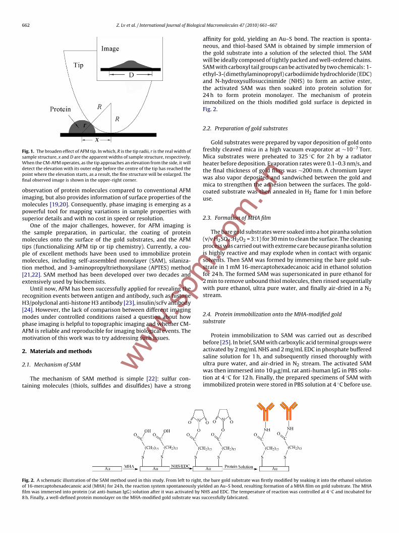
Upon CM-AFM, it is noteworthy that the AFM tip will intro-duce broaden effect as illustrated in Fig. 1. A round shape for tipis assumed as revealed by electron micrographs. A half spherestructure is assumed as a protein molecule immobilized on thiolmodified gold substrate. When the tip scans over the surface, itconstantly contacts with the protein layer: as the tip approachesan elevation from the side of the protein contour, it will detect theelevation with its outer edge before the centre of the tip reachesthe point where the elevation starts [18], such effect introduced inthe CM-AFM is called broaden effect.
x2 + R2 = (r + R)2 (1)
x =√
2Rr + r2 (2)
D = 2x = 2√
2Rr + r2 (3)
where R is the tip radii, r is the real width of protein molecule, xand D are the apparent widths of protein molecule, respectively.The observed image of the protein is illustrated in the upper-rightcorner of Fig. 1.
In recent years, based on the TM-AFM, a novel extension imag-ing mode termed phase imaging has been explored, in which case,the phase lag of the cantilever oscillation relative to the drive signal,accompanied with topography data, is monitored. Phase imagingnot only highlights edges to obtain better contrast and clearer
662 Z. Lv et al. / International Journal of Biologica
Fig. 1. The broaden effect of AFM tip. In which, R is the tip radii, r is the real width ofsWdpfi
oimps
tmtpmt[e
rH[mpAm
2
2
t
Fofi8
m
ample structure, x and D are the apparent widths of sample structure, respectively.hen the CM-AFM operates, as the tip approaches an elevation from the side, it willetect the elevation with its outer edge before the centre of the tip has reached theoint where the elevation starts, as a result, the fine structure will be enlarged. Thenal observed image is shown in the upper-right corner.
bservation of protein molecules compared to conventional AFMmaging, but also provides information of surface properties of the
olecules [19,20]. Consequently, phase imaging is emerging as aowerful tool for mapping variations in sample properties withuperior details and with no cost in speed or resolution.
One of the major challenges, however, for AFM imaging ishe sample preparation, in particular, the coating of protein
olecules onto the surface of the gold substrates, and the AFMips (functionalizing AFM tip or tip chemistry). Currently, a cou-le of excellent methods have been used to immobilize proteinolecules, including self-assembled monolayer (SAM), silaniza-
ion method, and 3-aminopropyltriethoxysilane (APTES) method21,22]. SAM method has been developed over two decades andxtensively used by biochemists.
Until now, AFM has been successfully applied for revealing theecognition events between antigen and antibody, such as histone3/polyclonal anti-histone H3 antibody [23], insulin/scFv antibody
24]. However, the lack of comparison between different imagingodes under controlled conditions raised a question about how
hase imaging is helpful to topographic imaging and whether CM-FM is reliable and reproducible for imaging biological events. Theotivation of this work was to try addressing such issues.
. Materials and methods ww.sp
.1. Mechanism of SAM
The mechanism of SAM method is simple [22]: sulfur con-aining molecules (thiols, sulfides and disulfides) have a strong
ig. 2. A schematic illustration of the SAM method used in this study. From left to right,f 16-mercaptohexadecanoic acid (MHA) for 24 h, the reaction system spontaneously yielm was immersed into protein (rat anti-human IgG) solution after it was activated by Nh. Finally, a well-defined protein monolayer on the MHA-modified gold substrate was s
w
l Macromolecules 47 (2010) 661–667
affinity for gold, yielding an Au–S bond. The reaction is sponta-neous, and thiol-based SAM is obtained by simple immersion ofthe gold substrate into a solution of the selected thiol. The SAMwill be ideally composed of tightly packed and well-ordered chains.SAM with carboxyl tail groups can be activated by two chemicals: 1-ethyl-3-(dimethylaminopropyl) carbodiimide hydrochloride (EDC)and N-hydroxysulfosuccinimide (NHS) to form an active ester,the activated SAM was then soaked into protein solution for24 h to form protein monolayer. The mechanism of proteinimmobilized on the thiols modified gold surface is depicted inFig. 2.
2.2. Preparation of gold substrates
Gold substrates were prepared by vapor deposition of gold ontofreshly cleaved mica in a high vacuum evaporator at ∼10−7 Torr.Mica substrates were preheated to 325 ◦C for 2 h by a radiatorheater before deposition. Evaporation rates were 0.1–0.3 nm/s, andthe final thickness of gold films was ∼200 nm. A chromium layerwas also vapor deposited and sandwiched between the gold andmica to strengthen the adhesion between the surfaces. The gold-coated substrate was then annealed in H2 flame for 1 min beforeuse.
2.3. Formation of MHA film
The bare gold substrates were soaked into a hot piranha solution(v/v H2SO4:H2O2 = 3:1) for 30 min to clean the surface. The cleaningprocess was carried out with extreme care because piranha solutionis highly reactive and may explode when in contact with organicsolvents. Then SAM was formed by immersing the bare gold sub-strate in 1 mM 16-mercaptohexadecanoic acid in ethanol solutionfor 24 h. The formed SAM was supersonicated in pure ethanol for2 min to remove unbound thiol molecules, then rinsed sequentiallywith pure ethanol, ultra pure water, and finally air-dried in a N2stream.
2.4. Protein immobilization onto the MHA-modified goldsubstrate
Protein immobilization to SAM was carried out as describedbefore [25]. In brief, SAM with carboxylic acid terminal groups wereactivated by 2 mg/mL NHS and 2 mg/mL EDC in phosphate buffered
.com
.cn
saline solution for 1 h, and subsequently rinsed thoroughly withultra pure water, and air-dried in N2 stream. The activated SAMwas then immersed into 10 �g/mL rat anti-human IgG in PBS solu-tion at 4 ◦C for 12 h. Finally, the prepared specimens of SAM withimmobilized protein were stored in PBS solution at 4 ◦C before use.
the bare gold substrate was firstly modified by soaking it into the ethanol solutionlded an Au–S bond, resulting formation of a MHA film on gold substrate. The MHAHS and EDC. The temperature of reaction was controlled at 4 ◦C and incubated foruccessfully fabricated.

Z. Lv et al. / International Journal of Biological Macromolecules 47 (2010) 661–667 663
Fig. 3. Topographic images of the bare gold (a), the rat anti-human IgG monolayer (b), the human IgG/rat anti-human IgG complexes (c), the human IgG modified tip scannedacross the rat anti-human IgG monolayer (d), and the antigen-antibody complexes modified tip scanned across rat anti-human IgG monolayer (e) recorded by CM-AFM,respectively. The CM-AFM was operated in PBS buffer solution with typical scanning rate of 2 Hz and scanning size of 2 �m × 2 �m.
www.spm
.com
.cn

664 Z. Lv et al. / International Journal of Biologica
Fig. 4. Size (diameter) distribution histogram of the bare gold, the antibody mono-layer and further treated with free antigen were represented by red, blue, and blackbars, respectively. Typically, the particles of the bare gold, the antibody monolayera5(r
2
i3MbsfNw
2
pssniaoac
ftsrfiTr
2
ilttat
m
nd the antibody treated with antigen have diameters of 20–40 nm, 30–50 nm and0–90 nm, respectively. The solid lines are theoretical Gaussian distribution curves.For interpretation of the references to color in this figure legend, the reader iseferred to the web version of the article.).5. Tip functionalization
Protocol of tip functionalization is analogous to that of proteinmmobilization. First, the tip was cleaned in piranha solution for0 min and washed with ultra pure water, then modified with 1 mMHA through SAM method. The MHA-modified tips were activated
y EDC and NHS in PBS buffer solution to form active esters, thenubject to incubation in 10 �g/mL human IgG PBS solution at 4 ◦Cor 6 h. Finally, the specimens were stored in PBS solution at 4 ◦C.otably, if not specified, the tips used in this study were bare tipsithout modification.
.6. AFM imaging
All images were acquired using Benyuan CSPM 5000 scanningrobe microscope (Benyuan Co., China) equipped with a 26-�m Ecanner. Commercial Si3N4 cantilevers with gold coated on bothides were chosen. The cantilevers with triangular shape and withominal spring constant of 0.07 N/m (BudgetSensors) were used
n CM-AFM. For TM-AFM, the AFM tips with resonance frequencybout 70 kHz (Olympus) were employed. All AFM images wereperated in PBS buffer solution with typical scanning rate of 2.0 Hznd scanning size of 2 �m × 2 �m. A number of imaging cycles wereonducted for CM-AFM and TM-AFM.
The bare gold, the antibody monolayer, and further treated withree antigen, were imaged by CM-AFM and TM-AFM. The sizes ofhese three different particles were analyzed by CSPM Image 4.62oftware program (provided by the manufacturer). Another kind ofecognition profile was obtained by performing the antigen modi-ed AFM tip scanned over the antibody monolayer in CM-AFM. ForM-AFM, both topographic and phase images were simultaneouslyecorded by simply clicking the option of “phase imaging”.
.7. Control experiment for CM-AFM
Blocking experiment was conducted to confirm the specificnteraction between antigen and antibody, which was done as fol-
www.sp
ows: firstly, the antigen-fixed tips were incubated for 30 min inhe antibody solution to block the binding sites of antigen. Then,he topographic images of antibody monolayer scanned by thentigen-antibody complexes modified AFM tip were performed inhe conditions as in Section 2.6.
l Macromolecules 47 (2010) 661–667
2.8. Materials
16-Mercaptohexadecanoic acid (MHA), 1-ethyl-3-(dimethyla-minopropyl) carbodiimide hydrochloride (EDC), and N-hydroxysulfosuccinimide (NHS) were purchased fromSigma–Aldrich Chemical Co. and used as received. Phosphatebuffered saline (PBS, 140 mM NaCl, 3 mM KCl, pH 7.4) and ethanol(guaranteed grade) were purchased from Merck Co., and ultrapure water (resistivity of 18.2 M� cm) was obtained by Milliporepurification system. Human IgG and rat anti-human IgG werepurchased from Biosun Co. (China).
3. Results and discussion
3.1. Preparation of protein monolayers
Since the sensitivity and reproducibility of bioassays are influ-enced by the surface properties, protein immobilization needs tobe carefully considered. The SAM method is reliable for proteinimmobilization because it ensures the activity, mobility and sta-bility of protein molecules [18,26]. Firstly, it is known that 1 mMthiol and immersion time for 24 h are often sufficient for form-ing well-ordered thiol film [21]. Secondly, MHA with long carbonchain acted as a spacer to minimize the interference between pro-tein molecules and gold substrate [27]. Thirdly, immobilizationconditions were controlled at 4 ◦C and under PBS buffer solutionthoroughly. Moreover, mimicking the natural status of proteinmolecules requires that the modified protein monolayer should notjust provide optimal orientation but also with minimal steric hin-drance. Our previous experiments demonstrated the SAM methodis reproducible and the formed protein monolayer is well ordered[28].
3.2. Topographic images in CM-AFM demonstrate binding eventsbetween antigen and antibody
A topographic image of the bare gold was recorded in PBSbuffer solution using a bare AFM tip as a comparison (Fig. 3a). Theperiodic flat island-like structures were observed, suggesting gooduniformity for gold substrates. The antibody (rat anti-human IgG)molecules were covalently immobilized on thiols modified goldsubstrates by SAM method that yielded a high surface coverage.The topography of the obtained protein monolayer was imaged byCM-AFM (Fig. 3b). The surface shows a number of homogeneoussphere-like structures, which was expected from a densely packedprotein monolayer [29]. IgG (∼150 kDa) consists of three fragments,two separate and identical Fab fragments with active sites for anti-gen binding and one Fc fragment. The three dimensional structureis either T- or Y-shaped with a size of 14.2 nm × 8.5 nm × 3.8 nm,as revealed by X-ray diffraction [30] and acknowledged as largebiological molecule. The observed single sphere-like structure haslarger size than that of a single protein molecule, a fact due to thetip broaden effect and the densely package of protein molecules.The antibody monolayer was incubated with antigen for yieldingantigen–antibody complexes, and sequentially subject to CM-AFMimaging. Compared to the surface of the antibody monolayer, thesurface of the antigen–antibody complexes shows distinct struc-tures with different shapes and much larger size of the image profile(Fig. 3c), suggesting specific interaction of antigen–antibody andeffect of antigen–antibody complexes formation [31–33].
.com
.cn
Imaging with the antigen (human IgG) fixed AFM tip in con-ditions identical to previously used to scan across the proteinmonolayer resulted in unique images (Fig. 3d). These recognitionprofiles arose from the binding of antigen on the AFM tip to anti-body molecules on the substrate, which led to a deformation of
Z. Lv et al. / International Journal of Biological Macromolecules 47 (2010) 661–667 665
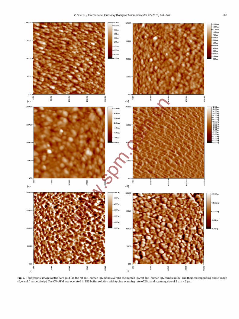
Fig. 5. Topographic images of the bare gold (a), the rat anti-human IgG monolayer (b), the human IgG/rat anti-human IgG complexes (c) and their corresponding phase image(d, e and f, respectively). The CM-AFM was operated in PBS buffer solution with typical scanning rate of 2 Hz and scanning size of 2 �m × 2 �m.
www.spm
.com
.cn

6 logica
twitbtbstrt
rteetbteibrpemee
3i
iattfrds(Etj[ttt(asa
3r
tbebtgai
m
66 Z. Lv et al. / International Journal of Bio
he antibody monolayer. The specificity of the recognition profilesas testified by an antibody blocking experiment. The AFM tip was
ncubated with free antibody solution led to the disappearance ofhe recognition profiles (Fig. 3e). This can be explained by free anti-ody binding to the active binding sites of the antigen molecules,hereby blocking recognition events between antigen and anti-ody. The antibody monolayer scanned with such modified AFM tiphows plateau-like structures (Fig. 3e). A reasonable explanation ishe antigen–antibody complexes modified AFM tip has a large tipadius, tip broaden effect would be heavily introduced when theip scans across the surface, finally obscures its “real” topography.
For obtaining high quality images in CM-AFM, some aspectsequire to be noticed: (1) biological materials are usually soft, andhe force applied to the cantilever is often lower than 100 pN, oth-rwise the AFM stylus will deform the biological sample to somextent; (2) when imaging in buffer solution, the long range elec-rostatic double-layer forces (several tens of nm) are governedy the charge density of both interacting surfaces, the pH andhe electrolyte composition of the buffer solution. As a result, thelectrostatic forces can be regulated by adjusting the pH and theonic strength of the buffer solution [34]. In present study, a PBSuffer solution was adopted for simulating the physiological envi-onment; (3) adjusting the instrumental parameters, such as “setoint” and “gain”, is essential to acquire high quality images. Orlse image artifacts will occur, such as tilt of the surface or defor-ation of sample [17]; (4) the AFM tip is prone to contamination,
specially at nano-scale. One should carefully address this issue forach experimental step.
.3. Size analysis of the observed nanostructures on CM-AFMmages
The size (diameter) of the nanostructures observed on themages of the bare gold, the antibody monolayer with or withoutntigen treatment was analyzed, respectively. Fig. 4 shows the his-ogram of the size distribution of the structures associated withhe three different materials. The mean size of the observed sur-ace structure on the bare gold substrate, which may serve as aeference, was found to be 31.65 (20–40) nm (Fig. 4, red bar). Aftereposition of antibody molecules onto the bare gold, the meanize of the observed surface structures increased 43.97 (30–50) nmFig. 4, blue bar). Considering the tip broaden effect according toq. (3) for a tip of R = 25 nm (data given by the tip manufacturer),his observed size is very close to the theoretical estimation of pro-ected size, 40.26 nm, for the IgG molecule of real size at 14.2 nm30], strongly suggesting that the observed structures are indeedhe antibody molecules immobilized onto the gold substrate. Afterreated with free antigen, however, the mean size of surface struc-ures on the antibody monolayer significantly increased to 67.550–90) nm (Fig. 4, black bar). This observed size corresponds tostructure of real size at 34 nm that is more than double of the
ize of antibody molecule itself, indicating that antigen bound tontibody and formed antibody–antigen complexes.
.4. Topographical and phase images in TM-AFM revealecognition events between antigen and antibody
One of the advantages of TM-AFM is a significant reduction ofhe lateral dragging force allowing more reproducible results toe obtained. This is favorable for imaging biological samples. How-ver, the resolution with TM-AFM is not superior to that of CM-AFM
www.sp
ecause the compression with the tapping is actually larger thanhe minimum applied force exerted by the cantilever [35]. Topo-raphic images of the bare gold, the antibody monolayer and thentibody monolayer treated with free antigen solution are shownn Fig. 5a, b and c, respectively. Typically, the surfaces of the bare
l Macromolecules 47 (2010) 661–667
gold, the antibody monolayer and the antigen–antibody complexesdisplay island-like structures (Fig. 5a), small spherical structures(Fig. 5b) and large spherical (about double size of observed anti-body molecule) structures (Fig. 5c), respectively. These results areconsistent with the CM-AFM topographic images. The observeddiscrepancies among three different surfaces indicate that the pro-tein molecules were adequately bound onto the gold substrate,and complexes were successfully formed because of the existenceof specific interactions between antigen and antibody. In addi-tion to topographic images, the phase images were simultaneouslyrecorded to verify and explain the surface properties. With respectto the phase images of the bare gold (Fig. 5d), the antibody mono-layer (Fig. 5e), and the antigen–antibody complexes (Fig. 5f), thegreater phase lag of the tip’s driven frequency, the brighter pixelsappear in the phase image [20]. All phase images show patch-likesurfaces but distinct nanostructures. The phase images are likely toserve as the clearer ‘topographical images’ and are often adoptedto observe fine surface nanostructures that cannot be visualized inthe topographic images [36]. Present study confirmed that phaseimages not only highlight edges but also give clearer phase con-trast. Thus, it is convincing that phase images can serve beyondtopographies.
Usually, two classes of factors may be responsible for the phaseshift of the oscillating cantilever. One is of instrumental parameters,such as the amplitude modulation feedback, the driving frequency,the tip’s sharpness and the spring constant [37–39]. The other is ofchemical or physical properties of the material surfaces, includingviscoelasticity, friction, adhesion and components [40–42]. To date,several theoretical models have been proposed to explain phasebehavior [37,43,44]. For example, Magonov and co-workers definedthe phase (angle) shift, �ϕ, between the free and interacting can-tilevers as [40]:
�ϕ = �
2− tan−1
(k
Q�
)≈ Q�
k(� ≤ k) (4)
where Q is the quality factor, k is the spring constant of a freely oscil-lating cantilever, and � is the overall force derivatives. For TM-AFM,the force derivative � is proportional to time-averaged contact areaA, determined by surface stiffness and morphological structure. Asofter material leads to a larger contact area A [35]. It is knownthat flexible protein molecules are soft biomaterials, whereas thegold substrates are stiff. According to Eq. (4), the phase shift of theantibody monolayer is larger than that of the bare gold substrates(1.71◦). Since the large contact area of the antigen–antibody com-plexes, the larger phase shift of the antigen–antibody complexes(20.58◦) was observed than that of the antibody monolayer (5.97◦).Our experimental results are consistent with these theoretical pre-dictions.
4. Conclusions
We herein present a study of recognition events betweenantigen and antibody molecules immobilized on SAM modifiedsubstrate and visualized by CM-AFM and TM-AFM. Both topo-graphic images by CM-AFM and TM-AFM, and phase images byTM-AFM were obtained. The topographic images of either the baregold substrate, the rat anti-human IgG monolayer, or the humanIgG/rat anti-human IgG complexes show significantly different sur-face structures, suggesting recognition events occurred betweenantibody and antigen. This is further supported by phase imageswith the phase angular variations, which are complementary to
.com
.cn
topographic images. Moreover, phase imaging can be served as areal time contrast enhancement technique to TM-AFM in termsof highlighting edges and clear observation of fine features. Sizeanalysis of the observed surface structures of different surfacecompositions demonstrated that the mean size of the observed
logica
setaabu
A
dRSd
R
[
[[
[[
[
[
[[
[[
[
[
[
[
[[
[[[
[
[
[
[
[
[[
[[
[
[Condens. Matter Mater. Phys. 61 (2000) 14179–14183..
Z. Lv et al. / International Journal of Bio
tructures on the antibody monolayers was very close to what isstimated for the actual size of the antibody molecule. These results,herefore, suggest that providing the sample is prepared appropri-tely and the instrumental parameters are set adequately, CM-AFMnd TM-AFM are capable of detecting recognition events betweeniological molecules, such as antigen and antibody, and could beseful tools in PPIs studies.
cknowledgements
This work was supported by the National Natural Science Foun-ation of China (Nos. 30670496, 30770529) and the Scientificesearch Foundation for the Returned Overseas Chinese Scholars,tate Education Ministry (2006-331) and the Natural Science Foun-ation Project of CQ CSTC (2006BB5017).
eferences
[1] F. Cecchet, A.S. Duwez, S. Gabriel, C. Jerome, R. Jerome, K. Glinel, S. Demoustier-Champagne, A.M. Jonas, B. Nysten, Anal. Chem. 79 (2007) 6488–6495.
[2] W. Lee, B.K. Oh, Y. Min Bae, S.H. Paek, W. Hong Lee, J.W. Choi, Biosens. Bioelec-tron. 19 (2002) 185–192.
[3] L. Li, S. Chen, S. Oh, S. Jiang, Anal. Chem. 74 (2002) 6017–6022.[4] P. Hinterdorfer, Y.F. Dufrene, Nat. Methods 3 (2006) 347–355.[5] H. Zhu, M. Bilgin, M. Snyder, Annu. Rev. Biochem. 72 (2003) 783–812.[6] W. Kusnezow, A. Jacob, A. Walijew, F. Diehl, J.D. Hoheisel, Proteomics 3 (2003)
254–264.[7] T.S. Tsapikouni, Y.F. Missirlis, Mater. Sci. Eng. B 152 (2008) 2–7.[8] R. Karisson, J. Mol. Recognit. 17 (2004) 151–161.[9] R. Pio, A. Martinez, E.J. Unsworth, J.A. Kowalak, J.A. Bengoechea, P.F. Zipfei, T.H.
Elsasser, F. Cuttitta, J. Biol. Chem. 276 (2001) 12292–12300.10] A. Roda, P. Pasini, M. Mirasoli, E. Michelini, M. Guardigli, Trends Biotechnol. 22
(2004) 295–303.11] N.C. Santos, M.A.R.B. Castanho, Biophys. Chem. 107 (2004) 133–149.12] D. Fotiadis, S. Scheuring, S.A. Muller, A. Engel, D.J. Muller, Micron 33 (2002)
385–397.13] G.U. Lee, D.A. Kidwell, R.J. Colton, Langmuir 10 (1994) 354–357.
14] C.D. Frisbie, L.F. Rosznyai, A. Noy, M.S. Wrighton, C.M. Lieber, Science 265 (1994)2071–2074.15] J.P. Cleveland, B. Anczykowski, A.E. Schmid, V.B. Elings, Appl. Phys. Lett. 72
(1998) 2613–2615.16] P. Parot, Y.F. Dufrene, P. Hinterdorfer, C. Le Grimellec, D. Navajas, J.L. Pellequer,
S. Scheuring, J. Mol. Recognit. 20 (2007) 418–431.
[
[[[
www.spm
l Macromolecules 47 (2010) 661–667 667
17] A. Alessandrini, P. Facci, Meas. Sci. Technol. 16 (2005) R65–R92.18] L. Guo, R. Wang, H. Xu, J. Liang, Physica. E: Low. Dimens. Syst. Nanostruct. 27
(2005) 240–244.19] G.K. Pang, K.Z. Baba-Kishi, A. Patel, Ultramicroscopy 81 (2000) 35–40.20] M. Stark, C. Moller, D.J. Muller, R. Guckenberger, Biophys. J. 80 (2001)
3009–3018.21] S. Ferretti, S. Paynter, D.A. Russell, K.E. Sapsford, D.J. Richardson, Trends Analyt.
Chem. 19 (2000) 530–540.22] J.C. Love, L.A. Estroff, J.K. Kriebel, R.G. Nuzzo, G.M. Whitesides, Chem. Rev. 105
(2005) 1103–1169.23] C. Stroh, H. Wang, R. Bash, B. Ashcroft, J. Nelson, H. Gruber, D. Lohr, S.M. Lindsay,
P. Hinterdorfer, Proc. Natl. Acad. Sci. U.S.A. 101 (2004) 12503–12507.24] W.D. Marcus, H. Wang, S.M. Lindsay, M.R. Sierks, Nanomedicine 4 (2008)
1–7.25] Z.J. Lv, J.H. Wang, G.P. Chen, L.H. Deng, Nanoscale Res. Lett. 5 (2010) 1032–1038.26] M. Lekka, A.J. Kulik, S. Jeney, J. Raczkowska, J. Lekki, A. Budkowski, L. Forro, J.
Chem. Phys. 123 (2005) 014702.27] D.K. Schwartz, Annu. Rev. Phys. Chem. 52 (2001) 107–137.28] Z.J. Lv, J.H. Wang, L.H. Deng, G.P. Chen, Nanoscale Res. Lett. 4 (2009) 1403–1408.29] A. Raab, W. Han, D. Badt, S.J. Smith-Gill, S.M. Lindsay, H. Schindler, P. Hinter-
dorfer, Nat. Biotechnol. 17 (1999) 901–905.30] E.W. Silverton, M.A. Navia, D.R. Davies, Proc. Natl. Acad. Sci. U.S.A. 74 (1977)
5140–5144.31] M.E. Browning-Kelley, K. Wadu-Mesthrige, V. Hari, G.Y. Liu, Langmuir 13 (1997)
343–350.32] P. Hammarstrom, M. Person, U. Carlsson, J. Biol. Chem. 276 (2001)
21765–21775.33] C.S. Neish, I.L. Martin, R.M. Henderson, J.M. Edwardson, Br. J. Pharmacol. 135
(2002) 1943–1950.34] S. Scheuring, S. Fotiadis, C. Moller, S.A. Muller, A. Engel, D.J. Muller, Single Mol.
2 (2001) 59–67.35] J. Yang, Cell Biochem. Biophys. 41 (2004) 435–449.36] F. Kienberger, L.T. Costa, R. Zhu, G. Kada, M. Reithmayer, L. Chtcheglova, C.
Rankl, A.B. Pacheco, S. Thalhammer, V. Pastushenko, W.M. Heckl, D. Blaas, P.Hinterdorfer, Biomaterials 28 (2007) 2403–2411.
37] R. Garcia, R. Perez, Surf. Sci. Rep. 47 (2002) 197–301.38] X. Chen, M.C. Davies, C.J. Roberts, S.B. Tendler, P.M. Williams, N.A. Burnham,
Surf. Sci. 460 (2000) 292–300.39] F.J. Giessibl, H. Bielefeldt, Phys. Rev. B: Condens. Matter Mater. Phys. 61 (2000)
9968–9971.40] P.J. De Pablo, J. Colchero, M. Luna, J. Gomez-Herrero, A.M. Baro, Phys. Rev. B:co
m.cn
41] R. Hillenbrand, M. Stark, R. Guckenberger, Appl. Phys. Lett. 76 (2000)3478–3480.
42] Z. Ye, X. Zhao, J. Microsc. 238 (2010) 27–35.43] S.N. Magonov, V. Elings, M.H. Whangbo, Surf. Sci. 375 (1997) L385–L391.44] A. Berquand, P.E. Mazeran, J.M. Laval, Surf. Sci. 523 (2003) 125–130.