Embed Size (px)
Citation preview
Trends in
TREIMM 1939 No. of Pages 15
Immunology OPEN ACCESS
Opinion
Afucosylated IgG responses inhumans – structural clues to the regulationof humoral immunity
Janita J. Oosterhoff ,1,2 Mads Delbo Larsen ,1,2 C. Ellen van der Schoot ,1,3 and Gestur Vidarsson 1,2,*
SignificanceIgG is normally fully fucosylated, andafucosylated IgG is only raised againstantigens associated with human cells,thus introducing a mechanism for fine-tuning IgG potency through increasedaffinity for FcγRIII. Unveiling the molecu-lar mechanisms underlying the forma-tion of afucosylated IgG can enhanceour understanding of infectious/non-infectious diseases and improved vac-cine design.
Highlights
Healthy immune responses require efficient protection without excessive inflam-mation. Recent discoveries on the degree of fucosylation of a human N-linkedglycan at a conserved site in the immunoglobulin IgG-Fc domain might addan additional regulatory layer to adaptive humoral immunity. Specifically,afucosylation of IgG-Fc enhances the interaction of IgG with FcγRIII and therebyits activity. Although plasma IgG is generally fucosylated, afucosylated IgG israised in responses to enveloped viruses and Plasmodium falciparum proteinsexpressed on infected erythrocytes, as well as during alloimmune responses.Moreover, while afucosylation can exacerbate some infectious diseases(e.g., COVID-19), it also correlates with traits of protective immunity againstmalaria and HIV-1. Herein we discuss the implications of IgG afucosylation forhealth and disease, as well as for vaccination.
Afucosylation of the IgG-Fc acts as a bi-nary switch for strong binding to Fc re-ceptors (FcγRIII) expressed on myeloidand natural killer cells, thereby steeringthe cellular response elicited by the IgG.
Although immune responses in humansgenerally produce fully fucosylated IgG,some antigen-specific IgG responsescan be predominantly afucosylated.
The consequences of afucosylated re-sponses vary depending on the setting.Afucosylated antigen-specific IgG leadsto immunopathology in SARS-CoV-2and dengue virus infections, and en-hanced severity in alloimmunity (preg-nancy and transplantation), whilecorrelating with control of HIV-1 infec-tions and protective traits againstmalaria.
Identifying how afucosylated responsesare regulated is vital for understandinghow these traits evolve in health and dis-ease, as well as in preventing or promot-ing these responses after vaccination.
1Immunoglobulin Research Laboratory,Department of ExperimentalImmunohematology, Sanquin Research,Amsterdam, The Netherlands
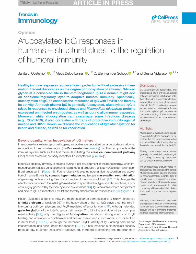
Beyond quantity: when fucosylation of IgG mattersIn response to a wide range of pathogens, antibodies are deposited on target surfaces, allowingrecognition of their constant region (the Fc domain; see Glossary) by other components of theimmune system such as the first molecule initiating the classical complement pathway(C1q) as well as cellular antibody receptors (Fc receptors) (Figure 1A) [1].
Extensive antibody diversity is created during B cell development in the bone marrow when im-munoglobulin variable gene segments rearrange and produce a unique variable domain in eachB cell precursor [1] (Figure 1B). Further diversity is created upon antigen recognition and activa-tion of mature B cells by somatic hypermutation and isotype class-switch recombinationof gene segments encoding the constant region of the immunoglobulin [1,2]. This diverges theeffector functions from the initial IgM-mediated to specialized isotype-specific functions, a pro-cess largely governed by the local cytokine environment [3,4]. IgG can activate both complementand bind to IgG-Fc receptors (FcγRs) and thereby shape immune responses [2,5,6] (Figure 1C).
Recent evidence underlines how the monosaccharide composition of a highly conservedN-linked glycan at position 297 in the heavy chain of human IgG plays a central role infine-tuning both complement and FcγR-mediated effector functions [7]. Although elevatedgalactosylation of the IgG-Fc glycan stimulates C1q binding and downstream comple-ment activity [8,9], only the degree of fucosylation has shown strong effects on FcγRbinding and activation in biochemical and cellular assays and in vivo models, as describedin detail later [8,10–12]. Although the increased FcγRIII affinity of IgG lacking core fucose(afucosylation) has been known for decades [10,11], it has remained a biochemical curiositybecause IgG is almost exclusively fucosylated, therefore questioning the importance of
Trends in Immunology, Month 2022, Vol. xx, No. xx https://doi.org/10.1016/j.it.2022.08.001 1© 2022 The Authors. Published by Elsevier Ltd. This is an open access article under the CC BY-NC-ND license (http://creativecommons.org/licenses/by-nc-nd/4.0/).
2Department of Biomolecular MassSpectrometry and Proteomics, UtrechtInstitute for Pharmaceutical Sciencesand Bijvoet Center for BiomolecularResearch, Utrecht University, Utrecht,The Netherlands3Landsteiner Laboratory, AmsterdamUniversity Medical Center (UMC),University of Amsterdam, Amsterdam,The Netherlands
*Correspondence:[email protected] (G. Vidarsson).
Trends in ImmunologyOPEN ACCESS
afucosylated IgGs in human immunity, and limiting the interest to mainly biotechnical andtherapeutic exploitation of this phenomenon in cancer [12,13]. However, a growing bodyof evidence highlights that some antigen-specific IgG responses can be predominantlyafucosylated, including responses to alloantigens, enveloped viruses, and Plasmodiumfalciparum proteins, which can be either beneficial or detrimental depending on the setting[14–28]. In this article we focus on the biological and clinical significance of antibodyglycosylation in humans, including fucosylation, that affect FcγR-mediated effector func-tions; we allude to a possible regulatory mechanism of afucosylated antibody responsesby introducing a novel evolutionary hypothesis which might help to explain why self mem-brane-bound antigens are a necessity for afucosylated IgG responses to occur. Lastly,we examine the possibility of exploiting IgG afucosylation for putative vaccine development.
N-linked IgG glycosylation and Fc effector functionsN-linked glycan structures are not directly encoded by the genome but are constructed throughenzymatic pathways. Following initial assembly on a lipid anchor at the cytoplasmic site of the en-doplasmic reticulum (ER) membrane, glycans are flipped to the luminal side for further extensionto form N-glycan precursors (reviewed in [29]). Glycan precursors are cotranslationally attachedto proteins at accessible consensus motifs (NxS/T; x being any amino acid except proline) byoligosaccharyltransferase complexes and are further modified by a coordinated series ofglycosyltransferases and glycosidases as the protein moves from the endoplasmic reticulum(ER) to the Golgi apparatus [29].
VH
Variable region (Fab)Constant region (Fc)
FucoseN-acetylglucosamine (GlcNAc)
Sialic acidGalactose
Mannose
CH1
CH2
CH3
VL
CL
(B) (C)
Increased affinityfor afucosylated IgG
VDJ recombination of VH and VL
IgG subclasses
Asn297-glycan composition
IgG1 IgG2 IgG3 IgG4
(A) Immunoglobulin G (IgG) Diversity Effector functions
Fc receptor binding
C1q binding IgG1≈IgG3IgG2IgG4
FcγRI FcγRIIa FcγRIIb FcγRIIIa FcγRIIIb
Bisecting GlcNAc
Increased binding tohexamerized, galactosylated IgG1
Core structure
FUT8
MGAT3
B4GALT1 ST6GAL1
V D J H
Asn297
TrendsTrends inin ImmunologyImmunology
Figure 1. Diversity and effector functions of human IgG. (A) Schematic representation of IgG with variable (Fab) and constant (Fc) regions in the green and orangepanels, respectively. The localization of the conserved Asn297 glycan in the Fc domain (CH2) is indicated [2]. (B) Antibody diversity arises during B cell development whenrearrangement of variable, diversity, and joining gene regions [V(D)J] results in a diverse repertoire of antigen-specificities (upper panel) [2]. Upon B cell activation, B cellsundergo isotype switching allowing them to produce antibodies of the IgG class, which is further subdivided into IgG1, IgG2, IgG3 and IgG4 subclasses (middle panel) [2].The variable composition of the Asn297 glycan also contributes to IgG diversity, and different glycosyltransferases are responsible for enzymatic addition of residues to thecore structure (GlcNAc4Man3) [30]. These modifications include the addition of galactose (B4GALT1), sialic acid (ST6GALT), bisecting GlcNAc (MGAT3), and core fucose(FUT8) (lower panel) [30]. (C) The diverse repertoire of IgG shapes effector functions, including the interaction of IgG-Fc with C1q, to activate the complement system, andFcγRs, to elicit antibody-dependent cellular cytotoxicity and phagocytosis [antibody-dependent cellular cytotoxicity (ADCC) and antibody-dependent cellular phagocytosis(ADCP)] [2]. The affinities of the different IgG subclasses for effector molecules are indicated by the individual color and thickness of the arrows [5]. Only the Asn162 glycanpresent in FcγRIIIa is shown as a biantennary glycan, although all FcγRs containmultipleN-linked glycans and also some of high-mannose structures. Abbreviations: CH1–3, immunoglobulin heavy-chain constant regions 1–3; CL, light-chain constant region; VH, heavy-chain variable region; VL, light-chain variable region.
2 Trends in Immunology, Month 2022, Vol. xx, No. xx

GlossaryAffinity maturation: the process bywhich B cells increase their affinity forcognate antigen through randommutations of the variable region.Alloantigens/alloimmunization:immune response towards antigen fromthe same species towards a protein(alloantibody) other than that of theresponding individual.Antibody-dependent cellularcytotoxicity (ADCC) andantibody-dependent cellularphagocytosis (ADCP): Fc effectorfunctions induced upon engagement ofFc receptors on immune cells byopsonized targets resulting incytotoxicity or phagocytosis of targets,respectively.Antibody-dependent enhancement(ADE): the phenomenon whereantibody binding enhances viral entryand/or replication.Avidity-docking platform: a platformcreated by multiple target-bound IgGsthat increases the overall binding strengthbetween IgG-opsonized cells andFcγR-expressing effector cells, beyondthose of individual IgG–FcγR pairs, as aresult of themultivalency of the interaction.This also applies for C1q binding.Bisecting GlcNAc: aN-acetylglucosamine (GlcNAc)extension on theN-glycan core structurebetween the two N-glycan branches.Classical complement pathway: thepathway of the complement cascadethat is optimally activated upon theformation of IgG hexamers on targetsurfaces that allow binding of therecognition molecule C1q.Class-switch recombination: agenetic rearrangement of the constantgene segments of antibodies in B cellsthat leads to antibody class-switching.Dengue hemorrhagic fever (DHF): arare complication of dengue virus infectioncharacterized by high fever and damageto lymph and blood vessels, leading tosevere bleeding and organ failure.Dengue shock syndrome (DSS): alife-threatening complication in denguedisease defined as hypotension andnarrow pulse pressure.Elite controllers: individuals infectedwith HIV-1 who maintain undetectableviral loads in the absence of therapy.Fab regions: fragment antigen binding,the variable domain of an antibody that isresponsible for antigen binding.Fc domain: fragment crystallizable, theconstant domain of an antibody that is
Trends in ImmunologyOPEN ACCESS
The N-linked glycosylation motif at Asn297 in IgG is highly conserved [7]. The IgG-Fc glycan con-sists of an N-acetylglucosamine (GlcNAc) and a mannose biantennary core structure. These canbe modified by addition of a fucose residue to the primary GlcNAc, a bisecting GlcNAc to thecore structure, galactose residues to the antennary GlcNAcs, and sialic acid to galactose resi-dues [30] (Figure 1B). These modifications allow the formation of up to 36 different glycans thatare readily detectable in human plasma [13]. In addition to the conserved Fc N-glycosylationsite, 10–20%of IgGs also possessN-glycosylation sites in Fab regions, which are formed duringaffinity maturation, and influence the affinity of antibodies for antigens [31].
Residues proximal to the hinge region in the CH2 domain of IgG contain different but overlap-ping binding sites for C1q and FcγRs [32]. Although interactions between IgG and C1q re-quire an avidity-docking platform of Fc:Fc-oligomerized IgG, optimally forming hexamersfor binding of C1q and subsequent complement activation [33], FcγR activation requiresless or differently structured clustering of IgG on effector cells [34]. Humans have several ac-tivating FcγRs (FcγRI, FcγRIIa, FcγRIIc, FcγRIIIa, and FcγRIIIb) and at least one inhibitory re-ceptor (FcγRIIb) [35]. The integrated signal from these receptor–IgG-Fc interactions directsthe type and strength of the effector functions, such as antibody-dependent cellularcytotoxicity (ADCC) and antibody-dependent cellular phagocytosis (ADCP), in naturalkiller (NK) cells and various myeloid cells which express a selection of these receptors [36].The Fc glycan is essential for IgG effector function as it maintains the structure of the Fc do-main required for both FcγR and C1q docking [37,38]; indeed, removal of the IgG-Fc glycanabrogates Fc effector functions [39–41]. Furthermore, the exact composition of the IgG-Fcglycan affects its interaction with C1q and FcγRs, and thereby shapes IgG effector activity,as described later [8].
In summary, the N-linked glycan of IgG-Fc is required for IgG effector functions and its composi-tion fine-tunes Fc effector functions.
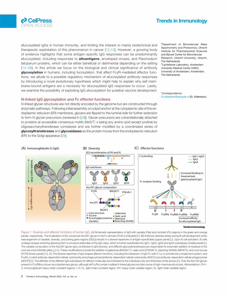
Fc fucosylationIn recent decades studies have demonstrated that omitting the core fucose of the Fc glycanenhances FcγRIII activity, as evidenced from the improved ADCC capacity of afucosylatedIgG compared with fucosylated IgG against multiple cellular targets, using human peripheralblood mononuclear cells (PBMCs) or NK cells as effector cells [10,11]. Because of this,afucosylated IgG is currently being exploited to increase the efficacy of therapeuticantibodies [12].
A unique glycan at Asn162 in FcγRIIIa and FcγRIIIb [42,43] collides with the core fucose andlimits the accessibility of the FcγRIII-glycan which needs to be accommodated within the con-formational space of the IgG-Fc, thereby leading to suboptimal IgG binding [44–46]. As a con-sequence, afucosylated IgG has an up to 40-fold increased affinity compared with fucosylatedIgG, as evidenced from surface plasmon resonance (SPR) experiments, and this explainsthe enhanced ADCC activity [11,45,47–49] (Figure 2). The functional effect on NK cell ADCCin many cases has even exceeded the aforementioned 40-fold affinity increase, most likelydue to the increased avidity of the interaction [10,11]. The higher affinity of afucosylated IgGfor FcγRIIIb correlates with enhanced phagocytosis of human platelets and bacteria byFcγRIIIb+ neutrophils, but also reduces ADCC of human cancer cells by neutrophils[14,50,51]. The latter is presumably because less IgG is available for FcγRIIa activation,which receptor-blocking experiments have shown to be essential for neutrophil-mediatedADCC [50,51]. Afucosylation of IgG thus functions as a binary switch which increases the po-tency of IgG by enhanced affinity to FcγRIII.
Trends in Immunology, Month 2022, Vol. xx, No. xx 3

responsible for binding to effectormolecules of the immune system.Fetal and neonatal alloimmunethrombocytopenia (FNAIT): a bleed-ing disorder during pregnancy causedby maternal alloantibodies that targetpaternally inherited antigens on theplatelets of the fetus/newborn.Fucosylation: the addition of a fucoseresidue to a glycan/sugar branch.Galactosylation: the addition of agalactose residue to a glycan/sugarbranch.Glycosidases: enzymes that catalyzethe hydrolysis of glycosidic bonds duringthe processing or catabolism ofoligosaccharides.Glycosylation: a post-translationalmodification in which sugar branchesare attached to other macromoleculesincluding proteins.Glycosyltransferases: enzymes thatcatalyze the transfer ofmonosaccharides to carbohydratesduring the synthesis of oligosaccharidesby creating a glycosidic bond.Hemolytic disease of the fetus andnewborn (HDFN): a bleeding disorderthat can occur when maternalalloantibodies against red blood cells(RBCs) cross the placenta and mediatethe destruction of cells expressing thetargeted antigen.N-linked glycans: oligosaccharidesattached to asparagine (Asn, N) residuesat a consensus motif N-x-S/T where x isany amino acid except proline.Non-neutralizing antibodies:antibodies that bind to pathogenswithout disturbing receptor–ligandinteractions necessary for infection; theycan (potentially) have protective ordisease-enhancing effects throughFc-mediated effector functions.Pyroptosis: inflammatory lyticprogrammed cell death, often triggeredby intracellular pathogens, thatculminates in activation of caspases andthe inflammasome leading toproinflammatory cytokine release andcellular lysis.RhD and K: alloantigens on RBCs, alsoknown as blood group systems.Sialoside ligands: ligands with sialicacid-containing carbohydrates.Sialylation: the addition of a sialic acidresidue to a glycan/sugar branch.Somatic hypermutation:hypermutation in the variable generegions of antibodies in fullydeveloped B cells contributing toaffinity maturation of the antibodyproduced by the cell.
Trends in ImmunologyOPEN ACCESS
Fc galactosylation, sialylation, and bisectionAlthough no effect was initially found on NK cell-mediated ADCC activity against various cell linesby Fc galactosylation [10,52], recent SPR and chromatography data demonstrate that Fcgalactosylation doubles the affinity of afucosylated IgG for FcγRIIIa, whereas it has less effecton fucosylated IgG [8,53,54]. Moreover, studies emphasize that galactosylation enhances thehexamerization potential of IgG; indeed, increased C1q binding and complement activationhave been demonstrated onmultiple solid-phase assays and cellular targets with human comple-ment [8,9,55,56]. Although difficult to isolate from galactosylation, Fc sialylation appears to mar-ginally enhance C1q binding and complement activation [8,56,57], whereas only negligible effectsof FcγR and C1q binding have been reported that are attributable to the bisecting GlcNAc [8].
Changes in IgG glycosylation with agingPlasma IgG-Fc glycans of human adults are ~94% fucosylated, 40% galactosylated, 8% bisected,and 4% sialylated (where both the degrees of galactosylation and sialylation refer to antenna occu-pation of the IgG-Fc glycan) [58]. Fc galactosylation and sialylation decrease with age in bothmalesand females, the latter exhibiting the most prominent reduction during menopause, but a transientincrease occurs during pregnancy, suggesting hormonal regulation of these traits [58,59].
By contrast, the degree of bisecting GlcNAc and Fc fucosylation is largely unchanged throughoutthe human lifespan [58]. Newborns harbor almost exclusively maternal IgG, and hence IgG glyco-sylation of neonates is virtually identical to that of the mother [60,61], although minor differenceshave been reported, possibly caused by methodological differences [62]. As the infant’s own IgGdisplaces the maternal IgG, IgG-Fc fucosylation of the infant reaches almost 100% 1 month afterbirth [61]. The fucosylation degree gradually decreases to 94% at ~20 years of age and only de-creases marginally thereafter [58,61]. As explained later, this decrease of fucosylation with age ispossibly due to the accumulation of afucosylated IgG acquired during immune responses againstpathogens that stimulate the formation of afucosylated IgG [27]. The degree of bisecting GlcNAcgenerally correlates negatively with the degree of fucosylation, suggesting that bisection may ste-rically inhibit fucosylation [61].
Thus, Fc glycosylation of IgG changes throughout life. Although the galactosylation and sialylationof IgGmay be partly hormonally regulated, the changes in IgG-Fc fucosylation likely arise from theaccumulation of antigen-specific IgG responses with decreased Fc fucosylation.
Afucosylated IgG responses in alloimmunity and infectious diseasesDespite the low abundance and relatively stable concentrations of afucosylated IgG in plasma,multiple studies of antigen-specific IgG, all using affinity-purified IgG andmass spectrometry anal-ysis, recently highlighted the clinical importance of some antigen-specific afucosylated IgG re-sponses, which were also verified by FcγRIIIa-dependent serological methods in severe acuterespiratory syndrome coronavirus 2 (SARS-CoV-2) responses [14–28,63–65]. Theseafucosylated IgG responses include those directed towards alloantigens (Table 1), which cancontribute to alloimmune-mediated diseases (Box 1), against enveloped viruses, and againstP. falciparum proteins expressed on red blood cells (RBCs), all responses in which the degreeof afucosylation can influence disease severity [14–28,63–65].
Fetal and neonatal alloimmune thrombocytopeniaFetal and neonatal alloimmune thrombocytopenia (FNAIT) is caused by alloantibodiesagainst fetal human platelet antigens (HPAs) induced by maternal exposure to fetal cells carryinga paternally derived incompatible alloantigen [66]. More than 80% of FNAIT cases are estimatedto result from HPA-1a incompatibility [66]. HPA-1a-specific alloantibodies can be found in ~0.2%
4 Trends in Immunology, Month 2022, Vol. xx, No. xx

Surface plasmon resonance (SPR):a label-free analytical technique formeasuring binding kinetics and affinity ofmolecular interactions.Toll-like receptors (TLRs): immunereceptors expressed by a wide range ofimmune cells that sense pathogen-associated molecular patterns.
Trends in ImmunologyOPEN ACCESS
of pregnancies in the Caucasian population, and these result in thrombocytopenia (<150 × 109
thrombocytes/l) in ~50% of cases with HPA-1a-specific alloantibodies [66]. Severe FNAIT ismuch more rare (~1–2% of HPA-1a-immunized cases) and is characterized by internal bleeding,especially intracranial hemorrhage (ICH), often resulting in lifelong disabilities or death [66]. Fcfucosylation of HPA-1a-specific IgG shows great variation, from virtually 100% down to ~10%[14,15,21]. Decreased Fc fucosylation causes enhanced phagocytosis of platelets in vitro byFcγRIIIa+ monocytes and FcγRIIIb+ neutrophils, both likely contributing to enhanced plateletphagocytosis in the fetal liver and spleen [22,45]. In agreement, antibody titers and platelet countsdo not strictly correlate with ICH incidence, whereas HPA-1a-specific IgG-Fc fucosylation corre-lates positively with neonatal platelet counts and negatively with disease severity [14,15,67]. Fur-thermore, Fc fucosylation was found to be the strongest predictor of bleeding severity, followedby elevated galactosylation and antibody titers [15]. As HPA-1a-specific IgG afucosylation is sta-ble during pregnancy and in subsequent pregnancies for at least a decade, afucosylation couldpotentially identify pregnancies at risk [14,15].
Hemolytic disease of the fetus and newbornSimilarly to immune responses against platelets, IgG afucosylation has also been reported in caseswhere these antibodies are directed against alloantigens on RBCs, causing hemolytic disease of
Target cell
Cytotoxicity (ADCC)
NK-cell
Phagocytosis (ADCP)
ADCC/ADCP
Total IgG (afucosylated)
Total IgG (fucosylated)
Antigen specific IgG (afucosylated)
Antigen specific IgG (fucosylated)
Effector cell
Plasma - steady state
Effector cell
Plasma - immune response
IgG Asn297 (fucosylated)
FcyRIIIa Asn162 Monocyte
Target cell
Target cell
TrendsTrends inin ImmunologyImmunology
Figure 2. Afucosylation of antigen-specific IgG can affect steady-state FcγRIII occupancy and effector activities in humans. Under steady-state conditions(left panel), most FcγRIII are occupied by afucosylated IgGs with random specificities (depicted in green, ~6% of total plasma IgG) as their affinity is higher than theirfucosylated counterparts (depicted in gray, ~94% of total plasma IgG) [48,58]. During effector responses (right panel), fucosylated IgG (in blue) displaces an aspecificafucosylated IgG already occupying FcγRIII less efficiently, due to differences in affinities [48]. By contrast, afucosylated IgG response (in orange) will displace anaspecific afucosylated IgG more easily, resulting in strong antibody-dependent cellular cytotoxicity and phagocytosis (ADCC/ADCP) activity [48]. Abbreviations: ADCC,antibody-dependent cellular cytotoxicity; ADCP, antibody-dependent cellular phagocytosis; NK, natural killer.
Trends in Immunology, Month 2022, Vol. xx, No. xx 5

Table 1. Characteristics of afucosylated IgG responses in humans
Antigen Degree ofmaximumafucosylation
Interindividualspread
Stability ofresponse
Consequences fordisease
Vaccine responsea Refs
Alloimmune diseases
Pregnancy HPA-1a ↑↑↑ High Stable for up to7 yearspost-delivery
Correlates with increaseddisease severity in FNAIT
N/A [14,15,21]
HPA-3a,HPA-5b
↑↑↑ Unknown Unknown Unknown N/A [21]
RhD ↑↑↑ High Stable for up to9 yearspost-delivery
Associated with a highrisk of HDFN
N/A [22–24]
K ↑↑↑ High Unknown Associated with severefetal anemia
N/A [24,25]
Transplantation HLA ↑↑↑ High Unknown Associated withantibody-mediated graftrejection
N/A [20]
Enveloped viruses
HIV Envelopeprotein
↑ Low Stable for atleast 3 years
Feature of HIV elitecontrollers
Unknown [26,27]
Dengue virus Envelopeprotein
↑↑ Intermediate Unknown Associated with diseaseprogression
Unknown [28]
Cytomegalovirus Antigenmixture
↑↑↑ High Stable for up toa decade
Unknown Unknown [27]
Hepatitis B virus HBsAg ↑ Intermediate Unknown Unknown Soluble HBsAgprotein subunitvaccine inducesfucosylatedHBsAg-specific IgG,unlike naturalinfection
[27]
Measles virus(morbillivirusmeasles virus)
Antigenmixture
↑ Low Stable formultipledecades
Unknown Live attenuatedviruses induceafucosylatedantigen-specific IgG,similarly to naturalinfection
[27]
Mumps virus(mumpsorthorubulavirus)
Antigenmixture
↑↑ Low Stable formultipledecades
Unknown Live attenuatedviruses induceafucosylatedantigen-specific IgG,similarly to naturalinfection
[27]
SARS-CoV-2 Spikeprotein
↑↑ Intermediate Transientafucosylation afterseroconversion;long-termunknown
Correlates with systemicimmunopathology
mRNA vaccineinduces afucosylatedspike-specific IgG,similarly to naturalinfection
[16,17,27,63,64]
Respiratorysyncytial virus
Antigenmixture
Low Unknown Correlates with NK cellactivity
Unknown [19]
Intracellular parasites
Plasmodiumfalciparum
VAR2CSA ↑↑↑ High Present for atleast a decade;decreases withantigenexposure
Protection acquired in aparity-dependent mannercorrelates with decreasingFc fucosylation levels withincreasing exposure
Soluble VAR2CSAprotein subunit vaccineinduces fucosylatedantigen-specific IgG,unlike natural infection
[18]
Trends in ImmunologyOPEN ACCESS
6 Trends in Immunology, Month 2022, Vol. xx, No. xx
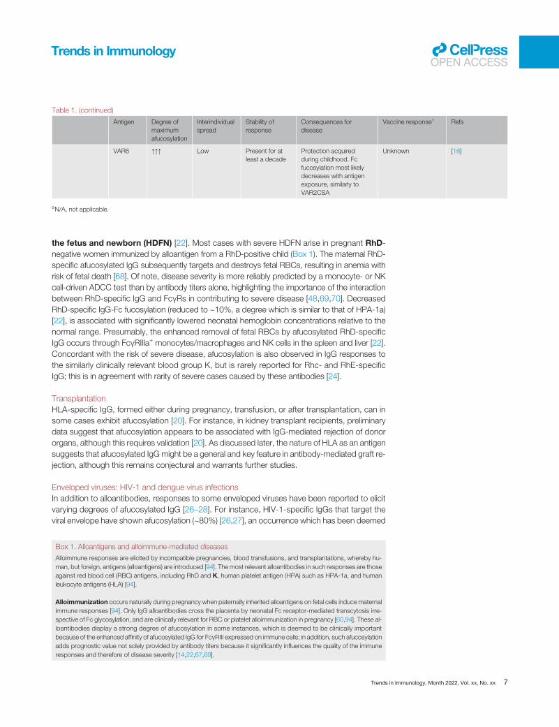
Table 1. (continued)
Antigen Degree ofmaximumafucosylation
Interindividualspread
Stability ofresponse
Consequences fordisease
Vaccine responsea Refs
VAR6 ↑↑↑ Low Present for atleast a decade
Protection acquiredduring childhood. Fcfucosylation most likelydecreases with antigenexposure, similarly toVAR2CSA
Unknown [18]
aN/A, not applicable.
Trends in ImmunologyOPEN ACCESS
the fetus and newborn (HDFN) [22]. Most cases with severe HDFN arise in pregnant RhD-negative women immunized by alloantigen from a RhD-positive child (Box 1). The maternal RhD-specific afucosylated IgG subsequently targets and destroys fetal RBCs, resulting in anemia withrisk of fetal death [68]. Of note, disease severity is more reliably predicted by a monocyte- or NKcell-driven ADCC test than by antibody titers alone, highlighting the importance of the interactionbetween RhD-specific IgG and FcγRs in contributing to severe disease [48,69,70]. DecreasedRhD-specific IgG-Fc fucosylation (reduced to ~10%, a degree which is similar to that of HPA-1a)[22], is associated with significantly lowered neonatal hemoglobin concentrations relative to thenormal range. Presumably, the enhanced removal of fetal RBCs by afucosylated RhD-specificIgG occurs through FcγRIIIa+ monocytes/macrophages and NK cells in the spleen and liver [22].Concordant with the risk of severe disease, afucosylation is also observed in IgG responses tothe similarly clinically relevant blood group K, but is rarely reported for Rhc- and RhE-specificIgG; this is in agreement with rarity of severe cases caused by these antibodies [24].
TransplantationHLA-specific IgG, formed either during pregnancy, transfusion, or after transplantation, can insome cases exhibit afucosylation [20]. For instance, in kidney transplant recipients, preliminarydata suggest that afucosylation appears to be associated with IgG-mediated rejection of donororgans, although this requires validation [20]. As discussed later, the nature of HLA as an antigensuggests that afucosylated IgG might be a general and key feature in antibody-mediated graft re-jection, although this remains conjectural and warrants further studies.
Enveloped viruses: HIV-1 and dengue virus infectionsIn addition to alloantibodies, responses to some enveloped viruses have been reported to elicitvarying degrees of afucosylated IgG [26–28]. For instance, HIV-1-specific IgGs that target theviral envelope have shown afucosylation (~80%) [26,27], an occurrence which has been deemed
Box 1. Alloantigens and alloimmune-mediated diseases
Alloimmune responses are elicited by incompatible pregnancies, blood transfusions, and transplantations, whereby hu-man, but foreign, antigens (alloantigens) are introduced [94]. The most relevant alloantibodies in such responses are thoseagainst red blood cell (RBC) antigens, including RhD and K, human platelet antigen (HPA) such as HPA-1a, and humanleukocyte antigens (HLA) [94].
Alloimmunization occurs naturally during pregnancy when paternally inherited alloantigens on fetal cells induce maternalimmune responses [94]. Only IgG alloantibodies cross the placenta by neonatal Fc receptor-mediated transcytosis irre-spective of Fc glycosylation, and are clinically relevant for RBC or platelet alloimmunization in pregnancy [60,94]. These al-loantibodies display a strong degree of afucosylation in some instances, which is deemed to be clinically importantbecause of the enhanced affinity of afucosylated IgG for FcγRIII expressed on immune cells; in addition, such afucosylationadds prognostic value not solely provided by antibody titers because it significantly influences the quality of the immuneresponses and therefore of disease severity [14,22,67,69].
Trends in Immunology, Month 2022, Vol. xx, No. xx 7

Trends in ImmunologyOPEN ACCESS
to be more prominent in HIV-1-infected elite controllers compared with non-elite controllers.For the former, strong NK cell-mediated ADCC responses against HIV-1-infected human CD4+
T cells correlated with viral inhibition in vitro [26], suggesting that afucosylation of HIV-1-specificIgGs is associated with protection.
In contrast to HIV-1, decreased Fc fucosylation of IgG directed against antigens of the viral enve-lope of dengue virus is associated with disease escalation [28]. Moreover, non-neutralizingantibodies from heterotypic dengue virus serotypes are considered to be risk factors for devel-oping dengue hemorrhagic fever (DHF) and dengue shock syndrome (DSS) because theyenhance viral uptake, replication, and the production of proinflammatory cytokines, mediated byFcγRs expressed on immune cells, referred to as antibody-dependent enhancement (ADE)[28,71]. In agreement with the increased affinity of afucosylated IgGs for FcγRIII and the factthat ADE often relies on FcγR, the higher degree of afucosylation of antigen-specific IgGs isassociated with DHF and DSS, as evidenced by the elevated concentrations of afucosylateddengue-specific IgG in patients with DHF/DSS, thus suggesting a pathogenic role of afucosylatednon-neutralizing IgGs in these diseases [28,72].
Enveloped viruses: cytomegalovirus, hepatitis B virus, respiratory syncytial virus, measles virus,and mumps virus infectionsOther enveloped viruses also elicit afucosylated IgG immune responses [27]. Although the extentof afucosylated IgGs that are generated against respiratory syncytial virus (RSV) and hepatitis Bvirus (HBV) are minor, they vary (<80% fucosylation) and correlate with NK cell activation, stimu-lated by immobilized RSV-specific IgG [19,27]. The extent of afucosylated IgGs against measles(morbillivirus measles virus) and mumps virus (mumps orthorubulavirus) are similar to those elic-ited by dengue virus and HIV-1, whereas cytomegalovirus (CMV) stimulates the strongestafucosylated IgG responses of all viruses studied so far, with fucosylation levels resemblingthose of RhD- and HPA-1a-specific IgGs, and these can remain stable for at least a decade[14,15,23,27]. Moreover, the responses to measles and mumps viruses that have been exam-ined several decades after the primary response also reveal stable Fc fucosylation profiles forthese antibodies. However, the clinical implications of stably afucosylated IgG for protectionand disease progression during these viral infections remain unknown [27].
Enveloped virus: SARS-CoV-2 infectionsThe pathogenesis of coronavirus disease 2019 (COVID-19), caused by SARS-CoV-2, is highlyvariable, ranging from asymptomatic to severe respiratory failure, and fatal outcomes are linkedto excessive immune activation [73,74]. Afucosylated IgGs targeting the viral membrane spikeprotein have been reported, particularly in hospitalized patients [16,17,27,63]. The most promi-nent afucosylation (reduced to ~60%) coincided with seroconversion and admission to the inten-sive care unit (ICU), after which fucosylation can increase to >90% within weeks followingrecovery of individuals who did not succumb to the first inflammatory wave [27]. The disease pro-gression in individuals with afucosylated IgG might be explained by increased binding of viral im-mune complexes to FcγRIII-bearing cells, leading to potent activation of the latter, and whichcould have potentially led to excessive release of proinflammatory cytokines associated with se-vere COVID-19. In partial agreement with this hypothesis, in vitro stimulation of monocyte-derivedalveolar type macrophages from healthy donors with afucosylated IgG immune complexes, in thepresence of a Toll-like receptor (TLR) stimulus, has been reported to enhance FcγRIIa- andFcγRIIIa-dependent secretion of proinflammatory cytokines [17,27].
Moreover, recent data suggest that CD16+ monocytes are a primary target for the uptake andsterile RNA replication of IgG-opsonized SARS-CoV-2 through FcγRIIIa, thereby triggering
8 Trends in Immunology, Month 2022, Vol. xx, No. xx

Trends in ImmunologyOPEN ACCESS
inflammasome activation and pyroptosis, and supporting the idea that afucosylated IgGs fuelstrong proinflammatory responses [75,76]. In addition, interleukin (IL)-6 and C-reactive proteinplasma concentrations have been reported to be elevated in COVID-19 ICU patients, but not inindividuals with mild symptoms, and the former inversely correlate with Fc fucosylation ofspike-specific IgG [27]. This suggests that afucosylated IgG early in the responsemight cause ex-cessive FcγRIIIa stimulation of cells such as alveolar macrophages, presumably contributing tosystemic immunopathology – that might partly resolve as the degree of Fc fucosylation increases,a phenomenon which remains to be directly validated [27]. Indeed, as studies of responsesagainst other viruses so far have all been carried out after the primary response has ceased[26–28], it remains unclear whether transient afucosylation, as observed for SARS-CoV-2, is ageneral phenomenon.
Plasmodium falciparum parasite infectionsImmune responses towards the protozoan parasite P. falciparum, that is responsible for mostmalaria cases in sub-Saharan Africa, also show markedly reduced fucosylation of IgG againstparasite antigens expressed on infected RBCs [18]. These proteins, members of theP. falciparum erythrocyte membrane protein-1 (PfEMP1) family, are required for adhesion of in-fected RBCs to host receptors on endothelium and tissues, and are therefore necessary for par-asite survival [77]. PfEMP1 antigen VAR6-specific IgG has shownmarked afucosylation, whereasIgG against the merozoite protein glutamate-rich protein (GLURP), which is not displayed on hostRBCs, has not [18]. In contrast to alloimmune responses and antiviral responses, IgG specific forthe pregnancy-restricted PfEMP1 VAR2CSA displays exposure-dependent Fc fucosylation be-cause the fucosylation decreases with increasing parity – a proxy for possible exposures toVAR2CSA in endemic areas [18]. This might offer a putative molecular mechanism to explainthe correlation between parity number and reduced risk of pregnancy-associated malaria [78].Of note, the afucosylation in the responses to PfEMP1s is more prominent than that observedin IgGs against enveloped viruses (~25% vs ~85% fucosylation, respectively) [18]. As the degreeof fucosylation of PfEMP1-specific IgG correlates with features known to associate with protec-tion (parity), it is tempting to assign decreased fucosylation as a protective trait, although furthervalidation is needed [18,78].
In summary, alloimmune responses against RBCs and platelets, as well as responses againstenveloped viruses and specific P. falciparum antigens, can elicit afucosylated IgGs, and these ap-pear to be of clinical importance, although the underlying mechanisms remain to be investigated.Of note, the gradual decrease of Fc fucosylation during adolescence coincides with the accumu-lation of immunity to enveloped viruses and other intracellular pathogens, suggesting that theseantigens form a unique category that stimulates B cells to produce afucosylated IgGs, whichare otherwise rare.
The importance of antigen context for Fc fucosylationMost immune responses induce fully fucosylated IgG, in agreement with plasma IgG findings of~94% IgG fucosylation [58]. This includes responses against bacterial polysaccharides (andprotein-conjugated vaccines thereof), inactivated influenza virus, tetanus toxoid vaccine(Clostridium tetani), and citrullinated proteins [79–82] (Figure 3A, Key figure). Conversely,afucosylated IgG responses share a common feature in that the antigen is localized in the hostcell membrane [14,18,20,22,26–28]. For instance, in FNAIT, HDFN, and P. falciparum infections,the antigens localize to platelet- or RBC membranes (Figure 3B). Similarly, during infections withenveloped viruses, viral antigens are exposed on the host membrane after fusion of the viral en-velope during invasion and when virions are assembled for viral budding [83] (Figure 3C). Basedon the similarity of IgG-Fc fucosylation in these responses, we recently proposed that antigen
Trends in Immunology, Month 2022, Vol. xx, No. xx 9

Key figure
Hypothetical model illustrating the possible role of antigen context-induced alteration of immunesignaling and the production of afucosylated IgG in humans
(A) (C)
Bacteria
B cell B cell
Adeno/mRNA shuttles
Infected cellForeign/alloantigen
Self
Recognition of self
Non-enveloped virus
Malaria parasite
Enveloped virus
Soluble antigens Antigens from enveloped viruses
Budding Fusion
(B) Self-membrane antigens (D) Vaccine design
TrendsTrends inin ImmunologyImmunology
Figure 3. (A) Soluble antigens, derived from non-enveloped viruses or bacteria, signal through the B cell receptor (BCR) resulting in B cell activation and the production offucosylated antigen-specific IgG [79–82]. (B) For alloantigens on platelets and red blood cells (RBCs) (not shown), and Plasmodium falciparum proteins expressed oninfected RBCs, we propose that antigen recognition by the BCR is accompanied by a so far unknown receptor–ligand pair that provides a signal for 'self'. Signalingthrough these complexes is required for the induction of afucosylated IgG [14,18,20,22]. (C) Similarly, responses towards enveloped viral infections resembleresponses described in (B) for alloantigens and P. falciparum proteins, as these antigens are also self-membrane-bound [27]. Self-recognition might either occur fromthe virion (left) if segments of the host cell membrane are included in the viral envelope, or (right) from virus-infected cells as the viral envelope fuses with the hostmembrane [83], both resulting in the production of afucosylated antigen-specific IgG [26–28]. (D) The potency of afucosylated IgG might be harnessed in vaccinedesign by mimicking natural antigen display as depicted in (B) and (C). Using mRNA templates or viral shuttles for transcription in host cells, foreign antigens might beexpressed in the context of a self-membrane which could induce afucosylated IgG [64]. Abbreviation: adeno, adenovirus.
Trends in ImmunologyOPEN ACCESS
recognition by B cells in the context of a host membrane activates a so far unknown receptor–ligand pairs which could be necessary for the induction of afucosylated IgG [27] (Figure 3). How-ever, although membrane sensing is simple and plausible (Box 2 and see Figure I in Box 2), it isinsufficient per se – as illustrated by the large interindividual and interantigen variation of IgGafucosylation, suggesting that more factors are involved [14,18,20,22,26–28].
Genome-wide association studies have implied that the transcription regulator IKAROSmight ac-count for small differences in IgG fucosylation in B cells, whereas the transcription factor HNF-1αmight control the expression of fucosyltransferases in hepatic cells, suggesting that proteinfucosylation is likely controlled on the transcriptomic level [84,85]. Furthermore, during HIV-1
Box 2. Receptor–ligand pairs involved in regulating B cell activation and differentiation
Fine-tuning of B cell responses is provided by a vast array of regulatory receptor–ligand pairs [4] (Figure IA–D). Upon antigen engagement, B cell receptors (BCRs) clusteron the membrane and initiate signaling pathways that are essential for B cell survival, proliferation, and differentiation [95]. In addition, B cell-specific receptors such asCD19 and CD21, MHC class II, and CD40 are important surface molecules for B cell activation [4]. Furthermore, T helper cells and dendritic cells modulate the B cellresponse by secreting various cytokines including IL-4, interferon-γ, and transforming growth factor-β which regulate B cell differentiation and antibody features suchas isotype switching [4]. Other cytokines such as IL-21 and IL-17, as well as co-stimulation through Toll-like receptors (TLRs), also play a vital role in B cell activation andshaping the antibody response [4,96,97]. During antigen binding by BCRs in the context of human B cell membranes, receptor–ligand pairs, including known 'self-markers' such as CD47 or sialoside ligands, and their receptors SIRP-γ and CD22, respectively, might serve as putative checkpoints for the expression ofafucosylated IgGs, given that self-membrane association appears to be a commonality of antigens that can stimulate afucosylated IgGs [27,98–100] (Figure IE,F).
10 Trends in Immunology, Month 2022, Vol. xx, No. xx

CD40-L
R
L
(E)
(D)
C3b
CD81
PAMPs (e.g. LPS, ssRNA, CpG, flagellin)
CD47
CD22SIRP-γ
Sialosideligand
Cytokines (IL-4, IFNγ, TGFβ, IL-21, IL17)
(C)(B)(A)
(F)
??
TLR1-9
T-cell
B-cell
IgG fucosylation
B-cell
Infected cell
BCR
BCR CD21
CD19
CD40 MHC-II
CD4
TCR
?
B-cell regulatory receptor-ligand pairs
B-cell sensing of ‘self’
Proliferation, survival, isotype switching, somatic hypermutation, IgG glycosylation
Receptor Ligand Ligand expression
PD1 PD-1L/2L Dendri�c cells, macrophages, T-cells, B-cells, epithelial cells
CD72 CD100 T-cells
LIR1 HLA-I Nucleated cells
CEACAM1 CEACAM Leukocytes, epithelial cells, endothelial cells
BAFFR Transmembrane BAFF Macrophages, monocytes, dendri�ccells
LAIR1 Collagen ECM component
TrendsTrends inin ImmunologyImmunology
Figure I. Immunoregulatory receptor–ligand pairs involved in fine-tuning the B cell response in humans. (A–D) Recognition of antigen by B cell receptors(BCRs) results in BCR clustering and crosslinking with the CD19–CD21 coreceptor complex thereby increasing BCRmediated signaling [4,95]. In addition, signaling viaToll-like receptors (TLRs), CD40–CD40L, and cytokine receptors is important for B cell differentiation, isotype switching, and IgG glycosylation [4]. (E) A similar signalingaxis might be involved in regulating IgG fucosylation through sensing of ‘self’ (e.g., CD47 or sialoside ligands sensed by B cells) [98,99] (F), but other receptors andligands, depicted in (E) as R and L, respectively, might also be involved depending on the infected cell type, thus driving the switch to producing afucosylated IgG[100]. Abbreviations: CpG, CpG-rich DNA; ECM, extracellular matrix; IL, interleukin; LPS, lipopolysaccharide; PAMP, pathogen-associated molecular pattern;ssRNA, single-stranded RNA; TCR, T cell receptor; TGF, transforming growth factor.
Trends in ImmunologyOPEN ACCESS
Trends in Immunology, Month 2022, Vol. xx, No. xx 11
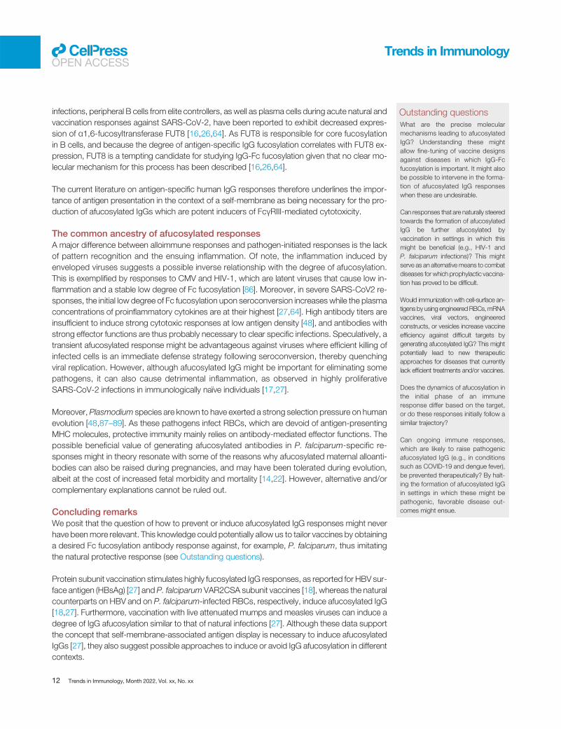
Outstanding questionsWhat are the precise molecularmechanisms leading to afucosylatedIgG? Understanding these mightallow fine-tuning of vaccine designsagainst diseases in which IgG-Fcfucosylation is important. It might alsobe possible to intervene in the forma-tion of afucosylated IgG responseswhen these are undesirable.
Can responses that are naturally steeredtowards the formation of afucosylatedIgG be further afucosylated byvaccination in settings in which thismight be beneficial (e.g., HIV-1 andP. falciparum infections)? This mightserve as an alternative means to combatdiseases for which prophylactic vaccina-tion has proved to be difficult.
Would immunization with cell-surface an-tigens by using engineeredRBCs,mRNAvaccines, viral vectors, engineeredconstructs, or vesicles increase vaccineefficiency against difficult targets bygenerating afucosylated IgG? This mightpotentially lead to new therapeuticapproaches for diseases that currentlylack efficient treatments and/or vaccines.
Does the dynamics of afucosylation inthe initial phase of an immuneresponse differ based on the target,or do these responses initially follow asimilar trajectory?
Can ongoing immune responses,which are likely to raise pathogenicafucosylated IgG (e.g., in conditionssuch as COVID-19 and dengue fever),be prevented therapeutically? By halt-ing the formation of afucosylated IgGin settings in which these might bepathogenic, favorable disease out-comes might ensue.
Trends in ImmunologyOPEN ACCESS
infections, peripheral B cells from elite controllers, as well as plasma cells during acute natural andvaccination responses against SARS-CoV-2, have been reported to exhibit decreased expres-sion of α1,6-fucosyltransferase FUT8 [16,26,64]. As FUT8 is responsible for core fucosylationin B cells, and because the degree of antigen-specific IgG fucosylation correlates with FUT8 ex-pression, FUT8 is a tempting candidate for studying IgG-Fc fucosylation given that no clear mo-lecular mechanism for this process has been described [16,26,64].
The current literature on antigen-specific human IgG responses therefore underlines the impor-tance of antigen presentation in the context of a self-membrane as being necessary for the pro-duction of afucosylated IgGs which are potent inducers of FcγRIII-mediated cytotoxicity.
The common ancestry of afucosylated responsesA major difference between alloimmune responses and pathogen-initiated responses is the lackof pattern recognition and the ensuing inflammation. Of note, the inflammation induced byenveloped viruses suggests a possible inverse relationship with the degree of afucosylation.This is exemplified by responses to CMV and HIV-1, which are latent viruses that cause low in-flammation and a stable low degree of Fc fucosylation [86]. Moreover, in severe SARS-CoV2 re-sponses, the initial low degree of Fc fucosylation upon seroconversion increases while the plasmaconcentrations of proinflammatory cytokines are at their highest [27,64]. High antibody titers areinsufficient to induce strong cytotoxic responses at low antigen density [48], and antibodies withstrong effector functions are thus probably necessary to clear specific infections. Speculatively, atransient afucosylated response might be advantageous against viruses where efficient killing ofinfected cells is an immediate defense strategy following seroconversion, thereby quenchingviral replication. However, although afucosylated IgG might be important for eliminating somepathogens, it can also cause detrimental inflammation, as observed in highly proliferativeSARS-CoV-2 infections in immunologically naïve individuals [17,27].
Moreover, Plasmodium species are known to have exerted a strong selection pressure on humanevolution [48,87–89]. As these pathogens infect RBCs, which are devoid of antigen-presentingMHC molecules, protective immunity mainly relies on antibody-mediated effector functions. Thepossible beneficial value of generating afucosylated antibodies in P. falciparum-specific re-sponses might in theory resonate with some of the reasons why afucosylated maternal alloanti-bodies can also be raised during pregnancies, and may have been tolerated during evolution,albeit at the cost of increased fetal morbidity and mortality [14,22]. However, alternative and/orcomplementary explanations cannot be ruled out.
Concluding remarksWe posit that the question of how to prevent or induce afucosylated IgG responses might neverhave beenmore relevant. This knowledge could potentially allow us to tailor vaccines by obtaininga desired Fc fucosylation antibody response against, for example, P. falciparum, thus imitatingthe natural protective response (see Outstanding questions).
Protein subunit vaccination stimulates highly fucosylated IgG responses, as reported for HBV sur-face antigen (HBsAg) [27] and P. falciparum VAR2CSA subunit vaccines [18], whereas the naturalcounterparts on HBV and on P. falciparum-infected RBCs, respectively, induce afucosylated IgG[18,27]. Furthermore, vaccination with live attenuated mumps and measles viruses can induce adegree of IgG afucosylation similar to that of natural infections [27]. Although these data supportthe concept that self-membrane-associated antigen display is necessary to induce afucosylatedIgGs [27], they also suggest possible approaches to induce or avoid IgG afucosylation in differentcontexts.
12 Trends in Immunology, Month 2022, Vol. xx, No. xx

Trends in ImmunologyOPEN ACCESS
For instance, in response to the COVID-19 pandemic, many parallel vaccine approaches weretaken, but, of the vaccine platforms so far approved, the majority provided mRNA templates fortranscription of the membrane-bound SARS-CoV-2 spike in host cells, thus imitating natural in-fection [27,90] (Figure 3D). In accordance with our hypothesized model describing how onlyself-membrane-bound antigens induce afucosylated IgG, naïve mRNA SARS-CoV-2 vaccineesresponded with transiently afucosylated spike-specific IgGs that increased within the firstweeks following seroconversion, and with kinetics similar to those of natural SARS-CoV2 infec-tions, potentially suggesting that shared signaling pathways are used in these contexts [64].Whether adenoviral COVID-19 vaccines behave similarly remains unknown. However, the currentdata on afucosylated antigen-specific IgGs as presented earlier suggest that the induction oflong-lived afucosylated IgGsmight require approaches different from those based onmRNA vac-cines, such as using engineered RBCs or specific local cytokine environments to induce stablyafucosylated IgGs [14,18,22,23,27].
Future research on afucosylated IgG responses is challenged by the lack of animal models whichwould be essential for expanding our understanding of the mechanisms behind such immune re-sponses. The Asn162 glycan of human FcγRIII is conserved in the orthologous mouse receptor(FcγRIV), and also provides discrimination between fucosylated and afucosylated mouse IgG[42], even across species; indeed, afucosylated human IgG has demonstrated improvedFcγRIV-mediated antitumor activity, relative to fucosylated IgG, in B16 melanoma mouse models[91]. However, afucosylated IgG is almost completely absent in inbred as well as outbred mice,which possibly excludes the mouse as a model organism for studying afucosylated IgG re-sponses [92,93]. Furthermore, the complete mechanism(s) behind afucosylated IgG responsesis probably complex, involving multiple immune cells that can be differentially regulated acrossspecies. Therefore, a robust in vivo model to study afucosylated IgG responses remains a chal-lenge and multiple obstacles need to be addressed.
In conclusion, a better understanding of the balance and regulation of afucosylated responses isa fascinating area of future research which holds potential for advancing our knowledge of hu-moral immunity in infectious and non-infectious diseases, as well as in improved vaccine designaiming to mimic the appropriate antigen display and environment.
AcknowledgmentsJ.J.O. and M.D.L. were supported by Landsteiner Stichting for Bloodtranfusion Research (LSBR) grants 1908 and 1721,
respectively, awarded to G.V.
Declaration of interestsThe authors declare no conflicts of interests.
References
1. Schroeder, H.W. and Cavacini, L. (2010) Structure and functionof immunoglobulins. J. Allergy Clin. Immunol. 125, S41–S522. Vidarsson, G. et al. (2014) IgG subclasses and allotypes: from
structure to effector functions. Front. Immunol. 5, 5203. Tesfaye, D.Y. et al. (2019) Targeting conventional dendritic cells
to fine-tune antibody responses. Front. Immunol. 10, 15294. Pone, E.J. et al. (2010) Toll-like receptors and B-cell receptors
synergize to induce immunoglobulin class-switch DNA recom-bination: relevance to microbial antibody responses. Crit. Rev.Immunol. 30, 1–29
5. Bruhns, P. et al. (2009) Specificity and affinity of humanFcgamma receptors and their polymorphic variants for humanIgG subclasses. Blood 113, 3716–3725
6. de Taeye, S.W. et al. (2020) FcγR binding and ADCC activity ofhuman IgG allotypes. Front. Immunol. 11, 740
7. Arnold, J.N. et al. (2007) The impact of glycosylation on the bi-ological function and structure of human immunoglobulins.Annu. Rev. Immunol. 25, 21–50
8. Dekkers, G. et al. (2017) Decoding the human immunoglobulinG-glycan repertoire reveals a spectrum of Fc-receptor- andcomplement-mediated-effector activities. Front. Immunol. 8,877
9. Peschke, B. et al. (2017) Fc-galactosylation of human immuno-globulin gamma isotypes improves C1q binding and enhancescomplement-dependent cytotoxicity. Front. Immunol. 8, 646
10. Shinkawa, T. et al. (2003) The absence of fucose but not thepresence of galactose or bisecting N-acetylglucosamine ofhuman IgG1 complex-type oligosaccharides shows the criticalrole of enhancing antibody-dependent cellular cytotoxicity.J. Biol. Chem. 278, 3466–3473
Trends in Immunology, Month 2022, Vol. xx, No. xx 13
Trends in ImmunologyOPEN ACCESS
11. Shields, R.L. et al. (2002) Lack of fucose on human IgG1 N-linked oligosaccharide improves binding to human FcgammaRIII and antibody-dependent cellular toxicity. J. Biol. Chem.277, 26733–26740
12. Pereira, N.A. et al. (2018) The 'less-is-more' in therapeutic anti-bodies: afucosylated anti-cancer antibodies with enhancedantibody-dependent cellular cytotoxicity. MAbs 10, 693–711
13. Wuhrer, M. et al. (2007) Glycosylation profiling of immunoglob-ulin G (IgG) subclasses from human serum. Proteomics 7,4070–4081
14. Kapur, R. et al. (2014) A prominent lack of IgG1-Fc fucosylationof platelet alloantibodies in pregnancy. Blood 123, 471–480
15. Sonneveld, M.E. et al. (2016) Glycosylation pattern of anti-platelet IgG is stable during pregnancy and predicts clinical out-come in alloimmune thrombocytopenia. Br. J. Haematol. 174,310–320
16. Chakraborty, S. et al. (2022) Early non-neutralizing, afucosylatedantibody responses are associated with COVID-19 severity. Sci.Transl. Med. 14, eabm7853
17. Hoepel, W. et al. (2021) High titers and low fucosylation of earlyhuman anti–SARS-CoV-2 IgG promote inflammation by alveolarmacrophages. Sci. Transl. Med. 13, eabf8654
18. Larsen, M.D. et al. (2021) Afucosylated Plasmodium falciparum-specific IgG is induced by infection but not by subunit vaccina-tion. Nat. Commun. 12, 5838
19. van Erp, E.A. et al. (2020) Natural killer cell activation by respira-tory syncytial virus-specific antibodies is decreased in infantswith severe respiratory infections and correlates with Fc-glycosylation. Clin. Transl. Immunol. 9, e1112
20. Bharadwaj, P. et al. (2022) Afucosylation of HLA-specific IgG1as a potential predictor of antibody pathogenicity in kidneytransplantation. MedRxiv Published online March 12, 2022.https://doi.org/10.1101/2022.03.09.22272152
21. Wuhrer, M. et al. (2009) Regulated glycosylation patterns of IgGduring alloimmune responses against human platelet antigens.J. Proteome Res. 8, 450–456
22. Kapur, R. et al. (2014) Low anti-RhD IgG-Fc-fucosylation inpregnancy: a new variable predicting severity in haemolytic dis-ease of the fetus and newborn. Br. J. Haematol. 166, 936–945
23. Kapur, R. et al. (2015) Prophylactic anti-D preparations displayvariable decreases in Fc-fucosylation of anti-D. Transfusion 55,553–562
24. Sonneveld, M.E. et al. (2017) Antigen specificity determinesanti-red blood cell IgG-Fc alloantibody glycosylation andthereby severity of haemolytic disease of the fetus and new-born. Br. J. Haematol. 176, 651–660
25. Sonneveld, M.E. et al. (2018) Fc-glycosylation in human IgG1and IgG3 is similar for both total and anti-red-blood cell anti-Kantibodies. Front. Immunol. 9, 31
26. Ackerman, M.E. et al. (2013) Natural variation in Fc glycosyla-tion of HIV-specific antibodies impacts antiviral activity. J. Clin.Invest. 123, 2183–2192
27. Larsen, M.D. et al. (2021) Afucosylated IgG characterizesenveloped viral responses and correlates with COVID-19 sever-ity. Science 371, eabc8378
28. Wang, T.T. et al. (2017) IgG antibodies to dengue enhanced forFcγRIIIA binding determine disease severity. Science 355,395–398
29. Schjoldager, K.T. et al. (2020) Global view of human protein gly-cosylation pathways and functions. Nat. Rev. Mol. Cell Biol. 21,729–749
30. Jennewein, M.F. and Alter, G. (2017) The immunoregulatoryroles of antibody glycosylation. Trends Immunol. 38, 358–372
31. van de Bovenkamp, F.S. et al. (2018) Adaptive antibody diver-sification through N-linked glycosylation of the immunoglobulinvariable region. Proc. Natl. Acad. Sci. U. S. A. 115, 1901–1906
32. de Taeye, S.W. et al. (2019) The ligands for human IgG and theireffector functions. Antibodies 8, 30
33. Diebolder, C.A. et al. (2014) Complement is activated by IgGhexamers assembled at the cell surface. Science 343, 1260–1263
34. Vidarsson, G. and van de Winkel, J.G.J. (1998) Fc receptor andcomplement receptor-mediated phagocytosis in host defence.Curr. Opin. Infect. Dis. 11, 271–278
35. Nimmerjahn, F. and Ravetch, J.V. (2008) Fcgamma receptors asregulators of immune responses. Nat. Rev. Immunol. 8, 34–47
36. Getahun, A. and Cambier, J.C. (2015) Of ITIMs, ITAMs, andITAMis: revisiting immunoglobulin Fc receptor signaling.Immunol. Rev. 268, 66–73
37. Krapp, S. et al. (2003) Structural analysis of human IgG-Fcglycoforms reveals a correlation between glycosylation andstructural integrity. J. Mol. Biol. 325, 979–989
38. Ganesh Subedi, A.P. et al. (2015) The structural role of antibodyN-glycosylation in receptor interactions. Structure 23,1573–1583
39. Wang, G. et al. (2016) Molecular basis of assembly and activa-tion of complement component C1 in complex with immuno-globulin G1 and antigen. Mol. Cell 63, 135–145
40. Lux, A. et al. (2013) Impact of immune complex size and glyco-sylation on IgG binding to human FcγRs. J. Immunol. 190,4315–4323
41. Jung, S.T. et al. (2011) Bypassing glycosylation: engineeringaglycosylated full-length IgG antibodies for human therapy.Curr. Opin. Biotechnol. 22, 858–867
42. Dekkers, G. et al. (2018) Conserved FcγR-glycan discriminatesbetween fucosylated and afucosylated IgG in humans andmice. Mol. Immunol. 94, 54–60
43. Ferrara, C. et al. (2006) The carbohydrate at FcγRIIIa Asn-162:an element required for high affinity binding to non-fucosylatedIgG glycoforms. J. Biol. Chem. 281, 5032–5036
44. Sakae, Y. et al. (2017) Conformational effects of N-glycan corefucosylation of immunoglobulin G Fc region on its interactionwith Fcγ receptor IIIa. Sci. Rep. 7, 13780
45. Ferrara, C. et al. (2011) Unique carbohydrate-carbohydrate in-teractions are required for high affinity binding betweenFcgammaRIII and antibodies lacking core fucose. Proc. Natl.Acad. Sci. U. S. A. 108, 12669–12674
46. Falconer, D.J. et al. (2018) Antibody fucosylation lowersFcγRIIIa/CD16a affinity by limiting the conformations sampledby the N162-glycan. ACS Chem. Biol. 13, 2179
47. Niwa, R. et al. (2005) IgG subclass-independent improvementof antibody-dependent cellular cytotoxicity by fucose removalfrom Asn297-linked oligosaccharides. J. Immunol. Methods306, 151–160
48. Temming, A.R.R. et al. (2019) Functional attributes of antibodies,effector cells, and target cells affecting NK cell-mediatedantibody-dependent cellular cytotoxicity. J. Immunol. 203,3126–3135
49. Karampatzakis, A. et al. (2021) Antibody afucosylation aug-ments CD16-mediated serial killing and IFNγ secretion byhuman natural killer cells. Front. Immunol. 12, 641521
50. Treffers, L.W. et al. (2019) FcγRIIIb restricts antibody-dependent destruction of cancer cells by human neutrophils.Front. Immunol. 10, 3124
51. Peipp, M. et al. (2008) Antibody fucosylation differentially im-pacts cytotoxicity mediated by NK and PMN effector cells.Blood 112, 2390–2399
52. Raju, T.S. (2008) Terminal sugars of Fc glycans influence anti-body effector functions of IgGs. Curr. Opin. Immunol. 20,471–478
53. Thomann, M. et al. (2015) In vitro glycoengineering of IgG1 andits effect on Fc receptor binding and ADCC activity. PLoS One10, e0134949
54. Kiyoshi, M. et al. (2018) Assessing the heterogeneity of the Fc-glycan of a therapeutic antibody using an engineeredFcγReceptor IIIa-immobilized column. Sci. Rep. 8, 3955
55. Wei, B. et al. (2021) Fc galactosylation follows consecutive re-action kinetics and enhances immunoglobulin G hexamerizationfor complement activation. MAbs 13, 1893427
56. van Osch, T.L.J. et al. (2021) Fc galactosylation promoteshexamerization of human IgG1, leading to enhanced classicalcomplement activation. J. Immunol. 207, 1545–1554
57. Quast, I. et al. (2015) Sialylation of IgG Fc domain impairscomplement-dependent cytotoxicity. J. Clin. Invest. 125,4160–4170
58. Baković, M.P. et al. (2013) High-throughput IgG Fc N-glycosylation profiling by mass spectrometry of glycopeptides.J. Proteome Res. 12, 821–831
59. van de Geijn, F.E. et al. (2009) Immunoglobulin Ggalactosylation and sialylation are associated with pregnancy-induced improvement of rheumatoid arthritis and the
14 Trends in Immunology, Month 2022, Vol. xx, No. xx

Trends in ImmunologyOPEN ACCESS
postpartum flare: results from a large prospective cohort study.Arthritis Res. Ther. 11, R193
60. Einarsdottir, H.K. et al. (2013) Comparison of the Fc glycosyla-tion of fetal and maternal immunoglobulin G. Glycoconj. J. 30,147–157
61. de Haan, N. et al. (2016) Changes in healthy human IgG Fc-glycosylation after birth and during early childhood. J. ProteomeRes. 15, 1853–1861
62. Jennewein, M.F. et al. (2019) Fc glycan-mediated regulation ofplacental antibody transfer. Cell 178, 202–215
63. Chakraborty, S. et al. (2021) Proinflammatory IgG Fc structuresin patients with severe COVID-19. Nat. Immunol. 22, 67–73
64. Van Coillie, J. et al. (2022) The BNT162b2 mRNA SARS-CoV-2vaccine induces transient afucosylated IgG1 in naive but notantigen-experienced vaccinees. BioRxiv Published onlineFebruary 15, 2022. https://doi.org/10.1101/2022.02.14.480353
65. Šuštić, T. et al. (2022) Immunoassay for quantification ofantigen-specific IgG fucosylation. EBioMedicine 81, 104109
66. Kamphuis, M.M. et al. (2010) Screening in pregnancy for fetal orneonatal alloimmune thrombocytopenia: systematic review.BJOG 117, 1335–1343
67. Tiller, H. et al. (2017) Current perspectives on fetal and neonatalalloimmune thrombocytopenia – increasing clinical concernsand new treatment opportunities. Int. J. Women's Health 9, 223
68. de Haas, M. et al. (2015) Haemolytic disease of the fetus andnewborn. Vox Sang. 109, 99–113
69. Oepkes, D. et al. (2001) Clinical value of an antibody-dependentcell-mediated cytotoxicity assay in the management of Rh Dalloimmunization. Am. J. Obstet. Gynecol. 184, 1015–1020
70. Hadley, A.G. (2002) Laboratory assays for predicting the sever-ity of haemolytic disease of the fetus and newborn. Transpl.Immunol. 10, 191–198
71. Guzman, M.G. et al. (2013) Secondary infection as a risk factorfor dengue hemorrhagic fever/dengue shock syndrome: An his-torical perspective and role of antibody-dependent enhance-ment of infection. Arch. Virol. 158, 1445–1459
72. Thulin, N.K. et al. (2020) Maternal anti-dengue IgG fucosylationpredicts susceptibility to dengue disease in infants. Cell Rep.31, 107642
73. Huang, C. et al. (2020) Clinical features of patients infected with2019 novel coronavirus in Wuhan, China. Lancet 395, 497–506
74. Hu, B. et al. (2021) Characteristics of SARS-CoV-2 and COVID-19. Nat. Rev. Microbiol. 19, 141–154
75. Sefik, E. et al. (2022) Inflammasome activation in infected mac-rophages drives COVID-19 pathology. Nature 606, 585–593
76. Junqueira, C. et al. (2022) FcγR-mediated SARS-CoV-2 infec-tion of monocytes activates inflammation. Nature 606, 576–584
77. Jensen, A.R. et al. (2020) Cerebral Plasmodium falciparum ma-laria: the role of PfEMP1 in its pathogenesis and immunity, andPfEMP1-based vaccines to prevent it. Immunol. Rev. 293,230–252
78. Salanti, A. et al. (2004) Evidence for the involvement ofVAR2CSA in pregnancy-associated malaria. J. Exp. Med.200, 1197–1203
79. Vestrheim, A.C. et al. (2014) A pilot study showing differences inglycosylation patterns of IgG subclasses induced by pneumo-coccal, meningococcal, and two types of influenza vaccines.Immun. Inflamm. Dis. 2, 76
80. Selman, M.H.J. et al. (2012) Changes in antigen-specific IgG1Fc N-glycosylation upon influenza and tetanus vaccination.Mol. Cell. Proteomics 11, 014563
81. Wang, T.T. et al. (2015) Anti-HA glycoforms drive B cell affinityselection and determine influenza vaccine efficacy. Cell 162,160–169
82. Rombouts, Y. et al. (2015) Anti-citrullinated protein antibodiesacquire a pro-inflammatory Fc glycosylation phenotype priorto the onset of rheumatoid arthritis. Ann. Rheum. Dis. 74,234–241
83. Takahashi, T. and Suzuki, T. (2011) Function of membrane raftsin viral lifecycles and host cellular response. Biochem. Res. Int.2011, 245090
84. Klarić, L. et al. (2020) Glycosylation of immunoglobulin G is reg-ulated by a large network of genes pleiotropic with inflammatorydiseases. Sci. Adv. 6, eaax0301
85. Lauc, G. et al. (2010) Genomics meets glycomics – the firstGWAS study of human N-glycome identifies HNF1A as a mas-ter regulator of plasma protein fucosylation. PLoS Genet. 6,e1001256
86. Christensen-Quick, A. et al. (2017) Cytomegalovirus and HIVpersistence: pouring gas on the fire. AIDS Res. Hum. Retrovir.33, S23–S30
87. Miller, L.H. et al. (1979) Interaction between cytochalasin B-treated malarial parasites and erythrocytes attachment andjunction formation. J. Exp. Med. 149, 172–184
88. Barnwell, J.W. et al. (1989) In vitro evaluation of the role of theDuffy blood group in erythrocyte invasion by Plasmodiumvivax. J. Exp. Med. 169, 1795–1802
89. Kwiatkowski, D.P. (2005) How malaria has affected the humangenome and what human genetics can teach us about malaria.Am. J. Hum. Genet. 77, 171–192
90. Grigoryan, L. and Pulendran, B. (2020) The immunology ofSARS-CoV-2 infections and vaccines. Semin. Immunol. 50,101422
91. Braster, R. et al. (2021) Afucosylated IgG targets FcγRIV for en-hanced tumor therapy in mice. Cancers (Basel) 13, 2372
92. de Haan, N. et al. (2017) The N-glycosylation of mouse immu-noglobulin G (IgG)-fragment crystallizable differs between IgGsubclasses and Strains. Front. Immunol. 8, 31
93. Krištić, J. et al. (2018) Profiling and genetic control of the murineimmunoglobulin G glycome. Nat. Chem. Biol. 14, 516–524
94. Sonneveld, M.E. et al. (2016) The elements steering pathogenesis inIgG-mediated alloimmune diseases. J. Clin. Immunol. 36, 76–81
95. Kwak, K. et al. (2019) B cell signaling in context. Nat. Immunol.20, 963–969
96. Wang, J. et al. (2011) Fc-glycosylation of IgG1 is modulated byB-cell stimuli. Mol. Cell. Proteomics 10, 04655
97. Cao, Y. et al. (2022) Cytokines in the immune microenvironmentchange the glycosylation of IgG by regulating intracellular glyco-syltransferases. Front. Immunol. 12, 6022
98. Paulson, J.C. et al. (2012) Siglecs as sensors of self in innate andadaptive immune responses. Ann. N. Y. Acad. Sci. 1253, 37–48
99. Brooke, G. et al. (2004) Human lymphocytes interact directlywith CD47 through a novel member of the signal regulatory pro-tein (SIRP) family. J. Immunol. 173, 2562–2570
100. Rubin, S.J.S. et al. (2019) B cell checkpoints in autoimmunerheumatic diseases. Nat. Rev. Rheumatol. 15, 303–315
Trends in Immunology, Month 2022, Vol. xx, No. xx 15