Embed Size (px)
Citation preview
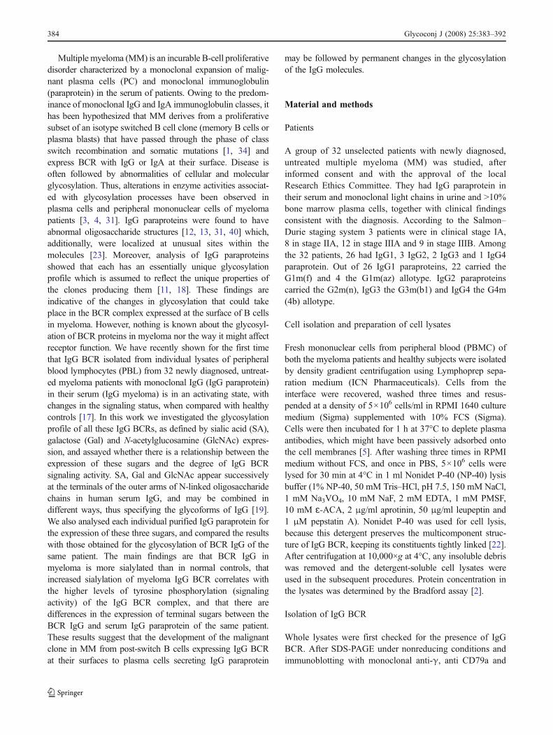
Glycosylation of IgG B cell receptor (IgG BCR) in multiplemyeloma: relationship between sialylation and the signalactivity of IgG BCR
Vesna Ilić & Nadežda Milošević-Jovčić & Sonja Petrović &
Dragana Marković & Gordana Stefanović &
Tatjana Ristić
Received: 14 December 2007 /Accepted: 17 December 2007 / Published online: 11 January 2008# Springer Science + Business Media, LLC 2007
Abstract Little is known about the glycosylation of theisotype switched B cell receptor (BCR) in multiplemyeloma, and the way it might affect receptor function.In this work IgG BCRs isolated from the individual lysatesof peripheral blood lymphocytes (PBL) of 32 patients withIgG multiple myeloma and healthy controls were investi-gated for the expression of sialic acid (SA), galactose (Gal)and N-acetylglucosamine (GlcNAc), the sugars known tospecify the glycoforms of human serum IgG. The degree ofglycosylation and signaling status of all 32 isolatedmyeloma IgG BCRs were correlated and compared withthe glycosylation of the IgG paraproteins isolated from seraof the same patients. It was shown that BCR IgG inmyeloma is more heavily sialylated when compared withnormal controls, that the increased sialylation of IgG BCRis associated with higher levels of tyrosine phosphorylation(signaling activity) of the IgG BCR supramolecularcomplex and that BCR IgG and serum IgG paraproteinfrom the same patient differed in all cases in the levels ofterminal sugar expression. The results suggest that thedevelopment of the malignant clone in MM from post-switch B cells expressing IgG BCR at their surfaces to
plasma cells secreting IgG paraprotein may be followed bypermanent glycosylation changes in the IgG molecules.
Keywords Multiple myeloma . IgG BCR .
Signaling activity . Glycosylation
Introduction
The B-cell antigen receptor (BCR) is a molecular complexcomposed of a membrane immunoglobulin of differentisotype (class, subclass) associated with Igα (CD79a) andIgβ (CD 79b) signal transducing subunits (co-receptors). Itbinds antigen and triggers the signaling events determiningthe survival, progression, expansion and activation of the Bcell [29, 30]. It also functions to endocytose bound antigenfor subsequent intracellular processing and presentationwith class II molecule [42]. Abnormalities in the BCR haveoften been associated with certain hematological malignanciesin which structural or folding defects as well as functionaldeficiency of some components of this receptor have beenobserved [27]. The BCR constituents are glycoproteins anddifferences in glycosylation in any of them may contribute tothe abnormal behavior of the whole receptor in a particulardisease. Recently, Vuillier et al. [41] reported defects inglycosylation and folding in the μ and CD79a (Igα) chainsassociated with the lower levels of surface IgM expression inchronic lymphocytic leukemia (CLL) B cells, while Zhuet al. [46], Zabalegui et al. [45] and Radcliffe et al. [35]found a high incidence of potential glycosylation sites in theantigen binding sites of the BCR in follicular lymphoma(FL). A potential role for the BCR oligosaccharides intumorigenesis, disease activity and progression has beensuggested [25, 35].
Glycoconj J (2008) 25:383–392DOI 10.1007/s10719-007-9101-9
V. Ilić :N. Milošević-Jovčić (*) : S. Petrović :D. MarkovićInstitute for Medical Research, University of Belgrade,Dr Subotića 4,Belgrade, Serbiae-mail: [email protected]
G. StefanovićSchool of Dentistry, University of Belgrade,Dr Subotića 8,Belgrade, Serbia
T. RistićCenter of Medical Biochemistry, Clinical Center Niš,Niš, Serbia
Multiple myeloma (MM) is an incurable B-cell proliferativedisorder characterized by a monoclonal expansion of malig-nant plasma cells (PC) and monoclonal immunoglobulin(paraprotein) in the serum of patients. Owing to the predom-inance of monoclonal IgG and IgA immunoglobulin classes, ithas been hypothesized that MM derives from a proliferativesubset of an isotype switched B cell clone (memory B cells orplasma blasts) that have passed through the phase of classswitch recombination and somatic mutations [1, 34] andexpress BCR with IgG or IgA at their surface. Disease isoften followed by abnormalities of cellular and molecularglycosylation. Thus, alterations in enzyme activities associat-ed with glycosylation processes have been observed inplasma cells and peripheral mononuclear cells of myelomapatients [3, 4, 31]. IgG paraproteins were found to haveabnormal oligosaccharide structures [12, 13, 31, 40] which,additionally, were localized at unusual sites within themolecules [23]. Moreover, analysis of IgG paraproteinsshowed that each has an essentially unique glycosylationprofile which is assumed to reflect the unique properties ofthe clones producing them [11, 18]. These findings areindicative of the changes in glycosylation that could takeplace in the BCR complex expressed at the surface of B cellsin myeloma. However, nothing is known about the glycosyl-ation of BCR proteins in myeloma nor the way it might affectreceptor function. We have recently shown for the first timethat IgG BCR isolated from individual lysates of peripheralblood lymphocytes (PBL) from 32 newly diagnosed, untreat-ed myeloma patients with monoclonal IgG (IgG paraprotein)in their serum (IgG myeloma) is in an activating state, withchanges in the signaling status, when compared with healthycontrols [17]. In this work we investigated the glycosylationprofile of all these IgG BCRs, as defined by sialic acid (SA),galactose (Gal) and N-acetylglucosamine (GlcNAc) expres-sion, and assayed whether there is a relationship between theexpression of these sugars and the degree of IgG BCRsignaling activity. SA, Gal and GlcNAc appear successivelyat the terminals of the outer arms of N-linked oligosaccharidechains in human serum IgG, and may be combined indifferent ways, thus specifying the glycoforms of IgG [19].We also analysed each individual purified IgG paraprotein forthe expression of these three sugars, and compared the resultswith those obtained for the glycosylation of BCR IgG of thesame patient. The main findings are that BCR IgG inmyeloma is more sialylated than in normal controls, thatincreased sialylation of myeloma IgG BCR correlates withthe higher levels of tyrosine phosphorylation (signalingactivity) of the IgG BCR complex, and that there aredifferences in the expression of terminal sugars between theBCR IgG and serum IgG paraprotein of the same patient.These results suggest that the development of the malignantclone in MM from post-switch B cells expressing IgG BCRat their surfaces to plasma cells secreting IgG paraprotein
may be followed by permanent changes in the glycosylationof the IgG molecules.
Material and methods
Patients
A group of 32 unselected patients with newly diagnosed,untreated multiple myeloma (MM) was studied, afterinformed consent and with the approval of the localResearch Ethics Committee. They had IgG paraprotein intheir serum and monoclonal light chains in urine and >10%bone marrow plasma cells, together with clinical findingsconsistent with the diagnosis. According to the Salmon–Durie staging system 3 patients were in clinical stage IA,8 in stage IIA, 12 in stage IIIA and 9 in stage IIIB. Amongthe 32 patients, 26 had IgG1, 3 IgG2, 2 IgG3 and 1 IgG4paraprotein. Out of 26 IgG1 paraproteins, 22 carried theG1m(f) and 4 the G1m(az) allotype. IgG2 paraproteinscarried the G2m(n), IgG3 the G3m(b1) and IgG4 the G4m(4b) allotype.
Cell isolation and preparation of cell lysates
Fresh mononuclear cells from peripheral blood (PBMC) ofboth the myeloma patients and healthy subjects were isolatedby density gradient centrifugation using Lymphoprep sepa-ration medium (ICN Pharmaceuticals). Cells from theinterface were recovered, washed three times and resus-pended at a density of 5×10⁶ cells/ml in RPMI 1640 culturemedium (Sigma) supplemented with 10% FCS (Sigma).Cells were then incubated for 1 h at 37°C to deplete plasmaantibodies, which might have been passively adsorbed ontothe cell membranes [5]. After washing three times in RPMImedium without FCS, and once in PBS, 5×10⁶ cells werelysed for 30 min at 4°C in 1 ml Nonidet P-40 (NP-40) lysisbuffer (1% NP-40, 50 mM Tris–HCl, pH 7.5, 150 mM NaCl,1 mM Na3VO4, 10 mM NaF, 2 mM EDTA, 1 mM PMSF,10 mM ɛ-ACA, 2 μg/ml aprotinin, 50 μg/ml leupeptin and1 μM pepstatin A). Nonidet P-40 was used for cell lysis,because this detergent preserves the multicomponent struc-ture of IgG BCR, keeping its constituents tightly linked [22].After centrifugation at 10,000×g at 4°C, any insoluble debriswas removed and the detergent-soluble cell lysates wereused in the subsequent procedures. Protein concentration inthe lysates was determined by the Bradford assay [2].
Isolation of IgG BCR
Whole lysates were first checked for the presence of IgGBCR. After SDS-PAGE under nonreducing conditions andimmunoblotting with monoclonal anti-γ, anti CD79a and
384 Glycoconj J (2008) 25:383–392
anti CD79b antibodies, a fraction of 195–220 kDa wasidentified as reactive with these reagents. IgG BCR wasthen isolated as briefly described [7]. Cleared lysatescontaining 1 mg proteins (in a volume of 1 ml) weresubjected to precipitation by adsorption to 100 μl of proteinG-Sepharose beads (Amersham Biotech). This sorbentreacts with the sole IgG isotype irrespective of the IgGsubclass. The lysates and beads were incubated at 4°C for3 h, with continuous shaking. The protein complexes werecollected by centrifugation and washed four times in NP-40lysis buffer. Bound proteins were then released by boilingthe samples in 250 μl of reducing SDS sample buffer.Normal IgG BCRs were isolated from NP-40 cell lysatesobtained from ten healthy subjects included as controlsafter they had given informed consent. Immunoblotanalysis of supernatants of protein G-treated lysates showedtraces of unbound IgG in only four lysate supernatants,suggesting that the vast majority of IgG BCRs present inthe lysates were isolated by the method used.
Molecular mass determination
Molecular size markers (Amersham Biotech) were sub-jected to 10% sodium dodecyl sulphate polyacrylamide gelelectrophoresis (SDS-PAGE). The retardation factor (Rf)was calculated by plotting the electrophoretic mobility ofeach marker against its molecular mass and a standardcurve was constructed.
Identification of BCR constituents
Samples of 10 μg isolated BCR (BCR isolates) weresubjected to 10% SDS-PAGE. Total proteins were visual-ized by a silver staining procedure. The BCR constituentswere identified after transfer to nitrocellulose membrane(Amersham Biotech). The membrane was blocked with 3%BSA in TBS and incubated at room temperature for 2 hwith the following primary antibodies: mouse monoclonalanti-human γ chain (MH16-1ME, CBL, The Netherlands),goat polyclonal anti-CD79a (sc-8502) and goat polyclonalanti-CD79b (sc-8504) (Santa Cruz Biotech, CA), for IgG,Igα co-receptor and Igβ co-receptor identification respec-tively. Horseradish peroxidase-conjugated anti-mouse oranti-goat IgG antibody (Amersham Biotech) was used forimmunodetection and, after incubation at room temperaturefor 1 h, BCR constituents were visualized with chloro-1-naphthol as the peroxidase substrate.
Analysis of protein tyrosine phosphorylation
IgG BCR isolates (10 μg) were loaded onto 10% SDSpolyacrylamide gel for electrophoresis. The size-separatedproteins were transferred to nitrocellulose membrane
(Amersham Biotech). To detect tyrosine-phosphorylatedsubstrates, membranes were incubated with mouse mono-clonal anti-phosphotyrosine antibody (PT-66, Sigma) atroom temperature for 2 h, followed by horseradishperoxidase-conjugated goat anti-mouse Ig secondary anti-body (Amersham Biotech) for 1 h at room temperature,using 4-chloro-1-naphthol as the peroxidase substrate. Inorder to verify the identity of the phosphorylated proteins ofinterest (co-receptors), blots were stripped of antibodies byincubation in stripping buffer (100 mM 2-ME, 2% SDS,62.5 mM Tris–HCl, pH 6.7) at 56°C for 30 min, washedextensively, reprobed with specific antibodies and thendeveloped using the enhanced chemiluminescence reagentsystem (Amersham Biotech) according to the manufac-turer's instructions.
Isolation of monoclonal serum IgG
IgG paraproteins were isolated from individual patients seraby ion exchange chromatography onQ Sepharose (AmershamBiosciences, Buckinghamshire, UK), in combination withaffinity purification on protein G-Sepharose [38]. IgG para-proteins were separated from fresh sera, and the sampleswere stored at −70°C until use. IgG subclasses of the isolatedparaproteins were defined by Western blot [36], using mousemonoclonal antibodies specific for γ1, γ2, γ3 and γ4 heavychains (Nordic, The Netherlands). Allotypic Gm markers ofthe isolated IgG paraproteins were determined by inhibition-ELISA [32].
Glycosylation analyses
The localization of sialic acid (SA), galactose (Gal) and N-acetylglucosamine (GlcNAc) within the isolated IgG BCRwas determined by lectin immunoblot assay [39]. Thisinvolved a combined procedure of SDS-PAGE underreduction conditions and lectin binding to the heavy andlight chains of BCR IgG and co-receptors, separated afterreducting. Biotinylated lectins (Vector, Burlingame, CA)from Sambucus nigra (SNA), Ricinus communis (RCA-I)and Griffonia (Bandeiraea) simplicifolia (BS-II) were usedto detect specifically α2-6 linked SA, β1-4 linked Gal andβ1-2 linked GlcNAc sugar residues, respectively. The sameanalyses were performed with IgG paraproteins isolatedfrom MM patients' sera. Sugar expression was quantitativelyestimated by measuring the peak areas obtained after densi-tometric scanning of blots using IMAGEQUANT software(San Diego, CA) and after lectin ratio determinations.
Statistical analyses
Software Prism Pad, version 3.0 (San Diego, CA) wasused for statistical analyses. Data were expressed as
Glycoconj J (2008) 25:383–392 385
means±standard deviations. Values of p≤0.05 wereconsidered significant.
Results
IgG BCR composition and phosphorylation status in MMpatients
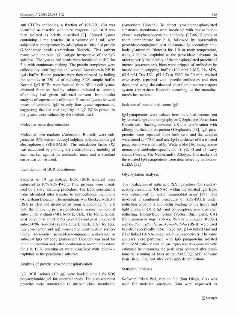
We have previously confirmed [17] the presence of IgGBCR and the completeness of its structure in all myelomaand control IgG BCR isolates, analysed in this work(Fig. 1).
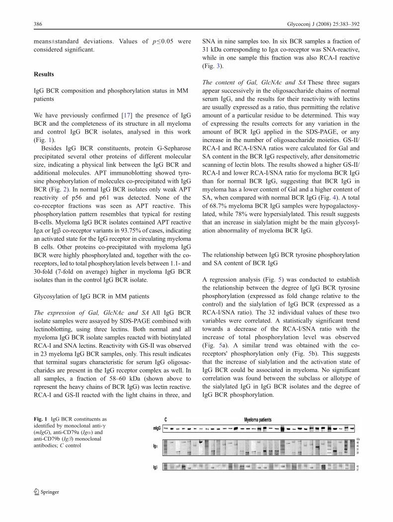
Besides IgG BCR constituents, protein G-Sepharoseprecipitated several other proteins of different molecularsize, indicating a physical link between the IgG BCR andadditional molecules. APT immunoblotting showed tyro-sine phosphorylation of molecules co-precipitated with IgGBCR (Fig. 2). In normal IgG BCR isolates only weak APTreactivity of p56 and p61 was detected. None of theco-receptor fractions was seen as APT reactive. Thisphosphorylation pattern resembles that typical for restingB-cells. Myeloma IgG BCR isolates contained APT reactiveIgα or Igβ co-receptor variants in 93.75% of cases, indicatingan activated state for the IgG receptor in circulating myelomaB cells. Other proteins co-precipitated with myeloma IgGBCR were highly phosphorylated and, together with the co-receptors, led to total phosphorylation levels between 1.1- and30-fold (7-fold on average) higher in myeloma IgG BCRisolates than in the control IgG BCR isolate.
Glycosylation of IgG BCR in MM patients
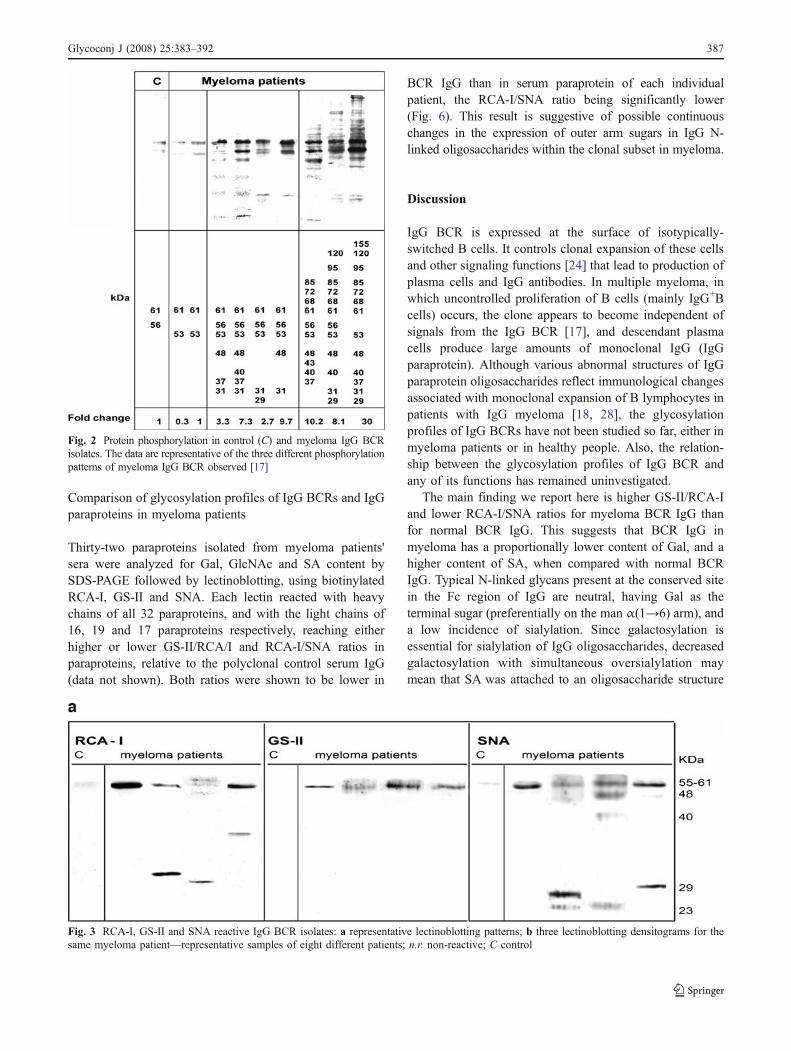
The expression of Gal, GlcNAc and SA All IgG BCRisolate samples were assayed by SDS-PAGE combined withlectinoblotting, using three lectins. Both normal and allmyeloma IgG BCR isolate samples reacted with biotinylatedRCA-I and SNA lectins. Reactivity with GS-II was observedin 23 myeloma IgG BCR samples, only. This result indicatesthat terminal sugars characteristic for serum IgG oligosac-charides are present in the IgG receptor complex as well. Inall samples, a fraction of 58–60 kDa (shown above torepresent the heavy chains of BCR IgG) was lectin reactive.RCA-I and GS-II reacted with the light chains in three, and
SNA in nine samples too. In six BCR samples a fraction of31 kDa corresponding to Igα co-receptor was SNA-reactive,while in one sample this fraction was also RCA-I reactive(Fig. 3).
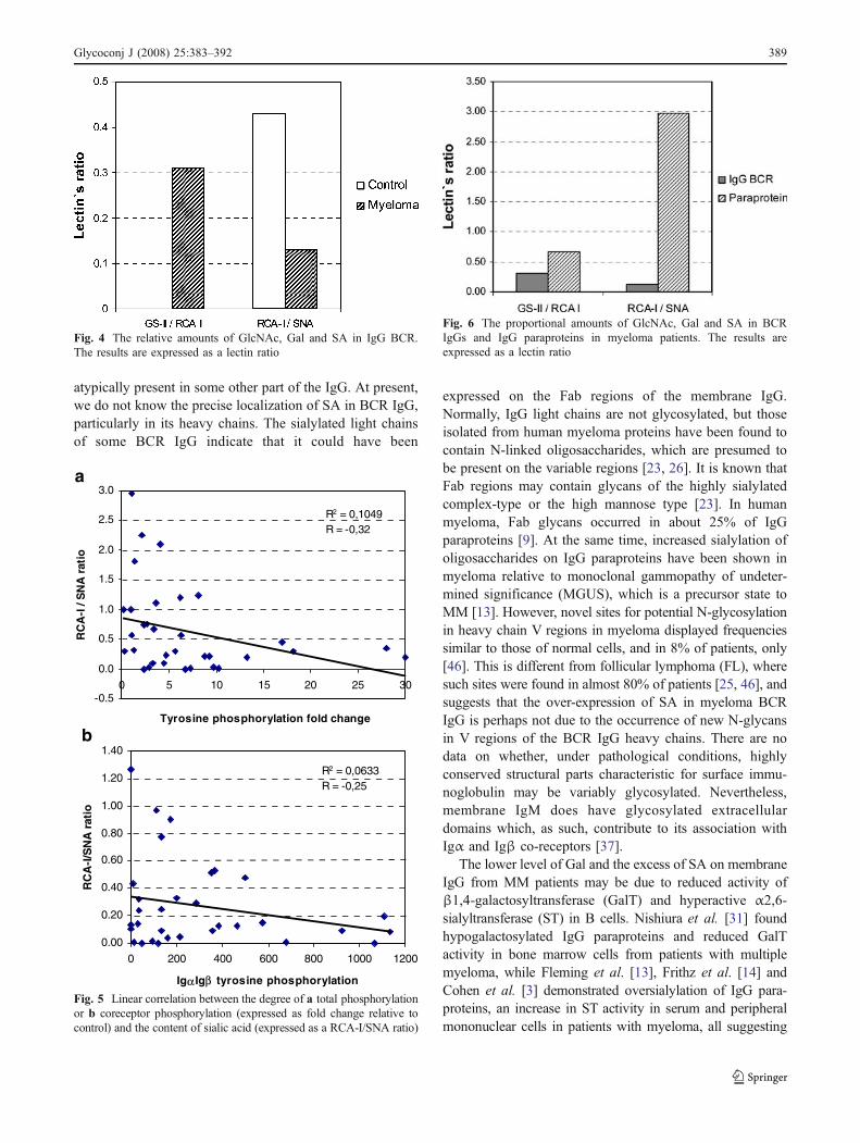
The content of Gal, GlcNAc and SA These three sugarsappear successively in the oligosaccharide chains of normalserum IgG, and the results for their reactivity with lectinsare usually expressed as a ratio, thus permitting the relativeamount of a particular residue to be determined. This wayof expressing the results corrects for any variation in theamount of BCR IgG applied in the SDS-PAGE, or anyincrease in the number of oligosaccharide moieties. GS-II/RCA-I and RCA-I/SNA ratios were calculated for Gal andSA content in the BCR IgG respectively, after densitometricscanning of lectin blots. The results showed a higher GS-II/RCA-I and lower RCA-I/SNA ratio for myeloma BCR IgGthan for normal BCR IgG, suggesting that BCR IgG inmyeloma has a lower content of Gal and a higher content ofSA, when compared with normal BCR IgG (Fig. 4). A totalof 68.7% myeloma BCR IgG samples were hypogalactosy-lated, while 78% were hypersialylated. This result suggeststhat an increase in sialylation might be the main glycosyl-ation abnormality of myeloma BCR IgG.
The relationship between IgG BCR tyrosine phosphorylationand SA content of BCR IgG
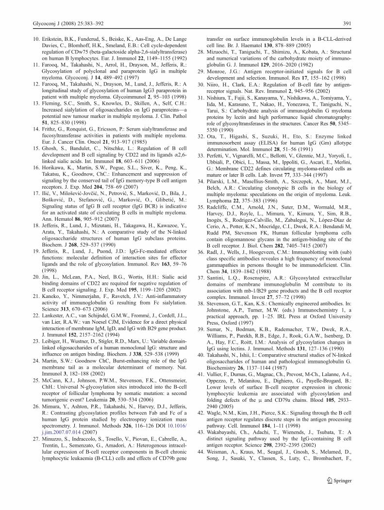
A regression analysis (Fig. 5) was conducted to establishthe relationship between the degree of IgG BCR tyrosinephosphorylation (expressed as fold change relative to thecontrol) and the sialylation of IgG BCR (expressed as aRCA-I/SNA ratio). The 32 individual values of these twovariables were correlated. A statistically significant trendtowards a decrease of the RCA-I/SNA ratio with theincrease of total phosphorylation level was observed(Fig. 5a). A similar trend was obtained with the co-receptors' phosphorylation only (Fig. 5b). This suggeststhat the increase of sialylation and the activation state ofIgG BCR could be associated in myeloma. No significantcorrelation was found between the subclass or allotype ofthe sialylated IgG in IgG BCR isolates and the degree ofIgG BCR phosphorylation.
Fig. 1 IgG BCR constituents asidentified by monoclonal anti-γ(mIgG), anti-CD79a (Igα) andanti-CD79b (Igβ) monoclonalantibodies; C control
386 Glycoconj J (2008) 25:383–392
Comparison of glycosylation profiles of IgG BCRs and IgGparaproteins in myeloma patients
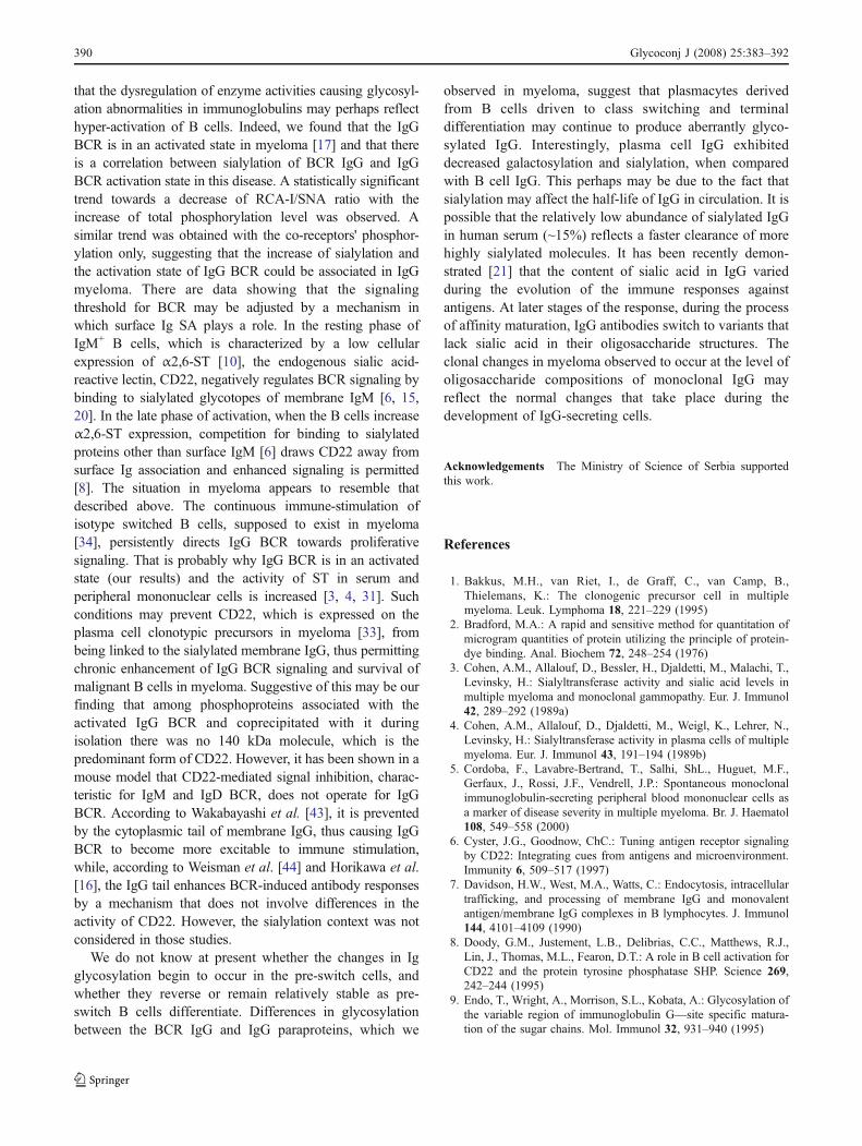
Thirty-two paraproteins isolated from myeloma patients'sera were analyzed for Gal, GlcNAc and SA content bySDS-PAGE followed by lectinoblotting, using biotinylatedRCA-I, GS-II and SNA. Each lectin reacted with heavychains of all 32 paraproteins, and with the light chains of16, 19 and 17 paraproteins respectively, reaching eitherhigher or lower GS-II/RCA/I and RCA-I/SNA ratios inparaproteins, relative to the polyclonal control serum IgG(data not shown). Both ratios were shown to be lower in
BCR IgG than in serum paraprotein of each individualpatient, the RCA-I/SNA ratio being significantly lower(Fig. 6). This result is suggestive of possible continuouschanges in the expression of outer arm sugars in IgG N-linked oligosaccharides within the clonal subset in myeloma.
Discussion
IgG BCR is expressed at the surface of isotypically-switched B cells. It controls clonal expansion of these cellsand other signaling functions [24] that lead to production ofplasma cells and IgG antibodies. In multiple myeloma, inwhich uncontrolled proliferation of B cells (mainly IgG+Bcells) occurs, the clone appears to become independent ofsignals from the IgG BCR [17], and descendant plasmacells produce large amounts of monoclonal IgG (IgGparaprotein). Although various abnormal structures of IgGparaprotein oligosaccharides reflect immunological changesassociated with monoclonal expansion of B lymphocytes inpatients with IgG myeloma [18, 28], the glycosylationprofiles of IgG BCRs have not been studied so far, either inmyeloma patients or in healthy people. Also, the relation-ship between the glycosylation profiles of IgG BCR andany of its functions has remained uninvestigated.
The main finding we report here is higher GS-II/RCA-Iand lower RCA-I/SNA ratios for myeloma BCR IgG thanfor normal BCR IgG. This suggests that BCR IgG inmyeloma has a proportionally lower content of Gal, and ahigher content of SA, when compared with normal BCRIgG. Typical N-linked glycans present at the conserved sitein the Fc region of IgG are neutral, having Gal as theterminal sugar (preferentially on the man α(1→6) arm), anda low incidence of sialylation. Since galactosylation isessential for sialylation of IgG oligosaccharides, decreasedgalactosylation with simultaneous oversialylation maymean that SA was attached to an oligosaccharide structure
Fig. 2 Protein phosphorylation in control (C) and myeloma IgG BCRisolates. The data are representative of the three different phosphorylationpatterns of myeloma IgG BCR observed [17]
Fig. 3 RCA-I, GS-II and SNA reactive IgG BCR isolates: a representative lectinoblotting patterns; b three lectinoblotting densitograms for thesame myeloma patient—representative samples of eight different patients; n.r. non-reactive; C control
Glycoconj J (2008) 25:383–392 387
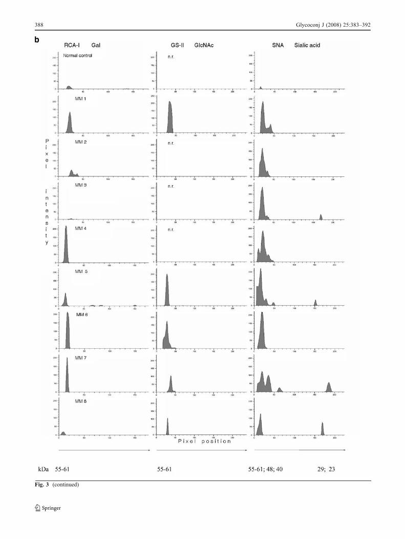
Fig. 3 (continued)
388 Glycoconj J (2008) 25:383–392
atypically present in some other part of the IgG. At present,we do not know the precise localization of SA in BCR IgG,particularly in its heavy chains. The sialylated light chainsof some BCR IgG indicate that it could have been
expressed on the Fab regions of the membrane IgG.Normally, IgG light chains are not glycosylated, but thoseisolated from human myeloma proteins have been found tocontain N-linked oligosaccharides, which are presumed tobe present on the variable regions [23, 26]. It is known thatFab regions may contain glycans of the highly sialylatedcomplex-type or the high mannose type [23]. In humanmyeloma, Fab glycans occurred in about 25% of IgGparaproteins [9]. At the same time, increased sialylation ofoligosaccharides on IgG paraproteins have been shown inmyeloma relative to monoclonal gammopathy of undeter-mined significance (MGUS), which is a precursor state toMM [13]. However, novel sites for potential N-glycosylationin heavy chain V regions in myeloma displayed frequenciessimilar to those of normal cells, and in 8% of patients, only[46]. This is different from follicular lymphoma (FL), wheresuch sites were found in almost 80% of patients [25, 46], andsuggests that the over-expression of SA in myeloma BCRIgG is perhaps not due to the occurrence of new N-glycansin V regions of the BCR IgG heavy chains. There are nodata on whether, under pathological conditions, highlyconserved structural parts characteristic for surface immu-noglobulin may be variably glycosylated. Nevertheless,membrane IgM does have glycosylated extracellulardomains which, as such, contribute to its association withIgα and Igβ co-receptors [37].
The lower level of Gal and the excess of SA on membraneIgG from MM patients may be due to reduced activity ofβ1,4-galactosyltransferase (GalT) and hyperactive α2,6-sialyltransferase (ST) in B cells. Nishiura et al. [31] foundhypogalactosylated IgG paraproteins and reduced GalTactivity in bone marrow cells from patients with multiplemyeloma, while Fleming et al. [13], Frithz et al. [14] andCohen et al. [3] demonstrated oversialylation of IgG para-proteins, an increase in ST activity in serum and peripheralmononuclear cells in patients with myeloma, all suggesting
Fig. 4 The relative amounts of GlcNAc, Gal and SA in IgG BCR.The results are expressed as a lectin ratio
R2 = 0,1049R = -0,32
-0.5
0.0
0.5
1.0
1.5
2.0
2.5
3.0
0 5 10 15 20 25 30
Tyrosine phosphorylation fold change
RC
A-I
/ S
NA
rat
io
R2 = 0,0633R = -0,25
0.00
0.20
0.40
0.60
0.80
1.00
1.20
1.40
0 200 400 600 800 1000 1200
Igα Igβ tyrosine phosphorylation
RC
A-I
/SN
A r
atio
a
b
Fig. 5 Linear correlation between the degree of a total phosphorylationor b coreceptor phosphorylation (expressed as fold change relative tocontrol) and the content of sialic acid (expressed as a RCA-I/SNA ratio)
Fig. 6 The proportional amounts of GlcNAc, Gal and SA in BCRIgGs and IgG paraproteins in myeloma patients. The results areexpressed as a lectin ratio
Glycoconj J (2008) 25:383–392 389
that the dysregulation of enzyme activities causing glycosyl-ation abnormalities in immunoglobulins may perhaps reflecthyper-activation of B cells. Indeed, we found that the IgGBCR is in an activated state in myeloma [17] and that thereis a correlation between sialylation of BCR IgG and IgGBCR activation state in this disease. A statistically significanttrend towards a decrease of RCA-I/SNA ratio with theincrease of total phosphorylation level was observed. Asimilar trend was obtained with the co-receptors' phosphor-ylation only, suggesting that the increase of sialylation andthe activation state of IgG BCR could be associated in IgGmyeloma. There are data showing that the signalingthreshold for BCR may be adjusted by a mechanism inwhich surface Ig SA plays a role. In the resting phase ofIgM+ B cells, which is characterized by a low cellularexpression of α2,6-ST [10], the endogenous sialic acid-reactive lectin, CD22, negatively regulates BCR signaling bybinding to sialylated glycotopes of membrane IgM [6, 15,20]. In the late phase of activation, when the B cells increaseα2,6-ST expression, competition for binding to sialylatedproteins other than surface IgM [6] draws CD22 away fromsurface Ig association and enhanced signaling is permitted[8]. The situation in myeloma appears to resemble thatdescribed above. The continuous immune-stimulation ofisotype switched B cells, supposed to exist in myeloma[34], persistently directs IgG BCR towards proliferativesignaling. That is probably why IgG BCR is in an activatedstate (our results) and the activity of ST in serum andperipheral mononuclear cells is increased [3, 4, 31]. Suchconditions may prevent CD22, which is expressed on theplasma cell clonotypic precursors in myeloma [33], frombeing linked to the sialylated membrane IgG, thus permittingchronic enhancement of IgG BCR signaling and survival ofmalignant B cells in myeloma. Suggestive of this may be ourfinding that among phosphoproteins associated with theactivated IgG BCR and coprecipitated with it duringisolation there was no 140 kDa molecule, which is thepredominant form of CD22. However, it has been shown in amouse model that CD22-mediated signal inhibition, charac-teristic for IgM and IgD BCR, does not operate for IgGBCR. According to Wakabayashi et al. [43], it is preventedby the cytoplasmic tail of membrane IgG, thus causing IgGBCR to become more excitable to immune stimulation,while, according to Weisman et al. [44] and Horikawa et al.[16], the IgG tail enhances BCR-induced antibody responsesby a mechanism that does not involve differences in theactivity of CD22. However, the sialylation context was notconsidered in those studies.
We do not know at present whether the changes in Igglycosylation begin to occur in the pre-switch cells, andwhether they reverse or remain relatively stable as pre-switch B cells differentiate. Differences in glycosylationbetween the BCR IgG and IgG paraproteins, which we
observed in myeloma, suggest that plasmacytes derivedfrom B cells driven to class switching and terminaldifferentiation may continue to produce aberrantly glyco-sylated IgG. Interestingly, plasma cell IgG exhibiteddecreased galactosylation and sialylation, when comparedwith B cell IgG. This perhaps may be due to the fact thatsialylation may affect the half-life of IgG in circulation. It ispossible that the relatively low abundance of sialylated IgGin human serum (~15%) reflects a faster clearance of morehighly sialylated molecules. It has been recently demon-strated [21] that the content of sialic acid in IgG variedduring the evolution of the immune responses againstantigens. At later stages of the response, during the processof affinity maturation, IgG antibodies switch to variants thatlack sialic acid in their oligosaccharide structures. Theclonal changes in myeloma observed to occur at the level ofoligosaccharide compositions of monoclonal IgG mayreflect the normal changes that take place during thedevelopment of IgG-secreting cells.
Acknowledgements The Ministry of Science of Serbia supportedthis work.
References
1. Bakkus, M.H., van Riet, I., de Graff, C., van Camp, B.,Thielemans, K.: The clonogenic precursor cell in multiplemyeloma. Leuk. Lymphoma 18, 221–229 (1995)
2. Bradford, M.A.: A rapid and sensitive method for quantitation ofmicrogram quantities of protein utilizing the principle of protein-dye binding. Anal. Biochem 72, 248–254 (1976)
3. Cohen, A.M., Allalouf, D., Bessler, H., Djaldetti, M., Malachi, T.,Levinsky, H.: Sialyltransferase activity and sialic acid levels inmultiple myeloma and monoclonal gammopathy. Eur. J. Immunol42, 289–292 (1989a)
4. Cohen, A.M., Allalouf, D., Djaldetti, M., Weigl, K., Lehrer, N.,Levinsky, H.: Sialyltransferase activity in plasma cells of multiplemyeloma. Eur. J. Immunol 43, 191–194 (1989b)
5. Cordoba, F., Lavabre-Bertrand, T., Salhi, ShL., Huguet, M.F.,Gerfaux, J., Rossi, J.F., Vendrell, J.P.: Spontaneous monoclonalimmunoglobulin-secreting peripheral blood mononuclear cells asa marker of disease severity in multiple myeloma. Br. J. Haematol108, 549–558 (2000)
6. Cyster, J.G., Goodnow, ChC.: Tuning antigen receptor signalingby CD22: Integrating cues from antigens and microenvironment.Immunity 6, 509–517 (1997)
7. Davidson, H.W., West, M.A., Watts, C.: Endocytosis, intracellulartrafficking, and processing of membrane IgG and monovalentantigen/membrane IgG complexes in B lymphocytes. J. Immunol144, 4101–4109 (1990)
8. Doody, G.M., Justement, L.B., Delibrias, C.C., Matthews, R.J.,Lin, J., Thomas, M.L., Fearon, D.T.: A role in B cell activation forCD22 and the protein tyrosine phosphatase SHP. Science 269,242–244 (1995)
9. Endo, T., Wright, A., Morrison, S.L., Kobata, A.: Glycosylation ofthe variable region of immunoglobulin G—site specific matura-tion of the sugar chains. Mol. Immunol 32, 931–940 (1995)
390 Glycoconj J (2008) 25:383–392
10. Erikstein, B.K., Funderud, S., Beiske, K., Aas-Eng, A., De LangeDavies, C., Blomhoff, H.K., Smeland, E.B.: Cell cycle-dependentregulation of CDw75 (beta-galactoside alpha-2,6-sialyltransferase)on human B lymphocytes. Eur. J. Immunol 22, 1149–1155 (1992)
11. Farooq, M., Takahashi, N., Arrol, H., Drayson, M., Jefferis, R.:Glycosylation of polyclonal and paraprotein IgG in multiplemyeloma. Glycoconj. J 14, 489–492 (1997)
12. Farooq, M., Takahashi, N., Drayson, M., Lund, J., Jefferis, R.: Alongitudinal study of glycosylation of human IgG3 paraprotein inpatient with multiple myeloma. Glycoimmunol 2, 95–103 (1998)
13. Fleming, S.C., Smith, S., Knowles, D., Skillen, A., Self, C.H.:Increased sialylation of oligosaccharides on IgG paraproteins—apotential new tumour marker in multiple myeloma. J. Clin. Pathol51, 825–830 (1998)
14. Frithz, G., Ronquist, G., Ericsson, P.: Serum sialyltransferase andfucosyltransferase activities in patients with multiple myeloma.Eur. J. Cancer Clin. Oncol 21, 913–917 (1985)
15. Ghosh, S., Bandulet, C., Nitschke, L.: Regulation of B celldevelopment and B cell signaling by CD22 and its ligands α2,6-linked sialic acids. Int. Immunol 18, 603–611 (2006)
16. Horikawa, K., Martin, S.W., Pogue, S.L., Siver, K., Peng, K.,Takatsu, K., Goodnow, ChC.: Enhancement and suppression ofsignaling by the conserved tail of IgG memory-type B cell antigenreceptors. J. Exp. Med 204, 758–69 (2007)
17. Ilić, V., Milošević-Jovčić, N., Petrović, S., Marković, D., Bila, J.,Bošković, D., Stefanović, G., Marković, O., Glibetić, M.:Signaling status of IgG B cell receptor (IgG BCR) is indicativefor an activated state of circulating B cells in multiple myeloma.Ann. Hematol 86, 905–912 (2007)
18. Jefferis, R., Lund, J., Mizutani, H., Takagawa, H., Kawazoe, Y.,Arata, Y., Takahashi, N.: A comparative study of the N-linkedoligosaccharide structures of human IgG subclass proteins.Biochem. J 268, 529–537 (1990)
19. Jefferis, R., Lund, J., Puond, J.D.: IgG-Fc-mediated effectorfunctions: molecular definition of interaction sites for effectorligands and the role of glycosylation. Immunol. Rev 163, 59–76(1998)
20. Jin, L., McLean, P.A., Neel, B.G., Wortis, H.H.: Sialic acidbinding domains of CD22 are required for negative regulation ofB cell receptor signaling. J. Exp. Med 195, 1199–1205 (2002)
21. Kaneko, Y., Nimmerjahn, F., Ravetch, J.V.: Anti-inflammatoryactivity of immunoglobulin G resulting from Fc sialylation.Science 313, 670–673 (2006)
22. Lankester, A.C., van Schijndel, G.M.W., Frommé, J., Cordell, J.L.,van Lier, R.A.W.: van Noesel CJM, Evidence for a direct physicalinteraction of membrane IgM, IgD, and IgG with B29 gene product.J. Immunol 152, 2157–2162 (1994)
23. Leibiger, H., Wustner, D., Stigler, R.D., Marx, U.: Variable domain-linked oligosaccharides of a human monoclonal IgG: structure andinfluence on antigen binding. Biochem. J 338, 529–538 (1999)
24. Martin, S.W.: Goodnow ChC, Burst-enhancing role of the IgGmembrane tail as a molecular determinant of memory. Nat.Immunol 3, 182–188 (2002)
25. McCann, K.J., Johnson, P.W.M., Stevenson, F.K., Ottensmeier,ChH.: Universal N-glycosylation sites introduced into the B-cellreceptor of follicular lymphoma by somatic mutation: a secondtumorigenic event? Leukemia 20, 530–534 (2006)
26. Mimura, Y., Ashton, P.R., Takahashi, N., Harvey, D.J., Jefferis,R.: Contrasting glycosylation profiles between Fab and Fc of ahuman IgG protein studied by electrospray ionization massspectrometry. J. Immunol. Methods 326, 116–126 DOI 10.1016/j.jim.2007.07.014 (2007)
27. Minuzzo, S., Indraccolo, S., Tosello, V., Piovan, E., Cabrelle, A.,Trentin, L., Semenzato, G., Amadori, A.: Heterogenous intracel-lular expression of B-cell receptor components in B-cell chroniclymphocytic leukaemia (B-CLL) cells and effects of CD79b gene
transfer on surface immunoglobulin levels in a B-CLL-derivedcell line. Br. J. Haematol 130, 878–889 (2005)
28. Mizuochi, T., Taniguchi, T., Shimizu, A., Kobata, A.: Structuraland numerical variations of the carbohydrate moiety of immuno-globulin G. J. Immunol 129, 2016–2020 (1982)
29. Monroe, J.G.: Antigen receptor-initiated signals for B celldevelopment and selection. Immunol. Res 17, 155–162 (1998)
30. Niiro, H., Clark, E.A.: Regulation of B-cell fate by antigen-receptor signals. Nat. Rev. Immunol 2, 945–956 (2002)
31. Nishiura, T., Fujii, S., Kanayama, Y., Nishikawa, A., Tomiyama, Y.,Iida, M., Karasuno, T., Nakao, H., Yonezawa, T., Taniguchi, N.,Tarui, S.: Carbohydrate analysis of immunoglobulin G myelomaproteins by lectin and high performance liquid chromatography:role of glycosyltransferases in the structures. Cancer Res 50, 5345–5350 (1990)
32. Ota, T., Higashi, S., Suzuki, H., Eto, S.: Enzyme linkedimmunosorbent assay (ELISA) for human IgG (Gm) allotypedetermination. Mol. Immunol 28, 51–56 (1991)
33. Perfetti, V., Vignarelli, M.C., Bellotti, V., Glennie, M.J., Yoryoli, I.,Ubbiali, P., Obici, L., Massa, M., Ippoliti, G., Ascari, E., Merlini,G.: Membrane CD22 defines circulating myeloma-related cells asmature or later B cells. Lab. Invest 77, 333–344 (1997)
34. Pilarski, L.M., Masellius-Smith, A., Szczepek, A., Mant, M.J.,Belch, A.R.: Circulating clonotypic B cells in the biology ofmultiple myeloma: speculations on the origin of myeloma. Leuk.Lymphoma 22, 375–383 (1996)
35. Radcliffe, C.M., Arnold, J.N., Suter, D.M., Wormald, M.R.,Harvey, D.J., Royle, L., Mimura, Y., Kimura, Y., Sim, R.B.,Inogès, S., Rodrigez-Calvillo, M., Zabalegui, N., López-Díaz deCerio, A., Potter, K.N., Mocridge, C.I., Dwek, R.A.: Bendandi M,Rudd PM, Stevenson FK, Human follicular lymphoma cellscontain oligomannose glycans in the antigen-binding site of theB cell receptor. J. Biol. Chem 282, 7405–7415 (2007)
36. Radl, J., Wells, J., Hoogeveen, C.M.: Immunoblotting with (sub)class specific antibodies reveales a high frequency of monoclonalgammapathies in persons thought to be immunodeficient. Clin.Chem 34, 1839–1842 (1988)
37. Santini, L.Q., Rosenspire, A.R.: Glycosylated extracellulardomains of membrane immunoglobulin M contribute to itsassociation with mb-1/B29 gene products and the B cell receptorcomplex. Immunol. Invest 27, 57–72 (1998)
38. Stevenson, G.T., Kan, K.S.: Chemically engineered antibodies. In:Johnstone, A.P., Turner, M.W. (eds.) Immunochemistry 1, apractical approach, pp. 1–25. IRL Press at Oxford UniversityPress, Oxford (1997)
39. Sumar, N., Bodman, K.B., Rademacher, T.W., Dwek, R.A.,Williams, P., Parekh, R.B., Edge, J., Rook, G.A.W., Isenberg, D.A., Hay, F.C., Roitt, I.M.: Analysis of glycosylation changes inIgG using lectins. J. Immunol. Methods 131, 127–136 (1990)
40. Takahashi, N., Ishii, I.: Comparative structural studies of N-linkedoligosaccharides of human and pathological immunoglobulin G.Biochemistry 26, 1137–1144 (1987)
41. Vullier, F., Dumas, G., Magnac, Ch., Prevost, M-Ch., Lalanne, A-I.,Oppezzo, P., Melanitou, E., Dighiero, G., Payelle-Brogard, B.:Lower levels of surface B-cell receptor expression in chroniclymphocytic leukemia are associated with glycosylation andfolding defects of the μ and CD79a chains. Blood 105, 2933–2940 (2005)
42. Wagle, N.M., Kim, J.H., Pierce, S.K.: Signaling through the B cellantigen receptor regulates discrete steps in the antigen processingpathway. Cell. Immunol 184, 1–11 (1998)
43. Wakabayashi, Ch., Adachi, T., Wienends, J., Tsubata, T.: Adistinct signaling pathway used by the IgG-containing B cellantigen receptor. Science 298, 2392–2395 (2002)
44. Weisman, A., Kraus, M., Seagal, J., Gnosh, S., Melamed, D.,Song, J., Sasaki, Y., Classen, S., Luty, C., Brombachert, F.,
Glycoconj J (2008) 25:383–392 391
Nitschke, L., Rajewski, K.: IgG1 B cell receptor signaling is inhibitedby CD22 and promotes the development of B cells whose survival isless dependent on Igα/β. J. Exp. Med 204, 747–758 (2007)
45. Zabalegui, N., de Cerio, A.L., Inoges, S., Rodriguez-Calvillo, M.,Perez-Calvo, J., Hernandez, M., Garcia-Foncillas, J., Martin-Algarra,S., Rocha, E., Bendandi, M.: Acquired potential N-glycosylation
sites within the tumor-specific immunoglobulin heavy chains of B-cell malignancies. Haematologica 89, 541–546 (2004)
46. Zhu, D.,McCarthy, H., Ottensmeier, ChH., Johnson, P., Hamblin, T.J.,Stevenson, F.K.: Acquisition of potential N-glycosylation sites inimmunoglobulin variable region by somatic mutation is distinctivefeature of follicular lymphoma. Blood 99, 2562–2568 (2002)
392 Glycoconj J (2008) 25:383–392