Embed Size (px)
Citation preview

Hindawi Publishing CorporationInternational Journal of Molecular ImagingVolume 2011, Article ID 283497, 10 pagesdoi:10.1155/2011/283497
Review Article
Towards In Vivo Imaging of Cancer Sialylation
Ivan Martinez-Duncker,1 Roberta Salinas-Marin,1 and Carlos Martinez-Duncker2
1 Human Glycobiology Laboratory, Science Faculty, Autonomous University of the State of Morelos, Avenida Universidad 1001,Col. Chamilpa, 62209 Cuernavaca, MOR, Mexico
2 The Molecular Imaging Commission of the Mexican Academy of Surgery, Zacatecas 230-502, Colonia Roma,06700 Mexico, DF, Mexico
Correspondence should be addressed to Carlos Martinez-Duncker, [email protected]
Received 2 May 2011; Accepted 14 June 2011
Academic Editor: Lie-Hang Shen
Copyright © 2011 Ivan Martinez-Duncker et al. This is an open access article distributed under the Creative Commons AttributionLicense, which permits unrestricted use, distribution, and reproduction in any medium, provided the original work is properlycited.
In vivo assessment of tumor glucose catabolism by positron emission tomography (PET) has become a highly valued study in themedical management of cancer. Emerging technologies offer the potential to evaluate in vivo another aspect of cancer carbohydratemetabolism related to the increased anabolic use of monosaccharides like sialic acid (Sia). Sia is used for the synthesis of sialylatedoligosaccharides in the cell surface that in cancer cells are overexpressed and positively associated to malignancy and worseprognosis because of their role in invasion and metastasis. This paper addresses the key points of the different strategies that havebeen developed to image Sia expression in vivo and the perspectives to translate it from the bench to the bedside where it wouldoffer the clinician highly valued complementary information on cancer carbohydrate metabolism that is currently unavailablein vivo.
1. Introduction
There is no doubt that in vivo imaging of glucose catabolismby positron emission tomography (PET) through the useof the PET tracer Fluorine-18-2-fluoro-2-deoxy-D-glucose(FDG) has revolutionized diagnosis and staging of cancer[1]. FDG, an analogue of glucose, is transported into cellsby glucose transporters and follows the glycolytic pathwaybeing phosphorylated by hexokinase into FDG-6-phosphate.At this point FDG is metabolically trapped because it cannotfollow the normal glycolytic pathway due to the substitutionof the 2′ hydroxyl group by Fluorine 18 (18F), causing itsaccumulation in the cell. Higher accumulation of FDG incancer cells compared to normal cells allows the imaging byPET of the “Warburg effect” or aerobic glycolysis which isuniquely observed in cancer [2]. The association of this effectto malignancy has made FDG an indispensable or at leasta very important imaging agent in the diagnosis, staging,restaging, and assessment of treatment in various types ofcancer including lung, colorectal, esophageal, stomach, headand neck, thyroid, cervical, ovarian, and breast cancers, aswell as melanoma and most types of lymphoma [1, 3].
The in vivo imaging of anabolic rather than cataboliccarbohydrate pathways has a potential usefulness in cancermanagement by offering the clinician complementary infor-mation on cancer carbohydrate metabolism that is currentlyunavailable in vivo. This paper addresses the key points ofdifferent strategies that have been developed to image thein vivo expression of sialylated oligosaccharides synthesizedby an essential cellular process known as glycosylation[4]. Glycosylation involves the synthesis and attachmentto proteins (glycoproteins) and lipids (glycolipids) of astructurally diverse group of oligosaccharides chains alsoknown as glycans that are synthesized in the endoplasmicreticulum and Golgi apparatus of all human cells. Along thispaper the role of sialylated antigens in the glycophenotypeof cancer, their biosynthesis, and emerging imaging toolsare described in text and figures as the foundations of apreliminary suggestion towards a new clinical approach intumor imaging.
Each type of human cell displays in its surface an arrayof glycans attached to proteins or lipids to form glycocon-jugates. The different types of monosaccharides and linkagecombinations can be found on glycans code for biological

2 International Journal of Molecular Imaging
information that translates into structural or functionalproperties of the glycoconjugate. The type and degree ofexpression of glycans is determined by the type and activitiesof the proteins involved in their synthesis that can bedifferent from one cell type to another and even differ tosome extent between the same types of cell. This dynamictemporospatial characteristic of glycosylation and the greatintrinsic information it can carry as a “glycan code” havebeen evolutionarily selected as an important mechanism forcells to communicate and enhance distinct physiologic andpathologic states, including malignant transformation [5].
The “glycan code” has been probed and imaged exten-sively in vitro mostly by the use of antibodies that recognizecarbohydrate epitopes or through carbohydrate-bindingproteins known as lectins [6]. In the case of cancer certaincharacteristics of the glycan code have been clearly associatedto increased tumor aggressiveness and worse prognosis. Oneof these being the overexpression of sialic acid (Sia) residuesthat are present in many characterized carbohydrate antigens[7].
2. Sialylated Antigens inthe Glycophenotype of Cancer
A substantial component that distinguishes malignantfrom benign glycophenotypes is the neoexpression oroverexpression of sialylated epitopes in the cell surfacethat include sialyl-Tn (STn; Siaα2→ 6GalNAc-T/S), sia-lyl T (ST; Siaα2-3Galβ1-3GalNAc-T/S), sialyl Lewis X(SLex; Siaα2,3Galβ1,4(Fucα1,3)GlcNAc), and sialyl LewisA (SLea/CA19-9; Sia2,3Galβ1,3(Fucα1,4)GlcNAc), Figure 1.Additionally, various tumors including small cell and non-small cell lung carcinomas, multiple myeloma, neuroblas-toma, and Wilms’ tumor express Sia as a homopolymercalled polysialic acid or PSA (Siaα2,8Sia) [8–12]. The neo-or overexpression of these sialylated antigens is mainlycaused by an increase in the expression of sialyltransferases(STs) and fucosyltransferases (FUTs), glycosyltransferaseenzymes responsible for their synthesis that add Sia andfucose residues to glycans, respectively. Additionally, effectiveincrease in antigen synthesis requires not only glycosyltrans-ferase overexpression but also adequate donor and accep-tor substrate synthesis which requires regulation of otherelements of the glycosylation machinery [13]. This increasein sialylated antigen expression during cancer progressionhas been associated to hypoxic conditions that develop in agrowing tumor and induce transcription of STs, FUTs, andnucleotide-sugar transporters that provide them with donorsubstrate [14–17].
Overexpression of sialylated antigens like SLea and SLex
plays an important role in the biology of cancer, particularlythe metastatic process. Overexpression of these antigens incirculating cancer cells allows them to attach to a familyof endogenous lectins called selectins that are expressedin endothelial cells of high endothelial venules (HEVs),but also platelets and leukocytes [18]. Selectins normallyparticipate in the first phase of what is known as theleukocyte adhesion cascade that allows leukocytes to roll
over the inflamed vascular endothelium through selectin-carbohydrate interactions mediated by ligands that expressSLex and SLea epitopes. Leukocyte rolling gives way to morestable interactions with endothelial integrins that allow themto transmigrate to the interstitial space to culminate animmune response [18]. Cancer cells that disperse from aprimary tumor and access the vascular compartment haveexploited this leukocyte adhesion cascade mechanism toattach to HEV and extravasate to initiate the establishmentof metastatic lesions. Also, overexpression of SLea/x antigenshas been found to facilitate tumor angiogenesis by mediatingcancer cell adhesion to endothelial cells [19, 20].
It is clear that increased expression of sialylated antigenson cancer cells is closely implicated in the process ofcancer progression, and more malignant cancer cells tendto have a more enhanced expression of these carbohydratedeterminants [19]. The numerous clinical statistics madeavailable to date show that the intensity of SLea/x and sialylTn expression on cancer cells significantly correlates with theprognosis of patients and are reviewed elsewhere [21–24]. Astatistically significant correlation between the postoperativepatient prognosis and SLea expression has been reportedfor colon and stomach cancers while its correlation withSLex expression has been reported for lung, breast, prostate,stomach, colon, and urinary bladder cancers [25]. Also, inboth small cell and nonsmall cell lung carcinomas, colorectalcancer, and multiple myeloma, the expression of PSA hasbeen correlated with tumor progression [8, 9, 11, 12, 26].Unfortunately, the determination of the sialylation status,particularly of solid tumors, is currently performed mainlyon tissue sections obtained from tumor biopsies limiting thenumber of possible assessments through time.
3. Sialic Acid and Its Biosynthesis
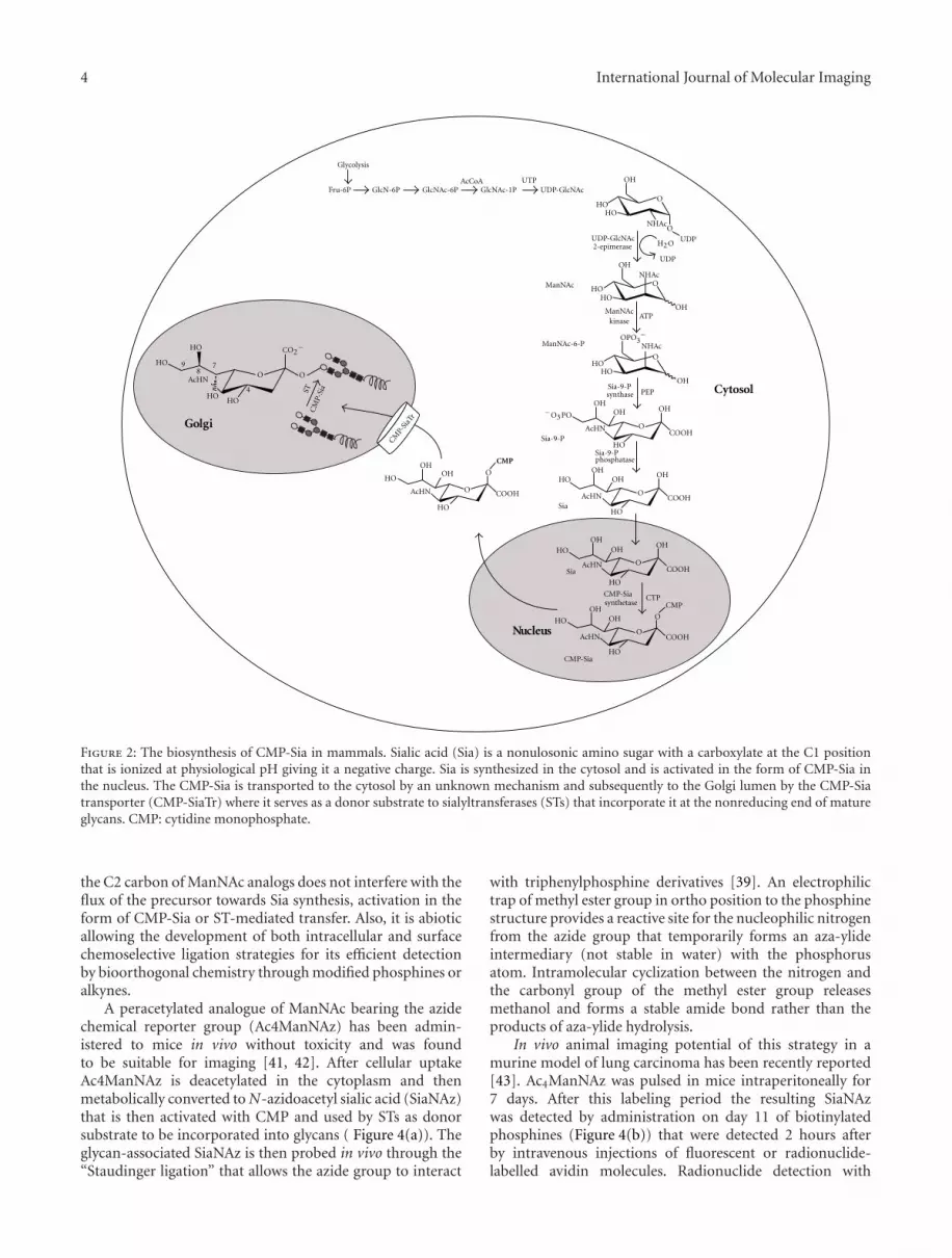
The name sialic acid collectively refers to a family of over50 naturally occurring sugars and a growing number ofsynthetic analogs. In humans the predominant Sia is N-acetylneuraminic acid [27]. Sia is a nonulosonic amino sugarwith a carboxylate at the C1 position that is ionized atphysiological pH giving it a negative charge (Figure 2). Siahas the potential for additional substitutions with acetyl,methyl, sulphate, and phosphate groups at the hydroxylgroups on the 4-, 7-, 8-, and 9-carbons that give it additionalproperties. The negative charge of Sia and its terminalposition in glycans have given it a predominant role indetermining the nature of glycan interactions involved inmany essential functions of human physiology.
The biosynthesis of Sia begins in the cytosol withthe formation of N-acetylmannosamine (ManNAc) from arelatively minor proportion of UDP-N-acetylglucosamine(UDP-GlcNAc), obtained form the extracellular environ-ment [28] or derived from GlcNAc via the action ofGlcNAc 2-epimerase [29] (Figure 2). In mammals, theManNAc is then phosphorylated to give ManNAc-6-phosphate (ManNAc-6P). The second step involves the con-densation of either ManNAc or MacNAc-6P with phospho-enolpyruvate (PEP) to give neuraminic acid or neuraminic

International Journal of Molecular Imaging 3
AcHN
GalNAc
HO
OH
O
COOH
NHAc
O
O
O
O OOO
O
O
2
6AcHN
COOH
HO
O
HO
OH
HO
OH
O
OH
OH
OH
O
O
OH
NHAc
O2 3
3
3
3
3
3
1
11
COOH
HO
HO
HO
O
O
HO
OHHO
HO
HO
HO
O
OHOH
O
OH
O O
O
OH
OH
O
OH
OH
2
2 11
4
4
Sia
Sia
SiaGlcNAc
AcHN
O
O
OH
Sia
Gal
Gal
FucFuc
OH
O
NHAc
ST
SLea
STn
Gal
AcHNNHAc
COOH
OH
OH
OHOH
OHOH
OH
OH
GlcNAc
SLex
H3CH3C
GalNAc
Figure 1: Chemical structures of sialylated antigens associated to malignancy. Sialyl-Tn (Siaα2→ 6GalNAc-T/S), sialyl T (Siaα2-3Galβ1-3GalNAc-T/S), sialyl Lewis X (SLex; Siaα2,3Galβ1,4(Fucα1,3)GlcNAc), and sialyl Lewis A (SLea; Siaα2,3Galβ1,3(Fucα1,4)GlcNAc).
acid-9P, respectively. In mammals, neuraminic acid-9P isthen dephosphorylated to generate neuraminic acid (Sia).Finally, in the nucleus the activation of Sia with cytidinemonophosphate (CMP-Sia) is generated with the use ofcytosine triphosphate (CTP) [30]. Once in the cytosol CMP-Sia is then transported to the Golgi lumen by a specifictransporter (CMP-SiaTr) where it is used as donor substrateby more than twenty human sialyltransferases (STs) thatincorporate it into the nonreducing end of glycans [16, 31,32].
Linkage of Sia by STs can be done to a terminalgalactose residue (Gal) via α2,6 or α2,3-linkage, to N-acetyl-galactosamine (GalNAc) or galactosamine residues viaα2,6-linkage, or to another Sia as an α2,8 homopolymer(Figure 3). The presence or absence of Sia as well as the typeof linkage it presents in the glycan have been selected inevolution as recognition characteristics that allow or hamperbiding of glycan ligands, thus determining important molec-ular interactions that affect cell behavior including cell-cell,cell-matrix, and cell-soluble molecule interactions that playroles in both physiological and pathological processes [7].
4. Sialic Acid Imaging In Vivo
Because of the clear association that has been establishedbetween sialylated antigen overexpression and tumor aggres-siveness, the imaging of tumor sialylation would offer aconcomitant or alternative study to PET-FDG imaging notonly to diagnose cancer but also to assess patient’s prognosis.To obtain the in vivo sialylation status of a tumor is a clinicalasset that must be translated from the bench to the bedside.There are at least two strategies for the in vivo imaging oftumor Sia expression. One involves metabolic labeling thatallows imaging of de novo synthesis of Sia, and the secondone involves native Sia recognition by imaging probes.
4.1. Metabolic Labeling. Metabolic labeling allows the studyof a metabolic pathway by administering to the cells com-pounds that are modified analogs of natural substrates forthat particular pathway. Modification of these compoundsaims at providing a means for their detection withoutinterfering with their metabolic use. An ideal candidatecompound for evaluating tumor sialylation in vivo has tobe cell permeable, has to be able to enter the CMP-Siabiosynthesis pathway at a committed stage, and althoughmodified has to be efficiently used by STs. The use of labelledSia or CMP-Sia is restricted because of the lack of plasmamembrane transporters and the negative charge of unmod-ified Sia that limits its cellular permeability. Uptake studieswith 18F-labelled sialic acids N-acetyl-3-[18F]fluorosialic acid(3-Sia) and N-acetyl-2-deoxy-2,3-difluorosialic acid (2,3-diSia) showed inefficient uptake and unsuitability for in vivoimaging [33]. An alternative to solve this problem is the useof modified permeable Sia analogs [34, 35] or permeableSia precursors that can be subsequently detected by imagingprobes. Sia precursors, particularly based on ManNAc, havebeen favored over Sia analogs for in vitro and in vivo imagingbecause of lower cost and synthetic tractability of ManNAcover Sia [36].
Of the different analogue precursors of Sia biosynthe-sis, ManNAc analogues are ideal candidates for metaboliclabeling because ManNAc is the first pathway intermediateto be committed to Sia biosynthesis, assuring that imaging ofthe labelled ManNAc analog reflects glycan Sia expression.And although ManNAc is permeable, ManNAc analogscan be further acetylated to increase passive membranepermeability [37].
Among many modifications that have been achievedon the ManNAc sugar with chemical reporters [38] thatallow its detection, only an azide-labelled bioorthogonalchemistry [39] has successfully been used to image in vivo itsincorporation as glycan-bound Sia [40]. The azide group at
4 International Journal of Molecular Imaging
O
HOHO
ONHAc
NHAc
NHAc
OH
UDP
O
UDP
O
O
O
HOHO
OH
OH
OHOH
OH
OH
ATP
ManNAc
ManNAc-6-P
HOHO
HO
HO
HO
OH
OH
OH
AcHN
AcHN
AcHN
H2O
O
OHOH
Sia-9-P
PEP
O
COOH
COOH
COOH
COOH
OH
HO
HO
HO
Sia
Sia
GlcNAc-1PNAc-6PUTP
GlcN-6PFru-6P
Glycolysis
UDP-GlcNAc
−O3PO
O
OH
AcHN
HO
OH
O
O
AcHN
HO
HO OH
CTP
O
COOHO
HO
HO
OH
CMP
AcHN
HOHO
879
4
Nucleus
CM
P-Si
aST
CMP-
SiaT
r
Cytosol
Golgi
CMP-Sia
AcCoA
UDP-GlcNAc2-epimerase
ManNAckinase
Sia-9-Psynthase
Sia-9-Pphosphatase
CO2−
CMP-Siaynthetases
CMP
OPO −3
Glc
Figure 2: The biosynthesis of CMP-Sia in mammals. Sialic acid (Sia) is a nonulosonic amino sugar with a carboxylate at the C1 positionthat is ionized at physiological pH giving it a negative charge. Sia is synthesized in the cytosol and is activated in the form of CMP-Sia inthe nucleus. The CMP-Sia is transported to the cytosol by an unknown mechanism and subsequently to the Golgi lumen by the CMP-Siatransporter (CMP-SiaTr) where it serves as a donor substrate to sialyltransferases (STs) that incorporate it at the nonreducing end of matureglycans. CMP: cytidine monophosphate.
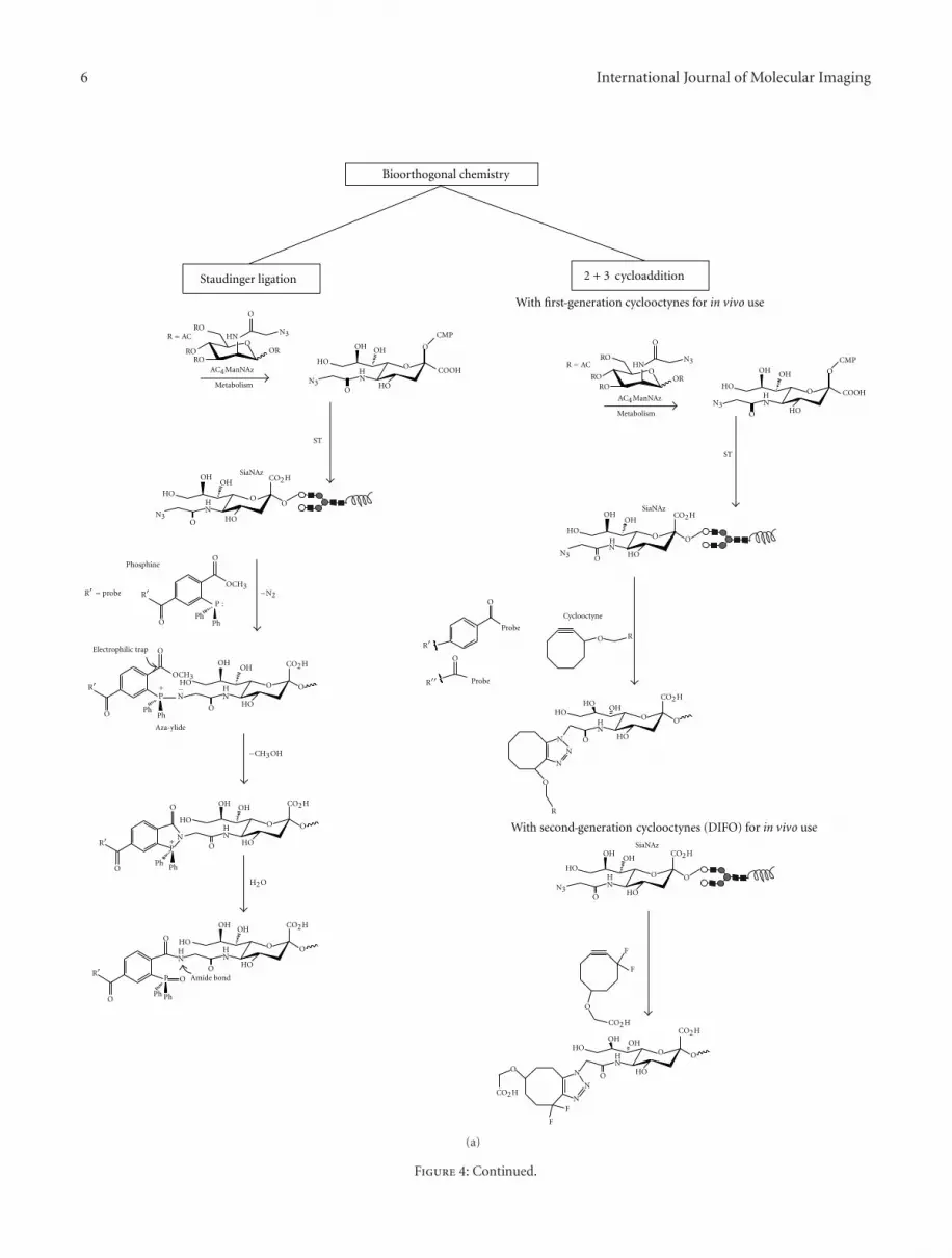
the C2 carbon of ManNAc analogs does not interfere with theflux of the precursor towards Sia synthesis, activation in theform of CMP-Sia or ST-mediated transfer. Also, it is abioticallowing the development of both intracellular and surfacechemoselective ligation strategies for its efficient detectionby bioorthogonal chemistry through modified phosphines oralkynes.
A peracetylated analogue of ManNAc bearing the azidechemical reporter group (Ac4ManNAz) has been admin-istered to mice in vivo without toxicity and was foundto be suitable for imaging [41, 42]. After cellular uptakeAc4ManNAz is deacetylated in the cytoplasm and thenmetabolically converted to N-azidoacetyl sialic acid (SiaNAz)that is then activated with CMP and used by STs as donorsubstrate to be incorporated into glycans ( Figure 4(a)). Theglycan-associated SiaNAz is then probed in vivo through the“Staudinger ligation” that allows the azide group to interact
with triphenylphosphine derivatives [39]. An electrophilictrap of methyl ester group in ortho position to the phosphinestructure provides a reactive site for the nucleophilic nitrogenfrom the azide group that temporarily forms an aza-ylideintermediary (not stable in water) with the phosphorusatom. Intramolecular cyclization between the nitrogen andthe carbonyl group of the methyl ester group releasesmethanol and forms a stable amide bond rather than theproducts of aza-ylide hydrolysis.
In vivo animal imaging potential of this strategy in amurine model of lung carcinoma has been recently reported[43]. Ac4ManNAz was pulsed in mice intraperitoneally for7 days. After this labeling period the resulting SiaNAzwas detected by administration on day 11 of biotinylatedphosphines (Figure 4(b)) that were detected 2 hours afterby intravenous injections of fluorescent or radionuclide-labelled avidin molecules. Radionuclide detection with

International Journal of Molecular Imaging 5
O
O
OH
α 2,3-ST3 Gal
α 2,6-ST6 Gal
OH OH OH
OHOH
OHOH
OROH
OAcHN
HO
HO
HO
HO
COOH
OCMP
CMP-Sia
Sia
α 2,8-ST8Sia
Gal
+
O
O
HO
HO HOHO
O
COOH
COOH
O
OH
2
33
8
8
+
CMP
+ CMP
+ CMP
6
62
HO
OR
COOH
AcHNHO
HOHO
COOH
COOH
2
O
AcHN
AcHN
HO HO
OR
2
8 OHO
NHR
OR
Galactosamine
R = Ac
R =HST6GalNAc
AcHN
HO HO
HO
O
NHROR
OO
HOHO
HO
HOHO
O
AcHNHO HO
or
N- cetylgalactosamine
HOH2C
a
Figure 3: Types of sialyltransferases and their main acceptor substrates. STs can link sialic acid (Sia) to terminal galactose (Gal) via α2,6 orα2,3-linkage or to galactosamine or N-acetyl-galactosamine (GalNAc) via α2,6-linkage. Also, Sia can be linked to the C8 position of anotherSia residue forming polysialic acids. CMP: cytidine monophosphate.
a SPECT camera which is the most closely related towhat human applications would use was achieved with anIndium-111-labelled Neutravidin-DOTA, showing a signifi-cant azido-labelled ManNAc-dependent increase in tumor-to-tissue contrast. As the authors of this paper suggest, thisstrategy requires further improvement by reducing it toa one-step probe approach by using phosphine moleculesmodified with gamma or positron emitting radioisotopesand with an improved hydrophilic nature of phosphines toreduce the nonspecific stromal binding that was observed inthis study. Also, other compounds like cyclooctynes couldbe used to detect azide groups via strain-promoted azide-alkyne cycloaddition (Figure 4(a)) [44]. The cyclooctynemolecule can be more easily modified to generate reactantswith enhanced kinetics. Some derivates possess a ring strainand electron-withdrawing fluorine substituents that togetherpromote the [3 + 2] dipolar cycloaddition with azidesto form regioisomeric mixtures of triazoles. This reactionhas higher reaction kinetics compared to the Staudingerligation [38] and has been further improved by synthesizingsecond-generation difluorinated cyclooctynes (DIFOs) thathave propargylic fluorine atoms that increase its interactionenergy with the azide (Figure 4(a)) [45]. These DIFOs havebeen successfully employed for dynamic in vivo imaging ofdeveloping zebrafish [41], and important advances have beenmade to improve their synthesis [46].
It is worth considering that fluorophore modified phos-phines or cyclooctynes could also be used for imaging of thesialylation status of superficial cancers like skin melanomaor colorectal cancer through fluorescent endoscopy. It isnoteworthy to mention that advances in positron emitters
through the Staudinger ligation have been achieved usingphosphine-substituted thioesters with 18F-fluoroethylazide[47].
4.2. Probes for Recognition of Native Sia. A second strategyconsists in using imaging probes that recognize native Sia inthe cell surface. Historically, sialic acids have been exploredin vitro using plant lectins like wheat germ agglutinin(WGA) that detects Sia independently of its type of linkage,Sambucus nigra lectin (SNA) that recognizes α2,6 Sia orMaackia amurensis aggluttinin (MAL) that recognizes α2,3Sia. Also, antibodies directed against SLex or SLea deter-minants have been used for many years; this is the caseof CA19-9 a tumoral marker used for detection of SLea
determinants in gastrointestinal tumors. These in vitro toolshave been difficult to translate in vivo not only becauseof their immunogenicity, that at least for antibodies couldbe circumvented by humanizing them, but also becauseof their molecular weight, that would restrict them to thevascular compartment. This vascular restriction would notbe a problem if the aim was to image the expression ofSia on the vascular network of a tumor, for example, orin chronic inflammatory diseases. In the case of lectins,immunogenicity could be avoided if human endogenoussialic-acid-recognizing lectins were to be used like siglecs orselectins, but again their use would probably be limited to thevascular compartment.
Nonetheless, other alternatives show promise mainly inthe field of magnetic resonance imaging for the detection ofnative Sia molecules (Figure 5). Frullano et al. reported the
6 International Journal of Molecular Imaging
H2O
R = ACRO
RORO
OH
OH
OH
OH
OHOH
OH
OH
OHOH
OHOR
HN
HO
HO
HO
HO
HO
HO
HO
HO
HO
HO
HO
HO
HO
HON
N
NH
NH
NN
N
N
N
N
N
H
NH
NH
NH
NH
CMP
COOH
ST
ST
AC4ManNAz
AC4ManNAz
Metabolism
Metabolism
SiaNAz
CO2H
CO2H
CO2H
CO2H
CO2H
Cyclooctyne
R
R
Probe
ProbeR
R
R
R
P :
OCH3
F
F
F
F
CO2H
R = probe −N2
Electrophilic trap
−CH3OH
Bioorthogonal chemistry
Staudinger ligation
O
O
O
O
O
O
O
O
O O
O
O
O
O
OO
O
O O
O
O
R
O
O
O
O O
O
O
O
O
O
O
O
O
O
N3
N3
R = ACRO
RORO
OR
HN
O
O
N3OH OH
HO
HONH
CMP
COOH
O
O
O
N3
N3 OHOH
HO
HO
HO
NH
CO2H
O O
ON3
N3
PhPh
Ph
PhPh
Ph
CO2H
OH OHCO2H
−P+
P+
HO
HONH
O
OR
O
O
O O
PhPh
OHOH
CO2H
P
OCH3
Amide bond
2 + 3 ycloadditionc
SiaNAz
SiaNAz
A a-ylidez
Phosp ineh
With first generation cyclooctynes for in vivo use
With second generation (DIFO) for in vivo usecyclooctynes
-
-
NH
(a)
Figure 4: Continued.

International Journal of Molecular Imaging 7
O
O
O
O
OO
O
OO
O
OO
H NH N
O H
OHOH
O H
H O
HO
H O
HO
NH
NH
NH
F
F
F
F
NH
O
3S
Avidin
+
Avidin
+
4
O
O
H N
NH
N H
OO
OO
3S 4
O
O
H N
NH
N H
O
3S 4
N N
N
OO
ON 3
OHOH
HO
HO
NH
CO2Me
Ph2P
CO2HHH
HH
HH
Biotin
R =R =
NH
NH
SiaNAz
Phosp ineh
RN H
Ph2P
CO2−
CO2−
Cyclooct nei
Detection of biotin- hosphine and biotin-difluorinated with labelled avidinecyclooctynesp
(b)
Figure 4: Reactions in bioorthogonal chemistry for detection of azide-modified sialic acid. (a) Two reactions involving bioorthogonalchemistry of azide-modified sialic acid (Sia) have been developed for in vivo imaging, the strain-promoted [3 + 2] cycloaddition, andthe Staudinger ligation. In both cases a peracetylated analogue of N-acetylmannosamine (Ac4ManNAz) enters cells by enhanced passivediffusion and is deacetylated by intracellular carboxylesterases. The resulting ManNAz molecule is then converted to N-azidoacetyl sialicacid (SiaNAz) in the cytosol and transported to the nucleus, where it is activated with cytidine monophosphate (CMP) to form CMP-SiaNAz. CMP-SiaNAz is then incorporated into glycans as SiaNAz by the action of STs in the Golgi. The Staudinger ligation allows azidegroup detection of SiaNAz with a phosphine-substituted ester. The harmful byproduct nucleophilic aza-ylide is captured by intramolecularcyclization by an electrophilic trap (methyl ester) within the phosphine structure allowing it to be used in living animals without physiologicalharm because phosphine oxide is not produced. Regarding the strain-promoted [3 + 2] cycloaddition of azides the cyclooctyne moleculepossesses a ring strain and electron-withdrawing fluorine substituents that together promote the [3 + 2] dipolar cycloaddition with azidesto form regioisomeric mixtures of triazoles. This reaction occurs more rapidly than the Staudinger ligation, can be used in vivo, and doesnot require auxiliary reagents. First- and second-generation cyclooctyne (DIFO) compounds have been used in [3 + 2] cycloaddition. (b)Modification of phosphines or cyclooctynes with biotin and subsequent detection with fluorescent or radioisotope labelled avidins have beenused for SiaNAz detection in vivo in animal models of lung cancer, a similar strategy could be employed using cyclooctynes.
synthesis of two lanthanide ion ligands denominated L1 (3,9-Bis{6-[(4,5-dihydroimidazol-2-yl)aminoethyl]-10-[2-(dihy-droxyboranylphenyl)]-2-oxo-3,6,9-triazadecyl}-6-carbox-ymethyl-,6,9-triazaundecanedioic Acid) and L2 (3,9-Bis[3-(dihydroxyboranylphenyl)-2-oxo-3,6-diazaheptyl]-6-carboxymethyl-3,6,9-triazaundecanedioic acid). These com-pounds were obtained by inclusion of a central chelatingunit based on the DTPA-bisamide structure (DTPA, diethyl-enetriamine pentaacetic acid). Molecular modeling studiesindicate that both compounds can allow two-site binding toSia through ester formation by interaction of the boronatefunction in the ligand with the geminal diol of Sia andan electrostatic interaction between a positively chargedaminoimidazolium (L1) or aminomethyl (L2) group presentin metaposition relative to the boronic function andthe carboxylate group of Sia [48]. Both gadolinium
complexed ligands showed high specific and reversiblebinding towards Sia. The dynamic equilibrium that thecompounds showed between their sialic-acid-bound andfree states would allow them to be excreted physiologically.Similarly, a thiourea-based synthetic receptor (2-{[3-(4-Nitrophenyl)ureido]methyl}phenylboronic acid) reportedby Regueiro-Figueroa et al. that contains phenylboronic acidfunctions also showed a high specificity towards Sia dueto cooperative binding through ester formation with thephenylboronic acid and hydrogen bond interactions betweenthe carboxylate group and the thiourea moiety (Figure 5)[49].
This native recognition approach compared to metaboliclabelling has the potential to recognize the type of Sialinkages and even carbohydrate antigens, offering additionalspecific information, but this aim requires further research.

8 International Journal of Molecular Imaging
O
H
HOHO
NHN
HN
HN
HN
HN
HN
HN
S
OH
O
HO
OH
OH
HOO
O O
O
O
CO−2Sia
8 79
4
Carboxylate anion
NH
B(OH)2
O2N
N
NH
NH
O
N
NN
N
N
N
HO
O
HN
HN
HN
(HO)2B
(HO)2B
Boronic function
Boronic function
Ligand L1
Ligand L2
Thiourea
Phenylboronic acid function
NN
N
OH
O
O
OH
O
B
B(OH)2
OH HO
AcHN
Diol groups
Functional group
Functional group
Thiourea based synthetic receptor with phenylboronic acid
Diethylenetriamine
Diethylenetriamine
-
Figure 5: Receptors for native recognition of Sia. The carboxylate and diol groups of sialic acid have been exploited forrecognition purposes by lanthanide ligand DTPA-bisamide compounds L1 (3,9-Bis{6-[(4,5-dihydroimidazol-2-yl)aminoethyl]-10-[2-(dihydroxyboranylphenyl)]-2-oxo-3,6,9-triazadecyl}-6-carboxymethyl-,6,9-triazaundecanedioic acid) and L2 (3,9-Bis[3-(dihydroxyboran-ylphenyl)-2-oxo-3,6-diazaheptyl]-6-carboxymethyl-3,6,9-triazaundecanedioic acid) bearing boronic and functional groups (L1: aminoim-idazolium and L2: aminomethyl). Also, a thiourea-based synthetic receptor (2-{[3-(4-Nitrophenyl)ureido]methyl}phenylboronic acid) hasbeen reported to show a high specificity towards Sia.
5. Conclusions
Although glycans equip the cell surface with efficient anddynamic signalling mechanisms that have been partiallydeciphered to convey important clinical information, glycansand glycan-mediated interactions have been rarely targeted
for in vivo imaging with some exceptions that particularlyinvolve quantification of liver function [50–52].
In vivo imaging of tumor sialylation is in the transitionstage from the bench to the bedside and is only limited byminor improvements in radioisotope probe synthesis to beused concomitantly or alternatively with FDG in the medical

International Journal of Molecular Imaging 9
management of cancer. The metabolic labelling with Siaprecursor analogs and detection offered by bioorthogonalchemistry offer at this moment a high value for clinical usein the near future. It is important to press for the continuingtechnological development of these strategies to assess assoon as possible their clinical value in diagnosis, staging,prognosis, and treatment response. Imaging of tumor sia-lylation brings together information of both catabolic andanabolic processes associated to malignancy which can onlyimprove patient management.
Acknowledgments
I. Martinez-Duncker and R. Salinas-Marin were supportedby CONACYT scholarship no. 217256 and Research ProjectGrant no. 57157.
References
[1] A. Zhu, D. Lee, and H. Shim, “Metabolic positron emis-sion tomography imaging in cancer detection and therapyresponse,” Seminars in Oncology, vol. 38, no. 1, pp. 55–69,2011.
[2] R. A. Gatenby and R. J. Gillies, “Why do cancers have highaerobic glycolysis?” Nature Reviews Cancer, vol. 4, no. 11, pp.891–899, 2004.
[3] C. Martınez-Duncker and L. M. Hurtado-Lopez, “ 18F-FDGPET of thyroid nodules with inconclusive cytologic results,”Journal of Nuclear Medicine, vol. 47, no. 9, p. 1555, 2006.
[4] A. Varki, Essentials of Glycobiology, vol. 29, Cold SpringHarbor Laboratory Press, Cold Spring Harbor, NY, USA, 2ndedition, 2009.
[5] A. Varki, R. Kannagi, and B. P. Toole, “Glycosylation changesin cancer,” in Essentials of Glycobiology, Cold Spring HarborLaboratory, New York, NY, USA, 2009.
[6] N. Sharon, “Lectins: carbohydrate-specific reagents and bio-logical recognition molecules,” The Journal of Biological Chem-istry, vol. 282, no. 5, pp. 2753–2764, 2007.
[7] N. M. Varki and A. Varki, “Diversity in cell surface sialicacid presentations: implications for biology and disease,”Laboratory Investigation, vol. 87, no. 9, pp. 851–857, 2007.
[8] F. Tanaka, Y. Otake, T. Nakagawa et al., “Prognostic signifi-cance of polysialic acid expression in resected non-small celllung cancer,” Cancer Research, vol. 61, no. 4, pp. 1666–1670,2001.
[9] E. P. Scheidegger, P. M. Lackie, J. Papay, and J. Roth, “In vitroand in vivo growth of clonal sublines of human small cell lungcarcinoma is modulated by polysialic acid of the neural celladhesion molecule,” Laboratory Investigation, vol. 70, no. 1, pp.95–106, 1994.
[10] J. Roth, C. Zuber, P. Wagner, I. Blaha, D. Bitter-Suermann, andP. U. Heitz, “Presence of the long chain form of polysialic acidof the neural cell adhesion molecule in Wilms’ tumor. Identi-fication of a cell adhesion molecule as an oncodevelopmentalantigen and implications for tumor histogenesis,” AmericanJournal of Pathology, vol. 133, no. 2, pp. 227–240, 1988.
[11] S. R. Smith, B. Auerbach, and L. Morgan, “Serum neural celladhesion molecule in multiple myeloma and other plasma celldisorders,” British Journal of Haematology, vol. 92, no. 1, pp.67–70, 1996.
[12] H. Hildebrandt, C. Becker, S. Gluer, H. Rosner, R. Gerardy-Schahn, and H. Rahmann, “Polysialic acid on the neural cell
adhesion molecule correlates with expression of polysialyl-transferases and promotes neuroblastoma cell growth,” CancerResearch, vol. 58, no. 4, pp. 779–784, 1998.
[13] F. Dall’Olio and M. Chiricolo, “Sialyltransferases in cancer,”Glycoconjugate Journal, vol. 18, no. 11-12, pp. 841–850, 2001.
[14] R. Kannagi, K. Sakuma, K. Miyazaki et al., “Altered expressionof glycan genes in cancers induced by epigenetic silencing andtumor hypoxia: clues in the ongoing search for new tumormarkers,” Cancer Science, vol. 101, no. 3, pp. 586–593, 2010.
[15] I. Martinez-Duncker, R. Mollicone, P. Codogno, and R. Oriol,“The nucleotide-sugar transporter family: a phylogeneticapproach,” Biochimie, vol. 85, no. 3-4, pp. 245–260, 2003.
[16] A. Harduin-Lepers, R. Mollicone, P. Delannoy, and R. Oriol,“The animal sialyltransferases and sialyltransferase-relatedgenes: a phylogenetic approach,” Glycobiology, vol. 15, no. 8,pp. 805–817, 2005.
[17] C. Javaud, F. Dupuy, A. Maftah, R. Julien, and J. M. Petit, “Thefucosyltransferase gene family: an amazing summary of theunderlying mechanisms of gene evolution,” Genetica, vol. 118,no. 2-3, pp. 157–170, 2003.
[18] H. F. Langer and T. Chavakis, “Leukocyte-endothelial inter-actions in inflammation,” Journal of Cellular and MolecularMedicine, vol. 13, no. 7, pp. 1211–1220, 2009.
[19] R. Kannagi, M. Izawa, T. Koike, K. Miyazaki, and N. Kimura,“Carbohydrate-mediated cell adhesion in cancer metastasisand angiogenesis,” Cancer Science, vol. 95, no. 5, pp. 377–384,2004.
[20] K. Konstantopoulos and S. N. Thomas, “Cancer cells in transit:the vascular interactions of tumor cells,” Annual Review ofBiomedical Engineering, vol. 11, pp. 177–202, 2009.
[21] A. Cazet, S. Julien, M. Bobowski et al., “Consequencesof the expression of sialylated antigens in breast cancer,”Carbohydrate Research, vol. 345, no. 10, pp. 1377–1383, 2010.
[22] T. Kawaguchi, “Cancer metastasis: characterization and iden-tification of the behavior of metastatic tumor cells and the celladhesion molecules, including carbohydrates,” Current DrugTargets Cardiovascular and Haematological Disorders, vol. 5,no. 1, pp. 39–64, 2005.
[23] H. Ura, R. Denno, K. Hirata, K. Yamaguchi, T. Yasoshima,and T. Shishido, “Close correlation between increased sialyl-lewis(x) expression and metastasis in human gastric carci-noma,” World Journal of Surgery, vol. 21, no. 7, pp. 773–776,1997.
[24] P.H. Wang, “Altered sialylation and its roles in gynecologiccancers,” Journal of Cancer Molecules, vol. 2, no. 3, pp. 107–116, 2006.
[25] R. Kannagi, “Carbohydrate antigen sialyl Lewis a—its patho-physiological significance and induction mechanism in cancerprogression,” Chang Gung Medical Journal, vol. 30, no. 3, pp.189–209, 2007.
[26] A. Fernandez-Briera, I. Garcıa-Parceiro, E. Cuevas, and E. Gil-Martın, “Effect of human colorectal carcinogenesis on theneural cell adhesion molecule expression and polysialylation,”Oncology, vol. 78, no. 3-4, pp. 196–204, 2010.
[27] N. M. Varki, E. Strobert, E. J. Dick Jr., K. Benirschke,and A. Varki, “Biomedical differences between human andnonhuman hominids: potential roles for uniquely humanaspects of sialic acid biology,” Annual Review of Pathology, vol.6, pp. 365–393, 2011.
[28] A. Varki, “Radioactive tracer techniques in the sequencing ofglycoprotein oligosaccharides,” FASEB Journal, vol. 5, no. 2,pp. 226–235, 1991.

10 International Journal of Molecular Imaging
[29] I. Maru, Y. Ohta, K. Murata, and Y. Tsukada, “Molecu-lar cloning and identification of N-acyl-D-glucosamine 2-epimerase from porcine kidney as a renin-binding protein,”Journal of Biological Chemistry, vol. 271, no. 27, pp. 16294–16299, 1996.
[30] M. E. Tanner, “The enzymes of sialic acid biosynthesis,”Bioorganic Chemistry, vol. 33, no. 3, pp. 216–228, 2005.
[31] M. Eckhardt and R. Gerardy-Schahn, “Molecular cloning ofthe hamster CMP-sialic acid transporter,” European Journal ofBiochemistry, vol. 248, no. 1, pp. 187–192, 1997.
[32] I. Martinez-Duncker, T. Dupre, V. Piller et al., “Geneticcomplementation reveals a novel human congenital disorderof glycosylation of type II, due to inactivation of the GolgiCMP-sialic acid transporter,” Blood, vol. 105, no. 7, pp. 2671–2676, 2005.
[33] K. Ishiwata, T. Ido, T. Nakajima, H. Ohrui, I. Kijima-Suda, and M. Itoh, “Tumor uptake study of 18F-labeled N-acetylneuraminic acids,” International Journal of RadiationApplications and Instrumentation, vol. 17, no. 4, pp. 363–367,1990.
[34] C. Oetke, R. Brossmer, L. R. Mantey et al., “Versatile biosyn-thetic engineering of sialic acid in living cells using syntheticsialic acid analogues,” Journal of Biological Chemistry, vol. 277,no. 8, pp. 6688–6695, 2002.
[35] M. Bardor, D. H. Nguyen, S. Diaz, and A. Varki, “Mechanismof uptake and incorporation of the non-human sialic acidN-glycolylneuraminic acid into human cells,” Journal ofBiological Chemistry, vol. 280, no. 6, pp. 4228–4237, 2005.
[36] J. Du, M. A. Meledeo, Z. Wang, H. S. Khanna, V. D. Paruchuri,and K. J. Yarema, “Metabolic glycoengineering: sialic acid andbeyond,” Glycobiology, vol. 19, no. 12, pp. 1382–1401, 2009.
[37] M. B. Jones, H. Teng, J. K. Rhee, N. Lahar, G. Baskaran,and K. J. Yarema, “Characterization of the cellular uptakeand metabolic conversion of acetylated N-acetylmannosamine(ManNAc) analogues to sialic acids,” Biotechnology and Bio-engineering, vol. 85, no. 4, pp. 394–405, 2004.
[38] T. L. Hsu, S. R. Hanson, K. Kishikawa, S. K. Wang, M. Sawa,and C. H. Wong, “Alkynyl sugar analogs for the labeling andvisualization of glycoconjugates in cells,” Proceedings of theNational Academy of Sciences of the United States of America,vol. 104, no. 8, pp. 2614–2619, 2007.
[39] E. Saxon and C. R. Bertozzi, “Cell surface engineering by amodified Staudinger reaction,” Science, vol. 287, no. 5460, pp.2007–2010, 2000.
[40] J. A. Prescher, D. H. Dube, and C. R. Bertozzi, “Chemicalremodelling of cell surfaces in living animals,” Nature, vol. 430,no. 7002, pp. 873–877, 2004.
[41] S. T. Laughlin, J. M. Baskin, S. L. Amacher, and C. R.Bertozzi, “In vivo imaging of membrane-associated glycans indeveloping zebrafish,” Science, vol. 320, no. 5876, pp. 664–667,2008.
[42] P. V. Chang, X. Chen, C. Smyrniotis et al., “Metabolielabeling of sialic acids in living animals with alkynyl sugars,”Angewandte Chemie, vol. 48, no. 22, pp. 4030–4033, 2009.
[43] A. A. Neves, H. Stockmann, R. R. Harmston et al., “Imagingsialylated tumor cell glycans in vivo,” The FASEB Journal, vol.25, no. 8, pp. 2528–2537, 2011.
[44] N. J. Agard, J. A. Prescher, and C. R. Bertozzi, “A strain-promoted [3 + 2] azide-alkyne cycloaddition for covalentmodification of biomolecules in living systems,” Journal of theAmerican Chemical Society, vol. 126, no. 46, pp. 15046–15047,2004.
[45] J. M. Baskin, J. A. Prescher, S. T. Laughlin et al., “Copper-freeclick chemistry for dynamic in vivo imaging,” Proceedings of theNational Academy of Sciences of the United States of America,vol. 104, no. 43, pp. 16793–16797, 2007.
[46] J. A. Codelli, J. M. Baskin, N. J. Agard, and C. R. Bertozzi,“Second-generation difluorinated cyclooctynes for copper-free click chemistry,” Journal of the American Chemical Society,vol. 130, no. 34, pp. 11486–11493, 2008.
[47] L. Carroll, S. Boldon, R. Bejot, J. E. Moore, J. Declerck, andV. Gouverneur, “The traceless Staudinger ligation for indirect18F- radiolabelling,” Organic and Biomolecular Chemistry, vol.9, no. 1, pp. 136–140, 2011.
[48] L. Frullano, J. Rohovec, S. Aime et al., “Towards targeted MRI:new MRI contrast agents for sialic acid detection,” Chemistry,vol. 10, no. 20, pp. 5205–5217, 2004.
[49] M. Regueiro-Figueroa, K. Djanashvili, D. Esteban-Gomez, A.de Blas, C. Platas-Iglesias, and T. Rodrıguez-Blas, “Towardsselective recognition of sialic acid through simultaneousbinding to its cis-Diol and carboxylate functions,” EuropeanJournal of Organic Chemistry, vol. 2010, no. 17, pp. 3237–3248,2010.
[50] P. Chaumet-Riffaud, I. Martinez-Duncker, A. L. Marty et al.,“Synthesis and application of lactosylated, 99mTc chelatingalbumin for measurement of liver function,” BioconjugateChemistry, vol. 21, no. 4, pp. 589–596, 2010.
[51] J. M. Jeong, M. K. Hong, J. Lee et al., “99mTc-neolactosylatedhuman serum albumin for imaging the hepatic asialoglyco-protein receptor,” Bioconjugate Chemistry, vol. 15, no. 4, pp.850–855, 2004.
[52] S. Kim, J. M. Jeong, M. K. Hong et al., “Differential receptortargeting of liver cells using 99mTc-neoglycosylated humanserum albumins,” Archives of Pharmacal Research, vol. 31, no.1, pp. 60–66, 2008.


