Embed Size (px)
Citation preview

In situ atomic force microscopy of partially demineralizedhuman dentin collagen fibrils
Stefan Habelitz,a,* Mehdi Balooch,b Sally J. Marshall,a Guive Balooch,a
and Grayson W. Marshall, Jr.a
a Department of Preventive and Restorative Dental Sciences, University of California, 707 Parnassus Avenue D-2260, San Francisco, CA 94143, USAb Department of Chemistry and Materials Science, Lawrence Livermore National Laboratory, Livermore, CA 94550, USA
Received 26 December 2001, and in revised form 29 May 2002
Abstract
Dentin collagen fibrils were studied in situ by atomic force microscopy (AFM). New data on size distribution and the axial repeat
distance of hydrated and dehydrated collagen type I fibrils are presented. Polished dentin disks from third molars were partially
demineralized with citric acid, leaving proteins and the collagen matrix. At this stage collagen fibrils were not resolved by AFM, but
after exposure to NaOClaq for 100–240 s, and presumably due to the removal of noncollagenous proteins, individual collagen fibrils
and the fibril network of dentin connected to the mineralized substrate were revealed. High-aspect-ratio silicon tips in tapping mode
were used to image the soft fibril network. Hydrated fibrils showed three distinct groups of diameters: 100, 91, and 83 nm and a
narrow distribution of the axial repeat distance at 67 nm. Dehydration resulted in a broad distribution of the fibril diameters be-
tween 75 and 105 nm and a division of the axial repeat distance into three groups at 67, 62, and 57 nm. Subfibrillar features (4 nm)
were observed on hydrated and dehydrated fibrils. The gap depth between the thick and thin repeating segments of the fibrils varied
from 3 to 7 nm. Phase mode revealed mineral particles on the transition from the gap to the overlap zone of the fibrils. This method
appears to be a powerful tool for the analysis of fibrillar collagen structures in calcified tissues and may aid in understanding the
differences in collagen affected by chemical treatments or by diseases. � 2002 Elsevier Science (USA). All rights reserved.
Keywords: Dentin; Collagen; Atomic force microscopy; Hydration
1. Introduction
Dentin is the calcified tissue that forms the internalbulk of the tooth, lying between the enamel and the pulpchamber. It is a hydrated biological composite consist-ing of tubules, which were the pathways of the formativeodontoblastic cells, the peritubular dentin, a highlymineralized zone surrounding the tubules, and the in-tertubular dentin (Ten Cate, 1994). The latter has anestimated composition of approximately 50wt% mineralphase, 40wt% organic phase, and 10wt% aqueous fluidsand is similar to the composition of bone (Weiner andWagner, 1998). Ninety weight percent of the organicphase in dentin is collagen, which is almost exclusivelytype I (Goldberg and Takagi, 1993; Linde and Robins,
1988). Type I collagen forms a fibrous three-dimensionalnetwork which builds up the dentin matrix. Comparedto bone, the collagen matrix in dentin is more interwo-ven with numerous crossings of fibrils (Kramer, 1951).Noncollagenous proteins, mostly glycoproteins andproteoglycans, cover the collagen fibrils and are associ-ated with the inorganic phase. In particular, phospho-proteins are believed to be critical for inducing mineralnucleation and for binding to calcium phosphates(Begue-Kirn et al., 1998; Dahl et al., 1998; MacDougallet al., 1992; Saito et al., 1998). The mineral in dentin is acarbonated apatite, which is similar to the mineral inbone and calcified tendon and is located either in thegaps between collagen molecules (intrafibrillar) or at-tached to the collagen fibrils (extrafibrillar) (Lees et al.,1997; Wassen et al., 2000). Plate-like and cylindricalmorphologies have been reported for the apatite crystalsin dentin with dimensions between 20 and 5 nm (Ar-senault, 1989; Kinney et al., 2001; LeGeros, 1991).
Journal of Structural Biology 138 (2002) 227–236
www.academicpress.com
Journal of
StructuralBiology
* Corresponding author. Fax: +415-476-0858.
E-mail address: [email protected] (S. Habelitz).
1047-8477/02/$ - see front matter � 2002 Elsevier Science (USA). All rights reserved.
PII: S1047 -8477 (02 )00029-1
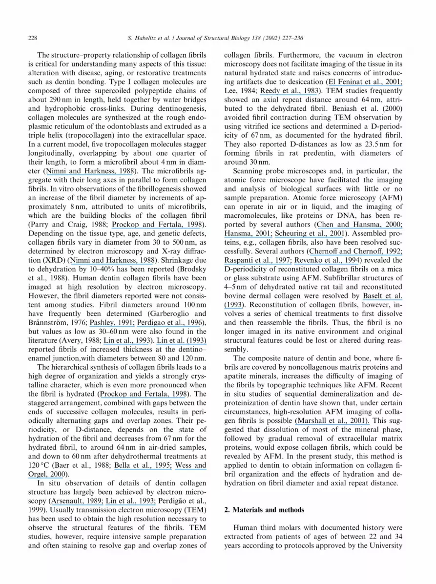
The structure–property relationship of collagen fibrilsis critical for understanding many aspects of this tissue:alteration with disease, aging, or restorative treatmentssuch as dentin bonding. Type I collagen molecules arecomposed of three supercoiled polypeptide chains ofabout 290 nm in length, held together by water bridgesand hydrophobic cross-links. During dentinogenesis,collagen molecules are synthesized at the rough endo-plasmic reticulum of the odontoblasts and extruded as atriple helix (tropocollagen) into the extracellular space.In a current model, five tropocollagen molecules staggerlongitudinally, overlapping by about one quarter oftheir length, to form a microfibril about 4 nm in diam-eter (Nimni and Harkness, 1988). The microfibrils ag-gregate with their long axes in parallel to form collagenfibrils. In vitro observations of the fibrillogenesis showedan increase of the fibril diameter by increments of ap-proximately 8 nm, attributed to units of microfibrils,which are the building blocks of the collagen fibril(Parry and Craig, 1988; Prockop and Fertala, 1998).Depending on the tissue type, age, and genetic defects,collagen fibrils vary in diameter from 30 to 500 nm, asdetermined by electron microscopy and X-ray diffrac-tion (XRD) (Nimni and Harkness, 1988). Shrinkage dueto dehydration by 10–40% has been reported (Brodskyet al., 1988). Human dentin collagen fibrils have beenimaged at high resolution by electron microscopy.However, the fibril diameters reported were not consis-tent among studies. Fibril diameters around 100 nmhave frequently been determined (Garberoglio andBr�aannstr€oom, 1976; Pashley, 1991; Perdigao et al., 1996),but values as low as 30–60 nm were also found in theliterature (Avery, 1988; Lin et al., 1993). Lin et al. (1993)reported fibrils of increased thickness at the dentino–enamel junction,with diameters between 80 and 120 nm.
The hierarchical synthesis of collagen fibrils leads to ahigh degree of organization and yields a strongly crys-talline character, which is even more pronounced whenthe fibril is hydrated (Prockop and Fertala, 1998). Thestaggered arrangement, combined with gaps between theends of successive collagen molecules, results in peri-odically alternating gaps and overlap zones. Their pe-riodicity, or D-distance, depends on the state ofhydration of the fibril and decreases from 67 nm for thehydrated fibril, to around 64 nm in air-dried samples,and down to 60 nm after dehydrothermal treatments at120 �C (Baer et al., 1988; Bella et al., 1995; Wess andOrgel, 2000).
In situ observation of details of dentin collagenstructure has largely been achieved by electron micro-scopy (Arsenault, 1989; Lin et al., 1993; Perdig~aao et al.,1999). Usually transmission electron microscopy (TEM)has been used to obtain the high resolution necessary toobserve the structural features of the fibrils. TEMstudies, however, require intensive sample preparationand often staining to resolve gap and overlap zones of
collagen fibrils. Furthermore, the vacuum in electronmicroscopy does not facilitate imaging of the tissue in itsnatural hydrated state and raises concerns of introduc-ing artifacts due to desiccation (El Feninat et al., 2001;Lee, 1984; Reedy et al., 1983). TEM studies frequentlyshowed an axial repeat distance around 64 nm, attri-buted to the dehydrated fibril. Beniash et al. (2000)avoided fibril contraction during TEM observation byusing vitrified ice sections and determined a D-period-icity of 67 nm, as documented for the hydrated fibril.They also reported D-distances as low as 23.5 nm forforming fibrils in rat predentin, with diameters ofaround 30 nm.
Scanning probe microscopes and, in particular, theatomic force microscope have facilitated the imagingand analysis of biological surfaces with little or nosample preparation. Atomic force microscopy (AFM)can operate in air or in liquid, and the imaging ofmacromolecules, like proteins or DNA, has been re-ported by several authors (Chen and Hansma, 2000;Hansma, 2001; Scheuring et al., 2001). Assembled pro-teins, e.g., collagen fibrils, also have been resolved suc-cessfully. Several authors (Chernoff and Chernoff, 1992;Raspanti et al., 1997; Revenko et al., 1994) revealed theD-periodicity of reconstituted collagen fibrils on a micaor glass substrate using AFM. Subfibrillar structures of4–5 nm of dehydrated native rat tail and reconstitutedbovine dermal collagen were resolved by Baselt et al.(1993). Reconstitution of collagen fibrils, however, in-volves a series of chemical treatments to first dissolveand then reassemble the fibrils. Thus, the fibril is nolonger imaged in its native environment and originalstructural features could be lost or altered during reas-sembly.
The composite nature of dentin and bone, where fi-brils are covered by noncollagenous matrix proteins andapatite minerals, increases the difficulty of imaging ofthe fibrils by topographic techniques like AFM. Recentin situ studies of sequential demineralization and de-proteinization of dentin have shown that, under certaincircumstances, high-resolution AFM imaging of colla-gen fibrils is possible (Marshall et al., 2001). This sug-gested that dissolution of most of the mineral phase,followed by gradual removal of extracellular matrixproteins, would expose collagen fibrils, which could berevealed by AFM. In the present study, this method isapplied to dentin to obtain information on collagen fi-bril organization and the effects of hydration and de-hydration on fibril diameter and axial repeat distance.
2. Materials and methods
Human third molars with documented history wereextracted from patients of ages of between 22 and 34years according to protocols approved by the University
228 S. Habelitz et al. / Journal of Structural Biology 138 (2002) 227–236

of California, San Francisco Committee on HumanResearch. Teeth were sterilized by c-radiation andstored in deionized water at 4 �C until prepared (Whiteet al., 1994). Sagittal midcoronal sections of six teeth(thickness of 2mm) were prepared by polishing througha series of SiO2 papers and with water-based diamondpaste to 0:25lm (Buehler, Lake Bluff, IL). Ultrasonictreatments in water for 10 s were used to clean the sur-face. Specimens were etched with 10 vol% citric acidðC6H8O7Þ for 15 s. The application of acids such as citricacid dissolves the mineral in calcified tissues and leavesthe organic phases behind. Dentin etching is a commontechnique for providing a better substrate for bonding todental adhesives (Marshall et al., 1997). Subsequently,specimens were treated with an aqueous solution of6.5 vol% sodium hypochlorite (NaOClaq) at time inter-vals of 10–20 s for up to 300 s total time (Marshall et al.,2001). Sodium hypochlorite is used as a cleansing, dis-infective, nonspecific deproteinizing agent in endodontictreatments. In aqueous solutions superoxide radicals(O�
2 ) are formed and induce one-electron oxidations thatfragment long peptide chains of proteins. NaOClaq alsois believed to chlorinate protein terminal groups, form-ing N-chloroamines, which are then broken down (DiRenzo et al., 2001; Pereira et al., 1973).
AFM images in the midcoronal area of demineralizeddentin were obtained at each NaOCl treatment interval.Once the collagen fibril network was observed in contactmode at large scan sizes (about 10� 10lm), particularareas of the specimen were studied at high resolution intapping mode. AFM studies were performed on a MultiMode Nanoscope III using a calibrated vertical-engage‘‘JV’’ piezo-scanner (Digital Instruments, Santa Bar-bara, CA). Contact mode images were obtained inliquid with Si3N4-tips (NP), (Digital Instruments). Forhigh-resolution images in air, high-aspect-ratio Si-tips(Nanosensor, Wetzlar, Germany) with a radius of ap-proximately 10 nm were used at resonance frequenciesnear 330 kHz. Specimens were dehydrated by air-blow-ing (5 s) and continued drying in air for at least 1 h be-fore imaging. Hydrated samples were imaged in Hanksbalanced salts solution using Nanoprobe SPM tips (FIBSpike) at a resonance frequency around 165 kHz. Tap-ping and phase mode images were recorded simulta-neously at scanning rates as low as 0.3Hz for scan sizesbelow 1� 1lm. Phase mode imaging is sensitive to theelastic properties of the substrate. The phase lag, whichis recorded during scanning in tapping mode, is used tocreate a phase image, which is based on both the to-pography and the elastic response of the substrate to thetapping tip (Burnham et al., 1997).
Collagen fibril diameter and axial repeat distancewere measured from section analysis of images usingdata that had been modified only by plane fitting. Sec-tion analysis was used to determine the step height be-tween the gap and overlap zones of individual fibrils.
The collagen fibril diameter was preferentially deter-mined from fibrils that exposed their complete diameter,by measuring the radius of curvature across the overlapzone, assuming a circular transverse shape. A total of395 and 180 diameters of individual fibrils were analyzedin the hydrated and dehydrated states, respectively.Measurements of fibril dimensions were corrected forthe tip radius. The dimensions necessary to calculate theradius of the AFM silicon tips were determined fromhigh-resolution SEM images (Hitachi, model S-4500with cooled cathode field emission capability with na-nometer resolution). A parabola of the form Y ¼ aX 2 þbX þ c was fitted to the profile using standard least-squares software. The tip radius (r) at its apex is givenby 1=ð2aÞ (typically a 5 to 10-nm radius of curvature).The fibril diameters obtained from the radius of curva-ture were corrected for tip broadening by the equatione ¼ 2r, where e is the error in the horizontal dimension(Takeyasu et al., 1996).
Axial repeat distances of individual collagen fibrilswere determined from AFM images by Fourier trans-form analysis (AFM software 4.48r, Digital Instru-ments) of spectral frequencies along a line placedparallel onto the fibril axes. A total of 322 and 363 pe-riodicities of fibrils were measured in the hydrated anddehydrated states, respectively. The values obtained forthe diameter and periodicity of each fibril were roundedto the full nanometer.
3. Results
Figs. 1a–d show a series of 10� 10-lm contact modeimages of hydrated human intertubular dentin afterdifferent chemical treatments. The polished and un-treated tooth specimen of Fig. 1a shows its characteristicstructure (Marshall et al., 1997). A layer of highermineralization, the peritubular dentin, lines tubules ofabout 1lm in diameter. The matrix phase between thetubules is intertubular dentin, consisting of extracellularmatrix proteins and carbonated apatite mineral. Fig. 1bshows the microstructure of human dentin after acidtreatment with 10 vol% citric acid for 15 s (Marshallet al., 1993). Dentinal tubule diameters are increased dueto the recession of the peritubular dentin. As shown inan earlier study, this treatment demineralized about700 nm deep into the intertubular dentin (Marshallet al., 2001). The demineralized layer consists predomi-nantly of collagen and other matrix proteins. In the nextstep, 6.5 vol% NaOClaq (bleach) was applied to etchedspecimens, and their surfaces were imaged at time in-tervals of 10–20 s. A dense network of fibrils becamevisible after an accumulation of 70 s of treatment with6.5% NaOClaq. The image quality of the fibrillarnetwork improved with bleaching time and appearedto be optimized for times between 100 and 200 s
S. Habelitz et al. / Journal of Structural Biology 138 (2002) 227–236 229

(Figs. 1c and d). The number of fibrils decreased withbleaching time. NaOClaq treatments for more than 240 sled to the total removal of collagen fibrils, leaving be-hind a ‘‘moth-eaten’’ appearance of dentin caused by thedemineralization and deproteinization of dentin thatexposed dentinal tubules, microtubules, and numerousinterconnections (Marshall et al., 2001).
Specimens, which were treated with NaOClaq fortimes from 110 to 200 s and exhibited the fibrillar net-work of collagen in the contact mode using a Si3N4-tip,were then examined in air and buffered solutions using ahigh-aspect-ratio Si-tip in tapping mode. Fig. 2 showsan overview of the fibril organization and their randomorientation at a scan size of 2:3lm. At this magnifica-tion, the fibril periodic pattern appeared. Fig. 3a is amixed-mode surface plot obtained in liquid exhibitingthe fibrillar structure and the axial repeat pattern ofcollagen type I. Interlocking of gap and overlap regionsof neighboring fibrils is observed. Fig. 3b is a surfaceplot of dentin collagen obtained in air after drying. Theaxial repeat pattern remains after dehydration. Fig. 4contains topographic (a) and phase mode (b) imagesobtained in liquid on demineralized and bleached dentinof the same area. Both image modes exhibit the char-acteristic structure of collagen fibrils and are suitable formeasuring the dimensions of fibril features. Phase mode
imaging, however, also is sensitive to the elastic pro-perties of the substrate, and differences in the stiffness ofthe substrate can be revealed (Behrend et al., 1999). Thephase mode image in Fig. 4b shows two darker spots onthe transition from the gap to the overlap zone of the
Fig. 2. Tapping mode AFM image of dentin collagen fibrils, obtained
from demineralized specimen (15 s citric acid) treated with 6.5 vol%
NaOClaq for 130 s and in air, showing the repeat pattern and the
random distribution of fibrils in intertubular dentin.
Fig. 1. Contact mode AFM images of the occlusal section of human dentin. (a) Untreated polished sample showing dentin tubules surrounded by
peritubular dentin and the intertubular matrix; (b) after partial demineralization by 10 vol% citric acid for 15 s, peritubular dentin is dissolved;
(c) subsequently treated with 6.5 vol% NaOClaq for 100 s; and (d) subsequently treated with 6.5 vol% NaOClaq for 200 s. Collagen fibrils are revealed.
All images were obtained in liquid.
230 S. Habelitz et al. / Journal of Structural Biology 138 (2002) 227–236

Fig. 4. AFM images of dentin collagen fibrils obtained in liquid. (a) Tapping mode image: gap and overlap zones of adjacent fibrils interlock; (b)
phase mode image reveals the presence of mineral particles attached to fibrils.
Fig. 3. Mixed-mode surface plots of tapping and phase mode AFM images of dentin collagen fibrils, obtained (a) in liquid and (b) in air. The axial
repeat pattern is still present after dehydration.
Fig. 5. Tapping mode AFM image obtained in liquid of individual gap zone of collagen fibril and adjacent overlap zones. Section analysis across the
diameter of fibril overlap zone reveals subfibrillar structure. Bumps at about 4 nm in distance are associated with microfibrils, the building blocks of
collagen type I.
S. Habelitz et al. / Journal of Structural Biology 138 (2002) 227–236 231

collagen fibrils, which indicate areas of different elasticproperties of the substrate. Fig. 5 shows, in a 100� 100-nm scanning image, an individual gap zone between twooverlap zones of a hydrated collagen fibril. The sectionprofile across the diameter of an overlap zone revealed asubfibrillar structure. Irregular patterns of bumps 2 nmin height appeared at distances of 3.0–4.5 nm from eachother. Subfibrillar features of similar sizes also wereobserved after dehydration. However, they were notpresent on all fibrils regardless of their state of hydra-tion.
Images with scan sizes below 1:0� 1:0lm were usedfor section analysis to determine step height and fibrildiameter, applying the deconvolution method describedabove. The axial repeat distance was obtained by Fou-rier analysis (Fig. 6). The step height between the gapand the overlap zones varied between 3 and 7 nm. Fig.7a shows the distribution of dentin collagen fibril diam-eters. Analysis of hydrated collagen revealed threegroups of fibril diameters with narrow normal distri-butions at 83, 91, and 100 nm. Dehydrated collagen fi-brils showed a broad distribution of axial diametersbetween 80 and 105 nm. Fig. 7b shows measurements ofthe axial repeat distance of collagen fibrils. Axial repeatdistances of hydrated collagen fibrils appeared in therange of 54–75 nm, but were predominantly observedwithin a narrow distribution at 67–68 nm. Drying of thespecimens in air resulted in a division of the collagenfibrils into three groups, with axial repeat distancesdistributed around 57, 62, and 67 nm. The correlationcoefficients for axial repeat distances and diameters ofhydrated and dehydrated fibrils were 0.17 and )0.7,respectively, showing that the data for the hydrated fi-bril are not correlated, while dehydration affects fibrildiameter and repeat distance in a reciprocal way. Thefibril diameter increases with decreasing axial repeatdistance.
4. Discussion
In this study, a chemical process was used to exposetype I collagen fibrils of midcoronal dentin. Polisheddentin sections prepared from documented human thirdmolars were partially demineralized by citric acid. Acidetching is a common procedure to dissolve the calcium-phosphate mineral phases in dentin in order to form aporous collageneous surface layer, which allows thepenetration of a monomer that forms a layer of collagen
Fig. 6. Section analysis along the axis of a dehydrated fibril shows
wave-like repeat pattern. The periodicity of the gap and the overlap
zone (D-distance) was determined by Fourier analysis at 67.9 nm. The
step height between the gap and the overlap zone was about 4 nm.
Fig. 7. (a) Plot of the frequency distribution of fibril diameters: three distinguished sizes are observed (83, 91, and 100nm) for the hydrated fibrils,
while dehydration caused a broad distribution between 80 and 100nm; (b) Distribution of the axial repeat distances: hydrated fibrils show a narrow
distribution around 67–68nm, while dehydration induced grouping of fibril repeat distances at 57, 62, and 67 nm.
232 S. Habelitz et al. / Journal of Structural Biology 138 (2002) 227–236
and polymer, resulting in a superior interface for thebonding of dental restorations. As shown by severalauthors (Kinney et al., 1996; Marshall et al., 1997), acidetching produces three zones consisting of a collagen-rich layer superficial to a zone containing a gradient ofmineral concentration, which covers the undeminera-lized dentin substrate. After partial demineralization,samples were immersed into 6.5 vol% NaOClaq, whichgradually removed proteins of the extracellular matrix inthe collagen-rich zone. At time intervals between 110 and200 s, the fibrous network of collagen was revealed in situusing AFM. The organization of the collagen frameworkand the structure of individual collagen fibrils were an-alyzed. Images at lower resolution showed a randomdistribution of fibril orientation within the plane imaged(Fig. 2). The fibril axial repeat pattern did not disappearafter dehydration (Fig. 3), which is contrary to obser-vations made by El Feninat et al. (2001). The reportedlayered arrangement of the fibrous network with layersoriented perpendicular to the tubule axes could not beshown due to the limitation of two-dimensional imaging(Kramer, 1951; Wang and Weiner, 1998). However, wefrequently observed at higher resolutions (scan sizes be-low 1:5� 1:5lm) that adjacent fibrils were interlocked.When fibrils lie parallel to each other, as in Fig. 4, the gapzones come in contact with the overlap zones of adjacentfibrils and thus interlock with each other. It is not certainif this interlocking of fibrils is native to the undeminer-alized dentin, since after demineralization fibrils are lessconstrained and might be likely to rearrange into such aconfiguration. However, it shows that dentin collagenfibrils have the ability to interlock, which is differentfrom observations in tendons, in which fibrils are alignedparallel in a way that gap zones are in contact with thegap zones and overlap zones with the overlap zones ofthe adjacent fibrils (Arsenault, 1989; Baer et al., 1988;Landis et al., 1991).
Phase mode images at high resolution (Fig. 4b)showed features of about 20 nm located at the transitionbetween the gap and the overlap zone of collagen fibrils.Phase mode becomes sensitive to differences in theelastic response of the tip to the substrate when the set-point amplitude is moderate, but an influence from ad-hesive interactions and topography cannot be avoidedcompletely (Behrend et al., 1999). The contrast of theseparticles on collagen is stronger in phase mode than thecontrast shown in the topographic mode (Fig. 3a). Toobtain such strong contrast as that shown in Fig. 4b, theparticles must differ substantially in their elasticity fromthe underlying organic substrate. We therefore assumethat these features represent residual mineral particleson collagen fibrils, which confirms that the acid etchingprocedure only partially demineralized the substrate.These particles preferentially appeared in the transitionfrom the gap to the overlap zone, which might be relatedto the location of particular noncollageneous proteins,
e.g., phosphophoryn, that reportedly induce mineral-ization in collageneous tissues (Begue-Kirn et al., 1998;Dahl et al., 1998; MacDougall et al., 1992; Saito et al.,2000).
Subfibrillar structures could be revealed in air and inliquid only at scan sizes below 300� 300 nm. Bumps ofabout 4 nm in width were detected on the surface ofcross-sections through the fibril diameter (Fig. 5). Inagreement with observations by Baselt et al. (2001), weattribute these nano-features to the presence of micro-fibrils. In contrast to Baselt et al. (2001), who foundsubfibrillar structures only if fibrils had been dehy-drated, we observed these features also on hydrated fi-brils, where they appeared even more pronounced thanon dehydrated specimens. Due to the relatively highroughness of etched and bleached dentin surfaces,imaging of these nano-features is difficult and theirresolution might predominantly depend on the quality(sharpness and aspect ratio) of the tip, but not neces-sarily on structural changes induced by hydration.The possibility that these 4-nm bumps are artifactual inorigin cannot be ruled out completely at this time.However, the consistency of the size of these bumps atdifferent scan sizes suggests that they are more likely realfeatures associated with the fibril substructure.
New data on the effect of hydration on the fibril struc-ture have been obtained. Analyses of tapping mode AFMimages exhibited significant differences in the distributionof fibril diameter and axial repeat distance, depending onthe state of hydration of the substrate. Hydrated and de-hydrated collagen fibrils varied in their fibril diametersbetween 75 and 105 nm. While the dehydrated fibrilsshowed a broad distribution between these values, hy-drated fibrils showed a grouping at particular diameters.As shown in Fig. 7a, narrow distributions occurred at di-ameters of 83, 91, or 100 nm when fibrils were hydrated.According to Holmes et al. (2001), a fibril cross-section ofeach of these groups would contain approximately 3200,3550, or 3900 collagen molecules, respectively. Precisemeasurements of the collagen fibril diameter in dentin aredifficult to obtain by electron microscopy, since the ran-dom orientation of the fibril makes observations of cross-sections perpendicular to the fibril’s axis difficult. Thismight be a reason for the wide range of diameters(30–120 nm) reported in the literature (Avery, 1988; Linet al., 1993). In this studywe could confirmmeasurementsof dentin collagen made by electron microscopy fromseveral authors (Garberoglio and Br�aannstr€oom, 1976;Pashley, 1991; Perdigao et al., 1996) who observed diam-eters between 50 and 100 nm for dehydrated fibrils. Incontrast, hydratedfibrils showed theuniqueoccurrence ofdistribution in groups. In the literature, collagen fibrilgrowth has been reported to occur in increments ofapproximately 8 nm, which was attributed to units ofmicrofibrils added during fibrilogenesis (Prockop andFertala, 1998). Our findings showed that the mean diam-
S. Habelitz et al. / Journal of Structural Biology 138 (2002) 227–236 233

eters of the groups differed by 8 or 9 nm, supporting thetheory of microfibrils as structural building blocks of col-lagen fibrils.
Data for distribution functions of the fibril repeatdistance were obtained in liquid and dry environments(Fig. 7b). The repeat distance of hydrated fibrils pre-dominantly occurred between 67 and 68 nm, in goodagreement with values observed by other authors (Baseltet al., 1993; Chernoff and Chernoff, 1992). It also sup-ports the use of type I collagen specimens as standardsfor equipment calibration (Wess and Orgel, 2000). Afterdehydration, the periodicity shortened for some fibrilsand showed an overall wider distribution. Besides the67-nm repeat, two further groups of fibrils with peri-odicities around 62 and 57 nm occurred. According toXRD and NMR studies (Bella et al., 1995; Price et al.,1997), hydrated fibrils have a higher degree of structuralorganization or crystallinity. In accordance, our findingsof a well-defined axial repeat distance at 67–68 nm and adistribution of the diameter in three defined groupssupport the observation of an increased structural orderin the hydrated fibril. In contrast, dehydration inducesstructural disorder and mechanical stresses. Adsorbedand chemically bound water evaporates, which desta-bilizes the quarternary structure of collagen moleculesand lowers the degree of organization in the fibril (Bellaet al., 1995; Price et al., 1997; Wess and Orgel, 2000). Itis assumed that the decreased structural order in thedehydrated fibril causes the fibril diameter to spreadover a wide range, between 75 and 105 nm. However, acertain structural arrangement must exist, since the axialrepeat pattern remained and showed a distribution inthree groups. Furthermore, the effect of partial demin-eralization and the effect of fibril dissolution fromNaOCl on the fibril diameter has to be taken into con-sideration. Acid etching dissolves the mineral phasegradually. It is assumed that the extrafibrillar mineral isdissolved first, while the intrafibrillar mineral is pro-tected by the collagen molecules and dissolves moreslowly. Hence, it is likely that after etching, intrafibrillarmineral is inhomogenously distributed, which couldcause the sometimes irregular appearance of the fibrilsegments. A test for correlation showed that a decreasedaxial periodicity of the fibril is associated with an in-creased fibril diameter. Hence, it is assumed thatshrinking of the fibril’s long axis is compensated for by awidening of its diameter. However, we did not observethe fibril diameter to increase significantly above100 nm. Eventually fibrils that still contain most of theirintrafibrillar mineral are less affected by dehydration. Incontrast, fibrils that are completely demineralized mightbe altered greatly by dehydration, since the structuralsupport by the mineral is missing. It is assumed thatthese fibrils are more prone to contract in their longeraxis and to bulge out in their diameter as a result ofdehydration.
NaOCl attacks proteins nonspecifically and collagenfibrils dissolve during the bleaching process. Hence thefibril diameter can be altered by this treatment. In orderto minimize these effects we are currently developing atechnique to avoid major alterations of the nativefibrillar structure of dentin collagen. This method willinclude EDTA treatments to dissolve the mineral phasewithout altering dentin proteins and the use of specificenzymes to digest particular noncollageneous proteins indentin.
5. Conclusions
Etching and controlled deproteinization enabled thein situ imaging of the fibrous network of partiallydemineralized dentin collagen by AFM. Fully hydratedand dehydrated type I collagen fibrils were analyzed athigh resolution and information on the organization ofthe fibrillar network of collagen was obtained. Dehy-dration induced significant structural changes to the fi-brils. While three major groups of fibril diameters wereobserved when the substrate was hydrated, fibril diam-eter spread with a broad distribution between 75 and105 nm after drying. A narrow distribution of the axialrepeat distance between 67 and 68 nm was present inhydrated fibrils. Dehydration caused splitting into threegroups of axial periodicities at 57, 62, and 67 nm. Thisnew method provides additional insight into the struc-ture and organization of dentin collagen and may con-tribute to a better understanding of alterations incollagens induced by chemical or biochemical treat-ments, age, or diseases. Modeling of the fibril structureusing this data is encouraged to better understand theeffect of dehydration on the molecular level.
Acknowledgments
AFM-images of Figs. 1c and d are courtesy of Dr. NilY€uucel, Department of Preventive and Restorative Den-tal Sciences, University of California, San Francisco.This research was supported by NIH/NIDCR, GrantPO1 DE09859.
References
Arsenault, A.L., 1989. A comparative electron microscopic study of
apatite crystals in collagen fibrils of rat bone, dentin and calcified
turkey leg tendons. Bone Miner. 6, 165–177.
Avery, J.K., 1988. Oral Development and Histology. B.C. Dekker Inc.,
Toronto, Philadelphia.
Baer, E., Cassidy, J.J., Hiltner, A., 1988. Hierachical structure of
collagen and its relationship to the physical properties of tendon.
In: Nimni, M.E. (Ed.), Collagen: vol. II, Biochemistry and
Biomechanics. CRC Press, Boca Raton, FL, p. 5v.
234 S. Habelitz et al. / Journal of Structural Biology 138 (2002) 227–236

Baselt, D.R., Revel, J.P., Baldeschwieler, J.D., 1993. Subfibrillar
structure of type I collagen observed by atomic force microscopy.
Biophys. J. 65, 2644–2655.
Begue-Kirn, C., Krebsbach, P.H., Bartlett, J.D., Butler, W.T., 1998.
Dentin sialoprotein, dentin phosphoprotein, enamelysin and amel-
oblastin: tooth-specific molecules that are distinctively expressed
during murine dental differentiation. Eur. J. Oral Sci. 106,
963–970.
Behrend, O.P., Odoni, L., Loubet, J.L., Burnham, N.A., 1999. Phase
imaging: deep or superficial? Appl. Phys. Lett. 75, 2551–2553.
Bella, J., Brodsky, B., Berman, H.M., 1995. Hydration structure of a
collagen peptide. Structure 3, 893–906.
Brodsky, B., Tanaka, S., Eikenberry, E.F., 1988. X-ray diffraction as a
tool for studying collagen structure. In: Nimni, M.E. (Ed.),
Collagen: vol. I, Biochemistry. CRC Press, Boca Raton, FL, p. 5v.
Burnham, N.A., Behrend, O.P., Oulevey, F., Gremaudi, G., Gallo,
P.J., Gourdon, D., Dupas, E., Kulik, A.J., Pollock, H.M.,
Briggs, G.A.D., 1997. How does a tip tap? Nanotechnology 8,
67–75.
Chen, C.H., Hansma, H.G., 2000. Basement membrane macromole-
cules: insights from atomic force microscopy. J. Struct. Biol. 131,
44–55.
Chernoff, E.A.G., Chernoff, D.A., 1992. Atomic force microscope
images of collagen fibers. J. Vac. Sci. Technol. A 10, 596–599.
Dahl, T., Sabsay, B., Veis, A., 1998. Type I collagen–phosphophoryn
interactions: specificity of the monomer–monomer binding. J.
Struct. Biol. 123, 162–168.
Di Renzo, M., Ellis, T.H., Sacher, E., Stangel, I., 2001. A photoacou-
stic FTIRS study of the chemical modifications of human dentin
surfaces: II. Deproteination. Biomaterials 22, 793–797.
El Feninat, F., Ellis, T.H., Sacher, E., Stangel, I., 2001. A tapping
mode AFM study of collapse and denaturation in dentinal
collagen. Dent. Mater. 17, 284–288.
Garberoglio, R., Br�aannstr€oo m, M., 1976. Scanning electron micro-
scopic investigation of human dentinal tubules. Arch. Oral Biol. 21,
355–362.
Goldberg, M., Takagi, M., 1993. Dentine proteoglycans: composition,
ultrastructure and functions. Histochem. J. 25, 781–806.
Hansma, H.G., 2001. Surface biology of DNA by atomic force
microscopy. Annu. Rev. Phys. Chem. 52, 71–92.
Holmes,D.F.,Graham,H.K., Trotter, J.A.,Kadler,K.E., 2001. STEM/
TEM studies of collagen fibril assembly. Micron 32, 273–285.
Kinney, J.H., Haupt, D.L., Balooch, M., White, J.M., Bell, W.L.,
Marshall, S.J., Marshall Jr., G.W., 1996. The threshold effects
of Nd and Ho: YAG laser-induced surface modification on
demineralization of dentin surfaces. J. Dent. Res. 75, 1388–
1395.
Kinney, J.H., Pople, J.A., Marshall, G.W., Marshall, S.J., 2001.
Collagen orientation and crystallite size in human dentin: a small
angle X-ray scattering study. Calcif. Tissue Int. 69, 31–37.
Kramer, I.R.H., 1951. The distribution of collagen fibrils in the dentin
matrix. Br. Dent. J. 91, 1–7.
Landis, W.J., Moradian-Oldak, J., Weiner, S., 1991. Topographic
imaging of mineral and collagen in the calcifying turkey tendon.
Connect. Tissue Res. 25, 181–196.
Lee, R.M.K.W., 1984. A critical appraisal of the effects of fixation,
dehydration and embedding on cell volume. In: Revel, J.-P. (Ed.),
The Science of Biological Specimen Preparation forMicroscopy and
Microanalysis. AFM Scanning Electron Microscopy, Chicago.
Lees, S., Capel, M., Hukins, D.W., Mook, H.A., 1997. Effect of
sodium chloride solutions on mineralized and unmineralized turkey
leg tendon. Calcif. Tissue Int. 61, 74–76.
LeGeros, R.Z., 1991. Calcium phosphates in oral biology and
medicine. Monogr. Oral Sci. 15, 1–201.
Lin, C.P., Douglas, W.H., Erlandsen, S.L., 1993. Scanning electron
microscopy of type I collagen at the dentin–enamel junction of
human teeth. J. Histochem. Cytochem. 41, 381–388.
Linde, A., Robins, S.P., 1988. Quantitative assessment of collagen
crosslinks in dissected predentin and dentin. Coll. Relat. Res. 8,
443–450.
MacDougall, M., Zeichner-David, M., Murray, J., Crall, M., Davis,
A., Slavkin, H., 1992. Dentin phosphoprotein gene locus is not
associated with dentinogenesis imperfecta types II and III. Am. J.
Hum. Genet. 50, 190–194.
Marshall, G.W., Y€uucel, N., Balooch, M., Kinney, J.H., Habelitz, S.,
Marshall, S.J., 2001. Sodium hypochlorite alterations of dentin and
dentin collagen. Surf. Sci. 491, 444–455.
Marshall Jr., G.W., Balooch, M., Tench, R.J., Kinney, J.H., Marshall,
S.J., 1993. Atomic force microscopy of acid effects on dentin. Dent.
Mater. 9, 265–268.
Marshall Jr., G.W., Marshall, S.J., Kinney, J.H., Balooch, M., 1997.
The dentin substrate: structure and properties related to bonding.
J. Dent. 25, 441–458.
Nimni, M.E., Harkness, R.D., 1988. Molecular structures and
functions of collagens. In: Nimni, M.E. (Ed.), Collagen: vol. I,
Biochemistry. CRC Press, Boca Raton, FL, pp. 1–79.
Parry, D.A.D., Craig, A.S., 1988. Collagen fibrils during development.
In: Nimni, M.E. (Ed.), Collagen. CRC Press, Boca Raton, FL, pp.
1–20.
Pashley, D.H., 1991. Clinical correlations of dentin structure and
function. J. Prosthet. Dent. 66, 777–781.
Perdigao, J., Lambrechts, P., van Meerbeek, B., Tome, A.R.,
Vanherle, G., Lopes, A.B., 1996. Morphological field emission–
SEM study of the effect of six phosphoric acid etching agents on
human dentin. Dent. Mater. 12, 262–271.
Perdig~aao, J., Thompson, J.Y., Toledano, M., Osorio, R., 1999. An
ultra-morphological characterization of collagen-depleted etched
dentin. Am. J. Dent. 12, 250–255.
Pereira, W.E., Hoyano, Y., Summons, R.E., Bacon, V.A., Duffield,
A.M., 1973. Chlorination studies. II. The reaction of aqueous
hypochlorous acid with a-amino acids and dipeptides. Biochim.
Biophys. Acta 313, 170–180.
Price, R.I., Lees, S., Kirschner, D.A., 1997. X-ray diffraction analysis
of tendon collagen at ambient and cryogenic temperatures: role of
hydration. Int. J. Biol. Macromol. 20, 23–33.
Prockop, D.J., Fertala, A., 1998. The collagen fibril: the almost
crystalline structure. J. Struct. Biol. 122, 111–118.
Raspanti, M., Alessandrini, A., Ottani, V., Ruggeri, A., 1997. Direct
visualization of collagen-bound proteoglycans by tapping-mode
atomic force microscopy. J. Struct. Biol. 119, 118–122.
Reedy, M.C., Reedy, M.K., Goody, R.S., 1983. Co-ordinated electron
microscopy and X-ray studies of glycerinated insect flight muscle.
II. Electron microscopy and image reconstruction of muscle fibres
fixed in rigor, in ATP and in AMPPNP. J. Muscle Res. Cell Motil.
4, 55–81.
Revenko, I., Sommer, F., Minh, D.T., Garrone, R., Franc, J.M., 1994.
Atomic force microscopy study of the collagen fibre structure. Biol.
Cell 80, 67–69.
Saito, T., Yamauchi, M., Abiko, Y., Matsuda, K., Crenshaw, M.A.,
2000. In vitro apatite induction by phosphophoryn immobilized
on modified collagen fibrils. J. Bone Miner. Res. 15, 1615–
1619.
Saito, T., Yamauchi, M., Crenshaw, M.A., 1998. Apatite induction
by insoluble dentin collagen. J. Bone Miner. Res. 13, 265–
270.
Scheuring, S., Fotiadis, D., Moller, C., Muller, S.A., Engel, A., Muller,
D.J., 2001. Single proteins observed by atomic force microscopy.
Single Mol. 2, 59–67.
Takeyasu, K., Omote, H., Nettikadan, S., Tokumasu, F., Iwamoto-
Kihara, A., Futai, M., 1996. Molecular imaging of Escherichia coli
F0F1-ATPase in reconstituted membranes using atomic force
microscopy. FEBS Lett. 392, 110–113.
Ten Cate, A.R., 1994. Oral histology: Development, Structure, and
Function, fourth ed. Mosby, St. Louis.
S. Habelitz et al. / Journal of Structural Biology 138 (2002) 227–236 235

Wang, R., Weiner, S., 1998. Human root dentin: structural anisotropy
and Vickers microhardness isotropy. Connect. Tissue Res. 39, 269–
279.
Wassen, M.H., Lammens, J., Tekoppele, J.M., Sakkers, R.J., Liu, Z.,
Verbout, A.J., Bank, R.A., 2000. Collagen structure regulates fibril
mineralization in osteogenesis as revealed by cross-link patterns in
calcifying callus. J. Bone Miner. Res. 15, 1776–1785.
Weiner, S., Wagner, H.D., 1998. The material bone: structure–mechan-
ical function relations. Annu. Rev. Mater. Sci. 28, 271–298.
Wess, T.J., Orgel, J.P., 2000. Changes in collagen structure: drying,
dehydrothermal treatment and relation to long term deterioration.
Thermochim. Acta 365, 119–128.
White, J.M., Goodis, H.E., Marshall, S.J., Marshall, G.W., 1994.
Sterilization of teeth by gamma radiation. J. Dent. Res. 73, 1560–1567.
236 S. Habelitz et al. / Journal of Structural Biology 138 (2002) 227–236


