Embed Size (px)
Citation preview

The Lichen Connections of Black Fungi
Lucia Muggia • Cecile Gueidan •
Kerry Knudsen • Gary Perlmutter •
Martin Grube
Received: 4 September 2012 / Accepted: 2 November 2012
� Springer Science+Business Media Dordrecht 2012
Abstract Many black meristematic fungi persist on
rock surfaces—hostile and exposed habitats where
high doses of radiation and periods of desiccation
alternate with rain and temperature extremes. To cope
with these extremes, rock-inhabiting black fungi show
phenotypic plasticity and produce melanin as cell wall
pigments. The rather slow growth rate seems to be an
additional prerequisite to oligotrophic conditions. At
least some of these fungi can undergo facultative,
lichen-like associations with photoautotrophs. Certain
genera presenting different lifestyles are phylogenetic
related among the superclass Dothideomyceta. In this
paper, we focus on the genus Lichenothelia, which
includes border-line lichens, that is, associations of
melanised fungi with algae without forming proper
lichen thalli. We provide a first phylogenetic hypoth-
esis to show that Lichenothelia belongs to the super-
class Dothideomyceta. Further, culture experiments
revealed the presence of co-occurring fungi in Liche-
nothelia thalli. These fungi are related to plant
pathogenic fungi (Mycosphaerellaceae) and to other
rock-inhabiting lineages (Teratosphaeriaceae). The
Lichenothelia thallus-forming fungi represent there-
fore consortia of different black fungal strains. Our
results suggest a common link between rock-inhabit-
ing meristematic and lichen-forming lifestyles of
ascomycetous fungi.
Keywords Algae � Borderline lichens �
Dothideomyceta � Lichenicolous � Lichenothelia �
Symbiosis
Introduction
The rock-inhabiting oligotrophic lifestyle of melan-
ized fungi is found in the large lineages of Dothideo-
mycetes and Chaetothyriomycetidae. Rock surfaces
are the most abundant natural substrates for oligo-
trophic black fungi, being colonized in all climatic
zones, including the most hostile habitats on Earth
L. Muggia (&) � M. Grube
Institute of Plant Sciences, Karl-Franzens-University
Graz, Holteigasse 6, 8010 Graz, Austria
e-mail: [email protected]
L. Muggia
Department of Life Science, University of Trieste, Via L.
Giorgieri 10, Trieste, Italy
C. Gueidan
Department of Life Science, The Natural History
Museum, Cromwell Road, SW7 5BD London, UK
K. Knudsen
The Herbarium, Department of Botany and Plant
Sciences, University of California, Riverside, CA 92521,
USA
G. Perlmutter
UNC Herbarium, North Carolina Botanical Garden,
University of North Carolina, Chapel Hill,
NC 27599-3280, USA
123
Mycopathologia
DOI 10.1007/s11046-012-9598-8

such as Antarctic dry valleys, the Atacama desert or
summits in the Himalayas [33]. Owing to their high
desiccation-tolerance, black fungi seem to prevail
under poikilohydric conditions when water is only
temporarily available (humidity, rain and condensa-
tion). This stress-tolerant lifestyle apparently evolved
early in the Ascomycota, when exposed rocks were the
primary substrates on land. Recent phylogenetic
timing suggests that the rock-inhabiting lifestyle arose
much earlier in Dothideomycetes than in Chaetothyri-
ales [14].
It is still not well understood how rock-inhabiting
black fungi gain nutrients and energy for growth. In
many habitats, external sources of fixed carbon and
other nutrients could be derived from the atmosphere
by rain or animal droppings, but this hardly applies to
the most harsh environments where black fungi are
present, such as the McMurdo Dry Valleys or the
Makalu cliff in the Himalaya at 7,400 m altitude
(material in herbarium GZU). Alternatively, associa-
tionwith other stress-tolerantmicroorganisms could be
a symbiotic strategy to gain nutrients. Attachment of
melanized fungi to microscopic algae and cyanobac-
teria has been repeatedly observed. A direct involve-
ment of black fungi in fungal–algal interactions was
earlier described as a ‘‘balanced algal parasitism’’ [42].
When black fungi are co-cultured with algae, some
rock-inhabiting species can develop lichen-like struc-
tures in only a few months [13]. The co-culture of
Nostoc sp. with a rock-inhabiting fungus (Sarcinomy-
ces sp.) resulted in specific arrangements of both
organisms, which was seen as an indicator of a specific
interaction by Gorbushina and Broughton [11]. These
authors also regarded rock surfaces as a ‘‘symbiotic
playground’’, where detrimental interactions between
species (antibiosis) are selected against [8, 12].
Black fungi and lichens often co-occur on the same
pieces of rock, and in arid habitats, black fungi
frequently even colonize lichens. Harutyunyan et al.
[18] isolated several strains that belong to melanized
fungi of different genera, such as Mycosphaerella,
Rhinocladiella and Capnobotryella. Their closest
relatives have diverse ecological relationships and
include human and plant pathogens. Some of the
lichen-associated black fungi might also interact with
algae from their hosts. Brunauer et al. [3] axenically
co-cultured a black fungus isolated from a lichen
thallus with various lichen algae. Observations
revealed the development of a lichen-like fungal
plectenchyme covering the algae and attaching to the
algal cells, especially with those of the native lichen
host [3].
Associations of fungi and algae which do not result
in a stratified lichen thallus structure, but in a poorly
structured fungal–algal consortium, have been termed
‘‘borderline lichens’’ [13, 24]. Nonetheless, distinctive
structures and/or sexual fungal fruiting bodies may
develop in borderline lichens. Because these fruiting
bodies represent recognizable phenotypic characters,
they have been studied by lichenologists. Only few
black fungal species form characteristic morphologi-
cal thallus shapes without sexual reproduction: the
thalli of Cystocoleus ebeneus and Racodium rupestre,
in contrast to most other lichens, are not shaped by the
fungal partners. In these species, an algal thread—
representing the genus Trentepholia—is densely en-
caged by fungal hyphae with melanised cell walls.
Muggia et al. [28] found that these two sterile genera
do not form a monophyletic group but are related with
other members in Capnodiales, an order also including
human opportunists and plant pathogens.
In this paper, we present the first phylogenetic data
on the peculiar genusLichenothelia [19].Lichenothelia
is a cosmopolitan genus of rock-inhabiting melanised
fungi that encompasses currently 24 species (http://
www.mycobank.org/). Some species, which have been
found in association with algae or with lichen thalli,
produce fertile structures with asci and ascospores, but
these are not sufficient for proper phylogenetic classi-
fication and species concept in Lichenothelia is argu-
able. The phylogenetic relationships between species
of this genus and with other rock-inhabiting and lich-
enized fungal lineages have not been elucidated so far
and are the aim of the present study.
Materials and Methods
Sampling and Molecular Analyses
Lichenothelia spp. specimens were collected in the
period 2009–2011 and are stored in GZU, NCU and
UCR. Thirty specimens were selected for DNA
extraction, among which we included the generic type
Lichenothelia scopularia (Nyl.) D. Hawksw. We
selected Lichenothelia species from both lichenico-
lous (Lichenothelia convexa and L. tenuissima) and
saxicolous (Lichenothelia sp. and L. cf. calcarea)
Mycopathologia
123

habit; among the samples, six thalli were found
associated with algae. The samples which were
successfully sequenced are reported in Table 1. The
material was analyzed under stereo and light micro-
scope, and digital images of the samples were acquired
with a ZeissAxioCam MRc5 digital camera fitted to
the microscopes. Images of both herbarium samples
and cultures (Fig. 1a-d) were digitally optimized using
the CombineZM software (open source image pro-
cessing software available at www.hadleyweb.pwp.
blueyonder.co.uk/). The photographs were further
refined with Adobe Photoshop 7.0, and the plate was
prepared with CorelDRAW X4.
The samples were carefully dissected under the
stereo-microscope and prepared for DNA extraction.
Small groups of ascomata or, if these were rare or
lacking, about 0.5 cm2 of the dry crustose melanised
thalli were detached from the rock substratewith a sterile
razor blade. The fungalmaterialwas always taken froma
single area of the rock. The dry fungal material was
transferred into a 1.5-ml tube and pulverized with metal
beads using a TissueLyserII (Retsch). The DNA was
extracted according the protocol ofCubero et al. [6]. The
phylogenetic affiliation of Lichenothelia was studied
with sequences of the nuclear large and partial nuclear
small ribosomal subunits (nucLSU and nucSSU). The
nucLSU fragment was obtained in two pieces using
primers ITS1F [9] and LR5 for the first half, and LR7
[43] and LR3R for the second (http://www.biology.
duke.edu/fungi/mycolab/primers.htm). The nucSSU
locus was amplified using the primers nuSSU0072 and
nuSSU0852 [10]. Gene amplifications followed touch-
down PCR conditions as in previous studies [29, 30].
Both complementary strands were sequenced, and
sequencing was run by Macrogen Inc. (Amsterdam,
Netherlands). The sequences were assembled and edited
in BioEdit [17].
Culture Isolation
Axenic cultures of the Lichenothelia fungus were
prepared from specimens collected since up to
5 months. Three samples for which enough material
was available were selected for culture isolation
(Lichenothelia sp. L985, L986 and Lichenothelia cf.
calcarea L1323; all associated with algae).The mode
of isolation and inoculation followed the method
described by Yamamoto et al. [44] and Stocker-
Worgotter [41], with somemodifications depending on
the type of thallus. An area of the thallus of about
1–2 cm2 was selected and was washed by pipetting
with bi-distilled sterile water and Tween80 to remove
possible external contaminations such as bacteria and
yeasts [4]. The thalluswas then carefully detachedwith
a sterile razor blade, and the melanized fragments of
the hyphae and ascomata were taken with a sterile
needle and inoculated in slanted tubes or on small agar
plates. Each tube contained one single inoculum,
whereas up to four inocula were put on one petri plate.
Agar plates and tubes were sealed with parafilm to
avoid desiccation of themedium andwere incubated in
a growing chamber at 20 �C, with a light–dark regime
of 14–10 hours with light intensity of 60–100 lmol
photons m-2 s-1 and 60 % humidity. Cultures and the
following subcultures were set on the malt yeast (MY,
[1]), Trebouxia (TM, [1]) and Lilly and Barnett’s
(LBM, [25]) media. Each sample was inoculated on up
to five slanted tubes or agar plates of each medium.
Inocula were checked weekly for contamination. After
1 month, the inocula reached about 0.5 cm in diameter
and it was possible to subculture them and to prepare
them for DNA extraction and sequencing. The DNA
extraction protocol followed Cubero et al. [6]; the
identity of the cultures was checked by sequencing the
same nuclear loci (nucLSU and nucSSU) selected to
study the Lichenothelia specimens. PCR and sequenc-
ing followed as described above.
Alignment and Phylogenetic Analyses
The identity of the new generated sequences was
checked with sequences available in the GenBank
database. Sequences obtained from herbarium speci-
mens and sequences obtained from the cultures showed
a high similarity with taxa from the Dothideomycetes.
Therefore, we included in our dataset selected taxa of
the new established superclass Dothideomyceta (Doth-
ideomycetes and Arthoniomycetes, [32, 39]) in order
to cover a broad phylogenetic range of taxa (Table 2).
The majority of the selected taxa were from a recent
study by Ruibal et al. [38]. Symbiotaphrina buchneri
and S. kochii were selected as outgroups. Sequence
alignments were prepared manually in BioEdit; sepa-
rate alignments were originally prepared for nucLSU
and nucSSU. Ambiguous regions and introns were
excluded from the alignments. For a number of
specimens, we were unable to generate sequences for
both selected loci, and for other taxa, sequences were
Mycopathologia
123

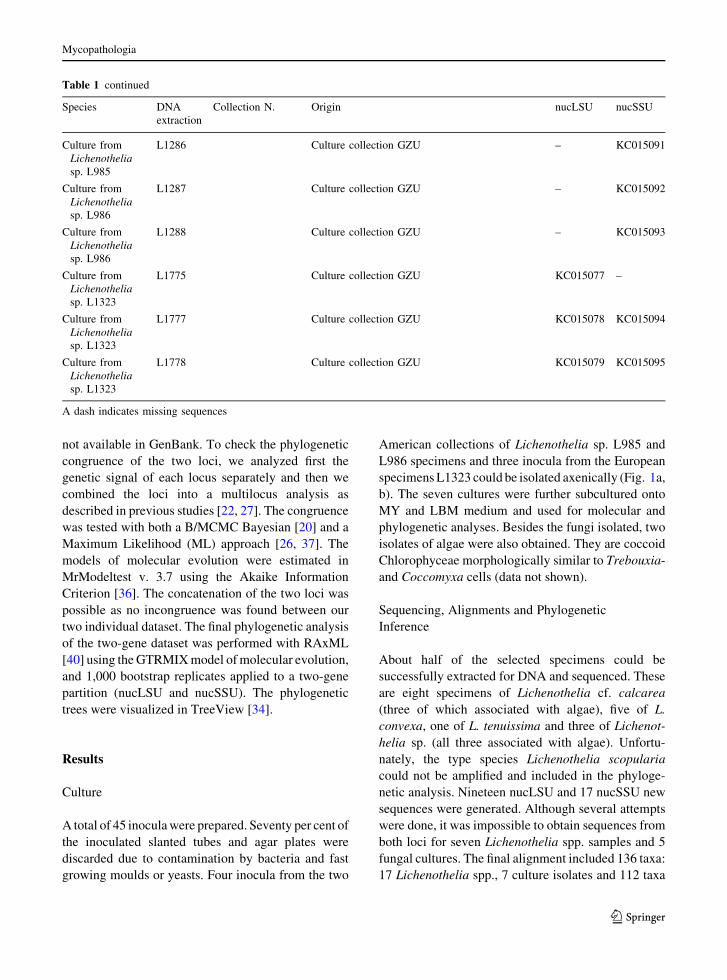
Table 1 Origin data and sequence accession numbers of analyzed Lichenothelia specimens and black fungi newly isolated in culture
Species DNA
extraction
Collection N. Origin nucLSU nucSSU
Lichenothelia cf.
calcarea
L1296 Knudsen K. 13482
(UCR
1778KK64)
USA, California, Riverside County, Mojave
Desert, Joshua Tree National Park, 33�5702200N/
116�0005500W
KC015060 KC015080
Lichenothelia cf.
calcarea
L1323 Muggia L. (GZU) Europe, Czech Republic, South Moravia,
Moravsky Krumlov, on the hill of Kaple Svateho
Floriana.
KC015061 KC015081
Lichenothelia cf.
calcarea
L1324 Muggia L. (GZU) Europe, Czech Republic, South Moravia,
Moravsky Krumlov, on the hill of Kaple Svateho
Floriana
KC015062 KC015082
Lichenothelia cf.
calcarea
L1706 Knudsen K.
13079.2 (UCR
1782KK64)
USA, California, Riverside County, Mojave
Desert, Joshua Tree National Park, 34�0101400N/
116�1002900W
KC015063 –
Lichenothelia cf.
calcarea
L1707 Knudsen K. 12670
(UCR
1576KK64)
USA, California, Riverside County, Mojave
Desert, Joshua Tree National Park, 33�5502100N/
116�0203500W
KC015064 –
Lichenothelia cf.
calcarea
L1708 Knudsen K. 12672
(UCR
1503KK64)
USA, California, Riverside County, Mojave
Desert, Joshua Tree National Park, 33�5502100N/
116�0203500W
KC015065 –
Lichenothelia cf.
calcarea
L1715 Knudsen K.
13079.2 (UCR
1782KK64)
USA, California, Riverside County, Mojave
Desert, Joshua Tree National Park, 34�0101400N/
116�1002900W
KC015066 –
Lichenothelia cf.
calcarea
L1717 Knudsen K. 13482
(UCR
1778KK64)
USA, California, Riverside County, Mojave
Desert, Joshua Tree National Park, 33�5702200N/
116�0005500W
KC015067 –
Lichenothelia
convexa
L1606 Knudsen K. 12564
(UCR1 67675)
USA, California, Riverside County, Mojave
Desert, Joshua Tree National Park, 33�5602000N/
116�0405800W
KC015068 KC015083
Lichenothelia
convexa
L1607 Knudsen K. 12452
(URC
1304KK64)
Europe, Czech Republic, Pitkovice, 50�0102600N/
14�3402100E
KC015069 KC015084
Lichenothelia
convexa
L1608 Knudsen K. 12452
(URC
1304KK64)
Europe, Czech Republic, Pitkovice, 50�0102600N/
14�3402100E
KC015070 KC015085
Lichenothelia
convexa
L1609 Knudsen K. 12452
(URC
1304KK64)
Europe, Czech Republic, Pitkovice, 50�0102600N/
14�3402100E
KC015071 KC015086
Lichenothelia
convexa
L1702 Knudsen K.
14252.2 (UCR
597KK64)
USA, California, Riverside County, Mojave
Desert, Joshua Tree National Park, 33�5300800N/
116�0602500W
KC015072 –
Lichenothelia
tenuissima
– Knudsen K. 10406
(UCR 197485)
USA, California, San Bernardino County, San
Bernardino National Forest, 34�1202900N/
116�4300300W
KC015073 –
Lichenothelia sp. L984 Perlmutter G. 2617
(NCU)
USA, North Carolina, Wake County, on granitic
boulder, 35�4404800N/78�2502700W
KC015074 KC015087
Lichenothelia sp. L985 Perlmutter G. 2621
(NCU)
USA, North Carolina, Wake County, on granitic
boulder, 35�4404800N/78�2502700W
KC015075 KC015088
Lichenothelia sp. L986 Perlmutter G. 2620
(NCU)
USA, North Carolina, Wake County, on granitic
boulder, 35�4404800N/78�2502700W
KC015076 KC015089
Culture from
Lichenothelia
sp. L985
L1285 Culture collection GZU – KC015090
Mycopathologia
123
not available in GenBank. To check the phylogenetic
congruence of the two loci, we analyzed first the
genetic signal of each locus separately and then we
combined the loci into a multilocus analysis as
described in previous studies [22, 27]. The congruence
was tested with both a B/MCMC Bayesian [20] and a
Maximum Likelihood (ML) approach [26, 37]. The
models of molecular evolution were estimated in
MrModeltest v. 3.7 using the Akaike Information
Criterion [36]. The concatenation of the two loci was
possible as no incongruence was found between our
two individual dataset. The final phylogenetic analysis
of the two-gene dataset was performed with RAxML
[40] using theGTRMIXmodel ofmolecular evolution,
and 1,000 bootstrap replicates applied to a two-gene
partition (nucLSU and nucSSU). The phylogenetic
trees were visualized in TreeView [34].
Results
Culture
A total of 45 inoculawere prepared. Seventy per cent of
the inoculated slanted tubes and agar plates were
discarded due to contamination by bacteria and fast
growing moulds or yeasts. Four inocula from the two
American collections of Lichenothelia sp. L985 and
L986 specimens and three inocula from the European
specimensL1323 could be isolated axenically (Fig. 1a,
b). The seven cultures were further subcultured onto
MY and LBM medium and used for molecular and
phylogenetic analyses. Besides the fungi isolated, two
isolates of algae were also obtained. They are coccoid
Chlorophyceae morphologically similar to Trebouxia-
and Coccomyxa cells (data not shown).
Sequencing, Alignments and Phylogenetic
Inference
About half of the selected specimens could be
successfully extracted for DNA and sequenced. These
are eight specimens of Lichenothelia cf. calcarea
(three of which associated with algae), five of L.
convexa, one of L. tenuissima and three of Lichenot-
helia sp. (all three associated with algae). Unfortu-
nately, the type species Lichenothelia scopularia
could not be amplified and included in the phyloge-
netic analysis. Nineteen nucLSU and 17 nucSSU new
sequences were generated. Although several attempts
were done, it was impossible to obtain sequences from
both loci for seven Lichenothelia spp. samples and 5
fungal cultures. The final alignment included 136 taxa:
17 Lichenothelia spp., 7 culture isolates and 112 taxa
Table 1 continued
Species DNA
extraction
Collection N. Origin nucLSU nucSSU
Culture from
Lichenothelia
sp. L985
L1286 Culture collection GZU – KC015091
Culture from
Lichenothelia
sp. L986
L1287 Culture collection GZU – KC015092
Culture from
Lichenothelia
sp. L986
L1288 Culture collection GZU – KC015093
Culture from
Lichenothelia
sp. L1323
L1775 Culture collection GZU KC015077 –
Culture from
Lichenothelia
sp. L1323
L1777 Culture collection GZU KC015078 KC015094
Culture from
Lichenothelia
sp. L1323
L1778 Culture collection GZU KC015079 KC015095
A dash indicates missing sequences
Mycopathologia
123

selected within the Dothideomyceta. Among the 112
taxa retrieved from GenBank, 13 had sequences for
only one locus. After exclusion of introns and regions
with ambiguous size, the final combined alignment
contained 1,960 nucleotide, 1,252 for nucLSU and 708
for the partial nucSSU subunit.
Our results (Fig. 2) are congruent with previous
phylogenetic inferences reported by Ruibal et al. [38],
Schoch et al. [39] and Nelsen et al. [31, 32]. Parts of the
backbonephylogeny, especially basal branches, are still
little supported, but family and order clades of
Dothideomyceta are well resolved. Myriangiales and
Dothideales are sister group (67 %), yet their sister
relationship with Capnodiales has low support. In
Capnodiales, the family Teratosphaeriaceae splits into
two monophyletic clades Teratosphaeriaceae (1) and
(2). The majority of rock-inhabiting fungi (RIF) is
placed within Capnodiales; RIF are also included in
Arthoniales, Dothideales, Myriangiales and are at the
base of the newly recovered Lichenothelia-group. Two
rock isolates TRN456 and TRN529, which formed a
separate clade, the ‘‘unknown lineage 1’’ in Ruibal et al.
[38], are nestedwithinArthoniales, supporting the sister
relationship found by Ruibal et al. [38]. The lichen
family Trypetheliaceae is on a long branch sister to
Arthoniales; this relationship has here low support and
may be an artefact, as suggested in Ruibal et al. [38].
The present phylogenetic inference shows one new
clade, although not well supported as a monophyletic
lineage, including Lichenothelia cf. calcarea, L.
convexa and Lichenothelia sp. All the specimens
associated with algae are included in this clade. Four
Fig. 1 Habit of Lichenothelia sp. and cultured black fungi. a,
b habit of Lichenothelia sp. specimens from the Lichenothelia-
group (Fig. 2; Table 1): aLichenothelia sp. L986, bLichenothelia
sp. L1296. c, d culture of black fungi isolated from specimens of
Lichenothelia sp.: c culture of black fungus L1286 of clade I
(Fig. 2), d culture of black fungus L1777 of clade II (Fig. 2).
Bar = 0.5 cm
Mycopathologia
123

Table 2 NCBI accessions of taxa included in the phylogenetic analysis of Fig. 2
Taxon Sample ID nucLSU nucSSU
Anisomeridium polypori AFTOL 101 – DQ782877
Arthonia caesia AFTOL 775 FJ469668 –
Arthopyrenia salicis CBS 368.94 AY538339 AY538333
Astrothelium cinnamomeum AFTOL 110 AY584652 AY584676
Bimuria novae-zelandae CBS 107.79/AFTOL 931 AY016356 AY016338
Botryosphaeria dothidea CBS 115476/AFTOL 946 DQ678051 DQ677998
Capnobotryella renispora CBS 214.90 EU019248 Y18698
Capnodiales sp. CBS 191364 GU323215 GU561840
Capnodium coffeae CBS 147.52/AFTOL 939 DQ247808 DQ247801
Catenulostoma abietis CBS 459.93/AFTOL 2210 DQ678092 DQ678040
Cladosporium cladosporioides CBS170.54/AFTOL 1289 DQ678057 DQ678004
Cladosporium sp. CBS180.53/AFTOL 1035 AY016367 AY016351
Columnosphaeria fagi (1) CBS 171.93/AFTOL 1582 AY016359 AY016342
Columnosphaeria fagi (2) CBS 584.75/AFTOL 912 DQ470956 DQ471004
Coniosporium apollinis CBS 100218 GU250898 GU250919
Coniosporium apollinis CBS 352.97 GU250895 GU250916
Coniosporium apollinis CBS 109865 GU250900 GU250921
Coniosporium apollinis CBS 109867 GU250901 –
Coniosporium apollinis CBS 100213 GU250896 GU250917
Coniosporium apollinis CBS 109860 GU250899 GU250920
Coniosporium apollinis CBS 100214 GU250897 GU250918
Coniosporium uncinatum CBS 123158/A35 GU250925 GU250933
Coniosporium uncinatum CBS 100219 GU250903 GU250923
Coniosporium uncinatum CBS 100212 GU250902 GU250922
Cryomyces antarcticus CCFEE 536 GU250365 GU250321
Cryomyces minteri CBS 116302/CCFEE 5187 GU250369 DQ066714
Cystocoleus ebeneus L348 (GZU, Hafellner 41566) EU048580 EU048573
Davidiella tassiana CBS 399.80/AFTOL 1591 DQ678074 DQ678022
Delphiniella strobiligena CBS 735.71/AFTOL 1257 DQ470977 DQ471029
Dendrographa leucophaea AFTOL 308 AY548810 AY548803
Dendrographa minor AFTOL 355 AF279382 AF279381
Dendryphiella arenaria CBS 181.58/AFTOL 995 DQ470971 DQ471022
Devriesia streliziae CBS 122379 GU296146 GU301810
Dothidea insculpta CBS 189.58/AFTOL 921 DQ247802 DQ247810
Dothiora cannabinae CBS 737.71/AFTOL 1359 DQ470984 DQ479933
Elasticomyces elasticus CBS 122540/CCFEE 5320 GU250376 GU250333
Elsinoe centrolobi CBS 222.50/AFTOL 1854 DQ678094 DQ678041
Elsinoe phaseoli CBS 165.31/AFTOL 1855 DQ678095 DQ678042
Farlowiella carmichaelina CBS 206.36/AFTOL 1787 AY541492 AY541482
Friedmanniomyces endolithicus CCFEE 524 GU250364 DQ066715
Gloniopsis praelonga CBS 112415 FJ161173 FJ161134
Guinardia bidwellii CBS 237.48/AFTOL 1618 DQ678085 DQ678034
Helicomyces roseus CBS 283.51/AFTOL 1613 DQ678083 DQ678032
Hysteropatella clavispora CBS 247.34/AFTOL 1305 AY541493 DQ678006
Hysteropatella elliptica CBS 935.97/AFTOL 1790 DQ767657 EF495114
Mycopathologia
123

Table 2 continued
Taxon Sample ID nucLSU nucSSU
Kirschsteiniothelia aethiops (1) CBS 109.53/AFTOL 925 AY016361 AY016344
Kirschsteiniothelia aethiops (2) DAOM 231155/AFTOL 273 DQ678046 DQ677996
Laurera megasperma AFTOL 2094 FJ267702 GU561841
Lecanactis abietina AFTOL 305 AY548812 AY548805
Leptosphaeria maculans DAOM 229267/AFTOL 277 DQ4709646 DQ470993
Lophium mytilinum CBS 269.34/AFTOL 1609 DQ678081 DQ678030
Macrophomina phaseolina CBS 227.33/AFTOL 1783 DQ678088 DQ678037
Microxyphium citri CBS 451.66 GU301848 GU296177
Mycosphaerella euripotami JK 5586J GU301852 GZ479761
Mycosphaerella fijiensis OSC 100622/AFTOL 2021 DQ678098 DQ767652
Mycosphaerella graminicola CBS 292.38/AFTOL 1615 DQ678084 DQ678033
Mycosphaerella punctiformis CBS 113265/AFTOL 942 DQ470968 DQ471017
Myriangium duriaei CBS 260.36/AFTOL 1304 DQ678059 AY016347
Mytilinidion resinicola CBS 304.34 FJ161185 FJ161145
Neofusicoccum ribis CBS 115475/AFTOL 1232 DQ678053 DQ678000
Opegrapha dolomiticola AFTOL 993 – DQ883706
Patellaria atrata CBS 958.97 GU301855 GU296181
Phaeosclera dematioides CBS 157.81 GU301858 GU296184
Phaeotrichum benjaminii CBS 541.72/AFTOL 1184 AY004340 AY016348
Pleospora herbarum CBS 541.72/AFTOL 940 DQ247804 DQ247812
Pleosporales sp. CBS 101277 – GU456309
Preussia terricola DAOM 230091/AFTOL 282 AY544686 AY544726
Racodium rupestre L242 (TSB 37932) EU048582 EU048577
Rhythidhysterium rufulum CBS 306.38 FJ469672 AF164375
Roccella fuciformis AFTOL 126 AY584654 AY584678
Roccellographa cretacea AFTOL 93 DQ883696 DQ883705
Sarcinomyces crustaceus CBS 156.89 GU250893 –
Schismatomma decolorans AFTOL 307 AY548815 AY548809
Scorias spongiosa CBS 325.33/AFTOL 1594 DQ678075 DQ678024
Simoniella variegata AFTOL 80 – AY584669
Sirodesmium olivaceum CBS 395.59 GU250915 GU250904
Stylodothis puccinioides CBS 193.58 AY004342 AY016353
Sydowia polyspora CBS 116.29/AFTOL 1300 DQ678058 DQ678005
Symbiotaphrina buchneri CBS 6902 FJ176887 FJ176831
Symbiotaphrina kochii CBS 250.77 AY227719 FJ176833
Teratosphaeria associata CBS 112224 GU301874 GU296200
Tripospermum myrti CBS 437.68 GU323216 –
Trypethelium nitidiusculum AFTOL 2099 FJ267701 GU561842
Tubeufia cerea CBS 254.75/AFTOL 1316 DQ470982 DQ471034
Tubeufia paludosa CBS 245.49/AFTOL 1589 DQ767654 DQ767649
Tyrannosorus pinicola CBS 124.88/AFTOL 1235 DQ470974 DQ471025
Westerdykella cylindrica CBS 454.72/AFTOL 1037 AY004343 AY016355
Rock isolate A6 – GU250924 GU250932
Rock isolate A73 – GU250926 GU250934
Rock isolate AN1 – GU250927 GU250935
Mycopathologia
123

subclades are identified; Lichenothelia cf. calcarea
and Lichenothelia convexa seem to be paraphyletic.
One additional specimen of L. cf. calcarea (L1717) is
basal to Teratosphaeriaceae (2), and L. tenussima is
basal to Myriangiales and Dothideales. These two
specimens are reported in parenthesis because they
might represent other fungal contaminants which have
been amplified instead of the true Lichenothelia
fungus. The sister relationship of the main Lichenot-
helia-group with Coniosporium apollinis- and C.
uncinatum-groups is poorly supported. Still these
three groups remain phylogenetically distinct from
other known lineages of Dothideomycetes.
In contrast, sequences obtained from cultured
specimens group in two distinct clades, clade I and
clade II: clade I represents the specimens from USA,
clade II those from Europe (Table 1). Clade I is nested
within Teratosphaeriaceae (2) and is closely related to
RIFs, to the lichenized Cystocoleus ebeneus and to the
two plant pathogens Tripospermum myrti and Devrie-
sia streliziae. Clade II is supported by a long branch in
Mycosphaerellaceae and is related to plant pathogenic
species of Mycosphaerella.
Discussion
The present study gives new insights into phylogenetic
relationships among Dothideomyceta fungi [39] with
different lifestyles, such as rock-inhabiting fungi
(RIF), melanised lichenized fungi and plant patho-
gens. Our phylogenetic inference reveals three new
fungal lineages in this class and provides for the first
time a phylogenetic placement of the enigmatic
lichen-like genus Lichenothelia. On the basis of
morphological characters, at the time of its descrip-
tion, this genus was believed to represents a link
between the dothidealean and lecanoralean fungi [19].
The results show that the majority of Lichenothelia
specimens form one clade in the Dothideomyceta
whose sister-group relationship with Coniosporium
apollinis and C. uncinatum is, however, only poorly
Table 2 continued
Taxon Sample ID nucLSU nucSSU
Rock isolate AN13 – GU250928 GU250936
Rock isolate CCFEE5211 – GU250371 GU250419
Rock isolate TRN5 CBS 118762 GU323956 GU323988
Rock isolate TRN11 CBS 118281 GU323957 –
Rock isolate TRN42 CBS 117958 GU323958 –
Rock isolate TRN62 CBS 118305 GU323961 GU323991
Rock isolate TRN66 CBS 118306 GU323962 GU323992
Rock isolate TRN77 CBS 118287 GU323963 GU323993
Rock isolate TRN80 CBS 118286 GU323965 GU323995
Rock isolate TRN87 CBS 118290 GU323966 GU323996
Rock isolate TRN111 CBS 118294 GU323967 GU324028
Rock isolate TRN123 CBS 117932 GU323970 GU323999
Rock isolate TRN124 CBS 118283 GU323971 GU324000
Rock isolate TRN137 CBS 118300 GU323973 GU324002
Rock isolate TRN138 CBS 118301 GU323974 GU324003
Rock isolate TRN142 CBS 118302 GU323975 GU324004
Rock isolate TRN153 CBS 118330 GU323977 GU324006
Rock isolate TRN235 CBS 118605 GU323979 –
Rock isolate TRN267 CBS 118769 – GU324043
Rock isolate TRN268 CBS 119305 GU323981 –
Rock isolate TRN456 – GU323986 GU324015
Rock isolate TRN529 – GU323987 GU324016
A dash indicates missing sequences
Mycopathologia
123

Mycopathologia
123

supported. Despite the occasional presence of several
fungal species in one sample (see below), the genetic
identity of Lichenothelia seems convincing, as spec-
imens coming from different localities group all
together. In contrast, the placement of two additional
Lichenothelia samples at the base of Teratosphaeria-
cea (2) and of Myriangiales and Dothideales might be
due to the amplification of contaminating fungi. Our
preliminary results were obtained from a fairly
restricted number of taxa, and no data could so far
be obtained for the generic type species Lichenothelia
scopularia. A broader taxon and gene sampling (such
as mtSSU—for which already few sequences could be
obtained—or protein coding genes) will be needed to
corroborate the monophyly of Lichenothelia.
The phenotypically similarity between Lichenothe-
lia and the subgenera Lichenostigma and Licheno-
gramma raised the question whether they should be
recognized as distinct groups [7, 21, 23, 35]. Despite
the striking morphological similarities of these two
subgenera with Lichenothelia, they have been sepa-
rated due to subtle variations of thalli and fruiting
bodies [5, 15]. All the three genera are known to be
both lichenicolous fungi and to grow on bare rock
surfaces (such as Lichenostigma saxicola, [23]).
Therefore, common ascomata structure and spores
types, variable presence of vegetative hyphae and
shared lifestyles could represent traits assignable to
one homogenous fungal lineage. According to mor-
phological similarities, especially in the ascus struc-
ture, they were classified in the order Arthoniales, as
their own family Lichenotheliaceae [2, 5, 8, 16, 21].
The inclusion of Lichenostigma and Lichenogramma
species in future studies will clarify this unresolved
taxonomic issue. Our results, in any case, place
Lichenothelia in a new lineage within the Dothideo-
mycetes, but not in the Arthoniales.
Owing to the inconspicuous thallus these fungi
produce, the taxa are not frequently collected in the
field bymycologists or lichenologists. Therefore, fresh
collections are rarely available for molecular studies.
Even then, the fungi of interest are either directly
sequenced or isolated and cultured to obtain sufficient
amount of mycelium to extract genomic DNA.
Contaminant fungi which are cryptically associated
with the specimens grow sometimes more efficiently
in cultures than the apparently slower growing Liche-
nothelia species. These additional or co-occurring
fungi can bias the final results. Their presence is
confirmed in this study by the fungi represented in
clades I and II (Fig. 1). Both clades contain cultured
isolates from two geographic origins. The revelation
of co-occurring fungi in Lichenothelia thalli demon-
strates that thallus-forming black fungi frequently
represent consortia of different black fungal strains.
These additional fungi are related to plant pathogenic
fungi (Mycosphaerellaceae) and to other rock-inhab-
iting lineages (Teratosphaeriaceae).
Our results suggest a common link between rock-
inhabiting meristematic and lichen-forming lifestyles
in ascomycetous fungi. Lichenized lineages are scat-
tered in the Dothideomyceta [31, 32]. However,
compared to the larger and predominantly lichenized
lineages of Arthoniales and Trypeteliales, the thallus
of other lichenized species within this superclass is
usually poorly developed or simple and crust-like.
Cystocoleus and Racodium are two monospecific
lichen genera forming own lineages within Capnodi-
ales [28]. Their thallus is formed by colonies of
filamentous Trentepohlia-algae which are densely
entwined by coherent and dark-pigmented fungal
hyphae. In previous studies, we found that other black
fungi are frequently found in lichens [18] and that at
least some of these lichen-associated fungi might also
interact specifically with the lichen’s algae [3].
Meristematic growth is a common feature of pheno-
plasticity found in diverse rock-inhabiting black fungi.
We hypothesize that the ability to form coherent
cellular structures (or ‘‘pseudo-tissues’’), in some
oligotrophic rock-inhabiting fungi, could represent a
pre-adaptation in the evolution of the lichen thallus.
Lifestyle transition in fungi can be studied through
several approaches. Alternatively, to statistically eval-
uating the phylogenetic history with discrete catego-
ries, we show here, with Lichenothelia as one example,
that there could be a smooth transition from rock-
inhabiting lifestyle to lichenized lifestyle.Whether this
includes even an incipient stage of lichenicolous habit
still need further investigations. We think that in this
ecologically versatile group of oligotrophic fungi,
lifestyle expression could depend to some extent on
Fig. 2 Phylogenetic relationships of Lichenothelia spp. and
other rock-inhabiting fungi within known lineages of Dothide-
omyceta [38]: maximum likelihood analyses of the combined
nucLSU and nucSSU loci. Branches with bootstrap support
above 75 % are in bold, other bootstrap supports above 60 % are
reported above or aside the corresponding branches. Lichenized
taxa are labelled by an asterisk
b
Mycopathologia
123

ecological settings. Along with the notion of a
‘‘symbiotic playground’’ put forward by Gorbushina
and Broughton [11], mutualistic symbiotic relations
could then be selected against detrimental interactions,
especially under oligotrophic conditions. These inter-
actions then involve self-sustaining interactions with
algae or cyanobacteria, as well as the presence of black
fungal commensals in pre-formed thallus structures of
related and unrelated lichen-forming fungi.
Acknowledgments LM and MG are grateful to the Austrian
Science Foundation for financial support (FWF P24114). We
thank Cene Gostincar and Josef Hafellner for constructive
discussions and Jana Kocourkova for field co-work.
References
1. Ahmadjian V. The lichen symbiosis. Massachusetts:
Blaisdell Publishing Company; 1967.
2. Athienza V, Hawksworth DL. Lichenothelia renobalesiana
sp. nov. (Lichenotheliaceae), for a lichenicolous ascomy-
cete confused with Polycoccum opulentum (Dacampia-
ceae). The Lichenologist. 2008;40:87–96.
3. Brunauer G, Blaha J, Hager A, Turk R, Stocker-Worgotter
E, Grube M. An isolated lichenicolous fungus forms
lichenoid structures when co-cultured with various coccoid
algae. Symbiosis. 2007;44:127–36.
4. Bubrick P, Galun M. Spore to spore resynthesis of Xanth-
oria parietina. The Lichenologist. 1986;18:47–9.
5. Catalayud V, Naverro-Rosines P, Hafellner J. A synopsis of
Lichenostigma subgen. Lichenogramma (Arthoniales), with
a key to the species. Myc Res. 2002;106:1230–42.
6. Cubero OF, Crespo A, Fatehi J, Bridge PD. DNA extraction
and PCR amplification method suitable for fresh, herbarium
stored and lichenized fungi. Plant SystEvol. 1999;217:243–9.
7. Fernadez-Brime S, Llimona X, Navarro-Rosines P. Li-
chenostigma rupicolae (Lichenotheliaceae), a new liche-
nocolous species growing on Pertusaria rupicola. The
Lichenologist. 2010;42:241–7.
8. Friedmann EI, Kappen L, Meyer MA, Nienow JA. Long-
term productivity in the cryptoendolithic microbial com-
munity of the Ross Desert, Antarctica. Microb Ecol.
1993;25:51–69.
9. Gardes M, Bruns TD. ITS primers with enhanced specificity
for basidiomycetes. Application for the identification of
mycorrhizae and rust. Mol Ecol. 1993;2:113–8.
10. Gargas A, Taylor JW. Polymerase chain reaction (PCR)
primers for amplifying, sequencing nuclear 18S rDNA from
lichenized fungi. Mycologia. 1992;84:589–92.
11. Gorbushina AA, Broughton WJ. Microbiology of the
atmosphere-rock interface: how biological interactions and
physical stresses modulate a sophisticated microbial eco-
system. Ann Rev Microbiol. 2009;63:431–50.
12. Gorbushina AA, Beck A, Schulte A. Microcolonial rock
inhabiting fungi and lichen photobionts: evidence for
mutualistic interactions. Myc Res. 2005;109:1288–96.
13. Gorbushina AA, Whitehead K, Dornieden T, Niesse A,
Schulte A, Hedges JI. Black fungal colonies as units of
survival: hyphal mycosporines synthesized by rock dwell-
ing microcolonial fungi. Can J Bot. 2003;81:131–8.
14. Gueidan C, Ruibal C, De Hoog GS, Schneider H. Rock-
inhabiting fungi originated during periods of dry climate in
the late Devonian and middle Triassic. Fun Biol. 2011;115:
987–96.
15. Hafellener J. Studien uber lichenicole Pilze und Flechten II.
Lichenostigma maureri gen et spec. nov., ein in den Ostal-
pen haufiger lichenicoler Pilz (Ascomycota, Arthoniales).
Herzogia. 1982;6:299–308.
16. Halici MG, Kocakaya M, Aksoy A. Lichenostigma anatol-
icum sp. nov. (Ascomycota, Lichenotheliaceae) on a brown
Acarospora from central Turkey. Mycotaxon. 2009;108:
67–72.
17. Hall TA. BioEdit: a user friendly biological sequence
alignment editor and analysis program for Windows 95/98/
NT. Nuc Ac Symp Ser. 1999;41:95–8.
18. Harutyunyan S, Muggia L, Grube M. Black fungi in lichens
from seasonally arid habitats. Stud Mycol. 2008;61:83–90.
19. Hawksworth DL. Lichenothelia, a new genus for the
Microthelia aterrima group. The Lichenologist. 1981;13:
141–53.
20. Huelsenbeck JP, Ronquist F. MRBAYES 3: Bayesian
phylogenetic inference under mixed models. Bioinformat-
ics. 2003;19:1572–4.
21. Ihlen PG. A new species of Lichenostigma (Lichenothelia-
ceae, Arthoniales) from Scandinavia. The Lichenologist.
2004;36:183–9.
22. Kauff F, Lutzoni F. Phylogeny of the Gyalectales and Os-
tropales (Ascomycota, Fungi): among and within order
relationships based on nuclear ribosomal RNA small and
large subunits. Mol Phyl Evol. 2002;25:138–56.
23. Knudsen K, Kocourkova J. A new Lichenostigma species
(genus incertae sedis) from southern California. The Bry-
ologist. 2010;113:229–34.
24. Kohlmeyer J, Hawksworth DL, Volkmann-Kohlmeyer B.
Observations on two marine and maritime ‘‘borderline’’
lichens:Mastodia tessellata and Collemopsidium pelvetiae.
Myc Progr. 2004;3:51–6.
25. Lilly VG, Barnett HL. Physiology of fungi. New York:
McGrow-Hill; 1951.
26. Meson-Gamer R, Kellogg E. Testing for phylogenetic
conflict among molecular dataset in the tribe Triticeae
(Gramiae). Syst Biol. 1996;45:524–45.
27. Miadlikowska J, Kauff F, Hofstetter V, Fraker E, Grube M,
Hafellner J, Reeb V, Hodkinson BP, Kukwa M, Lucking R,
et al. New insights into classification and evolution of the
Lecanoromycetes (Pezizomycotina, Ascomycota) from
phylogenetic analyses of three ribosomal RNA- and two
protein-coding genes. Mycologia. 2006;98:1088–103.
28. Muggia L, Hafellner J, Wirtz N, Hawksworth DL, GrubeM.
The sterile microfilamentous lichenized fungi Cystocoleus
ebeneus and Racodium rupestre are relatives of plant
pathogens and clinically important dothidealean fungi.
Mycol Res. 2008;112:50–6.
29. Muggia L, Gueidan C, Grube M. Phylogenetic placement of
some morphologically unusual members of Verrucariales.
Mycologia. 2010;102:835–46.
Mycopathologia
123

30. Muggia L, Nelson P, Wheeler T, Yakovchenko LS, Tøns-
berg T, Spribille T. Convergent evolution of a symbiotic
duet: the case of the lichen genus Polychidium (Peltigerales,
Ascomycota). Am J Bot. 2011;98:1647–56.
31. Nelsen MP, Lucking R, Mbatchou JS, Andrew CJ, Spiel-
mann AA, Lumbsch HT. New insights into relationships of
lichen-forming Dothideomycetes. Fun Div. 2011;51:
155–62.
32. Nelsen MP, Lucking R, Grube M, Mbatchou JS, Muggia L,
Plata ER, Lumbsch HT. Unravelling the phylogenetic
relationships of lichenised fungi in Dothideomyceta. Stud
Mycol. 2009;64:135–44.
33. Onofri S, Selbmann L, de Hoog GS, Grube M, Barreca D,
Ruisi S, Zucconi L. Evolution and adaptation of fungi at
boundaries of life. Adv Space Res. 2007;40:657–1664.
34. Page RDM. TREEVIEW: an application to display phylo-
genetic trees on personal computers. Comput Appl Biosci.
1996;12:357–8.
35. Perez-Ortega S, Catalayud V. Lichenostigma epirupestre, a
new lichenicolous species on Pertusaria from Spain. My-
cotaxon. 2009;107:189–95.
36. Posada D, Crandall KA. MODELTEST: testing the model
of DNA substitution. Bioinf Appl Notes. 1998;14:817–8.
37. Reeb V, Lutzoni F, Roux C. Contribution of RPB2 to
multilocus phylogenetic studies of the euascomycetes
(Pezizomycotina, Fungi) with special emphasis on the
lichen-forming Acarosporaceae and evolution of polyspory.
Mol Phyl Evol. 2004;32:1036–60.
38. Ruibal C, Gueidan C, Selbmann L, Gorbushina AA, Crous
PW, Groenewald JZ, Muggia L, Grube M, Isola D, Schoch
CL, Staley JT, Lutzoni F, de Hoog GS. Phylogeny of rock-
inhabiting fungi related to Dothideomycetes. Stud Mycol.
2009;64:123–33.
39. Schoch CL, Crous PW, Groenewald JZ, Boehm EWA,
Burgess TI, et al. A class-wide phylogenetic assessment of
Dothideomycetes. Stud Myc. 2009;64:1–15.
40. Stamatakis A, Ludwig T, Meier H. RAxML-iii: a fast pro-
gram for maximum likelihood-based inference of large
phylogenetic trees. Bioinformatics. 2005;21:456–63.
41. Stocker-Worgotter E. Investigating the production of sec-
ondary compounds in cultured lichen mycobionts. In:
Kanner I, Beckett RP, Varma AK, editors. Protocol in
lichenology, culturing biochemistry, ecophysiology and use
in biomonitoring. Berlin: Springer; 2002. p. 296–306.
42. Turian G. Coniosporium aeroalgicolum sp. nov.—a
dematiaceous fungus living in balanced parasitism with
aerial algae. Bulletin de la Societe Botanique Suisse.
1977;87:19–24.
43. Vilgalys R, Hester M. Rapid genetic identification and
mapping of enzymatically amplified ribosomal DNA from
several Cryptococcus species. J Bacter. 1990;172:4238–46.
44. Yamamoto Y, Kinoshita Y, Yoshimura I. Culture of thallus
fragments and re-differentiation of lichens. In: Kanner I,
Beckett RP, Varma AK, editors. Protocol in lichenology,
culturing biochemistry, ecophysiology and use in biomon-
itoring. Berlin: Springer; 2002. p. 34–46.
Mycopathologia
123


