Embed Size (px)
Citation preview
Natural variation in Caenorhabditis briggsae mitochondrial formand function suggests a novel model of organelle dynamics
Kiley A. Hicksa, Dee R. Denverb, and Suzanne Estesa,§
Kiley A. Hicks: [email protected]; Dee R. Denver: [email protected] Department, Portland State University, 1719 SW 10th Ave., Portland, OR 97201, USAbDepartment of Zoology and Center for Genome Research and Biocomputing, Oregon StateUniversity, Corvallis, OR 97331, USA
AbstractMitochondrial functioning and morphology are known to be connected through cycles of organellefusion and fission that depend upon mitochondrial membrane potential (ΔΨM); however, we lackan understanding of the features and dynamics of natural mitochondrial populations. Using datafrom our recent study of univariate mitochondrial phenotypic variation in Caenorhabditis briggsaenematodes, we analyzed patterns of phenotypic correlation for 24 mitochondrial traits. Ourfindings support a role for ΔΨM in shaping mitochondrial dynamics, but no role formitochondrial ROS. Further, our study suggests a novel model of mitochondrial populationdynamics dependent upon cellular environmental context and with implications for mitochondrialgenome integrity.
Keywordscomplex I; electron transport chain; membrane potential; mitochondrial dynamics; nad5Δ;reactive oxygen species
1. IntroductionMitochondria are dynamic organelles that participate in continuous cycles of fusion, fissionand autophagy within the cells of nearly all eukaryotic organisms. These cycles serve to linkmitochondrial shape to organelle function (Chen et al., 2005; Duvezin-Caubet et al., 2006)as well as each mitochondrion to the larger mitochondrial population (Hyde et al., 2010).Mitochondria perform several functions vital to eukaryotic life, including bioenergy (ATP)production and regulation of calcium homeostasis and apoptosis, nearly all of which dependupon the process of oxidative phosphorylation at the mitochondrial electron transport chain(ETC). Electron transfer through functional protein complexes of the ETC is coupled to thepumping of protons across the mitochondrial inner membrane, which establishes amitochondrial membrane potential (ΔΨM). This ΔΨM provides the potential energy togenerate ATP and serves to control fusion-fission cycles (Twig, Elorza, et al., 2008), both of
© 2012 Elsevier B.V. and Mitochondria Research Society. All rights reserved.§Corresponding author, SE: [email protected]; mailing address: 1719 SW 10th Ave., Portland, OR 97201; tel: 503-725-8782, fax:503-725-3888.
Publisher's Disclaimer: This is a PDF file of an unedited manuscript that has been accepted for publication. As a service to ourcustomers we are providing this early version of the manuscript. The manuscript will undergo copyediting, typesetting, and review ofthe resulting proof before it is published in its final citable form. Please note that during the production process errors may bediscovered which could affect the content, and all legal disclaimers that apply to the journal pertain.
NIH Public AccessAuthor ManuscriptMitochondrion. Author manuscript; available in PMC 2014 January 01.
Published in final edited form as:Mitochondrion. 2013 January ; 13(1): 44–51. doi:10.1016/j.mito.2012.12.006.
NIH
-PA Author Manuscript
NIH
-PA Author Manuscript
NIH
-PA Author Manuscript
which are necessary for mitosis, fuel sensing, autophagy and other processes (Graef &Nunnari, 2011; Mitra et al., 2009; Molina et al., 2009). A natural consequence of ETCfunction is the occasional leakage of electrons onto molecular oxygen, which can generatereactive oxygen species (ROS) (Raha & Robinson, 2000). Under normal circumstances,excess ROS are scavenged by various antioxidants before they can damage importantmacromolecules (Imlay, 2008; Sedensky & Morgan, 2006); however, impairment of theETC often results in elevated ROS production (Dingley et al., 2009; Grad & Lemire, 2004;Verkaart et al., 2007) and oxidative damage of proteins and nucleic acids (Wanagat et al.,2001; Yang et al., 2007), along with depressed ΔΨM (Gaskova et al., 2007; Lemire et al.,2009; Ventura et al., 2006) and altered mitochondrial dynamics (Ichishita et al., 2008).
Findings like those above highlight the integration between mitochondrial function,morphology and the fusion-fission cycle, and many recent studies have aimed to reveal themechanistic bases of these relationships (Chen et al., 2005; Chen & Chan, 2005; Palermo etal., 2007; Pham et al., 2004; Westermann, 2012; Wikstrom et al., 2009; Yasuda et al., 2011).Mitochondrial ROS level does not appear to be related consistently to mitochondrialmorphology or dynamics. For example, elevated mitochondrial ROS levels have beenassociated with both increased (Koopman et al., 2005) and decreased mitochondrialbranching (Grünewald et al., 2010; Pletjushkina et al., 2006). Conversely, studies examiningmitochondrial form and function in isolated cells and/or mutant organisms reveal a directlink between ΔΨM and mitochondrial morphology, such that higher ΔΨM inducesorganellar elongation (Ishihara et al., 2003; Legros et al., 2002) and loss of ΔΨM causessevere fragmentation of the mitochondrial network (Duvezin-Caubet et al., 2006; Song etal., 2007). Many of these mitochondrial shape changes are mediated by an altered balancebetween mitochondrial fusion and fission (Chen et al., 2005; Okamoto & Shaw, 2005),which is increasingly appreciated to have a role in human disease. For instance, abnormalfusion-fission cycles are characteristic of neurodegenerative disorders including Parkinson’sand Alzheimer’s disease (Irrcher et al., 2010; Su et al., 2010; Trimmer et al., 2000; Wang etal., 2008; Winklhofer & Haass, 2010). Many of the genes and cellular intermediatesinvolved in mitochondrial dynamics (and its imbalance) have now been identified andcharacterized (Dimmer et al., 2002; Griffin et al., 2006; Ishihara et al., 2003; Lee et al.,2004; Meeusen et al., 2004; Scorrano, 2005; Yaffe, 1999). Based on such work, Twig andcolleagues proposed that the fusion-fission-apoptosis cycle creates a “quality control axis”that acts to maintain mitochondrial integrity (Twig, Hyde, et al., 2008). In their model,persistently depolarized mitochondria (those with low ΔΨM) are segregated from thefunctional group by their inability to fuse. In this way low-functioning mitochondria – andperhaps, damaged mitochondrial genomes – are weeded out and an overall healthierorganelle population is thus maintained (Bess, et al., 2012; Hyde et al., 2010; Kowald &Kirkwood, 2011; Meyer & Bess, 2012; Twig, Hyde, et al., 2008).
Despite the abundance of research focused on the dynamics of individual mitochondria (i.e.,fusion and fission cycles), less attention has been devoted to the population-level behaviorsof these organelles. A recent review highlights various “global” (cellular) and “local”(individual mitochondrion) controls thought to influence mitochondrial fusion and fission,suggesting that the collective mitochondrial population can indeed respond to cellular cues(Hyde et al., 2010). For example, mitochondria have been observed to undergo concertedhyper-fusion during G1-S phase of the cell cycle, and subsequent hyper-fragmentation as thecell progresses into S phase (Hyde et al., 2010). Still, we have a limited understanding of thebiological roles of mitochondrial fission-fusion cycling and its organism-levelconsequences, and little information regarding the features and dynamics of mitochondrialpopulations and how these might influence individual mitochondrial form and function.Further, although we have some information about the patterns of relationship betweencertain mitochondrial phenotypes (e.g., ΔΨM and organelle elongation), no comprehensive
Hicks et al. Page 2
Mitochondrion. Author manuscript; available in PMC 2014 January 01.
NIH
-PA Author Manuscript
NIH
-PA Author Manuscript
NIH
-PA Author Manuscript
survey of such phenotypes has been conducted within live organisms. Finally, the extent towhich research on cell lines and genetic fusion-fission mutants will apply to naturalpopulations of organisms remains unknown.
Caenorhabditis nematodes have emerged as important models for studying the underlyingcauses of mitochondrial ETC dysfunction and its associated biological consequences.Mitochondrial metabolism and ETC function are known to be extremely similar in wormsand mammals (reviewed in Dimmer et al., 2002; Westermann, 2010). Also, nematodes havehighly differentiated tissues and a transparent cuticle that make them amenable to liveimaging studies. Caenorhabditis briggsae in particular offers many advantages formitochondrial biology research including its substantial mitochondrial genetic (Howe &Denver, 2008) and phenotypic (Clark et al., 2012; Cutter et al., 2010; Estes et al., 2011;Hicks et al., 2012; Raboin et al., 2010; Ross et al., 2011) diversity. C. briggsae exhibit acosmopolitan distribution and mitochondrial genetic analyses group its known naturalisolates into three major phylogeographic clades corresponding to latitude of origin (Figure1 in Howe & Denver, 2008) (Fig. 1). Recent work indicates that isolates within these cladesare likely adapted to local thermal regimes (Jovelin & Cutter, 2011; Prasad et al., 2011). Wefound that phylogenetic membership also accounts for among-isolate variation in severalmitochondrial form and function traits; this was particularly true for ΔΨM, which was anextremely reliable predictor of clade membership (Hicks et al., 2012). Further, C. briggsaeappear especially prone to acquiring mitochondrial deletion mutations (Howe et al., 2010), aprocess that has likely contributed to its high levels of standing mitochondrial geneticdiversity. Indeed, many natural populations of C. briggsae harbor a large deletion (nad5Δ)within their mitochondrial genomes that removes half of the NADH-dehydrogenase 5 (nad5)gene (Figure 1 in Howe & Denver, 2008), which encodes an integral subunit of ETCcomplex I. nad5Δ-bearing genomes were recently shown to behave as selfish geneticelements (Clark et al., 2012) and levels of nad5Δ heteroplasmy (the average number ofdeletion-bearing genomes per individual) are known to vary from zero to over 50% amonggeographically-segregated isolates of C. briggsae (Estes et al., 2011; Howe & Denver,2008). Recent work showed that nad5Δ level was unrelated to isolate-specific variation inΔΨM, ROS, and aspects of mitochondrial morphology (Hicks et al., 2012), but that it islikely to be detrimental to nematode health and fitness at high (< ~40%) heteroplasmy levels(Estes et al., 2011; Howe & Denver, 2008). In summary, its extensive genetic andsubcellular phenotypic variation makes C. briggsae a promising natural system in which toinvestigate individual- and population-level mitochondrial dynamics.
We present a reanalysis of data from our recent study of variation in C. briggsaemitochondrial form and function (Hicks et al., 2012), which quantified 24 mitochondrialphenotypes including ROS level, ΔΨM and aspects of organelle morphology on replicatelive worms from 10 natural isolates of C. briggsae (Fig. 1). This work reported univariateanalyses of these traits to describe standing levels of phenotypic variation among clades andisolates and focused its interpretation on phylogeographic patterns of phenotypic variation.By contrast, the current study reports a systematic evaluation of the bivariate relationshipsbetween all mitochondrial phenotypes from the combined dataset. Our aim was to examinethe connections between mitochondrial physiology and dynamics within a natural system..Our findings support a major role for ΔΨM in shaping mitochondrial dynamics. Based onprevious studies and current models of mitochondrial dynamics, we expected to observemore punctate morphologies among low-ΔΨM mitochondria due to their reduced rates offusion. Conversely, we expected that mitochondria with high ΔΨM would maintain thecanonical elongated shape. Our findings were in agreement with both of these expectationsand provide general support for Twig’s model (Twig, Hyde, et al., 2008) of mitochondrialdynamics. Furthermore, our results suggest an addition to this model in which individual
Hicks et al. Page 3
Mitochondrion. Author manuscript; available in PMC 2014 January 01.
NIH
-PA Author Manuscript
NIH
-PA Author Manuscript
NIH
-PA Author Manuscript

organelles respond to their functional environment; i.e., the average ΔΨM of thesurrounding mitochondrial population.
2. Materials and Methods2.1. Nematode strains
We use data from our recent study (Hicks et al., 2012) in which an array of mitochondrialphenotypes were measured for ten natural isolates of Caenorhabditis briggsae nematodes(Fig. 1). Nuclear (Cutter et al., 2006; Jovelin & Cutter, 2011) and mitochondrial (Raboin etal., 2010) phylogenetic analyses place most C. briggsae natural isolates into three majorphylogeographic clades that are latitudinally distinct, referred to as the Kenyan, Temperate,and Tropical clades (Fig. 1). Tropical clade isolates are found in tropical latitudes andcontain substantial genetic diversity (Cutter et al., 2006), while temperate clade isolatesinhabit northern latitudes and exhibit little genetic diversity (Cutter et al., 2006). Severalnuclear polymorphisms distinguish Kenyan clade isolates from both temperate and tropicalclade strains (Dolgin et al., 2008).The isolates used in this study were chosen to representthese three major phylogeographic clades, and to encompass the full range of known nad5Δheteroplasmy level – from zero to ~50% deletion-bearing genomes. Briefly, the appearanceof nad5Δ depends upon the presence of a mitochondrial pseudogene - Ψnad5-2 (see Figure1 in Howe & Denver, 2008). The two Temperate clade isolates (PB800 and EG4181) harbora compensatory Ψnad5-2 allele that limits the recurrent formation of nad5Δ; the twoKenyan clade isolates (ED3101 and ED3092) completely lack Ψnad5-2, which precludesformation of nad5Δ (Howe & Denver, 2008). C. briggsae strains and the evolutionarygenetics of nad5Δ have been described in further detail elsewhere (Estes et al., 2011; Hickset al., 2012; Howe & Denver, 2008).
2.2. Sample preparation and image analysisFor detailed methods regarding nematode sample preparation, image acquisition andanalysis, please refer to (Dingley et al., 2009; Estes et al., 2011; Hicks et al., 2012). Briefly,data for all mitochondrial traits were obtained by analyzing confocal images of thepharyngeal bulb region of young adult nematodes. Worms were incubated with 10 μmconcentrations of the mitochondria-targeted fluorescent dye(s) appropriate for eachexperiment (below). After 24 hours, worms were washed free of dye, paralyzed usinglevamisole, and imaged using a high-resolution wide-field confocal microscope (AdvancedLight Microscopy Core, Oregon Health and Science University). All images weredeconvolved prior to analysis and all image analysis was performed using ImageJ software(NIH).
The relative intensity of MitoSox Red (Molecular Probes, Eugene, OR) fluorescence fromthe terminal pharyngeal bulb of each worm was used to quantify relative ROS levels.Zielonka and Kalyanaraman (2010) determined that MitoSOX Red quantifies total levels ofmitochondrial oxidants when used in conjunction with microscopic analysis (Zielonka &Kalyanaraman, 2010). Final ROS levels for each isolate were calculated as the differencebetween pharyngeal bulb intensity in labeled and unlabeled control worms from each isolate.Dye-based ROS measurements reflect both the rates of ROS generation and ROSscavenging by antioxidant enzymes or small molecules, and thus give a comprehensive viewof the level of oxidative stress experienced by an organism. Supporting this claim, acomparison of our ROS measurements with a survey of oxidative DNA damage (frequencyof 8-oxo-dG lesions) conducted on a set of C. elegans mutation-accumulation lines (Denveret al., 2009; Denver et al., 2012) was highly positively correlated (Spearman’s ρ1,6=0.943,P<=0.05) (J. Joyner Matos, K. Hicks, D. Denver, S. Estes, C. Baer, unpubl.). Finally, wefind no relationship between pharyngeal pumping rates and ROS or ΔΨM (Estes et al.,
Hicks et al. Page 4
Mitochondrion. Author manuscript; available in PMC 2014 January 01.
NIH
-PA Author Manuscript
NIH
-PA Author Manuscript
NIH
-PA Author Manuscript
2011; Hicks et al., 2012) indicating that our measures are not biased by variation in the ratesof dye uptake by feeding.
Relative ΔΨM levels were quantified using MitoTracker Red CMXRos (Molecular Probes),a dye that localizes exclusively to polarized organelles (Pendergrass et al., 2004). The ΔΨMassays were performed concurrently with those of mitochondrial morphology by co-labelingworms with the ΔΨM-dependent probe MitoTracker Red CMXRos, and with MitoTrackerGreen FM (Molecular Probes), which accumulates within all mitochondria regardless oftheir respiration state (Pendergrass et al., 2004). This experimental setup allowed us todetect state-specific mitochondrial traits, such as shape changes occurring only indepolarized mitochondria, and to directly correlate mitochondrial ΔΨM and morphologytraits. Unfortunately, the spectral similarities between the ROS and ΔΨM probes make itnecessary to use separate images for ROS analysis. Thus, associations between ROS and allother mitochondrial traits should be interpreted with caution.
Finally, as previously discussed (Hicks et al., 2012), we failed to co-label nematodes treatedas above with either DAPI or Hoechst 33342 (Sigma), which would have allowed us tovisualize cell nuclei and thereby assess the intracellular distributions of mitochondria.(Appropriate GFP fusions are not yet available for C. briggsae.) Both dyes noticeablyinterfered with the fluorescence of the above MitoTracker dyes in C. briggsae (Hicks, pers.obs.). Our study therefore focuses on properties of individual mitochondria andmitochondrial populations within the pharyngeal bulb organ.
2.3. Trait descriptions and statistical analysisA total of 24 mitochondrial form and function traits were analyzed (Table S1). Briefly,relative mitochondrial membrane potential (ΔΨM max) served as an indicator ofmitochondrial functionality. Functional mitochondria were distinguished by theirquantifiable uptake of MitoTracker Red CMXRos, while nonfunctional mitochondria tookup untraceable amounts of MitoTracker Red CMXRos. Relative reactive oxygen specieslevel (ROS max) further characterized mitochondrial activity. Maximum rather than meanΔΨM and ROS levels were used because we previously found a significant effect oflevamisole (the cholinergic agonist used to paralyze nematodes for image acquisition) onmean but not maximum ROS levels (Hicks et al., 2012). Additionally, we scored ten traitsthat describe aspects of either the functional (subscript F), nonfunctional (subscript N), orthe total (subscript T) pharyngeal mitochondrial population: the combined area of themitochondrial population (AFP, ANP, ATP), the ratio of the area of functional tononfunctional mitochondria (AFP/NP), and the percentage of the total mitochondrial area thatis functional (AFP/TP), the number of organelles (NF, NN, NT), the ratio of functional tononfunctional organelles (NF/N), and the percentage of functional mitochondria (NF/T).Differences in mitochondrial morphology were measured using the area (AF, AN), aspectratio (ARF, ARN), and circularity (CF, CN) of individual mitochondria. Aspect ratioquantifies elongation and has a minimal value of 1, which corresponds to a perfect circle(Russ, 2002). Circularity will also equal 1 when the measured object is a perfect circle, butdecreases to 0 as the object becomes more branched (Russ, 2002). Because circularitycannot accurately be measured for extremely small objects (ImageJ website), we omittedfrom all analyses mitochondria smaller than 2 pixels. Finally, the variance (subscript V) incircularity (CFV, CNV) and aspect ratio (ARFV, ARNV) measured the degree of heterogeneitywithin the mitochondrial population of each nematode. See Hicks, et al. (2012) for furtherdescription of image processing.
Because our data often violated assumptions of the Pearson product-moment correlation(e.g., normally distributed data, monotonic bivariate relationships), we characterizedcorrelations among mitochondrial form and function characters by calculating Spearman
Hicks et al. Page 5
Mitochondrion. Author manuscript; available in PMC 2014 January 01.
NIH
-PA Author Manuscript
NIH
-PA Author Manuscript
NIH
-PA Author Manuscript

rank-order correlation coefficients between each pair of traits as in Huang et al. (2004).Because ROS levels were measured on different sets of nematodes than all other traits, wemeasured isolate-mean correlations for these pairs of traits. All statistical analysis wasperformed in JMP 9 (SAS Institute, Cary, NC).
3. Results3.1. Mitochondrial trait associations
We analyzed the relationships between pairs of mitochondrial traits (Table S2), individualmeasurements of which were originally obtained by Hicks et al. (2012), for all naturalisolates following (Estes et al., 2011). First, no significant correlations between ROS andany other mitochondrial trait were revealed (data not shown). Maximum ΔΨM was,however, statistically related to a number of other characters. ΔΨM was positively related toaspect ratio of functional mitochondria (ARF) (ρ = 0.208, P<=0.01), meaning that isolateswith higher maximum ΔΨM tended to have more elongated mitochondria. Similarly,maximum ΔΨM was also weakly negatively correlated to the circularity of functionalmitochondria (CF) (ρ = −0.248, P<=0.01), suggesting that worms with higher maximumΔΨM fluorescence also tended to have less circular – or more branched – organelles. It isimportant to note that maximum ΔΨM was necessarily positively correlated to traits relatedto functional mitochondrial area (AF, AFP, and ATP) since individuals with higher scores forthese traits had necessarily taken up more membrane-potential dependent dye and thus hadhigher values for ΔΨM.
A number of the other correlations were expected due to the nature of the measurements(e.g., between traits describing the number of mitochondria and those describing thecombined area of mitochondria populations); however, a systematic survey of the remaining(statistically significant) correlations revealed consistent patterns of relationship between themajor classes of mitochondrial traits (shape, area, and number), which can be summarized asfollows:
(1) As expected, circularity (CN and CF) demonstrated a strong negative correlation withaspect ratio (ARN and ARF) for both nonfunctional and functional mitochondria (ρ =−0.811, P<=0.001 and ρ = −0.801, P<=0.001, respectively). Figure 2A shows therelationship between these two traits for functional mitochondria. Circularity responds tochanges in surface irregularities (or the amount of branching) of each mitochondrionwhereas aspect ratio responds to the elongation of organelles (Koopman et al., 2005; Russ,2002). This negative relationship must certainly owe itself largely to the fact that morecircular mitochondria are less elongate; however, it also implies that mitochondrialbranching was rare and that deviations from perfect circularity were most often achieved byelongation rather than by branching for all organelles regardless of their functional status.
(2) In isolates containing a higher ratio of polarized mitochondria (higher scores for NF/N orNF/T), all mitochondria were larger (NF/N and AF: ρ = 0.317, P<=0.001; NF/N and AN: ρ =0.168, P<=0.05), less circular (NF/N and CF: ρ = −0.276, P<=0.001; NF/N and CN: ρ =−0.196, P<=0.05) (e.g., Fig. 2B shows this pattern for functional mitochondria), and moreelongate (NF/N and ARF: ρ = 0.183, P<=0.05; NF/N and ARN: ρ = 0.188, P<=0.05)regardless of their functional state. (Correlations between NF/T and other listed traits followsimilar trends; Table S2.) However, only the depolarized mitochondria in these isolates weresignificantly more variable with regard to circularity (NF/N and CNV: ρ = 0.190, P<=0.05).Similarly, as the area of individual functional mitochondria (AF) or the combined area of thefunctional mitochondrial population (AFP) increased, all mitochondria became less circular(AFP and CF: ρ = −0.509, P<=0.001; AFP and CN: ρ = −0.216, P<=0.001) and more elongate
Hicks et al. Page 6
Mitochondrion. Author manuscript; available in PMC 2014 January 01.
NIH
-PA Author Manuscript
NIH
-PA Author Manuscript
NIH
-PA Author Manuscript

(AFP and ARF: ρ = 0.256, P<=0.001; AFP and CN: ρ = 0.160, P<=0.05. (Correlationsbetween AF and other listed traits follow similar trends; Table S2).
(3) In isolates with more nonfunctional/depolarized mitochondria (higher scores for NN), thenonfunctional mitochondria in these isolates become less elongate and more uniform withrespect to this trait (NN and ARN: ρ = −0.200, P<=0.01; NN and ARNV: ρ = −0.241,P<=0.01). Conversely, the polarized mitochondria in these isolates were more variable withregard to elongation (NN and ARFV: ρ = 0.154, P<=0.05). As the area of individualnonfunctional mitochondria (AN) or the area of the nonfunctional mitochondrial population(ANP) increased, nonfunctional mitochondria responded by deviating from circularity, (ANPand CN: ρ= −0.316, P<=0.001, AN and CN: ρ = −0.698, P<=0.001; Fig. 2C) in waysindicative of elongation or branching (c.f., Koopman et al., 2005), and by becoming morevariable with respect to circularity (AN and CNV: ρ= 0.409, P<=0.001, Fig. 2D). Because weomitted mitochondria smaller than 2 pixels from all analyses (Hicks et al., 2012 andMaterials and Methods), the strength of these correlations and the fact that they apply onlyto depolarized mitochondria suggest that they should not be influenced by any size-relatedbias.
Finally, we note that regressions of average mitochondrial trait correlations for each isolateon isolate-specific nad5Δ heteroplasmy revealed no evidence that any of the mitochondrialphenotypic associations were related to nad5Δ level; however, because our estimates ofaverage nad5Δ for each C. briggsae isolate were obtained from a different set of worms thanthose phenotyped (Hicks et al., 2012), the biological meaning of these tests is questionable.
4. Discussion4.1. Implications for mitochondrial dynamics
We performed a systematic evaluation of phenotypic correlations among mitochondrialtraits originally reported in (Hicks et al., 2012) with the aim of uncovering patterns thatdescribe the relationships between mitochondrial physiology and morphology. While somestudies have indicated that ROS production can be associated with dramatic ultra-structuraltransformations in mitochondria (Koopman et al., 2005; Liot et al., 2009; Wang et al., 2008),our analyses failed to clearly relate net ROS levels with any alterations in mitochondrialshape or population structure. Again, a caveat prohibiting further interpretation of this resultis that ROS was necessarily measured on different individual nematodes than all othermitochondrial traits (see section 2.2). Thus, estimates of ROS were obtained from differentsets of mitochondria than those describing ΔΨM and morphology traits.
In agreement with previous studies (Ishihara et al., 2003; Mattenberger et al., 2003; Twig,Elorza, et al., 2008), our findings suggest a central role for ΔΨM in shaping mitochondrialmorphology, manifested in its relationship with several aspects of mitochondrial shape andpopulation structure (Table S2). Further, our analysis of mitochondrial trait associationsrevealed that, regardless of their functional state, mitochondria appear to respond differentlydepending on their functional neighborhood; i.e., whether they co-occur with otherorganelles within the nematode pharynx that are mainly polarized or depolarized. Themorphology and physiological state of individual mitochondria is dependent on ΔΨM(Ishihara et al., 2003; Miceli et al., 2011; Twig & Shirihai, 2011). This makes sense asseveral critical organellar functions are contingent upon ΔΨM, including mitochondrialfusion and ATP and ROS production rates (Gaskova et al., 2007; Ishihara et al., 2003;Murphy, 2009). Recent work has shown that mitochondrial fusion is brief and accompaniedby fission (Twig, Elorza, et al., 2008; Wikstrom et al., 2009) and that normal cycles offusion and fission are necessary to maintain the canonical ovoid mitochondrial shape (Chen& Chan, 2005; Kageyama et al., 2011). The model of mitochondrial life cycles proposed by
Hicks et al. Page 7
Mitochondrion. Author manuscript; available in PMC 2014 January 01.
NIH
-PA Author Manuscript
NIH
-PA Author Manuscript
NIH
-PA Author Manuscript

Twig, et al. (2008) connects mitochondrial morphology and function by suggesting that,following a fusion-fission cycle, one daughter mitochondrion remains polarized while theother is transiently depolarized. The transiently depolarized daughter will either regainΔΨM (if it contains a sufficient number of functional ETC components) and resume itsparticipation in the fusion-fission cycle, or it will remain depolarized and undergo fissionand eventual mitophagic degradation (Fig. 1 in Twig et al., 2008). Our data suggest that suchfusion-cycling occurs within the context of a larger mitochondrial population that is itselfeither more or less polarized (Fig. 3). Specifically, we find that both polarized anddepolarized mitochondria are more elongate (less fragmented) in C. briggsae isolatescontaining more mitochondria with high ΔΨM (higher values for NF/N and AFP), althoughdepolarized organelles are slightly more variable in shape than polarized organelles.Conversely, when they inhabit less functional isolates (those with more mitochondria withlow ΔΨM), both polarized and depolarized mitochondria become increasinglyheterogeneous in shape, but depolarized mitochondria become overall more fragmented(Table S2). Placing these data within the context of Twig et al.’s (2008) model, we proposethat a majority of the mitochondria in isolates with higher average ΔΨM will exhibit the“typical” ovoid shape by maintaining normal rates of fusion-fission cycles. Transientlydepolarized mitochondria in this environment will be more likely to recover membranepolarization after fission, helping to maintain a large polarized mitochondrial population.Conversely, isolates with lower ΔΨM will suffer a reduced frequency of fusion-fissioncycling and display increased shape heterogeneity in the entire mitochondrial population.Transiently depolarized mitochondria in this environment will be less likely to harborfunctional ETC products and will more often join the persistently depolarized population,which is unable to undergo fusion. Polarized mitochondria in these isolates will thenexperience reduced numbers of fusion “mates” – in essence, an intracellular Allee effect(Allee, 1931), which will contribute to further reduced rates of fusion-fission cycling andlead to increased shape heterogeneity of all mitochondrial morphs (Fig. 3).
4.2. Implications for mitochondrial genome integrity and evolutionIt has been suggested that damaged mitochondrial genomes may be preferentially shunted tothe depolarized daughter organelle – the one more often destined for degradation (Kowald &Kirkwood, 2011; Twig, Hyde, et al., 2008). If this is the case, mitochondrial fusion-fissioncycling may have a critical role to play in maintaining mtDNA genome stability and couldconceivably contribute to intra- and interspecific differences in mtDNA mutation rates andheteroplasmy levels. Recent work shows that the removal of damaged mtDNA in C. elegansrequires mitochondrial fusion (Bess et al., 2012; Meyer & Bess, 2012). Because the fusion-fission cycle relies on ΔΨM, alterations to ΔΨM that are unrelated to mtDNA quality (i.e.,that weaken the link between mitochondrial genotype and phenotype) could reduce theefficacy of the selective process (Twig, Hyde, et al., 2008). Hicks, et al. (2012) determinedthat much of the measured variation in mitochondrial phenotypes – and especially that ofΔΨM - related to the phylogeographic clade membership of particular C. briggsae isolates,rather than to nad5Δ level (Tables S1 and S2 in Hicks et al., 2012). Specifically, Tropicalisolates tended to have the lowest values for ΔΨM followed by Kenyan isolates, andTemperate isolates exhibited the highest ΔΨM (Figure 3G in Hicks et al., 2012). This led tothe hypothesis that Tropical C. briggsae isolates may have adaptively reduced their ΔΨM inorder to counter increased ROS levels brought on by high temperatures (Brand, 2000; Hickset al., 2012). Because C. briggsae are ectotherms, the external temperature can directlyinfluence their physiology; higher temperatures can increase nematode metabolism and ROSgeneration. Additionally, Tropical clade worms contain fewer total mitochondria (NT andNF) within the focal area (pharyngeal bulb) compared to both the Temperate and Kenyanclades (Tukey HSD, α = 0.05; (Figure 3H in Hicks et al., 2012)). Since cold-adaptedectotherms often exhibit increased mitochondrial density (Morley et al., 2009), the reduction
Hicks et al. Page 8
Mitochondrion. Author manuscript; available in PMC 2014 January 01.
NIH
-PA Author Manuscript
NIH
-PA Author Manuscript
NIH
-PA Author Manuscript

in mitochondrial number within Tropical clade isolates is also consistent with an adaptiveresponse to heat. If it is indeed the case that Tropical C. briggsae isolates have adaptivelyreduced their ΔΨM, they may experience a reduced efficiency of selection allowing theamplification of damaged mtDNA molecules – such as those bearing nad5Δ. Such adynamic could help to explain the counter-intuitive finding that Tropical C. briggsae isolatesdisplay higher average nad5Δ heteroplasmy levels despite having larger effective populationsizes (and presumably more efficient natural selection) than Temperate clade isolates thathave fixed compensatory mutations preventing nad5Δ accumulation (Howe & Denver,2008). In other words, the Tropical isolate’s adaptive reduction of ΔΨM in response to hightemperature may interfere with the selective removal of nad5Δ by the mitochondrial fusion-fission cycle. Alternatively, because nad5Δ affects a component of mitochondrial ETCcomplex I that is putatively involved in H+ pumping (Janssen et al., 2006; Lenaz et al.,2006), the deletion may itself reduce ΔΨM and provide a ROS avoidance mechanism thatdoes not directly produce heat (unlike mitochondrial uncoupling) - thus conferring a directbenefit to Tropical C. briggsae isolates (Brand, 2000; Iser et al., 2005). Either scenarioimplies that different evolutionary pressures may be shaping the subcellular phenotypes ofdifferent C. briggsae populations and suggests fruitful avenues for experimental work.
4.3. ConclusionsOur analysis suggests that ΔΨM, but not mitochondrial ROS level, has a major role inshaping mitochondrial dynamics within natural populations of C. briggsae nematodes. Wealso identified a set of correlations that may describe a global control mechanism formitochondrial dynamics. In particular, our findings suggest a model of mitochondrialpopulation dynamics in which cellular environmental context dependency – and inparticular, whether the mitochondrial population is mainly polarized or depolarized – is akey feature. To our knowledge, ours is the first study to connect natural variation insubcellular phenotypes to a model of mitochondrial dynamics. The model is also congruentwith recent work highlighting the importance of both organellar and cellular influence onmitochondrial fusion-fission processes (Hyde et al., 2010; Kowald & Kirkwood, 2011), butrobust tests will require experimental confirmation of several assumptions, includingwhether ΔΨM correlates linearly to fission-fusion ability. The natural phenotypic andgenetic variation within the C. briggsae system will be advantageous for further study in thisarea (e.g., partitioning of the genetic and cellular environmental components of observedmitochondrial phenotypic variation). With particular regard to C. briggsae evolution, ourfindings highlight the need for future work to understand what if any role mitochondrialfission-fusion dynamics play in mediating transmission of nad5Δ-bearing genomes andadaptation to local thermal and other conditions.
Supplementary MaterialRefer to Web version on PubMed Central for supplementary material.
AcknowledgmentsWe thank A. Snyder (Advanced Light Microscopy Core, Oregon Health and Science University) for generoustechnical advice on confocal imaging and analysis. Thanks also to A. Coleman-Hulbert and S. Smith for labassistance. This work was funded by National Institutes of Health grant 5 R01 GM087628-02 to DRD and SE, PSUFaculty Enhancement grant to SE, and American Heart Association Predoctoral Fellowship 11PRE4880069 toKAH.
Abbreviations
ATP adenosine triphosphate
Hicks et al. Page 9
Mitochondrion. Author manuscript; available in PMC 2014 January 01.
NIH
-PA Author Manuscript
NIH
-PA Author Manuscript
NIH
-PA Author Manuscript

ETC electron transport chain
ΔΨM mitochondrial membrane potential
nad5 NADH-dehydrogenase 5
ROS reactive oxygen species
ReferencesAllee WC. Co-operation among animals. Am. J. Sociol. 1931; 37:386–398.
Brand MD. Uncoupling to survive? The role of mitochondrial inefficiency in ageing. Exp. Gerontol.2000; 35:811–820. [PubMed: 11053672]
Bess AS, Crocker TL, Ryde IT, Meyer JN. Mitochondrial dynamics and autophagy aid in removal ofpersistent mitochondrial DNA damage in Caenorhabditis elegans. Nucleic Acids Res. 2012;40:7916–7931. [PubMed: 22718972]
Chen H, Chan DC. Emerging functions of mammalian mitochondrial fusion and fission. Hum. Mol.Genet. 2005; 14(Spec No):R283–R289. [PubMed: 16244327]
Chen H, Chomyn A, Chan DC. Disruption of fusion results in mitochondrial heterogeneity anddysfunction. J. Biol. Chem. 2005; 280:26185–26192. [PubMed: 15899901]
Clark KA, Howe DK, Gafner K, Kusuma D, Ping S, Estes S, Denver DR. Selfish little circles,transmission bias and evolution of large deletion-bearing mitochondrial DNA in Caenorhabditisbriggsae nematodes. PLoS One. 2012; 7:e41433. [PubMed: 22859984]
Cutter AD, Félix MA, Barriere A, Charlesworth D. Patterns of nucleotide polymorphism distinguishtemperate and tropical wild isolates of Caenorhabditis briggsae. Genetics. 2006; 173:2021–2031.[PubMed: 16783011]
Cutter AD, Yan W, Tsvetkov N, Sunil S, Felix MA. Molecular population genetics and phenotypicsensitivity to ethanol for a globally diverse sample of the nematode Caenorhabditis briggsae. Mol.Ecol. 2010; 19:798–809. [PubMed: 20088888]
Denver DR, Dolan PC, Wilhelm LJ, Sung W, Lucas-Lledo JI, Howe DK, Lewis SC, Okamoto K,Thomas WK, Lynch M, Baer CF. A genome-wide view of Caenorhabditis elegans base-substitutionmutation processes. Proc. Natl. Acad. Sci. U S A. 2009; 106:16310–16314. [PubMed: 19805298]
Denver DR, Wilhelm LJ, Howe DK, Gafner K, Dolan PC, Baer CF. Variation in base-substitutionmutation in experimental and natural lineages of Caenorhabditis nematodes. Genome Biology andEvolution. 2012; 4:513–522. [PubMed: 22436997]
Dimmer KS, Fritz S, Fuchs F, Messerschmitt M, Weinbach N, Neupert W, Westermann B. Geneticbasis of mitochondrial function and morphology in Saccharomyces cerevisiae. Mol. Biol. Cell.2002; 13:847–853. [PubMed: 11907266]
Dingley S, Polyak E, Lightfoot R, Ostrovsky J, Rao M, Greco T, Ischiropoulos H, Falk MJ.Mitochondrial respiratory chain dysfunction variably increases oxidant stress in Caenorhabditiselegans. Mitochondrion. 2009; 10:125–136. [PubMed: 19900588]
Dolgin ES, Félix MA, Cutter AD. Hakuna Nematoda: genetic and phenotypic diversity in Africanisolates of Caenorhabditis elegans and C. briggsae. Heredity. 2008; 100:304–315. [PubMed:18073782]
Duvezin-Caubet S, Jagasia R, Wagener J, Hofmann S, Trifunovic A, Hansson A, Chomyn A, BauerMF, Attardi G, Larsson NG, Neupert W, Reichert AS. Proteolytic processing of OPA1 linksmitochondrial dysfunction to alterations in mitochondrial morphology. J. Biol. Chem. 2006;281:37972–37979. [PubMed: 17003040]
Estes S, Coleman-Hulbert AL, Hicks KA, de Haan G, Martha SR, Knapp JB, Smith SW, Stein KC,Denver DR. Natural variation in life history and aging phenotypes is associated with mitochondrialDNA deletion frequency in Caenorhabditis briggsae. BMC Evol. Biol. 2011; 11:11. [PubMed:21226948]
Hicks et al. Page 10
Mitochondrion. Author manuscript; available in PMC 2014 January 01.
NIH
-PA Author Manuscript
NIH
-PA Author Manuscript
NIH
-PA Author Manuscript

Gaskova D, DeCorby A, Lemire BD. DiS-C3(3) monitoring of in vivo mitochondrial membranepotential in C. elegans. Biochem. Biophys. Res. Commun. 2007; 354:814–819. [PubMed:17266929]
Grad LI, Lemire BD. Mitochondrial complex I mutations in Caenorhabditis elegans producecytochrome c oxidase deficiency, oxidative stress and vitamin-responsive lactic acidosis. Hum.Mol. Genet. 2004; 13:303–314. [PubMed: 14662656]
Graef M, Nunnari J. Mitochondria regulate autophagy by conserved signalling pathways. EMBO J.2011; 30:2101–2114. [PubMed: 21468027]
Griffin EE, Detmer SA, Chan DC. Molecular mechanism of mitochondrial membrane fusion. Biochim.Biophys. Acta. 2006; 1763:482–489. [PubMed: 16571363]
Grünewald A, Voges L, Rakovic A, Kasten M, Vandebona H, Hemmelmann C, Lohmann K, OrolickiS, Ramirez A, Schapira AHV, Pramstaller PP, Sue CM, Klein C. Mutant Parkin impairsmitochondrial function and morphology in human fibroblasts. PloS ONE. 2010; 5:e12962.[PubMed: 20885945]
Hicks KA, Howe DK, Leung A, Denver DR, Estes S. In Vivo Quantification Reveals ExtensiveNatural Variation in Mitochondrial Form and Function in Caenorhabditis briggsae. PLoS ONE.2012; 7:e43837. [PubMed: 22952781]
Howe DK, Baer CF, Denver DR. High rate of large deletions in Caenorhabditis briggsae mitochondrialgenome mutation processes. Genome Biology and Evolution. 2010; 2010:29–38.
Howe DK, Denver DR. Muller’s Ratchet and compensatory mutation in Caenorhabditis briggsaemitochondrial genome evolution. BMC Evol. Biol. 2008; 8:62. [PubMed: 18302772]
Huang C, Xiong C, Kornfeld K. Measurements of age-related changes of physiological processes thatpredict lifespan of Caenorhabditis elegans. Proc. Natl. Acad. Sci. U S A. 2004; 101:8084–8089.[PubMed: 15141086]
Hyde BB, Twig G, Shirihai OS. Organellar vs. cellular control of mitochondrial dynamics. Sem. CellDev. Biol. 2010; 21:575–581.
Ichishita R, Tanaka K, Sugiura Y, Sayano T, Mihara K, Oka T. An RNAi screen for mitochondrialproteins required to maintain the morphology of the organelle in Caenorhabditis elegans. J.Biochem. 2008; 143:449–454. [PubMed: 18174190]
Imlay JA. Cellular defenses against superoxide and hydrogen peroxide. Annu. Rev.of Biochem. 2008;77:755–776. [PubMed: 18173371]
Irrcher I, Aleyasin H, Seifert EL, Hewitt SJ, Chhabra S, Phillips M, Lutz AK, Rousseaux MW,Bevilacqua L, Jahani-Asl A, Callaghan S, MacLaurin JG, Winklhofer KF, Rizzu P, Rippstein P, etal. Loss of the Parkinson’s disease-linked gene DJ-1 perturbs mitochondrial dynamics. Hum. Mol.Genet. 2010; 19:3734–3746. [PubMed: 20639397]
Iser WB, Kim D, Bachman E, Wolkow C. Examination of the requirement for ucp- 4, a putativehomolog of mammalian uncoupling proteins, for stress tolerance and longevity in C. elegans.Mech. of Ageing Dev. 2005; 126:1090–1096.
Ishihara N, Jofuku A, Eura Y, Mihara Katsuyoshi. Regulation of mitochondrial morphology bymembrane potential, and DRP1-dependent division and FZO1-dependent fusion reaction inmammalian cells. Biochem. Biophys. Res. Commun. 2003; 301:891–898. [PubMed: 12589796]
Janssen RJ, Nijtmans LG, van den Heuvel LP, Smeitink JA. Mitochondrial complex I, structure,function and pathology. J. Inherit. Metab. Dis. 2006; 29:499–515. [PubMed: 16838076]
Jovelin R, Cutter AD. MicroRNA sequence variation potentially contributes to within-speciesfunctional divergence in the nematode Caenorhabditis briggsae. Genetics. 2011; 189:967–976.[PubMed: 21890738]
Kageyama Y, Zhang Z, Sesaki H. Mitochondrial division, molecular machinery and physiologicalfunctions. Curr. Opin. Cell Biol. 2011; 23:427–434. [PubMed: 21565481]
Knott AB, Bossy-Wetzel E. Impairing the mitochondrial fission and fusion balance, a new mechanismof neurodegeneration. Ann. NY Acad. Sci. 2008; 1147:283–292. [PubMed: 19076450]
Koopman WJ, Verkaart S, Visch HJ, van der Westhuizen FH, Murphy MP, van den Heuvel LW,Smeitink JA, Willems PH. Inhibition of complex I of the electron transport chain causes O2-. -mediated mitochondrial outgrowth. Am. J. Physiol. Cell Physiol. 2005; 288:1440–1450.
Hicks et al. Page 11
Mitochondrion. Author manuscript; available in PMC 2014 January 01.
NIH
-PA Author Manuscript
NIH
-PA Author Manuscript
NIH
-PA Author Manuscript

Kowald A, Kirkwood TB. Evolution of the mitochondrial fusion-fission cycle and its role in aging.Proc. Natl. Acad. Sci. U S A. 2011; 108:10237–10242. [PubMed: 21646529]
Lee Y, Jeong S-Y, Karbowski M, Smith CL, Youle RJ. Roles of the mammalian mitochondrial fissionand fusion mediators Fis1, Drp1, and Opa1 in apoptosis. Mol. Biol. Cell. 2004; 15:5001–5011.[PubMed: 15356267]
Legros F, Lombès A, Frachon P, Rojo M. Mitochondrial fusion in human cells is efficient, requires theinner membrane potential, and is mediated by mitofusins. Mol. Biol. Cell. 2002; 13:4343–4354.[PubMed: 12475957]
Lemire BD, Behrendt M, DeCorby A, Gaskova D. C. elegans longevity pathways converge to decreasemitochondrial membrane potential. Mech. Ageing Dev. 2009; 130:461–465.
Lenaz G, Fato R, Genova ML, Bergamini C, Bianchi C, Biondi A. Mitochondrial Complex I, structuraland functional aspects. Biochim. Biophys. Acta. 2006; 1757:1406–1420. [PubMed: 16828051]
Liot G, Bossy B, Lubitz S, Kushnareva Y, Sejbuk N, Bossy-Wetzel E. Complex II inhibition by 3-NPcauses mitochondrial fragmentation and neuronal cell death via an NMDA- and ROS-dependentpathway. Cell Death and Differ. 2009; 16:899–909.
Mattenberger Y, James DI, Martinou JC. Fusion of mitochondria in mammalian cells is dependent onthe mitochondrial inner membrane potential and independent of microtubules or actin. FEBS Lett.2003; 538:53–59. [PubMed: 12633852]
Meeusen S, McCaffery JM, Nunnari J. Mitochondrial fusion intermediates revealed in vitro. Science.2004; 305:1747–1752. [PubMed: 15297626]
Meyer JN, Bess AS. Involvement of autophagy and mitochondrial dynamics in determining the fateand effects of irreparable mitochondrial DNA damage. Autophagy. 2012; 8:12.
Miceli MV, Jiang JC, Tiwari A, Rodriguez-Quiñones JF, Jazwinski SM. Loss of mitochondrialmembrane potential triggers the retrograde response extending yeast replicative lifespan. Frontiersin Genetics. 2011; 2:102. [PubMed: 22303396]
Mitra K, Wunder C, Roysam B, Lin G, Lippincott-Schwartz J. A hyperfused mitochondrial stateachieved at G1-S regulates cyclin E buildup and entry into S phase. Proc. Natl. Acad. Sci. U S A.2009; 106:11960–11965. [PubMed: 19617534]
Molina AJ, Wikstrom JD, Stiles Linsey, Las Guy, Mohamed Hibo, Elorza A, Walzer Gil, Twig G,Katz Steve, Corkey Barbara E, Shirihai OS. Mitochondrial networking protects beta-cells fromnutrient-induced apoptosis. Diabetes. 2009; 58:2303–2315. [PubMed: 19581419]
Morley SA, Lurman GJ, Skepper JN, Portner HO, Peck LS. Thermal plasticity of mitochondria, alatitudinal comparison between Southern Ocean molluscs. Comp. Biochem. Physiol. A. Mol.Integr. Physiol. 2009; 152:423–430. [PubMed: 19100332]
Murphy MP. How mitochondria produce reactive oxygen species. Biochem. J. 2009; 417:1–13.[PubMed: 19061483]
Okamoto K, Shaw JM. Mitochondrial morphology and dynamics in yeast and multicellular eukaryotes.Annu. Rev. Genet. 2005; 39:503–536. [PubMed: 16285870]
Palermo V, Falcone C, Mazzoni C. Apoptosis and aging in mitochondrial morphology mutants of S.cerevisiae. Folia. Microbiol. (Praha). 2007; 52:479–483. [PubMed: 18298044]
Pendergrass W, Wolf N, Poot M. Efficacy of MitoTracker Green and CMX rosamine to measurechanges in mitochondrial membrane potentials in living cells and tissues. Cytometry A. 2004;61:162–169. [PubMed: 15382028]
Pham NA, Richardson T, Cameron J, Chue B, Robinson BH. Altered mitochondrial structure andmotion dynamics in living cells with energy metabolism defects revealed by real time microscopeimaging. Microsc. Microanal. 2004; 10:247–260. [PubMed: 15306050]
Pletjushkina OY, Lyamzaev KG, Popova EN, Nepryakhina OK, Ivanova OY, Domnina LV, ChernyakBV, Skulachev VP. Effect of oxidative stress on dynamics of mitochondrial reticulum. Biochim.Biophys. Acta. 2006; 1757:518–524. [PubMed: 16829229]
Prasad A, Croydon-Sugarman MJF, Murray RL, Cutter AD. Temperaturedependent fecundityassociates with latitude in Caenorhabditis briggsae. Evolution. 2011; 65:52–63. [PubMed:20731713]
Hicks et al. Page 12
Mitochondrion. Author manuscript; available in PMC 2014 January 01.
NIH
-PA Author Manuscript
NIH
-PA Author Manuscript
NIH
-PA Author Manuscript

Raboin MJ, Timko AF, Howe DK, Félix MA, Denver DR. Evolution of Caenorhabditis mitochondrialgenome pseudogenes and Caenorhabditis briggsae natural isolates. Mol. Biol. Evol. 2010;27:1087–1096. [PubMed: 20026478]
Raha S, Robinson BH. Mitochondria, oxygen free radicals, disease and ageing. Trends Biochem. Sci.2000; 25:502–508. [PubMed: 11050436]
Ross JA, Koboldt DC, Staisch JE, Chamberlin HM, Gupta BP, Miller RD, Baird SE, Haag ES.Caenorhabditis briggsae recombinant inbred line genotypes reveal inter-strain incompatibility andthe evolution of recombination. PLoS Genetics. 2011; 7:e1002174. [PubMed: 21779179]
Russ, JC. The Image Processing Handbook. Boca Raton: CRC Press, LLC; 2002.
Scorrano L. Proteins that fuse and fragment mitochondria in apoptosis, con-fissing a deadly con-fusion? J. Bioenerg. Biomembr. 2005; 37:165–170. [PubMed: 16167173]
Sedensky MM, Morgan PG. Mitochondrial respiration and reactive oxygen species in C. elegans. Exp.Gerontol. 2006; 41:957–967. [PubMed: 16919906]
Song Z, Chen H, Fiket M, Alexander C, Chan DC. OPA1 processing controls mitochondrial fusion andis regulated by mRNA splicing, membrane potential, and Yme1L. J. Cell Biol. 2007; 178:749–755. [PubMed: 17709429]
Su B, Wang Xinglong, Zheng L, Perry G, Smith MA, Zhu X. Abnormal mitochondrial dynamics andneurodegenerative diseases. Biochim. Biophys. Acta. 2010; 1802:135–142. [PubMed: 19799998]
Trimmer PA, Swerdlow RH, Parks JK, Keeney P, Bennett JP Jr, Miller SW, Davis RE, Parker WD Jr.Abnormal mitochondrial morphology in sporadic Parkinson’s and Alzheimer's disease cybrid celllines. Exp. Neurol. 2000; 162:37–50. [PubMed: 10716887]
Twig G, Elorza A, Molina AJ, Mohamed H, Wikstrom JD, Walzer G, Stiles L, Haigh SE, Katz S, LasG, Alroy J, Wu M, Py BF, Yuan J, Deeney JT, et al. Fission and selective fusion governmitochondrial segregation and elimination by autophagy. EMBO J. 2008; 27:433–446. [PubMed:18200046]
Twig G, Hyde BB, Shirihai OS. Mitochondrial fusion, fission and autophagy as a quality control axis,the bioenergetic view. Biochim. Biophys. Acta. 2008; 1777:1092–1097. [PubMed: 18519024]
Twig G, Shirihai OS. The interplay between mitochondrial dynamics and mitophagy. Antiox. RedoxSignaling. 2011; 14:1939–1951.
Ventura N, Rea SL, Testi R. Long-lived C. elegans Mitochondrial mutants as a model for humanmitochondrial-associated diseases. Exp. Gerontol. 2006; 41:974–991. [PubMed: 16945497]
Verkaart S, Koopman WJ, van Emst-de Vries SE, Nijtmans LGJ, van den Heuvel LWPJ, SmeitinkJAM, Willems PHGM. Superoxide production is inversely related to complex I activity ininherited complex I deficiency. Biochim. Biophys. Acta-Molecular Basis of Disease. 2007;1772:373–381.
Wanagat J, Cao Z, Pathare P, Aiken JM. Mitochondrial DNA deletion mutations colocalize withsegmental electron transport system abnormalities, muscle fiber atrophy, fiber splitting, andoxidative damage in sarcopenia. FASEB J. 2001; 15:322–332. [PubMed: 11156948]
Wang W, Fang H, Groom L, Cheng A, Zhang W, Liu J, Wang X, Li K, Han P, Zheng M, Yin J,Mattson MP, Kao JP, Lakatta EG, Sheu SS, et al. Superoxide flashes in single mitochondria. Cell.2008; 134:279–290. [PubMed: 18662543]
Wang, Xinglong; Su, B.; Fujioka, H.; Zhu, X. Dynamin-like protein 1 reduction underliesmitochondrial morphology and distribution abnormalities in fibroblasts from sporadic Alzheimer’sdisease patients. The Am. J. Pathol. 2008; 173:470–482.
Westermann B. Bioenergetic role of mitochondrial fusion and fission. Biochim. Biophys. Acta. 2012;10:1833–1838.
Westermann B. Mitochondrial dynamics in model organisms, What yeasts, worms and flies havetaughtus about fusion and fission of mitochondria. Sem. Cell Dev. Biol. 2010; 21:542–549.
Wikstrom JD, Twig G, Shirihai OS. What can mitochondrial heterogeneity tell us about mitochondrialdynamics and autophagy? The Int. J. Biochem. Cell Biol. 2009; 41:1914–1927.
Winklhofer KF, Haass C. Mitochondrial dysfunction in Parkinson’s disease. Biochim. Biophys. Acta.2010; 1802:29–44. [PubMed: 19733240]
Yaffe MP. The Machinery of Mitochondrial Inheritance and Behavior. Science. 1999; 283:1493–1497.[PubMed: 10066164]
Hicks et al. Page 13
Mitochondrion. Author manuscript; available in PMC 2014 January 01.
NIH
-PA Author Manuscript
NIH
-PA Author Manuscript
NIH
-PA Author Manuscript

Yang W, Li J, Hekimi S. A Measurable increase in oxidative damage due to reduction in superoxidedetoxification fails to shorten the life span of long-lived mitochondrial mutants of Caenorhabditiselegans. Genetics. 2007; 177:2063–2074. [PubMed: 18073424]
Yasuda K, Hartman PS, Ishii T, Suda H, Akatsuka A, Shoyama T, Miyazawa M, Ishii N.Interrelationships between mitochondrial fusion, energy metabolism and oxidative stress duringdevelopment in Caenorhabditis elegans Biochem. Biophys. Res. Commun. 2011; 404:751–755.
Zielonka J, Kalyanaraman B. Hydroethidine- and MitoSOX-derived red fluorescence is not a reliableindicator of intracellular superoxide formation, another inconvenient truth. Free. Radic. Biol. Med.2010; 48:983–1001.
Hicks et al. Page 14
Mitochondrion. Author manuscript; available in PMC 2014 January 01.
NIH
-PA Author Manuscript
NIH
-PA Author Manuscript
NIH
-PA Author Manuscript
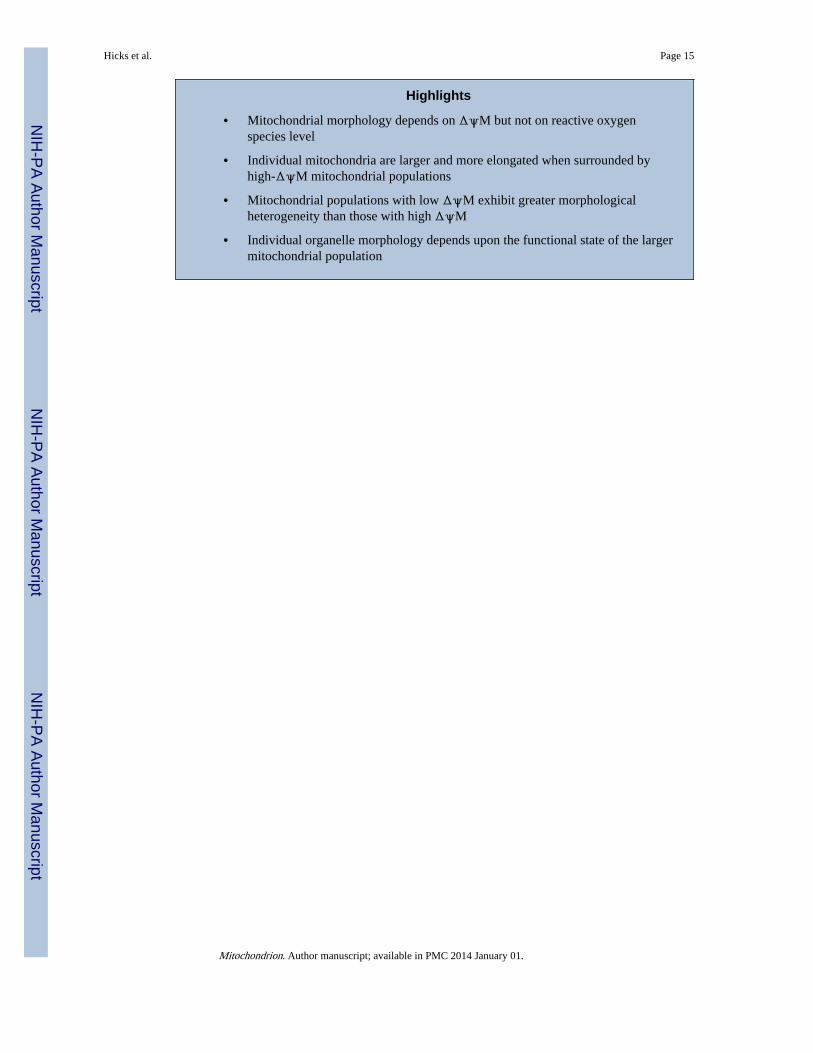
Highlights
• Mitochondrial morphology depends on ΔψM but not on reactive oxygenspecies level
• Individual mitochondria are larger and more elongated when surrounded byhigh-ΔψM mitochondrial populations
• Mitochondrial populations with low ΔψM exhibit greater morphologicalheterogeneity than those with high ΔψM
• Individual organelle morphology depends upon the functional state of the largermitochondrial population
Hicks et al. Page 15
Mitochondrion. Author manuscript; available in PMC 2014 January 01.
NIH
-PA Author Manuscript
NIH
-PA Author Manuscript
NIH
-PA Author Manuscript

Figure 1. C. briggsae natural isolatesPhylogenetic relationship, geographic origin and average nad5Δ heteroplasmy level(numbers above branches) of C. briggsae isolates included in this study (modified fromHowe & Denver, 2008). nad5Δ heteroplasmy levels were determined using qPCR anddescribe the average percentage of nad5Δ-deletion bearing genomes within individualworms from each isolate (Howe & Denver, 2008). GL = global superclade; KE = Kenyaclade; TE and TR = temperate and tropical subclades of GL; C(+) = isolates bearing thecompensatory Ψnad5Δ-2 allele. Note that we assayed the natural HK104 isolate here ratherthan the inbred line reported in Estes et al. (2011), which evolved high nad5Δ levels in thelab (see Section 2.1).
Hicks et al. Page 16
Mitochondrion. Author manuscript; available in PMC 2014 January 01.
NIH
-PA Author Manuscript
NIH
-PA Author Manuscript
NIH
-PA Author Manuscript

Figure 2. Examples of bivariate relationships of mitochondrial phenotypesPatterns of relationship between traits describing mitochondrial size, morphology, andwithin-individual variance are shown. All measurements were made on the same set ofconfocal images (see Section 2.2) and each point represents the bivariate phenotype for anindividual nematode (N=167–170). A) Aspect ratio is negatively related to circularity withinthe functional mitochondria of individual worms (ρ=-0.806, P<=0.0001). Removing oneoutlier (black symbol) decreases the correlation slightly (ρ=-0.797, P<=0.0001). B) As thetwo-dimensional area of the total functional mitochondrial population increases, functionalmitochondria become less circular (ρ=-0.509, P<=0.0001). C) As the two-dimensional areaof individual nonfunctional mitochondria increases, these mitochondria become less circular(ρ=-0.698, P<=0.0001). D) Individual nonfunctional mitochondria become more variablewith respect to circularity as their two-dimensional area increases (ρ=-0.409, P<=0.0001).When two outliers (black symbols) are removed, the correlation remains unchanged.Outliers were determined by calculating Mahalanobis distances for the correlation betweentraits of each sample and visually identifying extreme outliers.
Hicks et al. Page 17
Mitochondrion. Author manuscript; available in PMC 2014 January 01.
NIH
-PA Author Manuscript
NIH
-PA Author Manuscript
NIH
-PA Author Manuscript

Figure 3. A context-dependent model of mitochondrial dynamics in which mitochondria respondto the functional state of their intracellular environmentWe propose that an organism has three types of mitochondrial populations, polarized,transiently depolarized, and persistently depolarized. The polarized population is capable ofundergoing fusion (white arrows) while the persistently depolarized population is not (whiteblunted arrow). The transiently depolarized mitochondria are produced after a fusion-fissioncycle and will either regain sufficient ΔψM and join the polarized/fusing population (grayarrow) or, if they are unable to regain ΔψM, join the persistently depolarized population(black arrow) (Twig et al., 2008). We propose that mitochondria in a more functionalenvironment (higher ΔψM, at left) are more likely to regain ΔψM and join the polarized/fusing population. Normal fusion-fission cycles will maintain a majority of mitochondria inthe canonical ovoid morph. Here, many depolarized mitochondria are destined to recoverΔψM and rejoin the fusing population. Conversely, mitochondria in a less functionalenvironment (lower ΔψM, at right) are less likely to regain ΔψM and will therefore join thepersistently depolarized/non-fusing population resulting in fewer polarized mitochondria.Here, lower than normal rates of fusion and fission will increase the shape heterogeneity ofall mitochondria.
Hicks et al. Page 18
Mitochondrion. Author manuscript; available in PMC 2014 January 01.
NIH
-PA Author Manuscript
NIH
-PA Author Manuscript
NIH
-PA Author Manuscript


