Embed Size (px)
Citation preview

Gustatory behaviour in Caenorhabditis elegans
Renate K Hukema

ISBN-10: 90-9021186-1 ISBN-13: 978-90-9021186-2 Cover: Stephanie van Baaijen The studies presented in this thesis were performed in the Department of Cell Biology and Genetics of the Erasmus MC in Rotterdam, The Netherlands. Printing of this thesis was partially supported by the J.E. Jurriaanse Stichting.

Gustatory behaviour in Caenorhabditis elegans
Smaak in Caenorhabditis elegans
proefschrift ter verkrijging van de graad van
doctor aan de Erasmus Universiteit Rotterdam
op gezag van de rector magnificus Prof.dr. S.W.J. Lamberts
en volgens besluit van het College voor Promoties.
de openbare verdediging zal plaatsvinden op
woensdag 29 november 2006 om 13.45 uur
door
Renate Kirstin Hukema
geboren te Gorinchem

Promotiecommissie Promotor: Prof.dr. F.G. Grosveld Overige leden: Prof.dr. C.I. de Zeeuw Dr.ir. D.N. Meijer Dr.ir. N.J. Galjart Copromotor: Dr. G. Jansen

Contents
Outline of this thesis
6
1 Introduction
7
2 Two genetic pathways involving TRPV channel-, G protein-, and MAP kinase-signalling mediate chemotaxis to NaCl in C. elegans
57
3 Antagonistic sensory cues generate gustatory plasticity in Caenorhabditis elegans
77
4 Gustatory plasticity in C. elegans involves the neurotransmitters glutamate, serotonin, dopamine, and octopamine
103
5 Candidate gene approach identifies genes involved in the responses of Caenorhabditis elegans to NaCl
127
6 Discussion and future directions
153
Summary 165 Samenvatting 169 Dankwoord 173 Curriculum Vitae 176

Outline
6
Outline of this thesis
It is essential for the survival of an organism that it interacts with its environment. The animal has to be able to distinguish hazardous situations from possible food sources. Salt sensing is important for finding food and also for water-homeostasis. However, relatively little is known about the molecular mechanisms of salt taste.
The nematode C. elegans is an ideal model-organism to study the genetics of behaviour (Brenner, 1974). It is capable of sensing salts and we discriminate three different responses: it is attracted to low salt concentrations (Ward, 1973; Dusenbery et al., 1974), it avoids high salt concentrations that give osmotic problems (Culotti & Russell, 1978), and the response to NaCl shows plasticity: normally attractive salt concentrations are avoided after pre-exposure to salt (Saeki et al., 2001; Jansen et al., 2002).
The goal of this study was to unravel the molecular mechanisms and the cellular circuitry behind the different responses of C. elegans to NaCl. A candidate gene approach was used in which mutants for 123 genes were tested for their role in salt responses. A complete overview of all genes tested is given in the Chapter 5. We found 22 genes involved in attraction, 57 genes involved in avoidance and 87 genes involved in gustatory plasticity. Several of the most interesting results were followed up and are discussed in this thesis.
In Chapter 1, a summary is given of what is known about the molecular mechanisms of taste, an introduction to C. elegans as a model organism, and a brief overview of the C. elegans nervous system and neurotransmission. Furthermore, various sensory behaviours of C. elegans and their plasticity are discussed.
Chapter 2 focuses on chemotaxis to NaCl and discusses five newly identified genes, which function in two genetic pathways that mediate NaCl chemotaxis. Chapter 3 discusses the role of G protein signalling in gustatory plasticity and identifies multiple neurons as well as a genetic pathway involved in this process. Chapter 4 discusses the roles of different neurotransmitters in the different responses of C. elegans to NaCl. An overall discussion and summary are given in the final chapter. Taken together, this study provides new insights into the processes of NaCl chemotaxis and its plasticity in C. elegans. We expect that these insights can be extrapolated to mammals.

Chapter 1
Introduction
1

Chapter 1 8
Gustation
1.1.1 Hard-wiring Organisms need to interact with their environment in order to avoid
hazardous situations and to be able to find food. Taste is very important for finding food. Organisms have to distinguish between attractive or good, and toxic or nasty compounds. The gustatory system distinguishes only a few taste categories, unlike for example the olfactory or auditory system. In humans, taste is categorized into five modalities: sweet, bitter, salt, sour, and umami. Umami was first described in 1908 as the taste of glutamate in dried kelp (Yamaguchi & Ninomiya, 2000). In general, sweet, umami, and low salt concentrations are associated with positive behaviours and lead to food acceptance. In contrast, bitter and sour lead to avoidance behaviours. Studies in rats indicate the existence of a sixth taste modality: a taste for dietary fat (Gilbertson et al., 1997; Tsuruta et al., 1999; Kitagawa & Shingai, 2001). Unfortunately, relatively little is known about the exact processes involved in taste. What is known about the gustatory system in humans is mostly concerned with the hard wiring of the system.
In Drosophila, taste is mediated by sensory neurons on the proboscis, internal mouthpart organs, legs and wings, and oviposter (Stocker, 1994). The sensory structures for taste are innervated by two to four gustatory neurons and a single mechanosensory neuron (Falk et al., 1976). Unlike the mammalian gustatory system, taste information in Drosophila is directly relayed to the brain. The adult Drosophila brain contains about 100,000 neurons, with the cell bodies in an outer shell surrounding a fibrous core. The subesophageal ganglion, which is the most ventral region of the brain, receives gustatory projections (Stocker & Schorderet, 1981; Rajashekar & Singh, 1994). This subesophageal ganglion contains a map of the different taste organs. Both position and quality of taste are represented in this map (Thorne et al., 2004; Wang et al., 2004).
In vertebrates, taste begins on the tongue and palate where epithelial-derived taste cells detect chemical cues (Reviewed in Lindemann, 1996). Three morphological structures of taste detecting papillae are topographically arranged on the tongue. Fungiform papillae decorate the anterior two-thirds of the tongue, foliate papillae are on the lateral edges, and circumvillate papillae are found on the posterior two-thirds. These mammalian taste cells are not classic neurons; they do not send axonal projections to the brain. Instead, primary gustatory fibres contact multiple taste cells; these cells can be located within different taste buds. The fibres that carry taste information make their synapses centrally in the medulla, in a

Introduction 9
slender line of cells called the nucleus of the solitary tract. It is not quite clear how sensory taste information is further processed in
the brain. Three different models have been proposed to describe how sensory information is processed in vertebrates (Laurent, 1999; Smith & St.John 1999; Scott, 2004): (I) The labelled line model; in this model neurons respond to selective cues in the periphery and this information remains segregated in the brain. (II) The mixed line model; in this model neurons respond to multiple stimuli with different levels of activity. (III) The third model predicts that the precise firing pattern of action potentials conveys information about the nature of the sensory stimuli. Experiments in animal models have shown that cells respond selectively to different taste modalities, demonstrating that taste information is segregated in the periphery. This is most consistent with the labelled-line model of taste coding (Scott, 2005).
1.1.2 Signal transduction Relatively little is known about the signal transduction mechanisms
involved in taste. Genomic database searches identified a family of 68 candidate gustatory receptors in Drosophila (Clyne et al., 2000; Dunipace et al., 2001; Scott et al., 2001; Robertson et al., 2003). These gustatory receptors do not show sequence homology to members of the mammalian T1R or T2R taste receptor families. Neither do they show homology to C. elegans chemoreceptors. The Drosophila Gr5a receptor was found to specifically recognize trehalose (Dahanukar et al., 2001; Ueno et al., 2001; Chyb et al., 2003). Cells that contain this trehalose-receptor recognize many sugars besides trehalose. Therefore, these cells are expected to express additional receptors (Dahanukar et al., 2001; Ueno et al., 2001; Chyb et al., 2003; Wang et al., 2004). A second population of cells can be defined by the expression of the Gr66a receptor. Multiple different Gr66a expressing cells exist and they can specifically recognize bitter compounds (Thorne et al., 2004; Wang et al., 2004; Marella et al., 2006). The observation that there is a specific population of cells necessary for the detection of bitter and another population of cells to specifically recognize sweet, shows a striking resemblance to the organization of gustatory cells in mammals (Scott, 2005). By expressing an exogenous ligand-gated ion channel in the taste cells and using in vivo imaging with G-CaMP, a fluorescent Ca2+ probe, it was shown that cellular activity is sufficient to drive taste behaviour (Marella et al., 2006).
Thus far, in mammals two taste receptor families have been identified. The first family of taste receptors identified is the T1R family, which consists of three

Chapter 1 10
genes (Hoon et al., 1999; Bachmanov et al., 2001; Kitagawa et al., 2001; Max et al., 2001; Montmayeur et al., 2001; Nelson et al., 2001; Sainz et al., 2001). The T1R receptors belong to the family of type C G protein coupled receptors (GPCRs), mostly related to pheromone receptors and metabotropic glutamate receptors. Receptors from this family function as homo- and heterodimers to detect their ligands (Pin et al., 2005). The heterodimer of T1R2 plus T1R3 seems to detect all natural sugars and artificial sweeteners, and also functions as the umami receptor in humans (Nelson et al., 2001; Li et al., 2002; Damak et al., 2003; Zhao et al., 2003). The T1R2 extracellular amino terminal domain binds most sugars, the T1R3 transmembrane carboxy terminal domain binds some artificial sweeteners, and the T1R2 transmembrane carboxy terminal domain binds G proteins (Xu et al., 2004; Jiang et al., 2004, 2005a, 2005b).
The second family of taste receptors that has been identified in mammals is the T2R family of receptors (Adler et al., 2000; Matsunami et al., 2000). This family consists of 25 genes in humans and 35 genes in mouse and rat (Go et al., 2005). T2Rs have a short extracellular amino terminal domain and are distantly related to mammalian V1R pheromone receptors and opsins. T2Rs function as receptors for different bitter compounds (Chandrashekar et al., 2000; Bufe et al., 2002; Behrens et al., 2004). Expression studies showed that multiple T2Rs could be expressed in a single taste cell, suggesting that bitter cells recognize multiple compounds and no distinction can be made between these different bitter compounds (Adler et al., 2000; Mueller et al., 2005).
No co-expression has been found of T1R1, T1R2, and T2Rs. Therefore no cell has been found that detects sugars, amino acids (umami), as well as bitter compounds (Hoon et al., 1999; Adler et al., 2000; Nelson et al., 2001). The differential expression of different taste receptors argues that there is a topographic map of taste sensitivity on the tongue. However, the tongue is not clearly segregated into different regions that exclusively recognize different tastes (Scott, 2005). There is no clear evidence that any kind of spatial segregation of sensitivities contributes to a neuronal representation of taste quality, although there are some slight differences in sensitivity across the tongue and palate, especially in rodents (Smith & Margolskee, 2001).
The first downstream signalling molecule involved in taste that has been identified is Gustducin, a Gα protein preferentially expressed in the tongue (McLaughlin et al., 1992). Mice lacking Gustducin show reduced responses to sugars, amino acids, and bitter compounds both in behavioural assays and electrophysiological analyses. This argues that Gustducin is a critical transducer of

Introduction 11
taste in general (Wong et al., 1996; Glendinning et al., 2005). Activated Gustducin causes changes in cAMP levels. Downstream of the G protein, phospholipase 2B and the TRPM5 cationic channels, which are selectively expressed in taste cells, are required for the detection of sugars, amino acids, and bitter compounds (Rossler et al., 1998; Asano-Miyoshi et al., 2000; Asano-Miyoshi et al., 2001; Perez et al., 2002; Zhang et al., 2003). Overall, the signalling pathway emerging for sweet, umami, and bitter taste is that GPCRs activate G proteins and PLC, leading to activation of transient receptor potential (TRP) channels and depolarisation of the cell.
Recently, also in Drosophila, G protein signalling was shown to be involved in sugar reception (Ishimoto et al., 2006). Knockout of the Gγ subunit 1 resulted in lowered responses to sugars, both behaviourally and electrophysiologically. This indicates that indeed the Gγ subunit is involved in tasting sugar, but it also shows that another pathway exists.
All signal transduction molecules discussed thus far play a role in tasting
sugars, amino acids, and bitter compounds. Signal transduction of salt and sour taste appears to use other signal transduction pathways that may not be mediated by G protein signalling, since the absence of Gustducin, PLC, or TRP5M does not have any effect on salt or sour perception (Wong et al., 1996; Zhang et al., 2003). In contrast, ion channels have been proposed to mediate the detection of these taste modalities (Reviewed in Lindemann, 1996; Boughter & Smith, 1998; Smith & Margolskee, 2001).
The sense of sour is mediated by acids or protons that permeate through cells and potentially influence ion channel activity (DeSimone et al., 2001). Acid-sensing or proton channels that have been suggested include an H+-gated Ca2+ channel (Miyamoto et al., 1998), the inhibition of a K+ channel (Kinnamon & Roper, 1988), an amiloride sensitive Na+ channel (Gilbertson et al., 1993), a Cl- channel (Miyamoto et al., 1998), an acid-sensing degenerin (Ugawa et al., 1998; Liu & Simon, 2001), and a hyperpolarisation-activated cyclic nucleotide-gated channel (Stevens et al., 2001).
The detection of salts involves two types of taste receptors in mammals (Frank et al., 1983; Stewart et al., 1997): a Na+ specific receptor and a second receptor that does not discriminate between Na+, K+, and NH4+. In the anterior tongue of humans the Na+ sensitive receptor in the fungiform taste receptor cells is the amiloride sensitive epithelial channel (ENaC). Amiloride is a diuretic that specifically blocks ENaCs. The effects of amiloride on gustatory responses were not limited to NaCl. Suppression of responses to KCl, HCl, and saccharine also

Chapter 1 12
occurred. No gustatory responses to amiloride itself were found (Halpern, 1998; Reviewed in Lindemann, 2001). However, a significant part of the taste responses to NaCl is amiloride insensitive. The amiloride insensitive salt taste receptor is a constitutively active non-selective cation channel derived from the VR1 gene (Lyall et al., 2004). VR1 knockout mice demonstrate no functional amiloride insensitive salt taste. The differential distribution of the ENaC and the amiloride insensitive transduction pathways vary widely among species. However, any downstream signalling remains unknown.
1.2 Behavioural plasticity
An organims is continuously exposed to various stimuli among which taste, therefore the nervous system shows plasticiy in order to cope with any changes. Plasticity may occur at many sites in the nervous system, both at the molecular and cellular level, and eventually at the behavioural level. Virtually all animals demonstrate a degree of behavioural plasticity. One form of behavioural plasticity is learning, which may be defined as an adaptive change in behaviour caused by experience.
There are a number of different types of learning, ranging from relatively simple to very complex (Table 1). Simple forms of behavioural plasticity are habituation, sensitisation, and adaptation; a more complex form is conditioning; and even more complex forms of learning are latent learning, observational learning, and imprinting. Ultimately, learning can lead to memory, which involves long-term potentiation (LTP) and long-term depression (LTD).
The simplest form of behavioural plasticity is habituation, which is defined
as the decrease in a behavioural response as a result of repeated presentation of a stimulus. Habituation is a universal phenomenon and involves changes in the intensity of a response, not the nature of the response itself. Habituation can be divided into short-term and long-term habituation, in which short-term habituation may involve a decrease in the amount of neurotransmitter released and long-term habituation may involve a decrease in the number of active synapses as well as a decreased output at a given synaps. Long-term habituation is dependent on protein synthesis, whereas short-term habituation is not. Adaptation may have the same effect on a response, albeit that habituation can be reversed by an independent harsh stimulus and adaptation cannot.

Introduction 13
In contrast to habituation, sensitisation is defined as the enhancement of a response after exposure to a strong stimulus, different from the stimulus that elicits the response itself. In contrast with habituation, sensitisation increases the chance of neurotransmitter being released, thereby increasing the strength of a synaps. Long-term training can produce long-lasting sensitisation. The effects can be eliminated by desensitisation.
Table 1: Different types of behavioural plasticity (Shepherd GM, Neurobiology 3rd ed.)
Types of learning
Simple
Habituation Sensitisation Adaptation
Associative
Classical or passive Operant or instrumental One-trial or aversion
Complex
Latent Observational Imprinting
Conditioning, which is a more complex form of behavioural plasticity, can
de divided into classical conditioning or associative learning and operant conditioning. Associative learning uses a molecular mechanism similar to that of sensitisation. In this form of learning an animal associates a neutral stimulus with a second stimulus that is either punishing or rewarding. The most famous example of associative learning is an experiment performed by Pavlov in the early 1900’s. A dog was conditioned by offering food with a simultaneous sound of a bell. After conditioning, the dog started producing saliva in anticipation of food whenever the bell sounded.
In associative learning or classical conditioning, the animal is a passive participant. By contrast, an animal may be asked to learn a task or solve a problem; this is called operant or instrumental conditioning. Since the animal usually makes mistakes before learning the task, it is also referred to as trial-and-error learning. In this form of behavioural plasticity, the strength of the response depends on the strength of the reward or punishment.

Chapter 1 14
A special form of conditioning is aversion learning, which differs in several aspects from normal conditioning. In aversion learning, the taste for food may be lost when it is suspected to be sick making. It is also referred to as one-trial learning. Unfortunately almost nothing is known about the mechanisms that mediate this type of learning.
Very complex forms of behavioural plasticity are latent learning,
observational learning, and imprinting. Latent learning helps an animal to perform better in operant tasks and occurs when an animal is introduced to a new environment. In a new environment an animal normally starts exploring without reward or punishment. Observational learning occurs when an animal observes another animal performing a task. After this observation the animal learns the task more rapidly. Finally, imprinting is a process in which a preference is set in the nervous system at juvenile stages. An example of imprinting is the process whereby a young animal forms a behavioural attachment to a parent.
Ultimately, learning can lead to the formation of long-term or associative memory. Memory may be defined as the storage and recall of previous experience. In mammals, the main model for memory involves LTP and LTD. LTP is a long lasting enhancement of synaptic transmission first reported by Bliss and Lomo (Bliss & Lomo, 1973), it can be induced by high frequency stimulation of neurons. In contrast to LTP, LTD is a long lasting reduction in synaptic transmission and can be induced by delivering low frequency stimulations to neurons.
1.3 Caenorhabditis elegans as a model organism
The 1 mm large soil nematode Caenorhabditis elegans was first used in the 1970’s to study the genetics of development and behaviour (Figure 1; Brenner, 1974). Since then studies using this model organism have provided valuable insights into the molecular mechanisms that govern various biological processes. The natural isolate N2 from Bristol is referred to as the standard wild type strain.
C. elegans develops in four larval stages (L1-L4) to become either a self-fertilising hermaphrodite or a male. The hermaphrodites are useful to maintain strains, but the occasional males allow genetic crosses. Each larval stage is terminated by a moult, since the animal is ensheated by a tough impermeable elastic cuticle. The nematode has a short generation time. Hermaphrodites have a lifespan of about two weeks and lay approximately 300 eggs. The development to an adult depends on the temperature and therefore can be manipulated by shifting

Introduction 15
temperatures. It takes about three days to become an adult at 20°C. Under harsh conditions L2 larvae can also develop to an alternative third stage, referred to as the dauer stage. These dauer larvae show an increased lifespan and can survive starvation and overcrowding conditions. Dauer larvae can recover from this alternative stage and develop to adults when environmental conditions improve.
Figure 1: Wild type C. elegans.
C. elegans is used as a model organism because of its simplicity; genetic screens are fast and cheap and can be done on large scale. Moreover, many biological processes have been conserved from worm to man. In addition, several genetic tools are available. Importantly, the complete genome, which consists of five pairs of autosomes and one pair of sex chromosomes, has been fully sequenced and annotated (The C. elegans Sequencing Consortium, 1998). Different strains with single nucleotide polymorphisms (SNPs) compared to wild type Bristol N2, for example the Hawaiian strain CB4856, have been isolated and sequenced (Koch et al., 2000; Wicks et al., 2001). These SNPs can be used to map random mutations in the genome (Wicks et al., 2001). Mutations can be obtained by using transposon or chemical mutagenesis (Zwaal et al., 1993; Jansen et al., 1997; Liu et al., 1999). Genes can also be knocked down using RNAi techniques either by injection or by feeding (Kamath et al., 2003). Transgenic animals can be generated within a few days by germ line transformation (Mello et al, 1991). A last advantage of C. elegans is its transparency, which allows in vivo imaging techniques with all kinds of fluorescent constructs and dyes.
The development of C. elegans has been extensively studied and the complete embryonic lineage has been determined (Sulston et al., 1983; Schnabel et al., 1997). Hermaphrodites consist of a fixed number of 959 somatic cells. Together these cells form various tissues like the cuticle, muscles, the pharynx, the gut, the reproductive system, and the nervous system. Most of the neurons in C. elegans are located around the pharynx where their processes form a ring called the nerve ring (White et al., 1986). The nerve ring is the central region of processing that contains around 175 nerve endings. Running from the nerve ring is a set of longitudinal bundles that connect the nerve ring to sensory neurons, motor neurons and several

Chapter 1 16
small ganglia in the tail. Contacts are made using gap junctions, chemical synapses, or neuromuscular junctions. Neurons have simple branching structures, which appear to be largely invariant between different animals and can be assigned into 118 neuronal classes based on their morphology and connectivity (White et al., 1986). The invariant positions and connections of all 302 neurons that make up the nervous system have been determined (White et al., 1986). This allows studying the cellular circuits that play a role in specific behaviours. Even the role of a single neuron in behaviour can be studied. This is a main advantage compared to other ‘simple’ model organisms, which all still have very complex nervous systems; for example Drosophila has 100,000 neurons. Despite its simplicity, C. elegans shows various rather complex behaviours (reviewed in Hobert, 2003). 1.4 C. elegans sensory behaviour
C. elegans shows all sensory behaviours, except for hearing and seeing. It can respond to touch, osmolarity, temperature, oxygen, and a variety of water-soluble and volatile chemicals. The sensory receptor cells of C. elegans are arranged in sensilla. Each sensillum contains a number of ciliated nerve endings and two non-neuronal cells: a sheath cell, which is a glia-like cell enveloping the sensory endings and a socket cell, which joins the sensillum to the hypodermis (Ward et al., 1975; Ware et al., 1975). The amphids are two large sensilla, one on the left and one on the right side of the head. They have an opening to the environment formed by a channel of the sheath and socket cells, through the cuticle. Two analogous, but simpler structures are located in the tail. These are called the phasmids. Together, the amphids and phasmids have been shown to be the main chemoreceptive organs (Bargmann et al., 1990; Hilliard et al., 2002).
Three classes of sensory neurons exist in the amphids (White et al., 1986). The first class consists of cells with one or two slender cilia directly exposed to the environment through the amphid pore. These are the eight neurons ADF, ADL, ASE, ASG, ASH, ASI, ASJ, and ASK, which detect water-soluble compounds (Bargmann et al., 1990; Bargmann & Horvitz, 1991; Kaplan & Horvitz, 1993; Troemel et al., 1995). The second class includes three cell types – AWA, AWB, and AWC – with flattened branched cilia near the amphid pore, enclosed by sheath cells. These neurons are involved in the detection of volatile chemicals (Bargmann et al., 1993). The third class contains only one cell type: the ADF neurons. The ADF neurons have complex brush like structures at the sensory ending embedded in the amphid sheath cells for the detection of temperature changes (Mori & Ohshima,

Introduction 17
1995). The functions of the amphid sensory neurons are summarised in Table 2. The neurons of the amphids have synaptic outputs that are mainly focussed onto four interneurons: AIA, AIB, AIY, and AIZ (White et al., 1986).
Laser ablation studies have shown that the amphid neurons are not needed for postembryonic viability. However, when all eight types of amphid sensory neurons were killed with a laser, C. elegans shows no chemotaxis (Bargmann & Horvitz, 1991). No two types of the amphid sensory neurons have identical functions, except for the left and right cell of one type. Most neurons recognize several different chemical cues. The ASH neurons can even sense stimuli from different modalities, such as chemicals and touch.
For a long time the left and right cells of a neuron pair were thought to be functionally identical. However, the left and right members of the AWC and ASE neuron pairs have been shown to be functionally different and to express different genes. The fate of the left and right AWC neuron is randomly determined. The decision for left or right is made by a signalling pathway using Ca2+ and MAP kinases (Troemel et al., 1999; Wang et al., 2001; Sagasti et al., 2001; Tanaka-Hino et al., 2002; Davies et al., 2003; Chuang & Bargmann, 2005).
Table 2: The roles of the 12 pairs of amphid sensory neurons in C. elegans. Adapted from Bargmann et al., 1990.
Amphid sensory neurons ASE Na+, Cl-, K+, cAMP, biotin, lysine, Cu2+, Cd2+, protons ADF Na+, Cl-, cAMP, biotin, protons, dauer ASG Na+, Cl-, cAMP, biotin, lysine, dauer ASI Na+, Cl-, cAMP, biotin, lysine, dauer ASJ dauer
Taste
ASK lysine, dauer
ASH osmolarity, touch, odorants, heavy metals, protons, bitter compounds
ADL octanol, Cu2+, Cd2+
Nociception
AWB 2-nonanone AWA diacetyl, pyrazine, 2,4,5-trimethylthiazole Olfaction
AWC benzaldehyde, butanone, isoamylalcohol, 2,3-pentanedione, 2,4,5-trimethylthiazole
Thermosensation AFD temperature
The fates of the left and right ASE neurons, the main salt sensing cells, are
regulated as well. Functionally, the left ASE neuron is mainly sensitive to Na+, whereas the right ASE neuron is mainly sensitive to Cl- and K-. The lim-6 homeobox gene is required for this functional asymmetry and also for the ability to distinguish Na+ from Cl- (Pierce-Shimomura et al., 2001). lim-6 regulates the

Chapter 1 18
asymmetric expression of several guanylate cyclases (gcy’s) (Hobert et al., 1999). It is required to repress expression of the ASER specific guanylate cyclase gcy-5 in ASEL through homeobox gene cog-1 and Groucho homologue unc-37 (Chang et al., 2003). The guanylate cyclases gcy-6 and gcy-7 are specifically expressed in ASEL independent of lim-6. In addition, left/right asymmetry of ASE neurons is controlled by the microRNA lsy-6, which in turn is activated by the zinc-finger transcription factor die-1 and in turn represses cog-1 activity in ASEL (Johnston & Hobert, 2003; Chang et al., 2003).
1.4.1 Taste
C. elegans can detect and discriminate between a wide range of water-
soluble compounds: cyclic nucleotides – cAMP and cGMP -, biotin, anions – Cl-, Br- I-, SO4-, NO3- -, cations – Na+, Li+, K+, Mg2+, Ca2+ -, and amino acids – lysine, cysteine and histidine (Dusenbery et al., 1974). C. elegans can even discriminate compounds that are sensed by the same cell, suggesting discrimination occurs within a single cell (Bargmann et al., 1990). Animals can chemotax by placing their head in a particular relationship to the sensed (gradient of) attractant, rather than maximizing the speed at which it approaches the attractant (Ward, 1973).
Three different assays exist to analyse chemotaxis of C. elegans to water-soluble compounds. The first analyses of chemotaxis were done using gradient assays (Ward, 1973). In these assays shallow gradients were formed by radial diffusion from a point-source of an attractant and subsequently the fraction of worms that accumulate in the centre of the gradient was determined (Figure 2A). In these assays it is not quite clear to which concentration the animals respond. A second assay uses a steep instead of a shallow gradient, and gives a choice between two concentrations or two different attractants by the use of quadrant plates (Wicks et al., 2000; Jansen et al., 2002). Different quadrants of these plates can be filled with agar of different constitution (Figure 2B). The dispersal of worms over the different quadrants is a measure for the attraction to a compound. The third assay is called a step response (Miller et al., 2005). Animals are individually placed on a thin porous membrane over a pair of inverted showerheads. Each showerhead emits a solution with a different concentration of the compound tested. Stepwise, temporal changes in concentration are delivered by sliding the showerheads relatively to the animal. Behaviour is scored as the transitions that occurred between four states: forward, reversal, omega turn, and other. These are different states of the locomotory program of C. elegans.

Introduction 19
Figure 2: Schematic overview of soluble compound chemotaxis assay plates. (A) gradient chemotaxis assay plate. (B) quadrant chemotaxis assay plate.
Laser ablation studies showed that the ASE neurons are both necessary
and sufficient for chemotaxis to water-soluble compounds, such as cAMP, biotin, Na+, and Cl- (Bargmann & Horvitz, 1991). After killing the ASE neurons, a residual response was left, which was completely abolished after killing the ADF, ASG, and ASI neurons. However, killing the ADF, ASG, ASI neurons, and leaving ASE neurons intact, had no effect on chemotaxis. This suggests that these cells have redundant functions. To abolish chemotaxis to lysine not only the ASE, ASG, and ASI neurons, but also the ASK neurons had to be killed. Taken together, the laser ablation experiments showed that the ASE neurons are most important, and in addition the ASG and ASI neurons sense lysine, cAMP and other compounds. The ADF neurons sense cAMP, biotin, NaCl, but not lysine. The ASK neurons only sense lysine (Bargmann et al., 1990). However, in these behavioural assays the compounds were tested as ammonium or acetate salts. In the shallow gradient assays animals showed no response to NH4Ac. However, other analyses showed that C. elegans is strongly attracted to NH4Ac (Wicks et al., 2000; Jansen et al., 2002; Chang et al., 2004), therefore it is not exactly clear to what compounds the animals responded in the previous laser ablation experiments.
Several screens were performed to find genes involved in chemotaxis to water-soluble compounds using the gradient assays. Many of the mutations found in these screens affected the structure of the ciliated neurons (Dusenbery et al., 1975; Lewis & Hodgkin, 1977; Perkins et al, 1986; Bargmann & Horvitz, 1991). This indicates that the ciliated neurons are required for normal chemotaxis, but it does not yield any information about the signal transduction mechanisms involved. In addition, some of the mutations found in the screens affected the fate of specific amphid sensory neurons, for example, the C2H2-type zinc finger transcription factor che-1, which is needed for normal function of the ASE neurons (Uchida et al., 2003).
A few genes from the genetic screens that were cloned encode signalling molecules. The tax-2 and tax-4 genes encode β and α type subunits of a cGMP gated channel and the tax-6 gene encodes the A subunit of the calcium activated

Chapter 1 20
phosphatase calcineurin (Coburn & Bargmann, 1996; Komatsu et al., 1996; Kuhara et al., 2002). This suggests that the second messengers cGMP and calcium are important for chemotaxis to NaCl in C. elegans.
In mammals salts are sensed by amiloride sensitive ENaC channels or via TRPV channels (Heck et al., 1984; Canessa et al., 1994; Lyall et al., 2004). The C. elegans genome contains 28 ENaC channels and five TRPV channels (Colbert et al., 1997; Tobin et al., 2002; Goodman & Schwarz, 2003). However, none of the ENaCs have been implicated in taste in C. elegans thus far. Only a few mutant strains exist for the ENaCs and most of those cannot be tested in behavioural assays, since they have uncoordinated phenotypes. Moreover, amiloride did not affect salt chemotaxis (Hukema & Jansen, unpublished results). In addition, also the TRPV channel subunits have not been implicated in salt chemotaxis (Jansen et al., 2002).
Finally, RNAi techniques revealed a role for kvs-1 and mps-genes in chemotaxis to biotin and lysine (Bianchi et al., 2003). mps-genes encode Mink related peptides (MiRPs) that associate with an voltage-gated K+ channel encoded by kvs-1. kvs-1 and the mps-genes are expressed in the ASE and ADF neurons, which both play a role in chemotaxis (Bianchi et al., 2003; Park et al., 2005; Bargmann & Horvitz, 1991). Analysis of loss-of-function or deletion mutants for kvs-1 and the mps-genes will have to further elucidate the exact role of the genes in chemotaxis to NaCl.
1.4.2 Olfaction The AWA and AWC amphid sensory neurons are used to detect attractive volatile chemicals, the AWB, ASH, and ADL neurons are used to avoid repellent volatile chemicals (Bargmann et al., 1993; Troemel et al., 1995; 1997). Olfactory cues are most likely detected by GPCRs, one of which has been identified as ODR-10 (Sengupta et al., 1996). Each olfactory neuron expresses a certain repertoire of GPCRs. This repertoire defines the animal’s capacity to respond to a certain compound and also defines the nature of its response, either attraction or avoidance (Troemel et al., 1997). Detection of odorants can be tested using chemotaxis assays in which animals can move towards or away from a point source of an odorant on an agar plate (Bargmann et al., 1993; Troemel et al., 1997). Using these behavioural assays it has been found that the AWA and AWC neurons use different signal transduction machineries.

Introduction 21
In the AWA neurons the Gα protein ODR-3 constitutes the main signal for odorant detection (Roayaie et al., 1998). GPA-3 and GOA-1 also mediate stimulatory signals in the AWA neurons, whereas GPA-5 mediates an inhibitory signal (Lans et al., 2004; Matsuki et al., 2006). Downstream of the G proteins the TRP channel subunit OSM-9 is required (Colbert et al., 1997). OSM-9 can form a channel together with the other TRP channel subunit OCR-2 and they promote each other’s localization to the cilia (Tobin et al., 2002). Polyunsaturated fatty acids (PUFAs) are thought to be able to modulate TRP channels. Indeed, it appears that PUFAs function as endogenous modulators of TRP channels in olfactory signalling in the AWA neurons (Kahn-Kirby et al., 2004). Moreover, the nPKCε/η TTX-4 and the PKCδ/θ TPA-1 are both required for AWA mediated olfaction. These proteins are both activated by the DAG-analog PMA (Okochi et al., 2005) and are also involved in PUFA signalling. A schematic overview of olfactory signalling in the AWA neurons is given in Figure 3A. Figure 3: Different signal transduction pathways involved in sensory behaviours in C. elegans. (A) Olfactory signalling in the AWA neurons. (B) Olfactory signalling in the AWC neurons. (C) Aerotaxis signalling in the AQR, PQR, URX neurons. (D) Thermotaxis signalling in the ADF neurons. For details and references see text.
In the AWC neurons olfaction is mediated by the second messenger cGMP. First, the odorant signal is transduced by Gα proteins. The main signalling route requires ODR-3, but GPA-3 and GPA-13 act redundantly in attraction to AWC-sensed odorants. In addition, GPA-2 has an inhibitory as well as stimulatory function (Roayaie et al., 1998; Lans et al., 2004). Downstream of the G proteins the guanylate cyclases DAF-11 and ODR-1 are required for normal AWC function in

Chapter 1 22
olfaction (Bargmann et al., 1993; Vowels and Thomas, 1994; Birnby et al., 2000; L’Etoile & Bargmann, 2000). In turn, cGMP can activate a cGMP-gated channel formed by the β and α subunits TAX-2 and TAX-4 (Coburn & Bargmann, 1996; Komatsu et al., 1996). TAX-2 and TAX-4 co-localise to the cilia of the AWC neurons, making it likely that they form a hetero-multimeric channel (Komatsu et al., 1996; 1999). Downstream of the cGMP signalling, a MAP kinase pathway may function in the AWC neurons, since LET-60 Ras is required for odorant detection, which is dependent on TAX-2/TAX-4 and the voltage-activated Ca2+ channel subunit UNC-2 (Hirotsu et al., 2000). A schematic overview of olfactory signalling in the AWC neurons is given in Figure 3B.
1.4.3 Nociception C. elegans is capable of avoiding all kinds of repulsive stimuli, such as touch and noxious chemicals, which is generally referred to as nociception. The first sensory modality to be explored in detail was mechanosensation. Both harsh and light touch of the body elicit avoidance responses (Chalfie et al., 1981). Animals reverse and change direction if a mechanical stimulus is applied to the anterior boy. In contrast, when the stimulus is applied to the posterior body, animals accelerate their forward movement. The neural circuitry for touch avoidance has been traced from the sensory neurons to the motor neurons (Chalfie et al., 1985). Gentle touch is transduced via the ALM, AVM, PLM, and PVM sensory neurons; the PVD neurons are responsible for strong mechanical touch responses; and the ASH, FLP, OLQ, and IL1 neurons are involved in nose touch responses (reviewed in Goodman, 2006). Common to all different forms of mechanosensation is that the AVA and AVD interneurons are involved in forward movement and the AVB and PVC interneurons are involved in reversals (Chalfie et al., 1985; Kaplan & Horvitz, 1993; Wicks & Rankin, 1995).
Mechanosensation is transduced by DEG/ENaC channels and TRP channels. The main DEG/ENaC ion channel complex involved in transduction of touch responses comprises four different molecules: the degenerins MEC-4 and MEC-10, the somatostatin-like protein MEC-2, and the paraoxonase like protein MEC-6 (Chelur et al., 2002; Goodman et al., 2002). Light touch to the nose is sensed by the ASH neurons (Kaplan & Horvitz, 1993) and requires the release of glutamate from these ASH neurons to their command interneurons, as well as the TRP channel subunits OSM-9 and OCR-2 (Colbert et al., 1997; Mellem et al., 2002).

Introduction 23
The ASH sensory neurons are polymodal and are involved in a range of avoidance responses. They do not only detect mechanical stimuli, but they are involved in the response to a range of chemical repellents as well. Wild type animals avoid sugars and salts in high concentrations (Culotti & Russell, 1978), detergents (Dusenbery, 1974; Hilliard et al., 2005), heavy metals such as Cd2+ and Cu2+ (Sambongi et al., 1999), pH lower than 4 (Sambongi et al., 2000), and all kinds of bitter compounds such as denatonium and quinine (Tajima et al., 2001; Hilliard et al., 2004). The avoidance of bitter compounds requires the Gα protein GPA-3 and the unknown protein QUI-1, which are not needed for touch avoidance (Hilliard et al., 2004). Besides the ASH neurons, other neurons are also important for avoidance. The ADL neurons contribute to avoidance of high osmolarity, octanol, Cd2+, and Cu2+ (Bargmann et al., 1990; Troemel et al., 1997; Sambongi et al., 1999) and the phasmid sensory neurons are involved in SDS avoidance (Hilliard et al., 2002). In addition, the AWB neurons play a role in the detection and avoidance of volatile chemicals (Troemel et al., 1997). Distinct pathways in the ASH neurons exist for touch response and osmotic avoidance (Hart et al., 1999). osm-10 encodes for a novel cytosolic protein and osm-10 mutant animals show defects in osmotic avoidance, but no effect was found on the response to nose touch or volatile chemicals. Further evidence for the existence of distinct pathways came from the analysis of grk-2 mutants (Fukuto et al., 2004). The grk-2 gene encodes a G protein coupled receptor kinase, and loss-of-function of grk-2 results in a defect in the avoidance of octanol. In contrast, nose touch avoidance was normal in grk-2 mutants.
The different pathways in the ASH neurons for touch and osmotic avoidance both transduce their signal to postsynaptic cells with the neurotransmitter glutamate. The different levels of glutamate are released in the different responses, resulting in different glutamate receptors being activated on the post-synaptic neurons (Mellem et al., 2002). The signal transduction involved in the release of glutamate requires G proteins and TRP channels. The Gα protein ODR-3 as well as the TRP channel OSM-9 is essential for ASH mediated avoidance responses (Colbert et al., 1997; Roayaie et al., 1998; Tobin et al., 2002). OSM-9 probably forms a polymodal channel together with OCR-2, since mutation of ocr-2 leads to similar avoidance defects as osm-9 mutation. Activity of this OSM-9/OCR-2 channel is probably regulated by PUFAs. Mutant animals with defects in PUFA synthesis are indeed defective in avoidance of hyperosmolarity, heavy metals, volatile repellents, and touch (Kahn-Kirby et al., 2004).

Chapter 1 24
1.4.4 Oxygen sensation
Social feeding was first recognized as a variation in feeding behaviour in cultures of different natural isolates of C. elegans. Wild type N2 animals are solitary feeders; in contrast, the Hawaiian strain CB4856 shows social feeding. This difference in feeding behaviour is caused by the expression of different NPY-1 isoforms. NPY-1 is a neuropeptide receptor that exists in two different isoforms in the wild. One isoform, NPR-1 215F, is found exclusively in social strains, while the other isoform, NPR-1 215V, is found exclusively in solitary strains (de Bono & Bargmann, 1998). FLP-18 and FLP-21 are ligands for NPR-1 and differentially activate the two different isoforms. Overexpression of flp-21 can transform social animals into solitary feeders (Rogers et al., 2003). In addition, aversive inputs from the ASH and ADL neurons, using the TRP channel subunits OSM-9 and OCR-2, play a role in social feeding (Coates & de Bono, 2002). Later, it was found that C. elegans is also capable of sensing oxygen gradients in its environment (Gray et al., 2004). It turned out that the different social feeding behaviours are the result of different preference of oxygen levels by the different strains.
Oxygen related behaviours can be tested in aerotaxis assays. In these assays animals are placed in a gas-phase oxygen gradient and are allowed to move freely on an agar surface (Gray et al., 2004). Using these assays it was found that C. elegans has a preference for 5-12% oxygen, and avoids higher and lower concentrations. Avoidance of higher oxygen levels requires cGMP signalling via guanylate cyclases - GCY-35 and GCY-36 – and the cGMP gated channel TAX-2/TAX-4. The guanylate cyclases are activated by a drop in oxygen levels, which results in depolarisation of AQR, PQR, and URX body cavity neurons. Expression of GCY-35 and GCY-36 in olfactory neurons transforms these neurons into oxygen sensors. This implied that expression of these guanylate cyclases determines the oxygen sensing capacity of the neurons. Previously it was found that NPR-1 functions in the body cavity neurons and that its activity is modulated by TAX-2/TAX-4 (Coates & de Bono, 2002). These results indicate that aerotaxis and social feeding indeed use the same mechanisms. A schematic overview of aerotaxis signalling in the AQR, PQR, URX neurons is given in Figure 3C.
The response to oxygen is plastic. Culturing at a lower percentage of 1-2%
oxygen for a few hours resets the preferred oxygen concentration, seeking instead of avoiding 0-5% oxygen. The body cavity neurons are needed for this plasticity of

Introduction 25
aerotaxis as well (Gray et al., 2004; Cheung et al., 2005). GCY-35 and TAX-4 mediate oxygen sensation in the same sensory neurons that control social feeding behaviour. Social feeding occurs only when oxygen levels exceed preferred levels and seems to be a strategy for responding to hyperoxygenic environments (Gray et al., 2004).
1.4.5 Thermotaxis
C. elegans can also respond to changes in its surrounding temperature: thermotaxis (Hedgecock & Russell, 1975). Thermotaxis behaviour can be studied by observing the tracks of individual animals in radial thermal gradients. When exposed to a thermal gradient, wild type animals migrate to their cultivation temperature and move isothermally.
The neuronal circuitry for thermotaxis in C. elegans has been identified by laser ablation experiments (Mori & Ohshima, 1995). Ablation of the AFD sensory neurons resulted in severe thermotaxis defects. The AFD neurons transmit their signal to the AIY and AIZ interneurons, since ablation of the AIY interneurons resulted in a cryophilic or cold seeking phenotype and ablation of AIZ interneurons results in a thermophilic or heat seeking phenotype. These interneurons further transmit the thermotaxis signal to command interneurons, of which the RIA interneurons may be important for the integration of the thermotaxis signals. Besides the hard-wiring, several genes that are important for the signal transduction in thermotaxis have been identified. Three guanylate cyclases - gcy-8, gcy-18, and gcy-23 – seem to function redundantly in thermotaxis (Inada et al., 2006). These guanylate cyclase, which are expressed exclusively in the AFD neurons, produce cGMP. The second messenger cGMP may in turn activate the cGMP gated channel, formed by the TAX-2 and TAX-4 subunits, which is essential for thermotaxis (Coburn & Bargmann, 1996; Komatsu et al., 1996). Activation of the TAX-2/TAX-4 channels probably raises the Ca2+ levels in the cell, which in turn activates downstream signalling molecules. One of the downstream targets may be the Ca2+/calmodulin-dependent kinase CMK-1 that is required for modulation of normal thermotaxis (Satterlee et al., 2004). In contrast, the calcineurin subunit A TAX-6 has been shown to negatively regulate the neuronal activity of the AFD neurons (Kuhara et al., 2002). A schematic summary of thermotaxis signalling in the ADF neurons is given in Figure 3D.

Chapter 1 26
1.4.6 Pheromones Pheromones are signals used for communication between individuals of the same species. C. elegans also produces a pheromone, called dauer pheromone (Jeong et al., 2005), which is continuously secreted. Upon overcrowding very high concentrations of the dauer pheromone cause the animals to enter the dauer stage (Albert et al., 1981), an alternative developmental stage with distinct features and extended lifespan. The pheromone induces dauer larva formation at the second molt and inhibits recovery from the dauer stage in a dose-dependent manner (Golden & Riddle, 1982). Dauer-formation signal transduction involves two endocrine pathways: the insulin and the TGFβ pathway, which are regulated by G protein signalling (Riddle et al., 1981; Vowels & Thomas, 1994; Zwaal et al., 1997; Gerish et al., 2001; Li et al., 2003). The amphid sensory neurons are also involved in dauer formation (Bargmann et al., 1990). Killing the ADF, ASG, and ASI neurons results in dauer formation regardless of crowding conditions or food-availability, signals that normally induce dauer formation. The ADF and ASI neurons appear to act redundantly in this process. The ASJ neurons are crucial for dauer recovery, which is not affected by the ADF, ASG, and ASI neurons. This suggests that dauer formation and recovery are clearly distinct mechanisms. 1.5 Behavioural plasticity in C. elegans All different forms of sensory behaviour discussed above show plasticity (reviewed in Hobert, 2003), allowing us to dissect the molecular pathways involved in learning behaviours.
1.5.1 Gustatory plasticity
The response of C. elegans to salts is plastic. Two different behavioural assays have been developed to study this gustatory plasticity. The first plasticity assay uses gradient plates to test chemotaxis behaviour. In this assay animals are conditioned on NGM-plates for four hours. When animals are starved on plates containing NaCl during the four hour conditioning, they show dramatically reduced chemotaxis to NaCl in the subsequent gradient assay (Saeki et al., 2001). This conditioning requires both the presence of NaCl and the absence of food, indicating that it is not merely adaptation or habituation. While chemotaxis to

Introduction 27
volatile chemicals is unaffected by pre-exposure to NaCl in the absence of food, it does affect chemotaxis to other water-soluble compounds. The other way around, conditioning with other water-soluble compounds, such as glucose and sorbitol, did not affect chemotaxis to NaCl. Exogenous serotonin blocked the reduction in chemotaxis after starvation in the absence of food. Thus, serotonin may mediate the food signal in this behavioural assay. Thus far, one gene has been identified that is involved in this process: hen-1, encoding an LDL receptor motif containing protein. hen-1 mutant animals showed a weaker behavioural change after conditioning than wild types (Ishihara et al., 2002). This protein seems to be necessary specifically for the integration of different signals, since detection of NaCl is normal in hen-1 mutant animals. The other assay developed to test gustatory plasticity is based on the quadrant chemotaxis assay (Jansen et al., 2002). In these assays animals are pre-exposed in a buffer containing NaCl and subsequently animals are tested on quadrant plates for their chemotaxis behaviour. Prolonged exposure of wild type animals in a buffer abolishes chemotaxis, and even results in avoidance of the salt in a time and concentrations dependent manner. In addition, this behaviour is reversible and partly salt specific. Optimal pre-exposure conditions for this assay are a pre-exposure of 15 minutes in a buffer containing 100 mM NaCl and subsequently testing chemotaxis behaviour to 25 mM NaCl.
Using the quadrant gustatory plasticity assay, it was found that the Gγ subunit GPC-1 is involved in gustatory plasticity (Jansen et al., 2002). GPC-1 is needed for avoidance after pre-exposure to NaAc, NaCl, and NH4Cl, whereas chemotaxis to these compounds was normal in naïve gpc-1 mutant animals. The Gγ subunit is expressed in the ADL, ASI, ASJ, ASH, AFD, AWB, and PHB neurons, of which the ASI and ASH neurons have been shown to play a role in salt responses (Bargmann et al., 1990; Jansen et al., 2002). Thus, it seems likely that GPC-1 functions in these neurons in gustatory plasticity. Two other genes previously implicated in olfactory adaptation, osm-9 and adp-1, are also involved in gustatory plasticity (Colbert & Bargmann, 1995; Colbert et al., 1997; Jansen et al., 2002).
1.5.2 Olfactory adaptation
The responses to olfactory cues are regulated in a dynamic way by behavioural context and the animal’s previous experience. Prolonged exposure to an odorant leads to a decreased response to that odorant, called olfactory

Chapter 1 28
adaptation. Olfactory adaptation is odorant selective; animals can adapt independently to different odorants sensed by a single pair of olfactory neurons (Colbert & Bargmann, 1995). Olfactory adaptation, and also recovery from adaptation, increases upon starvation. The effect of starvation is antagonised by exogenous serotonin (Colbert & Bargmann, 1997). In olfactory adaptation, the signal of serotonin is not transmitted through GOA-1 as has been shown for egg-laying (Matsuki et al., 2006). However, a balance of the Gα proteins GOA-1 and EGL-30 was found to regulate olfactory adaptation in the AWC neurons. Elevated levels of diacylglycerol (DAG) result in a defect in olfactory adaptation. DAG levels can be elevated either by exogenous addition of a DAG analogue or by mutating genes from the GOA-1 pathway (Matsuki et al., 2006).
Further evidence for the role of G proteins in olfactory adaptation came from arr-1 mutants, which show significant defects in olfactory adaptation and recovery from adaptation (Palmitessa et al., 2005). In contrast, overexpression of ARR-1 enhances olfactory adaptation. arr-1 encodes an arrestin, which is thought to regulate G protein activity and often function together with G protein receptor kinases (GRKs). C. elegans has two GRKs, GRK-1, and GRK-2, which are broadly expressed. It seems that GRK-2 is not involved in olfactory adaptation, it remains to be seen whether GRK-1 is involved (Fukuto et al., 2004).
Olfactory adaptation can be inhibited by EGTA, which chelates extracellular Ca2+, suggesting that olfactory adaptation requires calcium signalling (Colbert & Bargmann, 1995). The importance of calcium in olfactory adaptation was further stressed by the finding that the TRP channel OSM-9 as well as the calcineurin A subunit TAX-6 are involved in this process (Coburn & Bargmann, 1996; Kuhara et al., 2002). OSM-9 functions in the AWA neurons in olfaction together with OCR-2, but in neurons that express only oms-9 and not ocr-2, OSM-9 resides in the cell body instead of the cilia. In the cell body OSM-9 seems to function in adaptation rather than in sensation (Tobin et al., 2002). tax-6 mutant animals showed enhanced adaptation, which is suppressed by mutation of osm-9. Thus, tax-6 might negatively regulate the response to odorants by regulating OSM-9 dependent adaptation.
In addition, using a forward genetic screen an additional olfactory adaptation pathway was found. This pathway uses the unknown protein ADP-1. adp-1 mutant animals show a defect in adaptation to odorants, but not in detection of odorants, suggesting that sensation and adaptation are distinct processes. Moreover, adp-1; osm-9 double mutant animals have stronger defects than either

Introduction 29
single mutant, suggesting that osm-9 and adp-1 function in separate pathways (Colbert & Bargmann, 1995).
Not only calcium, but also the second messenger cGMP plays a role in olfactory adaptation, since overexpression of ODR-1 disrupts adaptation to the odorant butanone (L’ Etoile et al., 2002). The guanylate cyclase ODR-1 produces cGMP, which can activate for example the cGMP dependent kinase EGL-4. EGL-4 activity is needed for adaptation only in adult animals and requires a predicted NLS after longer odorant exposure, suggesting that nuclear translocation triggers long-term adaptation (L’Etoile et al., 2002). In the nucleus EGL-4 could transmit its signal to the transcription factor TBX-2. This protein is mostly localized to the cytoplasm, but upon olfactory adaptation it is translocated to the nucleus (Miyahera et al., 2004). In addition, EGL-4 acts presynaptically in cholinergic neurons and acts to regulate serotonin levels indirectly in dauer formation (Daniels et al., 2000). Perhaps EGL-4 can regulate serotonin levels in olfactory adaptation as well, since serotonin antagonizes the enhancing effect of starvation on olfactory adaptation.
Other studies have revealed even more complex forms of plasticity of C. elegans olfactory responses: habituation, aversion learning, state dependency, and imprinting.
Adaptation has been defined as a decrement in the behavioural response due to sensory fatigue, which cannot be dishabituated. On the other hand, habituation is a form of associative learning in which the decrease in response can be readily reversed. In C. elegans, olfactory habituation can be reversed by harsh treatment, such as centrifugation or vortexing (Bernhard & van der Kooy, 2001; Morrison & van der Kooy, 2001). Adaptation is thought to be the result of pre-exposure to high concentrations of an odorant, whereas habituation is the result of pre-exposure to low concentrations (Bernhard & van der Kooy, 2001). Unfortunately, very little is known about the molecular processes involved in olfactory habituation.
C. elegans can also modify its olfactory preferences after exposure to pathogenic bacteria, resulting in avoidance of odorants from the pathogen and increased attraction to odorants from familiar nonpathogenic bacteria (Zhang & Bargmann, 2005). Exposure to pathogenic bacteria increases serotonin levels in the ADF neurons by transcriptional and post-transcriptional mechanisms. Serotonin functions through the MOD-1 serotonin receptor, which is expressed in sensory interneurons, to promote aversion learning.

Chapter 1 30
If olfactory adaptation is acquired during ethanol administration, the adaptation is subsequently displayed only if the ethanol stimulus is present again. This behaviour was called state-dependency and is a form of classical conditioning. cat-1 and cat-2 mutant animals are impaired in this process, indicating a function for dopamine and/or serotonin, since CAT-1 and CAT-2 are involved in the transport and synthesis of these neurotransmitters, respectively (Duerr et al., 1999; Lints & Emmons et al., 1999; Bettinger & McIntire, 2004).
A different form of olfactory plasticity is imprinting (Remy & Hobert, 2005). Olfactory imprinting is a process in which the exposure of animals to olfactory cues during a specific and restricted time window leaves a permanent memory that shapes the animal’s behaviour upon encountering the olfactory cue at later stages. The imprint is associated with favourable growth conditions and is generated at early juvenile stages. Olfactory imprinting uses a single interneuron pair that is postsynaptic to olfactory neurons: AIY.
Thus far, very little is known about the molecular mechanisms behind these complex forms of olfactory learning and it is not quite clear whether different mechanisms are involved. However, it is very remarkable that most of them seem to involve serotonin signalling.
1.5.3 Tap withdrawal habituation Many studies on learning behaviour in C. elegans have been done using the tap withdrawal habituation assay. C. elegans swims backwards in response to a tap delivered to the petri dish it is cultured on. This tap withdrawal response can be both habituated and sensitised(Rankin et al., 1990). Both habituation and recovery are dependent on the frequency of stimulation and the interstimuli interval (ISI). Short ISI produce greater response decrement than long ISI; short ISI induced effects recovers more rapidly than those induced by long ISI. This suggests long term and short term ISI stimulation initiate different processes (Rankin & Broster, 1992).
C. elegans can retain memory for habituation training for over 24 hours. This long-term retention was blocked by heat shock, resulting in a stop of protein synthesis, between training blocks (Beck & Rankin, 1995). Retention of habituation can be affected by associations and context cues can serve as facilitory associative components for retention of habituation training (Rankin, 2000).
The neural circuit that underlies habituation was determined by laser ablating individual neurons and examining the resulting effect on the tap

Introduction 31
withdrawal response (Chalfie et al., 1985; Wicks & Rankin, 1995; 1996). Initial genetic analyses showed that the neurotransmitter glutamate is important in this form of learning and memory in C. elegans. Mutants for the glutamate transporter EAT-4 show normal tap withdrawal responses, but showed more rapid and complete habituation than wild type animals (Rankin & Wicks, 2000).
Additionally, group reared animals show a much stronger response to mechanical stimuli than animals raised in isolation. Glutamate is also involved in this process, since the glutamate receptor GLR-1 is needed for this effect (Rose et al., 2005). Serotonin was found to be important in olfactory learning, and in the tap withdrawal habituation glutamate is the neurotransmitter involved. Thus, both serotonin and glutamate are important neurotransmitters in behavioural plasticity in C. elegans, similar to what was found for learning in vertebrates.
1.5.4 Thermotaxis associative learning
Thermotaxis shows plasticity, since the temperature preference of well-fed
animals can be reset by cultivating these animals only for a few hours at another temperature (Mohri et al., 2005). Besides, animals disperse from their preferred temperature after starvation and overcrowding, suggesting that animals can associate their cultivation temperature with environmental cues, such as food availability. The change in feeding state quickly stimulates a switch between attraction to and avoidance of the memorized temperature. However, the acquisition of this new temperature memory establishes more slowly than normal. In a forward screen several aho (abnormal hunger orientation) mutants were isolated that are defective in starvation induced cultivation temperature avoidance. Unfortunately, these genes have not been cloned yet. Exogenous serotonin and octopamine can mimic the well-fed state and the starved state of the animal, respectively (Mohri et al., 2005).
The LDL receptor motif protein HEN-1, which is involved in gustatory plasticity, is also needed in thermotaxis associative learning (Ishihara et al., 2002). The HEN-1 protein can be secreted and works non-cell-autonomously. It seems that this protein has a general function in the integration of different signals. Furthermore, it has been shown that temperature avoidance is dependent on Ca2+ and requires a functional calcium-binding sensor NCS-1 (Gomez et al., 2001). The temperature associative learning defect of ncs-1 mutants could be rescued by expressing the ncs-1 gene in the AIY interneuron, not in the temperature sensing neuron AFD. Overexpression of ncs-1 improved memory and resulted in enhanced

Chapter 1 32
association with temperature. Thus, the levels of NCS-1 in the animal determine the association between starvation and temperature.
1.6 Neurotransmission
The discussed behaviours and their plasticity all involve multiple neurons, requiring signalling between these different cells. A neurotransmitter is a substance that is released from the presynaptic nerve terminal and subsequently changes the properties of the postsynaptic cell by binding to specific receptors. Over the years different categories of neurotransmitters have been found to function in the vertebrate nervous system. These categories are acetylcholine, the biogenic amines, the amino acids glutamate and GABA, and neuropeptides.
Although the C. elegans nervous system has only 302 neurons and it is relatively simple, the diversity of classical neurotransmitters seems comparable to that in vertebrate nervous systems. Almost all known mammalian neurotransmitters are also found in C. elegans except for (nor)epinephrine (Rand et al., 2000). However, it has been suggested that the functions of epinephrine and norepinephrine are taken over by tyramine and octopamine in invertebrates (Roeder et al., 2003).
1.6.1 Acetylcholine
In vertebrates, acetylcholine is the excitatory neurotransmitter at neuromuscular junctions, but it also acts in pain and some chemosensory pathways. Acetylcholine is synthesized in nerve terminals from acetyl coenzyme A and choline, in a reaction that is catalysed by choline acetyltransferase (Figure 4A). Like all neurotransmitters, acetylcholine is packed into vesicles that are released in a Ca2+ dependent manner. Postsynaptically the signal of acetylcholine is transduced via either nicotinic or muscarinic receptors. Nicotinic acetylcholine receptors are ionotropic receptors and muscarinic receptors are GPCRs. The postsynaptic actions of acetylcholine are not terminated by reuptake, like with other neurotransmitters, but in contrast acetylcholine is hydrolysed by the enzyme acetylcholinesterase. This enzyme hydrolyses acetylcholine into acetate and choline. Subsequently, choline is taken up by cholinergic nerve terminals.
In C. elegans, acetylcholine is the main excitatory neurotransmitter and the only one that is essential for viability (Brenner, 1974; Rand et al., 2000). Approximately 115 neurons in C. elegans appear to be cholinergic; almost all of

Introduction 33
these are motor neurons. Moreover, animals are paralysed by cholinesterase inhibitors and nicotinic agonists. Taken together, it seems that acetylcholine mediates locomotion.
In C. elegans at least 40 genes with similarity to nicotinic subunits have been identified, although not all have been shown to be authentic nicotinic receptors (Squire et al., 1995; Ballivet et al., 1996; Bargmann, 1998). These receptor genes can be categorized into five groups based on sequence similarity. In addition, three muscarinic acetylcholine receptors have been identified in C. elegans: GAR-1, -2, and -3, of which GAR-3 is most similar to mammalian muscarinic receptors (Park et al., 2003). Genes involved in acetylcholine metabolism have also been found in C. elegans: CHA-1 is an acetyltransferase and UNC-17 is a choline transporter (Rand & Russell, 1984; Rand, 1989; Alfonso et al., 1993; 1994).
1.6.2 Glutamate
Glutamate is generally thought to be the most important transmitter for
normal vertebrate brain function. Nearly all excitatory neurons in the central nervous system are glutamatergic. It has been estimated that half of the synapses in the brain release this amino acid. The precursor of glutamate in synaptic terminals is glutamine, which is released by glial cells and metabolised to glutamate by the mitochondrial enzyme glutaminase (Figure 4B). Subsequently, glutamate is packed into vesicles, which are released in a Ca2+ dependent manner. Postsynaptically, glutamate can bind either to ionotropic glutamate receptors (iGluRs) or metabotropic glutamate receptors. iGluRs comprise a family of diverse ion channels formed by a tetrameric arrangement of receptor subunits. The 18 subunits described in mammals are divided into different groups: NMDA and non-NMDA receptors. The family of non-NMDA receptors is subdivided into AMPA, kainate, and delta subtypes. Functional ligand-gated receptors are formed by heteromerisation of subunits from a single subtype. Excess glutamate is removed from the synaptic cleft by transporters present in both gial cells and presynaptic terminals. Glial cells contain the enzyme glutamine synthetase, which converts glutamate back into glutamine.
Ten genes that encode putative glutamate receptor subunits have been identified in C. elegans. The subunits encoded by these genes belong either to non-NMDA (GLR1-8) or the NMDA (NMR-1 and NMR-2) class (Maricq et al., 1995; Hart et al., 1995; Brockie et al., 2001). The EAT-4 glutamate transporter was found to

Chapter 1 34
affect the amount of glutamate released from glutamatergic neurons. It is hypothesized to influence glutamate release by modulating the activity of glutaminase (Lee et al., 1999).
Most of the phenotypes associated with glutamate in C. elegans came from the analysis of glr-1 AMPA-type glutamate receptor mutant animals and eat-4 glutamate transporter mutant animals. Glutamate is released by many sensory and interneurons in C. elegans and some behavioural circuits involving glutamate have been identified. The response to light mechanical stimuli of the head is mediated by the ASH neurons. These neurons produce glutamate and activate the interneurons through the GLR-1 postsynaptic glutamate receptor (Hart et al., 1995; Maricq et al., 1995; Mellem et al., 2002; Chao et al., 2004). The response to strong mechanical stimuli in the anterior body is mediated by the ALM and AVM sensory neurons through the AVR-15 postsynaptic glutamate gated channel (Lee et al., 1999). Also the effects of M3 motor neurons on pharyngeal muscle is mediated through AVR-15 (Li et al., 1997; Dent et al., 2000). The glutamate transporter EAT-4 is involved in habituation of the tap withdrawal response, since eat-4 mutants habituate more rapidly than wild types, recover more slowly, and show no dishabituation (Rankin et al., 2000; Rose et al., 2002; 2003). NMDA receptors provide currents that modulate the frequency of movement and reversals during foraging (Brockie et al., 2001). Osmotic avoidance involves NMDA and non-NMDA receptors (Kaplan & Horvitz, 1993; Mellem et al., 2002). Furthermore, glutamate plays a role in control of locomotion (Zheng et al., 1999).
1.6.3 GABA
Most inhibitory neurons in the vertebrate brain use γ-aminobutyric acid (GABA) as a neurotransmitter. GABA has the property to inhibit the ability of mammalian neurons to fire action potentials (Curtis et al., 1995). GABA is present in high concentrations throughout the .nervous system, but also in other tissues. It is most commonly found in local-circuit interneurons, where it is synthesized from glutamate by the enzyme glutamic acid decarboxylase, which requires the cofactor pyridoxal phosphate for activity (Figure 4C). After GABA containing vesicles have been released, GABA can bind two types of receptors: GABAA receptors are ligand-gated ion channels, and GABAB receptors are metabotropic receptors. The mechanism of removal of GABA from the synaptic cleft is similar to that of glutamate: both neurons and glial cells have high affinity transporters for GABA. Eventually, GABA is converted to succinate by two different enzymes.

Introduction 35
Figure 4: Overview of the biosynthesis of neurotransmitters with the belonging enzymes. (A) Synthesis of acetylcholine. (B) Synthesis of glutamate. (C) Synthesis of GABA. (D) Synthesis of dopamine. (E) Synthesis of octopamine. (F) Synthesis of serotonin.
In C. elegans multiple genes have been found needed for GABA function.
These genes include the unc-25 glutamic acid decarboxylase (Jin et al., 1999), unc-47 GABA-transporter (McIntire et al., 1997), snf-11 GABA transporter (Mullen et al., 2006), unc-49 inhibitory GABAA receptor (Bamber et al., 1999), and the exp-1 excitatory channel receptor (Beg & Jorgensen, 2003). Examination of the C. elegans genome reveals several more receptors, including GABAB receptors (Jorgensen, 2005).
Although GABA is the most abundant neurotransmitter in the vertebrate brain, in C. elegans only 26 neurons are GABAergic: 6 DD neurons, 13 VD neurons, 4 RME neurons, RIS, AVL, and AVB (McIntire et al., 1993). Three phenotypes are linked to the GABAergic nervous system in C. elegans: (I) the DD and VD neurons have an inhibitory role in locomotion; (II) the RME neurons are associated with

Chapter 1 36
foraging behaviour; (III) the AVL and DVB neurons have an excitatory role in the defecation cycle. No phenotype has been associated with the loss of the RIS interneuron.
Taken together, there are three main differences of the C. elegans GABAergic nervous system compared to that in vertebrates. First, GABA acts in neuromuscular junctions in C. elegans, whereas it functions in the central nervous system in vertebrates. Second, less than 10% of C. elegans neurons are GABAergic, in contrast to 30-40% of the neurons in vertebrates. Third, GABA has an inhibitory role in vertebrates, but can function excitatory as well as inhibitory in C. elegans. No GABA-gated cation channel related to EXP-1 has been found in the vertebrate genome.
1.6.4 Biogenic amines
The group of biogenic amines contains the catecholamines – octopamine,
dopamine, norepinephrine, and epinephrine – and serotonin. All catecholamines are derived from a common precursor, the amino acid tyrosine (Figure 4D,E). The first rate-limiting step in the synthesis of catecholamines is catalysed by tyrosine hydroxylase and results in the synthesis of dihydrozyphenylalanine (DOPA). Dopamine is produced from DOPA by DOPA-decarboxylase and it is present in several brain regions, but its major role is in the control of body movements (Figure 4D). Next, a third enzyme, dopamine β-hydroxylase, converts dopamine into norepinephrine. This neurotransmitter mainly functions in neurons important in modulating sleep and wakefulness. Epinephrine is present at much lower levels in the brain and is synthesized by phenylethanolamine-N-methyltransferase from norepinephrine. Some neurons in the brain are thought to use epinephrine as a neurotransmitter, but its function remains unknown. Not all cells that release catecholamines express all biosynthetic enzymes. The biogenic amine octopamine is also derived from tyrosine, but its synthesis requires other enzymes: tyrosine decarboxylase converts tyrosine into tyramine and tyramine β-hydroxylase converts tyramine into octopamine (Figure 4E).
Serotonin is synthesized from the amino acid tryptophan. Tryptophan is taken up by neurons and hydroxylated in a reaction catalysed by the enzymes tryptophan-5-hydroxylase and 5-hydroxytryptophan decarboxylase or aromatic amine acid decarboxylase (Figure 4F). Interestingly, DOPA decarboxylase and 5-hydroxytryptophan decarboxylase seem to be identical. Thus, the synthesis of both dopamine and serotonin requires the same enzymes.

Introduction 37
After carrying out its function in the synaptic cleft, the catecholamines – dopamine, norepinephrine, and epinephrine – and serotonin are removed from the synaptic cleft by reuptake into the nerve terminals or into surrounding glial cells by an Na+ dependent transporter. There are two major enzymes involved in the catabolism of catecholamines: mono-amine oxidase and catechol O-methyltransferase, which both are targets for numerous psychotic drugs.
In C. elegans the genes encoding for the components of dopamine, serotonin, and octopamine synthesis have been identified. The enzymes encoded by bas-1 and cat-4 are involved in both serotonin and dopamine synthesis, cat-2 in needed for dopamine synthesis and tph-1 is specifically needed for the synthesis of serotonin (Figure 4D, F; Loer & Kenyon, 1993; Lints & Emmons, 1999; Sze et al., 2000). Octopamine biosynthesis requires the enzymes encoded by the genes tdc-1 and tbh-1 (Figure 4E; Alkema et al., 2005).
The C. elegans genome contains several serotonin and dopamine receptors and transporters. Dopamine and serotonin are transported into the synaps by the synaptic vesicular monoamine transporter CAT-1 (Duerr et al., 1999). dat-1 and mod-5 encode for dopamine and serotonin reuptake transporters, respectively (Ranganathan et al., 2001; Jayanthi et al., 1998). In a BLAST search for biogenic amine receptors in the C. elegans genome several GPCRs have been found, among which the SER-1 and SER-4 serotonin receptors, the SER-2 tyramine receptor (Rex & Kominiecki, 2002), and the DOP-1, DOP-2, and DOP-3 dopamine receptors (Tsalik et al., 2003; Chase et al., 2004). Furthermore, a serotonin-gated chloride channel MOD-1 was found (Ranganathan et al., 2000).
1.6.4.1 Dopamine
In C. elegans, dopamine was found in eight sensory neurons: the CEPV,
CEPD, ADE, and PDE neurons (Sulston et al., 1975). Ablation of dopaminergic neurons as well as application of a dopamine receptor agonist eliminates area restricted search behaviour (Hills et al., 2004) and it modulated the plasticity of mechanosensation (Sanyal et al., 2004). In addition, dopamine deficits result in difficulties in sensing and responding to the presence of food (Loer and Kenyon, 1993; Sawin et al., 2000). Exogenous dopamine inhibits locomotion and egg laying (Horvitz, 1982; Dempsey et al, 2005). Dopamine acts extrasynaptically to modulate locomotion rate by activating D1 and D2 receptors, which are co-expressed in cholinergic neurons (Chase et al., 2004).

Chapter 1 38
1.6.4.2 Serotonin In C. elegans seven neurons from four different classes can be stained with
antibodies against serotonin: 2 NSM neurons, 2 ADF neurons, RIH, and 2 AIM neurons (Horvitz, 1982; Desai et al., 1988; Loer and Kenyon, 1993; Duerr et al., 1999). However, only two classes are capable of de novo synthesis of serotonin: the ADF and NSM neurons (Sze et al., 2000). Serotonin regulates egg laying, neuronal migration, pharyngeal pumping, male mating, but also the responses to starvation (Loer and Kenyon, 1993; Weinschenker et al., 1995; Schafer et al., 1996; Sawin et al., 2000; Sze et al., 2000; Hardaker et al., 2001; Kindt et al., 2002; Nuttley et al., 2002; Niacaris and Avery, 2003; Shyn et al., 2003; Chao et al., 2004; Dempsey et al., 2005; Mohri et al., 2005). Remarkable is the role that serotonin has in a variety of assays involving behavioural plasticity: in thermotaxis, salt-chemotaxis, and olfaction (Colbert & Bargmann, 1997; Saeki et al., 2001; Bettinger & McIntire, 2004; Mohri et al., 2005; Zhang & Bargmann, 2005).
1.6.4.3 Octopamine and Tyramine
In C. elegans, exogenous octopamine inhibits egg laying and stimulates
locomotion (Horvitz, 1982 ; Alkema et al., 2005). Moreover, octopamine is involved in the signal transduction of food signals in thermotaxis. Animals disperse from their cultivation temperature after starvation and overcrowding, which can be induced by exogenous octopamine (Mohri et al., 2005).
Tyramine is a precursor of octopamine. Tyramine was found to influence egg laying, modulated reversal behaviours, but also suppresses head oscillations (Alkema et al., 2005). Overall, tyramine influences other behaviours than octopamine, indicating that tyramine can function as a neurotransmitter on its own. 1.6.5 Neuropeptides
A neuropeptide is a small peptide that can act as a neurotransmitter or neuromodulator. Neuropeptides are frequently released as co-transmitters with other classical neurotransmitters, of which they can modulate the impact. They are derived from large precursor or proprotein molecules which can be enzymatically cleaved and post-translationally modified to yield multiple copies of the same peptide or copies of several different bioactive peptides. It is important to note that

Introduction 39
the synthesis of neuropeptides requires gene activation, DNA transcription and RNA translation, with the prepropeptide being transported from the ribosomes to the Golgi apparatus, where it is processed into propeptides, which can be transported from the soma to synaptic sites. This in contrast to the classical neurotransmitters, which can be synthesized by cytosolic enzymes at the synaptic sites.
Critical in the cleavage of proproteins into active neuropeptides are proprotein convertases. C. elegans has two such proprotein convertases: EGL-3 that regulates mechanosensory behaviours, and EGL-21 that affects locomotion behaviour (Kass et al., 2001; Jacob & Kaplan, 2003). The C. elegans genome contains 23 predicted FMRFamide-like (flp) peptide genes and 32 predicted neuropeptide-like (nlp) genes. Recently, 21 peptides derived from formerly predicted neuropeptide-like genes, 28 predicted FMRFamide-related peptides (FaRPs), and 11 novel peptides were identified, but their receptors remain to be identified (Li et al., 1999; Husson et al., 2005). 1.7 References Adler E, Hoon MA, Mueller KL, Chandrashekar J, Ryba NJP, Zuker CS (2000) A novel
family of mammalian taste receptors. Cell 100: 693-702 Albert PS, Brown SJ, Riddle DL (1981) Sensory control of dauer larva formation in
Caenorhabditis elegans. J Comp Neuro, 198: 435-451 Alfonso A, Grundahl K, Duerr JS, Han H-P, Rand JB (1993) The Caenorhabditis elegans unc-17
gene: a putative vesicular acetylcholine transporter. Science 261: 617-619 Alfonso A, Grundahl K, McManus JR, Asbury JM, Rand JB (1994) Alternative splicing leads
to two cholinergic proteins in Caenorhabditis elegans. J Mol Biol 241: 627-630 Alkema MJ, Hunter-Ensor M, Ringstad N, Horvitz HR (2005) Tyramine Functions
independently of octopamine in the Caenorhabditis elegans nervous system. Neuron 46: 247-60
Asano-Miyoshi M, Abe K, Emori Y (2000) Co-expression of calcium signaling components in vertebrate taste bud cells. Neurosci Letters 283: 61-64
Asano-Miyoshi M, Abe K, Emori Y (2001) IP3 receptor type 3 and PLCβ2 are co-expressed with taste receptors T1R and T2R in rat taste bud cells. Chem Senses 26: 259-265
Bachmanov AA, Li X, Reed DR, Ohmen JD, Li S, Chen Z, Tordoff MG, De Jong PJ, Wu C, West CB, Chatterjee A, Ross DA, Beauchamp GK (2001) Positional cloning of the mouse saccharin preference (Sac) locus. Chem Senses 26: 925-933

Chapter 1 40
Ballivet M, Alliod C, Bertrand S, Bertrand D (1996) Nicotinic acetylcholine receptors in the nematode Caenorhabditis elegans. J Mol Biol 258: 261-269
Bamber BA, Beg AA, Twyman RE, Jorgensen EM (1999) The Caenorhabditis elegans unc-49 locus encodes multiple subunits of a heteromultimeric GABA receptor. J Neurosci 19: 5348-5359
Bargmann CI (1998) Neurobiology of the Caenorhabditis elegans genome. Science 282: 2028-2033
Bargmann CI, Hartwieg E, Horvitz HR (1993) Odorant-selective genes and neurons mediate olfaction in C. elegans. Cell 74: 515-527
Bargmann CI, Horvitz HR (1991) Chemosensory neurons with overlapping functions direct chemotaxis to multiple chemicals in C. elegans. Neuron 7: 729-742
Bargmann CI, Thomas JH, Horvitz HR (1990) Chemosensory cell function in the behavior and development of Caenorhabditis elegans. Cold Spring Harb Symp Quant Biol 55: 529-538
Beck CD, Rankin CH (1995) Heat shock disrupts long-term memory consolidation in Caenorhabditis elegans. Learn Mem 2:161-77
Beg AA, Jorgensen EM (2003) EXP-1 is an excitatory GABA-gated cation channel. Nat Neurosci 6:1145-1152
Behrens M, Brockhoff A, Kuhn C, Bufe B, Winnig M, Meyerhof W (2004) The human taste receptor hTAS2R14 responds to a variety of different bitter compounds. Biochem Biophys Res Comm 319: 479-485
Bernhard N, van der Kooy D (2000) A behavioural and genetic dissection of two forms of olfactory plasticit in Caenorhabditis elegans: adaptation and habituation. Learn Mem 7: 199-212
Bettinger JC, McIntire SL (2004) State-dependency in C. elegans. Genes Brain Behav 7: 199-212 Bianchi L, Kwok SM, Driscoll M, Sesti F (2003) A potassium channel-MiRP complex controls
neurosensory function in Caenorhabditis elegans. J Biol Chem 278: 12415-12424 Birnby DA, Link EM, Vowels JJ, Tian H, Colacurcio PL, Thomas JH (2000) A transmembrane
guanylyl cyclase (DAF-11) and Hsp90 (DAF-21) regulate a common set of chemosensory behaviors in Caenorhabditis elegans. Genetics 155: 85-104
Bliss TV, Lomo T (1973) Long-lasting potentiation of synaptic transmission in the dentate area of the anaesthetized rabbit following stimulation of the perforant path. J Physiol 232: 357-374
Boughter JD Jr, Smith DV (1998) Amiloride blocks acid responses in NaCl-best gustatory neurons of the hamster solitary nucleus. J Neurophysiol 80: 1362-1372
Brenner S (1974) The genetics of Caenorhabditis elegans. Genetics 77: 71-94

Introduction 41
Brockie PJ, Mellem JE, Hills T, Madsen DM, Maricq AV (2001) The C. elegans glutamate receptor subunit NMR-1 is required for slow NMDA-activated currents that regulate reversal frequency during locomotion. Neuron 31: 617-630
Bufe B, Hofmann T, Krautwurst D, Raguse J-D, Meyerhof W (2002) The human TAS2R16 receptor mediates bitter taste in response to β-glucopyranosides. Nat Genetics 32: 397-401
Canessa CM, Schild L, Buell G, Thorens B, Gautschi I, Horisberger JD, Rossier BC (1994) Amiloride-sensitive epithelial Na+ channel is made of three homologous subunits. Nature 367: 463–467
Chalfie M, Sulston J (1981) Developmental genetics of the mechanosensory neurons of Caenorhabditis elegans. Dev Biol 82: 358-370
Chalfie M, Sulston JE, White JG, Southgate E, Thomson JN, Brenner S (1985) The neural circuit for touch sensitivity in Caenorhabditis elegans. J Neurosci 5: 956-964
Chandrashekar J, Mueller KL, Hoon MA, Adler E, Feng L, Guo W, Zuker CS, Ryba NJP (2000) T2Rs function as bitter taste receptors. Cell 100: 703-711.
Chang S, Johnston Jr RJ, Froskjaer-Jensen C, Lockery S, Hobert O (2004) MicroRNAs act sequentially and asymmetrically to control chemosensory laterality in the nematode. Nature 430: 785-789
Chang S, Johnston RJ, Hobert O (2003) A transcriptional regulatory cascade that controls left/right asymmetry in chemosensory neurons of C. elegans. Genes Dev 17: 2123-2137
Chao MY, Komatsu H, Fukuto HS, Dionne HM, Hart AC (2004) Feeding status and serotonin rapidly and reversible modulate a Caenorhabditis elegans chemosensory circuit. Proc. Natl. Acad. Sci. U S A 101: 15512-15517
Chase DL, Pepper JS, Koelle MR (2004) Mechanisms of extrasynaptic dopamine signaling in Caenorhabditis elegans. Nat. Neurosci. 10: 1096-1103
Chelur DS, Ernstrom GG, Goodman MB, Yao CA, Chen L, O’Hagan R, Chalfie M (2002) The mechanosensory protein MEC-6 is a subunit of the C. elegans touch-cell degenerin channel. Nature 420: 669-673
Cheung BHH, Cohen M, Rogers C, Albayram O, de Bono M (2005) Experience-dependent modulation of C. elegans behavior in ambient oxygen. Curr Biol 15: 905-917
Chuang CY, Bargmann CI (2005) A toll-interleukin 1 repeat protein at the synapses specifies asymmetric odorant receptor expression via ASK1 MAPKKK signaling. Genes Dev 19: 270-281
Chyb S, Wickens A, Dahanukart A, Carlson JR (2003) Drosophila Gr5a encodes a taste receptor tuned to trehalose. Proc Natl Acad Sci U S A 100: 14526-14530
Clyne PJ, Warr CG, Carlson JR (2000) Candidate taste receptors in Drosophila. Science 287: 1830-1834

Chapter 1 42
Coates JC, de Bono M (2002) Antagonistic pathways in neurons exposed to body fluid regulate social feeding in Caenorhabditis elegans. Nature 419: 925-929
Coburn CM, Bargmann CI (1996) A putative cyclic nucleotide-gated channel is required for sensory development and function in C. elegans. Neuron 17: 695-706
Colbert HA, Bargmann CI (1995) Odorant-specific adaptation pathways penerate olfactory plasticity in C. elegans. Neuron 14: 803-812
Colbert HA, Bargmann CI (1997) Environmental signals modulate olfactory acuity, discrimination, and memory in Caenorhabditis elegans. Learn Mem 4: 179-191
Colbert HA, Smith TL, Bargmann CI (1997) OSM-9, a novel protein with structural similarity to channels, is required for olfaction, mechanosensation, and olfactory adaptation in Caenorhabditis elegans. J Neurosci 17: 8259-8269
Culotti JG, Russell RL (1978) Osmotic avoidance defective mutants of the nematode Caenorhabditis elegans. Genetics 90: 243-256
Curtis DR (1995) GABA synapses in the brain. Trends Neurosci 18: 263 Dahanukar A, Foster K, Van der Goes van Naters WM, Carlson JR (2001) A Gr receptor is
required for response to the sugar trehalose in taste neurons of Drosophila. Nat Neurosci 4: 1182-1186
Damak S, Rong M, Kokrashvili Z, Varadarajan V, Zou S, Jiang P, Margolskee RF, Yasumatsu K, Ninomiya Y (2003) Detection of sweet and umami taste in the absence of taste receptor T1r3. Science 301: 850-853
Daniels SA, Ailion M, Thomas JH, Senupta P (2000) egl-4 acts trhough a transforming growth factor β SMAD pathway in Caenohabditis elegans to regulate multiple neuronal circuits in response to sensory cues. Genetics 156: 123-141
Davies AG, Pierce-Shinomura JT, Kim H, Van Hoven MK, Thiele TR, Bonci A, Bargmann CI, McIntire SL (2003) A central role of the BK potassium channel in behavioral responses to ethanol in C. elegans. Cell 115: 655-666
de Bono M, Bargmann CI (1998) Natural variation in a neuropeptide Y receptor homolog modifies social behavior and food response in C. elegans. Cell 94: 679-689
Dempsey CM, Mackenzie SM, Gargus A, Blanco G, Sze JY (2005) Serotonin (5HT), fluoxetine, imipramine and dopamine target distinct 5HT receptor signaling to modulate Caenorhabditis elegans egg-laying behavior. Genetics 2005 169: 1425-1436
Dent JA, Smith MM, Vassilatis DK, Avery L (2000) The genetics of ivermectin resistance in Caenorhabditis elegans. Proc Natl Acad Sci U S A 97: 2674-2679
Desai C, Garriga G, McIntire SL, Horvitz HR (1988) A genetic pathway for the development of the Caenorhabditis elegans HSN motor neurons. Nature 336: 638–646
DeSimone JA, Lyall V, Heck GL, Feldman GM (2001) Acid detection by taste receptor cells. Respir Physiol 129: 231-245

Introduction 43
Duerr JS, Frisby DL, Gaskin J, Duke A, Asermely K, Huddleston D, Eiden LE, Rand JB (1999) The cat-1 gene of Caenorhabditis elegans encodes a vesicular monoamine transporter required for specific monoamine-dependent behaviors. J Neurosci 19: 72-84
Dunipace L, Meister S, McNealy C, Amrein H (2001) Spatially restricted expression of candidate taste receptors in the Drosophila gustatory system. Curr Biol 11: 822-835
Dusenbery DB (1974) Analysis of chemotaxis in the nematode Caenorhabditis elegans. J Exp Zool 188: 41-47
Dusenbery DB, Scheridan RE, Russell RL (1975) Chemotaxis-defective mutants of the nematode Caenorhabditis elegans. Genetics 80: 297-309
Falk R, Bleiser Avivi N, Atidia J (1976) Labellar taste organs of Drosophila melanogaster. J Morpholy 150: 327-341
Frank ME, Contreras RJ, Hettinger TP (1983) Nerve fibers sensitive to ionic taste stimuli in chorda tympani of the rat. J Neurophysiol 50: 941-960
Fukuto HS, Ferkey DM, Apicella AJ, Lans H, Sharmeen T, Chen W, Lefkowitz RJ, Jansen G, Schafer WR, Hart AC (2004) G protein-coupled receptor kinase function is essential for chemosensation in C. elegans. Neuron 42: 581-593
Gerish BC, Weitzel C, Kober-Eisermann C, Rotters V, Antebi A (2001) A hormonal signaling pathway influencing C. elegans metabolism, reproductive development, and lfie span. Dev Cell 1: 841-851
Gilbertson TA, Fontenot DT, Liu L, Zhang H, Monroe WT (1997) Fatty acid modulation of K+ channels in taste receptor cells: gustatory cues for dietary fat. Am J Physiol 272: C1203-1210
Gilbertson TA, Roper SD, Kinnamon SC (1993) Proton currents through amiloride-sensitive Na+ channels in isolated hamster taste cells: Enhancement by vasopressin and cAMP. Neuron 10: 931-942
Glendinning JI, Bloom LD, Onishi M, Zheng KH, Damak S, Margolskee RF, Spector AC (2005) Contribution of β-Gustducin to taste-guided licking responses of mice. Chem Senses 30: 299-316
Go Y, Satta Y, Takahata N, Takenaka O (2005) Lineage-specific loss of function of bitter taste receptor genes in humans and nonhuman primates. Genetics 170: 313-326
Golden JW, Riddle DL (1982) A pheromone influences larval development in the nematode Caenorhabditis elegans. Science 218: 578-580
Gomez M, De Castro E, Guarin E, Sasakura H, Kuhara A, Mori I, Bartfai T, Bargmann CI, Nef P (2001) Ca2+ signaling via the neuronal calcium sensor-1 regulates associative learning and memory in C. elegans. Neuron 30: 241-248
Goodman MB (2006) Mechanosensation. WormBook, ed. The C. elegans Research Community, WormBook, doi/10.1895/wormbook.1.62.1, htttp://www.wormbook.org.

Chapter 1 44
Goodman MB, Ernstrom GG, Chelur DS, O'Hagan R, Yao CA, Chalfie M (2002) MEC-2 regulates C. elegans DEG/ENaC channels needed for mechanosensation. Nature 415: 1039-1042
Goodman MB, Schwarz EM (2003) Transducing touch in Caenorhabditis elegans. Annu Rev Physiol 65: 429-452
Gray JM, Karow DS, Lu H, Chang AJ, Chang JS, Ellis RE, Marletta MA, Bargmann CI (2004) Oxygen sensation and social feeding mediated by a C. elegans guanylate cyclase homologue. Nature 430: 317-322
Halpern BP (1998) Amiloride and vertebrate gustatory responses to NaCl. Neurosci Biobehav Rev 23: 5-47
Hardaker LA, Singer E, Kerr R, Zhou G, Schafer WR (2001) Serotonin modulates locomotory behavior and coordinates egg-laying and movement in Caenorhabditis elegans. J Neurobiol 49: 303-313
Hart AC, Kass J, Shapiro JE, Kaplan JM (1999) Distinct signaling pathways mediate touch and osmosensory responses in a polymodal sensory neuron. J Neurosci 19: 1952-1958
Hart AC, Sims S, Kaplan JM (1995) Synaptic code for sensory modalities revealed by C. elegans GLR-1 glutamate receptor. Nature 378: 82-85
Heck GL, Mierson S, DeSimone JA (1984) Salt taste transduction occurs through an amiloride-sensitive sodium transport pathway. Science 223: 403-405
Hedgecock EM, Russel RL (1975) Normal and mutant thermotaxis in the nematode Caenorhabditis elegans. Proc Natl Acad Sci U S A 72: 4061-4065
Hilliard MA, Apicella AJ, Kerr R, Suzuki H, Bazzicalupo P, Schafer WR (2005) In vivo imaging of C. elegans ASH neurons: Cellular response and adaptation to chemical repellents. EMBO J 24: 63-72
Hilliard MA, Bargmann CI, Bazzicalupo P (2002) C. elegans responds to chemical repellents by integrating sensory inputs from the head and the tail. Curr Biol 12: 730-734
Hilliard MA, Bergamasco C, Arbucci S, Plasterk RH, Bazzicalupo P (2004) Worms taste bitter: ASH neurons, QUI-1, GPA-3 and ODR-3 mediate quinine avoidance in Caenorhabditis elegans. EMBO J 23: 1101-1111
Hills T, Brockie PJ, Maricq AV (2004) Dopamine and glutamate control area-restricted search behavior in Caenorhabditis elegans. J Neurosci 24: 1217-1225
Hirotsu T, Saeki S, Yamamoto M, Iino Y (2000) The Ras-MAPK pathway is important for olfaction in Caenorhabditis elegans. Nature 404: 289-293
Hobert O (2003) Behavioral plasticity in C. elegans: paradigms, circuits, genes. J Neurobiol 54: 203-223

Introduction 45
Hobert O, Tessmar K, Ruvkun G (1999) The Caenorhabditis elegans lim-6 LIM homeobox gene regulates neurite outgrowth and function of particular GABAergic neurons. Development 126: 1547-1562
Hoon MA, Adler E, Lindemeier J, Battey JF, Ryba NJP, Zuker CS (1999) Putative mammalian taste receptors: A class of taste-specific GPCRs with distinct topographic selectivity. Cell 96: 541-551
Horvitz HR, Chalfie M, Trent C, Sulston JE, Evans PD (1982) Serotonin and octopamine in the nematode Caenorhabditis elegans. Science 216: 1012–1014
Husson SJ, Clynen E, Baggerman G, De Loof A, Schoofs L (2005) Discovering neuropeptides in Caenorhabditis elegans by two dimensional liquid chromatography and mass spectrometry. Biochem Biophys Res Commun 335:76-86
Inada H, Ito H, Satterlee J, Sengupta P, Matsumoto K, Mori I (2006) Identification of guanylyl cyclases that function in thermosensory neurons of Caenorhabditis elegans. Genetics 172: 2239-2252
Ishihara T, Iino Y, Mohri A, Mori I, Gengyo-Ando K, Mitani S, Katsura I (2002) HEN-1, a secretory protein with an LDL receptor motif, regulates sensory integration and learning in Caenorhabditis elegans. Cell 109: 639-649
Ishimoto H, Takahashi K, Ueda R, Tanimura T (2006) G-protein gamma subunit 1 is required for sugar reception in Drosophila. EMBO J 24: 3259-3265
Jacob TC, Kaplan JM (2003) The EGL-21 carboxypeptidase E facilitates acetylcholine release at Caenorhabditis elegans neuromuscular junctions. J Neurosci 23: 2122-2130
Jansen G, Hazendonk E, Thijssen KL, Plasterk RH (1997) Reverse genetics by chemical mutagenesis in Caenorhabditis elegans. Nat Genet 17: 119-121
Jansen G, Weinkove D, Plasterk RHA (2002) The G-protein gamma subunit gpc-1 of the nematode C. elegans is involved in taste adaptation. EMBO J 21: 986-994
Jayanthi LD, Apparsundaram S, Malone MD, Ward E, Miller DM, Eppler M, Blakely RD (1998) The Caenorhabditis elegans gene T23G55 encodes an antidepressant- and cocaine-sensitive dopamine transporter. Mol Pharmacol 54: 601-609
Jeong PY, Jung M, Yim YH, Kim H, Park M, Hong E, Lee W, Kim YH, Kim K, Paik YK (2005) Chemical structure and biological activity of the Caenorhabditis elegans dauer-inducing pheromone. Nature 433: 541-545
Jiang P, Snyder LA, Max M, Ji Q, Liu Z, Benard LMJ, Margolskee RF (2004) The cysteine-rich region of T1R3 determines responses to intensely sweet proteins. J Biol Chem 279: 45068-45075
Jiang P, Zhao B, Liu Z, Snyder LA, Benard LMJ, Margolskee RF, Max M, Cui M, Osman R (2005) Lactisole interacts with the transmembrane domains of human T1R3 to inhibit sweet taste. J Biol Chem 280: 15238-15246

Chapter 1 46
Jiang P, Zhao B, Snyder LA, Benard LMJ, Max M, Margolskee RF, Cui M, Osman R (2005) Identification of the cyclamate interaction site within the transmembrane domain of the human sweet taste receptor subunit T1R3. J Biol Chem 280: 34296-34305
Jin Y, Jorgensen E, Hartwieg E, Horvitz RH (1999) The Caenorhabditis elegans gene unc-25 encodes glutamic acid decarboxylase and is required for synaptic transmission but not synaptic development. J Neurosci 19: 539–548
Johnston RJ, Hobert O (2003) A microRNA controlling left/right neuronal asymmetry in Caenorhabditis elegans. Nature 426: 845-849
Jorgensen EM (2005) GABA. WormBook, ed. The C. elegans Research Community, WormBook, doi/10.1895/wormbook.1.7.1, http://www.wormbook.org.
Kahn-Kirby AH, Dantzker JL, Apicella AJ, Schafer WR, Browse J, Bargmann CI, Watts JL (2004) Specific polyunsaturated fatty acids drive TRPV-dependent sensory signaling in vivo. Cell 119: 889-900
Kamath RS, Fraser AG, Dong Y, Poulin G, Durbin R, Gotta M, Kanapin A, Le Bot N, Moreno S, Sohrmann M, Welchman DP, Zipperlen P, Ahringer J (2003) Systemic functional analysis of the Caenorhabditis elegans genome using RNAi. Nature 421: 231-237
Kaplan JM, Horvitz HR (1993) A dual mechanosensory and chemosensory neuron in Caenorhabditis elegans. Proc Natl Acad Sci U S A 90: 2227-2231
Kass J, Jacob TC, Kim P, Kaplan JM (2001) The EGL-3 proprotein convertase regulates mechanosensory responses of Caenorhabditis elegans. J Neurosci 21: 9265-9272
Kindt KS, Tam T, Whiteman S, Schafer WR (2002) Serotonin promotes Go-dependent neuronal migration in Caenorhabditis elegans. Curr Biol 12: 1738-1747
Kinnamon SC, Roper SD (1988) Membrane properties of isolated mudpuppy taste cells. J Gen Physiol 91: 351-371
Kitagawa J, Shingai T (2001) Responses of pharyngeal taste nerve fibers to fatty acids in rats. Chem Senses 26: 760
Kitagawa M, Kusakabe Y, Miura H, Ninomiya Y, Hino A (2001) Molecular genetic identification of a candidate receptor gene for sweet taste. Biochem Biophys Res Comm 283: 236-242
Koch R, van Luenen HG, van der Horst M, Thijssen KL, Plasterk RH. (2000) Single nucleotide polymorphisms in wild isolates of Caenorhabditis elegans. Genome Res 10: 1690-1696
Komatsu H Jin YH, L’Etoile N, Mori I, Bargmann CI, Akaike N, Ohshima Y (1999) Functional reconstruction of a heteromeric cyclic nucleotide-gated channel of Caenorhabditis elegans in cultured cells. Brain Res 821: 160-168

Introduction 47
Komatsu H, Mori I, Rhee JS, Akaike N, Ohshima Y (1996) Mutations in a cyclic nucleotide-gated channel lead to abnormal thermosensation and chemosensation in C. elegans. Neuron 17: 707-718
Kuhara A, Inada H, Katsura I, Mori I (2002) Negative regulation and gain control of sensory neurons by the C. elegans calcineurin TAX-6. Neuron 33: 751-763
Lans H, Rademakers S, Jansen G (2004) A network of stimulatory and inhibitory Gα subunits regulate olfaction in Caenorhabditis elegans. Genetics 167: 1677-1687
Laurent G (1999) A systems perspective on early olfactory coding. Science 286: 723-728 Lee RY, Sawin ER, Chalfie M, Horvitz HR, Avery L (1999) EAT-4, a homolog of a
mammalian sodium-dependent inorganic phosphate cotransporter, is necessary for glutamatergic neurotransmission in Caenorhabditis elegans. J Neurosci 19: 159-167
L'Etoile ND, Bargmann CI (2000) Olfaction and odor discrimination are mediated by the C. elegans guanylyl cyclase ODR-1. Neuron 25: 575-586
L'Etoile ND, Coburn CM, Eastham J, Kistler A, Gallegos G, Bargmann CI (2002) The cyclic GMP-dependent protein kinase EGL-4 regulates olfactory adaptation in C. elegans. Neuron 36: 1079-1089
Lewis JA, Hodgkin JA (1977) Specific neuroanatomical changes in chemosensory mutants of the nematode Caenorhabditis elegans. J Comp Neurol 172: 489-510
Li C, Nelson LS, Kim K, Nathoo A, Hart AC (1999) Neuropeptide gene families in the nematode Caenorhabditis elegans. Ann NY Acad Sci 897:239–252
Li H, Avery L, Denk W, Hess GP (1997) Identification of chemical synapses in the pharynx of Caenorhabditis elegans. Proc Natl Acad Sci U S A 94: 5912-5916
Li W, Kennedy SG, Ruvkun G (2003) daf-28 encodes a C. elegans insulin superfamily member that is regulated by environmental cues and acts in the DAF-2 signaling pathway. Genes Dev 17: 844-858
Li X, Staszewski L, Xu H, Durick K, Zoller M, Adler E (2002) Human receptors for sweet and umami taste. Proc Natl Acad Sci U S A 99: 4692-4696
Lindemann B (1996) Taste reception. Phys Rev 76 719-766 Lindemann B (2001) Receptors and transduction in taste. Nature 413: 219-225 Lints R, Emmons SW (1999) Patterning of dopaminergic neurotransmitter identity among
Caenorhabditis elegans ray sensory neurons by a TGFβ family signaling pathway and a Hox gene. Development 126: 5819-5831
Liu L, Simon SA (2001) Acidic stimuli activates two distinct pathways in taste receptor cells from rat fungiform papillae. Brain Res 923: 58-70
Liu LX, Spoerke JM, Mulligan EL, Chen J, Reardon B, Westlund B, Sun L, Abel K, Armstrong B, Hardiman G, King J, McGague L, Basson M, Clover R, Johnson CD

Chapter 1 48
(1999) High-throughput isolation of Caenorhabditis elegans deletion mutants. Genome Res 9: 859-867
Loer CM, Kenyon CJ (1993) Serotonin-deficient mutants and male mating behavior in the nematode Caenorhabditis elegans. J Neurosci 13: 5407–5417
Lyall V, Heck GL, Phan T-HT, Alam RI, Russell OF, Malik SA, DeSimone JA, Vinnikova AK, Ghosh S, Bigbee JW (2004) The mammalian amiloride-insensitive non-specific salt taste receptor is a vanilloid receptor-1 variant. J Physiol 558: 147-159
Marella S, Fischler W, Kong P, Asgarian S, Rueckert E, Scott K (2006) Imaging taste responses in the fly brain reveals a functional map of taste category and behavior. Neuron 49: 285-295
Maricq AV, Peckol E, Driscoll M, Bargmann CI (1995) Mechanosensory signaling in C. elegans mediated by the GLR-1 glutamate receptor. Nature 378: 78-81
Matsuki M, Kunitomo H, Iino Y (2006) Goα regulates olfactory adaptation by antagonizing Gqα-DAG signaling in Caenorhabditis elegans. Proc Natl Acad Sci U S A 103: 1112-1117
Matsunami H, Montmayeur J-P, Buck LB (2000) A family of candidate taste receptors in human and mouse. Nature 404: 601-604
Max M, Shanker YG, Huang L, Rong M, Liu Z, Campagne F, Weinstein H, Damak S, Margolskee RF (2001) as1r3, encoding a new candidate taste receptor, is allelic to the sweet responsiveness locus Sac. Nat Genetics 1: 58-63
McIntire SL, Jorgensen E, Kaplan J, Horvitz HR (1993) The GABAergic nervous system of Caenorhabditis elegans. Nature 364: 337-341
McIntire SL, Reimer RJ, Schuske K, Edwards RH, Jorgensen EM (1997) Identification and characterization of the vesicular GABA transporter. Nature 389: 870-876
McLaughlin SK, McKinnon PJ, Margolskee RF (1992) Gustducin is a taste-cell-specific G protein closely related to the transducins. Nature 357: 563-569
Mellem JE, Brockie PJ, Zheng Y, Madsen DM, Maricq AV (2002) Decoding of polymodal sensory stimuli by postsynaptic glutamate receptors in C. elegans. Neuron 36: 933-944
Mello CC, Kramer JM, Stinchcomb D, Ambros V (1991) Efficient gene transfer in C. elegans: extrachromosomal maintenance and integration of transforming sequences. EMBO J 10: 3959-3970
Miller AC, Thiele TR, Faumont S, Moravec ML, Lockery SR (2005) Step-Response Analysis of Chemotaxis in Caenorhabditis elegans. J Neurosci 25: 3369 –3378
Miyahera K, Suzuki N, Ishihara T, Tsuchiya E, Katsura I (2004) TBX-2/TBX3 transcriptional factor homologue controls olfactory adaptation in Caenorhabditis elegans. J Neurobiol 58: 392-402
Miyamoto T, Fujiyama R, Okada Y, Sato T (1998) Sour transduction involves activation of NPPB-sensitive conductance in mouse taste cells. J Neurophysiol 80: 1852-1859

Introduction 49
Mohri A, Kodama E, Kimura KD, Koike M, Mizuno T, Mori I (2005) Genetic control of temperature preference in the nematode Caenorhabditis elegans. Genetics 169: 1437-1450
Montmayeur J-P, Liberles SD, Matsunami H, Buck LB (2001) A candidate taste receptor gene near a sweet taste locus. Nat Neurosci 4: 492-498
Mori I, Ohshima Y (1995) Neural regulation of thermotaxis in Caenorhabditis elegans. Nature 376: 344-348
Morrison GE, van der Kooy D (2001) A mutation in the AMPA-type glutamate receptor, glr-1, blocks olfactory associative and nonassociative learning in Caenorhabditis elegans. Behav Neurosci 115: 640-649
Mueller KL, Chandrashekar J, Zuker CS, Hoon MA, Erlenbach I, Ryba NJP (2005) The receptors and coding logic for bitter taste. Nature 434: 225-229
Mullen GP, Mathews EA, Saxena P, Fields SD, McManus JR, Moulder G, Barstead RJ, Quick MW, Rand JB (2006) The Caenorhabditis elegans snf-11 Gene Encodes a Sodium-dependent GABA Transporter Required for Clearance of Synaptic GABA. Mol Biol Cell 17: 3021-3030
Nelson G, Hoon MA, Chandrashekar J, Zhang Y, Ryba NJP, Zuker CS (2001) Mammalian sweet taste receptors. Cell 106: 381-390
Niacaris T, Avery L (2003) Serotonin regulates repolarization of the C. elegans pharyngeal muscle. J Exp Biol 206: 223-231
Nuttley WM, Atkinson-Leadbeater, van der Kooy D (2002) Serotonin mediates food-odor associative learning in the nematode Caenorhabditis elegans. Proc Natl Acad Sci U S A 99: 12449-12454
Okochi Y, Kimura KD, Ohta A, Mori I (2005) Diverse regulation of sensory signalling by C. elegans ηPKC-ε/η TTX-4. EMBO J 24: 2127-2137
Palmitessa A, Hess HA, Bany IA, Kim Y, Koelle MR, Benovic JL (2005) C. elegans arrestin regulates neural G protein signalling and olfactory adaptation and recovery. J Biol Chem 280: 24649-24662
Park KH, Hernandez L, Cai S-Q, Wang Y, Sesti F (2005) A Family of K+ Channel Ancillary Subunits Regulate Taste Sensitivity in Caenorhabditis elegans. J Biol Chem 290: 21893–21899
Park YS, Kim S, Shin Y, Choi B, Cho NJ (2003) Alternative splicing of the muscarinic acetylcholine receptor GAR-3 in Caenorhabditis elegans. Biochem Biophys Res Commun 308: 961-965
Perez CA, Huang L, Rong M, Kozak JA, Preuss AK, Zhang H, Max M, Margolskee RF (2002) A transient receptor potential channel expressed in taste receptor cells. Nat Neurosci 5: 1169-1176

Chapter 1 50
Perkins LA, Hedgecock EM, Thomson JN, Culotti JG (1986) Mutant sensory cilia in the nematode Caenorhabditis elegans. Dev Biol 117: 456-487
Pierce-Shimomura JT, Faumont S, Gaston MR, Pearson BJ, Lockery SR (2001) The homeobox gene lim-6 is required fordistinct chemosensory representations in C. elegans. Nature 410: 694-698
Pin J-P, Kniazeff J, Liu J, Binet V, Goudet C, Rondard P, Prézeau L (2005) Allosteric functioning of dimeric class C G-protein-coupled receptors. FEBS J 272: 2947-2955
Rajashekhar KP, Singh RN (1994) Neuroarchitecture of the tritocerebrum of Drosophila melanogaster. J Comp Neurol 349: 633-645
Rand JB (1989) Genetic analysis of the cha-1 – unc-17 gene complex in Caenorhabditis. Genetics 122: 73-80
Rand JB, Duerr JS, Frisby DL (2000) Neurogenetics of vesicular transporters in C. elegans. FASEB J 14: 2414-2422
Rand JB, Russell RL (1984) Choline acetyltransferase-deficient mutants of the nematode Caenorhabditis elegans. Genetics 106: 227-248
Ranganathan R, Cannon SC, Horvitz HR (2000) MOD-1 is a serotonin-gated chloride channel that modulates locomotory behaviour in C. elegans. Nature 408: 470-475
Ranganathan R, Sawin ER, Trent C, Horvitz HR (2001) Mutations in the Caenorhabditis elegans serotonin reuptake transporter MOD-5 reveal serotonin-dependent and-independent activities of fluoxetine. J Neurosci 21: 5871-5884
Rankin CH (2000) Context conditioning in habituation in the nematode Caenorhabditis elegans. Behav Neurosci 114: 496-505
Rankin CH, Beck CD, Chiba CM (1990) Caenorhabditis elegans: a new model system for the study of learning and memory. Behav Brain Res 37: 89-92
Rankin CH, Broster BS (1992) Factors affecting habituation and recovery from habituation in the nematode Caenorhabditis elegans. Behav Neurosci 106: 239-249
Rankin CH, Gannon T, Wicks SR (2000) Developmental analysis of habituation in the nematode C. elegans. Dev Psychobiol 36: 261-270
Rankin CH, Wicks SR (2000) Mutations of the Caenorhabditis elegans brain-specific inorganic phosphate transporter eat-4 affect habituation of the tap-withdrawal response without affecting the response itself. J Neurosci 20: 4337-4344
Remy JJ, Hobert O (2005) An interneuronal chemoreceptor required for olfactory imprinting in C. elegans. Science 309: 787-790
Rex E, Komuniecki RW (2002) Characterization of a tyramine receptor from Caenorhabditis elegans. J Neurochem 82: 1352-1359
Riddle DL, Swanson MM, Albert PS (1981) Interacting genes in nematode dauer larva formation. Nature 290: 668-671
Introduction 51
Roayaie K, Crump JG, Sagasti A, Bargmann CI (1998) The Gα protein ODR-3 mediates olfactory and nociceptive function and controls cilium morphogenesis in C. elegans olfactory neurons. Neuron 20: 55-67
Robertson HM, Warr CG, Carlson JR (2003) Molecular evolution of the insect chemoreceptor gene superfamily in Drosophila melanogaster. Proc Natl Acad Sci U S A 100: 14537-14542
Roeder T, Seifert M, Kähler C, Gewecke M (2003) Tyramine and octopamine: antagonistic modulators of behavior and metabolism. Arch Insect Biochem Physiol 54: 1-13
Rogers C, Reale V, Kim K, Chatwin H, Li C, Evans P, de Bono M (2003) Inhibition of Caenorhabditis elegans social feeding by FMRFamide-related peptide activation of NPR-1. Nat Neurosci 6: 1178-1185
Rose JK, Kaun KR, Chen SH, Rankin CH (2003) GLR-1, a non-NMDA glutamate receptor homolog, is critical for long-term memory in Caenorhabditis elegans. J Neurosci 23: 9595-9599
Rose JK, Kaun KR, Rankin CH (2002) A new group-training procedure for habituation demonstrates that presynaptic glutamate release contributes to long-term memory in Caenorhabditis elegans. Learn Mem 9: 130-137
Rose JK, Sangha S, Rai S, Norman KR, Rankin CH (2005) Decreased sensory stimulation reduces behavioral responding, retards development, and alters neuronal connectivity in Caenorhabditis elegans. J Neurosci 25: 7159-7168
Rossler P, Kroner C, Freitag J, Noe J, Breer H (1998) Identification of a phospholipase C beta subtype in rat taste cells. Eur J Cell Biol 77: 253–261
Saeki S, Yamamoto M, Iino Y (2001) Plasticity of chemotaxis revealed by paired presentation of a chemoattractant and starvation in the nematode Caenorhabditis elegans. J Exp Biol 204: 1757-1764
Sagasti A, Hisamoto N, Hyodo J, Takana-Hino M, Matsumo K, Bargmann CI (2001) The CaMKII UNC-43 activates the MAPKKK NSY-1 to execute a lateral aisnaling decision required for asymmetric olfactory neuron fates. Cell 105: 221-232
Sainz E, Korley JN, Battey JF, Sullivan SL (2001) Identification of a novel member of the T1R family of putative taste receptors. J Neurochem 77: 896-903
Sambongi Y, Nagae T, Liu Y, Yoshimizu T, Takeda K, Wada Y, Futai M (1999) Sensing of cadmium and copper ions by externally exposed ADL, ASE, and ASH neurons elicits avoidance response in Caenorhabditis elegans. Neuroreport 10: 753-757
Sambongi Y, Takeda K, Wakabayashi T, Ueda I, Wada Y, Futai M (2000) Caenorhabditis elegans senses protons through amphid chemosensory neurons: proton signals elicit avoidance behavior. Neuroreport 11: 2229-2232
Sanyal S, Wintle RF, Kindt KS, Nuttley WM, Arvan R, Fitzmaurice P, Bigras E, Merz DC, Hebert TE, van der Kooy D, Schafer WR, Culotti JG, Van Tol HH (2004) Dopamine

Chapter 1 52
modulates the plasticity of mechanosensory responses in Caenorhabditis elegans. EMBO J 23: 473-482
Satterlee JS, Ryu WS, Sengupta P (2004) The CMK-1 CaMKI and the TAX-4 Cyclic nucleotide-gated channel regulate thermosensory neuron gene expression and function in C. elegans. Curr Biol 14: 62-68
Sawin ER, Ranganathan R, Horvitz HR (2000) C. elegans locomotory rate is modulated by the environment through a dopaminergic pathway and by experience through a serotonergic pathway. Neuron 26: 619-631
Schafer WR, Sanchez BM, Kenyon CJ (1996) Genes affecting sensitivity to serotonin in Caenorhabditis elegans. Genetics 143: 1219-1230
Schnabel R, Hutter H, Moerman D, Schnabel H (1997) Assessing normal embryogenesis in Caenorhabditis elegans using a 4D microscope: variability of development and regional specification. Dev Biol 184: 234-265
Scott K (2004) The sweet and the bitter of mammalian taste. Curr Opin Neurobiol 14: 423-427 Scott K (2005) Taste recognition: Food for thought. Neuron 48: 455-464 Scott K, Brady RJ, Cravchik A, Morozov P, Rzhetsky A, Zuker C, Axel R (2001) A
chemosensory gene family encoding candidate gustatory and olfactory receptors in Drosophila. Cell 104: 661-673
Sengupta P, Chou JH, Bargmann CI (1996) odr-10 encodes a seven transmembrane domain olfactory receptor required for responses to the odorant diacetyl. Cell 84: 899-909
Shyn SI, Kerr R, Schafer WR (2003) Serotonin and Go modulate functional states of neurons and muscles controlling C. elegans egg-laying behavior. Curr Biol 13: 1910-1915
Smith DV, Margolskee RF (2001) Making sense of taste. Scientific American 284: 32-39 Smith DV, St. John SJ (1999) Neural coding of gustatory information. Curr Opin Neurobiol 9:
427-435 Squire MD, Tornoe C, Baylis HA, Fleming JT, Barnard EA, Sattelle DB (1995) Molecular
cloning and functional co-expression of a Caenorhabditis elegans nicotinic acetylcholine receptor subunit acr-2. Receptors Channels 3: 107-115
Stevens DR, Selfert R, Bufe B, Müller F, Kremmer E, Gauss R, Meyerhof W, Kaupp UB, Lindemann B (2001) Hyperpolarization-activated channels HCN1 and HCN4 mediate responses to sour stimuli. Nature 413: 631-635
Stewart RE, DeSimone JA, Hill DL (1997) New perspectives in a gustatory physiology: transduction, development, and plasticity. Am J Physiol 272: C1-C26
Stocker RF (1994) The organization of the chemosensory system in Drosophila melanogaster: A review. Cell Tiss Res 275: 3-26
Stocker RF, Schorderet M (1981) Cobalt filling of sensory projections from internal and external mouthparts in Drosophila. Cell Tiss Res 216: 513-523

Introduction 53
Sulston J, Dew M, Brenner S (1975) Dopaminergic neurons in the nematode Caenorhabditis elegans. J Comp Neurol 163: 215-226
Sulston JE Schirenberg E, White JG, Thomson JN (1983) The embryonic cell lineage of the nematode Caenorhabditis elegans. Dev Biol 100: 64-119
Sze JY, Victor M, Loer C, Shi Y, Ruvkun G (2000) Food and metabolic signalling defects in a Caenorhabditis elegans serotonin-synthesis mutant. Nature 403: 560-564
Tajima T, Watanabe N, Kogawa Y, Takiguchi N, Kato J, Ikeda T, Kuroda A, Ohtake H (2001) Chemotaxis of the nematode Caenorhabditis elegans toward cycloheximide and quinine hydrochloride. J Biosci Bioeng 91: 322-324
Tanaka-Hino M, Sagasti A, Hisamoto N, Kawasaki M, Nakano S, Ninomiya-Tsuji J, Bargmann CI, Matsumoto K (2002) SEK-1 MAPKK mediates Ca2+ signaling to determine neuronal asymmetric development in Caenorhabditis elegans. EMBO Rep 3: 56-62
The C. elegans Sequencing Consortium (1998) Genome sequence of the nematode C. elegans: a platform for investigating biology. Science 282: 2012-2018
Thorne N, Chromey C, Bray S, Amrein H (2004) Taste perception and coding in Drosophila. Curr Biol 14: 1065-1079
Tobin D, Madsen D, Kahn-Kirby A, Peckol E, Moulder G, Barstead R, Maricq A, Bargmann C (2002) Combinatorial expression of TRPV channel proteins defines their sensory functions and subcellular localization in C. elegans neurons. Neuron 35: 307-318
Troemel ER, Chou JH, Dwyer ND, Colbert HA, Bargmann CI (1995) Divergent seven transmembrane receptors are candidate chemosensory receptors in C. elegans. Cell 83: 207-218
Troemel ER, Kimmel BE, Bargmann CI (1997) Reprogramming chemotaxis responses: sensory neurons define olfactory preferences in C. elegans. Cell 91: 161-169
Troemel ER, Sagasti A, Bargmann CI (1999) Lateral signaling mediated by axon contact and calcium entry regulates asymmetric odorant receptor receptor expression in C. elegans. Cell 99: 387-398
Tsalik EL, Niacaris T, Wenick AS, Pau K, Avery L, Hobert O (2003) LIM homeobox gene-dependent expression of biogenic amine receptors in restricted regions of the C. elegans nervous system. Dev Biol 263: 81-102
Tsuruta M, Kawada T, Fukuwatari T, Fushiki T (1999) The orosensory recognition of long-chain fatty acids in rats. Physiol Behav 66: 285–288
Uchida O, Nakano H, Koga M, Oshima Y (2003) The C. elegans che-1 gene encodes a zinc finger transcription factor required for specification of the ASE chemosensory neurons. Development 130: 1215-1224

Chapter 1 54
Ueno K, Ohta M, Morita H, Mikuni Y, Nakajima S, Yamamoto K, Isono K (2001) Trehalose sensitivity in Drosophila correlates with mutations in and expression of the gustatory receptor gene Gr5a. Curr Biol 11: 1451-1455
Ugawa S, Minami Y, Guo W, Saishin Y, Takatsuji K, Yamamoto T, Tohyama M, Shimaria S (1998) Receptor that leaves a sour taste in the mouth. Nature 395: 555-556
Vowels JJ, Thomas JH (1994) Multiple chemosensory defects in daf-11 and daf-21 mutants of Caenorhabditis elegans. Genetics 138: 303-316
Wang Z, Singhvi A, Kong P, Scott K (2004) Taste representations in the Drosophila brain. Cell 117: 981-991
Wang ZW, Saifee O, Nonet ML, Salkoff L (2001) SLO-1 potassium channels control quantal content of neurotransmitter release at the C. elegans neuromuscular junction. Neuron 32: 867-881
Ward S (1973) Chemotaxis by the nematode Caenorhabditis elegans: identification of attractants and analysis of the response by use of mutants. Proc Natl Acad Sci U S A 70: 817-821
Ward S, Thomson N, White JG, Brenner S (1975) Electron microscopical reconstruction of the anterior sensory anatomy of the nematode Caenorhabditis elegans. J Comp Neurol 160: 313-337
Ware RW, Clark D, Crossland K, Russell RL (1975) The nerve ring of the nematode C. elegans: sensory input and motor output. J Comp Neurol 162: 71-110
Weinschenker D, Garriga G, Thomas JH (1995) Genetic and pharmacological analysis of neurotransmitters controlling egg laying in C. elegans. J Neurosci 15: 6975-6985
White JG, Southgate JN, Thomson JN, Brenner S (1986) The structure of the nervous system of the nematode Caenorhabditis elegans. Phil Trans R Soc Lond B 314: 1-340
Wicks SR, de Vries CJ, van Luenen HG, Plasterk RH (2000) CHE-3, a cytosolic dynein heavy chain, is required for sensory cilia structure and function in Caenorhabditis elegans. Dev Biol 221: 295-307
Wicks SR, Rankin CH (1995) Integration of mechanosensory stimuli in Caenorhabditis elegans. J Neurosci 15: 2434-2444
Wicks SR, Rankin CH (1996) The integration of antagonistic reflexes revealed by laser ablation of identified neurons determines habituation kinetics of the Caenorhabditis elegans tap withdrawal response. J Comp Physiol 179: 675-85
Wicks SR, Yeh RT, Gish WR, Waterston RH, Plasterk RH (2001) Rapid gene mapping in Caenorhabditis elegans using a high density polymorphism map. Nat Genet 28: 160-164
Wong GT, Gannon KS, Margolskee RF (1996) Transduction of bitter and sweet taste by gustducin. Nature 381: 796-800

Introduction 55
Xu H, Staszewski L, Tang H, Zoller M, Li X, Adler E (2004) Different functional roles of T1R subunits in the heteromeric taste receptors. Proc Natl Acad Sci U S A 101: 14258-14263
Yamaguchi S, Ninomiya K (2000) Umami and food palatability. J Nutr 130: 921S-926S Zhang Y, Chandrashekar J, Mueller KL, Cook B, Zuker CS, Hoon MA, Ryba NJP, Wu D
(2003) Coding of sweet, bitter, and umami tastes: Different receptor cells sharing similar signaling pathways. Cell 112: 293-301
Zhang Y, Lu H, Bargmann CI (2005) Pathogenic bacteria induce Aversive olfactory learning in Caenorhabditis elegans. Nature 438: 179-184
Zhao GQ, Zhang Y, Chandrashekar J, Zuker CS, Hoon MA, Erlenbach I, Ryba NJP (2003) The receptors for mammalian sweet and umami taste. Cell 115: 255-266
Zheng Y, Brockie PJ, Mellem JE, Madsen DM, Maricq AV (1999) Neuronal control of locomotion in C. elegans is modified by a dominant mutation in the GLR-1 ionotropic glutamate receptor. Neuron 24: 347-361
Zwaal RR, Broeks A, van Meurs J, Groenen JT, Plasterk RH (1993) Target-selected gene inactivation in Caenorhabditis elegans by using a frozen transposon insertion mutant bank. Proc Natl Acad Sci U S A 90: 7431-7435
Zwaal RR, Mendel JE, Sternberg PW, Plasterk RH (1997) Two neuronal G proteins are involved in chemosensation of the Caenorhabditis elegans dauer-inducing pheromone. Genetics 145: 715-727


Chapter 2
Two genetic pathways involving TRPV channel-, G protein-,
and MAP kinase-signalling mediate chemotaxis to NaCl
in C. elegans
Renate K Hukema, Suzanne Rademakers, Gert Jansen
2
Manuscript in preparation

Chapter 2 58
Abstract
Not much is known about the molecular mechanisms of NaCl detection. Thus far, two ion channels have been shown to play a role in this process in mammals: an epithelial Na+ channel (ENaC) and a transient receptor potential channel TRPV. We use Caenorhabditis elegans to unravel the molecular mechanisms of NaCl detection. Previous studies have shown that NaCl chemo-attraction in C. elegans requires cGMP and Ca2+ signalling via the cyclic nucleotide gated channel TAX-2/TAX-4 and the calcineurin A subunit TAX-6. Here we identify five new molecules involved in chemo-attraction to NaCl in C. elegans and position them in two different genetic pathways. One pathway involves the genes tax-4 and tax-6, and the newly identified mitogen activated protein (MAP) kinases: nsy-1 and sek-1. The second pathway consists of tax-2, as well as the newly identified TRPV channel osm-9, the Gα subunit odr-3, and the guanylate cyclase gcy-35. Our findings show overlap in the molecular mechanisms of NaCl detection between C. elegans and mammals, since both use signalling via TRPV channels. In addition, we provide the first evidence for the involvement of G protein- and MAP kinase signalling in NaCl chemotaxis. Introduction
Salt taste is essential for water homeostasis and can serve as a possible food cue. Very little is known about the molecular mechanisms of how salts are detected (reviewed in Lindemann, 2001). In mammals salts appear to be sensed by two different mechanisms. The first uses the amiloride sensitive epithelial Na+ channel (ENaC) (Heck et al., 1984; Canessa et al., 1994), the second an amiloride insensitive pathway involving the transient receptor potential (TRP) channel VR1 (Lyall et al., 2004). It has been proposed that Na+ influx through these channels can activate the cell leading to neurotransmitter release. However, other signalling molecules have not yet been identified.
We use the model organism C. elegans to study the molecular mechanisms of salt chemotaxis using the quadrant assay (Wicks et al., 2000; Jansen et al., 2002). In this assay animals, washed with a buffer, are placed on petri dishes divided into four quadrants with plastic spacers. Each quadrant is filled with buffered agar with a certain concentration of a compound. The dispersal of animals over the quadrants with or without the compound serves as a measure of attraction or repulsion of the compound. Using this assay, we can discriminate three responses

Chemotaxis 59
of C. elegans to NaCl. First, animals are attracted to NaCl concentrations up to 200 mM, and second they avoid higher concentrations. The third response is called gustatory plasticity: avoidance of normally attractive NaCl concentrations after pre-exposure to NaCl.
C. elegans uses only 12 pairs of sensory neurons located in the head, the amphid sensory neurons, to sense its environment (Bargmann et al., 1990). The most important salt sensing cells in C. elegans are the ASE neurons (Bargmann & Horvitz, 1991). These neurons are essential for NaCl chemotaxis also in quadrant assays (Hukema et al., 2006). However, three other pairs of neurons have redundant functions in salt detection: the ADF, ASG, and ASI neurons (Bargmann & Horvitz, 1991). In addition, the ASE, ASI, ADF and ASH neurons have a role in gustatory plasticity. Avoidance of high NaCl concentrations is mediated by the nociceptive ASH neurons.
Several genes involved in chemotaxis to NaCl have been found in genetic screens using gradient assays, in which animals are exposed to a shallow gradient of NaCl (Ward, 1973; Dusenbery et al., 1975). These genes include the cGMP gated channel subunit genes tax-2 and tax-4, and the calcineurin subunit A tax-6 (Coburn & Bargmann, 1996; Komatsu et al., 1996; Kuhara et al., 2002). We confirmed the role of these genes in the quadrant assays and in addition found that also the calcineurin subunit B CNB-1 and the neuronal Ca2+ sensor NCS-1 are involved in attraction to NaCl (Hukema et al., 2006). Taken together, this shows that the second messengers cGMP and Ca2+ are important for chemotaxis to NaCl in C. elegans.
In this study, we identified five new genes that play a role in chemotaxis to NaCl. We show that these genes function in two genetic pathways. The first pathway consists of the tax-4 and tax-6 genes, as well as the newly identified MAP kinase genes nsy-1 and sek-1. The second pathway includes tax-2 as well as genes for the TRP channel subunit osm-9, the Gα subunit odr-3, and the guanylate cyclase gcy-35. Our finding that the TRPV channel subunit OSM-9 is required for chemotaxis to salt in C. elegans shows a striking similarity to salt taste in mammals, in which the TRPV channel VR1 has been found to mediate salt responses (Lyall et al., 2004). In addition, we provide the first evidence that G protein and MAP kinase signalling play a role in NaCl chemotaxis.

Chapter 2 60
Results TAX-2 and TAX-4 function in parallel pathways
Previously it was found that chemotaxis to salts involves the second messenger cGMP (Coburn & Bargmann, 1996; Komatsu et al., 1996). We confirmed the role of the cGMP gated channel (CNG) subunits TAX-2 and TAX-4 also in the quadrant assay (Hukema et al., 2006). In this assay tax-2 and tax-4 mutant animals show a very similar defect in chemotaxis to NaCl (Figure 1A): they show no response to 0.1-1 mM NaCl, but there still is a significant, albeit strongly reduced, response to higher NaCl concentrations (10-100 mM). Since TAX-2 and TAX-4 function together in many processes (Coburn & Bargmann, 1996; Komatsu et al., 1996), we wondered if tax-2 and tax-4 function in the same genetic pathway in NaCl chemotaxis. Therefore, we tested tax-2; tax-4 double mutant animals for their response to NaCl. To our surprise, loss of both tax-2 and tax-4 completely abolished chemotaxis to NaCl (Figure 1A), suggesting that tax-2 and tax-4 function, at least partially, in parallel genetic pathways.
Figure 1: tax-2, tax-4, and tax-6 function, at least partially, in two genetic pathways in NaCl chemotaxis. (A) tax-2 (p<0.05) and tax-4 (p<0.01) mutant animals show reduced chemotaxis to NaCl, but do not significantly differ from each other (p>0.05). Chemotaxis to NaCl is completely abolished in tax-2; tax-4 double mutant animals (p<0.05 when compared to either of the two single mutant animals), suggesting that tax-2 and tax-4 function, at least partially, in parallel genetic pathways. (B) tax-4 and tax-6 mutant animals show a similar defect in chemotaxis to NaCl (p<0.05). tax-4; tax-6 double mutant animals do not significantly differ from tax-4 mutant animals (p>0.05), suggesting that tax-4 and tax-6 function in the same genetic pathway. Indicated are the averages of at least 4 assays ± s.e.m.
Previous studies have shown that the calcineurin A subunit TAX-6 plays a
role in salt chemotaxis (Kuhara et al., 2002; Hukema et al., 2006). We found that tax-6 mutant animals show a very similar response to NaCl as tax-4 mutant animals

Chemotaxis 61
(Figure 1B). To determine whether tax-6 functions in the same pathway as tax-4, we tested tax-4; tax-6 double mutant animals for their response to NaCl. We found that tax-4; tax-6 double mutant animals show the same response to NaCl as either of the single mutants, suggesting that they function in the same genetic pathway. The TAX-6 calcineurin subunit A is a Ca2+-activated phosphatase, which might be activated by Ca2+ entering the cell via a cGMP-gated channel that includes TAX-4 and perhaps other CNG subunits.
MAP kinase function in chemotaxis
The implication of the phosphatase calcineurin TAX-6/CNB-1 in salt chemotaxis triggered us to test various kinase mutant strains in our quadrant chemotaxis assay (Kuhara et al., 2002; Hukema et al., 2006). The C. elegans genome contains 438 predicted kinases (Manning, 2005) several of which have been shown to function in sensory signalling. Of these kinases involved in sensory signalling, we tested 18 different mutants, including mitogen activated protein (MAP) kinases – jkk-1, jnk-1, pmk-3, nsy-1, and sek-1 – (Kawasaki et al., 1999; Berman et al., 2001; Sagasti et al., 2001; Villanueva et al., 2001; Tanaka-Hino et al., 2002), diacylglycerol kinases – dkg-1, dgk-2, and dgk-3 – (Lynch et al., 1995; Hadju-Cronin et al., 1999; Nurrish et al., 1999; Espelt et al., 2005), Ca2+-Calmodulin dependent (CaM) protein kinases – cmk-1 and unc-43 – (Eto et al., 1999; Reiner et al.,1999), G protein coupled receptor kinases – grk-1 and grk-2 – (Fukuto et al., 2004), kinases involved in IP3 signalling – age-1, pdk-1, and ppk-2 – (Paradis et al., 1999; Wolkow et al., 2002), and others – egl-4, kin-13, and pkc-2 – (Land et al., 1994; Islas-Trejo et al., 1997; L’Etoile et al., 2002). The role of three of these kinases– dgk-1, egl-4, and grk-2 – in the response of C. elegans to NaCl has been reported previously (Hukema et al., 2006). Unfortunately, unc-43 loss- and gain-of-function animals could not be tested in our behavioural assays, because of their severe locomotion defect. The behaviours of the other kinase mutants were tested in the quadrant NaCl chemotaxis assay; most mutants showed the same response as wild type animals (Table 1).
Interestingly, nsy-1 and sek-1 mutant animals showed strong defects in chemotaxis to NaCl. We found the defect using two alleles for both genes (Figure 2A, results not shown). Both showed the same behaviour: no response to 0.1-1 mM NaCl, but a clear, albeit significantly reduced, response to 10-100 mM NaCl (Figure 2A). The MAPKKK NSY-1 and the MAPKK SEK-1 function together in a pathway that regulates the asymmetric expression of str-2 in the AWC neurons, and in a pathway that regulates the innate immune response. In both cases it has been reported that NSY-1 functions upstream of SEK-1 and activates it by

Chapter 2 62
phosphorylation (Sagasti et al., 2001; Kim et al., 2002; Tanaka-Hino et al., 2002). Since the responses of nsy-1 and sek-1 mutant animals were very similar, these genes might also function in the same pathway in NaCl chemotaxis. Analysis of nsy-1; sek-1 double mutant animals indeed showed the same defect in chemotaxis to NaCl as nsy-1 and sek-1 single mutants, suggesting they function in the same genetic pathway (Figure 2A). Table 1: Chemotaxis response of mutant animals for kinases involved in sensory signalling in C. elegans.
gene product response to 0.1-100 mM NaCl
reference
age-1 (hx546) II PI3-kinase WT Wolkow et al., 2002 cmk-1 (ok287) IV CaMK I WT Eto et al., 1999
dgk-1 (nu62) (sy428) x
diacylglycerol kinase
WT Hadju-Cronin et al., 1999 ; Nurrish
et al., 1999; Hukema et al., 2006
dgk-2 (gk142) x diacylglycerol
kinase WT Espelt et al., 2005
dgk-3 (gk110) III diacylglycerol
kinase WT Lynch et al., 1995; Espelt et al., 2005
egl-4 (ky95) (n479)
(ok1105) IV
cGMP-dependent protein kinase
WT L’Etoile et al., 2002 ; Hukema et al.,
2006
grk-1 (ok1239) I G protein coupled
receptor kinase WT Fukuto et al., 2004
grk-2 (rt97) III G protein coupled
receptor kinase <<< Fukuto et al., 2004; Hukema et al.,
2006
jkk-1 (km2) x jnk kinase WT Kawasaki et al., 1999; Villanueva et
al., 2001
jnk-1 (gk7) IV Jun N-terminal
Kinase WT
Kawasaki et al., 1999; Villanueva et al., 2001
kin-13 (ok563) V protein kinase C WT Land et al.,1994
pdk-1 (mg142) x IP3 dependent
kinase WT Paradis et al., 1999
pkc-2 (ok328) x protein kinase C WT Islas-Trejo et al., 1997 pmk-3 (ok169) IV MAP kinase (p38) WT Berman et al., 2001
ppk-2 (pk1343) III PIP kinase WT D. Weinkove, personal
communication nsy-1 (ag3) (ok593) II
MAPKKK <<< Sagasti et al., 2001
sek-1 (ag1) (km4) x
MAPKK <<< Tanaka-Hino et al., 2002
unc-43 (e408) (n498gf) IV
CaMK II ND Reiner et al., 1999
The response of nsy-1 mutant animals also resembles the response of tax-4
mutant animals. Therefore, we analysed the response to NaCl of nsy-1; tax-4 double mutant animals. No additional defect in chemotaxis was found in nsy-1;

Chemotaxis 63
tax-4 double mutants, suggesting that nsy-1 functions in the same genetic pathway as tax-4 (Figure 2B). We propose that tax-4, tax-6, nsy-1 and sek-1 function in the same genetic pathway that regulates chemotaxis to NaCl.
Figure 2: A new role for MAP kinase signalling in chemotaxis to NaCl. (A) Both nsy-1 (ok593) and sek-1 (km4) mutant animals show defects in chemotaxis to 1-100 mM NaCl (p<0.05). nsy-1; sek-1 double mutant animals show a defect in chemotaxis to 0.1-100 mM NaCl (p<0.05 when compared to wild type), which is not significantly different from nsy-1 and sek-1 single mutant animals (p>0.05). (B) nsy-1; tax-4 double mutant animals show a defect in chemotaxis to 1-100 mM NaCl, which is not significantly different from tax-4 single mutant animals (p>0.05). These results suggest that nsy-1, sek-1, and tax-4 function in the same genetic pathway. Indicated are the averages of at least 4 assays ± s.e.m.
The TRPV channel subunit OSM-9 functions in NaCl chemotaxis
In mammals the TRPV channel subunit VR1 plays a role in the response to NaCl (Lyall et al., 2004). We found a role for the TRPV channel subunit OSM-9 in gustatory plasticity (Hukema et al., 2006). Moreover, osm-9 is expressed in the ASE, ADF, ASI, ASG and ASH neurons that are involved in various responses to NaCl (Bargmann & Horvitz, 1991; Colbert et al., 1997). We wondered if OSM-9 is also involved in attraction to NaCl, although osm-9 single mutant animals show no defect in chemotaxis to 0.1-100 mM NaCl (Hukema et al., 2006; Figure 3). We speculated that a function of osm-9 in NaCl chemotaxis could be masked by the fact that there are two parallel pathways that mediate chemotaxis to NaCl. Therefore, we tested osm-9; tax-6 double mutant animals for their response to NaCl. To our surprise, attraction to NaCl in these double mutants was completely abolished (Figure 3). This finding confirms the notion that there are two parallel genetic pathways that mediate chemotaxis to NaCl. In addition, our results show that, like in mammals, TRPV channels are involved in salt taste in C. elegans.

Chapter 2 64
Figure 3: The TRPV channel subunit OSM-9 functions in chemotaxis to NaCl. osm-9 single mutant animals show wild type responses to 0.1-100 mM NaCl (p>0.05). Chemotaxis to 10-100 mM NaCl is completely abolished in osm-9; tax-6 double mutant animals (p<0.001 when compared to both single mutants), suggesting that tax-6 and osm-9 function
in parallel genetic pathways. Indicated are the averages of at least 4 assays ± s.e.m. G protein signalling in chemotaxis
The Gα subunit ODR-3 is often found to function in the same pathways as OSM-9. Moreover, we found that ODR-3 plays an important role in gustatory plasticity, where it functions in the ADF neurons (Hukema et al., 2006). The ADF neurons also play a role in attraction to NaCl (Bargmann & Horvitz, 1991). As a consequence we posed that the Gα protein ODR-3 might also function in the ADF neurons in chemotaxis to NaCl.
odr-3 mutant animals show wild type chemotaxis to 0.1-100 mM NaCl (Hukema et al., 2006; Figure 4). However, chemotaxis was completely abolished in tax-4; odr-3 and tax-6; odr-3 double mutant animals (Figure 4A,B), suggesting that odr-3 functions in a genetic pathway parallel to the tax-4, tax-6 pathway. In contrast, tax-2; odr-3 double mutant animals showed the same response to NaCl as tax-2 single mutant animals (Figure 4C), suggesting that tax-2 and odr-3 function in the same genetic pathway, in parallel to the tax-4, tax-6 pathway.
We assumed that odr-3 and osm-9 would function in the same genetic
pathway, since both single mutant animals show wild type chemotaxis and both function in parallel to the tax-4, tax-6 pathway. This hypothesis was confirmed by the chemotaxis behaviour of osm-9; odr-3 double mutant animals, since they showed the same, essentially wild type, response to 0.1-100 mM NaCl, similar to either single mutant (results not shown). Taken together, our results suggest that two genetic pathways in chemotaxis to NaCl exist. One pathway includes tax-4, tax-6, nsy-1, and sek-1 and functions in parallel to the second pathway that includes tax-2, odr-3, and osm-9.
Thus far, no role for G protein signal transduction in attraction to NaCl
had been described. Here we show that the Gα protein ODR-3 is involved in this

Chemotaxis 65
Figure 4: The Gα protein ODR-3 functions in chemotaxis to NaCl. odr-3 single mutant animals show wild type responses to 0.1-100 mM NaCl (p>0.05). (A) Chemotaxis to 0.1-100 mM NaCl is completely abolished in tax-4; odr-3 double mutant animals (p<0.05 when compared to tax-4 single mutant animals for 10-100 mM). (B) Chemotaxis to 0.1-100 mM NaCl is completely abolished in tax-6; odr-3 double mutant animals (p<0.0001 when compared to tax-6 single mutant animals for 10-100 mM). (C) tax-2; odr-3 double mutants show a defect in chemotaxis (p<0.01 when compared to wild type), which is the same as that of tax-2 single mutants (p>0.05). These results suggest that odr-3 functions in the same genetic pathway as tax-2, in parallel to tax-4, and tax-6. (D) Expression of odr-3 in the ADF neurons of tax-4; odr-3 double mutants rescued chemotaxis to NaCl (p<0.05). Shown are the responses of two strains, which show very similar responses. Indicated are the averages of at least 4 assays ± s.e.m. process. odr-3 is expressed in the AWA, AWB, AWC, ADF and ASH neurons (Roayaie et al., 1998), of which only the ADF neurons have been implicated in attraction to NaCl (Bargmann & Horvitz, 1991). We have shown that ODR-3 is required in the ADF neurons in gustatory plasticity (Hukema et al., 2006). Hence, we expressed odr-3 specifically in the ADF neurons of tax-4; odr-3 double mutant animals. Expression of odr-3 in the ADF neurons partially restored NaCl chemotaxis in tax-4; odr-3 mutant animals (Figure 4D), suggesting that ODR-3 functions in the ADF neurons in chemotaxis to NaCl.

Chapter 2 66
cGMP signalling in chemotaxis Previous studies have shown that TAX-4 also plays a role in aerotaxis, the response of C. elegans to oxygen. TAX-4 mediates aerotaxis together with the guanylate cyclase GCY-35 in the body cavity neurons AQR, PQR, and URX (Gray et al., 2004). We found that gcy-35 also plays a role in the AQR, PQR, and URX neurons in gustatory plasticity (Hukema et al., 2006). Since gcy-35 is involved in salt responses, we proposed that, like tax-4, it might also be of importance in attraction. gcy-35 mutant animals showed wild type attraction to 0.1-100 mM NaCl (Hukema et al., 2006; Figure 5). In contrast, gcy-35; tax-4 double mutant animals do not show any response to 0.1-100 mM NaCl (Figure 5A). Thus, the guanylate cyclase GCY-35 is involved in attraction to NaCl, and probably functions in a genetic pathway parallel to tax-4. Next, we asked whether gcy-35 functions in the same genetic pathway as tax-2 by analysing the behaviour of tax-2; gcy-35 double mutant animals. As expected, the response to 100 mM NaCl of tax-2; gcy-35 mutant animals was very similar to the response of tax-2 mutant animals, although the double mutant animals showed less chemotaxis to 10 mM NaCl as tax-2 single mutant animals (Figure 5B). To further confirm that gcy-35 function in the tax-2 pathway we analysed the behaviour of gcy-35; odr-3 and gcy-35; osm-9 double mutant animals. The responses of these double mutant animals to 0.1-100 mM NaCl were indistinguishable from the wild type response, just like the responses of the different single mutant animals (Figure 5C,D). Taken together, our results suggest that gcy-35 functions in the same genetic pathway as tax-2, odr-3 and osm-9 in parallel to the tax-4, tax-6, nsy-1, sek-1 pathway.
Discussion
We have identified five new genes that play a role in chemotaxis to NaCl. Our results suggest that these genes function in two different genetic pathways. The first genetic pathway contains the CNG channel subunit tax-2, the TRPV channel subunit osm-9, the Gα subunit odr-3, and the guanylate cyclase gcy-35; the second genetic pathway contains the CNG channel subunit tax-4, the calcineurin subunit tax-6, the MAPKKK nsy-1 and the MAPKK sek-1 (Figure 6).
In mammals, salts are thought to be detected via amiloride sensitive ENaCs, where ion influx leads to depolarisation of the cell and neurotransmitter release. In addition, salts are probably detected by the TRPV1 channel, of which

Chemotaxis 67
Figure 5: A role for the guanylate cyclase GCY-35 in chemotaxis to NaCl. gcy-35 single mutant animals show wild type responses to 0.1-100 mM NaCl (p>0.05). (A) Chemotaxis to 0.1-100 mM NaCl is completely abolished in gcy-35; tax-4 double mutant animals (p<0.05 when compared to tax-4 single mutant animals for 10-100 mM). (B) Chemotaxis of tax-2; gcy-35 double mutants to 100 mM NaCl does not significantly differ from tax-2 single mutants (p>0.05). (C) gcy-35; odr-3 double mutant animals show wild type chemotaxis to 0.1-100 mM NaCl (p>0.05 compared to wild type, and gcy-35 or odr-3 single mutant animals). (D) gcy-35; osm-9 double mutant animals show wild type chemotaxis to 0.1-100 mM NaCl (p>0.05 compared to wild type, and gcy-35 or osm-9 single mutant animals). These results suggest that gcy-35 functions in the same genetic pathway as tax-2, odr-3, and osm-9, in parallel to tax-4. Indicated are the averages of at least 4 assays ± s.e.m. the molecular mechanisms remain to be elucidated (Lyall et al., 2004). Our finding that the TRPV channel subunit OSM-9 is involved in chemotaxis to NaCl, provides evidence that the molecular mechanism used by C. elegans to sense NaCl overlaps with the mammalian salt sensation mechanism. However, at present it is unknown what the molecular function of OSM-9 in NaCl detection is. OSM-9 could function as a salt receptor in the ASE neurons or other sensory neurons involved in chemotaxis to NaCl, in which OSM-9 is expressed. Since loss of function of OSM-9 alone does not affect NaCl chemotaxis, OSM-9 probably functions in parallel to another, unknown NaCl receptor. The C. elegans genome contains 28 ENaCs (Goodman & Schwarz, 2003), thus far none of these have been shown to affect chemotaxis to NaCl, however effects could be masked by functional redundancy.

Chapter 2 68
Alternatively, OSM-9 might function as a downstream signalling molecule and participate in a G protein signalling cascade as is used in olfaction and nociception (Colbert et al., 1997; Roayaie et al., 1998). G proteins have been implicated in several taste responses in mammals but not in salt detection. The Gα subunit gustducin is preferentially expressed in the tongue (McLaughlin et al., 1992), and is involved in the detection of sugar, amino acids, and bitter compounds (Wong et al., 1996; Glendenning et al., 2005). In addition, a recent study showed that in Drosophila a Gγ subunit is involved in sugar detection (Ishimoto et al., 2006). We have for the first time identified a G protein that is involved in chemotaxis to NaCl. The Gα protein subunit ODR-3 is involved in all three responses to NaCl that we distinguish. ODR-3 is needed in the ASH neurons in avoidance of high salt concentrations and it is needed in the ADF neurons in gustatory plasticity (Roayaie et al., 1998; Hukema et al., 2006). Here, we have shown that ODR-3 is also needed in the ADF neurons for chemotaxis to NaCl. This suggests that also GPCRs might be involved in the detection of salts just like for sugars, amino acids, and bitter compounds. Alternatively, ODR-3 could function downstream of other receptor types, such as the TRPV channel, or it could play a role in modulating the activity of NaCl detecting channels. Previously it has been shown that Ras/MAPK signalling is involved in olfaction in C. elegans (Hirotsu et al., 2000). In general, neuronal MAPK signalling is mostly involved in the integration of signals, and it has been shown in multiple model systems that MAPK signalling is involved in learning and memory (reviewed in Sharma & Carew, 2004; Sweatt, 2004; Blackwell, 2006). In this study, we have for the first time shown that MAPK signalling is also involved in chemotaxis to NaCl in C. elegans. Both the MAPKKK NSY-1 and the MAPKK SEK-1 are required for normal chemotaxis to NaCl. Our results suggest that both MAPK function in the same genetic pathway and therefore it seems likely that NSY-1 phosphorylates and thereby activates SEK-1. The MAP kinases NSY-1 and SEK-1 regulate asymmetric cell-fate of the AWC neurons (Sagasti et al., 2001; Tanaka-Hino et al., 2002). It is very well possible that they have a similar function in the asymmetry of the ASE neurons or other NaCl sensing neurons. However, no such effects of NSY-1 and SEK-1 on the ASE neurons are known. Alternatively, the MAP kinases could function in signalling rather than influencing cell fate. It is difficult to speculate where in the cellular circuitry involved in chemotaxis to NaCl the MAP kinases might function. Their

Chemotaxis 69
broad expression patterns suggest they could have a function in the sensory neurons, but they could also function more downstream in the integration of signals.
Previously the second messenger cGMP has been implicated in chemotaxis to NaCl (Coburn & Bargmann, 1996; Komatsu et al., 1996; Birnby et al., 2000). Here we identify a role for the guanylate cyclase gcy-35 in chemotaxis to NaCl. gcy-35 is not expressed in the sensory neurons that are involved in chemotaxis to NaCl (Bargmann & Horvitz, 1991; Cheung et al., 2004). However, we have previously shown that gcy-35 is needed in the AQR, PQR, URX neurons for gustatory plasticity (Hukema et al., 2006). Perhaps gcy-35 also functions in these neurons in chemotaxis to NaCl. However, it is unclear how these neurons contribute to NaCl chemotaxis.
Both tax-2 and tax-4 CNG mutant animals show the same defect in chemotaxis to NaCl. Surprisingly, the double mutant showed an additional defect, suggesting these CNG subunits function, at least partially, in different pathways. We conceived two possible explanations for these observations. The first possibility is that tax-2 and tax-4 function in two separate pathways (Figure 6, model I): One pathway in which TAX-2 forms a functional channel together with a CNG channel α subunit other than TAX-4. The C. elegans genome contains five putative CNG α subunits, including TAX-4, CNG-1, and CNG-3 (Komatsu et al., 1996; Cho et al., 2004; 2005). CNG-1 and CNG-3 are likely candidates to form a channel with TAX-2, since both are expressed in the amphid sensory neurons, CNG-3 is even expressed in the ASE neurons (Cho et al., 2004; 2005). The second pathway would use the α subunit TAX-4, which can form a functional channel on its own, since no alternative β subunit genes have been identified in the C. elegans genome. Heterologous expression of TAX-4 in cell culture has shown that this protein can indeed form a functional channel on its own (Komatsu et al., 1999).
A second, and to our opinion more likely, explanation would be that TAX-
2 and TAX-4 do function in the same pathway and form the main NaCl chemotaxis pathway (Figure 6, model II). Also in other behaviours TAX-2 and TAX-4 function together (Coburn & Bargmann, 1996; Komatsu et al., 1996). We propose that in addition to this pathway a second redundant pathway exists, which signals via tax-2 together with another CNG α subunit, e.g. CNG-1 or CNG-3, allowing signalling in response to high NaCl concentrations in tax-4 mutant animals. The response to

Chapter 2 70
NaCl seen in tax-2 mutant animals can be explained by the fact that TAX-4 can form a functional channel on its own. This model also explains that osm-9, odr-3, and gcy-35 mutant animals show wild type chemotaxis to NaCl, since mutations in these genes only affect a secondary signalling pathway.
Figure 6: Two genetic pathways mediate chemotaxis to 0.1-100 mM NaCl in C. elegans. One pathway includes tax-4, tax-6, nsy-1, and sek-1; the other pathway includes tax-2, osm-9, odr-3, and gcy-35. We propose two models explaining these pathways. In model I tax-2 functions in a pathway parallel to tax-4. The CNGβ subunit tax-2 needs
another α subunit to form a functional channel; tax-4 can form a functional channel on its own. In model II TAX-2/TAX-4 together constitute the main salt sensing pathway; in addition, there is a second redundant pathway including tax-2, which needs an α subunit other than tax-4, to form a functional channel.
Unfortunately, our data do not allow us to order the here identified genes in a putative signalling cascade, although it is tempting to do so based on the molecular function of the encoded proteins. In our favourite working model, TAX-2 and TAX-4 function together in the ASE neurons. Activation of the TAX-2/TAX-4 channel leads to Ca2+ influx, which can in turn lead to activation of calcineurin TAX-6 and the MAP kinases NSY-1 and SEK-1, however these latter proteins may also function in other cells. Another TAX-2 channel, together with another CNG α subunit than TAX-4, could also function in the ASE neurons or in other neurons involved in salt chemotaxis, such as the ADF neurons. In our assay, the ASE neurons are essential for NaCl detection, although other assays indicate that also other neurons contribute to NaCl detection. We find that signals from the ADF neurons contribute to NaCl detection. It is unclear if the ADF neurons are directly involved in NaCl detection. It is also possible that they function downstream of the main salt sensing ASE neurons. Perhaps signals from the ASE neurons are required to allow NaCl detection by other neurons, such as the ADF neurons. The Gα subunit ODR-3 functions in the ADF neurons. In the AQR, PQR, URX neurons gcy-35 might function to activate TAX-4, as it does in aerotaxis (Gray et al., 2004). OSM-

Chemotaxis 71
9 might function as salt receptor either in the ASE or the ADF neurons, or it could function downstream of ODR-3 in the ADF neurons.
The overlap we find between salt detection in C. elegans and mammals, suggests that molecular genetic studies in C. elegans can provide us more information about the molecular mechanisms of mammalian salt taste. Very little is known about the signal transduction involved in salt detection in mammals, but it is intriguing to note that many other sensory signalling pathways in mammals also use TRPV channel subunits, G proteins, and cGMP and Ca2+ signalling as we have shown to be involved in salt detection in C. elegans in this study. Materials and Methods Strains, genetics and germline transformation
Strains used in this work are tax-2 (p671), gcy-35 (ok769), grk-1 (ok1239), age-1 (hx546), nsy-1 (ag3) (ok593), grk-2 (rt97), dgk-3 (gk110), ppk-2 (pk1343), tax-4 (p678), cmk-1 (ok287), egl-4 (ky95) (n479) (ok584), jnk-1 (gk7), pmk-3 (ok169), osm-9 (ky10), tax-6 (p675), unc-43 (e408) (n498), kin-13 (ok563), odr-3 (n1605), dgk-1 (nu62) (sy428), dgk-2 (gk142), jnk-1 (km2), pdk-1 (mg142), pkc-2 (ok328), sek-1 (ag1) (km4). Wild type C. elegans used were the strain Bristol N2. Rescue of odr-3 expression in the ADF neurons was done using a srh-142::odr-3 construct, as described before, injected at a concentration of 25 ng/µl (Sagasti et al., 1999; Lans et al., 2004). Germline transformation was performed as described (Mello et al., 1991). We used an elt-2::GFP construct as co-injection marker (Fukushige et al., 1999). Behavioural assays Chemotaxis towards NaCl was assessed as described before (Wicks et al., 2000; Jansen et al., 2002; Hukema et al., 2006). A chemotaxis index was calculated: (A-C)/(A+C), where A is the number of animals at the quadrants with NaCl, and C is the number of animals at the quadrants without attractant. Statistical significance was determined using the two-tailed t-test. Error bars represent s.e.m. Acknowledgements
We thank T Stiernagle, the Caenorhabditis Genetics Center, The C. elegans Knock-out Consortium, M. de Bono, and I. Mori for C. elegans strains. This work was funded by the Centre for Biomedical Genetics and the Royal Dutch Academy of Sciences. G.J. is a Royal Dutch Academy of Sciences Fellow.

Chapter 2 72
References Bargmann CI, Horvitz HR (1991) Chemosensory neurons with overlapping functions direct
chemotaxis to multiple chemicals in C. elegans. Neuron 7: 729-742 Bargmann CI, Thomas JH, Horvitz HR (1990) Chemosensory cell function in the behavior
and development of Caenorhabditis elegans. Cold Spring Harb Symp Quant Biol 55: 529-538
Berman K, McKay J, Avery L, Cobb M (2001) Isolation and characterization of pmk-(1-3): three p38 homologs in Caenorhabditis elegans. Mol Cell Biol Res Commun 4: 337-344
Birnby DA, Link EM, Vowels JJ, Tian H, Colacurcio PL, Thomas JH (2000) A transmembrane guanylyl cyclase (DAF-11) and Hsp90 (DAF-21) regulate a common set of chemosensory behaviors in caenorhabditis elegans. Genetics 155: 85-104
Blackwell KT (2006) Subcellular, cellular, and circuit mechanisms underlying classical conditioning in Hermissenda crassicornis. Anat Rec B New Anat 289: 25-37
Canessa CM, Schild L, Buell G, Thorens B, Gautschi I, Horisberger JD, Rossier BC (1994) Amiloride-sensitive epithelial Na+ channel is made of three homologous subunits. Nature 367: 463–467
Cheung BHH, Arellano-Carbajal F, Rybicki I, de Bono M (2004) Soluble guanylate cyclases act in neurons exposed to the body fluid to promote C. elegans aggregation behavior. Curr Biol 14: 1105-1111
Cho S-W, Cho J-H, Song H-O, Park C-S (2005) Identification and characterization of a putative cyclic nucleotide-gated channel, cng-1, in C. elegans. Mol Cells 19: 149-154
Cho S-W, Choi KY, Park C-S (2004) A new putative cyclic nucleotide-gated channel gene, cng-3, is critical for thermotolerance in Caenorhabditis elegans. Biochem. Biophys Res Comm 325: 525-531
Coburn CM, Bargmann CI (1996) A putative cyclic nucleotide-gated channel is required for sensory development and function in C. elegans. Neuron 17: 695-706
Colbert HA, Smith TL, Bargmann CI (1997) OSM-9, a novel protein with structural similarity to channels, is required for olfaction, mechanosensation, and olfactory adaptation in Caenorhabditis elegans. J Neurosci 17: 8259-8269
Dusenbery DB, Scheridan RE, Russell RL (1975) Chemotaxis-defective mutants of the nematode Caenorhabditis elegans. Genetics 80: 297-309
Espelt MV, Estevez AY, Yin X, Strange K (2005) Oscillatory Ca2+ signaling in the isolated Caenorhabditis elegans intestine: role of the onositol-1,4,5-trisphosphate receptor and phospholipases Cβ and γ. J Gen Physiol 126: 379-392
Chemotaxis 73
Eto K, Takahashi N, Kimura Y, Masuho Y, Arai K, Muramatsu MA, Tokumitsu H (1999) Ca2+/Calmodulin-dependent protein kinase cascade in Caenorhabditis elegans. Implication in transcriptional activation. J Biol Chem 274: 22556-22562
Fukushige T, Hendzel MJ, Bazett-Jones DP, McGhee J (1999) Direct visualization of the elt-2 gut-specific GATA factor binding to a target promoter inside the living Caenorhabditis elegans. Proc Natl Acad Sci USA 96: 11883-11888
Fukuto HS, Ferkey DM, Apicella AJ, Lans H, Sharmeen T, Chen W, Lefkowitz RJ, Jansen G, Schafer WR, Hart AC (2004) G protein-coupled receptor kinase function is essential for chemosensation in C. elegans. Neuron 42: 581-593
Glendinning JI, Bloom LD, Onishi M, Zheng KH, Damak S, Margolskee RF, Spector AC (2005) Contribution of β-Gustducin to taste-guided licking responses of mice. Chem Senses 30: 299-316
Goodman MB, Schwarz EM (2003) Transducing touch in Caenorhabditis elegans. Annu Rev Physiol 65: 429-452
Gray JM, Karow DS, Lu H, Chang AJ, Chang JS, Ellis RE, Marletta MA, Bargmann CI (2004) Oxygen sensation and social feeding mediated by a C. elegans guanylate cyclase homologue. Nature 430: 317-322
Hadju-Cronin YM, Chen WJ, Patikoglou G, Koelle MR, Sternberg PW (1999) Antagonism between Goα and Gqα in Caenorhabditis elegans: the RGS protein EAT-16 is necessary for Goα signaling and regulates Gqα activity. Genes Dev 13: 1780-1793
Heck GL, Mierson S, DeSimone JA (1984) Salt taste transduction occurs through an amiloridesensitive sodium transport pathway. Science 223: 403–405
Hirotsu T, Saeki S, Yamamoto M, Iino Y (2000) The Ras-MAPK pathway is important for olfaction in Caenorhabditis elegans. Nature 404: 289-293
Hukema RK, Rademakers S, Dekkers MPJ, Burghoorn J, Jansen G (2006) Antagonistic sensory cues generate gustatory plasticity in Caenorhabditis elegans. EMBO J 25: 312-322
Ishimoto H, Takahashi K, Ueda R, Tanimura T (2006) G-protein gamma subunit 1 is required for sugar reception in Drosophila. EMBO J 24: 3259-3265
Islas-Trejo A, Land M, Tcherepanova I, Freedman JH, Rubin CS (1997) Structure and expression of the Caenorhabditis elegans protein kinase C2 gene. Origins and regulated expression of a family of Ca2+-activated protein kinase C isoforms. J Biol Chem 272: 6629-6640
Jansen G, Weinkove D, Plasterk RHA (2002) The G-protein γ subunit gpc-1 of the nematode C. elegans is involved in taste adaptation. EMBO J 21: 986-994
Kawasaki M, Hisamoto N, Iino Y, Yamamoto M, Ninomiya-Tsuji J, Matsumoto K (1999) A Caenorhabditis elegans JNK signal transduction pathway regulates coordinated movement via type-D GABAergic motor neurons. EMBO J 18: 3604-3615

Chapter 2 74
Kim DH, Feinbaum R, Alloing G, Emerson FE, Garsin DA, Inoue H, Tanaka-Hino M, Hisamoto N, Matsumoto K, Tan MW, Ausubel FM (2002) A conserved p38 MAP kinase pathway in Caenorhabditis elegans innate immunity. Science 297: 623-626
Komatsu H Jin YH, L’Etoile N, Mori I, Bargmann CI, Akaike N, Ohshima Y (1999) Functional reconstruction of a heteromeric cyclic nucleotide-gated channel of Caenorhabditis elegans in cultured cells. Brain Res 821: 160-168
Komatsu H, Mori I, Rhee JS, Akaike N, Ohshima Y (1996) Mutations in a cyclic nucleotide-gated channel lead to abnormal thermosensation and chemosensation in C. elegans. Neuron 17: 707-718
Kuhara A, Inada H, Katsura I, Mori I (2002) Negative regulation and gain control of sensory neurons by the C. elegans calcineurin TAX-6. Neuron 33: 751-763
Land M, Islas-Trejo A, Freedman JH, Rubin CS (1994) Structure and expression of a novel, neuronal protein kinase C (PKC1B) from Caenorhabditis elegans. PKC1B is expressed selectively in neurons that receive, transmit, and process environmental signals. J Biol Chem 269: 9234-9244
Lans H, Rademakers S, Jansen G (2004) A network of stimulatory and inhibitory Gα subunits regulate olfaction in Caenorhabditis elegans. Genetics 167: 1677-1687
L'Etoile ND, Coburn CM, Eastham J, Kistler A, Gallegos G, Bargmann CI (2002) The cyclic GMP-dependent protein kinase EGL-4 regulates olfactory adaptation in C. elegans. Neuron 36: 1079-1089
Lindemann B (2001) Receptors and transduction in taste. Nature 413: 219-225 Lyall V, Heck GL, Vinnikova AK, Ghosh S, Phan TH, Alam RI, Russell OF, Malik SA, Bigbee
JW, DeSimone JA (2004) The mammalian amiloride-insensitive non-specific salt taste receptor is a vanilloid receptor-1 variant. J Physiol 558: 147-159
Lynch AS, Briggs D, Hope IA (1995) Developmental expression pattern screen for genes predicted in the C. elegans genome sequencing project. Nat Genet 11: 309-313
McLaughlin SK, McKinnon PJ, Margolskee RF (1992) Gustducin is a taste-cell-specific G protein closely related to the transducins. Nature 357: 563-569
Manning G (2005) Genomic overview of protein kinases. WormBook, ed. The C. elegans Research Community, WormBook, doi/10.1895/wormbook.1.60.1, http://www.wormbook.org.
Mello CC, Kramer JM, Stinchcomb D, Ambros V (1991) Efficient gene transfer in C. elegans: extrachromosomal maintenance and integration of transforming sequences. EMBO J 10: 3959-3970
Nurrish S, Segalat L, Kaplan JM (1999) Serotonin inhibition of synaptic transmission: Gα0 decreases the abundance of UNC-13 at release sites. Neuron 24: 231-242

Chemotaxis 75
Paradis S, Ailion M, Toker A, Thomas JH, Ruvkun G (1999) A PDK1 homolog is necessary and sufficient to transduce AGE-1 PI3 kinase signals that regulate diapause in Caenorhabditis elegans. Genes Dev 13: 1438-1452
Reiner DJ, Newton EM, Tian H, Thomas JH (1999) Diverse behavioural defects caused by mutations in Caenorhabditis elegans unc-43 CaM kinase II. Nature 402: 199-203
Roayaie K, Crump JG, Sagasti A, Bargmann CI (1998) The G alpha protein ODR-3 mediates olfactory and nociceptive function and controls cilium morphogenesis in C. elegans olfactory neurons. Neuron 20: 55-67
Sagasti A, Hisamoto N, Hyodo J, Tanaka-Hino M, Matsumoto K, Bargmann CI (2001) The CaMKII UNC-43 activates the MAPKKK NSY-1 to execute a lateral signaling decision required for asymmetric olfactory neuron fates. Cell 105: 221-232
Sagasti A, Hobert O, Troemel ER, Ruvkun G, Bargmann CI (1999) Alternative olfactory neuron fates are specified by the LIM homeobox gene lim-4. Genes Dev 15: 1794-1806
Sharma SK, Carew TJ (2004) The roles of MAPK cascades in synaptic plasticity and memory in Aplysia: facilitatory effects and inhibitory constraints. Learn Mem 11: 373-378
Sweatt JD (2004) Mitogen-activated protein kinases in synaptic plasticity and memory. Curr Opin Neurobiol 14: 311-317
Tanaka-Hino M, Sagasti A, Hisamoto N, Kawasaki M, Nakano S, Ninomiya-Tsuji J, Bargmann CI, Matsumoto K (2002) SEK-1 MAPKK mediates Ca2+ signaling to determine neuronal asymmetric development in Caenorhabditis elegans. EMBO Rep 3: 56-62
Villanueva A, Lozano J, Morales A, Lin X, Deng X, Hengartner MO, Kolesnick RN (2001) jkk-1 and mek-1 regulate body movement coordination and response to heavy metals through jnk-1 in Caenorhabditis elegans. EMBO J 20: 5114-5128
Ward S (1973) Chemotaxis by the nematode Caenorhabditis elegans: identification of attractants and analysis of the response by use of mutants. Proc Natl Acad Sci U S A 70: 817-821
White JG, Southgate JN, Thomson JN, Brenner S (1986) The structure of the nervous system of the nematode Caenorhabditis elegans. Phil Trans R Soc Lond B 314: 1-340
Wicks SR, de Vries CJ, van Luenen HG, Plasterk RH (2000) CHE-3, a cytosolic dynein heavy chain, is required for sensory cilia structure and function in Caenorhabditis elegans. Dev Biol 221: 295-307
Wolkow CA, Munoz MJ, Riddle DL, Ruvkun G (2002) Insulin receptor substrate and p55 orthologous adaptor proteins function in the Caenorhabditis elegans daf-2/insulin-like signaling pathway. J Biol Chem 277: 49591-49597
Wong GT, Gannon KS, Margolskee RF (1996) Transduction of bitter and sweet taste by gustducin. Nature 381: 796-800


Chapter 3
Antagonistic sensory cues generate gustatory plasticity in
Caenorhabditis elegans
Renate K. Hukema, Suzanne Rademakers, Martijn Dekkers, Jan Burghoorn,
Gert Jansen
3
Published in EMBO J (2006) 25: 312-322

Chapter 3 78
Abstract
Caenorhabditis elegans shows chemo-attraction to 0.1 – 200 mM NaCl, avoidance of higher NaCl concentrations, and avoidance of otherwise attractive NaCl concentrations after prolonged exposure to NaCl (gustatory plasticity). Previous studies have shown that the ASE and ASH sensory neurons primarily mediate attraction and avoidance of NaCl, respectively. Here we show that balances between at least four sensory cell types, ASE, ASI, ASH, ADF and perhaps ADL, modulate the response to NaCl. Our results suggest that two NaCl-attraction signalling pathways exist, one of which uses Ca2+/cGMP signalling. In addition, we provide evidence that attraction to NaCl is antagonized by G protein signalling in the ASH neurons, which is desensitised by the G protein coupled receptor kinase GRK-2. Finally, the response to NaCl is modulated by G protein signalling in the ASI and ADF neurons, a second G protein pathway in ASH and cGMP signalling in neurons exposed to the body fluid.
Introduction
Salt taste is essential for ion and water homeostasis, however our understanding of the molecular mechanisms of salt perception is relatively limited (Lindemann, 2001). In rodents and Drosophila, salts are thought to be primarily sensed by cation influx through degenerin/epithelial Na+ channels (DEG/ENaC), leading to membrane depolarisation (Lindemann, 2001; Liu et al., 2003). In humans, the contribution of ENaC channels is less pronounced and salts are thought to be detected by a vanilloid receptor-1 (TRPV1) variant, a member of the transient receptor potential (TRP) cation channel family (Lyall et al., 2004). We use the nematode Caenorhabditis elegans to unravel the molecular mechanisms that mediate the response to NaCl.
C. elegans shows chemo-attraction to low NaCl and avoidance of high NaCl concentrations (Dusenbery, 1974; Ward, 1973; Culotti & Russell, 1978). Chemo-attraction to salts is mediated by four pairs of amphid sensory neurons (ADF, ASE, ASG and ASI), of which the ASE cells are most important (Bargmann & Horvitz, 1991). Several genes involved in salt detection have been identified. These include the guanylate cyclase DAF-11, the cGMP gated cation channel subunits TAX-2 and TAX-4 and the calcineurin A subunit TAX-6, suggesting that C. elegans uses cGMP and Ca2+ signalling in salt detection (Birnby et al., 2000; Coburn & Bargmann, 1996; Komatsu et al., 1996; Kuhara et al., 2002). C. elegans avoids NaCl concentrations above 200 mM (RKH & GJ, unpublished results). This response is thought to be
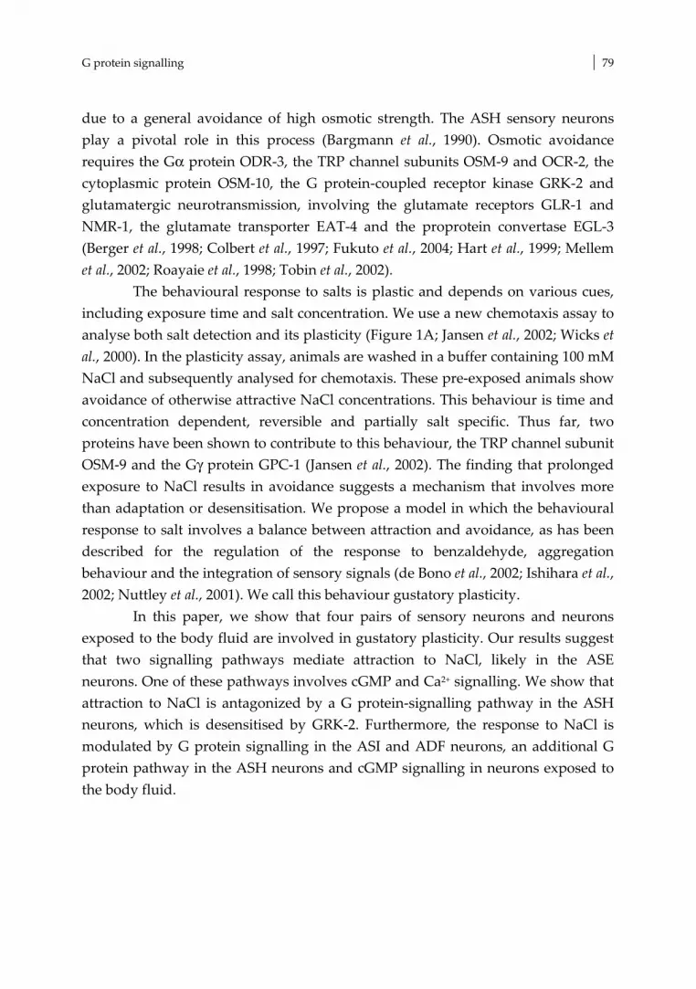
G protein signalling 79
due to a general avoidance of high osmotic strength. The ASH sensory neurons play a pivotal role in this process (Bargmann et al., 1990). Osmotic avoidance requires the Gα protein ODR-3, the TRP channel subunits OSM-9 and OCR-2, the cytoplasmic protein OSM-10, the G protein-coupled receptor kinase GRK-2 and glutamatergic neurotransmission, involving the glutamate receptors GLR-1 and NMR-1, the glutamate transporter EAT-4 and the proprotein convertase EGL-3 (Berger et al., 1998; Colbert et al., 1997; Fukuto et al., 2004; Hart et al., 1999; Mellem et al., 2002; Roayaie et al., 1998; Tobin et al., 2002).
The behavioural response to salts is plastic and depends on various cues, including exposure time and salt concentration. We use a new chemotaxis assay to analyse both salt detection and its plasticity (Figure 1A; Jansen et al., 2002; Wicks et al., 2000). In the plasticity assay, animals are washed in a buffer containing 100 mM NaCl and subsequently analysed for chemotaxis. These pre-exposed animals show avoidance of otherwise attractive NaCl concentrations. This behaviour is time and concentration dependent, reversible and partially salt specific. Thus far, two proteins have been shown to contribute to this behaviour, the TRP channel subunit OSM-9 and the Gγ protein GPC-1 (Jansen et al., 2002). The finding that prolonged exposure to NaCl results in avoidance suggests a mechanism that involves more than adaptation or desensitisation. We propose a model in which the behavioural response to salt involves a balance between attraction and avoidance, as has been described for the regulation of the response to benzaldehyde, aggregation behaviour and the integration of sensory signals (de Bono et al., 2002; Ishihara et al., 2002; Nuttley et al., 2001). We call this behaviour gustatory plasticity.
In this paper, we show that four pairs of sensory neurons and neurons exposed to the body fluid are involved in gustatory plasticity. Our results suggest that two signalling pathways mediate attraction to NaCl, likely in the ASE neurons. One of these pathways involves cGMP and Ca2+ signalling. We show that attraction to NaCl is antagonized by a G protein-signalling pathway in the ASH neurons, which is desensitised by GRK-2. Furthermore, the response to NaCl is modulated by G protein signalling in the ASI and ADF neurons, an additional G protein pathway in the ASH neurons and cGMP signalling in neurons exposed to the body fluid.
Chapter 3 80
Results Antagonistic inputs from three pairs of sensory neurons modulate gustatory behaviour
Previously, it was shown that the ASE neurons act as primary sensory neurons for NaCl detection (Bargmann & Horvitz, 1991). To confirm this role of the ASE cells, we tested the behaviour of che-1 and ceh-36 mutant animals. che-1 mutants lack functional ASE neurons due to mutation of a zinc finger protein similar to the Drosophila GLASS transcription factor (Uchida et al., 2003). che-1 is predominantly expressed in the ASE neurons; only occasionally expression was observed in other neurons (Uchida et al., 2003). ceh-36 encodes an Otx-related homeodomain protein expressed in the ASE and the AWC neurons, which specifies the identities of these neurons (Lanjuin et al., 2003). Both che-1 and ceh-36 mutant animals showed no significant chemotaxis to NaCl (0.1 – 100 mM, Figure 1B). In contrast, these animals showed wild type or even increased avoidance of 1 M NaCl (Figure 1C). Surprisingly, che-1 and ceh-36 animals showed no response to NaCl after pre-exposure to 100 mM NaCl (Figure 1D). The chemotaxis defects observed in che-1 animals could be rescued by introduction of the wild-type che-1 gene as a transgene (Figure 1B, D). Our results confirm that the ASE neurons are essential for chemotaxis to NaCl, also in our assay, and that these cells are not required for avoidance of 1 M NaCl (Bargmann & Horvitz, 1991; Uchida et al., 2003). Importantly, our results indicate that signalling via the ASE neurons is essential for gustatory plasticity.
To identify additional cells involved in gustatory plasticity we used the Gγ subunit, GPC-1. gpc-1 mutants showed reduced avoidance of, or even attraction to NaCl after pre-exposure (Jansen et al., 2002). gpc-1 is not expressed in the ASE neurons, but it is expressed in three other pairs of neurons involved in salt perception (Jansen et al., 2002): the ASI cells, which have a minor function in attraction to salts (Bargmann & Horvitz, 1991), and the ADL and ASH nociceptive neurons (Bargmann et al., 1990). Introduction of the wild type gpc-1 gene as a transgene in gpc-1 animals could restore the response to NaCl after pre-exposure (Figure 2A; Jansen et al., 2002). However, we never obtained full rescue of the defect. Since overexpression of gpc-1 induced a gustatory plasticity defect (Jansen et al., 2002), we surmise that probably the levels of GPC-1 are crucial for the behavioural response. Specific expression of GPC-1 in the ASI or ASH

G protein signalling 81
Figure 1: Both chemo-attraction to NaCl and gustatory plasticity require the ASE neurons. (A) Worms are washed in a low salt buffer or a buffer containing 100 mM NaCl and tested for chemotaxis. The assay plates contain two quadrants with only buffered agar (C) and two quadrants with buffered agar and NaCl (A). We use 0.1, 1, 10 or 100 mM NaCl to test for chemo-attraction, 1 M NaCl to test for avoidance and 25 mM NaCl in the gustatory plasticity assay. The distribution of the animals over the quadrants is determined after 10 minutes, and the chemotaxis index is calculated. (B) ASE function is required for the detection of NaCl, since chemotaxis to NaCl was abolished in che-1 and ceh-36 mutants (p<0.001). Chemotaxis could be restored by introduction of a che-1 transgene (che-1 rescue, p<0.001). (C) che-1 and ceh-36 animals strongly avoided 1 M NaCl. (D) che-1 and ceh-36 animals showed no significant response to NaCl after pre-exposure to 100 mM NaCl (p<0.001 compared to wild type animals). The che-1 defect
could be rescued by introduction of a che-1 transgene (che-1 rescue). In all figures: panels show mean + s.e.m.; n > 4 for all assays. Open bars indicate chemotaxis to 25 mM NaCl; gray bars indicate the response to 25 mM NaCl after pre-exposure to 100 mM NaCl; black bars show the response to 1 M NaCl. neurons partially restored avoidance after pre-exposure in gpc-1 mutants (Figure 2A). Expression of GPC-1 both in the ASI and ASH neurons did not yield better rescue (results not shown). These results suggest that ASI and ASH do not have additive functions, however since gpc-1 expression levels seem a crucial

Chapter 3 82
determinant in this behaviour these experiments are not conclusive. Expression of GPC-1 in the ADL and ASH nociceptive neurons resulted in better rescue than expression in the ASH neurons alone (Figure 2A), suggesting that GPC-1 probably also functions in ADL. Expression of gpc-1 in the ASE neurons did not restore gustatory plasticity (Figure 2A), suggesting that GPC-1 does not function non-cell-autonomously. Our findings indicate that GPC-1 acts both in the ASI and ASH neurons, and probably also in the ADL neurons, to modulate the response to NaCl. Avoidance of 1 M NaCl is not affected in gpc-1 animals, suggesting that separate pathways exist in the nociceptive neurons for osmotic avoidance and the modulation of the response to attractive NaCl concentrations. Figure 2: Identification of sensory neurons involved in gustatory plasticity. (A) Avoidance after pre-exposure could be restored by introducing the wild type gpc-1 gene in gpc-1 animals (genomic), or by expressing gpc-1 in the ASI, ASH or ASH and ADL neurons (p<0.01). Expression in ASE did not rescue the plasticity defect (p>0.05). (B) Expression of mec-4d/mec-2 constructs in the ASI neurons, in the ASH/ADL nociceptive neurons, or the ASH neurons significantly impaired gustatory plasticity (p<0.01).
To validate the importance of the ASI, ADL and ASH neurons in gustatory
plasticity, we expressed a dominant mutant DEG/ENaC channel MEC-4d in specific neurons to inactivate these cells (Harbinder et al., 1997). To increase the efficacy we co-expressed mec-2 constructs, since MEC-2 has been shown to increase MEC-4d activity (Goodman et al., 2002). Expression of the mec-4d/mec-2 constructs in the ASI neurons strongly affected gustatory plasticity, while expression in the ASH or ADL cells only mildly interfered with plasticity (Figure 2B). Expression of the mec-4d/mec-2 constructs in the ASI, ASH or ADL neurons did not affect salt detection (Figure 2B, results not shown). The mec-4d/mec-2 data confirm that the ASI, ASH and probably ADL neurons are involved in gustatory plasticity.

G protein signalling 83
Our results indicate that gustatory plasticity is the result of integration of input from three to four pairs of sensory neurons. Signalling via the ASE cells is essential for both attraction to salt and avoidance after pre-exposure. However, the ASE neurons are not involved in avoidance of 1 M NaCl. This response is mediated by the ASH neurons (Bargmann et al., 1990). Avoidance after pre-exposure involves cues from the ASI and ASH cells, and probably the ADL neurons. G protein signalling in the ADF neurons modulates gustatory plasticity
Our finding that the Gγ subunit GPC-1 plays a role in gustatory plasticity prompted us to test mutants of the Gα subunits expressed in the gustatory neurons in our behavioural assays. None of the sensory Gα mutants showed a defect in chemotaxis to NaCl (Table I). However, two Gα mutants showed impaired gustatory plasticity: GPA-1 and ODR-3 (Figure 3A, B).
Thus far, no function has been described for GPA-1 (Jansen et al., 1999). We found that gpa-1 animals have a defect in gustatory plasticity, whereas they showed wild type avoidance of 1 M NaCl (results not shown). The behaviour of gpa-1 animals was very similar to that of gpc-1 animals, and GPA-1 is expressed in the ASI neurons and the nociceptive neurons (Jansen et al., 1999), suggesting that GPA-1 might function together with GPC-1 in these neurons. To test this hypothesis we generated a gpa-1 gpc-1 double mutant. The behaviour of the double mutant was not significantly different from the behaviour of the two single mutants (Figure 3A), indicating that gpa-1 and gpc-1 function in the same genetic pathway that modulates gustatory plasticity.
ODR-3 is essential for olfaction and chemo-avoidance and is expressed in the AWA, AWB and AWC olfactory neurons and in the ADF and ASH neurons (Roayaie et al., 1998). odr-3 animals showed a very strong gustatory plasticity defect (Figure 3B). To test if this is caused by the osmotic avoidance defect of odr-3 animals we tested if expression of the odr-3 gene in the ASH neurons could restore gustatory plasticity. Expression of ODR-3 in the ASH neurons of odr-3 animals fully restored avoidance of 1 M NaCl (Figure 3C), confirming that ODR-3 in the ASH neurons is essential for this response (Roayaie et al., 1998). However, avoidance after pre-exposure could not be restored by expressing odr-3 in the ASH neurons (Figure 3B), indicating that ODR-3 mediated signalling in the ASH neurons is not sufficient for gustatory plasticity. To test if the ADF neurons are involved in gustatory plasticity, we expressed the odr-3 gene in these neurons.

Chapter 3 84
Table 1: Overview of strains tested for salt perception. gene reference function taste plasticity arr-1 (ok401) Fukuto et al., 2004 arrestin WT -0.12 ± 0.17*** ceh-36(ks86) Lanjuin et al., 2003 transcription factor m*** 0.13 ± 0.03*** che-1 (p679) Uchida et al., 2000 transcription factor m 0.01 ± 0.02*** cnb-1 (jh103) Bandyopadhyay et al., 2002 calcineurin B m* 0.74 ± 0.04** daf-11 (m47) Birnby et al., 2000 gyanylate cyclase WT WT dgk-1 (sy428) (nu62)
Hadju-Cronin et al., 1999; Nurrish et al., 1999
diacylglycerol kinase WT WT
0.72 ± 0.05*** 0.27 ± 0.10***
egl-4 (ky95) (ok1105)
Daniels et al.., 2000 PKG WT WT
0.06 ± 0.06 *** -0.06 ± 0.08 ***
fat-1 (wa9) Watts & Browse, 2002 ω-3 fatty acyl desaturase WT -0.02 ± 0.16** fat-3 (wa22) Watts & Browse, 2002 δ-6 fatty acid desaturase WT 0.20 ± 0.06*** fat-4 (wa14) (ok958)
Watts & Browse, 2002 δ-5 fatty acid desaturase WT WT
0.31 ± 0.09*** 0.10 ± 0.03***
gcy-32 (ok995) Coates & de Boneo, 2002 guanylate cyclase WT WT gcy-35 (ok769) Cheung et al., 2004 gyanylate cyclase WT -0.10 ± 0.10* gpa-1 (pk15) Jansen et al., 1999 Gα subunit WT 0.17 ± 0.06** gpa-2 (pk16) Zwaal et al., 1997 Gα subunit WT WT gpa-3 (pk35) Zwaal et al., 1997 Gα subunit WT WT gpa-4 (pk381) Jansen et al., 1999 Gα subunit WT WT gpa-10 (pk362) Jansen et al., 1999 Gα subunit WT WT gpa-11 (pk349) Jansen et al., 1999 Gα subunit WT WT gpa-13 (pk1270) Jansen et al., 1999 Gα subunit WT WT gpa-14 (pk347) Jansen et al., 1999 Gα subunit WT WT gpa-15 (pk477) Jansen et al., 1999 Gα subunit WT WT grk-2 (rt97) Fukuto et al., 2004 GRK m*** -0.61 ± 0.06***# itr-1 (sa73) Baylis et al., 1999; Dal Santo et al., 1999 IP3 receptor WT 0.82 ± 0.06*** ncs-1 (qa406) Gomez et al., 2001 Ca2+ sensor m** -0.13 ± 0.14** ocr-1 (ok132) (ak46)
Tobin et al., 2002 TRPV channel WT WT
-0.20 ± 0.08*** -0.25 ± 0.14***
ocr-2 (ak47) Tobin et al., 2002 TRPV channel WT -0.18 ± 0.12*** odr-3 (n1605) Roayaie et al., 1998 Gα subunit WT 0.85 ± 0.04*** osm-9 (ky10) Colbert et al., 1997 TRPV channel WT 0.06 ± 0.05**
pdl-1 (gk157) Smith & Pilgrim, personal communication
phosphodiesterase WT 0.13 ± 0.09 **
tax-2 (p671) Coburn & Bargmann, 1996 cGMP-gated channel m*** 0.83 ± 0.04*** tax-4 (p678) Komatsu et al., 1996 cGMP-gated channel m*** 0.83 ± 0.03*** tax-6 (p675) Kuhara et al., 2002 calcineurin A m*** 0.64 ± 0.05***
32 Mutant strains were tested for chemotaxis to 0.1, 1, 10 and 100 mM NaCl (taste) or chemotaxis to 25 mM NaCl after pre-exposure to 100 mM NaCl (plasticity) and their responses were compared to wild type animals: * = p < 0.05 ; ** = p < 0.01; *** = p < 0.001; unc = locomotion defect; these strains did not perform well in the assay; # pre-exposure for only 5 minutes, response stronger than wild types.

G protein signalling 85
ADF specific expression of odr-3 restored avoidance after pre-exposure (Figure 3B), but did not restore avoidance of 1 M NaCl (Figure 3C). Expression of odr-3 in both ASH and ADF did not yield better rescue, but reproduced the ASH specific rescue of avoidance of 1 M NaCl and ADF specific rescue of gustatory plasticity (results not shown). Hence, ODR-3 functions in two separate processes. It mediates avoidance of 1 M NaCl in the ASH neurons and it mediates a signal in the ADF neurons that modulates gustatory plasticity.
Figure 3: Gα subunits GPA-1 and ODR-3 modulate gustatory plasticity. (A) GPA-1 is involved in gustatory plasticity (p<0.0001) and functions in the same genetic pathway as GPC-1, since gpa-1 gpc-1 animals showed the same response as the single mutants (p>0.05) (B) Also ODR-3 is involved in gustatory plasticity (p<0.0001). Avoidance after pre-exposure could be restored by expressing ODR-3 in the ADF neurons (p<0.0001), but not by expressing it in the ASH neurons (p>0.05). (C) ODR-3 signalling mediates avoidance of 1 M NaCl (p<0.0001). Avoidance could be restored by expressing ODR-3 in the ASH neurons (p<0.0001), but not by expressing it in the ADF neurons (p>0.05).
Excessive avoidance in grk-2 animals counterbalances chemo-attraction to NaCl
G protein signal transduction can be modulated by G protein coupled receptor kinases (GRKs) and arrestins. Studies in mammals and Drosophila have shown that GRKs inhibit G protein signalling by phosphorylating activated G Protein Coupled Receptors (GPCRs) followed by arrestin binding and receptor internalisation (Ferguson, 2001). In addition to this classical function, recent studies indicate a role for GRKs and arrestins as signal transduction molecules (Perry & Lefkowitz, 2002; Kim et al., 2005; Ren et al., 2005). The C. elegans genome contains two GRKs, grk-1 and grk-2, and one arrestin gene, arr-1 (Fukuto et al., 2004). Loss-of-function of grk-2 impairs the response to attractive odorants and several repellents, including odorants, quinine and 1-4 M glycerol, but seems to have no or

Chapter 3 86
little involvement in olfactory adaptation. These results suggest that GRK-2 functions as a signal transduction molecule in the nociceptive neurons and olfactory neurons (Fukuto et al., 2004).
Loss-of-function of grk-2 severely impaired chemotaxis to NaCl (Figure 4A). However, grk-2 animals did show avoidance after pre-exposure (Figure 4B), indicating that these animals can detect NaCl. When these animals were only briefly pre-exposed to NaCl they even showed enhanced avoidance after pre-exposure (Figure 4C). Since the ASE cells are essential for both salt taste and gustatory plasticity, we surmised that GRK-2 does not function in the ASE neurons, but in the nociceptive neurons instead. We first tested the response of grk-2 animals to 1 M NaCl. Surprisingly, grk-2 animals showed strong avoidance of 1 M NaCl (Figure 4D), in contrast to the previously reported defect in avoidance of 1-4 M glycerol (Fukuto et al., 2004). These results suggest that different signalling routes exist for avoidance of high concentrations of glycerol and NaCl. We next tested if specific expression of grk-2 in the nociceptive neurons ADL and ASH could restore chemotaxis of the grk-2 mutants. Indeed, a srb-6::grk-2 construct restored the response to 1 - 100 mM NaCl partially (gjEx601 and 2 other strains) or fully (gjEx602) (Figure 4A). Probably, gjEx601 animals did not express sufficient GRK-2 in the nociceptive neurons for full rescue, since avoidance of 1M NaCl was not restored (Figure 4C). In contrast, gjEx602 animals probably over-expressed GRK-2, diminishing the avoidance responses (Figure 4A and D), and restoring chemo-attraction. To further confirm GRK-2 function in ASH, we also used an osm-10::grk-2 construct to express grk-2 in the ASH and ASI neurons. Also this construct could restore the NaCl chemo-attraction defect of grk-2 animals (gjEx639 in Figure 4A, B and D). In addition, specific expression of grk-2 in the ASH neurons could restore avoidance of octanol and 1 M glycerol (results not shown). In contrast, expression of GRK-2 in interneurons, but not the sensory neurons, using a glr-1::grk-2 construct did not restore chemotaxis to NaCl (results not shown). Our results suggest that GRK-2 modulates the strength of the NaCl avoidance signal mediated by the nociceptive neurons. Such a function fits with a classical function for GRK-2 in desensitisation of the NaCl avoidance signalling pathway (Figure 7), suggesting that GRK-2 has two different functions in separate signalling pathways in the ASH cells. Moreover, these results show that signals derived from the nociceptive neurons counterbalance chemo-attraction to NaCl mediated by the ASE neurons.

G protein signalling 87
Figure 4: Excessive avoidance in grk-2 animals counterbalances chemo-attraction to NaCl (A) Chemotaxis to 0.1 –100 mM NaCl was impaired in grk-2 animals (p<0.0001). The grk-2 defect could be restored by expressing GRK-2 in the nociceptive neurons in strains grk-2 gjEx601 (p<0.05), gjEx602 (p<0.001), and gjEx639 (p<0.001). (A) grk-2 mutants showed no chemo-attraction to 25 mM NaCl, but strong avoidance after pre-exposure to 100 mM NaCl. Expression of GRK-2 in the nociceptive neurons restored chemo-attraction in grk-2 gjEx601 animals (p<0.001) and grk-2 gjEx639 animals ((p<0.0001). grk-2 gjEx602 animals showed even better rescue of chemo-attraction (p<0.0001), but impaired avoidance after pre-exposure (p<0.0001). Mutation of arr-1 significantly reduced avoidance after pre-exposure (p<0.001), to a similar level as loss-of gpa-1. The behaviour of the gpa-1 arr-1 double mutant suggests that both genes function in the same pathway (p>0.05). (C) grk-2 animals showed enhanced
avoidance after pre-exposure to NaCl for only 5 minutes (p<0.001). (D) grk-2 animals showed strong avoidance of 1 M NaCl, which was not affected in grk-2 gjEx601 and gjEx639 animals (p>0.05), but impaired in grk-2 gjEx602 animals (p<0.001).
The C. elegans arrestin, arr-1, is required for adaptation and recovery from
adaptation to various odorants (Palmitessa et al., 2005). In our assays, mutation of arr-1 reduced avoidance after pre-exposure, while chemo-attraction or avoidance

Chapter 3 88
responses were not affected (Figure 4B, results not shown). These results are not in agreement with a function of ARR-1 in receptor or channel internalisation in the ASE or nociceptive neurons. Rather, arr-1 animals displayed behavioural deficits comparable to those observed in gpc-1 and gpa-1 animals. Indeed, gpa-1 arr-1 double mutant animals showed the same response after pre-exposure as the two single mutants (Figure 4B), indicating that these genes function in the same genetic pathway. However, arr-1 is broadly expressed in the nervous system of C. elegans (Fukuto et al., 2004; Palmitessa et al., 2005). Therefore, it is unclear in which cells ARR-1 functions in gustatory plasticity. Gustatory plasticity requires cGMP signalling in the body cavity neurons
A frequently used second messenger in G protein signal transduction is cGMP. cGMP can be generated by guanylate cyclases and it can activate other signalling molecules such as cGMP-gated channels or protein kinases (PKG). Previous studies have shown that cGMP signalling plays an important role in chemotaxis to NaCl. Mutation of the guanylate cyclase DAF-11 and the cGMP-gated channel subunits TAX-2 and TAX-4 abolish chemotaxis to NaCl (Birnby et al.., 2000; Coburn & Bargmann, 1996; Komatsu et al.., 1996). Surprisingly, mutation of daf-11 did not affect chemotaxis to NaCl in our assay (Figure 5A), whereas mutation of tax-2 and tax-4 strongly affected chemotaxis to 0.1-10 mM NaCl. However, these latter mutants still showed strong chemo-attraction to 100 mM NaCl (Figure 5A). These results confirm that cGMP signalling is involved in chemotaxis to NaCl, although it is unclear which proteins activate the TAX-2/TAX-4 channel. In addition, our results suggest that another pathway exists for the detection of NaCl, which functions in parallel to the cGMP pathway.
Mutations in tax-2 and tax-4 also abolish gustatory plasticity (results not shown). However, since these proteins play important roles in chemotaxis to NaCl, we cannot conclude whether TAX-2 and TAX-4 function in gustatory plasticity. Analysis of other cGMP signalling molecules did indicate a role of cGMP signalling in gustatory plasticity. Mutations in the cGMP dependent kinase EGL-4 and the phosphodiesterase delta-like protein PDL-1 affected gustatory plasticity (Figure 5C). EGL-4 is expressed in a large number of neurons (Fujiwara et al., 2002; Hirose et al., 2003) and PDL-1, responsible for cGMP breakdown, shows pan-neuronal expression (J. Smith & D. Pilgrim, personal communication). Hence, further cell-specific rescue and epistasis analysis are needed to determine the specific functions of these proteins.

G protein signalling 89
Figure 5: Gustatory plasticity requires cGMP (A) Salt detection was not affected in daf-11 animals (p>0.05), but significantly impaired in tax-2 and tax-4 animals (p<0.01 at 1 and 10 mM NaCl). These mutants still responded to 100 mM NaCl. (B) Mutation of gcy-35 affected gustatory plasticity (p<0.001). This defect could be rescued by expression of gcy-35 in the AQR, PQR, URX neurons (p<0.0001). Expression of a gcy-32::egl-2(gf) construct to inhibit the activity of AQR, PQR and URX neurons reduced avoidance after pre-exposure (p<0.05).Analysis of a gpc-1 gcy-35 double mutant strain indicated that these genes function in the same genetic pathway in gustatory plasticity (p>0.05 compared to the single mutants). (C) Mutation of the cGMP signalling molecules egl-4 (p<0.001) and pdl-1 (p<0.01) affected gustatory plasticity, whereas mutation of gcy-35 did not (p>0.05).
We also identified a guanylate cyclase that plays a role in gustatory plasticity. Mutations in the soluble guanylate cyclase gcy-35 affected this behaviour (Figure 5B), but did not affect chemo-attraction or avoidance of high NaCl concentrations. gcy-35 is expressed, among others, in the body cavity neurons AQR, PQR and URX (Cheung et al., 2004; Gray et al., 2004). These neurons are directly exposed to the pseudocoelomic body fluid (White et al., 1986) and regulate social feeding or aggregation behaviour (Coates & de Bono, 2002). We tested whether GCY-35 functions in these body cavity neurons during plasticity by specific expression of gcy-35 in these neurons. Indeed, avoidance after pre-

Chapter 3 90
exposure was restored in gcy-35 animals expressing a gcy-32::gcy-35 construct (Figure 5B). We did not obtain full rescue, suggesting that gcy-35 might also function in other cells in gustatory plasticity.
To confirm that the AQR, PQR and URX cells are involved in gustatory
plasticity we expressed a gain-of-function mutant form of the eag-type potassium channel EGL-2 in these neurons, using a gcy-32::egl-2(gf) construct. Expression of this construct has been shown to inhibit neuronal activity (Coates & de Bono, 2002). We found that expression of EGL-2(gf) in these neurons significantly reduced avoidance after pre-exposure to 100 mM NaCl, without affecting chemotaxis (Figure 5B, results not shown). These results show that the AQR, PQR and URX body cavity neurons mediate a signal that modulates the response to NaCl.
The phenotype of the gcy-35 animals was very comparable to that of the arr-1, gpa-1 and gpc-1 animals. To test if also gcy-35 functions in the same genetic pathway, we tested the behaviour of gpc-1 gcy-35 double mutant animals. The double mutant strain behaved as the two single mutants (Figure 5B). Therefore, we hypothesize that cGMP signalling via gcy-35 in the AQR, PQR and/or URX neurons functions in the same genetic pathway as gpc-1, gpa-1 and arr-1 to modulate the response to NaCl after prolonged exposure.
Gustatory plasticity requires Ca2+ signalling
Another frequently used second messenger is Ca2+. Ca2+ levels in the cell can be increased by influx via transient receptor potential (TRP) channels or cGMP-gated channels, and by depletion of intracellular stores after IP3-receptor activation. Increased Ca2+ levels can subsequently activate various downstream effector molecules, including calcineurin and the neuronal calcium sensor NCS-1. Previous studies have shown that Ca2+ signalling is important for chemotaxis to NaCl, since mutation of the cGMP-gated channel TAX-2/TAX-4, which upon activation leads to Ca2+ influx, affects chemotaxis to NaCl (Coburn & Bargmann, 1996; Komatsu et al., 1996; Figure 5A). In addition, it was shown that mutation of the calcineurin A subunit TAX-6 affects chemotaxis to NaCl (Kuhara et al., 2002). Calcineurin is a phosphatase consisting of two subunits A and B, encoded by tax-6 and cnb-1, respectively (Bandyopadhyay et al., 2002; Kuhara et al., 2002). We tested tax-6 and cnb-1 mutants in our assays, as well as ncs-1 mutants, which lack a functional neuronal calcium sensor 1 (Gomez et al., 2001). All three mutants showed chemotaxis defects comparable to those of the tax-2 and tax-4 mutants

G protein signalling 91
Figure 6: Gutatory plasticity requires Ca2+ signalling. (A) Salt detection was significantly impaired in tax-6, cnb-1 and ncs-1 animals (p<0.01 at 1 and 10 mM NaCl). These mutants still responded to 100 mM NaCl. The behaviour of tax-4 tax-6 double mutants was not significantly different from the behaviour of the two single mutants (p>0.05). (B) TRP channel mutants osm-9, ocr-1 and ocr-2 showed defects in plasticity (p<0.001). TRP-channel double mutants showed similar defects to the single mutants (p>0.05). The TRP-channel triple mutant showed a very small additive effect of inactivation of all three subunits (p<0.05, compared to ocr-1 and ocr-2 animals). The TRP channels function in the same genetic pathway as gpc-1, since the response of the gpc-1 osm-9 double did not differ significantly from the single mutants (p>0.05). (C) itr-1, dgk-1, fat-1, fat-3 and fat-4 showed defects in plasticity (p<0.001). FAT-4 functions in the same pathway as OSM-9 and GPC-1, since no significant difference could be detected between double and single mutants (p>0.05). (D) Avoidance of 1 M NaCl was not reduced in fat-1, fat-3, fat-4, or dgk-1 animals. (Figure 5A, 6A): strongly reduced chemotaxis to low NaCl concentrations (0.1-10 mM), but stronegr or even wild type responses to 100 mM NaCl. Also these mutants showed defects in gustatory plasticity, however, because of their function in chemotaxis, their role in gustatory plasticity remains unclear.
The similarities of the NaCl chemotaxis phenotypes of the tax-2, tax-4, tax-6, cnb-1 and ncs-1 animals suggested that these genes might function in the same

Chapter 3 92
pathway. In agreement with this hypothesis, we observed no significant difference between the responses of the tax-4 tax-6 double mutant animals and the two single mutants (Figure 6A). The expression patterns of these genes overlap only in the ASE neurons (Bandyopadhyay et al., 2002; Coburn & Bargmann, 1996; Gomez et al., 2001; Komatsu et al., 1996; Kuhara et al., 2002). Since the ASE neurons are essential for NaCl detection, it is likely that the cGMP and Ca2+ signalling molecules for the detection of NaCl act in the ASE neurons. However, at present we cannot rule out a function for these proteins in other sensory neurons or interneurons. Moreover, it is unclear which signals activate the cGMP/Ca2+ NaCl detection pathway.
Besides cGMP-gated channels also TRP channels mediate Ca2+ influx. We
have previously shown a function for the TRPV channel subunit OSM-9 in gustatory plasticity (Jansen et al., 2002). Here we show that also the OCR-1 and OCR-2 TRPV channel subunits are involved (Figure 6B). The responses of the three TRPV channel mutants did not differ significantly from each other, although the defect seems a little more severe in the osm-9 animals (Figure 6B). In addition, the behaviour of doubles between these three mutants did not differ significantly from the single mutants (Figure 6B), suggesting that these three subunits function in the same genetic pathway. In the ocr-1, ocr-2, osm-9 triple mutant we observed a very small additive effect of inactivation of all three TRP channel subunits compared to the ocr-1 and ocr-2 single mutants (Figure 6B).
The plasticity defects of the TRPV channel mutants were very similar to the defects of the gpa-1, gpc-1, arr-1 and gcy-35 mutants. The behaviour of gpc-1 osm-9 double mutant animals was not significantly different from the single mutants (Figure 6B), suggesting that also the TRPV channel subunits function in this genetic gustatory plasticity pathway. The expression patterns of these TRPV genes are restricted to subsets of sensory neurons, overlapping with neurons that play a role in gustatory plasticity: ASH and ADL (Colbert et al., 1997; Tobin et al., 2002). TRP channels can be activated in various ways, including by phospholipase C (PLC) dependent mechanisms (Montell et al., 2002). Activation of PLC by G proteins leads to the production of inositol (1,4,5) trisphosphate (IP3) and diacylglycerol (DAG). IP3 can activate IP3 receptors resulting in Ca2+ release from intracellular stores, which can activate TRP channels. TRP channels can also be activated by DAG, or its derivatives, polyunsaturated fatty acids (PUFAs; Kahn-Kirby et al., 2004). We tested if these signalling molecules are involved in gustatory plasticity.
Mutation of both the IP3 receptor ITR-1 (Baylis et al., 1999; Dal Santo et al., 1999) and the diacylglycerol kinase DGK-1, essential for reduction of DAG levels

G protein signalling 93
(Hajdu-Cronin et al., 1999; Nurrish et al., 1999), strongly reduced avoidance after pre-exposure (Figure 6C). However, these mutations did not affect chemo-attraction to 0.1 – 100 mM NaCl or avoidance of 1 M NaCl (Figure 6C, D, results not shown), indicating that IP3 and DAG signalling are only involved in the plasticity response. It is possible that these second messengers activate TRP channels, but they could also affect other signalling routes such as the Goα/Gqα network, which is involved in gustatory plasticity as well (RKH and GJ, unpublished results).
To test if PUFAs are involved in gustatory plasticity, we tested three lipid desaturase mutants that affect PUFA synthesis, fat-1, fat-3 and fat-4 (Watts & Browse, 2002). Recently, strong avoidance defects were described for fat-3 mutant animals, including a defect in avoidance of 1 M glycerol; mild or no defects were observed for fat-1 or fat-4 animals (Kahn-Kirby et al., 2004). In our assays, the PUFA synthesis mutants showed no defect in avoidance of 1 M NaCl (Figure 6D), again indicating that separate pathways exist for the detection of 1 M glycerol and 1 M NaCl. However, all three mutants showed aberrant behaviour in our gustatory plasticity assay (Figure 6C), indicating that PUFAs play a role in this process.
Since the PUFA synthesis mutants showed a very comparable behaviour to gpa-1, gpc-1, arr-1, osm-9 and gcy-35 animals, we analysed the behaviour of gpc-1 fat-4 and osm-9 fat-4 double mutants. The behaviour of these double mutants did not differ significantly from the behaviour of the single mutants, indicating that these genes all function in the same genetic pathway (Figure 6C). Discussion Model for gustatory plasticity in C. elegans
We can discriminate three responses of C. elegans to NaCl: first, chemotaxis to NaCl concentrations between 0.1 – 200 mM, second, avoidance of NaCl above 200 mM, and third, avoidance of otherwise attractive NaCl concentrations (25 mM) after pre-exposure to 100 mM NaCl. Data presented here and by others indicate that these responses require input from at least 4 chemosensory neurons and the neurons exposed to the body fluid. We propose a model to explain the different gustatory responses (Figure 7).
The ASE neurons are essential for chemotaxis to low NaCl concentrations (Figure 7). Chemo-attraction is antagonized by avoidance, mediated by the ASH neurons (Figure 7). The ASH neurons are not activated at low NaCl concentrations, but become activated at high NaCl concentrations (Hilliard et al., 2004). It is unclear

Chapter 3 94
Figure 7: Model for NaCl detection and its plasticity in C. elegans. NaCl is detected by the ASE sensory neurons, probably using cGMP/Ca2+ signalling and an unknown signalling pathway. Attraction is antagonized by avoidance mediated by the ASH, using ODR-3, OSM-9, OCR2 and the inhibitor GRK-2. We propose that upon prolonged exposure to attractive NaCl concentrations an ASE derived signal sensitises the ASH, ADL, ASI and ADF neurons, resulting in avoidance of normally attractive NaCl concentrations. We have shown that the ADF neurons use ODR-3, and the ASI neurons, a second pathway in the ASH neurons and probably the ADL neurons use GPC-1. Integration of these signals ultimately leads to the behavioural response of the animal.
where the ASE and ASH derived signals are integrated and where the choice between attraction and avoidance is made. It is also not known why avoidance is preferred over attraction at NaCl concentrations above 200 mM. Perhaps ASE signalling is blocked at high salt concentrations, or alternatively, preference for avoidance might be the default state, e.g. determined by the wiring of the nervous system. Our model suggests that also the ADF, ADL and ASI neurons are not or only weakly activated at low NaCl concentrations. We propose that upon prolonged exposure to 100 mM NaCl the ASE neurons produce a signal (blue arrows in Figure 7) that sensitises the ADF, ADL, ASI and ASH neurons, resulting in avoidance of otherwise attractive NaCl concentrations (black arrows in Figure

G protein signalling 95
7). We suggest that the ASE neurons signal via the body cavity neurons, AQR, PQR and URX, but it is also possible that the body cavity neurons function downstream of the ADF, ADL, ASI and ASH neurons.
Based on the cellular circuit described above, expression patterns and our cell specific rescue experiments, we can place several molecules in our model (Figure 7). We propose that stimulation of the ASE neurons by low NaCl concentrations activates a cGMP and Ca2+ signalling pathway and another unknown pathway. Avoidance of 1 M NaCl, mediated by the ASH neurons, requires the Gα subunit ODR-3 and the TRP channel subunits OSM-9 and OCR-2, and is inhibited by GRK-2. ODR-3 also functions in the ADF neurons in gustatory plasticity, where it might either transduce the avoidance signal, or alternatively transduce the ASE derived sensitising signal.
In addition, we have identified a genetic pathway that mediates gustatory plasticity. This pathway involves the Gγ GPC-1, the Gα GPA-1, the arrestin ARR-1, the TRPV channel subunits OSM-9, OCR-1 and OCR-2, PUFA signalling (FAT-4) and the guanylate cyclase GCY-35. It is unclear if these proteins function in the same cells, but our and previously published data are consistent with a function for GPC-1 in the ASI, ASH and perhaps ADL neurons (based on rescue experiments), GPA-1 and OSM-9 in these same neurons (based on expression patterns, Colbert et al., 1997; Jansen et al., 1999), OCR-1 in ADL and OCR-2 in ADL, ASH and ADF (Tobin et al., 2002) and GCY-35 in the AQR, PQR and URX body cavity neurons (based on rescue experiments). We cannot discriminate if these proteins transduce the avoidance response or function in a pathway that mediates the ASE derived sensitising signal. As indicated, part of this model is based on previously described expression patterns. Most of these expression patterns have been derived using GFP reporter constructs. These constructs may not fully represent the gene’s expression pattern. Therefore, additional cell-specific rescue experiments will be needed to confirm our model. Materials and methods Strains, genetics and germline transformation
Strains used in this work are listed in Table I. Wild type C. elegans were strain Bristol N2. Germline transformation and transgene integration were performed as described (Mello et al., 1991). We used an elt-2::GFP construct (30 ng/μl) as co-injection marker (Fukushige et al., 1999). Rescue of che-1, gpc-1, odr-3 and grk-2 and cell inactivation using mec-4d and mec-2 or egl-2(gf) was tested using

Chapter 3 96
5 or more transgenic strains for each clone injected at various concentrations (1 – 100 ng/μl). Although cell specific expression of mec-2/mec-4d or egl-2gf did affect gustatory plasticity we did not observe neuronal degeneration. Molecular biology
Details of plasmid construction are available on request. Promoters used for cell-specific rescue or cell inactivation were: flp-5 (ASE; Li et al., 1999), gpa-4 (ASI; Jansen et al., 1999), gpa-11 (ADL, ASH; Jansen et al., 1999), gpa-13 (ADF, ASH, AWC; Jansen et al., 1999), sra-6 (ASH, ASI faint; Troemel et al., 1995), srb-6 (ADL, ASH; Troemel et al., 1995), glr-1 (17 classes of neurons; Hart et al., 1995; Maricq et al., 1995), srh-142 (ADF; Sagasti et al., 1999). Expression patterns were confirmed using GFP-fusion constructs driven by the same promoter. Behavioural assays
Chemotaxis towards NaCl and gustatory plasticity were assessed as described before (Wicks et al., 2000; Jansen et al., 2002), using 0.1, 1, 10 or 100 mM or 1 M NaCl; after pre-exposure to 100 mM NaCl animals were tested for chemotaxis to 25 mM NaCl. A chemotaxis-index was calculated: (A-C)/(A+C); where A is the number of worms at the quadrants with NaCl, C is the number of worms at the quadrants without attractant. Statistical significance was determined using the two-tailed t-test. Error bars represent the s.e.m. Acknowledgements
We thank T. Stiernagle, the Caenorhabditis Genetics Center and the C. elegans Knock-out Consortium, C. Bargmann, J. Dent, A. Hart, N. L’Etoile, M. Koelle, and R. Plasterk for C. elegans strains; C. Bargmann, M. de Bono, J. McGhee, A. Hart and C. Li for constructs; H. Fukuto, D. Ferkey and A. Hart, D. Karow, J. Gray, C. Bargmann and M. Marletta, J. Smith and D. Pilgrim and S. Xu and P. Sternberg for the communication of unpublished results; J. Watts for suggestions; K. Ackema, H. Lans and J. Zareno for discussions and comments on the manuscript. This work was funded by the Centre for Biomedical Genetics and the Royal Dutch Academy of Sciences. G.J. is a Royal Dutch Academy of Sciences Fellow.

G protein signalling 97
References
Bandyopadhyay J, Lee J, Lee JI, Yu JR, Jee C, Cho JH, Jung S, Lee MH, Zannoni S, Singson A, Kim do H, Koo HS, Ahnn J (2002) Calcineurin, a calcium/calmodulin-dependent protein phosphatase, is involved in movement, fertility, egg laying, and growth in Caenorhabditis elegans. Mol Biol Cell 13: 3281-3293
Bargmann CI, Horvitz HR (1991) Chemosensory neurons with overlapping functions direct chemotaxis to multiple chemicals in C. elegans. Neuron 7: 729-742
Bargmann CI, Thomas JH, Horvitz HR (1990) Chemosensory cell function in the behavior and development of Caenorhabditis elegans. Cold Spring Harb Symp Quant Biol 55: 529-538
Baylis HA, Furuichi T, Yoshikawa F, Mikoshiba K, Sattelle DB (1999) Inositol 1,4,5-trisphosphate receptors are strongly expressed in the nervous system, pharynx, intestine, gonad and excretory cell of Caenorhabditis elegans and are encoded by a single gene (itr-1). J Mol Biol 294: 467-476
Berger AJ, Hart AC, Kaplan JM (1998) Gαs-Induced neurodegeneration in Caenorhabditis elegans. J Neurosci 18: 2871-2880
Birnby DA, Link EM, Vowels JJ, Tian H, Colacurcio PL, Thomas JH (2000) A transmembrane guanylyl cyclase (DAF-11) and Hsp90 (DAF-21) regulate a common set of chemosensory behaviors in Caenorhabditis elegans. Genetics 155: 85-104
Cheung BH, Arellano-Carbajal F, Rybicki I, de Bono M (2004) Soluble guanylate cyclases act in neurons exposed to the body fluid to promote C. elegans aggregation behavior. Curr Biol 14: 1105-1111
Coates JC, de Bono M (2002) Antagonistic pathways in neurons exposed to body fluid regulate social feeding in Caenorhabditis elegans. Nature 419: 925-929
Coburn CM, Bargmann CI (1996) A putative cyclic nucleotide-gated channel is required for sensory development and function in C. elegans. Neuron 17: 695-706
Colbert HA, Smith TL, Bargmann CI (1997) OSM-9, a novel protein with structural similarity to channels, is required for olfaction, mechanosensation, and olfactory adaptation in Caenorhabditis elegans. J Neurosci 17: 8259-8269
Culotti JG, Russell RL (1978) Osmotic avoidance defective mutants of the nematode Caenorhabditis elegans. Genetics 90: 243-256.
Dal Santo P, Logan MA, Chisholm AD, Jorgensen EM (1999) The inositol trisphosphate receptor regulates a 50-second behavioral rhythm in C. elegans. Cell 98: 757-767
Daniels SA, Ailion M, Thomas JH, Sengupta P (2000) egl-4 acts through a transforming growth factor-β.SMAD pathway in Caenorhabditis elegans to regulate multiple neuronal circuits in response to sensory cues. Genetics 156: 123-141

Chapter 3 98
de Bono M, Tobin DM, Davis MW, Avery L, Bargmann CI (2002). Social feeding in Caenorhabditis elegans is induced by neurons that detect aversive stimuli. Nature 419: 899-903
Dusenbery DB, Scheridan RE, Russell RL (1975) Chemotaxis-defective mutants of the nematode Caenorhabditis elegans. Genetics 80: 297-309
Ferguson SS (2001) Evolving concepts in G protein-coupled receptor endocytosis: the role in receptor desensitization and signaling. Pharmacol Rev 53: 1-24
Fujiwara M, Sengupta P, McIntire SL (2002) Regulation of body size and behavioral state of C. elegans by sensory perception and the EGL-4 cGMP-dependent protein kinase. Neuron 36: 1091-1102
Fukushige T, Hendzel MJ, Bazett-Jones DP, McGhee J (1999) Direct visualization of the elt-2 gut-specific GATA factor binding to a target promoter inside the living Caenorhabditis elegans. Proc Natl Acad Sci USA 96: 11883-11888
Fukuto HS, Ferkey DM, Apicella AJ, Lans H, Sharmeen T, Chen W, Lefkowitz RJ, Jansen G, Schafer WR, Hart AC (2004) G protein-coupled receptor kinase function is essential for chemosensation in C. elegans. Neuron 42: 581-593
Gomez M, De Castro E, Guarin E, Sasakura H, Kuhara A, Mori I, Bartfai T, Bargmann CI, Nef P (2001) Ca2+ signaling via the neuronal calcium sensor-1 regulates associative learning and memory in C. elegans. Neuron 30: 241-248
Goodman MB, Ernstrom GG, Chelur DS, O'Hagan R, Yao CA, Chalfie M (2002) MEC-2 regulates C. elegans DEG/ENaC channels needed for mechanosensation. Nature 415: 1039-1042
Gray JM, Karow DS, Lu H, Chang AJ, Chang JS, Ellis RE, Marletta MA, Bargmann CI (2004) Oxygen sensation and social feeding mediated by a C. elegans guanylate cyclase homologue. Nature 430: 317-322
Hadju-Cronin YM, Chen WJ, Patikoglou G, Koelle MR, Sternberg PW (1999) Antagonism between Goα and Gqα in Caenorhabditis elegans: the RGS protein EAT-16 is necessary for Goα signaling and regulates Gqα activity. Genes Dev 13: 1780-1793
Harbinder S, Tavernarakis N, Herndon LA, Kinnell M, Xu SQ, Fire A, Driscoll M (1997) Genetically targeted cell disruption in Caenorhabditis elegans. Proc Natl Acad Sci U S A 94: 13128-13133
Hart AC, Kass J, Shapiro JE, Kaplan JM (1999) Distinct signaling pathways mediate touch and osmosensory responses in a polymodal sensory neuron. J Neurosci 19: 1952-1958
Hart AC, Sims S, Kaplan JM (1995) Synaptic code for sensory modalities revealed by C. elegans GLR-1 glutamate receptor. Nature 378: 82-85

G protein signalling 99
Hilliard MA, Apicella AJ, Kerr R, Suzuki H, Bazzicalupo P, Schafer WR, (2004) In vivo imaging of C. elegans ASH neurons: cellular response and adaptation to chemical repellents. EMBO J 24:63-72
Hirose T, Nakano Y, Nagamatsu Y, Misumi T, Ohta H, Ohshima Y (2003) Cyclic GMP-dependent protein kinase EGL-4 controls body size and lifespan in C. elegans. Development 130: 1089-1099
Ishihara T, Iino Y, Mohri A, Mori I, Gengyo-Ando K, Mitani S, Katsura I (2002) HEN-1, a secretory protein with an LDL receptor motif, regulates sensory integration and learning in Caenorhabditis elegans. Cell 109: 639-649
Jansen G, Thijssen KL, Werner P, van der Horst M, Hazendonk E, Plasterk RH (1999) The complete family of genes encoding G proteins of Caenorhabditis elegans. Nat Genet 21: 414-419
Jansen G, Weinkove D, Plasterk RHA (2002) The G-protein gamma subunit gpc-1 of the nematode C. elegans is involved in taste adaptation. EMBO J 21: 986-994
Kahn-Kirby AH, Dantzker JL, Apicella AJ, Schafer WR, Browse J, Bargmann CI, Watts JL (2004) Specific polyunsaturated fatty acids drive TRPV-dependent sensory signaling in vivo. Cell 119: 889-900
Kim J, Ahn S, Ren XR, Whalen EJ, Reiter E, Wei H, Lefkowitz RJ (2005) Functional antagonism of different G protein-coupled receptor kinases for beta-arrestin-mediated angiotensin II receptor signaling. Proc Natl Acad Sci U S A 102: 1442-1447
Komatsu H, Mori I, Rhee JS, Akaike N, Ohshima Y (1996) Mutations in a cyclic nucleotide-gated channel lead to abnormal thermosensation and chemosensation in C. elegans. Neuron 17: 707-718
Kuhara A, Inada H, Katsura I, Mori I (2002) Negative regulation and gain control of sensory neurons by the C. elegans calcineurin TAX-6. Neuron 33: 751-763
Lanjuin A, VanHoven MK, Bargmann CI, Thompson JK, Sengupta P (2003) Otx/otd homeobox genes specify distinct sensory neuron identities in C. elegans. Dev Cell 5:621-633
Li C, Kim K, Nelson LS (1999) FMRFamide-related neuropeptide gene family in Caenorhabditis elegans. Brain Res 848: 26-34
Lindemann B (2001) Receptors and transduction in taste. Nature 413: 219-225 Liu L, Leonard AS, Motto DG, Feller MA, Price MP, Johnson WA, Welsh MJ (2003)
Contribution of Drosophila DEG/ENaC genes to salt taste. Neuron 39: 133-146 Lyall V, Heck GL, Vinnikova AK, Ghosh S, Phan TH, Alam RI, Russell OF, Malik SA, Bigbee
JW, DeSimone JA (2004) The mammalian amiloride-insensitive non-specific salt taste receptor is a vanilloid receptor-1 variant. J Physiol 558: 147-159

Chapter 3 100
Maricq AV, Peckol E, Driscoll M, Bargmann CI (1995) Mechanosensory signaling in C. elegans mediated by the GLR-1 glutamate receptor. Nature 378: 78-81
Mellem JE, Brockie PJ, Zheng Y, Madsen DM, Maricq AV (2002) Decoding of polymodal sensory stimuli by postsynaptic glutamate receptors in C. elegans. Neuron 36: 933-944
Mello CC, Kramer JM, Stinchcomb D, Ambros V (1991) Efficient gene transfer in C. elegans: extrachromosomal maintenance and integration of transforming sequences. EMBO J 10: 3959-3970
Montell DJ, Birnbaumer L, Flockerzi V (2002) The TRP channels, a remarkably functional family. Cell 108: 595-598
Nurrish S, Segalat L, Kaplan JM (1999) Serotonin inhibition of synaptic transmission: Goα decreases the abundance of UNC-13 at release sites. Neuron 24: 231-242
Nuttley WM, Harbinder S, van der Kooy D (2001) Regulation of distinct attractive and aversive mechanisms mediating benzaldehyde chemotaxis in Caenorhabditis elegans. Learn Mem 8: 170-181
Palmitessa A, Hess HA, Bany LA, Kim Y-M, Koelle MR, Benovic JL (2005) C. elegans arrestin regulates neural G protein signalling and olfactory adaptation and recovery. J Biol Chem 280: 24649-24662
Perry SJ, Lefkowitz RJ (2002) Arresting developments in hepthahelical receptor signaling and regulation. Trends Cell Biol 12: 130-138
Ren XR, Reiter E, Ahn S, Kim J, Chen W, Lefkowitz RJ (2005) Different G protein-coupled receptor kinases govern G protein and beta-arrestin-mediated signaling of V2 vasopressin receptor. Proc Natl Acad Sci U S A 102: 1448-1453
Roayaie K, Crump JG, Sagasti A, Bargmann CI (1998) The Gα protein ODR-3 mediates olfactory and nociceptive function and controls cilium morphogenesis in C. elegans olfactory neurons. Neuron 20: 55-67
Sagasti A, Hobert O, Troemel ER, Ruvkun G, Bargmann CI (1999) Alternative olfactory neuron fates are specified by the LIM homeobox gene lim-4. Genes Dev 15: 1794-1806
Tobin D, Madsen D, Kahn-Kirby A, Peckol E, Moulder G, Barstead R, Maricq A, Bargmann C (2002) Combinatorial expression of TRPV channel proteins defines their sensory functions and subcellular localization in C. elegans neurons. Neuron 35: 307-318
Troemel ER, Chou JH, Dwyer ND, Colbert HA, Bargmann CI (1995) Divergent seven transmembrane receptors are candidate chemosensory receptors in C. elegans. Cell 83: 207-218
Uchida O, Nakano H, Koga M, Oshima Y (2003) The C. elegans che-1 gene encodes a zinc finger transcription factor required for specification of the ASE chemosensory neurons. Development 130: 1215-1224

G protein signalling 101
Ward S (1973) Chemotaxis by the nematode Caenorhabditis elegans: identification of attractants and analysis of the response by use of mutants. Proc Natl Acad Sci USA 70: 817-821
Watts JL, Browse J (2002) Genetic dissection of polyunsaturated fatty acid synthesis in Caenorhabditis elegans. Proc Natl Acad Sci USA 99: 5854-5859
White JG, Southgate JN, Thomson JN, Brenner S (1986) The structure of the nervous system of the nematode Caenorhabditis elegans. Phil Trans R Soc Lond B 314: 1-340
Wicks SR, de Vries CJ, van Luenen HG, Plasterk RH (2000) CHE-3, a cytosolic dynein heavy chain, is required for sensory cilia structure and function in Caenorhabditis elegans. Dev Biol 221: 295-307
Zwaal RR, Mendel JE, Sternberg PW, Plasterk RHA (1997) Two neuronal G proteins are involved in chemosensation of the Caenorhabditis elegans dauer-inducing phenormone. Genetics 145: 715-727


Chapter 4
Gustatory plasticity in C. elegans involves the
neurotransmitters glutamate, serotonin, dopamine,
and octopamine
Renate K Hukema, Suzanne Rademakers, Gert Jansen
4
Manuscript in preparation

Chapter 4 104
Abstract
Caenorhabditis elegans shows chemo-attraction to 0.1-200 mM NaCl, avoidance of higher NaCl concentrations and avoidance of otherwise attractive NaCl concentrations after prolonged exposure to NaCl, which is called gustatory plasticity. Here we show that C. elegans only shows avoidance of relatively low salt concentrations after pre-exposure to salts in the absence of food, suggesting that in gustatory plasticity C. elegans associates low NaCl concentrations to this cue. In addition, starvation enhances gustatory plasticity. In a previous study we identified several sensory neurons and many genes involved in gustatory plasticity. Here we identify several neurotransmitter systems used in gustatory plasticity. We show that serotonin, dopamine, and octopamine are involved in gustatory plasticity. Both serotonin and dopamine are acutely needed during the gustatory plasticity response. In addition, serotonin is required during development. We found a role for dopamine in GABAergic neurons and the DA and DB motor neurons. Moreover, we found that glutamate is necessary for chemotaxis to NaCl and also for gustatory plasticity. Surprisingly, of all mutants that affect neurotransmission tested only loss-of-function of the glutamate gated Cl- channel avr-15, the propotein convertase egl-3, and the dopamine reuptake transporter dat-1 affect starvation enhanced gustatory plasticity. Introduction
Mammals are attracted to low concentrations and repelled by high concentrations of NaCl. Also the nematode C. elegans shows attraction to relatively low NaCl concentrations and avoids higher NaCl concentrations (Ward, 1973; Culotti & Russell, 1978). In addition, C. elegans avoids otherwise attractive NaCl concentrations after pre-exposure to NaCl (Jansen et al., 2002). We call this latter response gustatory plasticity. We try to decipher the molecular mechanisms and cellular circuitry that mediate gustatory plasticity.
C. elegans senses chemical from its environment using only eleven pairs of sensory neurons: the amphid sensory neurons (Bargmann & Horvitz, 1991). Of the amphid sensory neurons the ASE neurons are essential for chemotaxis to NaCl (Bargmann & Horvitz, 1991). The ADF, ASG, and ASI neurons have a redundant role in chemotaxis to NaCl (Bargmann & Horvitz, 1991). Avoidance of high salt concentrations is mediated by the ASH nociceptive neurons (Bargmann & Horvitz, 1991). Gustatory plasticity involves signals from at least five pairs of sensory

Neurotransmission 105
neurons (Hukema et al., 2006). The ASE neurons are essential, but also signals from the ADF, ASI, ASH, and probably ADL neurons are involved.
Relatively little is known about the molecular mechanisms of NaCl perception. Thus far, the cGMP gated channel TAX-2/TAX-4, the calcineurin TAX-6/CNB-1, and the neuronal calcium sensor NCS-1 have been shown to be involved (Coburn & Bargmann, 1996; Komatsu et al., 1996; Kuhara et al., 2002; Hukema et al., 2006). It is likely that these molecules function in the ASE neurons, but this has not been proven yet. Avoidance of high NaCl concentrations is generally thought to be due to avoidance of high osmotic strength. Osmotic avoidance requires the Gα subunit ODR-3, the TRP channel subunits OSM-9 and OCR-2, the cytoplasmic protein OSM-10, the G protein coupled receptor kinase GRK-2, and glutamatergic neurotransmission involving the glutamate receptors GLR-1 and NMR-1, the glutamate transporter EAT-4, and the proprotein convertase EGL-3 (Colbert et al., 1997; Berger et al., 1998; Roayaie et al., 1998; Hart et al., 1999; Mellem et al., 2002; Tobin et al., 2002; Fukuto et al., 2004). Many genes have been found to function in gustatory plasticity (Hukema et al., 2006). Some of these genes have been arranged in a genetic pathway, including the arrestin arr-1, G protein subunits gpa-1 and gpc-1, the guanylate cyclase gcy-35, the TRPV channel subunit osm-9, and fat-4 that is needed for the synthesis of polyunsaturated fatty acids. Of these genes, gpc-1 is required in the ASI, ASH, and probably ADL neurons. gcy-35 functions in the AQR, PQR, URX neurons. In addition, G protein signalling via ODR-3 is required in the ADF neurons, and GRK-2 functions in the ASH neurons.
Gustatory plasticity depends on the time of pre-exposure and the concentration of NaCl during pre-exposure (Jansen et al., 2002). However, since pre-exposure not only reduces attraction to NaCl, but even results in avoidance, we surmised that this behaviour involves more than adaptation or desensitisation and that perhaps additional cues are required to induce gustatory plasticity. Previously, NaCl chemotaxis was reported to be affected by starvation (Saeki et al., 2001). Serotonin can block the starvation effect on chemotaxis in these gradient assays. The plasticity of several other behaviours that have been described in C. elegans is also modulated by food signals: olfactory adaptation, basal slowing responses, and thermotaxis (Colbert & Bargmann, 1997; Sawin et al., 2000; Mohri et al., 2005). Starvation enhances basal slowing responses and olfactory adaptation; it also causes animals to avoid their cultivation temperature. In plasticity of thermotaxis exogenous serotonin and octopamine can mimic the well-fed state and the starved state of the animal, respectively (Mohri et al., 2005). Also the effect of starvation on olfactory adaptation is antagonised by serotonin (Colbert &

Chapter 4 106
Bargmann, 1997). Moreover, C. elegans locomotory rate is modulated by serotonin as well as dopamine (Sawin et al., 2000).
In mammals, the biogenic amines dopamine and serotonin are important neurotransmitters involved in many behaviours and have been implicated in a variety of brain disorders. Serotonin and dopamine are synthesized from tyrosine and tryptophan respectively by a series of enzymatic reactions and subsequently transported in synaptic vesicles. After exocytosis, the neurotransmitters exert their effects on postsynaptic neurons by binding their receptors; excess neurotransmitter is taken up into postsynaptic neurons by reuptake transporters.
All results obtained thus far suggest that serotonin and dopamine synthesis and signalling are conserved in C. elegans. Serotonin is present in seven neurons (Horvitz et al., 1982), but it is synthesized only in the ADF and NSM neurons (Sze et al., 2000). Serotonin synthesis requires the tryptophan hydroxylase tph-1, the aromatic amino acid decarboxylase bas-1, and the tyrosine hydroxylase cat-2 (Lints & Emmons, 1999; Sze et al., 2000; Hare & Loer, 2004). Dopamine was found in eight sensory neurons (Sulston et al., 1975) and its synthesis requires the aromatic amino acid decarboxylase bas-1 and the GTP cyclohydroxylase cat-4 (Sawin et al., 2000; Hare & Loer, 2004). After synthesis both serotonin and dopamine are transported by the synaptic vesicular transporter CAT-1 (Duerr et al., 1999). Subsequently, the neurotransmitters can bind several receptors (Ranganathan et al., 2000; Tsalik et al., 2003; Chase et al., 2004), and they are removed from the synaptic cleft by MOD-5 and DAT-1 reuptake transporters (Jayanthi et al., 1998; Ranganathan et al., 2001). C. elegans synthesizes an additional biogenic amine, octopamine, which requires the tyrosin hydroxylase tdc-1 and the tyramine hydroxylase tbh-1 (Alkema et al., 2005).
Here, we show that gustatory plasticity requires absence of food. This suggests that in this assay C. elegans can associate low salt concentrations to the absence of food. Prolonged starvation enhances gustatory plasticity. We show that the neurotransmitters serotonin, dopamine, octopamine, and glutamate are required for gustatory plasticity. Glutamate is also important for attraction to NaCl. Surprisingly, all neurotransmitter mutants showed normal starvation enhanced gustatory plasticity except for avr-15 and dat-1 mutant animals, suggesting that glutamate and excess dopamine signalling can block this process.

Neurotransmission 107
Results Food signals modulate gustatory plasticity
Gustatory plasticity depends on the salt concentrations and exposure time used during pre-exposure (Jansen et al., 2002). We wondered if this response involves additional cues. Starvation and a food signal have been implicated in behavioural plasticity of C. elegans. To test if food is also important in gustatory plasticity, we pre-exposed animals to NaCl in absence or in the presence of food. To do this, we pre-exposed animals on agar-plates instead of in a buffer. If animals were pre-exposed to 100 mM NaCl in the presence of bacteria, chemotaxis to 25 mM NaCl was unaffected and the animals showed the same response as naïve animals (Figure 1A). However, if we pre-exposed animals to 100 mM NaCl on plates in the absence of bacteria, they avoided 25 mM NaCl (Figure 1A). Thus, it seems that absence of food is essential for gustatory plasticity, suggesting the C. elegans associates low NaCl concentrations with the absence of food.
Figure 1: Starvation is required for gustatory plasticity. (A) Wild type animals avoid NaCl after pre-exposure on plates in the absence of food, but not in the presence of food. (B) Starvation enhances avoidance after pre-exposure in wild type animals (p<0.05). gpc-1 and adp-1 mutant animals showed a defect in gustatory plasticity
(p<0.0001), but they showed wild type levels of starvation enhanced gustatory plasticity (p>0.05). Indicated are the averages of at least 4 assays ± s.e.m. Open bars indicate chemotaxis to 25 mM NaCl; grey bars indicate the response to 25 mM NaCl after pre-exposure to 100 mM NaCl; black bars indicates the response to 25 mM NaCl after pre-exposure to 100 mM NaCl after starvation.
Next we wondered if prolonged starvation of animals could modulate
gustatory plasticity. Therefore, we first starved animals for four hours on agar plates before testing them in the plasticity assay. Although pre-exposed animals show strong avoidance, four hours of starvation resulted in even stronger avoidance after pre-exposure (Figure 1B). We call this latter behaviour starvation

Chapter 4 108
enhanced gustatory plasticity. Chemotaxis to NaCl was not affected when animals were starved in the absence of NaCl (results not shown).
Subsequently, we tested the known gustatory plasticity mutant animals gpc-1 and adp-1 (Jansen et al., 2002). Both gpc-1 and adp-1 mutant animals showed a defect a gustatory plasticity when they were pre-exposed to 50 mM NaCl, but starvation before the assay resulted in wild type levels of avoidance after pre-exposure (Figure 1B). Again this shows that starvation leads to enhanced gustatory plasticity. However, this also shows that neither gpc-1 nor adp-1 mediate the starvation signal. Finally, these results show that neither GPC-1 nor ADP-1 are required for strong avoidance of low salt concentrations per se, suggesting that these proteins function in the process that integrates absence of food with presence of 100 mM NaCl.
Serotonin is involved in gustatory plasticity Various plastic behaviours of C. elegans are modulated by starvation (Colbert & Bargmann, 1997; Sawin et al., 2000; Mohri et al., 2005). In many of these behaviours the food signal is mediated by serotonin (Colbert & Bargmann, 1997; Mohri et al., 2005). Therefore, we tested if serotonin also acts as the food signal in gustatory plasticity. First, we analysed the behaviour of mutant animals that have defects in the synthesis of serotonin: bas-1 and cat-4 mutant animals, which have reduced serotonin as well as dopamine levels, and tph-1 mutant animals, which have reduced serotonin levels (Sawin et al., 2000; Sze et al., 2000). Unfortunately, tph-1 mutant animals did not perform well in our behavioural assays due to locomotion defects.
Gustatory plasticity was strongly affected in bas-1 and cat-4 mutant animals (Figure 2A). In addition, chemotaxis to NaCl was slightly, but significantly, stronger in bas-1 and cat-4 mutant animals (Figure 2A, B). These results suggest that serotonin and/or dopamine are involved in the response of C. elegans to NaCl .
To further confirm the role of serotonin in gustatory plasticity, we tested if
exogenous serotonin could restore avoidance after pre-exposure in bas-1 mutant animals. We performed a time series, in which bas-1 mutant animals were cultured on plates containing 2 mM of serotonin. Indeed, exogenous application of serotonin restored avoidance after pre-exposure in bas-1 mutant animals (Figure 2C). Incubations to 96 hours, i.e. including the parental generation, were needed, which implies that serotonin plays a role in development. However, exposure of

Neurotransmission 109
Figure 2: Serotonin is required for gustatory plasticity. (A) Mutants with defects in serotonin synthesis, bas-1 and cat-4, showed defects in gustatory plasticity (p<0.01), but showed strong avoidance after pre-exposure when they were starved. (B) Chemotaxis to 1-100 mM NaCl is enhanced in bas-1 and cat-4 mutant animals when compared to wild type animals (p<0.05). (C) Plasticity could partially be restored in bas-1 mutant animals by exogenously applying 2 mM dopamine for four hours prior to the assay (p<0.01). Gustatory plasticity of bas-1 mutant animals could only be restored by supplying 2 mM serotonin for 72 hours or longer (p<0.01). (D) cat-1 and mod-5 mutant animals both showed defects in gustatory plasticity (p<0.001), they did show gustatory plasticity after starvation. (E) mod-1 and ser-1 serotonin receptor mutant animals both showed defects in gustatory plasticity (p<0.01), but showed normal gustatory plasticity after starvation. Indicated are the averages of at least 4 assays ± s.e.m. Open bars indicate chemotaxis to 25 mM NaCl; grey bars indicate the response to 25 mM NaCl after pre-exposure to 100 mM NaCl; black bars indicates the response to 25 mM NaCl after pre-exposure to 100 mM NaCl after starvation. bas-1 mutant animals to serotonin for 96 hours, but not for the last four hours before the assays did not restore gustatory plasticity. Taken together, these results

Chapter 4 110
indicate that serotonin is acutely needed in the process of gustatory plasticity and that it is also required during development.
Serotonin transporters and receptors are involved in gustatory plasticity To further study the role of serotonin in gustatory plasticity, we analysed the behaviour of other serotonin signalling mutants . Serotonin levels are not only regulated by de novo synthesis, but also by its transport in the nerve terminal and by reuptake from the synaptic cleft. Therefore, we tested animals with mutations in the synaptic vesicular monoamine transporter CAT-1 (Duerr et al., 1999) and in the re-uptake transporter MOD-5 (Ranganathan et al., 2001). Behavioural phenotypes of cat-1 mutant animals reflect deficiency in biogenic amines (Duerr et al., 1999), whereas mod-5 mutant animals are hypersensitive to exogenous serotonin (Ranganathan et al., 2001). Both cat-1 and mod-5 mutant animals showed defects in gustatory plasticity (Figure 2D). This suggests that not only reduced levels of serotonin, but also too high levels of serotonin affect gustatory plasticity. Serotonin can have systemic effects on the nervous system regardless of where it is synthesized. Only two pairs of neurons synthesize serotonin de novo, but several other neurons have been shown to contain the neurotransmitter (Horvitz et al., 1982; Sze et al., 2000). Therefore, we next tested several serotonin receptor mutants: the serotonin gated Cl- channel MOD-1 (Ranganathan et al., 2000), and the serpentine receptors SER-1 and SER-4 (Tsalik et al., 2003). We observed defects in mod-1 and ser-1 mutant animals in avoidance after pre-exposure (Figure 2E). ser-4 mutant animals showed wild type responses (results not shown). Unfortunately, the neuronal expression patterns of the serotonin receptors have not been extensively studied. Serotonin is not required for starvation enhanced gustatory plasticity We next addressed the question if serotonin plays a role in starvation enhanced gustatory plasticity. Therefore, we starved the serotonin synthesis and serotonin signalling mutant animals before testing gustatory plasticity behaviour. Remarkably, bas-1, cat-4, cat-1, mod-5, mod-1, and ser-1 mutant animals were all capable of strong avoidance after pre-exposure when these animals were starved for four hours (Figure 2A, D, E). These results show that altered serotonin signalling, either increased or decreased, does not affect starvation enhanced avoidance after pre-exposure, suggesting that serotonin does not mediate the starvation signal in this assay, and that serotonin is not required for avoidance of low NaCl concentrations per se.

Neurotransmission 111
Dopamine is involved in gustatory plasticity In C. elegans dopamine is often found to act antagonistically to serotonin. In addition, bas-1 and cat-4 mutant animals do not only lack serotonin, but also have reduced dopamine levels. Therefore, we wondered if also dopamine might be involved in gustatory plasticity. First, we tested cat-2 mutant animals, which have reduced dopamine levels but normal serotonin levels (Lints & Emmons, 1999). cat-2 mutant animals showed normal chemotaxis to NaCl, however gustatory plasticity was strongly affected (Figure 3A). The defect in cat-2 animals was less strong than the defect of bas-1 and cat-4 mutant animals. Taken together, these results suggest that besides serotonin also dopamine is needed for gustatory plasticity. To confirm the role of dopamine we determined if exogenous dopamine could restore plasticity in bas-1 mutant animals. Culturing bas-1 mutant animals for only four hours on plates containing 2 mM dopamine restored gustatory plasticity (Figure 2C), indicating an acute need for dopamine in this process. Longer incubations times did not have additional effects on gustatory plasticity. Dopamine transporter and receptors involved in gustatory plasticity To further confirm the importance of dopamine in gustatory plasticity we analysed dat-1 mutant animals. dat-1 mutant animals are thought to have potentiated dopamine activity, since dat-1 encodes a dopamine re-uptake transporter (Jayanthi et al., 1998). Mutation of dat-1 resulted in a small but significant defect in chemotaxis to NaCl (Figure 3A,B). These results suggest that dopamine modulates the response of C. elegans to NaCl. However, it is also possible that this defect in chemotaxis is caused by a general effect of excessive dopamine signalling on the responsiveness of the animals.
dat-1 mutant animals also showed a defect in gustatory plasticity (Figure 3A). This defect of dat-1 mutant animals suggests that not only reduced levels of dopamine, but also enhanced activity of dopamine disrupt avoidance after pre-exposure. However, there is only a small defect in gustatory plasticity that could also be caused by the defect in chemotaxis to NaCl. Therefore these results are not conclusive. To analyse where dopamine might function in the nervous system, we also analysed the behaviour of dopamine receptor mutants. We tested the response of dop-1 and dop-2 D2-like dopamine receptor mutant animals, and dop-3 D1-like dopamine receptor mutant animals (Chase et al., 2004). All three mutants showed a

Chapter 4 112
Figure 3: Dopamine is required for gustatory plasticity. (A) cat-2 dopamine synthesis mutant animals showed a defect in gustatory plasticity (p<0.01), but enhanced avoidance after pre-exposure when starved. dat-1 mutant animals showed a small defect in gustatory plasticity, and avoidance was not affected by starvation. (B) Chemotaxis to 1-100 mM NaCl was enhanced in dat-1 mutant animals when compared to wild type animals (p<0.05). C) dop-1(vs100), dop-2(vs105), dop-3(vs106), as well as all possible double and triple mutants(showed
reduced gustatory plasticity (p<0.01). The behaviour of double and triple mutants did not differ significantly from the single mutants (p>0.05). dop-1, dop-2 and dop-3 mutant animals did show plasticity when starved. (D) Gustatory plasticity could be restored when expressing dop-3 under control of the unc-47 or the acr-2 promoter in dop-3 (vs106) mutant animals (p<0.05). Expression of dop-3 driven by the unc-17 promoter did not affect gustatory plasticity of dop-3 (vs106) mutant animals (p>0.05). Open bars indicate chemotaxis to 25 mM NaCl; grey bars indicate the response to 25 mM NaCl after pre-exposure to 100 mM NaCl; black bars indicates the response to 25 mM NaCl after pre-exposure to 100 mM NaCl after starvation. defect in gustatory plasticity (Figure 3C). No additive defects were found in dop-2; dop-1, dop-1; dop-3, dop-2; dop-3 double mutant animals, or in dop-2; dop-1; dop-3 triple mutant animals (Figure 3C). Taken together, this suggests that both D1-like

Neurotransmission 113
and D2-like dopamine receptors are involved in gustatory plasticity and that they function in the same genetic pathway. DOP-3 function in DA, DB, and GABAergic neurons To study in which neurons the D1-like dopamine receptor DOP-3 functions, we rescued expression of dop-3 in the GABA-ergic or the cholinergic neurons of dop-3 mutant animals (Figure 3D). Previously it has been shown that DOP-3 receptors function in these neurons to regulate movement (Chase et al., 2004). Expression of dop-3 in the GABAergic neurons using the unc-47 promoter partially rescued the plasticity defect of dop-3 mutant animals (Figure 3D). Also expression of dop-3 under control of the acr-2 promoter resulted in a stronger avoidance after pre-exposure in dop-3 mutant animals (Figure 3D). However, expression of dop-3 under control of the unc-17 promoter did not result in rescue of the plasticity defect of dop-3 mutant animals. The unc-47 promoter drives expression in GABAergic neurons, the acr-2 promoter drives expression in the cholinergic DA and DB motor neurons, and the unc-17 promoter drives expression in all cholinergic neurons (Alfonso et al., 1993; McIntire et al., 1997; Hallam et al., 2000). Taken together, these results suggest that DOP-3 functions in the GABAergic neurons, and in the DA and DB neurons, but seems not to function in the other cholinergic neurons. Since the unc-17 promoter also drives expression in the DA and DB neurons, we expected that gustatory plasticity would be rescued by expressing dop-3 using this promoter. Perhaps the expression levels of dop-3 in the DA and DB neurons were too low in these animals, or other cholinergic neurons could have antagonistic roles in gustatory plasticity. Dopamine is not required for starvation enhanced gustatory plasticity Next we tested gustatory plasticity behaviour of cat-2 mutant animals after starvation. Four hours starvation before the assays resulted in very strong avoidance after pre-exposure in cat-2 mutant animals (Figure 3A). Also dop-1, dop-2, and dop-3 mutant animals showed avoidance after pre-exposure after starvation, suggesting that also dopamine is not required for starvation enhanced gustatory plasticity and that dopamine is not required for avoidance of 25 mM NaCl per se (Figure 3C). In contrast, starvation of dat-1 mutant animals did not result in stronger avoidance after pre-exposure (Figure 3A). However, mutation of dat-1 also affected chemotaxis to NaCl, therefore these results are not conclusive.

Chapter 4 114
Octopamine is involved in gustatory plasticity Next we tested the role of octopamine in gustatory plasticity, since this neurotransmitter has been found to mediate the starvation signal in thermotaxis behaviour (Mohri et al., 2005). We analysed tbh-1 and tdc-1 mutant animals, which have a defect in the synthesis of octopamine (Alkema et al., 2005) and ser-2 mutant animals, which lack a tyramine receptor (Rex & Kominiecki, 2002). These mutant animals all showed wild type chemotaxis to NaCl, but defective gustatory plasticity (Figure 4). Thus, it seems that octopamine and/or tyramine function in gustatory plasticity. However, octopamine or tyramine are not required for starvation enhanced gustatory plasticity, since tbh-1, tdc-1, and ser-2 mutant animals all showed very strong avoidance after pre-exposure when they were starved (Figure 4).
Figure 4: The role of octopamine in gustatory plasticity. tbh-1(ok1196) and tdc-1(ok914) octopamine synthesis mutants , as well as ser-2 tyramine receptor mutants have defects in gustatory plasticity, but showed stronger avoidance after pre-exposure after starvation (p<0.001 compared to non-starved animals). This suggests that octopamine, and perhaps tyramine, are needed for gustatory plasticity, but they do
not transduce the starvation signal. Indicated are the averages of at least 4 assays ± s.e.m. Open bars indicate chemotaxis to 25 mM NaCl; grey bars indicate the response to 25 mM NaCl after pre-exposure to 100 mM NaCl; black bars indicates the response to 25 mM NaCl after pre-exposure to 100 mM NaCl after starvation. Neuropeptides are involved in the response to NaCl Next, we analysed the role of other neurotransmission systems in gustatory plasticity. Neuropeptides are small peptides cleaved from larger precursors, the proproteins. Neuropeptides are thought to be co-released with classic neurotransmitters and to modulate their function (reviewed in Kupfermann et al., 1991). Neuropeptides are involved in plasticity of the response of C. elegans to oxygen (Rogers et al., 2003). Several neuropeptides have been identified in C. elegans (Li et al., 1999; Nathoo et al., 2001; Husson et al., 2005). However, only very few loss-of-function alleles of these neuropeptides exist. Therefore, we studied the role of neuropeptides in gustatory plasticity using egl-3 mutant animals. egl-3 encodes a proprotein convertase that is involved in the processing of neuropeptides (Kass et al., 2001). egl-3 mutant animals showed wild type or even

Neurotransmission 115
enhanced chemo-attraction to 0.1-100 mM NaCl (Figure 5A). In addition, egl-3 mutant animals showed a severe defect in gustatory plasticity; they were still very strongly attracted to NaCl after pre-exposure (Figure 5B). On the other hand, starved egl-3 animals did show avoidance after pre-exposure, although not as strong as in wild type animals (Figure 5B). This suggests that neuropeptides transduce part of the starvation signal in gustatory plasticity. Alternatively, this defect in starvation enhanced gustatory plasticity could also indicate an important role for neuropeptides in avoidance of low salt concentrations. Figure 5: egl-3 is needed for gustatory plasticity and normal chemotaxis. (A) Chemotaxis to 0.1-100 mM NaCl was enhanced in egl-3(n150) mutant animals when compared to wild type animals (p<0.01). (B) egl-3(n150) mutant animals were attracted to NaCl after pre-exposure, but showed avoidance after starvation. Indicated are the averages of at least 4 assays ± s.e.m. Open bars indicate chemotaxis to 25 mM NaCl; grey bars indicate the response to 25 mM NaCl after pre-exposure to 100 mM NaCl; black bars indicates the response to 25 mM NaCl after pre-exposure to 100 mM NaCl after starvation. Glutamate is involved in chemotaxis to NaCl Next, we tested whether glutamate is involved in chemotaxis or gustatory plasticity, since glutamate is involved in various behavioural responses in several species. We tested several mutant strains with defects in glutamatergic signalling. First, we tested the role of eat-4, which encodes a putative transporter associated with synaptic vesicles and the regulation of transmitter release (Lee et al., 1999). Mutations in the eat-4 gene, which is expressed in at least 38 neurons, are known to affect behaviours mediated by glutamatergic signalling and seem not to affect behaviours mediated by other neurotransmitters (Lee et al., 1999). We found that mutation of eat-4 strongly affected chemo-attraction to NaCl (Figure 6A). This suggests that the neurotransmitter glutamate is involved in the response of C. elegans to NaCl. In addition, eat-4 mutant animals did not show avoidance after pre-exposure, although they were capable of avoiding 1 M NaCl (results not

Chapter 4 116
shown). This does not necessarily mean that eat-4 is involved in gustatory plasticity, since these animals have a defect in chemotaxis to NaCl. To confirm the role of glutamate in the response of C. elegans to NaCl, we tested mutants for the glutamate-gated chloride channels AVR-14 and AVR-15 (Dent et al., 1997; 2000). avr-14 mutant animals showed a defect in chemotaxis to NaCl, although not as severe as eat-4 mutant animals; avr-14 mutant animals still showed a small, but significantly reduced response to 10-100 mM NaCl (Figure 6B). These results are comparable to those found earlier for tax-2, tax-4, tax-6, and ncs-1 mutant animals (Hukema et al., 2006). In contrast, avr-15 mutant animals showed wild type or even stronger chemotaxis to NaCl (Figure 6B). Mutations in both genes also affect avoidance of 1 M NaCl; avr-15 mutant animals showed reduced avoidance, whereas avr-14 mutant animals showed stronger avoidance than wild type animals (results not shown). In addition, mutations in both genes seem to affect gustatory plasticity, since both avr-14 and avr-15 mutant animals showed attraction to NaCl after pre-exposure, instead of avoidance (Figure 6C). For avr-14 mutant animals these results are not conclusive, since these animals also showed defects in chemotaxis to NaCl. On the other hand, the defect in gustatory plasticity of the avr-15 mutant animals indicates that glutamate is also involved in gustatory plasticity. Since the AVR-14 and AVR-15 channel subunits seemed to have opposite functions in the response of C. elegans to NaCl, we analysed avr-14; avr-15 double mutant animals for their responses to NaCl. Surprisingly, avr-14; avr-15 double mutant animals showed wild type attraction to 0.1-100 mM NaCl and wild type gustatory plasticity (Figure 6B, C). The only response of the double mutant animals different from wild type was avoidance of 1 M NaCl, which was stronger than in wild type animals (results not shown). Taken together, the two subunits AVR-14 and AVR-15 seem to have antagonistic effects on salt perception in C. elegans. Unfortunately, the expression patterns of the two genes have not been extensively studied. Glutamate is involved in gustatory plasticity To further analyse the role of glutamate in gustatory plasticity, we also tested several mutants for ionotropic glutamate receptors. Ten genes that encode putative glutamate receptor subunits have been identified in C. elegans: eight non-NMDA (glr-1-8) and two NMDA (nmr-1 and nmr-2) class receptors (Maricq et al., 1995; Hart et al., 1995; Brockie et al., 2001). Mutation of glr-1, glr-2, or nmr-1 did not affect chemo-attraction to NaCl (Figure 6D). However, we did find reduced

Neurotransmission 117
Figure 6: The role of glutamate in NaCl perception. (A) eat-4 (ky5) mutants showed a strong defect in chemotaxis to 1-100 mM NaCl (p<0.001). (B) avr-14 mutants showed a decreased attraction to 1-100 mM NaCl (p<0.05), while avr-15 mutantsshowed enhanced attraction to 10-100 mM NaCl (p<0.001) when compared to wild types. However, avr-14; avr-15 double mutants showed wild type responses to 0.1-100 mM NaCl (p>0.05). (C) avr-14 mutants showed a defect in attraction to 25 mM NaCl (p<0.0001); avr-15 mutants showed wild stronger attraction to 25 mM NaCl when compared to wild type (p<0.05). Both avr-14 and avr-15 showed defects in gustatory plasticity (p<0.0001), but avr-14; avr-15 double mutants showed wild type avoidance after pre-exposure (p>0.05). (D) glr-1, glr-2, and nmr-1 glutamate receptor mutants showed defects in gustatory plasticity (p<0.001). glr-1; glr-2, glr-1; nmr-1, glr-2; nmr-1 double mutants also showed defects in plasticity when compared to wild types (p<0.001), but these defects were not significantly different from the single mutants (p>0.05). The defect of glr-1; glr-2; nmr-1 triple mutants was not significantly different from the glr-2 mutants (p>0.05). Attraction to 25 mM NaCl was not affected in these mutants (p>0.05). (E) Glutamate receptor mutants – avr-15, glr-1, glr-2, nmr-1 – showed stronger avoidance after pre-exposure after starvation (p<0. when compared to non-starved animals). Indicated are the averages of at least 4 assays ± s.e.m. Open bars indicate chemotaxis to 25 mM NaCl; grey bars indicate the response to 25 mM NaCl after pre-exposure to 100 mM NaCl; black bars indicates the response to 25 mM NaCl after pre-exposure to 100 mM NaCl after starvation.

Chapter 4 118
gustatory plasticity in the NMDA type glutamate receptor mutant nmr-1 and in the non-NMDA type glutamate receptor mutants glr-1 and glr-2 (Figure 6D). All three mutant strains show the same response to NaCl after pre-exposure. Next, we tested glr-2; glr-1, nmr-1; grl-1 and nmr-1; glr-2 double mutant animals and nmr-1; glr-1, glr-2 triple mutant animals to see whether possible stronger defects were masked by functional redundancy. However, we did not find increased defects in glr-1; glr-2, nmr-1; glr-1, or nmr-1; glr-2 double mutant animals (Figure 6D). Also the defect of nmr-1; glr-2; glr-1 triple mutant animals was not significantly different from that of the glr-2 single mutant animals (Figure 6D). Thus, it seems likely that these glutamate receptor subunits are part of the same genetic pathway involved in gustatory plasticity. Mutation of avr-15 reduces starvation enhanced gustatory plasticity We subsequently asked if glutamate mediates the signal that is required for enhanced gustatory plasticity after starvation. glr-1, glr-2, and nmr-1 mutant animals showed wild type plasticity responses when they were starved (Figure 6E). However, avr-15 mutant animals showed enhanced avoidance after pre-exposure when compared to non-starved avr-15 mutant animals, but this avoidance did not reach wild type levels (Figure 6E). These results suggest that either glutamate signalling via AVR-15 mediates the starvation signal, or that avr-15 and glutamate play a very important role in avoidance of 25 mM NaCl. Acetylcholine and GABA Finally, we tested if cholinergic and GABA-ergic neurotransmission is involved in gustatory plasticity or other responses to NaCl in C. elegans. However, we could not test the different mutant strains for cholinergic or GABA-ergic neurotransmission, since these mutant animals have uncoordinated phenotypes, as a result of which they did not perform well in our behavioural assays. Therefore we cannot confirm or rule out a role for these neurotransmitters in gustatory plasticity. Discussion C. elegans can show two responses to low salt concentrations: naïve animals are attracted to 0.1-200 mM NaCl, and animals pre-exposed to 100 mM NaCl avoid low salt concentrations (gustatory plasticity). Here we show that this

Neurotransmission 119
latter process requires the absence of food, suggesting that C. elegans can associate low salt concentrations with this cue. We also show that prolonged starvation can enhance avoidance after pre-exposure. Previously it was reported that plasticity of chemotaxis requires both the presence of NaCl and the absence of food (zaeki et al., 2001). In addition we identify several neurotransmitters involved in chemotaxis to NaCl, gustatory plasticity, as well as starvation enhanced gustatory plasticity. Neurotransmission in chemotaxis to NaCl involves glutamate In this study we identify several mutants that showed defects in chemotaxis to NaCl. First, we find that glutamate levels play a critical role in the response of C. elegans to NaCl. In eat-4 animals, which probably have strongly reduced glutamate neurotransmission, chemotaxis to NaCl was strongly reduced. However, the phenotypes of avr-14 and avr-15 glutamate gated Cl channel subunit mutants suggest that glutamate can both stimulate and inhibit chemotaxis to NaCl.
Second, chemotaxis to NaCl is modulated by neuropeptides since changes in neuropeptide levels in egl-3 mutants, which lack the proprotein convertase required to generate neuropeptides (Kass et al., 2001) resulted in stronger attraction to NaCl than in wild type animals.
Third, chemotaxis to NaCl is probably modulated by dopamine. bas-1 and cat-4 mutant animals, which have reduced serotonin and dopamine levels both show stronger attraction to NaCl than wild type animals. We propose that this is due to diminished dopamine signalling, since excessive dopamine signalling, in dat-1 mutant animals, reduces chemotaxis to NaCl. Neurotransmission in gustatory plasticity
In this study, we show that gustatory plasticity requires neurotransmission using serotonin, dopamine, octopamine, and glutamate. Serotonin and dopamine are often suggested to have antagonistic functions. However, we found that lack of either of these neurotransmitters reduces gustatory plasticity. We have shown that the gustatory plasticity defect of serotonin and/or dopamine synthesis mutants can be rescued by exogenously applying these compounds. Both serotonin and dopamine are acutely needed in the process of gustatory plasticity; serotonin also has a role in development. Serotonin is produced by two cells and dopamine is found in eight cells (Sulston et al., 1975; Sze et al., 2000). It is not exactly clear which of these neurons play a role in gustatory plasticity. However, we have previously shown that the ADF neurons, which produce serotonin, are involved in gustatory plasiticty
Chapter 4 120
(Hukema et al., 2006). We expect that serotonin released from the ADF neurons is required for gustatory plasticity.
A next step to unravel the mechanisms of neurotransmission in gustatory plasticity is to identify the receptors. This would allow the identification of the cells involved in the process. Both dopamine and serotonin can modulate neurons that are not necessarily located postsynaptically to the neurons that produce the compounds and thus they can function as systemic neuromodulators. The expression patterns of the receptors that transduce their signals might therefore tell us more about the specific site of action. Unfortunately, the expression patterns of the serotonin receptors we found to play a role in gustatory plasticity have not been described precisely. The dopamine receptors dop-1, dop-2, and dop-3 are expressed in a subset of interneurons (Chase et al., 2004). Our results show that dop-3 functions in the GABAergic neurons as well as the DA and DB motor neurons. Although we could not test whether GABA and acetylcholine are involved in gustatory plasticity, our results suggest that both GABAergic neurons and the cholinergic DA and DB motor neurons are involved in gustatory plasticity. Taken together, we propose a model in which, upon prolonged exposure in the absence of food, serotonin is produced by the ADF neurons and dopamine by unidentified neurons. These signals play a role in the integration of food and salt signals, leading to avoidance of low salt concentrations. It is not known on which cells serotonin acts. Our results suggest that dopamine acts more downstream in the cellular circuitry on GABAergic command interneurons and the DA and DB motor neurons, ultimately leading to gustatory plasticity. However, other signals can bypass the requirement of serotonin and dopamine resulting in strong avoidance of low salt concentrations after starvation. Changes in glutamate levels also affect gustatory plasticity. The glutamate signal is transduced via both NMDA (NMR-1) and non-NMDA like (GLR-1 and GLR-2) ionotropic glutamate receptors. Our results suggest that these receptors function in the same genetic pathway, since double and triple mutant animals for these receptors show the same defects as the single mutant animals. It could be that they even function in the same complex, since they are expressed in the same cells: the AVA, AVD, AVE, AVG, and PVC neurons. The glutamate gated Cl- channel subunits AVR-14 and AVR-15 are involved in gustatory plasticity, in which they seem to have opposite functions. The results for the avr-14 mutant animals are not conclusive, since they also show a defect in chemotaxis to NaCl. Gustatory plasticity is comparable to wild type

Neurotransmission 121
animals in avr-14; avr-15 double mutant animals and defective in both single mutant animals. It seems likely that the opposite effects of both channel subunits depend on their expression patterns. Unfortunately, the expression patterns of these genes have not been deduced to the exact cells. Changes in neuropeptide levels also disrupt gustatory plasticity, since egl-3 mutant animals do not show avoidance after pre-exposure. Neuropeptides can be co-released with classical neurotransmitters and thereby influence their activity. It seems possible that neuropeptides affect glutamate activity in gustatory plasticity as has been proposed for touch and osmotic stimuli (Mellem et al., 2002). Enhanced gustatory plasticity after starvation
Starvation for four hours before the assays strongly enhanced avoidance after pre-exposure. This process of starvation enhanced gustatory plasticity does not require serotonin, dopamine, or octopamine. Mutant animals with decreased signalling using these neurotransmitters all showed wild type levels of starvation enhanced gustatory plasticity, despite their defects in gustatory plasticity. This suggests that in these animals the mechanism that mediates avoidance of low salt concentrations is still intact, and that serotonin, dopamine, and octopamine play a role in the mechanism whereby C. elegans associates low food with salt. We propose that prolonged starvation bypasses or sensitises the mechanism that associates the presence of salt with absence of food, resulting in strong avoidance even in the absence of important neurotransmitters such as serotonin, dopamine and octopamine.
We found three mutants that affect starvation enhanced gustatory
plasticity. First, starvation did not enhance gustatory plasticity in dat-1 mutant animals. In dat-1 mutant animals dopamine signalling is prolonged because dopamine is not properly cleared from the synaptic cleft (Jayanthi et al., 1998). A possible explanation for the defect in starvation enhanced gustatory plasticity is that the excess dopamine blocks the starvation signal. Alternatively, the excess dopamine could result in less responsiveness of the animals in general, which could also explain the chemotaxis defect of dat-1 mutants.
Second, mutation of the glutamate gated Cl channel subunits avr-15 affected starvation enhanced gustatory plasticity. However, since mutation of avr-15 also affects gustatory plasticity, it is not clear if AVR-15 mediates the starvation signal or functions in the process that mediates avoidance of low salt concentrations per se.

Chapter 4 122
Third, egl-3 mutant animals showed the same response as avr-15 animals: no avoidance after pre-exposure in well-fed animals and reduced starvation enhanced gustatory plasticity. It is therefore unclear where neuropeptides function in this process. Materials and Methods
Strains used in this work are avr-14(ad1302), mod-5(n822), tdc-1(n3247), tdc-1(ok1197), cat-2(e1112), tph-1(mg280), nmr-1(ky4), adp-1(ky20) glr-1(n2461), glr-2(tm669), eat-4(ad806)(n2474)(ky5), bas-1(ad446), dat-1(tm903), ser-4(ok512), avr-15(ad1051), egl-3(n150)(n588), cat-4(e1141), dop-2(vs105)(ok1038), mod-1(ok103), cat-1(e1111), dop-1(vs100)(ok398), dop-3(vs106)(ok295), ser-1(ok345), tbh-1(n3247), tbh-1(ok914), ser-2(pk1357), and gpc-1(pk298). Wild type C. elegans used were the strain Bristol N2. Germline transformation was performed as described (Mello et al., 1991). We used an elt-2::GFP construct as co-injection marker (Fukushige et al., 1999). Promoters used for rescue of dop-3 mutant animals were: acr-2 (DA and DB motor neurons; Hallam et al., 2000), unc-17 (cholinergic neurons; Alfonso et al., 1993), and unc-47 (GABAergic neurons; McIntire et al., 1997). Plasmids were as described before (Chase et al., 2004). Behavioural assays
Chemotaxis towards NaCl was assessed as described before (Wicks et al., 2000; Jansen et al., 2002). To test if starvation affected gustatory plasticity animals were starved on CTX-plates for four hours before the assay. A chemotaxis index was calculated: (A-C)/(A+C), where A is the number of animals at the quadrants with NaCl, and C is the number of animals at the quadrants without attractant. Statistical significance was determined using the two-tailed t-test. Error bars represent s.e.m. Supplementation studies Rescue of serotonin and dopamine deficiencies were performed by culturing animals on NGM-plates containing either 2 mM of dopamine hydrochloride or 2 mM of serotonin creatinine sulfate complex dissolved in 0.1 N HCl. Animals were cultured on the plates for 4 or 72 hours before the assays were performed. In case of serotonin supplementation animals were also cultured for 96 hours before the assay. In the latter case also the parents of the animals tested were cultured on plates containing serotonin. The concentration of 2 mM for both

Neurotransmission 123
substances did not influence behaviour of wild type animals. Higher concentrations affected locomotion, resulting is poor performance in the behavioural assays, whereas lower concentrations were less effective. Acknowledgements
We thank Theresa Stiernagle, the Caenorhabditis Genetics Center and the C. elegans Knock-out Consortium, Mitani and NBRP C. elegans Japan, J. Dent, A. Hart, M. Alkema, D. Chase for C. elegans strains; Joseph Dent and Daniel Chase for constructs. This work was funded by the Centre for Biomedical Genetics and the Royal Netherlands Academy of Sciences. G.J. is a Royal Netherlands Academy of Sciences Fellow. References Alfonso A, Grundahl K, Duerr JS, Han H-P, Rand JB (1993) The Caenorhabditis elegans unc-17
gene: a putative vesicular acetylcholine transporter. Science 261: 617-619 Alkema MJ, Hunter-Ensor M, Ringstad N, Horvitz HR (2005) Tyramine functions
independently of octopamine in the Caenorhabditis elegans nervous system. Neuron 46: 247-60
Bargmann CI, Horvitz HR (1991) Chemosensory neurons with overlapping functions direct chemotaxis to multiple chemicals in C. elegans. Neuron 7: 729-742
Berger AJ, Hart AC, Kaplan JM (1998) Gαs-Induced neurodegeneration in Caenorhabditis elegans. J Neurosci 18: 2871-2880
Brockie PJ, Mellem JE, Hills T, Madsen DM, Maricq AV (2001) The C. elegans glutamate receptor subunit NMR-1 is required for slow NMDA-activated currents that regulate reversal frequency during locomotion. Neuron 31: 617-630
Chase DL, Pepper JS, Koelle MR (2004) Mechanisms of extrasynaptic dopamine signaling in Caenorhabditis elegans. Nat. Neurosci. 10: 1096-1103
Coburn CM, Bargmann CI (1996) A putative cyclic nucleotide-gated channel is required for sensory development and function in C. elegans. Neuron 17: 695-706
Colbert HA, Bargmann CI (1997) Environmental signals modulate olfactory acuity, discrimination, and memory in Caenorhabditis elegans. Learn Mem 4: 179-191
Colbert HA, Smith TL, Bargmann CI (1997) OSM-9, a novel protein with structural similarity to channels, is required for olfaction, mechanosensation, and olfactory adaptation in Caenorhabditis elegans. J Neurosci 17: 8259-8269
Culotti JG, Russell RL (1978) Osmotic avoidance defective mutants of the nematode Caenorhabditis elegans. Genetics 90: 243-256

Chapter 4 124
Dent JA, Davis MW, Avery L (1997) avr-15 encodes a chloride channel subunit that mediates inhibitory glutamatergic neurotransmission and ivermectin sensitivity in Caenorhabditis elegans. EMBO J 16: 5867-5879.
Dent JA, Smith MM, Vassilatis DK, Avery L (2000) The genetics of ivermectin resistance in Caenorhabditis elegans. Proc Natl Acad Sci U S A 97: 2674-2679
Duerr JS, Frisby DL, Gaskin J, Duke A, Asermely K, Huddleston D, Eiden LE, Rand JB (1999) The cat-1 gene of Caenorhabditis elegans encodes a vesicular monoamine transporter required for specific monoamine-dependent behaviors. J Neurosci 19: 72-84
Fukushige T, Hendzel MJ, Bazett-Jones DP, McGhee J (1999) Direct visualization of the elt-2 gut-specific GATA factor binding to a target promoter inside the living Caenorhabditis elegans. Proc Natl Acad Sci USA 96: 11883-11888
Fukuto HS, Ferkey DM, Apicella AJ, Lans H, Sharmeen T, Chen W, Lefkowitz RJ, Jansen G, Schafer WR, Hart AC (2004) G protein-coupled receptor kinase function is essential for chemosensation in C. elegans. Neuron 42: 581-593
Hallam S, Singer E, Waring D, Jin Y (2000) The C. elegans NeuroD homolog cnd-1 functions in multiple aspects of motor neuron fate specification. Development 127: 4239-4252
Hare EE, Loer CM (2004) Function and evolution of the serotonin-synthetic bas-1 gene and other aromatic amino acid decarboxylase genes in Caenorhabditis. BMC Neurosci 4: 24-44
Hart AC, Kass J, Shapiro JE, Kaplan JM (1999) Distinct signaling pathways mediate touch and osmosensory responses in a polymodal sensory neuron. J Neurosci 19: 1952-1958
Hart AC, Sims S, Kaplan JM (1995) Synaptic code for sensory modalities revealed by C. elegans GLR-1 glutamate receptor. Nature 378: 82-85
Horvitz HR, Chalfie M, Trent C, Sulston JE, Evans PD (1982) Serotonin and octopamine in the nematode Caenorhabditis elegans. Science 216: 1012–1014
Hukema RK, Rademakers S, Dekkers MPJ, Burghoorn J, Jansen G (2006) Antagonistic sensory cues generate gustatory plasticity in Caenorhabditis elegans. EMBO J 25: 312-322
Husson SJ, Clynen E, Baggerman G, De Loof A, Schoofs L (2005) Discovering neuropeptides in Caenorhabditis elegans by two dimensional liquid chromatography and mass spectrometry. Biochem Biophys Res Commun 335:76-86
Jansen G, Weinkove D, Plasterk RHA (2002) The G-protein gamma subunit gpc-1 of the nematode C. elegans is involved in taste adaptation. EMBO J 21: 986-994
Jayanthi LD, Apparsundaram S, Malone MD, Ward E, Miller DM, Eppler M, Blakely RD (1998) The Caenorhabditis elegans gene T23G55 encodes an antidepressant- and cocaine-sensitive dopamine transporter. Mo. Pharmacol 54: 601-609
Kass J, Jacob TC, Kim P, Kaplan JM (2001) The EGL-3 proprotein convertase regulates mechanosensory responses of Caenorhabditis elegans. J Neurosci 21: 9265-9272

Neurotransmission 125
Komatsu H, Mori I, Rhee JS, Akaike N, Ohshima Y (1996) Mutations in a cyclic nucleotide-gated channel lead to abnormal thermosensation and chemosensation in C. elegans. Neuron 17: 707-718
Kuhara A, Inada H, Katsura I, Mori I (2002) Negative regulation and gain control of sensory neurons by the C. elegans calcineurin TAX-6. Neuron 33: 751-763
Kupfermann I (1991) Functional studies of co-transmission. Physiol Rev 71: 683–732 Lee RYN, Sawin E, Chalfie M, Horvitz HR, Avery L (1999) EAT-4, a homolog of a
mammalian sodium-dependent inorganic phosphate cotransporter, is necessary for glutamatergic neurotransmission in Caenorhabditis elegans. J Neurosci 19: 159-167
Li C, Nelson LS, Kim K, Nathoo A, Hart AC (1999) Neuropeptide gene families in the nematode Caenorhabditis elegans. Ann NY Acad Sci 897:239–252
Lints R, Emmons SW (1999) Patterning of dopaminergic neurotransmitter identity among Caenorhabditis elegans ray sensory neurons by a TGFβ family signaling pathway and a Hox gene. Development 126: 5819-5831
Maricq AV, Peckol E, Driscoll M, Bargmann CI (1995) Mechanosensory signalling in C. elegans mediated by the GLR-1 glutamate receptor. Nature 378: 78-81
McIntire SL, Reimer RJ, Schuske K, Edwards RH, Jorgensen EM (1997) Identification and characterization of the vesicular GABA transporter. Nature 389: 870-876
Mellem JE, Brockie PJ, Zheng Y, Madsen DM, Maricq AV (2002) Decoding of polymodal sensory stimuli by postsynaptic glutamate receptors in C. elegans. Neuron 36: 933-944
Mello CC, Kramer JM, Stinchcomb D, Ambros V (1991) Efficient gene transfer in C. elegans: extrachromosomal maintenance and integration of transforming sequences. EMBO J 10: 3959-3970
Mohri A, Kodama E, Kimura KD, Koike M, Mizuno T, Mori I (2005) Genetic control of temperature preference in the nematode Caenorhabditis elegans. Genetics169: 1437-1450
Nathoo AN, Moeller RA, Westlund BA, Hart AC (2001) Identification of neuropeptide-like protein gene families in Caenorhabditis elegans and other species. Proc Natl Acad Sci U S A 98: 14000-14005
Ranganathan R, Cannon SC, Horvitz HR (2000) MOD-1 is a serotonin-gated chloride channel that modulates locomotory behaviour in C. elegans. Nature 408: 470-475
Ranganathan R, Sawin ER, Trent C, Horvitz HR (2001) Mutations in the Caenorhabditis elegans Serotonin reuptake transporter MOD-5 reveal serotonin-dependent and –independent activities of fluoxetine. J. Neurosci. 21: 5871-5884
Rex E, Komuniecki RW (2002) Characterization of a tyramine receptor from Caenorhabditis elegans. J Neurochem 82: 1352-1359

Chapter 4 126
Roayaie K, Crump JG, Sagasti A, Bargmann CI (1998) The Gα protein ODR-3 mediates olfactory and nociceptive function and controls cilium morphogenesis in C. elegans olfactory neurons. Neuron 20: 55-67
Rogers C, Reale V, Kim K, Chatwin H, Li C, Evans P, de Bono M (2003) Inhibition of Caenorhabditis elegans social feeding by FMRFamide-related peptide activation of NPR-1. Nat Neurosci 6: 1178-1185
Saeki S, Yamamoto M, Iino Y (2001) Plasticity of chemotaxis revealed by paired presentation of a chemoattractant and starvation in the nematode Caenorhabditis elegans. J Exp Biol 204: 1757-1764
Sawin ER, Ranganathan R, Horvitz HR (2000) C. elegans locomotory rate is modulated by the environment through a dopaminergic pathway and by experience through a serotonergic pathway Neuron 26: 619-631
Sulston J, Dew M, Brenner S (1975) Dopaminergic neurons in the nematode Caenorhabditis elegans. J Comp Neurol 163: 215-226
Sze JY, Victor M, Loer C, Shi Y, Ruvkun G (2000) Food and metabolic signalling defects in a Caenorhabditis elegans serotonin-synthesis mutant. Nature 403: 560-564
Tobin D, Madsen D, Kahn-Kirby A, Peckol E, Moulder G, Barstead R, Maricq A, Bargmann C (2002) Combinatorial expression of TRPV channel proteins defines their sensory functions and subcellular localization in C. elegans neurons. Neuron 35: 307-318
Tsalik EL, Niacaris T, Wenick AS, Pau K, Avery L, Hobert O (2003) LIM homeobox gene-dependent expression of biogenic amine receptors in restricted regions of the C. elegans nervous system. Dev Biol 263: 81-102
Ward S (1973) Chemotaxis by the nematode Caenorhabditis elegans: identification of attractants and analysis of the response by use of mutants. Proc Natl Acad Sci U S A 70: 817-821
Wicks SR, de Vries CJ, van Luenen HG, Plasterk RH (2000) CHE-3, a cytosolic dynein heavy chain, is required for sensory cilia structure and function in Caenorhabditis elegans. Dev Biol 221: 295-307

Chapter 5
Candidate gene approach identifies genes involved in the
responses of Caenorhabditis elegans to NaCl
Renate K Hukema, Gert Jansen
5

Chapter 5 128
Introduction
Salt taste is very important for the detection of food and it is essential for water homeostasis. In general, animals are attracted to low concentrations of salt and they avoid high concentrations, probably due to osmotic avoidance. These responses are plastic and can change depending on past experiences (reviewed in Scott et al., 2005). In mammals, salt is detected using two different receptors: amiloride sensitive epithelial Na+ channels (ENaCs) (Heck et al., 1984; Canessa et al., 1994), and transient receptor potential (TRP) channel subunits (Lyall et al., 2004).
We use the nematode C. elegans to study the different salt taste responses. Several assays exist to analyse chemotaxis behaviour of C. elegans (Ward et al., 1973; Wicks et al., 2000; Miller et al., 2005). We use the quadrant assays (Wicks et al., 2000), in which animals are given a choice between quadrants filled with buffered agar containing a concentration of a certain compound and quadrants filled with only buffered agar. Using these assays we can discriminate three responses to NaCl (Jansen et al., 2002; Hukema et al., 2006). First, C. elegans is attracted to low salt concentrations up to 200 mM; second, they avoid NaCl concentrations higher than 200 mM. Third, C. elegans shows gustatory plasticity: avoidance of normally attractive NaCl concentrations after prolonged pre-exposure to attractive salt concentrations.
Chemotaxis to NaCl involves four different amphid sensory neurons. The ASE neurons are the main salt sensing cells, but the ADF, ASG, and ASI neurons have a redundant role in chemotaxis to NaCl (Bargmann & Horvitz, 1991). In C. elegans no receptor has been identified, but several downstream molecules have been identified, including the cGMP gated channel subunits TAX-2, and TAX-4, the calcineurin A subunit TAX-6, and the guanylate cyclase DAF-11 (Coburn & Bargmann, 1996; Komatsu et al., 1996; Birnby et al., 2000; Kuhara et al., 2002).
Osmotic avoidance via the polymodal nociceptive ASH neurons (Bargmann & Horvitz, 1991) requires the Gα protein ODR-3, the TRP channel subunits OSM-9 and OCR-2, the cytoplasmic protein OSM-10, the G protein coupled receptor kinase GRK-2, and glutamatergic neurotransmission using glutamate receptors GLR-1 and NMR-1, glutamate transporter EAT-4, and proprotein convertase EGL-3 (Colbert et al., 1997; Berger et al., 1998; Roayaie et al., 1998; Hart et al., 1999; Mellem et al., 2002; Tobin et al., 2002; Fukuto et al., 2004).
Gustatory plasticity requires the Gγ protein GPC-1, the TRP channel subunit OSM-9 and the unknown protein ADP-1 (Jansen et al., 2002). GPC-1 is not

Candidate gene approach 129
expressed in the ASE neurons, suggesting other neurons are involved in gustatory plasticity (Jansen et al., 2002).
We used a candidate gene approach to identify genes involved in the three
responses of C. elegans to NaCl. We focussed our efforts on the identification of genes involved in gustatory plasticity. We tested many mutants, which we selected based on our knowledge of salt attraction and avoidance behaviours, and behavioural plasticity in both C. elegans and mammals. Taken together, this lead us to test genes involved in G protein signalling (Table 1-3), cGMP signalling (Table 4), Ca2+ signalling (Table 5), PUFA synthesis (Table 6), IP3 signalling (Table 7), MAP kinase signalling (Table 8), and some other genes involved in general signal transduction (Table 9). To find out which cells function in gustatory plasticity we tested mutant animals that specifically lack the function of certain cells (Table 10) and to unravel which neurotransmitters are involved we tested mutants in general neurotransmission (Table 11), GABAergic and cholinergic neurotransmission (Table 11), glutamatergic neurotransmission (Table 12), dopaminergic and serotonergic neurotransmission (Table 13), and finally octopaminergic neurotransmission (Table 14).
In total we tested mutant animals for 123 genes. Seven genes we could not test in our behavioural assays, because they did not perform due to their uncoordinated phenotypes (marked ND in Table1-14). We found 22 genes involved in attraction to NaCl, 57 genes involved in avoidance, and 87 genes involved in gustatory plasticity. Most of the mutants that have a defect in attraction to NaCl are also defective in gustatory plasticity. However, these results are not conclusive since we cannot rule out that a defect in chemotaxis could be the cause of the plasticity defect. Even though there are also mutants that have a defect in chemotaxis, but not in plasticity.
The most interesting results were studied further and are discussed in Chapter 2-4. The other results are summarised in Table 1-14. Further experiments will have to elucidate the precise role of these genes in the responses of C. elegans to NaCl.

Chapter 5 130
Table 1: G protein subunits expressed in amphid sensory neurons
gene product neuronal
expression pattern
reference attraction plasticity avoidance
gpa-1 (pk15) V
Gα subunit
ADL ASH ASI ASJ PHA PHB
Jansen et al., 1999
WT << >>>
gpa-2 (pk16) V
Gα subunit
AIA AWC I5 IL1L IL2L
OLL/URB M1 M5 PHA PHB
PVT
Zwaal et al., 1997
WT WT WT
gpa-3 (pk35) V
Gα subunit
ADF ADL AIZ ASE ASG ASH ASI ASJ ASK AWA AWC PHA PHB
Zwaal et al., 1997
WT WT >>>
gpa-4 (pk381) IV
Gα subunit
ASI Jansen et al.,
1999 WT WT >>>
gpa-10 (pk362) V
Gα subunit
ADF ASI ASJ Jansen et al.,
1999 WT WT >>>
gpa-11 (pk349) II
Gα subunit
ADL ASH Jansen et al.,
1999 WT WT WT
gpa-13 (pk1270) V
Gα subunit
ADF ASH AWC PHA
PHB
Jansen et al., 1999
WT WT <<<
gpa-14 (pk347) I
Gα subunit
ADE ALA ASH ASI ASJ
ASK AVA CAN DVA PHA PHB PVQ RIA
Jansen et al., 1999
WT WT WT
gpa-15 (pk477) I
Gα subunit
ADL ASH ASK PHA PHB
Jansen et al., 1999
WT WT >
odr-3 (n1605) V
Gα subunit
ADF ASH AWA AWB
AWC
Roayaie et al., 1998
WT <<< <<<
gpc-1 (pk298) x
Gγ subunit
ADL ASH AJ AFD ASI PHB
Jansen et al., 2002
WT <<< >>>

Candidate gene approach 131
Table 2: Genes from the Goα/Gqα network
gene product neuronal
expression pattern
reference attraction plasticity avoidance
goa-1 (n1134) (n363) I
Goα subunit all neurons Mendel et al.,
1995; Segalat et al., 1995
WT <<< WT
egl-30 (n686)
(ad806) I Gqα subunit all neurons
Brundage et al., 1995; Lackner et
al., 1999 ND >> >>>
gpb-2 (sa603)
(pk751) I Gβ subunit all neurons
Chase et al., 2001; van der Linden et al.,
2001; Robatzek et al., 2001
WT <<< WT
eat-16 (sa609) I
RGS protein all neurons
Hadju-Cronin et al., 1999;
Robatzek et al., 2000
WT <<< WT
egl-10 (n692)
(md176) V
RGS protein all neurons Koelle et al., 1996 WT > >>>
egl-8 (ok934)
(n488) V
phospholipase Cβ
all neurons Lackner et al., 1999; Miller et
al., 1999 ND <<< >>>
dgk-1 (nu62)
(sy428) x
diacylglycerol kinase
all neurons
Hadju-Cronin et al., 1999 ;
Nurrish et al., 1999
WT <<< WT

Chapter 5 132
Table 3: RGS proteins and other modulators of G protein activity
gene product neuronal
expression pattern
reference attraction plasticity avoidance
rgs-1 (nr2017)
III RGS protein all neurons Dong et al., 2000 WT WT >>>
rgs-2 (vs17) x
RGS protein ventral cord; head and tail
neurons Dong et al., 2000 WT << >>>
rgs-3 (vs19) II
RGS protein
ADL ASH ASJ ASK AWB AWC PHA
PHB
Dong et al., 2000 WT < ?
rgs-4 (vs93) II
RGS protein Dong et al., 2000 WT << ?
rgs-6 (vs62) x
RGS protein all neurons Dong et al., 2000 WT WT ?
rgs-8 (vs64) x
RGS protein Dong et al., 2000 ND ND ND
rgs-9 (vs94) x
RGS protein Dong et al., 2000 WT <<< ?
rgs-10/11 (vs109) x
RGS protein all neurons Dong et al., 2000 WT <<< ?
grk-1 (ok1239) I
G protein coupled receptor kinase
Fukuto et al.,
2004 WT << WT
grk-2 (rt97) III
G protein coupled receptor kinase
Fukuto et al., 2004
<<< >>> >>>
arr-1 (ok401) x
arrestin Palmitessa et al.,
2005 WT <<< >>>

Candidate gene approach 133
Table 4: Genes involved in cGMP signalling
gene product neuronal
expression pattern
reference attraction plasticity avoidance
daf-11 (m47) V
guanylate cyclase ASI ASJ ASK AWB AWC
Birnby et al., 2000
WT WT >>>
odr-1 (n1936) x
guanylate cyclase ASI ASJ ASK AWB AWC
L’Etoile & Bargmann, 2000
WT WT >>>
gcy-5 (ok921) (ok930) II
guanylate cyclase ASER Yu et al., 1997 WT <<< WT
gcy-7 (tm901) V
guanylate cyclase ASEL Yu et al., 1997 WT <<< WT
gcy-31 (ok296) x
guanylate cyclase BAG Yu et al., 1997 >> << WT
gcy-32 (ok995) V
guanylate cyclase AQR PQR URX Yu et al., 1997 WT WT WT
gcy-33 (ok232) V
guanylate cyclase BAG Yu et al., 1997 WT <<< >>>
gcy-35 (ok769) I
guanylate cyclase ALN AQR AVM BDU PLM PLN PQR SDQ URX
Yu et al., 1997 ; Cheung et al.,
2004 WT <<< >>>
gcy-36 (db42) (db66) x
guanylate cyclase AQR PQR URX Yu et al., 1997 WT << WT
gcy-37 (ok384) IV
guanylate cyclase AQR PQR URX Yu et al., 1997 > << WT
tax-2 (p671) (p694) (ks31)
I
CNG-channel β subunit
AFD ASE ASG ASJ ASI ASK AQR AWB
AWC BAG PQR
Coburn & Bargmann, 1996
<<< <<< >>>
tax-4 (p678)(ks11)
(ks28) III
CNG-channel α subuni
ASE ASG ASI ASJ ASK BAG
AWC URX
Komatsu et al., 1996
<<< <<< >>>
cng-3 (jh113) IV
CNG-channel α subuni
AFD ASI ASE AWB AWC
Cho et al., 2004 WT WT >>
egl-4 (ky95) (n479)
(ok1105) IV
cGMP-dependent protein kinase
ASE ASH ASK AVJ AWC DVB DVC PQR RIB RMDD RMDV SMDD SMDV
Stansberry et al., 2001; Hirose et
al., 2003 WT <<< >>>
eat-7 (ad450) IV
gain-of-function allele of eat-4
Raizen, personal communication
WT <<< WT
pdl-1 (gk157) II
phosphodiesterase
Smith & McKay, personal
communication; McKay et al.,
2003
WT << >>>

Chapter 5 134
Table 5: Genes involved in Ca2+ signalling
gene product neuronal
expression pattern
reference attraction plasticity avoidance
ocr-1 (ak46) (ok132)
V
TRPV channel subunit
ADL AWA Tobin et al., 2002 WT <<< <<
ocr-2 (ak47)
IV
TRPV channel subunit
ADF ADL ASH AWA PHA PHB
Tobin et al., 2002 WT <<< <<<
osm-9 (ky10)
IV
TRPV channel subunit
ADF ADL ASE ASG ASH ASI ASJ ASK AWA
AWC OLQ PHA PHB
Colbert et al., 1997
WT << <<<
trp-1 (ok323)
III
TRPC channel subunit
BAG, motor neurons,
interneurons
Colbert et al., 1997
WT << >>>
itr-1 (sa73)
IV IP3 receptor
LUA PDA nerve ring, ventral nerve cord
Bayliss et al., 1999; Dal Santo et al., 1999; Gower
et al., 2001
WT <<< >>>
tax-6 (p675) (jh107)
IV
calcineurin A subunit
ADF ADL AFD AIM ASE ASH ASI ASK AUA
AVA AVE AVK AWA AWC PHA
PHB RMDV
Kuhara et al., 2002
<<< <<< >>>
cnb-1 (jh103)
V
calcineurin B subunit
ADF ADL AFD AIM ASE ASH ASI ASK AUA
AVA AVE AVK AWA AWC PHA
PHB RMDV
Bandhyopadhyay et al., 2002
< << >>>
ncs-1 (qa406)
x
neuronal calcium sensor
ADF AFD AIY ASE ASG AVK
AWA AWB AWC BAG PHB RMG
Gomez et al., 2001 << << >>>

Candidate gene approach 135
Table 6: Genes involved in the synthesis of polyunsaturated fatty acids
gene product neuronal
expression pattern
reference attraction plasticity avoidance
elo-1 (gk48)
(wa7) IV
PUFA elongase
Watts &
Browse, 2002 WT <<< WT
fat-1 (wa9) IV
ω-3 fatty acyl desaturase
Watts &
Browse, 2002 WT <<< WT
fat-3 (wa22)
IV
δ-6 fatty acid desaturase
several head and tail neurons
Watts & Browse, 2002;
Watts et al., 2003
WT <<< WT
fat-4 (ok958) (wa14)
IV
δ-5 fatty acid desaturase
Watts &
Browse, 2002 WT <<< >>>
dgk-1 (nu62)
(sy428) x
diacylglycerol kinase
all neurons
Hadju-Cronin et al.,
1999 ; Nurrish et al.,
1999
WT <<< WT
dgk-2 (gk142) x
diacylglycerol kinase
Espelt et al.,
2005 WT WT WT
dgk-3 (gk110)
III
diacylglycerol kinase
4 nuclei of nerve ring; 8 nuclei of
head ganglia
Lynch et al., 1995; Espelt et al., 2005
WT WT WT
egl-8 (ok934)
(n488) V
phospholipase Cβ
all neurons Lackner et al., 1999; Miller et al., 1999
unc <<< >>>

Chapter 5 136
Table 7: Genes involved in IP3 signalling
gene product neuronal
expression pattern
reference attraction plasticity avoidance
itr-1 (sa73) IV
IP3 receptor LUA PDA nerve
ring, ventral nerve cord
Bayliss et al., 1999; Dal Santo
et al., 1999; Gower et al.,
2001
WT <<< >>>
age-1 (hx546) II
PI3-kinase Wolkow et al., 2002; McKay et
al., 2003 WT < ?
aap-1 (ok282) I
PI3-kinase adaptor subunit
neurons Wolkow et al.,
2002 WT < >>
daf-2 (e1370)
III
McKay et al., 2003
WT < ?
ppk-2 (pk1343)
III PIP kinase
Weinkove, personal
communicaiton; McKay et al.,
2003
WT WT ?
pdk-1 (mg142)
x
IP3 dependent
kinase
majority of neurons in head
and tail
Paradis et al., 1999
WT WT >
Table 8: MAP kinases
gene product neuronal
expression pattern
reference attraction plasticity avoidance
jnk-1 (gk7) IV
Jun N-terminal Kinase
ALM AVM BDU CAN HSN PDE
PVD SDQ
Kawasaki et al., 1999;
Villanueva et al., 2001
WT << ?
jkk-1 (km2) x jnk kinase
ALM AVM BDU CAN HSN PDE
PVD SDQ
Kawasaki et al., 1999;
Villanueva et al., 2001
WT <<< ?
pmk-3 (ok169) IV
MAP kinase (p38)
Berman et al.,
2003 WT WT ?
nsy-1 (ag3) (ok593) II MAPKKK several neurons
Sagasti et al., 2001 <<< WT ?
sek-1 (ag1) (km4) x
MAPKK several neurons Tanaka-Hino
et al., 2002 <<< WT ?

Candidate gene approach 137
Table 9: Genes involved in general signal transduction and behavioural plasticity
gene product neuronal
expression pattern
reference attraction plasticity avoidance
che-6 (e1126) IV
Lewis &
Hodgkin, 1977
WT WT WT
crh-1 (tz2) IV
CREB Kimura et al.,
2002 WT << >>>
cmk-1 (ok287) IV
CaMK I Eto et al., 1999 WT WT >>>
egl-2 (n693) (n2656) V
voltage-gated K+ channel
AFD ALN AQR ASE AWC BAG IL2 PLN PQR
URX
Weinschenker et al., 1995
WT <<< ?
hab-1 (cn308) I
Xu et al., 2002 < <<< ?
hen-1 (tm501) x
LDL receptor like
protein AIY ASE
Ishihara et al., 2002
WT <<< >>>
kin-13 (ok563) V
protein kinase C
ALM BDU PDE PHA PHB PLM PVD PVM SDQ
Land et al., 1994
WT <<
lin-17 (n3091) (e1456) I
frizzled Sawa et al.,
1996 WT << >>
lin-18 (e620) x
receptor tyrosine kinase
Inoue et al.¸2004
<< <<< >>>
osr-1 (rm1) (ok959) I
Solomon et
al., 2004 WT WT WT
pkc-2 (ok328) x
protein kinase C
Islas-Trejo et
al., 1997 WT WT >>>
unc-43 (e408)
(e382) III CaMK II
dorsal nerve cord, ventral nerve
cord, nerve ring
Reiner et al., 1999 ND ND ND

Chapter 5 138
Table 10: Genes required for the function of certain cells
gene product neuronal
expression pattern
reference attraction plasticity avoidance
che-1 (p679) I transcription
factor ASE
Uchida et al., 2003
<<< <<< >>>
ceh-36 (ks86) x transcription
factor ASE AWC
Lanjuin et al., 2003
<<< <<< >>>
cog-1 (sy275) II transcription
factor ADL ASE
ASJ Palmer et al., 2002 WT < ?
fkh-2 (ok683) x transcription
factor ADF AWB Molin et al., 2000 WT <<< WT
lim-4 (yz12) (ky403)x
transcription factor
AWB RMED RMEV
RMDD SAID SIAV SMDV
SMDD
Sagasti et al., 1999
<< <<< >>
lim-6 (nr2073) x transcription
factor
ASEL ASG AVL AWA
DVB RIS RME
Hobert et al., 1999
WT <<< WT
osm-6 (p811) V Collet et al., 1998
ND ND ND
osm-10 (n1602) III
ASH ASI PHA PHB
Hart et al., 1999
<< < <<<
ttx-3 (ot22) (mg158) x
transcription factor AIY
Hobert et al., 1997 ;
Altun-Gultekin et
al., 2001
WT <<< >>
unc-42 (e419) V transcription
factor
AIN ASH AVA AVD AVE AVH AVJ AVK RIV RMD SMB SAA
SIB
Baran et al., 1999
ND ND ND

Candidate gene approach 139
Table 11: Genes encoding proteins involved in general neurotransmission, peptidergic neurotransmission, GABAergic neurotransmission, and cholinergic neurotransmission
gene product neuronal
expression pattern
reference attraction plasticity avoidance
syd-1 (ju82) II
PDZ domain protein
all neurons Hallam et al., 2002
WT WT >>>
sad-1 (ky289) x
protein kinase all neurons Crump et al.,
2001 WT <<< >>>
egl-3 (n150) (n588)
(n589) V
proprotein convertase
ALM ASH AVM AVB AVD HSN PVC RIG SDQ
Kass et al., 2001 WT << >>>
flp-21 (ok889)
V
FMRFamide-related
neuropeptide (FaRP)
ADL ASE ASI ASH FLP MC M4 M2 URA
Rogers et al., 2003; Kim &
Li, 2004 WT < WT
nlp-1 (ok1469) (ok1470)
x
neuropeptide-like protein
ASI AWC BDU PHB
Li et al., 1999;
Nathoo et al., 2001
WT WT WT
unc-25 (n2379)
(e156) III
glutamic acid decarboxylase
AVL DVB DD RME RIS VD
McIntire et al., 1993;
Eastman et al., 1999; Jin et al., 1999
ND ND ND
unc-49 (e407)
(e382) III GABAA receptor head ganglia
Bamber et al., 1999
ND ND ND
cha-1 (n2411) (p1151)
IV
choline acetyltransferase
IL2 URA URB SAA SAB SIA SIB SMB SMD RMD RIM VA VB VC DA DB AS SDQ HSN
ALN PLN
Rand et al., 1989
ND ND ND

Chapter 5 140
Table 12: Genes involved in glutamatergic neurotransmission
gene product neuronal
expression pattern
reference attraction plasticity avoidance
avr-14 (ad1302) I
glutamate-gated chloride channel
head neurons Dent et al., 2000 << <<< >>>
avr-15 (ad1051) V
glutamate-gated chloride channel
head neurons Dent et al., 1997; 2000
WT <<< <<<
glr-1 (n2461) III
AMPA receptor
AIB AVA AVB AVD AVE AVJ AVG DVC PVC
PVQ RIG RIM RIS RMD RME SMD
URY
Maricg et al., 1995; Hart et
al., 1995; Zheng et al.,1999; Brockie et al.,2001a
WT << <<<
glr-2 (tm669) III
AMPA receptor
AIA AIB AVA AVD AVE AVG
PVC RIA RIG RIR RMDD RMDV
Brockie et al., 2001a
WT < WT
nmr-1 (ak4) II
NMDA receptor AVA AVD AVE AVG PVC RIM
Brockie et al., 2001b
WT <<< >>>
mgl-2 (tm355) I
metobotropic glutamate receptor
AIB AVE RIB RMDV RMDD
SMDV SMDD tail neurons
Katsura, personal
communication WT << ?
eat-4 (ad819) (n2474) (ky5) III
vesicular glutamate
transporter
ADA AIN ALM ASH ASK AUA AVJ AVM FLP IL1 LUA OLL
OLQ PVD PLM PVR
Lee et al., 1999 <<< <<< WT

Candidate gene approach 141
Table 13: Genes involved in dopaminergic and serotonergic neurotransmission
gene product neuronal
expression pattern
reference attraction plasticity avoidance
bas-1 (ad446)
III
aromatic amino acid decarboxylase
Hare & Loer,
2004 WT <<< >>>
cat-2 (e1112) II
tyrosine hydroxylase
ADE CEPV CEPD PDE
Lints & Emmons,
1999 WT << WT
cat-4 (e1141) V
GTP cyclohydroxylase I
Sawin et al.,
2000 WT <<< WT
tph-1 (mg280)
II
tryptophan hydroxylase
ADF AIM HSN NSM RIH
Sze et al., 2000
unc < ?
dop-1 (vs100)
(ok398) x
D2 dopamine receptor
AVM ALM ALN PLN PVQ RIS
Chase et al., 2004
WT <<< WT
dop-2 (vs105) (ok1038)
V
D2 dopamine receptor
PDA RIA SIA SIB Chase et al.,
2004 WT < WT
dop-3 (vs106)
(ok295) x
D1 dopamine receptor
Chase et al.,
2004 WT <<< <<
mod-1 (ok103) V
serotonin receptor Ranganathan
et al., 2000 WT << WT
ser-1 (ok345) x
serotonin receptor Tsalik et al.,
2003 WT < <
ser-4 (ok512) III
serotonin receptor DVA DVC PVT
RIB RIS Tsalik et al.,
2003 WT WT ?
mod-5 (n822) I
serotonin reuptake tranporter
Ranganathan
et al., 2001 WT << >>>
dat-1 (tm903)
III
dopamine reuptake transporter
ADE CEPV CEPD PDE
Jayanthi et al., 1998 < << WT
cat-1 (ok411)
(e1111) x
synaptic vesicular monoamine transporter
ADE ADF AIM AIY CAN CEP HSN NSM PDE RIH VC4 VC5
Duerr et al., 1999
WT <<< WT

Chapter 5 142
Table 14: Genes involved in octopaminergic and tyraminergic neurotransmission
gene product neuronal
expression pattern
reference attraction plasticity avoidance
tbh-1 (n3247) (ok1196) x
tyramine hydroxylase
RIC Alkema et al., 2005
WT <<< ?
tdc-1 (n3419) (ok914) II
tyrosin decarboxylase
RIC RIM Alkema et al., 2005
WT <<< ?
ser-2 (pk1357) x tyramine receptor
AIY AIZ ALN AVH AUA CAN DA9 LUA OLL PVC PVD RIA RIC RID SABC
SABD SDQ
Tsalik et al., 2003
WT <<< ?
Materials and Methods Strains used in this study are: LG I: aap-1(ok282), avr-14(ad1302), che-1(p679), eat-16(sa609), egl-30(n686)(ad806), gcy-35(ok769), goa-1(n1134)(n363), gpa-14(pk347), gpa-15(pk477), gpb-2(sa603)(pk751), grk-1(ok1239), hab-1(cn308), lin-17(n3091)(e1456), mgl-2(tm355), mod-5(n822), osr-1(rm1)(ok959), tax-2(p671)(p694)(ks31), tdc-1(n3419)(ok914) LG II: age-1(hx546), cat-2(e1112), cog-1(sy275), gcy-5(ok921)(ok930), gpa-11(pk349), nmr-1(ak4), nsy-1(ag3)(ok593), pdl-1(gk157), rgs-3(vs19), rgs-4(vs93), syd-1(ju82), tph-1(mg280) LG III: bas-1(ad446), daf-2(e1370), dat-1(tm903), dgk-3(gk110), eat-4(ad819)(n2474)(ky5),glr-1(n2461), glr-2(tm669), grk-2(rt97), osm-10(n1602), ppk-2(pk1343), rgs-1(nr2017), ser-4(ok512), tax-4(p678)(ks11)(ks28), trp-1(ok323), unc-25(n2379)(e156), unc-49(e407)(e382) LG IV: cha-1(n2411)(p1151), che-6(e1126), cmk-1(ok287), cng-3(jh113), crh-1(tz2), eat-7(ad450), egl-4(ky95)(n479)(ok1105), egl-21(n611)( n476), elo-1(gk48)(wa7), fat-1(wa9), fat-3(wa22), fat-4(ok958)(wa14), gcy-37(ok384), gpa-4(pk381), itr-1(sa73), jnk-1(gk7), ocr-2(ak47), osm-9(ky10), pmk-3(ok169), tax-6(p675)(jh107),unc-43(e408)(n498gf) LG V: avr-15(ad1051), cat-4(e1141), cnb-1(jh103), daf-11(m47), dop-2(vs105)(ok1038), egl-2(n693)(n2656), egl-3(n150)(n588)(n589), egl-8(ok934)(n488), egl-10(n692)(md176), flp-21(ok889), gcy-7(tm901), gcy-32(ok995), gcy-33(ok232), gpa-1(pk15), gpa-2(pk16), gpa-3(pk35), gpa-10(pk362), gpa-13(pk1270), kin-13(ok563), mod-1(ok103), ocr-1(ak46)(ok132), odr-3(n1605), osm-6(p811), unc-42(e419) LG x: arr-1(ok401), cat-1(e1111), ceh-36(ks86), dgk-1(nu62)(sy428), dgk-2(gk142), dop-1(vs100)(ok398), dop-3(vs106)(ok295), fkh-2(ok683), gcy-31(ok296),

Candidate gene approach 143
gcy-36(db42)(db66), gpc-1(pk298), hen-1(tm501), jkk-1(km2), lim-4(yz12)(ky403), lim-6(nr2073), lin-18(e620), ncs-1(qa406), nlp-1(ok1469)(ok1470), odr-1(n1936), pdk-1(mg142), pkc-2(ok328), rgs-2(vs17), rgs-6(vs62), rgs-8(vs64), rgs-9(vs94), rgs-10/11(vs109), sad-1(ky289),sek-1(ag1)(km4), ser-1(ok345), ser-2(pk1357), tbh-1(n3247)(ok1196), ttx-3(ot22)(mg158) Wild type C. elegans used were the strain Bristol N2. Behavioural assays
Chemotaxis towards NaCl was assessed as described before (Wicks et al., 2000; Jansen et al., 2002). All strains were tested for chemotaxis to 0.1, 1, 10, 25, and 100 mM NaCl, as well as 1 M NaCl and they were tested for chemotaxis to 25 mM NaCl after pre-exposure to 100 mM NaCl. A chemotaxis index was calculated: (A-C)/(A+C), where A is the number of animals at the quadrants with NaCl, and C is the number of animals at the quadrants without attractant. Statistical significance was determined using the two-tailed t-test. Error bars represent s.e.m. Acknowledgements
We thank Theresa Stiernagle, the Caenorhabditis Genetics Center and the C. elegans Knock-out Consortium, Mitani and NBRP C. elegans Japan, M. Alkema, C. Bargmann, D. Chase, M. de Bono, J. Dent, A. Hart, M. Koelle, N. L’Etoile, I. Mori, and R. Plasterk for C. elegans strains. This work was funded by the Centre for Biomedical Genetics and the Royal Dutch Academy of Sciences. G.J. is a Royal Dutch Academy of Sciences Fellow. References Alkema MJ, Hunter-Ensor M, Ringstad N, Horvitz HR (2005) Tyramine Functions
independently of octopamine in the Caenorhabditis elegans nervous system. Neuron 46: 247-60
Altun-Gultekin Z, Andachi Y, Tsalik EL, Pilgrim D, Kohara Y, Hobert O (2001) A regulatory cascade of three homeobox genes, ceh-10, ttx-3 and ceh-23, controls cell fate specification of a defined interneuron class in C. elegans. Development 128: 1951-1969
Bamber BA, Beg AA, Twyman RE, Jorgensen EM (1999) The Caenorhabditis elegans unc-49 locus encodes multiple subunits of a heteromultimeric GABA receptor. J Neurosci 19: 5348-5359
Bandyopadhyay J, Lee J, Lee JI, Yu JR, Jee C, Cho JH, Jung S, Lee MH, Zannoni S, Singson A, Kim do H, Koo HS, Ahnn J (2002) Calcineurin, a calcium/calmodulin-dependent

Chapter 5 144
protein phosphatase, is involved in movement, fertility, egg laying, and growth in Caenorhabditis elegans. Mol Biol Cell 13: 3281-3293
Baran R, Aronoff R, Garriga G (1999) The C. elegans homeodomain gene unc-42 regulates chemosensory and glutamate receptor expression. Development 126: 2241-2251
Bargmann CI, Horvitz HR (1991) Chemosensory neurons with overlapping functions direct chemotaxis to multiple chemicals in C. elegans. Neuron 7: 729-742
Bayliss HA, Furuichi T, Yoshikawa F, Mikoshiba K, Sattelle DB (1999) Inositol 1,4,5-trisphosphate receptors are strongly expressed in the nervous system, pharynx, intestine, gonad and excretory cell of Caenorhabditis elegans and are encoded by a single gene itr-1. J Mol Biol 294: 467-476
Berger AJ, Hart AC, Kaplan JM (1998) Gαs-Induced neurodegeneration in Caenorhabditis elegans. J Neurosci 18: 2871-2880
Berman K, McKay J, Avery L, Cobb M (2001) Isolation and characterization of pmk-(1-3): three p38 homologs in Caenorhabditis elegans. Mol Cell Biol Res Commun 4: 337-344
Birnby DA, Link EM, Vowels JJ, Tian H, Colacurcio PL, Thomas JH (2000) A transmembrane guanylyl cyclase (DAF-11) and Hsp90 (DAF-21) regulate a common set of chemosensory behaviors in caenorhabditis elegans. Genetics 155: 85-104
Brockie PJ, Madsen DM, Zheng Y, Mellem J, Maricq AV (2001a) Differential expression of glutamate receptor subunits in the nervous system of Caenorhabditis elegans and their regulation by the homeodomain protein UNC-42. J Neurosci 21: 1510-1522
Brockie PJ, Mellem JE, Hills T, Madsen DM, Maricq AV (2001b) The C. elegans glutamate receptor subunit NMR-1 is required for slow NMDA-activated currents that regulate reversal frequency during locomotion. Neuron 31: 617-630
Brundage L, Avery L, Katz A, Kim U-J, Mendel JE, Stenberg PW, Simon MI (1995) Mutations in a C. elegans Gqα gene disrupt movement, egg laying and viability. Neuron 16: 999-1009
Canessa CM, Schild L, Buell G, Thorens B, Gautschi I, Horisberger JD, Rossier BC (1994) Amiloride-sensitive epithelial Na+ channel is made of three homologous subunits. Nature 367: 463–467
Chase DL, Patikoglou GA, Koelle MR (2001) Two RGS proteins that inhibit Goα and Gqα signaling in C. elegans neurons require a Gβ5-like subunit for function. Curr Biol 11: 222-231
Chase DL, Pepper JS, Koelle MR (2004) Mechanisms of extrasynaptic dopamine signaling in Caenorhabditis elegans. Nat. Neurosci. 10: 1096-1103
Cheung BH, Arellano-Carbajal F, Rybicki I, de Bono M (2004) Soluble guanylate cyclases act in neurons exposed to the body fluid to promote C. elegans aggregation behavior. Curr Biol 14: 1105-1111

Candidate gene approach 145
Cho S-W, Choi KY, Park C-S (2004) A new putative cyclic nucleotide-gated channel gene, cng-3, is critical for thermotolerance in Caenorhabditis elegans. Biochem. Biophys. Res. Comm. 325: 525-531
Coburn CM, Bargmann CI (1996) A putative cyclic nucleotide-gated channel is required for sensory development and function in C. elegans. Neuron 17: 695-706
Colbert HA, Smith TL, Bargmann CI (1997) OSM-9, a novel protein with structural similarity to channels, is required for olfaction, mechanosensation, and olfactory adaptation in Caenorhabditis elegans. J Neurosci 17: 8259-8269
Collet J, Spike CA, Lundquist EA, Shaw JE, Herman RK (1998) Analysis of osm-6, a gene that affects sensory cilium structure and sensory neuron function in Caenorhabditis elegans. Genetics 148: 187-200
Crump JG, Zhen M, Jin Y, Bargmann CI (2001) The SAD-1 kinase regulates presynaptic vesicle clustering and axon termination. Neuron 29: 115-129
Dal Santo P, Logan MA, Chisholm AD, Jorgensen EM (1999) The inositol trisphosphate receptor regulates a 50-second behavioral rhythm in C. elegans. Cell 98: 757-767
Dent JA, Davis M, Avery L (1997) avr-15 encodes a chloride channel subunit that mediates inhibitory glutamatergic neurotransmission and ivermectin sensitivity in Caenorhabditis elegans. Embo J 16: 5867-5879
Dent JA, Smith MM, Vassilatis DK, Avery L (2000) The genetics of ivermectin resistance in Caenorhabditis elegans. Proc Natl Acad Sci U S A 97: 2674-2679
Dong MQ, Chase D, Patikoglou GA,Koelle MR (2000) Multiple RGS proteins alter neural G protein signaling to allow C. elegans to rapidly change behavior when fed. Genes Dev 14: 2003-2014
Duerr JS, Frisby DL, Gaskin J, Duke A, Asermely K, Huddleston D, Eiden LE, Rand JB (1999) The cat-1 gene of Caenorhabditis elegans encodes a vesicular monoamine transporter required for specific monoamine-dependent behaviors. J Neurosci 19: 72-84
Eastman C, Horvitz HR, Jin Y (1999) Coordinated transcriptional regulation of the unc-25 glutamic acid decarboxylase and the unc-47 GABA vesicular transporter by the Caenorhabditis elegans UNC-30 homeodomain protein. J Neurosci 19: 6225-6234
Espelt MV, Estevez AY, Yin X, Strange K (2005) Oscillatory Ca2+ signaling in the isolated Caenorhabditis elegans intestine: role of the inositol-1,4,5-trisphosphate receptor and phospholipases Cβ and γ. J Gen Physiol 126: 379-392
Eto K, Takahashi N, Kimura Y, Masuho Y, Arai K, Muramatsu MA, Tokumitsu H (1999) Ca2+/Calmodulin-dependent protein kinase cascade in Caenorhabditis elegans. Implication in transcriptional activation. J Biol Chem 274: 22556-22562

Chapter 5 146
Fukuto HS, Ferkey DM, Apicella AJ, Lans H, Sharmeen T, Chen W, Lefkowitz RJ, Jansen G, Schafer WR, Hart AC (2004) G protein-coupled receptor kinase function is essential for chemosensation in C. elegans. Neuron 42: 581-593
Gomez M, De Castro E, Guarin E, Sasakura H, Kuhara A, Mori I, Bartfai T, Bargmann CI, Nef P (2001) Ca2+ signaling via the neuronal calcium sensor-1 regulates associative learning and memory in C. elegans. Neuron 30: 241-248
Gower NJ, Temple GR, Schein JE, Marra M, Walker DS, Baylis HA. (2001) Dissection of the promoter region of the inositol 1,4,5-trisphosphate receptor gene, itr-1, in C. elegans: a molecular basis for cell-specific expression of IP3R isoforms. J Mol Biol 306: 145-157
Hadju-Cronin YM, Chen WJ, Patikoglou G, Koelle MR, Sternberg PW (1999) Antagonism between Goα and Gqα in Caenorhabditis elegans: the RGS protein EAT-16 is necessary for Goα signaling and regulates Gqα activity. Genes Dev 13: 1780-1793
Hallam SJ, Goncharov A, McEwen J, Baran R, Jin Y (2002) SYD-1, a presynaptic protein with PDZ, C2 and rhoGAP-like domains, specifies axon identity in C. elegans. Nat Neurosci 5: 1137-1146
Hare EE, Loer CM (2004) Function and evolution of the serotonin-synthetic bas-1 gene and other aromatic amino acid decarboxylase genes in Caenorhabditis. BMC Neurosci 4: 24-44
Hart AC, Kass J, Shapiro JE, Kaplan JM (1999) Distinct signaling pathways mediate touch and osmosensory responses in a polymodal sensory neuron. J Neurosci 19: 1952-1958
Hart AC, Sims S, Kaplan JM (1995) Synaptic code for sensory modalities revealed by C. elegans GLR-1 glutamate receptor. Nature 378: 82-85
Heck GL, Mierson S, DeSimone JA (1984) Salt taste transduction occurs through an amiloride sensitive sodium transport pathway. Science 223: 403–405
Hirose T, Nakano Y, Nagamatsu Y, Misumi T, Ohta H, Ohshima Y (2003) Cyclic GMP-dependent protein kinase EGL-4 controls body size and lifespan in C elegans. Development 130: 1089-1099.
Hobert O, Mori I, Yamashita Y, Honda H, Ohshima Y, Liu Y, Ruvkun G (1997) Regulation of interneuron function in the C. elegans thermoregulatory pathway by the ttx-3 LIM homeobox gene. Neuron 19: 345-357
Hobert O, Tessmar K, Ruvkun G (1999) The Caenorhabditis elegans lim-6 LIM homeobox gene regulates neurite outgrowth and function of particular GABAergic neurons. Development 126: 1547-1562
Hukema RK, Rademakers S, Dekkers MPJ, Burghoorn J, Jansen G (2006) Antagonistic sensory cues generate gustatory plasticity in Caenorhabditis elegans. EMBO J 25: 312-322

Candidate gene approach 147
Inoue T, Oz HS, Wiland D, Gharib S, Deshpande R, Hill RJ, Katz WS, Sternberg PW (2004) C. elegans LIN-18 Is a Ryk Ortholog and Functions in Parallel to LIN-17/Frizzled in Wnt Signaling. Cell 118: 795-806
Ishihara T, Iino Y, Mohri A, Mori I, Gengyo-Ando K, Mitani S, Katsura I (2002) HEN-1, a secretory protein with an LDL receptor motif, regulates sensory integration and learning in Caenorhabditis elegans. Cell 109: 639-649
Islas-Trejo A, Land M, Tcherepanova I, Freedman JH, Rubin CS (1997) Structure and expression of the Caenorhabditis elegans protein kinase C2 gene. Origins and regulated expression of a family of Ca2+-activated protein kinase C isoforms. J Biol Chem 272: 6629-6640
Jansen G, Thijssen KL, Werner P, van der Horst M, Hazendonk E, Plasterk RH (1999) The complete family of genes encoding G proteins of Caenorhabditis elegans. Nat Genet 21: 414-419
Jansen G, Weinkove D, Plasterk RHA (2002) The G-protein gamma subunit gpc-1 of the nematode C. elegans is involved in taste adaptation. EMBO J 21: 986-994
Jayanthi LD, Apparsundaram S, Malone MD, Ward E, Miller DM, Eppler M, Blakely RD (1998) The Caenorhabditis elegans gene T23G55 encodes an antidepressant- and cocaine-sensitive dopamine transporter. Mol Pharmacol 54: 601-609
Jin Y, Jorgensen E, Hartwieg E, Horvitz HR (1999) The Caenorhabditis elegans gene unc-25 encodes glutamic acid decarboxylase and is required for synaptic transmission but not synaptic development. J Neurosci 19: 539-548
Kass J, Jacob TC, Kim P, Kaplan JM (2001) The EGL-3 proprotein convertase regulates mechanosensory responses of Caenorhabditis elegans. J Neurosci 21: 9265-9272
Kawasaki M, Hisamoto N, Iino Y, Yamamoto M, Ninomiya-Tsuji J, Matsumoto K (1999) A Caenorhabditis elegans JNK signal transduction pathway regulates coordinated movement via type-D GABAergic motor neurons. EMBO J 18: 3604-3615
Kim K, Li C (2004) Expression and regulation of an FMRFamide-related neuropeptide gene family in Caenorhabditis elegans. J Comp Neurol 475: 540-550
Kimura Y, Corcoran EE, Eto K, Gengyo-Ando K, Muramatsu MA, Kobayashi R, Freedman JH, Mitani S, Hagiwara M, Means AR, Tokumitsu H (2002) A CaMK cascade activates CRE-mediated transcription in neurons of Caenorhabditis elegans. EMBO Rep 3: 962-966
Koelle MR,Horvitz HR (1996) EGL-10 regulates G protein signaling in the C. elegans nervous system and shares a conserved domain with many mammalian proteins. Cell 84: 115-125
Komatsu H, Mori I, Rhee JS, Akaike N, Ohshima Y (1996) Mutations in a cyclic nucleotide-gated channel lead to abnormal thermosensation and chemosensation in C. elegans. Neuron 17: 707-718

Chapter 5 148
Kuhara A, Inada H, Katsura I, Mori I (2002) Negative regulation and gain control of sensory neurons by the C. elegans calcineurin TAX-6. Neuron 33: 751-763
Lackner MR, Nurrish SJ, Kaplan JM (1999) Facilitation of synaptic transmission by EGL-30 Gqα and EGL-8 PLCβ: DAG binding to UNC-13 is required to stimulate acetylcholine release. Neuron 24: 335-346
Land M, Islas-Trejo A, Freedman JH, Rubin CS (1994) Structure and expression of a novel, neuronal protein kinase C (PKC1B) from Caenorhabditis elegans. PKC1B is expressed selectively in neurons that receive, transmit, and process environmental signals. J Biol Chem 269: 9234-9244
Lanjuin A, VanHoven MK, Bargmann CI, Thompson JK, Sengupta P (2003) Otx/otd homeobox genes specify distinct sensory neuron identities in C. elegans. Dev Cell 5:621-633
Lee RY, Sawin ER, Chalfie M, Horvitz HR, Avery L (1999) EAT-4, a homolog of a mammalian sodium-dependent inorganic phosphate cotransporter, is necessary for glutamatergic neurotransmission in caenorhabditis elegans. J Neurosci 19: 159-167
L'Etoile ND, Bargmann CI (2000) Olfaction and odor discrimination are mediated by the C. elegans guanylyl cyclase ODR-1. Neuron 25: 575-586
Lewis JA, Hodgkin JA (1977) Specific neuroanatomical changes in chemosensory mutants of the nematode C. elegans. J Comp Neurol 172: 489-510
Li C, Nelson LS, Kim KY, Nathoo A, Hart AC (1999) Neuropeptide gene families in the nematode Caenorhabditis elegans. Ann N Y Acad Sci 897: 239-252
Lints R, Emmons SW (1999) Patterning of dopaminergic neurotransmitter identity among Caenorhabditis elegans ray sensory neurons by a TGFβ family signaling pathway and a Hox gene. Development 126: 5819-5831
Lyall V, Heck GL, Vinnikova AK, Ghosh S, Phan TH, Alam RI, Russell OF, Malik SA, Bigbee JW, DeSimone JA (2004) The mammalian amiloride-insensitive non-specific salt taste receptor is a vanilloid receptor-1 variant. J Physiol 558: 147-159
Lynch AS, Briggs D, Hope IA (1995) Developmental expression pattern screen for genes predicted in the C. elegans genome sequencing project. Nat Genet 11: 309-313
Maricq AV, Peckol E, Driscoll M, Bargmann CI (1995) Mechanosensory signalling in C. elegans mediated by the GLR-1 glutamate receptor. Nature 378: 78-81
McIntire SL, Jorgensen E, Kaplan J, Horvitz HR (1993) The GABAergic nervous system of Caenorhabditis elegans. Nature 364: 337-341
McKay SJ, Johnsen R, Khattra J, Asano J, Baillie DL, Chan S, Dube N, Fang L, Goszczynski B, Ha E, Halfnight E, Hollebakken R, Huang P, Hung K, Jensen V, Jones SJ, Kai H, Li D, Mah A, Marra M, McGhee J, Newbury R, Pouzyrev A, Riddle DL, Sonnhammer E, Tian H, Tu D, Tyson JR, Vatcher G, Warner A, Wong K, Zhao Z, Moerman DG (2003)

Candidate gene approach 149
Gene expression profiling of cells, tissues, and developmental stages of the nematode C. elegans. Cold Spring Harb Symp Quant Biol 68: 159-169
Mellem JE, Brockie PJ, Zheng Y, Madsen DM, Maricq AV (2002) Decoding of polymodal sensory stimuli by postsynaptic glutamate receptors in C. elegans. Neuron 36: 933-944
Mendel JE, Korswagen HC, Liu KS, Hajdu-Cronin YM, Simon MI, Plasterk RH, Sternberg PW (1995) Participation of the protein Go in multiple aspects of behavior in C. elegans. Science 267: 1652-1655
Miller AC, Thiele TR, Faumont S, Moravec ML, Lockery SR (2005) Step-response analysis of chemotaxis in Caenorhabditis elegans. J Neurosci 25: 3369 –3378
Miller KG, Emerson MD, Rand JB (1999) Goα and diacylglycerol kinase negatively regulate the Gqα pathway in C. elegans. Neuron 24: 323-333
Molin L, Mounsey A, Aslam S, Bauer P, Young J, James M, Sharma-Oates A, Hope IA (2000) Evolutionary conservation of redundancy between a diverged pair of forkhead transcription factor homologues. Development 127: 4825-4835
Nathoo AN, Moeller RA, Westlund BA, Hart AC (2001) Identification of neuropeptide-like protein gene families in Caenorhabditis elegans and other species. Proc Natl Acad Sci U S A 98: 14000-14005
Nurrish S, Segalat L, Kaplan JM (1999) Serotonin inhibition of synaptic transmission: Gαo decreases the abundance of UNC-13 at release sites. Neuron 24: 231-242
Palmer RE, Inoue T, Sherwood DR, Jiang LI, Sternberg PW (2002) Caenorhabditis elegans cog-1 locus encodes GTX/Nkx6.1 homeodomain proteins and regulates multiple aspects of reproductive system development. Dev Biol 252: 202-213
Palmitessa A, Hess HA, Bany IA, Kim Y, Koelle MR, Benovic JL (2005) C. elegans arrestin regulates neural G protein signalling and olfactory adaptation and recovery. J Biol Chem 280: 24649-24662
Paradis S, Ailion M, Toker A, Thomas JH, Ruvkun G (1999) A PDK1 homolog is necessary and sufficient to transduce AGE-1 PI3 kinase signals that regulate diapause in Caenorhabditis elegans. Genes Dev 13: 1438-1452
Rand JB (1989) Genetic analysis of the cha-1 – unc-17 gene complex in Caenorhabditis. Genetics 122: 73-80
Ranganathan R, Cannon SC, Horvitz HR (2000) MOD-1 is a serotonin-gated chloride channel that modulates locomotory behaviour in C. elegans. Nature 408: 470-475
Ranganathan R, Sawin ER, Trent C, Horvitz HR (2001) Mutations in the Caenorhabditis elegans serotonin reuptake transporter MOD-5 reveal serotonin-dependent and -independent activities of fluoxetine. J Neurosci 21: 5871-5884
Reiner DJ, Newton EM, Tian H, Thomas JH (1999) Diverse behavioural defects caused by mutations in Caenorhabditis elegans unc-43 CaM kinase II. Nature 402: 199-203

Chapter 5 150
Roayaie K, Crump JG, Sagasti A, Bargmann CI (1998) The G alpha protein ODR-3 mediates olfactory and nociceptive function and controls cilium morphogenesis in C. elegans olfactory neurons. Neuron 20: 55-67
Robatzek M, Niacaris T, Steger K, Avery L, Thomas JH (2001) eat-11 encodes GPB-2, a Gβ5 ortholog that interacts with Goα and Gqα to regulate C. elegans behavior. Curr Biol 11: 288-293
Robatzek M, Thomas JH (2000) Calcium/calmodulin-dependent protein kinase II regulates Caenorhabditis elegans locomotion in concert with a Go/Gq signaling network. Genetics 156: 1069-1082
Rogers C, Reale V, Kim K, Chatwin H, Li C, Evans P, de Bono M (2003) Inhibition of Caenorhabditis elegans social feeding by FMRFamide-related peptide activation of NPR-1. Nat Neurosci 6: 1178-1185
Sagasti A, Hisamoto N, Hyodo J, Tanaka-Hino M, Matsumoto K, Bargmann CI (2001) The CaMKII UNC-43 activates the MAPKKK NSY-1 to execute a lateral signaling decision required for asymmetric olfactory neuron fates. Cell 105: 221-232
Sagasti A, Hobert O, Troemel ER, Ruvkun G, Bargmann CI (1999) Alternative olfactory neuron fates are specified by the LIM homeobox gene lim-4. Genes Dev 13: 1794-1806
Sawa H, Lobel L, Horvitz HR (1996) The Caenorhabditis elegans gene lin-17, which is required for certain asymmetric cell divisions, encodes a putative seven-transmembrane protein similar to the Drosophila frizzled protein. Genes Dev 10: 2189-2197
Sawin ER, Ranganathan R and Horvitz HR (2000) C. elegans locomotory rate is modulated by the environment through a dopaminergic pathway and by experience through a serotonergic pathway Neuron 26: 619-631
Scott K (2005) Taste recognition: Food for thought. Neuron 48: 455-464 Segalat L, Elkes DA, Kaplan JM (1995) Modulation of serotonin-controlled behaviors by Go
in Caenorhabditis elegans. Science 267: 1648-1651 Solomon A, Bandhakavi S, Jabbar S, Shah R, Beitel GJ, Morimoto RI (2004) Caenorhabditis
elegans OSR-1 regulates behavioral and physiological responses to hyperosmotic environments. Genetics 167: 161-170
Stansberry J, Baude EJ, Taylor MK, Chen PJ, Jin SW, Ellis RE, Uhler MD (2001) A cGMP-dependent protein kinase is implicated in wild-type motility in C. elegans. J Neurochem 76: 1177-1187
Sze JY, Victor M, Loer C, Shi Y, Ruvkun G (2000) Food and metabolic signalling defects in a Caenorhabditis elegans serotonin-synthesis mutant. Nature 403: 560-564
Tanaka-Hino M, Sagasti A, Hisamoto N, Kawasaki M, Nakano S, Ninomiya-Tsuji J, Bargmann CI, Matsumoto K (2002) SEK-1 MAPKK mediates Ca2+ signaling to

Candidate gene approach 151
determine neuronal asymmetric development in Caenorhabditis elegans. EMBO Rep 3: 56-62
Tobin D, Madsen D, Kahn-Kirby A, Peckol E, Moulder G, Barstead R, Maricq A, Bargmann C (2002) Combinatorial expression of TRPV channel proteins defines their sensory functions and subcellular localization in C. elegans neurons. Neuron 35: 307-318
Tsalik EL, Niacaris T, Wenick AS, Pau K, Avery L, Hobert O (2003) LIM homeobox gene-dependent expression of biogenic amine receptors in restricted regions of the C. elegans nervous system. Dev Biol 263: 81-102
Uchida O, Nakano H, Koga M, Oshima Y (2003) The C. elegans che-1 gene encodes a zinc finger transcription factor required for specification of the ASE chemosensory neurons. Development 130: 1215-1224
van der Linden AM, Simmer F, Cuppen E, Plasterk RH (2001) The G-protein beta-subunit GPB-2 in Caenorhabditis elegans regulates the Goα-Gqα signaling network through interactions with the regulator of G-protein signaling proteins EGL-10 and EAT-16. Genetics 158: 221-235
Villanueva A, Lozano J, Morales A, Lin X, Deng X, Hengartner MO, Kolesnick RN (2001) jkk-1 and mek-1 regulate body movement coordination and response to heavy metals through jnk-1 in Caenorhabditis elegans. EMBO J 20: 5114-5128
Ward S (1973) Chemotaxis by the nematode Caenorhabditis elegans: identification of attractants and analysis of the response by use of mutants. Proc Natl Acad Sci U S A 70: 817-821
Watts JL, Browse J (2002) Genetic dissection of polyunsaturated fatty acid synthesis in Caenorhabditis elegans. Proc Natl Acad Sci USA 99: 5854-5859
Watts JL, Phillips E, Griffing KR, Browse J (2003) Deficiencies in C20 polyunsaturated fatty acids cause behavioral and developmental defects in Caenorhabditis elegans fat-3 mutants. Genetics 163: 581-589
Weinschenker D, Garriga G, Thomas JH (1995) Genetic and pharmacological analysis of neurotransmitters controlling egg laying in C. elegans. J Neurosci 15: 6975-6985
Wicks SR, de Vries CJ, van Luenen HG, Plasterk RH (2000) CHE-3, a cytosolic dynein heavy chain, is required for sensory cilia structure and function in Caenorhabditis elegans. Dev Biol 221: 295-307
Wolkow CA, Munoz MJ, Riddle DL, Ruvkun G (2002) Insulin receptor substrate and p55 orthologous adaptor proteins function in the Caenorhabditis elegans daf-2/insulin-like signaling pathway. J Biol Chem 277: 49591-49597
Xu X, Sassa T, Kunoh K, Hosono R (2002) A mutant exhibiting abnormal habituation behavior in Caenorhabditis elegans. J Neurogenet 16: 29-44

Chapter 5 152
Yu S, Avery L, Baude E, Garbers DL (1997) Guanylyl cyclase expression in specific sensory neurons: a new family of chemosensory receptors. Proc Natl Acad Sci U S A 94: 3384-3387
Zheng Y, Brockie PJ, Mellem JE, Madsen DM, Maricq AV (1999) Neuronal control of locomotion in C. elegans is modified by a dominant mutation in the GLR-1 ionotropic glutamate receptor. Neuron 24: 347-361
Zwaal RR, Mendel JE, Sternberg PW, Plasterk RH (1997) Two neuronal G proteins are involved in chemosensation of the Caenorhabditis elegans Dauer-inducing pheromone. Genetics 145: 715-727

Chapter 6
Discussion & Future directions
6

Discussion & future directions 154
6.1 Behavioural assays We distinguish three responses of C. elegans to NaCl: attraction to low concentrations up to 200 mM, avoidance of higher NaCl concentrations, and gustatory plasticity. In this latter response, animals avoid normally attractive NaCl concentrations after pre-exposure in the absence of food. Different assays exist to test the response of C. elegans to NaCl: gradient assays, quadrant assays, and step response assays (Ward, 1973; Wicks et al., 2000; Jansen et al., 2002; Miller et al., 2005). The step response assays have only been used to test wild type behaviour and to study the role of the ASE neurons in this behaviour (Miller et al., 2005). Unfortunately, no mutant animals have been tested in these assays yet. Forward genetic screens have been done using the gradient assays, in which a shallow gradient is presented to the animals (Ward, 1973; Dusenbery et al., 1975). Unfortunately, it is not quite clear to which concentration of attractant the animals respond in these assays.
We have used the quadrant assays to analyse C. elegans chemotaxis behaviour. In these assays animals are given a choice between an attractant and no attractant, a steep gradient is established in these assays. Clearly, there is a difference between the gradient and quadrant assays, and they seem to test different behaviours, since different results can be obtained with the different assays. Many mutants show similar defects in both gradient and quadrant assays, i.e. tax-2, tax-4, tax-6 (Chapter 2, 3, 5; Coburn & Bargmann, 1996; Komatsu et al., 1996; Kuhara et al., 2002; Hukema et al., 2006). However, daf-11 mutant animals did show a defect in chemotaxis in the gradient assays, but showed wild type responses in our quadrant assays (Birnby et al., 2000; Hukema et al., 2006). Apparently, DAF-11 is necessary for navigation in a gradient of NaCl, but not for detection of NaCl per se. At present we do not know what the differences between the two behaviours are. This will probably become apparent when more mutant animals are found that show a defect in only one of the two assays. Gustatory plasticity can be tested using two different assays. The first assay tests chemotaxis behaviour, using gradient assays after pre-exposure on plates for several hours (Saeki et al., 2001). We use quadrant assays to test chemotaxis behaviour after pre-exposure in a buffer (Jansen et al., 2002). In both assays gustatory plasticity requires the presence of NaCl and the absence of food. Unfortunately, relatively few mutant animals have been tested using the ‘Saeki’-assays. Several mutants have been found in a forward screen using these assays, but only one cloned gene was found to be involved in gustatory plasticity when using these assays: hen-1 (Ishihara et al., 2002). We confirmed the role of hen-1 in

Discussion & future directions 155
gustatory plasticity using the quadrant assays. Thus far, no differences have been found in signalling involved in the two gustatory plasticity assays. 6.2 Chemotaxis to NaCl (Chapter 2) We have shown that chemotaxis to NaCl involves two genetic pathways. One pathway includes tax-4, tax-6, nsy-1, and sek-1; the other pathway includes tax-2, odr-3, osm-9, and gcy-35. The existence of these genetic pathways has been shown by analysing the chemotaxis behaviour of several double mutant animals. It would be interesting to test additional double mutant animals to further confirm the two genetic pathways. Our finding that the tax-2 and tax-4 CNG subunits functions, at least partially, in two genetic pathways, suggests that perhaps additional CNGs are involved in chemotaxis to naCl. Therefore, it would be interesting to test double mutant animals with CNG α-subunits other than TAX-4, to find out with which α subunit TAX-2 might function. Furthermore, it would be very interesting to study in which cells the eight genes from the two genetic pathways function. We have shown that the Gα subunit ODR-3 functions in the ADF neurons in chemotaxis to NaCl, but it remains unclear in which cells the other genes function. It seems most likely that they play a role in the amphid sensory neurons that are important in chemotaxis to NaCl: the ASE, ASI, ASG, and ADF neurons (Bargmann & Horvitz, 1990). Cell specific rescue experiments will have to confirm this. However, the guanylate cyclase GCY-35 is not expressed in these amphid sensory neurons. It is expressed in the AQR, PQR, URX neurons, in which it functions in gustatory plasticity, but it is also expressed in other cells (Cheung, et al., 2004; Hukema et al., 2006). Unfortunately, NSY-1 and SEK-1 are very broadly expressed, making it difficult to predict where these proteins might function. Finally, it will be important to determine how these signalling proteins function together molecularly. Further epistasis analysis together with molecular imaging tools, forward screens, as well as further candidate gene approaches will have to unravel the molecular mechanisms involved. 6.3 G protein signalling in gustatory plasticity (Chapter 3) We have shown that gustatory plasticity requires G protein signalling in at least four different sensory neurons. Using cell specific rescue experiments and by genetically inactivating cells, we found a role for the ASE, ASI, ASH, and ADF neurons, but also the AQR, PQR, and URX neurons, and perhaps the ADL neurons. A remaining question is what the exact roles of the different cells in

Discussion & future directions 156
gustatory plasticity are and which signals activate these cells. We proposed a model in which the ASE neurons detect low salt concentrations and sensitise the ADF, ADL, ASI, and ASH neurons, enabling these cells to detect low salt concentrations and mediate avoidance. The role of the AQR, PQR, URX body cavity neurons remains unclear, but they might mediate the sensitisation signal. Alternatively, the ASI and ADF neurons and perhaps the body cavity neurons could be involved in detecting the absence of food. Molecular imaging experiments might reveal the exact roles of the different cells in gustatory plasticity; these experiments might show whether a certain cell has an excitatory or inhibitory role in the process. We found that gpa-1, gpc-1, arr-1, osm-9, fat-4, and gcy-35 are all part of the same genetic pathway invovled in gustatory plasticity. odr-3 mutant animals show a more severe defect in plasticity, and grk-2 mutant animals show the opposite, they show enhanced gustatory plasticity. A very interesting question is how all these different genes interact. It would therefore be very interesting to analyse the behaviour of double mutants with grk-2. Preliminary results with grk-2; odr-3 double mutant animals show intermediate effects on chemotaxis, avoidance, and gustatory plasticity. Further analysis of double mutant animals with other G proteins and other gustatory plasticity mutants will have to elucidate the effects of GRK-2 on the salt responses of C. elegans. 6.4 Neurotransmission in the responses of C. elegans to NaCl (Chapter 4) We found several neurotransmitters involved in gustatory plasticity in C. elegans, some of which are also involved in chemotaxis to NaCl. Glutamate, serotonin, dopamine, and octopamine are all needed for gustatory plasticity. Glutamate is also required for chemotaxis to NaCl. It is interesting that these neurotransmitters have been implicated in behavioural plasticity and learning behaviours in several other species. One of the remaining questions is in which cells the neurotransmitters function. Serotonin can be taken up and used as a neurotransmitter by more cells than it is de novo synthesised in. Therefore it is useful to study in which cells the receptors are needed. We have already shown that the dopamine receptor DOP-3 is needed in GABAergic as well as cholinergic neurons for gustatory plasticity, but simliar analyses need to be done for the other receptors involved in gustatory plasticity as well. Another remaining question is how the starvation signal that causes enhanced gustatory plasticity is mediated. Dopamine, serotonin, and octopamine

Discussion & future directions 157
are not required, but there seems to be a role for glutamate. However, other molecules need to play a role as well. We propose dauer signalling via TGFβ and insulin might be required. 6.5 Candidate gene approach (Chapter 5) We have identified many genes involved in chemotaxis to and avoidance of NaCl, as well as gustatory plasticity using a candidate gene approach. In total we tested 123 different genes in our assays. We found 22 genes to affect chemotaxis to NaCl, 57 genes affected avoidance of 1 M NaCl, and 87 genes affected gustatory plasticity. We further studied the functions of several genes involved in G protein signalling and neurotransmission, but most genes remain to studied further. Although this candidate gene approach yielded much information about the responses of C. elegans to NaCl, more surprising and unexpected results might be obtained using a forward genetic screen. We tried to use such an approach to identify new genes involved in gustatory plasticity and identified several mutants. To identify the mutations that caused the behavioural defects we used SNP mapping with the Hawaiian strain CB4856. Unfortunately, the CB4856 strain shows a defect in gustatory plasticity, making it very difficult to distinguish mutant animals from the screen from CB4856 animals. We also tried to identify the mutation that causes the gustatory plasticity defect in CB4856 animals, but the our preliminary mapping data suggested that several loci are involved.
Previously, forward genetic screens have been performed to identify genes involved in chemotaxis to NaCl (Dusenbery et al., 1975). Unfortunately, many mutant animals picked up in these screens showed structural defects in the amphid sensory neurons (Perkins et al., 1986). Another approach would perhaps allow us to find genes encoding signalling molecules. Perhaps a slightly adapted quadrant assay can be used for such a screen. Until now all experiments have been done using NaCl, but we have found that responses to NH4Ac are different from those to NaCl. For example che-1 mutant animals do not respond to NaCl, but show wild type responses to NH4Ac. Using the quadrant assays we can give animals a choice between the two salts. In such an assay C. elegans prefers NH4Ac over NaCl. If we use such an assay in a forward genetic screen we might find animals that do show chemotaxis to salt, but have an altered preference. Perhaps using such a screen we can identify novel signalling molecules.
Most, but not all, the mutant animals that show a defect in attraction to NaCl, also show a defect in gustatory plasticity. However, these results are not conclusive. The defect in chemotaxis of these mutant animals could explain the

Discussion & future directions 158
defect in gustatory plasticity; if animals do not sense NaCl properly, pre-exposure might not result in avoidance. However, both nsy-1 and sek-1 mutant animals show defects in chemotaxis to NaCl, but show wild type gustatory plasticity.
tax-6 mutant animals do not show a change in chemotaxis behaviour after pre-exposure. These mutant animals also have a defect in chemotaxis to NaCl. However, a tax-6 gain-of-function allele showed normal chemotaxis to NaCl, but showed a defect in gustatory plasticity. This suggests that tax-6 is not only involved in chemotaxis to NaCl, but also in gustatory plasticity.
Remarkably, we found many genes involved in avoidance of 1M NaCl. Only 10 mutants showed less avoidance than wild type animals; 47 different strains showed enhanced avoidance of 1 M NaCl when compared to wild type animals. Most of these 47 strains also showed a defect in gustatory plasticity, they showed less avoidance after pre-exposure. This means that the defect in gustatory plasticity cannot be explained by diminished avoidance behaviour per se. A remaining question is why so many mutants show enhanced avoidance of 1 M NaCl. Our data suggest that the response of C. elegans to salt is determined by a balance between attraction and avoidance. It seems that this balance is easily disrupted, resulting in stronger avoidance. However, this is not in agreement with the fact that many of these mutants also show a defect in gustatory plasticity, which means they show less avoidance after pre-exposure. This suggests that avoidance of high NaCl concentrations and avoidance of normally attractive NaCl concentrations after pre-exposure use different mechanisms. 6.6 Cellular model
Gustatory plasticity seems to involve a balance between attraction to and avoidance of NaCl, which is shifted upon pre-exposure. Normal responses to NaCl could be explained by combining attraction and avoidance responses. C. elegans is attracted to low NaCl concentrations and it avoids concentrations higher than 200 mM. We propose the low salt concentrations can only activate the ASE neurons, which mediate attraction. It seems likely that at a certain NaCl concentration, of opproximately 100 mM, also the ASH neurons can be activated, and mediate avoidance. At 200 mM NaCl in half of the animals the ASH neurons are not activated, these animals are attracted. In the other half, the ASH neurons are activated and seems to overrule the ASE response leading to avoidance. At higher concentrations this balance shifts towards avoidance. We propose that after pre-exposure this balance between attraction and avoidance is shifted.

Discussion & future directions 159
It would be very interesting to construct an overall cellular model that explains the different responses of C. elegans to NaCl. In Chapter 3 we already proposed a model for gustatory plasticity, in which the ASE neurons detect NaCl, which is antagonised by avoidance mediated by the ASH neurons, depending on the concentration. We proposed that a signal from the ASE neurons sensitises the ASH, ADL, ASI, and ADF neurons, resulting in avoidance of normally attractive NaCl concentrations. The ADF neurons are not only involved in gustatory plasticity, but they also play a role in chemotaxis to NaCl. We suggest that the ASE neurons are essential for chemotaxis to NaCl and that an ASE-derived signal sensitises the ADF neurons and thereby activates a redundant pathway in these neurons. This could work in a similar fashion as in gustatory plasticity; in chemotaxis as well as in gustatory plasticity the ASE neurons are essential and seem to sensitise the ADF neurons. However, in chemotaxis to NaCl this results in attraction to NaCl and in gustatory plasticity it results in avoidance of the same concentrations of NaCl. Both processes require the Gα subunit ODR-3 in the ADF neurons. This suggests that the decision for a different response to NaCl after activation of the ADF neurons seems to be made downstream of ODR-3.
In our cell specific rescue experiments we have shown that in gustatory
plasticity the Gγ protein subunit GPC-1 functions in the ASI, ADL, and ASH neurons; the Gα protein subunit functions ODR-3 in the ADF neurons; the G protein coupled receptor kinase GRK-2 functions in the ASH neurons; and the guanylate cyclase GCY-35 functions in the AQR, PQR, URX neurons (Chapter 3). ODR-3 also functions in the ADF neurons in chemotaxis to NaCl (Chapter 2). The TRPV channel subunit OSM-9 as well as ODR-3 has been shown to function in the ASH neurons in osmotic avoidance (Colbert et al., 1997; Roayaie et al., 1998). Thus, these genes can be placed in a cellular model (Figure 1). It is very attractive to also place other genes in the cellular model. Of course rescue experiments will need to be done to confirm where the different genes function, but we will suggest where they function using their expression patterns. First, the genes involved in chemotaxis to NaCl - tax-2, tax-4, tax-6, cnb-1, and ncs-1 – are all expressed in the ASE neurons (Coburn & Bargmann, 1996; Komatsu et al., 1996; Gomez et al., 2001; Bandyopadhyay et al., 20002; Kuhara et al., 2002). Since the ASE neurons are essential for chemotaxis to NaCl, it seems probable that these genes function in the ASE neurons. These genes are also expressed in the ASI neurons, in which they could also function. The ASI neurons are at least involved in gustatory plasticity and also function in

Discussion & future directions 160
Figure 1: Cellular models for chemotaxis to NaCl and gustatory plasticity in C. elegans. (A) The ASE neurons are essential for chemotaxis to NaCl and sensitise the ADF neurons. (B) The exact role of the AQR, PQR, URX neurons in gustatory plasticity remains unknown, likely signal transduction molecules that function in these cells are GCY-35, TAX-2, TAX-4. (C) The role of the BAG neurons in the responses of C. elegans to NaCl remains unknown, but it seems likely that it uses GCY-31, GCY-33, TAX-2, TAX-4, and NCS-1. (D) The ASE neurons are essential for gustatory plasticity and probably sensitise the ASI, ADF, and ASH neurons, leading to avoidance of otherwise attractive NaCl concentrations after pre-exposure. See text for details. It remains unknown if ODR-3 function upstream or downstream of OSM-9 in the ADF neurons. Both possibilities are given in (A) and (D) and either of them could be relevant in both cases. chemotaxis to NaCl in the gradient assay (Bargmann & Horvitz, 1991; Hukema et al., 2006). The guanylate cyclases gcy-5 and gcy-7 are also expressed in the ASE neurons, in the right and left ASE neuron, respectively (Yu et al., 1997). Both gcy-5 and gcy-7 mutant animals show a defect in gustatory plasticity, but not in chemotaxis to NaCl. Transcription factors determining the difference between the left and right ASE neuron are also needed for gustatory plasticity: lim-6 and cog-1 (Hobert et al., 1999; Palmer et al., 2002). Unfortunately, we have not yet been able to

Discussion & future directions 161
determine the role of the left and right ASE neuron in the responses to NaCl in our assay. However, it seems likely that asymmetry is needed for gustatory plasticity.
The TRPV channel subunits OSM-9, OCR-1, and OCR-2 probably function in the ASH neurons in avoidance of NaCl as has been described for other sensory behaviours. However, OSM-9 and OCR-2 are also expressed in the ADF neurons, where they might function in chemotaxis as well as in plasticity (Colbert et al., 1997; Tobin et al., 2000). The TRPV channels can be activated by PUFA signalling or by Ca2+. This suggests that genes involved in the synthesis of PUFAs, such as dgk-1, and egl-8, might function in the same cells. However, since PUFAs can easily pass the cell membrane, it is also possible they are synthesized in another cell. Ca2+ levels in the cell can be raised by release from intracellular stores by ITR-1. We hypothesize that itr-1 and other IP3 signalling molecules (age-1, aap-1) function in the same cells as the TRPV channels in gustatory plasticity, and can activate the TRPV channels.
The AQR, PQR, URX neurons are also involved in gustatory plasticity (Chapter 3). We have shown that gcy-35 functions in these cells in gustatory plasticity, but other genes involved in cGMP signalling are expressed in these neurons as well: the cGMP gated channel subunits tax-2 and tax-4, the cGMP dependent kinase egl-4, and the guanylate cyclases gcy-36 and gcy-37 (Fifure 1B). These genes all display a defect in gustatory plasticity. In addition, there might be a role for the BAG neurons in chemotaxis to NaCl and its plasticity. The guanylate cyclases gcy-31 and gcy-33 are both expressed only in these neurons (Chapter 5; Yu et al., 1997). gcy-31 and gcy-33 mutant animals both have a defect in gustatory plasticity and gcy-31 mutant animals also show enhanced attraction to NaCl. Other genes involved in these responses to NaCl are also expressed in these neurons: the cGMP gated channel subunits tax-2 and tax-4, the TRPC channel subunit trp-1, the neuronal calcium sensor ncs-1, and the voltage gated K+ channel egl-2 (Figure 1C; Weinschenker et al., 1995; Coburn & Bargmann, 1996; Colbert et al., 1997; Gomez et al., 2001). Further downstream in the cellular circuitry integration of different signals from the sensory neurons will have to take place. Likely candidates for the first step of integration are the AIA and AIB interneurons (White et al., 1986). The neurotransmitters glutamate, dopamine, serotonin, octopamine, as well as neuropeptides are required for the integration. Furthermore, it is difficult ot determine where in the cellular circuitry these neurotransmitters exactly function. We have shown that the dopamine receptor DOP-3 functions in GABAergic neurons – the AVL, DVB, DD, RME, RIS, and VD neurons (McIntire et al., 1997) -

Discussion & future directions 162
and DA and DB motor neurons (Chapter 4). However, further roles of interneurons and motor neurons will have to be elucidated by cell specific rescue experiments, genetic inactivation of the neurons, or molecular imaging tools. 6.7 Comparison to mammalian behaviour Our results show two striking similarities between C. elegans and mammals. First, we found a role for a TRPV channel subunit in chemotaxis to NaCl in C. elegans. In mammals such a TRPV channel subunit has also been found to play a role in salt taste. Second, many molecules involved in gustatory plasticity in C. elegans, also play a role in behavioural plasticity in mammals. Different forms of learning in mammals require the neurotransmitters serotonin and glutamate. AMPA as well as NMDA receptors play a role in LTP, which also requires signalling via cAMP, protein kinase A, and CaM kinases. Postsynaptic changes require IP3 receptor activation via G protein mediated signalling through PLC. Long-lasting changes during learning involves transcription of CRE-linked genes via CREB, which is opposed by the phosphatase calcineurin. All these different molecules are also involved in gustatory plasticity. Thus far many cGMP signalling molecules are involved in plasticity in C. elegans, a role for cAMP remains to be tested. These similarities between the molecular mechanisms of salt taste and its plasticity of C. elegans and mammals suggests that also our other results may be extrapolated to mammals. Here, we show that G protein and MAPK signalling play important roles in salt taste in C. elegans. Similar molecules are involved in other taste responses in mammals. We speculate that there might be more overlap of salt and bitter or sweet taste in mammals than anticipated. References Bandyopadhyay J, Lee J, Lee JI, Yu JR, Jee C, Cho JH, Jung S, Lee MH, Zannoni S, Singson A,
Kim do H, Koo HS, Ahnn J (2002) Calcineurin, a calcium/calmodulin-dependent protein phosphatase, is involved in movement, fertility, egg laying, and growth in Caenorhabditis elegans. Mol Biol Cell 13: 3281-3293
Bargmann CI, Horvitz HR (1991) Chemosensory neurons with overlapping functions direct chemotaxis to multiple chemicals in C. elegans. Neuron 7: 729-742
Birnby DA, Link EM, Vowels JJ, Tian H, Colacurcio PL, Thomas JH (2000) A transmembrane guanylyl cyclase (DAF-11) and Hsp90 (DAF-21) regulate a common set of chemosensory behaviors in Caenorhabditis elegans. Genetics 155: 85-104

Discussion & future directions 163
Cheung BH, Arellano-Carbajal F, Rybicki I, de Bono M (2004) Soluble guanylate cyclases act in neurons exposed to the body fluid to promote C. elegans aggregation behavior. Curr Biol 14: 1105-1111
Coburn CM, Bargmann CI (1996) A putative cyclic nucleotide-gated channel is required for sensory development and function in C. elegans. Neuron 17: 695-706
Colbert HA, Smith TL, Bargmann CI (1997) OSM-9, a novel protein with structural similarity to channels, is required for olfaction, mechanosensation, and olfactory adaptation in Caenorhabditis elegans. J Neurosci 17: 8259-8269
Dusenbery DB, Scheridan RE, Russell RL (1975) Chemotaxis-defective mutants of the nematode Caenorhabditis elegans. Genetics 80: 297-309
Fukuto HS, Ferkey DM, Apicella AJ, Lans H, Sharmeen T, Chen W, Lefkowitz RJ, Jansen G, Schafer WR, Hart AC (2004) G protein-coupled receptor kinase function is essential for chemosensation in C. elegans. Neuron 42: 581-593
Gomez M, De Castro E, Guarin E, Sasakura H, Kuhara A, Mori I, Bartfai T, Bargmann CI, Nef P (2001) Ca2+ signaling via the neuronal calcium sensor-1 regulates associative learning and memory in C. elegans. Neuron 30: 241-248
Gray JM, Karow DS, Lu H, Chang AJ, Chang JS, Ellis RE, Marletta MA, Bargmann CI (2004) Oxygen sensation and social feeding mediated by a C. elegans guanylate cyclase homologue. Nature 430: 317-322
Hobert O, Tessmar K, Ruvkun G (1999) The Caenorhabditis elegans lim-6 LIM homeobox gene regulates neurite outgrowth and function of particular GABAergic neurons. Development 126: 1547-1562
Hukema RK, Rademakers S, Dekkers MPJ, Burghoorn J, Jansen G (2006) Antagonistic sensory cues generate gustatory plasticity in Caenorhabditis elegans. EMBO J 25: 312-322
Ishihara T, Iino Y, Mohri A, Mori I, Gengyo-Ando K, Mitani S, Katsura I (2002) HEN-1, a secretory protein with an LDL receptor motif, regulates sensory integration and learning in Caenorhabditis elegans. Cell 109: 639-649
Jansen G, Weinkove D, Plasterk RHA (2002) The G-protein gamma subunit gpc-1 of the nematode C. elegans is involved in taste adaptation. EMBO J 21: 986-994
Komatsu H, Mori I, Rhee JS, Akaike N, Ohshima Y (1996) Mutations in a cyclic nucleotide-gated channel lead to abnormal thermosensation and chemosensation in C. elegans. Neuron 17: 707-718
Kuhara A, Inada H, Katsura I, Mori I (2002) Negative regulation and gain control of sensory neurons by the C. elegans calcineurin TAX-6. Neuron 33: 751-763
McIntire SL, Reimer RJ, Schuske K, Edwards RH, Jorgensen EM (1997) Identification and characterization of the vesicular GABA transporter. Nature 389: 870-876

Discussion & future directions 164
Miller AC, Thiele TR, Faumont S, Moravec ML, Lockery SR (2005) Step-Response Analysis of Chemotaxis in Caenorhabditis elegans. J Neurosci 25: 3369 –3378
Palmer RE, Inoue T, Sherwood DR, Jiang LI, Sternberg PW (2002) Caenorhabditis elegans cog-1 locus encodes GTX/Nkx6.1 homeodomain proteins and regulates multiple aspects of reproductive system development. Dev Biol 252: 202-213
Perkins LA, Hedgecock EM, Thomson JN, Culotti JG (1986) Mutant sensory cilia in the nematode Caenorhabditis elegans. Dev Biol 117: 456-487
Roayaie K, Crump JG, Sagasti A, Bargmann CI (1998) The Gα protein ODR-3 mediates olfactory and nociceptive function and controls cilium morphogenesis in C. elegans olfactory neurons. Neuron 20: 55-67
Saeki S, Yamamoto M, Iino Y (2001) Plasticity of chemotaxis revealed by paired presentation of a chemoattractant and starvation in the nematode Caenorhabditis elegans. J Exp Biol 204: 1757-1764
Tobin D, Madsen D, Kahn-Kirby A, Peckol E, Moulder G, Barstead R, Maricq A, Bargmann C (2002) Combinatorial expression of TRPV channel proteins defines their sensory functions and subcellular localization in C. elegans neurons. Neuron 35: 307-318
Ward S (1973) Chemotaxis by the nematode Caenorhabditis elegans: identification of attractants and analysis of the response by use of mutants. Proc Natl Acad Sci U S A 70: 817-821
Weinschenker D, Garriga G, Thomas JH (1995) Genetic and pharmacological analysis of neurotransmitters controlling egg laying in C. elegans. J Neurosci 15: 6975-6985
White JG, Southgate JN, Thomson JN, Brenner S (1986) The structure of the nervous system of the nematode Caenorhabditis elegans. Phil Trans R Soc Lond B 314: 1-340
Wicks SR, de Vries CJ, van Luenen HG, Plasterk RH (2000) CHE-3, a cytosolic dynein heavy chain, is required for sensory cilia structure and function in Caenorhabditis elegans. Dev Biol 221: 295-307
Yu S, Avery L, Baude E, Garbers DL (1997) Guanylyl cyclase expression in specific sensory neurons: a new family of chemosensory receptors. Proc Natl Acad Sci U S A 94: 3384-3387

Summary

Summary 166
It is essential for the survival of an organism to interact with its environment. Taste can help an organism to distinguish between attractive and hazardous situations. Unfortunately, relatively little is known about the molecular mechanisms of salt taste. In the first chapter an introduction is given about what is known about taste in mammals and Drosophila, behavioural plasticity, C. elegans as a model organism, C. elegans sensory behaviour and its plasticity, and finally neurotransmission. C. elegans can sense water-soluble compounds, volatile compounds, touch, oxygen levels, temperature, and pheromones. It mainly uses the amphid sensory neurons for detection of its environment, of which the ASE neurons are the main salt sensing cells. Salt chemotaxis can be tested in three different assays: we use the quadrant assays in which animals are given a choice between an attractant and no attractant. Salt chemotaxis shows plasticity, which we can also test using quadrant assays. In this assay, chemotaxis behaviour is tested after pre-exposure of the animals in a buffer containing 100 mM salt, resulting in avoidance of normally attractive salt concentrations. This behaviour is called gustatory plasticity. In Chapter 2, we identify five new genes involved in NaCl chemotaxis and position them in two genetic pathways. Chemotaxis to NaCl requires the cGMP gated channel TAX-2/TAX-4 and calcineurin TAX-6. We identified a role for the MAPKKK NSY-1, the MAPKK SEK-1, the TRPV channel subunit OSM-9, the Gα protein subunit ODR-3, and the guanylate cyclase GCY-35. A first genetic pathway consists of tax-4, tax-6, nsy-1, and sek-1; the second genetic pathway contains tax-2, osm-9, odr-3, and gcy-35. Our findings show overlap of C. elegans NaCl detection with mammalian systems, since both use a TRPV channel subunit. In addition, to our knowledge we provide the first evidence for the involvement of G proteins and MAP kinases in NaCl detection. In Chapter 3 the role of G protein signalling in gustatory plasticity is discussed. We show that the ASE neurons are essential for gustatory plasticity as well as chemotaxis to NaCl. Gustatory plasticity involves a balance between the ASE, ASI, ASH, ADF, and perhaps ADL neurons. Our results suggest that attraction mediated by the ASE neurons is antagonized by G protein signalling in the ASH nociceptive neurons, which is desensitised by the G protein coupled receptor kinase GRK-2. The response to NaCl is modulated by G protein signalling in the ASI and ADF neurons, a second G protein pathway in the ASH neurons, and cGMP signalling in the AQR, PQR, URX neurons. Finally, we identify a genetic pathway involved in gustatory plasticity including the G proteins gpa-1 and gpc-1,

Summary 167
the arrestin arr-1, the TRPV channel subunit osm-9, the guanylate cyclase gcy-35, and fat-4, which is needed for the synthesis of polyunsaturated fatty acids. In Chapter 4 the roles of different neurotransmitters in the responses of C. elegans to NaCl are discussed. We found that C. elegans only shows gustatory plasticity when they are pre-exposed to NaCl in the absence of food. Prolonged starvation enhances gustatory plasticity. Glutamate is essential for chemotaxis to NaCl as well as gustatory plasticity, and seems to play a role in starvation enhanced gustatory plasticity, since avr-15 mutant animals do not show wild type levels of avoidance after pre-exposure when they are starved. In addition, we show that serotonin, dopamine, and octopamine are involved in gustatory plasticity, but they are not required for starvation enhanced gustatory plasticity. Chapter 5 gives a short overview of the candidate gene approach we used to identify genes involved in the responses to NaCl. In total, we tested mutant animals for 123 different genes. We found 22 genes involved in attraction to NaCl, 57 genes involved in avoidance of 1 M NaCl, and 87 genes involved in gustatory plasticity. These genes are involved in signal transduction via G proteins, Ca2+, cGMP, IP3, MAP kinases, and neurotransmission via neuropeptides, glutamate, serotonin, dopamine, and octopamine. Finally, Chapter 6 gives an overall discussion and future directions. This study contributes to the understanding of the different responses of C. elegans to NaCl. It shows genetic pathways involved in chemotaxis to NaCl as well as gustatory plasticity. We find overlap with mammalian systems and we expect that this study provides new insights in salt taste and behavioural plasticity in general.


Samenvatting

Samenvatting 170
Het is van essentieel belang voor een organisme om interactie met de omgeving aan te gaan. Smaak helpt een organisme onderscheid te maken tussen aantrekkelijke en gevaarlijke situaties. Helaas is er erg weinig bekend over de moleculaire mechanismen van smaak. In het eerste hoofdstuk van dit proefschrift wordt een inleiding gegeven over smaak in zoogdieren en Drosophila, plasticiteit van gedrag, C. elegans als een modelorganisme, het gedrag van C. elegans en de plasticiteit daarvan en als laatste neurotransmissie. C. elegans neemt wateroplosbare stoffen, vluchtige stoffen, tast, zuurstof niveaus, temperatuur en feromonen waar. Daarvoor gebruikt het de zogenaamde amphid sensorische neuronen, waarvan de ASE neuronen het belangrijkst zijn voor het detecteren van zout. Zout chemotaxis van C. elegans kan getest worden met behulp van drie verschillende assays; wij gebruiken kwadrant assays waarin de dieren een keus krijgen tussen een bepaalde wateroplosbare stof of niets. Zout chemotaxis vertoont plasticiteit, wat ook getest kan worden met behulp van kwadrant assays. In deze assay worden de dieren eerst gedurende 15 minuten blootgesteld aan 100 mM zout in een buffer en vervolgens wordt de reactie van de dieren 25 mM zout getest. Dit resulteert in aversie van normaal aantrekkelijke concentraties zout. Dit gedrag noemen we plasticiteit van smaak. In hoofdstuk 2 identificeren we vijf nieuwe genen betrokken bij zout chemotaxis en we plaatsen deze in twee genetische paden. Een ion kanaal dat wordt gereguleerd door cGMP gevormd door TAX-2 en TAX-4, en calcineurin TAX-6 zijn benodigd voor chemotaxis van NaCl. Bovendien hebben we een rol gevonden voor de MAPKKK NSY-1, de MAPKK SEK-1, het TRPV kanaal OSM-9, het Gα eiwit ODR-3 en de guanylaat cyclase GCY-35. Het ene genetische pad bevat tax-4, tax-6, nsy-1 en sek-1; het andere genetische pad bevat tax-2, osm-9, odr-3 en gcy-35. Onze bevindingen tonen een overeenkomst tussen de detectie van NaCl door C. elegans en zoogdieren, aangezien beiden hiervoor TRPV kanalen gebruiken. Bovendien tonen we voor het eerst aan dat MAP kinases en G eiwitten betrokken zijn bij de detectie van NaCl. In hoofdstuk 3 wordt de rol van G eiwit signaal transductie in de plasticiteit van smaal behandeld. We laten zien dat de ASE neuronen essentieel zijn voor detectie van NaCl, en voor de plasticiteit van smaak. Een balans in singalering tussen de ASE, ASI, ASH, ADF en misschien ook ADL neuronen bepaald de plasticiteit van smaak. Onze resultaten suggereren dat attractie gemedieerd wordt door de ASE neuronen en tegengewerkt wordt door G eiwit signaal transductie in de ASH neuronen, wat gedesensitiseerd wordt door de G eiwit gekoppelde receptor kinase GRK-2. De NaCl respons wordt gemoduleerd

Samenvatting 171
door G eiwit signaal transductie in de ASI en ADF neuronen, een tweede G eiwit gemedieerd pad in de ASH neuronen en cGMP signaal transductie in de AQR, PQR, URX neuronen. Ten slotte, hebben we een genetisch pad geïdentificeerd waarvan de G eiwitten gpa-1 and gpc-1, arrestin arr-1, het TRPV kanaal osm-9, het guanylaat cyclase gcy-35 en fat-4, dat nodig is voor de synthese van meervoudig onverzadigde vetzuren. In hoofdstuk 4 wordt de rol van verschillende neurotransmitters in de respons van C. elegans op zout besproken. We hebben aangetoond dat er plasticiteit van smaak optreedt als C. elegans wordt blootgesteld aan zout in afwezigheid van voedsel. Bovendien wordt de plasticiteit versterkt door de afwezigheid van voedsel voor een paar uur, oftewel verhongering. Glutamaat is essentieel voor chemotaxis van NaCl zowel als de plasticiteit daarvan. Bovendien speelt het een rol in door verhongering versterkte plasticiteit van smaak. avr-15 mutanten laten verminderde aversie van zout zien na eerdere blootstelling, ook als ze verhongerd zijn. Bovendien laten we zien dat serotonine, dopamine en octopamine betrokken zijn bij de plasticiteit van smaak, maar deze neurotransmitters zijn niet benodigd voor plasticiteit die versterkt wordt door verhongering. Hoofdstuk 5 geeft een overzicht van een kandidaat gen benadering die we hebben gebruikt om genen te identificeren, die betrokken zijn bij de verschillende responsen van C. elegans op zout. In totaal hebben we 123 verschillende genen getest. Daarvan waren 22 genen betrokken bij attractie naar NaCl, 57 genen waren betrokken bij aversie van hoge concentraties NaCl en 87 genen waren betrokken bij de plasticiteit van smaak. Deze genen zijn betrokken bij signaal transductie met behulp van G eiwitten, Ca2+, cGMP, IP3, MAP kinases en neurotransmissie via neuropeptiden, glutamaat, serotonine, dopamine en octopamine. Als laatste wordt in hoofdstuk 6 alles bediscussieerd en worden handreikingen voor toekomstige proeven gegeven. Deze studie draagt bij aan het begrijpen van de verschillende responsen van C. elegans op NaCl. Het laat genetische paden zien die betrokken zijn bij smaak en de plasticiteit daarvan. Bovendien vinden we overeenkomsten met het systeem in zoogdieren en we verwachten dat onze resultaten worden geextrapoleerd naar hogere organismen zoals de mens en muis. Zo zullen onze resultaten nieuwe inzichten geven in de mechanismen van zout smaak en plasticiteit van gedrag in het algemeen.


Dankwoord

Dankwoord 174
Ik ben er klaar mee… rest mij alleen nog de belangrijke opdracht mensen te bedanken, want ik had dit toch echt niet alleen kunnen doen, maar ook niet willen doen. Terwijl ik dan toch in clichés ga vervallen wil ik eerst iedereen bedanken die ook maar enigszins betrokken zijn geweest bij het tot stand komen van dit werk. Iedereen die ooit meegedacht heeft, mijn verhalen heeft aangehoord, of anderszins mij de ruimte heeft gegeven om dit te bereiken: dank! Toch zijn er altijd weer mensen die net even wat meer dank verdienen, dus voor hen een woordje extra.
Allereerst Gert heel veel dank voor je begeleiding en je samenwerking. Ik denk dat het heel erg goed is gegaan de afgelopen jaren. Ondanks dat ik volgens je eigen woorden toch altijd zeer kritisch ben, zijn we het eigenlijk altijd eens geweest. Maar ik geloof dan ook dat het wel heel erg moeilijk is om problemen met je te krijgen. Suzanne ondanks dat ik soms misschien wat veel ruimte in beslag nam, heb ik altijd erg veel ruimte van je gekregen. Je hebt heel veel van mijn proeven mogelijk gemaakt, zeker aan het eind. Ik kon zo’n beetje overal met je over praten, of ik viel je er in ieder geval mee lastig. Ik mag het nu dan toch een keertje zeggen: je bent geweldig! Martijn wij hebben veel samen gedeeld, waaronder de vele congressen en de bijbehorende reizen en etentjes. Het is heerlijk om met je in discussie te gaan over eigenlijk alles: mijn mogelijke politieke carrière, over thee, of toch maar weer over de wetenschap. Je was soms nog meer met mijn werk bezig dan ikzelf, met een geweldige ideeënstroom waar nooit een eind aan komt. Heel veel succes met je eigen werk! Hannes ook jij bedankt voor je discussies, of in ieder geval voor je vragen die me altijd de ruimte gaven om weer eens een pleidooi te houden over hoe volgens mij de wereld eruit moet zien. Ik ben blij dat ik je nog wat van de tegenwoordige muziek heb kunnen bijbrengen. Karin, of ik wil of niet weet ik nu toch veel meer van paarden. Ook die continue ruis op de achtergrond zal ik missen. Heel veel succes met je eigen promotie. Zeli bedankt voor het vele werk dat je verzet hebt, de eerste student blijft toch altijd wel speciaal. Ook Souad bedankt, ik heb heel veel van je geleerd. Ik hoop dat ik jullie het een en ander heb kunnen bijbrengen. Ook de andere mensen die op een of andere manier een tijdje op het lab hebben gewerkt wil ik bedanken: Noemí, Jessica, Eva, Najat, Rowin en Jan. Natuurlijk ook de hox-mensen: Laura, Judith, Begoña en Jeroen (gelukkig is er

Dankwoord 175
altijd nog een zekerheid in het leven, als jij aan het werk bent op het lab zullen we het weten door je gehoest, gefluit en gevloek). Ook heel belangrijk waren de dames van de spoelkeuken: Jopie, Fatima, Prema, Joke en Chenelva. Zonder jullie had ik dit werk niet kunnen doen. Ik wilde soms op de meest vreemde momenten mijn flessen laten autoclaveren. Ook Marieke, Jasperina en Melle bedankt voor het regelwerk. De mensen van lab 10.30…… het leven wordt toch een stuk interessanter met een appelflap erbij. En andere mensen van de afdeling bedankt voor leuke meeting, borrels, labdagen en etentjes. Dan de familie bedankt dat jullie altijd maar het geklaag hebben aangehoord, terwijl het toch meestal onduidelijk was waar het over ging. Kus, hele dikke kus voor zus. En Boaz, stiekem is spelen met lego of piratenschepen nog leuker dan wetenschap. Stephanie bedankt voor je ontwerp. Janneke je bent een enorme steun geweest en SuperrrrrLonneke dan zijn we toch allebei op onze eigen manier dr in ongeveer dezelfde tijd.
Renate

Curriculum Vitae 176
CV
Renate Kirstin Hukema
25 maart 1979
1991-1997 VMO CSG Oude Hoven, Gorinchem
1997-2002 Biologie, Universiteit Utrecht
Stages: Stofwisselingsfysiologie, Universiteit Utrecht
Rudolf Magnus Instituut, Universiteit Utrecht
Scriptie: Klinische Genetica, Erasmus MC Rotterdam
2002-2006 Promotieonderzoek
Afdeling Celbiologie & Genetica, Erasmus MC Rotterdam
Promotor: Prof. Dr. Frank Grosveld
Copromotor: Dr. Gert Jansen
Publications Hukema RK, Rademakers S, Dekkers MPJ, Burghoorn J, Jansen G (2006)
Antagonistic sensory cues generate gustatory plasticity in Caenorhabditis elegans. EMBO J 25: 312-322
Hukema RK, Rademakers S, Jansen G. Two genetic pathways involving TRPV channel-, G protein-, and MAP kinase-signalling mediate chemotaxis to NaCl in Caenorhabditis elegans. Manuscript in preparation
Hukema RK, Rademakers S, Jansen G. Gustatory plasticity in C. elegans involves the neurotransmitters glutamate, serotonin, dopamine, and octopamine. Manuscript in preparation


