Embed Size (px)
Citation preview
Cell fate specification during Caenorhabditis elegans
male tail development
Thesis by
Helen Marie Chamberlin
In Partial Fulfillment of the Requirements
for the Degree of
Doctor of Philosophy
California Institute of Technology
Pasadena, California
1994
(Submitted April 28, 1994)
ll
c 1994
Helen Marie Chamberlin
All Rights Reserved
iii
I dedicate this thesis
to the memory of my father, R. Eliot Chamberlin,
to the legacy of my grandfather, Ralph V. Chamberlin,
to the ghost of Calvin B. Bridges.

lV
Acknowledgments
This project would not have been possible without the contributions of
many members of the C. elegans community and their generosity with
strains, data, and advice. I thank Andrew Chisholm and Mike Herman for
sharing strains and for discussions of cell lineage and male tail development.
I especially thank John Sulston for completing the impossible.
There are many at Caltech who have been very helpful. Special thanks
to my advisor, Paul Sternberg. This project derives from his insight, and it
would have been impossible without him (who else would automatically
understand (or care about) what the aa lineage defect in an F-, u-, Y.p-,
B.a(l/r)p- B.a(l!r)pa-Zin-12(d) mutant male was?). I thank him for his
wisdom, advice, and encouragement. I would like to thank the members of
my committee, Eric Davidson, Scott Fraser, Ed Lewis, and especially Howard
Lipshitz, for their help and advice. I thank the many members of the
Sternberg lab for making an interesting and exciting work environment, and
for enduring my group meetings. I thank Kathie Liu for inspiring the
experiments described in Chapter 2. I thank Gladys Medina for pouring
millions of plates and still being able to be cheerful. I thank Tom Novak for
his skill at the lost art of storytelling, and for teaching me things I didn't even
know I needed to learn. I especially thank my best friends Russell Hill and
Gregg Jongeward for tutorials in Genetics, Life, and Baseball.
As a student, I thank my many teachers. I thank those who introduced
me to science as a career and encouraged me to go to graduate school: Rita
Emmerson and Bob Dustman at the Salt Lake VA Medical Center, Jane
v
MacFarlane, Ron Okimoto, and David Wolstenholme at the University of
Utah Biology Department.
Most of all, I thank my family. I thank my brother, Ralph, for
encouraging me to do science; he is a good role model. I thank my parents,
Eliot and Betty Chamberlin, for their support and encouragement throughout
the years. From them I have inherited my work habits, and my noteworthy
stubbornness. I regret that my father did not live to see the birth of my
thesis. I think that he might have liked it; it looks just like him.
VI
Abstract
The cells of the specialized mating structures of the nematode
Caenorhabditis elegans adult male tail develop from sex-specific divisions of
postembryonic blast cells. One male-specific blast cell, B, is the precursor to
all the cells of the copulatory spicules. Both cell interactions and autonomous
fate specification mechanisms are utilized in the B lineage to specify fate.
During development the anterior daughter ofB, B.a, generates four
distinct pairs of cells. Cell ablation experiments indicate that the cells of
each pair respond to positional cues provided by other male-specific blast
cells. F and U promote anterior fates, Y.p promotes some posterior fates, and
the B.a progeny promote posterior fates. The cells within each pair may also
interact.
The lin-3 I let-23 signalling pathway, identified for its function in C.
elegans hermaphrodite vulval induction, mediates the signal from F and U.
Reduction-of-function mutations in lin-3 (EGF-like signal), let-23 (receptor),
sem-5 (adaptor), let-60 (ras), or lin-45 (raf) disrupt the fates of the anterior
cells, and mimic F and U ablation. In addition, ectopically expressed lin-3
disrupts the fates of the posterior cells, and can promote anterior fates even
in the absence ofF and U.
A genetic screen of over 9000 mutagenized gametes recovered 22
mutations in 20 loci that disrupt fate specification in male tail lineages.
Seven of these mutations may represent new genes that play a role in male
tail development.
The first division of the B cell is asymmetric. The gene vab-3 is
required for specification of B.a fates, and it may represent a factor whose
Vll
activity is localized to the B.a cell via the gene lin-17. lin-17 acts both at the
first division of the B cell and at specific other cell divisions in the lineage.
Vlll
Table of Contents
Acknowledgments iv
Abstract vi
Chapter 1: Cell fate specification in development A-1
I. Introduction A-2
Autonomous and conditional. fate specification A-3
C. elegans embryonic development A-3
C. elegans vulval development A-9
Patterning in Drosophila embryogenesis A-14
Mesoderm/endoderm interaction in Drosophila A-16
Bristle formation in Drosophila larvae A-18
Cell interactions in Xenopus embryogenesis A-20
Integration of multiple signals A-24
C. elegans vulval development A-24
Mesoderm/endoderm interaction in Drosophila A-26
Xenopus embryogenesis A-27
Tissue culture A-29
II. Introduction to C. elegans male tail development A-30
Review of anatomy and normal development A-32
Cell interactions in male-specific blast cells A-34
Genetic analysis of male-specific blast development A-35
Specification of blast cells A-36
The first asymmetric division of the B cell A-37
Fate specification in the later lineage A-38
III. Overview of the thesis A-39
References
Figures
ix
A-60
Figure 1. C. elegans early embryogenesis A-42
Figure 2. C. elegans vulval development A-44
Figure 3. AlP patterning in the Drosophila epidermis A-46
Figure 4. Mesoderm/endoderm interaction in Drosophila A-48
Figure 5. Bristle formation in Drosophila larvae A-50
Figure 6. Cell interactions in the Xenopus embryo A-52
Figure 7. Adult C. elegans male tail A-54
Figure 8 . Male specific blast lineages A-56
Figure 9. Genes required forB cell development A-59
Chapter 2: Multiple cell interactions are required for fate B-1
specification during male spicule development
Introduction B-2
Materials and methods B-3
Results B-5
Summary of spicule development B-5
Interpretation of cell lineage data B-7
I. The male specific blast cells provide positional cues B-8
II. Activity ofB.a(l/r):xx cells on the cell pairs B-14
III. Interactions among identified positional cues B-17
IV. Differences between B.a(l/r)a and B.a(l/r)p B-22
Discussion B-23
I. Multiple cell interactions: a model B-23
II. Properties of the identified extracellular cues B-25
X
Ill. Equivalence groups and specification of the pairs B-26
IV. Signal integration: three general types B-27
References B-28
Tables and Figures
Table 1. Summary of lineages B-9
Table 1. Summary of lineages, enlarged B-31
Table 2. Summary ofF/U and Y.p ablation B-14
Table 3. Summary of aa defects after ablation of other B. a B-15
Table 4. Summary of ap/pa defects after ablation of other B.a B-15
Table 5. Summary ofpp defects after ablation of other B.a B-17
Table 6. Bilateral asymmetry in y/o fates B-23
Table 7. Summary of cells that provide positional cues B-26
Table 8. Differentiated fates of B progeny B-26
Figure 1. Comparison of adult intact and F-U- animals B-3
Figure 2. B lineage in intact and ablated animals B-4
Figure 3. The early divisions ofB B-5
Figure 4. Photo of early divisions of the B cell B-6
Figure 5. Abnormal lineages B-7
Figure 6. Effects ofF/U and Y.p ablation B-8
Figure 7. Photos of transformation of atop fate B-13
Figure 8. Photos of disrupted o following ablation ofY.p B-14
Figure 9. Effects of removal of other B.a progeny B-15
Figure 10. Transformation of pa toE fate B-16
Figure 11. Ablation ofmultiple positional cues: aa cells B-17
Figure 12. Ablation ofmultiple positional cues: ap/pa cells B-19
Figure 13. Ablation of multiple positional cues: pp cells B-20
Xl
Figure 14. Difference between B.arp and B.ara B-23
Figure 15. Model B-24
Figure 16. Lineage asymmetries B-27
Figure 17. Three general types of intercellular signals B-27
Chapter 3 : The lin-3 I let-23 pathway mediates inductive signalling C-1
during male spicule development
Introduction C-3
Materials and methods C-5
Results C-8
The lin-3 I let-23 pathway mediates the FlU signal C-8
Two activities are integrated at distinct steps C-11
lin-12 mediates a lateral interaction between pp cells C-12
Discussion C-13
The lin-3 I let-23 pathway mediates the FlU signal C-13
The role of lin-3 I let-23 in fate specification C-14
The role of lin-15 in the B lineage C-15
Integration of multiple signals C-15
References C-34
Tables and figures
Table 1. Disruption of cell interactions in the B lineage
Table 2. The role of lin-12 in the pp pair
Table 3. The role of lin-12 in the aa pair
Figure 1. Photos of adult male spicules
Figure 2. The lineage of the male B cell
Figure 3. Five signal model
C-19
C-21
C-23
C-25
C-27
C-29
X:ll
Figure 4. Photo of transformation of pa cells to E fate
Figure 5. Integration of lin-15 and Y.p cue into pathway
Chapter 4: A screen for C. elegans mutants defective in lineages of
male-specific blast cells
Introduction
Materials and methods
Results
Mutants with early B lineage defects
Mutants with mid-stage B lineage defects
Mutations that disrupt specification ofF and U
Other mutations
Mutants with late B lineage defects
Genes required for egglaying, vulval development
Other mutations that disrupt F and U
Other mutations
Mutants with other lineage defects
Discussion
References
Tables and Figures
C-31
C-33
D-1
D-3
D-4
D-11
D-11
D-12
D-12
D-14
D-14
D-14
D-16
D-17
D-18
D-18
D-37
Table 1. Mutations recovered in the screen D-23
Table 2. Screens used to isolate Mab Lin mutants D-25
Table 3. B lineage defects observed in male mutants D-27
Table 4. Phenotypes associated with let-23 mutations D-30
Figure 1. Genetic maps D-32
Figure 2. U , F, andY lineages in wild type and mutants D-34
Xlll
Chapter 5: Asymmetric cell divisions and the segregation of fate E-1
potential
Introduction
Materials and methods
Results
vab-3 is required for normal B.a fates
vab-3 and lin-17 mutant defects are additive
lin-17 has additional functions in B.a and B.p
Discussion
lin-17 and vab-3 act to specify fate in the B cell
lin-17 acts at distinct steps in the B lineage
References
Tables and Figures
Table 1. Number ofB progeny in L3 stage males
Table 2. Late B lineages in lin-17 mutants
Figure 1. B lineage abnormalities in vab-3 mutants
Figure 2. Photo of lineage defect in vab-3 mutants
Figure 3. B lineage in lin-17; vab-3 double mutants
Figure 4. B.p lineage defects in lin-17 mutants
Figure 5. Model for action of lin-17, lin-44, and vab-3
Chapter 6: Summary
Autonomous and conditional fate specification
Integration of multiple signals
References
E-2
E-3
E-3
E-3
E-4
E-5
E-7
E-7
E-8
E-31
E-12
E-14
E-16
E-23
E-25
E-28
E-30
F-1
F-2
F-5
F-8
A-1
Chapter 1
Cell fate specification in development: Cell lineage, cell interactions
and an introduction to Caenorhabditis elegans male tail
development
A-2
I. Introduction
A basic question of developmental biology is how a single cell divides to
produce two cells that are different from each other. In general, the fates of
the two cells can be different if either a factor or other information is
distributed unequally between the daughters (autonomous specification) or
the subsequent environment is different for the daughters (conditional
specification). To understand these basic building blocks of development, it is
necessary to understand the cellular and molecular processes that underlie
these two general mechanisms. What sort of factors are unequally
distributed, and how is their distribution established? What factors provide
the extracellular cues that make one environment different from another?
How do cells interpret these differences to produce different outcomes?
Finally, during development of a multicellular organism, how do different
steps and mechanisms of fate specification coordinate to produce a wide
variety of differentiated fates?
This review discusses aspects of cell fate specification in several
systems, including C. elegans, Drosophila, and Xenopus. I have focused on
two issues in developmental biology: (1) the stepwise specification of fate and
the interplay of different mechanisms in the development of multicellular
organisms, and (2) the integration and coordination of multiple signals that
can be involved in fate specification. This review concentrates on a few
examples for which there is a genetic and/or molecular handle on the factors
that specifically function to mediate cell interactions and fate specification.
A-3
The integral relationship between autonomous and conditional fate
specification
In general, when fate specification is mediated by a cell interaction,
there is a signalling source (cell or group of cells), and a population of cells
that are competent to respond to the signal. Because of signal localization or
other mechanisms, only a subset of those cells capable of responding actually
receive the signal and respond appropriately. Such mechanisms of fate
specification can make a developmental process more robust, as they result in
the production of the appropriate cells or structures in the appropriate
position even if there are slight variations in the position of precursor cells.
An important component of this event, however, is that the responding cells
must be competent to receive and interpret the signal. In other words, they
are not strictly naive, but already have the "fate" of a responding cell: they
possess the receptor(s), downstream machinery, and transcription factors
necessary for the response. In a developing multicellular organism where a
subset of cells are capable of responding to a particular signal but others are
not, such restrictions of potential must have occurred by yet another
(generally earlier) fate specification event. It is likely that multicellular
development comprises dozens of such sequential steps that involve both
autonomous and conditional mechanisms.
C. elegans embryonic development
During the first four cleavages of the development of the C. elegans
embryo, a series of unequal, stem cell-like divisions give rise to the six
founder cells of the major embryonic lineages: AB, MS, E, C, D, and P4 (Fig.
A-4
1). These cells differ from each other in size, in the tempo of their cell cycle,
in the number of progeny, and in the differentiated fates oftheir progeny
(Sulston, et al., 1983). Early embryogenesis has been studied using
mutations that result in maternal effect lethality, and cell ablation and
blastomere isolation experiments.
Establishing asymmetry in the first division
The first division of the zygote (PO) is asymmetric, producing the
larger, anterior AB cell, and the smaller, posterior P1 cell. In addition to the
difference in size, germ line specific granules (P granules) are unequally
distributed between the two cells (Strome, 1983). After fertilization the P
granules are distributed throughout the ooplasm, but they subsequently
become localized (or protected from degradation) in the posterior part of the
cell. In each cell division, the P granules are localized to the P daughter, such
that they are eventually segregated into P4, the germline precursor.
The phenomena of normal development suggest that asymmetric cell
division, combined with the localization of specific factors, may play an
important role in the first division, and genetic analysis is consistent with
this hypothesis. One class of mutations that disrupt the first asymmetric
division are mutations in the par genes (cell partitioning: par-1- par-5 ;
Kemphues, et al. , 1988). In embryos derived from par mutant mothers the
normally asymmetric first division is symmetric. The first cleavage results in
cells of approximately equal size, the P granules remain distributed
throughout the ooplasm, and the cleavages of both daughters are
synchronous. Thus the par genes are required for establishing
anterior/posterior polarity in the embryo, or mediating the subsequent
A-5
asymmetric cell divisions and localization of asymmetrically distributed
factors in a general way.
The par genes are required to globally establish asymmetry in the first
(an possibly subsequent) cell divisions of the embryo, and thus indirectly
mediate proper localization of specific factors required for fate specification.
Another set of genes is required for the specific localization of factors. mex-1
represents a gene of this class. The first cell division in embryos from mex-1
mutants is asymmetric. However, using cell lineage analysis and antibodies
against markers for cells from the MS lineage, Mello, et al. (1992) determined
that mex-1 is required for proper restriction of MS fate potential to the P1 cell
(precursor ofMS). The function of mex-1 is discussed further below.
Localization of MS potential to the EMS blastomere
The MS blastomere is the precursor to the cells of the posterior
pharynx and a set of body wall muscles (Sulston, et al., 1983). These
differentiated fates can be identified using specific antibodies (Priess and
Thomson, 1987; Miller, et al., 1983). In addition, the structures ofthe
posterior pharynx can be identified by morphology even in embryos that fail
to undergo elongation. Priess and colleagues have used these tools in genetic
screens for maternal effect lethal mutations that alter the number of cells
expressing these markers and thus disrupt specification of MS fate. Three
genes identified in such screens are skn-1, mex-1, and pie-1. Genetic analysis
suggests that mex-1 andpie-1 act in sequential cell divisions to localize the
activity of skn-1 to the EMS blast, and that skn-1 encodes a factor essential
for MS specification.

A-6
skn-1 (skinhead)
Mutations in skn-1 result in an absence of pharyngeal and intestinal
cells, and eliminate MS derived muscle (Bowerman, et al., 1992). Direct
lineage analysis indicated that in embryos derived from skn-1 mothers the
MS and E blastomeres produced cell lineages and differentiated fates (e.g.,
hypodermal cells) like the daughters of AB. The skn-1 gene encodes a novel
protein product with a domain similar in sequence to the DNA binding motif
found in the bZIP class of transcription factors. Antibody studies indicate
that SKN-1 is localized to P2 and EMS nuclei during normal embryogenesis
(Bowerman, et al., 1993).
pie-1 (pharynx in excess)
Mutations in mex-1 andpie-1 cause phenotypes superficially opposite
from those caused by mutant skn-1 : embryos from mutant mothers have
extra pharyngeal and muscle cells. In pie-1 mutants both presumptive EMS
and P2 behave like the normal EMS cell, producing lineages like EMS and
differentiated intestinal cells, pharyngeal cells, and muscle. pie-1; skn-1
double mutants produce none of these fates; presumptive EMS and P2 behave
like P2 blast cells. Thus, in normal development, pie-1 is required to restrict
the activity of skn-1 to the EMS blast cell. Since SKN-1 is present in the P2
blastomere, this regulation must occur at the level of protein function.
mex-1 (muscle in excess)
mex-1 mutants produce an even more striking phenotype: all four
granddaughter cells of presumptive AB, in addition to MS, divide and
produce progeny like the normal MS blastomere. As in pie-1 mutants, these
A-7
fates require skn-1. Defects in mex-1 andpie-1 are additive; in mex-1; pie-1
double mutants the four grand progeny of presumptive AB behave like MS
and both presumptive EMS and P2 behave like a normal EMS. These data
suggest that one function of mex-1 is to localize MS potential to the P1 cell at
the first division. The persistence ofSKN-1 in the nuclei ofpresumptive ABa
and ABp blastomeres in mex-1 mutants is consistent with this model. Both
mex-1 and pie-1 are also required for normal germ cell specification.
Cell interactions in the early embryo
The sibling of the MS cell is E , the precursor to all of the cells of the
intestine. Blastomere recombination experiments suggest that contact with
P2 is necessary and sufficient to induce E fate in the EMS blastomere
(Goldstein, 1992; 1993). Early removal ofP2 results in failure of presumptive
E to divide with the long cell cycle of the normal E cell and the absence of
intestinal markers in the differentiated progeny. In contrast, pairing of P2
with EMS can induce these features of E fate, and they are induced in the
daughter of EMS that lies next to P2 even if P2 is removed and placed on the
side of EMS opposite to its normal position. Placing P2 adjacent to the
daughters of AB does not induce E fate. Thus, during normal development,
potential to produce E fate is restricted to EMS, and the P2 cell induces E
fate on the side of EMS that it touches. Although it is not known what
factors restrict E fate to the EMS blastomere, mechanisms in addition to mex-
1 must play a role because absence of maternal mex-1 is not sufficient to
produce E-like progeny from the grandprogeny of AB adjacent to P2.
A-8
Blastomere rearrangement and cell ablation experiments have
identified the general regulatory capacity of the embryo as well as specific cell
interactions. The first experiment that hinted at the potential for fate
regulation in the embryo comes from the work of Priess and Thomson (1987).
By micromanipulation of the division of AB such that ABa and ABp change
places, they showed that these two cells are initially equivalent. Animals in
which these cells are rearranged are indistinguishable from unmanipulated
animals. Furthermore, by ablating the EMS blastomere they determined
that at least one set of normal ABa progeny (the cells of the anterior pharynx)
require an interaction with EMS (or its progeny) for proper fate specification.
Mutations inglp-1 result in a phenotype similar to EMS ablated animals
(Priess, et al., 1987), and this gene likely plays a role in the cell interaction
that makes ABa different from ABp. This interaction is also why skn-1
mutants lack the entire pharynx rather than just the posterior portion.
A series of cell ablation experiments by Schnabel (1991) have
uncovered additional cell interactions. He used cell lineage analysis as well
as antibody staining to suggest that the early embryo is highly interactive,
and that there are at least two major interactions, followed by several minor
interactions, that occur at the eight cell stage and later. First, MS is
necessary specifically for the normal development of AB progeny ABalp and
ABara. When MS or EMS (also ablated by Priess and Thomson, 1987) is
ablated, these lineages are disrupted (termed primary interaction 1, or 11).
Likewise, ablation of C or the precursor P2 disrupts the lineages of ABarp,
ABpla, and ABpra (12). For each of these interactions these are the cells (and
their progeny) that contact the MS and C cells (and progeny). For the EMS
A-9
interaction, these experiments extend the work of Priess and Thomson, as
they demonstrate that not just presumptive pharynx cells are specified by
interactions, but that the fate specification is at the level of specific
precursors. In general these experiments hint at the complexity of cell
interactions required for normal development.
Recent work of Schnabel (1994) that identifies cell interactions
required for normal muscle fate specification in the EMS lineage confirms
some of the secondary interactions identified during cell ablation
experiments. Specifically, the results suggest that the EMS cell inherits the
potential to produce muscle cells. However, the presence ofP2 and ABp can
suppress this potential, i.e., P2 and ABp, or their progeny, interact with EMS,
or its progeny, to inhibit muscle cell fates. A second, overlying interaction
from ABa or its progeny counters this inhibitory interaction to promote
muscle fates.
C. elegans vulval development
During C. elegans vulval development, the anchor cell (AC) in the
gonad produces a signal that induces three of six hypodermal cells (P3. p -
P8.p) termed vulval precursor cells (VPCs) to divide and produce vulval tissue
(reviewed in Sternberg, 1993). The most proximal cell (P6.p) produces a 1 a
lineage, and the next nearest cells (P5.p and P7.p) produce 2° lineages (the
uninduced VPC lineage is 3°). Thus an important step in vulval development
is the cell interaction between the AC and the VPCs. The specification of the
AC so that it is competent to produce the signal, and the VPCs so that they
are competent to respond to the signal, as well as to execute the proper
response, are also essential (Fig. 2).
A-10
Generation of the signalling cell: AC specification
In both C. elegans hermaphrodites and males the cells Z1 and Z4 are
the precursors of all of the cells of the somatic gonad. In a given
hermaphrodite, either the cell Zl.ppp or the cell Z4.aaa will be the AC; the
other cell will be the ventral uterine precursor cell VU3. These two cells
comprise an equivalence group. The cells have equivalent potential, and fate
specification is mediated by a lateral interaction between the two cells that
ensures that only one cell adopts the AC fate. If either cell is ablated, the
remaining cell invariably differentiates as the AC, indicating that AC
represents the 1 o fate for this equivalence group. The lateral interaction
between Zl.ppp and Z4.aaa is mediated by lin-12. Gain-of-function
mutations in lin-12 (lin-12(d)) result in both cells adopting the VU fate.
These animals are thus missing an AC and subsequently the interaction
between the AC and the VPCs is disrupted. Loss-of-function mutations (lin-
12(0)) result in both cells adopting the AC fate.
Generation of the responding cells: VPC specification
There are two aspects of fate specification for the VPCs that I will
consider here. The first requirement is the proper generation of the Pn.p
cells from the Pn precursors. The second is the differential specification of
the Pn cells (and subsequently the Pn.p cells) in the central body region (P3.p
- P8.p; the VPCs) from the more terminal Pn cells.
A-ll
Generation ofthe Pn.p cells
There are at least two requirements for the proper generation ofPn.p
cells. First the Pn cells must divide. Second, they must divide
asymmetrically to produce an anterior neuroblast and a posterior hypodermal
blast. Many genetic mutants-- considered to have a "generation Vul"
phenotype -- have been identified that fail to produce Pn. p cells (Ferguson
and Horvitz, 1985; Ferguson, et al., 1987). One gene identified by such
mutations is lin-26. A reduction-of-function mutation in lin-26 results in
both Pn.a and Pn. p cells behaving as a neuroblast like the normal Pn.a cell.
Thus the function of lin-26 is to make the Pn.p cells different from the Pn.a
cells. Loss-of-function mutations in lin-26 are lethal. Additional analysis of
lin-26 suggests that it functions in many tissues to specify hypodermal fates
(M. Labouesse, personal communication).
Regional specification ofPn.p cells to become VPCs
At hatching there are twelve P cells that migrate to the ventral cord
and form a linear anterior/posterior array. The twelve P cells produce similar
lineages, but there are distinct variations in specific lineages that correspond
to anterior/posterior position. Important for this discussion is the fact that
only the Pn.p cells in the central body region (P3.p- P8.p) are VPCs because
only these cells can produce vulval tissue. Although normally only the three
cells proximal to the AC (P5.p- P7.p) will produce vulval tissue, the other
three have the potential if any ofP5.p- P7.p are removed (Sulston and White,
1980) or if they are exposed to excessive or ectopic signal (Hill and Sternberg,
1992; Hill, et al., in preparation). lin-39 plays an important role in the
specification of this potential. Mutations in lin-39 result in a Vul animal
A-12
(Clark, et al., 1993). Although P3.p- P8.p cells are present, the Pn cells in
the central body region behave like their anterior neighbors. Therefore, lin-
39 functions to make the cells in the central body region different from their
anterior neighbors. lin-39 encodes a homeobox-containing protein and is a
member of the C. elegans homeotic complex.
The cell interaction between the AC and the VPCs
Ablation of the gonadal AC results in the absence of vulval tissue,
whereas ablation of the entire gonad except the AC can result in normal
vulval development (Kimble, et al., 1979). The AC signal (an epidermal
growth factor (EGF)-like protein encoded by the gene lin-3 (Hill and
Sternberg, 1992)) is both necessary and sufficient to promote the VPCs to
initiate vulval development. Genes that are necessary for the response to lin-
3 include let-23 (receptor) (Aroian, et al., 1990), sem-5 (adaptor) (Clark, et al.,
1992), let-60 (ras) (Han and Sternberg, 1990), and lin-45 (raf) (Han, et al.,
1993). Reduction-of-function mutations in any of these genes result in a
Vulvaless (Vul) phenotype where all six VPCs may produce hypodermis at the
expense of vulval tissue. Gain-of-function mutations in let-60 (Beitel, et al. ,
1990) and over-production ofLIN-3 (Hill and Sternberg, 1992) result in a
Multivulva (Muv) phenotype in which all six VPCs may produce vulval tissue.
Loss-of-function mutations at another locus, lin-15, also result in a Muv
phenotype (Ferguson and Horvitz, 1985; Huang, et al., 1994). Genetic
epistasis experiments (Ferguson, et al., 1987, and references above) indicate
that these genes act together in a signal transduction pathway.
A-13
Subsequent steps that occur in the VPCs
Lateral signalling
A second component of pattern formation during vulval development is
the establishment of the 2°1°2° pattern of vulval fates. During normal
development this pattern results from the coordination of proximity to the AC
("dose" of AC signal) and a lateral interaction among the induced VPCs that
acts to ensure that two adjacent cells do not adopt the 1 o fate. This lateral
interaction is mediated by the gene lin-12, and is discussed in more detail
below.
Sublineage execution and a subsequent role in cell interactions
Part of the 1° and 2° vulval fates is the execution of the proper lineage
associated with the fate. The gene lin-11 is required for execution of the
normal2° fate (Ferguson and Horvitz, 1985; Ferguson, et al., 1987; Freyd, et
al., 1990). The normal2° lineage is asymmetric: the daughter proximal to the
vulval opening produces three progeny, whereas the more distal daughter
produces four progeny. In lin-11 mutants both daughters behave like a
normal distal daughter and, among other characteristics, produce four
progeny. lin-11 encodes a protein with a homeodomain and a LIM domain (a
cysteine rich motiflikely involved in binding metals), and may represent a
transcription factor whose activity is asymmetrically distributed between the
two 2° daughters.
Two other genes are required for proper execution of the 2° fate: lin-17
and lin-18 (Ferguson and Horvitz, 1985; Ferguson, et al., 1987). lin-17 has a
function in mediating the asymmetric cell division of many cells (Sternberg
and Horvitz, 1988), whereas lin-18 function is more specific to the 2° vulval
A-14
lineages. However, mutations in both result in a similar lineage defect in
that the 2° lineage ofP7.p, but not P5.p, is primarily disrupted, with the
asymmetry of the P7. p 2° lineage reversed. Cell ablation experiments in lin-
12 mutants (in which all VPCs adopt 2° fates) suggest that cells in the
hermaphrodite gonad other than the AC provide a signal that orients the
P7.p 2° lineage toward the vulval opening, and that lin-18 is required for this
interaction (W. Katz and P. Sternberg, in preparation).
An interaction is also mediated by VPCs that adopt the 1 o fate. They
provide positional information for proper migration and branching of the
HSN neuronal axons (Garriga, et al. , 1993).
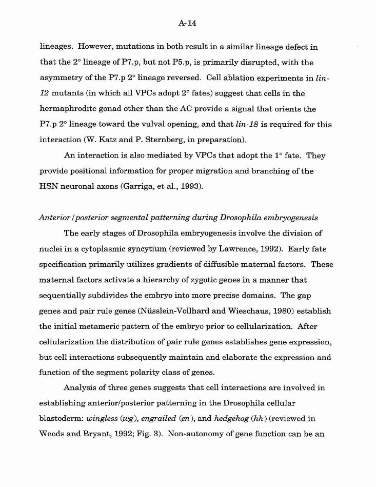
Anterior I posterior segmental patterning during Drosophila embryogenesis
The early stages of Drosophila embryogenesis involve the division of
nuclei in a cytoplasmic syncytium (reviewed by Lawrence, 1992). Early fate
specification primarily utilizes gradients of diffusible maternal factors. These
maternal factors activate a hierarchy of zygotic genes in a manner that
sequentially subdivides the embryo into more precise domains. The gap
genes and pair rule genes (Ni.isslein-Vollhard and Wieschaus, 1980) establish
the initial metameric pattern of the embryo prior to cellularization. After
cellularization the distribution of pair rule genes establishes gene expression,
but cell interactions subsequently maintain and elaborate the expression and
function of the segment polarity class of genes.
Analysis of three genes suggests that cell interactions are involved in
establishing anterior/posterior patterning in the Drosophila cellular
blastoderm: wingless (wg ), engrailed (en), and hedg ehog (hh) (reviewed in
Woods and Bryant, 1992; Fig. 3). Non-autonomy of gene function can be an
A-15
important indicator of the presence of cell interactions involved in fate
specification, and both wg and hh act in a non-autonomous fashion. Mutant
wg tissue in a heterozygous background can develop with a normal
phenotype, indicating that normal wg from adjacent cells can rescue the
mutant phenotype (Baker, 1988). In contrast, hh behaves non-autonomously
in what is termed a "domineering" manner (Mohler, 1988). In other words,
patches of hh mutant cells in a heterozygous background can cause an
abnormal phenotype in both wild type and mutant tissue. wg encodes a
secreted, diffusible protein, and hh encodes a transmembrane protein. en
encodes a homeodomain protein (Poole, et al., 1985), but it may be involved in
specifying the cell interactions mediated by en expressing cells, as well as
additional functions.
At the cellular blastoderm stage, each parasegment comprises a stripe,
four cells in diameter (reviewed by Akam, 1987). wg is expressed in the cells
anterior to each parasegmental border (Baker, 1987), and en and hh are
expressed adjacent in the cells posterior to each parasegmental boundary
(DiNardo and O'Farell, 1987; Mohler and Vani, 1992). This expression
pattern is established by the prior expression of pair rule genes. However,
the proper maintenance of these patterns depends on the reciprocal activity
of each of the genes (Bejsovec and Martinez Arias, 1991; Martinez Arias, et
al., 1988). Specifically, wg and en expression is initiated and then lost in
embryos mutant for the other gene. Likewise, hh expression is initiated and
then lost in en mutants (Mohler and Vani, 1992).
One model for the cell interactions following cellularization (Woods and
Bryant, 1992) proposes that wg is secreted from wg-expressing cells in
vesicles (a process that requires the armadillo protein; Gonzales, et al., 1991),
A-16
and is necessary for the maintenance of en expression. hh acts to promote wg
expression in cells immediately anterior to en expressing cells, possibly via
the gene patched (pte; Ingham, et al., 1991). In pte mutants supernumerary
cells express wg, so the normal interaction may involve negative regulation of
pte activity. Recent studies that use transgenic wg under control of a heat
shock promoter rule out the possibility that wg acts as a morphogen in
establishing fates within each segment (Sampedro, et al., 1993), and
subsequent models ascribe to wg primarily the function of establishing or
"sealing" the parasegmental boundaries (Lawrence and Sampedro, 1993).
Genetic studies of hh using a temperature sensitive allele suggest that hh has
two functions: the initial reciprocal induction function with wg in immediate
anterior cells, and a later function that acts at a distance over several cell
diameters (Heemskerk and DiNardo, 1994). In this second function hh
specifies distinct fates in a dose-dependent manner, and thus may act as a
morphogen. The key function of wg may be in the demarcation and
maintenance ofthe segmental boundary, with the segmental gradient
established by hh.
Specification of segmental identity across germ layers in Drosophila embryos
In addition to subdividing the embryo into repeated segmental units
along the anterior/posterior axis, zygotic genes establish the distinct identity
of each parasegment. The parasegmental identity is established by the genes
of the Antennapedia and Bithorax Complexes (Ant-C and BX-C): the
prototypes of the homeotic complexes in other metazoans (reviewed in Lewis,
1978; Peifer, et al., 1987; McGinnis and Krumlauf, 1992). The expression of
the genes of the Ant-C and BX-C is under the control of earlier acting nuclear
A-17
proteins involved in establishing the metameric pattern, but also maternal
genes that convey relative anterior/posterior position, and interactions among
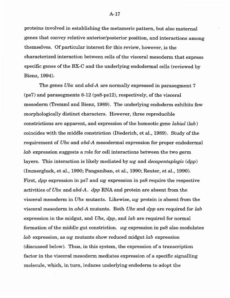
themselves. Of particular interest for this review, however, is the
characterized interaction between cells of the visceral mesoderm that express
specific genes of the BX-C and the underlying endodermal cells (reviewed by
Bienz, 1994).
The genes Ubx and abd-A are normally expressed in parasegment 7
(ps7) and parasegments 8-12 (ps8-ps12), respectively, of the visceral
mesoderm (Tremml and Bienz, 1989). The underlying endoderm exhibits few
morphologically distinct characters. However, three reproducible
constrictions are apparent, and expression of the homeotic gene labial (lab)
coincides with the middle constriction (Diederich, et al., 1989). Study ofthe
requirement of Ubx and abd-A mesodermal expression for proper endodermal
lab expression suggests a role for cell interactions between the two germ
layers. This interaction is likely mediated by wg and decapentaplegic (dpp)
(Immergluck, et al., 1990; Panganiban, et al., 1990; Reuter, et al., 1990).
First, dpp expression in ps7 and wg expression in ps8 require the respective
activities of Ubx and abd-A. dpp RNA and protein are absent from the
visceral mesoderm in Ubx mutants. Likewise, wg protein is absent from the
visceral mesoderm in abd-A mutants. Both Ubx and dpp are required for lab
expression in the midgut, and Ubx, dpp, and lab are required for normal
formation of the middle gut constriction. wg expression in ps8 also modulates
lab expression, as wg mutants show reduced midgut lab expression
(discussed below). Thus, in this system, the expression of a transcription
factor in the visceral mesoderm mediates expression of a specific signalling
molecule, which, in turn, induces underlying endoderm to adopt the
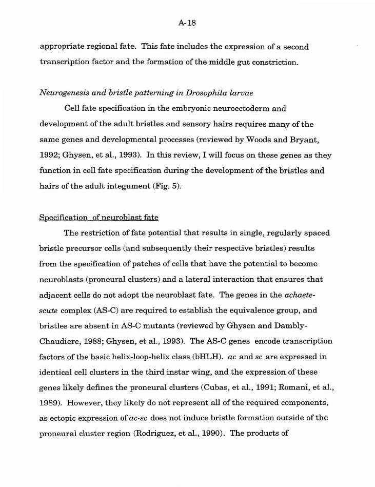
A-18
appropriate regional fate. This fate includes the expression of a second
transcription factor and the formation of the middle gut constriction.
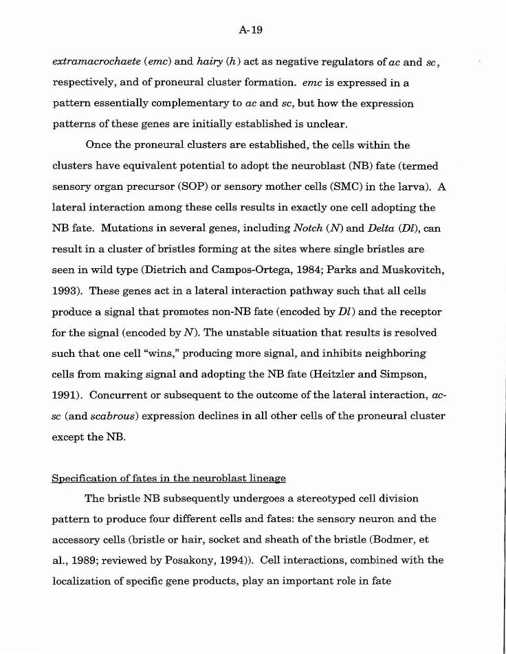
Neurogenesis and bristle patterning in Drosophila larvae
Cell fate specification in the embryonic neuroectoderm and
development of the adult bristles and sensory hairs requires many of the
same genes and developmental processes (reviewed by Woods and Bryant,
1992; Ghysen, et al., 1993). In this review, I will focus on these genes as they
function in cell fate specification during the development of the bristles and
hairs of the adult integument (Fig. 5).
Specification of neuroblast fate
The restriction of fate potential that results in single, regularly spaced
bristle precursor cells (and subsequently their respective bristles) results
from the specification of patches of cells that have the potential to become
neuroblasts (proneural clusters) and a lateral interaction that ensures that
adjacent cells do not adopt the neuroblast fate. The genes in the achaete
scute complex (AS-C) are required to establish the equivalence group, and
bristles are absent in AS-C mutants (reviewed by Ghysen and Dambly
Chaudiere, 1988; Ghysen, et al., 1993). The AS-C genes encode transcription
factors of the basic helix-loop-helix class (bHLH). ac and scare expressed in
identical cell clusters in the third instar wing, and the expression of these
genes likely defines the proneural clusters (Cubas, et al. , 1991; Romani, et al. ,
1989). However, they likely do not represent all of the required components,
a s ectopic expression of ac-sc does not induce bristle formation outside of the
proneural cluster region (Rodriguez, et al., 1990). The products of
A-19
extramacrochaete ( emc) and hairy (h) act as negative regulators of ac and sc,
respectively, and ofproneural cluster formation. emc is expressed in a
pattem essentially complementary to ac and sc, but how the expression
pattems of these genes are initially established is unclear.
Once the proneural clusters are established, the cells within the
clusters have equivalent potential to adopt the neuroblast (NB) fate (termed
sensory organ precursor (SOP) or sensory mother cells (SMC) in the larva). A
lateral interaction among these cells results in exactly one cell adopting the
NB fate. Mutations in several genes, including Notch (N) and Delta (Dl), can
result in a cluster of bristles forming at the sites where single bristles are
seen in wild type (Dietrich and Campos-Ortega, 1984; Parks and Muskovitch,
1993). These genes act in a lateral interaction pathway such that all cells
produce a signal that promotes non-NB fate (encoded by Dl) and the receptor
for the signal (encoded by N). The unstable situation that results is resolved
such that one cell "wins," producing more signal, and inhibits neighboring
cells from making signal and adopting the NB fate (Heitzler and Simpson,
1991). Concurrent or subsequent to the outcome of the lateral interaction, ac
sc (and scabrous) expression declines in all other cells of the proneural cluster
except the NB.
Specification of fates in the neuroblast lineage
The bristle NB subsequently undergoes a stereotyped cell division
pattem to produce four different cells and fates: the sensory neuron and the
accessory cells (bristle or hair, socket and sheath of the bristle (Bodmer, et
al., 1989; reviewed by Posakony, 1994)). Cell interactions, combined with the
localization of specific gene products, play an important role in fate
A-20
specification within this lineage. First, loss of Nor Dl function in the NB
results in bristle loss and causes all four cells to adopt the fate of the sensory
neuron (Hartenstein and Posakony, 1990; Parks and Muskovitch, 1993).
Partial reduction-of-function mutations can result in both daughters of the
NB producing a neuron and a sheath cell, so Nand Dl act at both cell
divisions in the lineage. Three additional genes are required: Hairless (H),
suppressor of Hairless (Su(H) ), and numb. These genes also function at each
division of the lineage to specify fate. Su(H) mutations result in defects
similar to theN and Dl defects (Schweisguth and Posakony, 1992), whereas
loss of H or numb function results in opposite defects (Bang, et al. , 1991;
Uemura, et al., 1989). These genes likely act together to ensure that the two
daughter cells from each division adopt fates different from each other. The
numb protein is localized and asymmetrically distributed during cell division
(Rhyu, et al., 1994), so the execution of the normal lineage involves both
asymmetric distribution of factors and cell interactions. It is not clear
whether the cell interactions mediated by N and Dl mediate the asymmetric
polarization of the precursor cell(s), or ifthe asymmetrically distributed
information is amplified and reinforced by lateral interactions between
siblings.
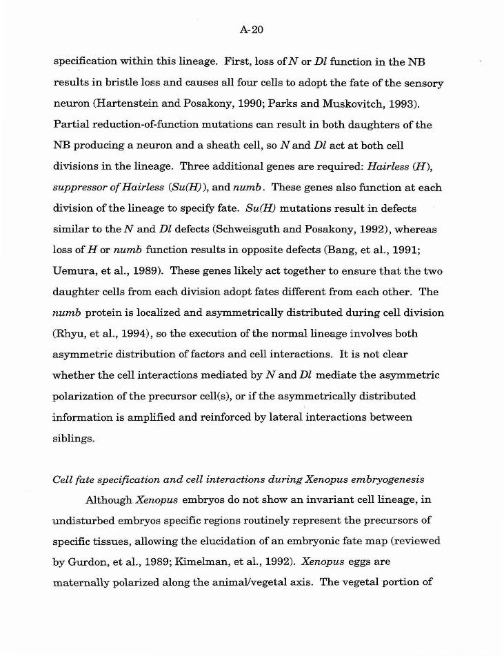
Cell fate specification and cell interactions during Xenopus embryogenesis
Although Xenopus embryos do not show an invariant cell lineage, in
undisturbed embryos specific regions routinely represent the precursors of
specific tissues, allowing the elucidation of an embryonic fate map (reviewed
by Gurdon, et al., 1989; Kimelman, et al., 1992). Xenopus eggs are
maternally polarized along the animal/vegetal axis. The vegetal portion of
A-21
the embryo contributes to the endoderm, the equatorial portion contributes to
the mesoderm, and the animal portion contributes to the ectoderm and
neuroectoderm. There is additional localization of fates as well. For
instance, mesoderm comprises a diverse group of differentiated fates, and
these fates do not arise in a homogeneous manner among the cells at the
equator. The dorsal/ventral axis of the embryo is established orthogonally to
the animal/vegetal axis, with the dorsal region established opposite the site of
sperm entry. I will consider two examples of cell interactions in Xenopus that
have been studied in detail: mesoderm induction and induction of the body
axis by the cells of the blastopore lip (Spemann organizer activity; Fig. 6).
The complexities of the multiple signal integration and the relationship
between the two signals is discussed below.
Mesoderm induction is defined by experiments that combine embryonic
animal caps (usually precursor to neuronal and epidermal fates) with vegetal
tissue (precursor to endodermal cell fates) (e.g., Gurdon, et al., 1985; reviewed
by Kimelman, et al., 1992). The presence of the vegetal cells can induce
mesodermal fates in the animal caps. Thus, the initial animal/vegetal
asymmetry in the oocyte provides the distinction of animal (epidermal) and
vegetal (endodermal) fates, and an inductive interaction promotes
mesodermal fates at the midline. However, there is a distinct difference
between the vegetal cells from the presumptive dorsal region and the cells
from the ventral region in the type of mesodermal fates that can be induced,
so a polarization of the vegetal region must exist. Such polarization could
result from a single signal from the vegetal pole that acts as a morphogen and
is maximally active in the presumptive dorsal region. In contrast, there could
A-22
be two signals: a generalized vegetal signal, and a localized signal that
induces dorsal mesoderm (Slack, et al., 1987; Smith, 1989; Woodland, 1989).
The Spemann organizer was identified in 1924 by the transplantation
of the dorsal blastopore lip from one amphibian into another, where the
transplanted tissue was able to induce a secondary body axis in which cells of
the host make up a large amount of the secondary axis tissue (reviewed in
Cooke, 1989; Kimelman, et al., 1992). Thus cell signalling mediates
establishment of the dorsal/ventral and anterior/posterior axes.
Recent work has identified some of the molecules that mediate these
cell interactions. The key molecules include the FGF class of molecules
(bFGF, aFGF, XeFGF), the TGF-13 class (activin, XTC-MIF), and Wnt-1 class
(Xwnt-8, noggin). The function ofthese molecules has been primarily
established by treating tissues with purified protein, so the nature of the
specific endogenous gene that mediates the signal may not yet be defined.
Nevertheless, the activity associated with the purified molecules indicates
that they are likely interacting with an endogenous signalling mechanism.
A basic FGF (bFGF) class of molecule is likely responsible for the
generalized mesoderm inducing function of vegetal cells. Addition of bFGF to
cultured animal caps can promote differentiation of ventral mesodermal
tissues (Kimelman and Kirschner, 1987; Slack, et al., 1987). However,
concentration ofbFGF may also play a role in fate specification, as animal
caps with injected bFGF RNA can form some dorsal mesodermal tissues
(Kimelman and Maas, 1992). The in vivo function of bFGF molecules is
corroborated by the observation that bFGF transcripts and proteins are
present in the Xenopus egg and embryo. In addition, a dominant negative
mutant form of the bFGF receptor can disrupt normal development,
A-23
producing tadpoles that lack posterior and trunk regions, and reduced
amounts of certain mesodermal tissue (Amaya, et al., 1991).
Activin is a member of the TGF-j3 class of proteins, and is a potent
inducer of mesoderm. Addition of activin to cultured animal caps can induce
both dorsal and ventral mesodermal tissues in a dose-dependent manner
(Green, et al., 1990). The function of the dorsal mesoderm to act as the
Spemann organizer can also be induced in animal caps by activin. Animal
caps cultured with activin can organize a secondary axis when transplanted
into host embryos (Cooke, 1989).
wnt proteins in Xenopus do not serve as mesodermal inducing agents,
but rather as modulators involved in both the induction of dorsal mesodermal
fates and in the conveyance of body axis information. Injection of wnt RNA
into oocytes or cells on the ventral side of cleavage stage embryos can induce
a secondary body axis (McMahon and Moon, 1989). wnt also acts in concert
with factors that induce mesoderm to change the responsiveness of animal
cap cells (Christian, et al., 1992). wnt alone can not induce mesodermal
differentiation. However, in the presence of wnt animal caps can respond to
bFGF by producing dorsal mesodermal tissues. In contrast, animal caps
without wnt treated to the same concentration ofbFGF produce mainly
ventral mesodermal tissues. noggin is not a wnt gene, but it may represent
the endogenous wnt activity. It is present both maternally and zygotically,
and zygotic transcription is localized to the dorsal mesodermal region (Smith
and Harland, 1992). It functions both to promote dorsal fates and as a
Spemann organizer (Smith and Harland, 1992; Smith, et al., 1993).
A-24
Fate specification and the integration of multiple signals
For two cells in which fate specification is mediated by cell
interactions, a single positional cue may be sufficient to make one cell
different from the other. However, study in several systems suggests that
cells are exposed to a number of extracellular cues that are integrated
concurrently to specify fate. Thus, the study of cell interactions during
development is not only the study of how a cell responds to a single signal,
but how it responds to a variety of signals, integrating them to produce a
specific outcome.
C. elegans vulval development
The primary cell interaction of vulval development is the AC signal
mediated by the lin-3 I let-23 signalling pathway. As suggested above,
however, this interaction is not the only event involved in proper pattern
formation during vulval development. There are at least two other
components -- the negative regulation mediated by a group of genes including
the lin-15 locus and the lateral interaction mediated by lin-12 --that also
play important roles (see Fig. 2). Since these three components of pattern
formation coordinate to produce the normal pattern of fates it is of interest to
understand how the responding cells integrate the three positional cues.
Loss-of-function (including molecular null) mutations in lin-15 result
in a Muv phenotype (Ferguson and Horvitz, 1985, 1989; Huang, et al., 1994).
Genetic analysis suggested that lin-15 is a complex locus with two
independently mutable activities, A and B. Mosaic analysis suggests that
normallin-15 activity in either the VPCs or the AC is not sufficient for the
A-25
wild type phenotype (i.e., lin-15 act in a cell non-autonomous fashion;
Herman and Hedgecock, 1990). lin-15 has been proposed to act in the
hypodermis surrounding the VPCs, hyp7. Molecular analysis indicates that
the two functions of lin-15 correspond to two distinct but coordinately
transcribed transcriptional units. lin-15 is one member of a group of genes
that also reflect these two activities. Animals bearing a mutation in either a
class A or a class B gene are phenotypically wild type. Animals homozygous
mutant for both a class A and a class B gene are Muv. Genetic epistasis
experiments suggest that lin-15 acts in parallel and antagonistically to lin-3
to negatively regulate let-23 . lin-3; lin-15 double mutants are Muv,
indicating that lin-3 is not necessary for the effect observed in lin-15 mutants
(i.e., lin-15 does not act by regulating or localizing lin-3 activity). let-23; lin-
15 double mutants are Vul, indicating that lin-15 acts to regulate the activity
of let-23. Transgenic extrachromasomal arrays of lin-3 that behave as a gain
of lin-3 function are similarly blocked by mutation in let-23 (Hill and
Sternberg, 1992). The effects ofboth lin-3 and lin-15 are thus integrated into
the same pathway at the let-23 receptor. lin-15 A and B proteins likely act,
either directly or indirectly, to negatively regulate let-23 receptor activity in
the absence of the specific, localized signal.
The integration of the AC signal and the lateral interaction is less
straightforward. However, the two pathways act coordinately to specify fate,
rather than one pathway directly regulating the other as in the lin-3 I let-23
pathway and lin-15. In lin-12(0) mutants 2° vulval fates are absent, but the
VPCs are still responsive to the AC signal (lin-3) (Greenwald, et al., 1983).
Thus lin-12 is not necessary for the function of the AC signal, and when both
pathways are not functional all cells adopt the 3° fate. Lateral interaction is
A-26
not necessarily essential for 2° fates, as 2° fates can be recovered from
isolated VPCs with the AC present (Sternberg and Horvitz, 1986) or following
a brief pulse of lin-3 from a heat shock promoter (Hill, et al., in preparation).
In lin-12(d) mutants all six VPCs adopt the 2° fate, even in the absence of the
AC (which is generally absent in lin-12(d) animals because of the earlier
ACNU decision). Thus the activated lin-12 is sufficient to bypass the
requirement for the AC signal to promote vulval fates. Nevertheless, in rare
lin-12(d) /lin-12(0) animals that have an AC, the cells are still responsive the
AC signal, and P6.p will produce a 1 o lineage (Stemberg and Horvitz, 1989).
Transgenic overexpression of lin-3 in otherwise normal animals can also
override the normal lateral interaction and promote adjacent 1 o cells (R. Hill,
pers. comm.). Finally, in experiments where both pathways are artificially
activated (overexpression of lin-3 in lin-12(d) I lin-12(0) animals), adjacent 1°
fates are possible, although at a much lower frequency than when lin-12 is
not mutant (R. Hill, pers. comm). Although in experimental conditions both
pathways can dominate in the formation of vulval fates, the two pathways
may normally act in parallel to result in the reproducible pattem of fates
required for vulval development. In the normal dose of AC signal, lin-12 may
play an important role in amplifying the differences between the proximal
P6. p cell and the more distal cells.
Drosophila endoderm induction
The expression of Ubx and dpp in the visceral mesoderm during
Drosophila development is required for normal endo~ermal development,
including expression of lab in the midgut. However, the spatial regulation of
expression is complex (see Fig. 4). wg expressed in ps8 of the visceral
A-27
mesoderm functions to modulate lab expression. In wg mutants the gradient
of lab expression, with the highest concentration near the posterior, is
reduced (Immergluck, et al., 1990). Ubx and dpp are also required for normal
wg expression. Thus, in this system, signals act sequentially (dpp -> wg) and
in parallel (dpp/wg ->lab) to induce regional specification. Signalling
feedback is also involved in producing the normal gene expression pattern.
For instance, both dpp and wg are required for normal Ubx expression
(Panganiban, et al., 1990). They also mediate feedback to reinforce the
normal pattern of gene expression in the signalling cells.
Xenopus mesoderm induction and body axis formation
Traditional models of mesoderm induction and body axis formation
suggest that functionally distinct processes and molecules mediate each step
(see Fig. 6.A). However, the complex developmental functions associated
with recently characterized molecules suggest that (1) although the two
processes are temporally distinct, they are likely not functionally
independent, and may use many of the same molecules, and (2) multiple
molecules may act in concert to mediate mesoderm induction and body axis
formation, and production of a variety of differentiated fates (Kimelman, et
al., 1992; see Fig. 6.B). The synergistic properties of wnt and bFGF in the
promotion of mesoderm suggests that these two signals may act in parallel to
promote mesodermal fates, with wnt activity localized to the presumptive
dorsal region of the embryo. An additional signal (''X", possibly mediated by
an activin) may also act in parallel in a partially redundant way with bFGF
because bFGF is neither necessary nor sufficient for normal anterior
structures such as head structures (Amaya, et al., 1991; Christian, et al.,
A-28
1992). One component of the model is that this activity normally functions
together with wnt. Recent work suggests that these signals may indeed
require synergistic function, as a dominant negative FGF receptor blocks full
activity of activin in promotion of mesodermal fates (Cornell and K.imelman,
1994; however, this experiment does not rule out that FGF and activin
pathways may act in series). Thus two generalized signals acting in concert
with a localized wnt activity induce mesoderm, as well as specify both relative
dorsal/ventral and anterior/posterior fate based on the concentration of wnt
activity. Head specification occurs at the domains of highest wnt activity
combined with "activin X" activity, trunk specification at domains with
moderate wnt and bFGF activity, and tail specification at domains with only
bFGF activity.
An additional cell interaction occurs during mesoderm induction
among the presumptive mesodermal cells. This positive lateral interaction
(termed the community effect) is required for specification of mesodermal
fates, and may act in parallel to the mesodermal inducing signals (Gurdon, et
al., 1984; Gurdon, 1988; Gurdon, et al., 1993). The community effect likely
functions in mesodermal cells to create homogeneity within cell populations,
but may also demarcate subpopulations of cells within a larger group. In this
function the community effect acts like the lateral interaction in vulval
development: it allows cells that detect subtle differences in the activity of
other signals to amplify those differences resulting in a discontinuity between
cell or tissue types.
A-29
Tissue culture
Most of the molecules proposed to act in providing parallel signal
information are often thought to function directly as signals in the traditional
sense. However, models that suggest that some of the key players may
actually function in more mundane cell biological roles (e.g., the proposed
function ofwg to "seal" the parasegmental boundaries during embryogenesis;
Lawrence and Sampedro, 1993) may be more accurate. Such molecules, while
perhaps less exciting, are no less important in cell interactions, especially as
modulators of primary signalling factors. Indeed, it is clear that the
extracellular matrix can play a critical role in what factors a cell is exposed
to, how the cell perceives those factors, and how it is capable of responding
(reviewed in Adams and Watt, 1993). As one example, embryonal carcinoma
(EC) cells grown on laminin or fibronectin layers respond to activin A or
bFGF mitogenically, whereas EC cells grown on plastic require such factors
for viability, but do not respond with cell division (Schubert and Kimura,
1991).
Developing cells are exposed to a variety of extracellular cues, and the
autonomous components ofthose cells (e.g., presence ofreceptors) allow the
specific response of the cells to these cues. Signals in the traditional sense
(e .g., diffusible factors that induce different fates) make up some of these
cues. However, as work continues in the direction ofhow cells integrate
multiple signals, the role of extracellular matrix molecules in modulating cell
interactions in vivo may become more apparent.
A-30
II. The C. elegans male tail as a model system to study specific
developmental questions
Male tail development provides a reproducible background in which to
study cell fate specification. The male specific blast cells, particularly the B
cell, produce fairly complex lineages, likely requiring several fate
specification steps. These lineages thus offer a good starting point to study
the stepwise specification of fate and the interplay of different mechanisms
important in development of multicellular organisms. Furthermore, my
experiments indicate (Chapter 2) that multiple cell interactions are
important for proper development of the B.a(llr )xx cells. Thus, this lineage is
also useful to study the integration and coordination of multiple signals
involved in fate specification. In these studies I have used the experimental
tools of cell ablation and genetics, coupled with direct observation of cell
lineage as an assay of fate. In this section I review the development of the
male tail as an introduction to the experimental system.
C. elegans as a system to study developmental biology
C. elegans provides a useful system for genetic analysis and the direct
and precise analysis of development. The ability to visualize cell nuclei and
their divisions, combined with the reproducibility of the lineage among
animals, has allowed the complete description of the normal cell division
pattern and the differentiated fates of all somatic cells (Sulston and Horvitz,
1977; Sulston, et al. , 1980; 1983). The cell lineage in C. elegans is essentially
invariant. However, because the extracellular environment for most
developing cells is as constant as the ancestry, the correlation of cell division
A-31
and fate does not necessarily indicate that fate specification is autonomous.
Although asymmetric cell divisions and segregated factors likely are
important for some cell divisions (e.g. , the first division of the zygote (Strome,
1983) ; the first division of the male B cell (Sternberg and Horvitz, 1988)), cell
interactions also play an important role in cell fate specification. For
example, the normal lineage includes several cells that have the potential to
adopt more than one fate (Sulston and Horvitz, 1977; Kimble and Hirsh,
1979; Sulston, et al., 1983). In addition, embryonic blast cell rearrangement
(Priess and Thomson, 1987; Wood, 1991) and isolation experiments
(Schierenberg, 1987; Goldstein, 1992; 1993) as well as embryonic (Schnabel,
1991; 1994; Bowerman, et al. , 1992) and postembryonic cell ablation
experiments (Sulston and White, 1980; Kimble, 1981; Chisholm and Hodgkin,
1989) have identified instances of cell interactions that specify cell fate. The
invariant cell lineage, rather than indicating a limited repertoire of fate
specification mechanisms, offers a reproducible background to study the
interplay of autonomous and conditional mechanisms.
Cell lineage as a marker for cell fate
In C. elegans the fate of precursor cells includes the cell lineage
(number, timing and axes of divisions) and the terminal differentiated fates
of the progeny produced by those divisions. Although both criteria are
important to understanding the fate of a precursor cell, for both cell ablation
and genetic studies I have focused on the former. In other analyses of
development in C. elegans , cell lineage has proven to be an accurate indicator
of fate choice (e.g., Horvitz, et al. , 1983; Kenyon, 1986; Sternberg and Horvitz,
1986; Schnabel, 1991; Bowerman, et al. , 1992).
A-32
Review of the anatomy and normal development of the C. elegans
male tail
At hatching, male and hermaphrodite C. elegans are morphologically
similar. Sexual dimorphism develops postembryonically as different blast
cells divide following sex-specific lineages (Sulston and Horvitz, 1977). The
C. elegans male tail consists of several structures (description and data of
Sulston and Horvitz, 1977 and Sulston, et al. , 1980): the fan, the hook, the
postcloacal sensilla, and the spicules (Fig. 7).
The fan comprises eighteen bilaterally symmetric ray sensilla
laminated between two layers of cuticle. The cells of the rays derive from
sex-specific divisions during the development of the lateral hypodermis cells
V5, V6, and T.
The hook and the associated sensillum lie directly anterior to the
cloacal opening. The cells of the hook sensillum derive from sex-specific
divisions during the development of the ventral hypodermal cells P10 and
P11.
The postcloacal sensilla are paired structures that reside posterior to
the cloaca. They are not associated with a morphologically distinct structure
as is the hook sensillum. The cells of the postcloacal sensilla derive primarily
from Y, a male specific blast cell (Fig. 8). In hermaphrodites, the cell that is
the male Y cell differentiates as a neuron (PDA). In males, the cell divides
asymmetrically, producing an anterior daughter (Y.a) that differentiates as a
neuron as the mother cell does in hermaphrodites, and a posterior daughter
(Y.p) that is the precursor to ten cells of the postcloacal sensilla. Other male
A-33
specific blast cells are F and U that produce several male-specific
interneurons, and B (discussed below). In general, the male-specific blast cell
lineages include one progeny that functions similar to the mother cell in
hermaphrodites, and several other male-specific neurons and hypodermal
cells. The work in this thesis focuses primarily on the development of the
male-specific blast cells.
The two spicules lie within the cloaca. The spicules are sensilla; each
includes two neurons with processes that run the length of the spicule to the
tip where they are open to the environment. The other cells ofthe spicules
are socket and sheath cells that provide structure and support. The sheath
cells secrete a specialized cuticle that is harder and more refractile than the
body cuticle. The spicules are attached proximally to protractor and retractor
sex muscles. During mating, after the male locates the hermaphrodite vulva,
the spicules are inserted into the vulval opening. The structure of the
spicules helps to anchor the male cloaca at the vulval opening, and the
neurons of the spicules coordinate sperm transfer (K. Liu and P. W.
Sternberg, in preparation).
All of the cells of the spicules derive from the male B cell (Fig. 8). In
hermaphrodites this cell is part of the dorsal rectal epithelium, and progeny
ofB.p retain this function during male postembryonic development. B.a
divides to produce the cells of the spicules, as well as some proctodea! cells,
two neurons of the postcloacal sensilla, and four cell deaths. The B cell
lineage consists of three different stages: early divisions, migration, and late
divisions. The early divisions take place primarily in the second larval stage
(L2), and result in ten progeny. A short-range migration of the eight B.a
progeny occurs in the late L2 stage. The late round of divisions begins in the
A-34
mid-L3 stage. The first division ofB is asymmetric and along an
approximately anterior/posterior axis. The larger anterior cell (B.a) is the
precursor of all cells of the spicules. B.a divides along a left/right axis,
establishing the bilateral symmetry of the spicules. This symmetry is broken
(during migration) and then re-established (during the late divisions) in the
progeny ofthese cells. B.al and B.ar each divide twice to produce a ring of
eight cells, one cell thick. These cells then migrate to form a ring of four cells,
two cells thick. The medial migration of the two dorsal (pp) and ventral (aa)
cells exhibits natural variation. In both cases, either the right or the left cell
can adopt the more anterior position, while the other cell adopts the more
posterior position. The cell that adopts the anterior position will produce a
different lineage than the cell that adopts the posterior position. The other
four B.a progeny migrate invariantly. The ap cells adopt the anterior
position (one on the left, one on the right), and the pa cells adopt the
posterior position.
Cell interactions during the development of the male-specific blast
cells
The variability of position and fate of specific aa and pp cells
suggested that each pair represents an equivalence group. Sulston and
White (1980) carried out a series of ablation experiments to better
understand the interactions involved in fate specification of t hese pairs.
Their experiments showed that following ablation of either the left or the
right aa cell, the remaining aa cell will migrate to the midline and produce
an a lineage. This establishes a fate as primary (1 °) and B fate as secondary
A-35
(2°). Even if the targeted cell is migrating to the anterior position when it is
ablated, the remaining cell will produce an a lineage. Similar experiments
with the pp cells could not establish 1° or 2° fate in the y/8 pair. However,
the variability of the B lineage from animal to animal, combined with these
preliminary cell ablation experiments, suggested that cell interactions may
play a role in establishing the fate of these cells.
Additional information about cell interactions during development of
the male-specific blast cells comes from the work of Chisholm and Hodgkin
(1989) . Work with the gene mab-9 (see below) suggested a possible
interaction between B and the F and U cells. Indeed, ablation of the B cell
prior to division resulted in disruption ofF and possibly U lineages.
Specifically, the asymmetric divisions ofF (such as the division: ofF.(l/r))
were reversed about 50% of the time, i.e., their orientation became random.
The data for U are less conclusive as the U lineage only shows evidence of
asymmetry in some animals. Thus the B cell, or its progeny, provide a
positional cue to the F and U cells that orients the asymmetric cell divisions.
Genetic analysis of the development of the male-specific blast cells
Genes that play roles in the development of the male-specific blast cells
have been identified both directly, by identifying mutations that disrupt male
tail development, and indirectly, by characterizing pleiotropic effects of
mutations disrupting fate specification in other tissues. Most of the genes
analyzed to date play a role in the development of the B cell (summarized in
Fig. 10). None of the genes functions in a truly sex-specific manner; rather,
their function in male tail development reflects their role in a developmental
A-36
process that is sex-specific. Although only a few genes have so far been
identified, they provide a glimpse of the genetic complexity in the
development of the male-specific blast cells.
Specification of blast cells
mab-9 (male abnormal)
Mutations in mab-9 were identified on the basis of the abnormal male
tail phenotype they confer (Hodgkin, 1983). Lineage analysis identified
several defects in mutant males: F, U, and B lineages were disrupted
(Chisholm and Hodgkin, 1989). The B lineage defect is consistent with the
interpretation that B is transformed to Y fate. The F and U lineage defects
reflect the disruption of the B cell, but also may indicate a function of mab-9
that makes F different from U. Analysis of mab-9 hermaphrodites indicated
that the hermaphrodite B cell also adopts a fate like the hermaphrodite Y
cell. Thus the function of mab-9 is to specify B fate and make it different
from Y. Its function is not sex-specific, but the sex-specific differences in
development make the defect more obvious in males than in hermaphrodites.
Since the B and Y cells are neither sisters nor lineal homologues, the reason
the function of only a single gene is necessary to distinguish them is not
straightforward. However, they are neighbors, so mab-9 may play a role in
establishing regional differences.
e{Jl-5 (egg-laying defective)
One gene that likely plays a role in the regional specification of fate is
egl-5. egl-5 is part of the C. elegans homeotic complex (Kenyon and Wang,
1991). Mutations in egl-5 disrupt the fate of many cells in the tail region of
the worm, including B (Chisholm, 1991). However, the B lineage defect in
A-37
egl-5 males does not appear to be a transformation of one fate to another, and
thus the specific function of egl-5 in the B lineage is unclear.
lin-12
One of the functions ofthe gene lin-12 is to specify the fate ofY, and
make it different from its lineal homolog, the neuron DA9. The function of
lin-12 is discussed further below.
Establishing the first asymmetric cell division of B
lin-17
lin-17 was originally identified for a subtle defect in vulval
development (Ferguson and Horvitz, 1985; Ferguson, et al. , 1987). However,
further analysis revealed that lin-17 functions in multiple postembryonic
lineages (Sternberg and Horvitz, 1988). The common theme among the lin-17
defects is that specific asymmetric divisions become symmetric. In the male
tail, lin-17 is required for proper development ofT, P10/11, and B. Normally,
the initial division ofB is asymmetric. Not only does B.a produce a different
cell lineage from B.p, but the B.a cell is larger than the B.p cell. In lin-17
mutants the B.a and B.p cells are generally equal in size, and both cells
produce an early lineage that is similar to the normal B.a lineage.
lin-44
Like mab-9, mutations in lin-44 were identified on the basis of male
tail defects (Herman and Horvitz, 1994). lin-44 is required for the
asymmetric division ofthe BandT cells. Unlike lin-17, mutations in lin-44
do not result in symmetric cell divisions, but rather a reversed polarity of the
asymmetric division. For example, normally the nucleus of B.a is always
larger than B.p. However, in lin-44 mutants, B.a was larger in only 20% of
A-38
animals scored, the cells were equal in size in 8% of animals, and B.p was
larger than B.a in 71% of animals (Herman and Horvitz, 1994). lin-17lin-44
double mutants display the lin-17 phenotype, suggesting that lin-17 is
necessary to establish the asymmetric division, while lin-44 is necessary to
orient the division.
Fate specification in the later lineage
lin-12
Mutations in lin-12 disrupt fate specification in a variety of
postembryonic lineages (Greenwald, et al., 1983). The two classes of
mutations in lin-12 --gain-of-function (lin-12(d)) and loss-of-function (lin-
12(0)) -- result in opposite phenotypes. In general, if the normal fates of two
cells are A and B, in lin-12(d) mutants both cells adopt the A fate, whereas in
lin-12(0) mutants both cells adopt the B fate. In several cases the two cells
involved are known to interact to ensure that each cell adopts a fate different
from the other. Thus lin-12 may mediate a lateral interaction (lateral
inhibition). The molecular similarity between lin-12 and other genes
involved in cell interactions (e.g., glp-1 in C. elegans and Notch in Drosophila)
is consistent with the function of lin-12 in cell interactions. In one lin-12-
mediated fate specification process (an interaction between two cells in the
hermaphrodite gonad that results in a choice between anchor cell fate and a
ventral uterine fate (the ACNU decision)) mosaic analysis indicates that lin-
12 acts in a cell-autonomous manner (Seydoux and Greenwald, 1989).
Mutations in lin-12 disrupt the B and Y lineages in males. In lin-12( d)
mutants there are two Y-like cells, whereas in lin-12(0) mutants there are no
Y cells. lin-12 likely mediates precursor fate specification between the Y cell
A-39
and its lineal homologue neuron DA9. However, there is no evidence that
these two cells interact to specify fate, as cell ablation experiments have not
uncovered any fate regulation for these cells (Greenwald, et al. , 1983;
Sulston, et al., 1983). In the B lineage lin-12 plays two roles: distinguishing
B.pa from B.pp and o from"{ in the pp pair. In lin-12(d) animals, both B.pa
and B.pp produce a lineage like the normal B.pa cell; in lin-12(0) animals
both cells produce a lineage like the normal B. pp cell. In contrast, lin-12( d)
mutants are normal in the B.a lineage. Only in lin-12(0) mutants do both pp
cells produce a "{lineage rather than an anterior "{ lineage and a posterior o lineage. Although this exception was originally thought to be due to the
possibility that the lin-12(d) mutation was not strong enough to produce the
opposite transformation, my results (see Chapter 3 ) suggest that this
exception is due to additional cell interactions. Nevertheless, the lineage
defect in lin-12(0) mutants also hints at the possible cell interactions in B
lineage specification.
III. Overview of the thesis
My work has centered on three issues: (1) the cell interactions that
establish fate among the eight intermediate B.a progeny, (2) fate specification
in the unequal cell divisions of both the B cell and specific cell divisions late
in the lineage, and (3) the identification and initial characterization of new
genes that may function at additional steps in the lineage. In Chapter 2, I
describe cell ablation experiments that identify five distinct cell interactions
required for proper fate specification in the eight intermediate B.a progeny.
Experiments in which multiple signalling sources are removed together also
A-40
address how these positional cues are integrated. In Chapter 3 I describe
experiments that indicate that the genes in the lin-3 I let-23 signalling
pathway mediate one of these positional cues from F and U. I extend the
experiments of Greenwald, et al. (1983) in analyzing the function of lin-12 in
lateral interactions between cells of equivalent potential. I also extend the
signal integration experiments of Chapter 2 with double mutant and cell
ablation experiments that address the integration of both lin-15 and the Y.p
positional cue into the lin-3 I let-23 signalling pathway. In Chapter 4, I
describe a genetic screen for mutants with abnormal male tail development.
This screen has identified additional genes that play a role in fate
specification in the B lineage. In Chapter 5 I describe experiments that
characterize the function of one of these genes, vab-3. vab-3 represents a
candidate factor distributed asymmetrically at the initial division of B that is
necessary for B .a fates. Experiments also identify its interaction with lin-17
at the first division ofB, and identify additional functions for lin-17 in
specific other asymmetric cell divisions in the B lineage.
A-41
Figures
Figure 1. Fate specification in C. elegans early embryogenesis. A. Relative
positions of blast cells at the four cell stage. B. Early embryonic lineage.
Cell interactions and genes that mediate specific steps of fate specification
are indicated. The par genes mediate the initial asymmetric cell division of
the zygote. mex-1 and pie-1 act sequentially to localize skn-1 activity to EMS.
Multiple cell interactions also occur among the early blast cells to specify fate
(see text).
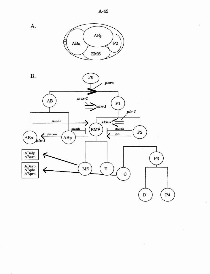
A-42
A.
B. pars
mex-1
~ "----ABarp ABpla ABpra
'\.. ~skn-1
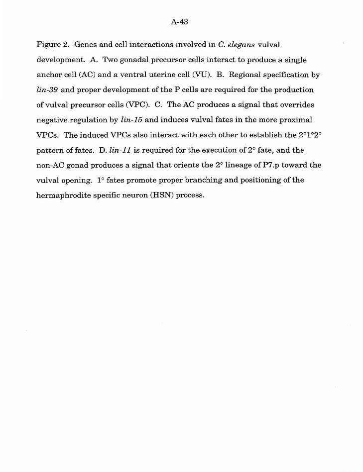
A-43
Figure 2. Genes and cell interactions involved in C. elegans vulval
development. A. Two gonadal precursor cells interact to produce a single
anchor cell (AC) and a ventral uterine cell (VU). B. Regional specification by
lin-39 and proper development of the P cells are required for the production
of vulval precursor cells (VPC). C. The AC produces a signal that overrides
negative regulation by lin-15 and induces vulval fates in the more proximal
VPCs. The induced VPCs also interact with each other to establish the 2°1°2°
pattern offates. D.lin-11 is required for the execution of2° fate, and the
non-AC gonad produces a signal that orients the 2° lineage of P7. p toward the
vulval opening. 1 o fates promote proper branching and positioning of the
hermaphrodite specific neuron (HSN) process.
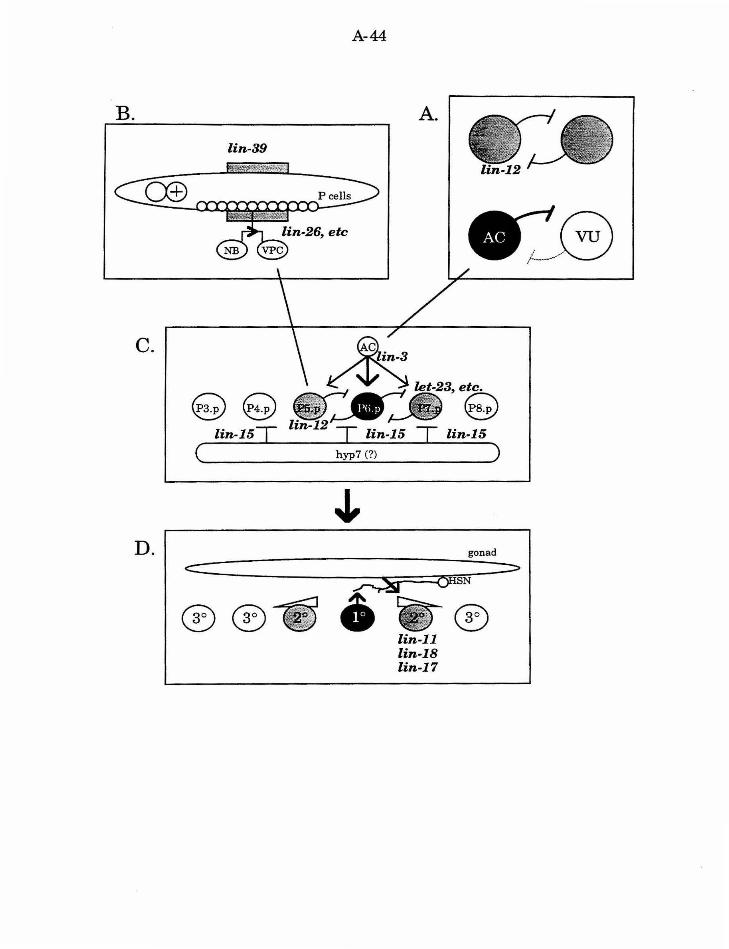
B.
c.
D.
A-44
A. lin-39
...,._ .. ..,..~- etc.
~<9 ~ lin-15T lin-15
c~------------~h~~-7~(?~) ----------~)
®®
gonad
6 {)HSN
lin-11 lin-18 lin-17
®
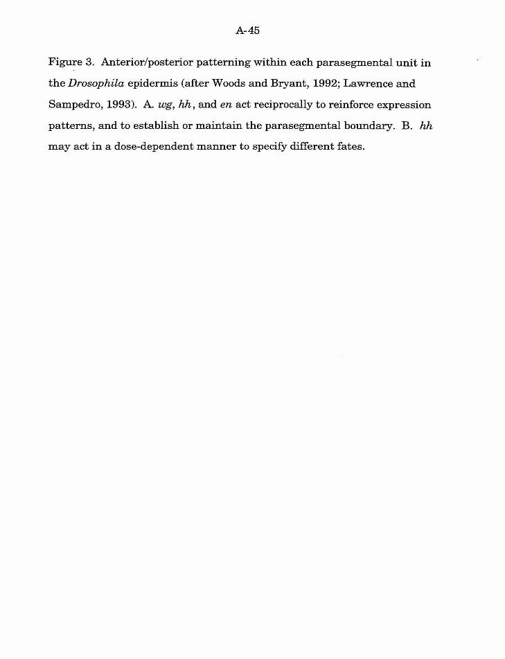
A-45
Figure 3. Anterior/posterior patterning within each parasegmental unit in
the Drosophila epidermis (after Woods and Bryant, 1992; Lawrence and
Sampedro, 1993). A. wg, hh, and en act reciprocally to reinforce expression
patterns, and to establish or maintain the parasegmental boundary. B. hh
may act in a dose-dependent manner to specify different fates.
A. anterior
A-46
parasegmental boundary
posterior
A-47
Figure 4. Regional specification of the Drosophila endoderm: the interaction
between the visceral mesoderm and the midgut (after Bienz, 1994). Ubx
expression is required for dpp expression, and abd-A expression is required
for wg expression in specific regions of the visceral mesoderm. dpp and wg
are also necessary for proper expression of lab in the midgut, and the middle
gut constriction. The pattern of expression of these genes is reinforced and
maintained by an elaborate series of feedback interactions.
E ~ 0 00 Cl)
s
A-48
A-49
Figure 5. Patterning of sensory organ precursors and the production of
bristle sensilla in the Drosophila epidermis (after Ghysen, et al., 1993;
Posakony, 1994). A. The expression of ac and sc (and their regulators)
establish the proneural cluster, a group of cells competent to adopt the
neuroblast (NB) fate. B. A lateral interaction among cells within the
proneural cluster ensures that only one cell adopts the NB fate. C. The NB
(or sensory organ precursor, SOP) executes a stereotyped lineage to produce
the four fates of the sensory organ: the socket (So), the shaft (bristle, Br), the
sheath (Sh), and the sensory neuron (N). Both cell interactions and localized
factors play a role in fate specification in this lineage.
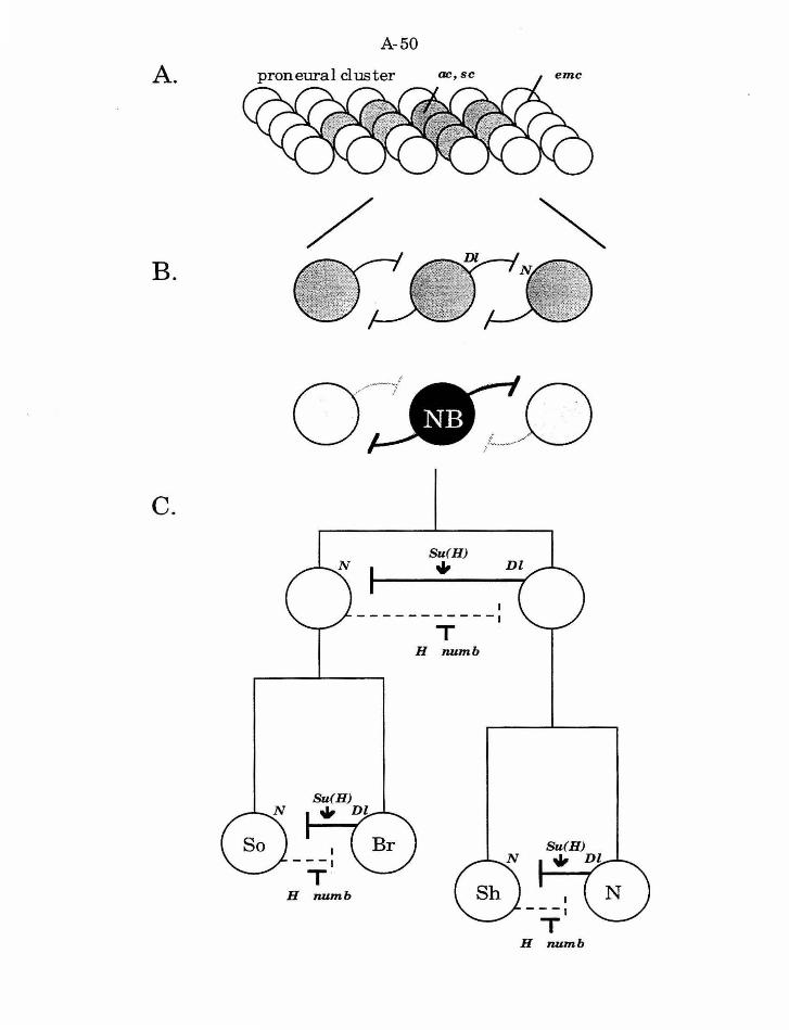
A.
B.
C.
A-50
/ i
0~---/
I ---1
T H 1Wmb
Su(H)
• I -----------1
T H 1Wmb
I ---1
T H 1ULmb
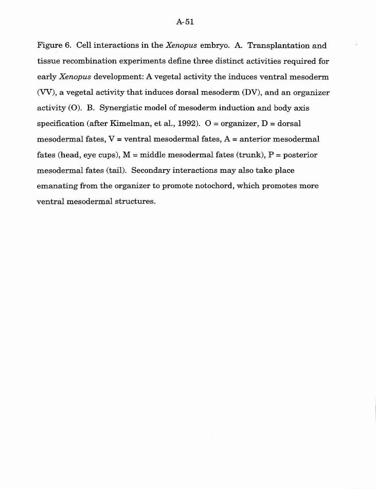
A-51
Figure 6. Cell interactions in the Xenopus embryo. A. Transplantation and
tissue recombination experiments define three distinct activities required for
early Xenopus development: A vegetal activity the induces ventral mesoderm
(VV), a vegetal activity that induces dorsal mesoderm (DV), and an organizer
activity (0). B. Synergistic model of mesoderm induction and body axis
specification (after Kimelman, et al., 1992). 0 =organizer, D =dorsal
mesodermal fates, V = ventral mesodermal fates, A = anterior mesodermal
fates (head, eye cups), M =middle mesodermal fates (trunk), P =posterior
mesodermal fates (tail). Secondary interactions may also take place
emanating from the organizer to promote notochord, which promotes more
ventral mesodermal structures.
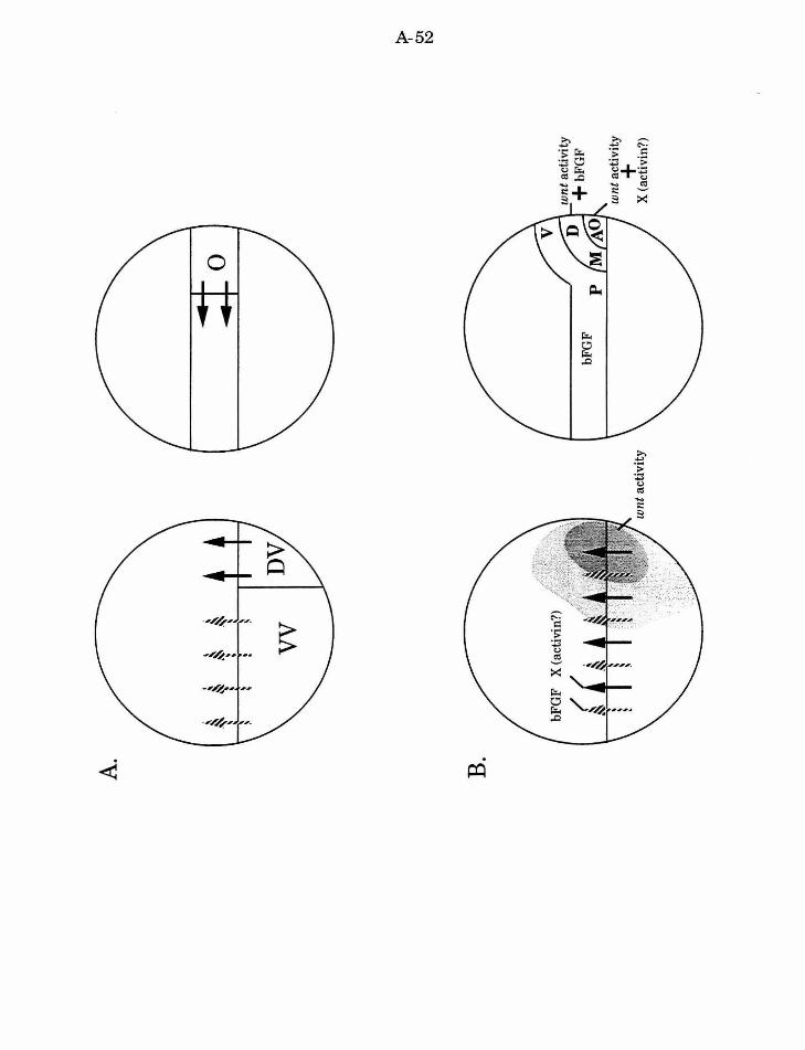
A.
. '
~ ~
·~
~ ~
~
~ ~
;::.
·~
~ ~
~ ~
B.
wnt
act
ivit
y
wn
t act
ivit
y +
bFG
F
wnt
act
ivit
y
+
X (a
ctiv
in?)
~
Ol
tv
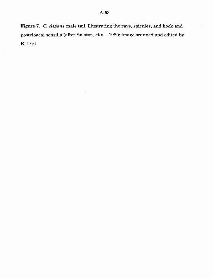
A-53
Figure 7. C. elegans male tail, illustrating the rays, spicules, and hook and
postcloacal sensilla (after Sulston, et al., 1980; image scanned and edited by
K. Liu).
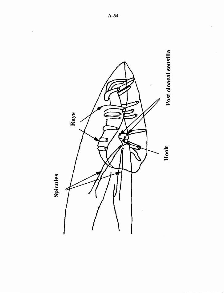
A-54
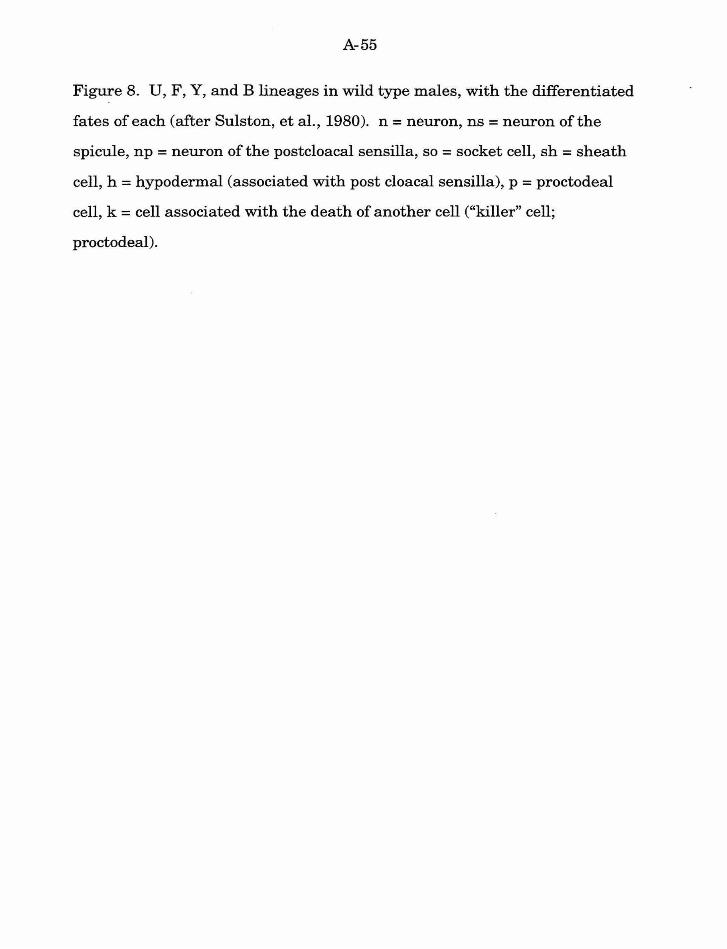
A-55
Figure 8. U, F, Y, and B lineages in wild type males, with the differentiated
fates of each (after Sulston, et al., 1980). n =neuron, ns =neuron of the
spicule, np = neuron of the postcloacal sensilla, so = socket cell, sh = sheath
cell, h =hypodermal (associated with post cloacal sensilla), p =proctodea!
cell, k = cell associated with the death of another cell ("killer" cell;
proctodea!).
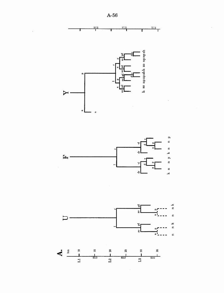
A.
hrs
u
F
y
10
p
15
n
20
I I
I ~
01
O
':l
l ~~
L
a av
pd
av
pd
25
av
pd
dr
vi
dr
vi
30
h so
n
pn
psh
h s
o n
pn
psh
la.l
~ 8
p 35
1
-,
I I
I I
I I
I I
. .
. .
k n
n P
k n
n p
n n
k n
n k
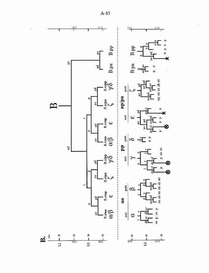
B. h
rs
Ll
1-
to
15
20
B.a
laa
B.a
lap
B
.alp
a B
.alp
p
alP
E
~
y/'5
a a
ant
post
an
t --
25
a ~
y
L3
L 3
0
~
d r
so
p so
p
35
ns s
h s
o ns
sh
so
ad B
.ara
a B
.ara
p
a!~
E
pp
po
st
ant
'5 E
rii p p
B
B.a
rpa
B.a
rpp
~ y/'
5
ap/p
a po
st
~
np n
s ns
sh
so
pv
ad
B.p
a
B.p
a
arlr
p p
B.p
p
.. , B
.pp
:p- 01
-.:
J
A-58
Figure 9. Genes required for normal development of the B cell.
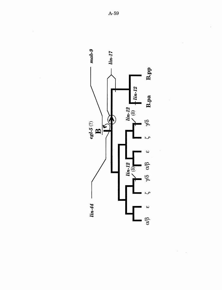
A-59
A-60
References
Adam.s, J. C. and Watt, F. M. (1993). Regulation of development and
differentiation by the extracellular matrix. Devt. 117, 1183-1198.
Akam, M. (1987). The molecular basis for metameric pattem in the
Drosophila embryo. Devt. 101, 1-22.
Amaya, E., Musci, T. J. and Kirschner, M. W. (1991). Expression of a
dominant negative mutant of the FGF receptor disrupts mesoderm formation
in Xenopus embryos. Cell 66,257-270.
Aroian, R. V., Koga, M., Mendel, J. E., Ohshima, Y. and Sternberg, P.
W. (1990). The let-23 gene necessary for Caenorhabditis elegans vulval
induction encodes a tyrosine kinase of the EGF receptor subfamily. Nature
348, 693-699.
Baker, N. E. (1987). Molecular cloning of sequences from wingless, a
segment polarity gene in Drosophila: the spatial distribution of a transcript
in embryos. EMBO J. 6, 1765-1773.
Baker, N. E. (1988). Embryonic and imaginal requirements for wingless, a
segment polarity gene in Drosophila . Dev. Biol. 125, 96-108.
Bang, A. G., Hartenstein, V. and Posakony, J. W. (1991). Hairless is
required for the development of adult sensory organ precursor cells. Devt. 111,
89-104.
Beitel, G., Clark, S. and Horvitz, H. R. (1990). The Caenorhabditis elegans
ras gene let-60 acts as a switch in the pathway of vulval induction. Nature
348' 503-509.
Bejsovec, A. and Martinez Arias, A. (1991). Roles of wingless in patterning
the larval epidermis of Drosophila. Devt. 113, 471-485.
A-61
Bienz, M. (1994). Homeotic genes and positional signalling in the Drosophila
viscera. TIG 10,22-26.
Bodmer, R., Caretto, R. and Jan, Y.-N. (1989). Neurogenesis of the
peripheral nervous system in Drosophila embryos: DNA replication patterns
and cell lineages. Neuron 3, 21-32.
Bowerman, B., Draper, B. W., Mello, C. C. and Priess, J. R. (1993). The
maternal gene skn-1 encodes a protein that is distributed unequally in early
C. elegans embryos. Cell 74, 443-452.
Bowerman, B., Eaton, B. A. and Priess, J. R. (1992). skn-1, a maternally
expressed gene required to specify the fate of ventral blastomeres in the early
C. elegans embryo. Cell 68, 1061-1075.
Bowerman, B., Tax, F. E., Thomas, J. H. and Priess, J. R. (1992). Cell
interactions involved in development ofbilaterally symmetrical intestinal
valve cells during embryogenesis in Caenorhabditis elegans. Develop. 116,
1113-1122.
Chisholm, A. (1991). Control of cell fate in the tail region of C. elegans by the
gene egl-5. Genes and Development 111, 921-932.
Chisholm, A. D. and Hodgkin, J. (1989). The mab-9 gene controls the fate
of B, the major male-specific blast cell in the tail region of Caenorhabditis
elegans. Genes & Deuel. 33, 1413-1423.
Christian, J. L., Olson, D. J. and Moon, R. T. (1992). Xwnt-8 modifies the
character of mesoderm induced by bFGF in isolated Xenopus ectoderm.
EMBO J 11, 33-41.
Clark, S. G., Chisholm, A. D. and Horvitz, H. R. (1993). Control of cell
fates in the central body region of C. elegans by the homeobox gene lin-39 .
Cell 74, 43-55.
A-62
Clark, S. G., Stern, M. J. and Horvitz, H. R. (1992). C. elegans cell
signalling gene sem-5 encodes a protein with SH2 and SH3 domains. Nature
356' 340-344.
Cooke, J. (1989). Mesoderm inducing factors and Spemann's organiser
phenomenon in amphibian development. Devt. 107,229-241.
Cornell, R. A. and Kimelman, D. (1994). Activin mediated mesoderm
induction requires FGF. Devt. 120, 453-462.
Cubas, P., de Celis, J.-F., Campuzano, S. and Modolell, J. (1991).
Proneural clusters of achaete-scute expression and the generation of sensory
organs in the Drosophila imaginal wing disc. Genes & Dev. 5, 996-1008.
Diederich, R. J., Merrill, V. K. L., Pultz, M.A. and Kaufman, T. C.
(1989). Isolation, structure, and expression of labial, a homeotic gene of the
Antennapedia complex involved in Drosophila head development. Genes Dev.
3, 399-414.
Dietrich, U. and Campos-Ortega, J. A. (1984). The expression of
neurogenic loci in imaginal epidermal cells of Drosophila melanogaster. J
Neurogenet. 1, 315-332.
DiNardo, S. and O'Farell, P. H. (1987). Establishment and refinement of
segmental pattern in the Drosophila embryo: spatial control of engrailed
expression by pair-rule genes. Genes Dev. 1, 1212-1225.
Ferguson, E. and Horvitz, H. R. (1985). Identification and characterization
of 22 genes that affect the vulval cell lineages of Caenorhabditis elegans.
Genetics 110, 17-72.
Ferguson, E. and Horvitz, H. R. (1989). The multivulva phenotype of
certain C. elegans mutants results from defects in two functionally-redundant
pathways. Genetics 123, 109-121.
A-63
Ferguson, E. L., Sternberg, P. W. and Horvitz, H. R. (1987). A genetic
pathway for the specification of the vulval cell lineages of Caenorhabditis
elegans. Nature 326, 259-267.
Freyd, G., Kim, S. K. and Horvitz, H. R. (1990). Novel cysteine-rich motif
and homeodomain in the product of the Caenorhabditis elegans cell lineage
gene lin-11 . Nature 344, 876-879.
Garriga, G., Desai, C. and Horvitz, H. R. (1993). Cell interactions control
the direction of outgrowth, branching and fasiculation of the HSN axons of
Caneorhabditis elegans. Develop. 117, 1071-1087.
Ghysen, A. and Dambly-Chaudiere, C. (1988). From DNA to form: the
achaete-scute complex. Genes Dev. 2, 495-501.
Ghysen, A., Dambly-Chaudiere, C., Jan, L. Y. and Jan, Y. N. (1993). Cell
interactions and gene interactions in peripheral neurogenesis. Genes Dev. 7,
723-733.
Goldstein, B. (1992). Induction of gut in Caenorhabditis elegans embryos.
Nature 357, 255257.
Goldstein, B. (1993). Establishment of gut fate in the E lineage of C.
elegans: the roles of lineage-dependent mechanisms and cell interactions.
Dev. 118, 1267-1277.
Gonzales, F., Swales, L., Bejsovec, A., Skaer, H. and Martinez Arias, A.
(1991). Secretion and movement of wingless protein in the epidermis ofthe
Drosophila embryo. Mech. Dev. 35, 43-54.
Green, J. B. A., Howes, G., Symes, K., Cooke, J. and Smith, J. C. (1990).
The biological effects ofXTC-MIF: quantitative comparison with Xenopus
bFGF. Devt. 108, 173-183.
A-64
Greenwald, I. S., Sternberg, P. W. and Horvitz, H. R. (1983). The lin-12
locus specifies cell fates in Caenorhabditis elegans. Cell 34, 435-444.
Gurdon, J. B. (1988). A community effect in animal development. Nature
336, 772-774.
Gurdon, J. B., Brennan, S., Fairman, S. and Mohun, T. J. (1984).
Transcription of muscle-specific actin genes in early Xenopus development:
Nuclear transplantation and cell dissociation. Cell 38, 691-700.
Gurdon, J. B., Fairman, S., Mohun, T. J. and Brennan, S. (1985). The
activation of muscle-specific actin genes in Xenopus development by an
induction between animal and vegetal cells of a blastula. Cell 41, 913-922.
Gurdon, J. B., Lemaire, P. and Kato, K. (1993). Community effects and
related phenomena in development. Cell 75, 831-834.
Gurdon, J. B., Mohun, T. J., Taylor, M. V. and Sharpe, C. R. (1989).
Embryonic induction and muscle gene activation. TIG 5, 51-56.
Han, M., Golden, A., Han, Y. and Sternberg, P. W. (1993). C. elegans lin-
45 raf gene participates in let-60 ras stimulated vulval differentiation. Nature
363, 133-140.
Han, M. and Sternberg, P. W. (1990). let-60, a gene that specifies cell fates
during C. elegans vulval induction, encodes a ras protein. Cell 63, 921-931.
Hartenstein, V. and Posak.ony, J. W. (1990). A dual function of the Notch
gene in Drosophila sensillum development. Dev. Biol. 142, 13-30.
Heemskerk, J. and DiNardo, S. (1994). Drosophila hedgehog acts as a
morphogen in cellular patterning. Cell 76 , 449-460.
Heitzler, P. and Simpson, P. (1991). The choice of cell fate in the epidermis
ofDrosophila. Cell 64, 1083-1092.
A-65
Herman, M.A. and Horvitz, H. R. (1994). The Caenorhabditis elegans gene
lin-44 controls the polarity of asymmetric cell divisions. Dev. in press.
Herman, R. K. and Hedgecock, E. M. (1990). The size of the C. elegans
vulval primordium is limited by lin-15 expression in surrounding
hypodermis. Nature 348, 169-171.
Hill, R. J., Katz, W. S. and Sternberg, P. W. (in preparation). The EGF
domain of Lin-3 is sufficient to induce vulval development.
Hill, R. J. and Sternberg, P. W. (1992). The lin-3 gene encodes an inductive
signal for vulval development in C. elegans. Nature 358, 470-476.
Hodgkin, J. (1983). Male phenotypes and mating efficiency in
Caenorhabditis elegans. Genetics 103,43-64.
Horvitz, H. R., Sternberg, P. W., Greenwald, I. S., Fixsen, W. and Ellis,
H. M. (1983). Mutations that affect neural cell lineages and cell fates during
the development of the nematode Caenorhabditis elegans. Cold Spring
Harbor Symp. Quant. Biol. 48, 453-463.
Huang, L. S., Tzou, P. and Sternberg, P. W. (1994). The lin-15 locus
encodes two negative regulators of C. elegans vulval development. MoZee.
Biol. Cell 5, -------.
Immergluck, K., Lawrence, P. A. and Bienz, M. (1990). Induction across
germ layers in Drosophila mediated by a genetic cascade. Cell 62 , 261-268.
Ingham, P. W., Taylor, A.M. and Nakano, Y. (1991). Role of the
Drosophila patched gene in positional signalling. Nature 353,
Kemphues, K. J., Priess, J. R., Morton, D. G. and Cheng, N. (1988).
Identification of genes required for cytoplasmic localization in early C.
elegans embryos. Cell 52, 311-320.
A-66
Kenyon, C. and Wang, B. (1991). A cluster of Antennapedia-class homeobox
genes in a nonsegmented animal. Science 253, 516-517.
Kenyon, C. J. (1986). A gene involved in the development of the posterior
body region of Caenorhabditis elegans. Cell 46, 477-487.
Kimble, J. (1981). Lineage alterations after ablation of cells in the somatic
gonad of Caenorhabditis elegans. Dev. Biol. 87, 286-300.
Kimble, J. and Hirsh, D. (1979). Post-embryonic cell lineages of the
hermaphrodite and male gonads in Caenorhabditis elegans. Dev. Biol. 70,
396-417.
Kimelman, D., Christian, J. L. and Moon, R. T. (1992). Synergistic
principles of development: overlapping patterning systems in Xenopus
mesoderm induction. Development 116, 1-9.
Kimelman, D. and Kirschner, M. (1987). Synergistic induction of
mesoderm by FGF and TGF-f3 and the identification of an mRNA coding for
FGF in the early Xenopus embryo. Cell 51, 869-877.
Kimelman, D. and Maas, A (1992). Induction of dorsal and ventral
mesoderm by ectopically expressed Xenopus basic fibroblast growth factor.
Devt. 114, 261-269.
Lawrence, P. A (1992). The making of a fly: the genetics of animal design.
Blackwell Scientific Publications, Oxford.
Lawrence, P. A and Sampedro, J. (1993). Drosophila segmentation: after
the first three hours. Devt. 119, 971-976.
Lewis, E. B. (1978). A gene complex controlling segmentation in Drosophila.
Nature 276 , 565-570.
A-67
Martinez Arias, A., Baker, N. E. and Ingham., P. W. (1988). Role of
segment polarity genes in the definition and maintenance of cell states in the
Drosophila embryo. Devt. 103, 157-170.
McGinnis, W. and Krumlauf, R. (1992). Homeobox genes and axial
patterning. Cell 68, 283-302.
McMahon, A. P. and Moon, R. T. (1989). Ectopic expression of the
protooncogene int-1 in xenopus embryos leads to duplication of the embryonic
axis. Cell 58, 1075-1084.
Mello, C. C., Draper, B. W., Krause, M., Weintraub, H. and Priess, J. R.
(1992). The pie-1 and mex-1 genes and matemal control of blastomere
identity in early C. elegans embryos. Cell 70, 163-176.
Miller, D. M., III, Ortiz, I., Berliner, G. C. and Epstein, H. F. (1983).
Differential localization of two myosins within nematode thick filaments. Cell
34, 477-490.
Mohler, J. (1988). Requirements for hedgehog, a segmental polarity gene, in
patterning larval and adult cuticle of Drosophila. Genetics 120, 1061-1072.
Mohler, J. and Vani, K. (1992). Molecular organization and embryonic
expression of the hedgehog gene involved in cell-cell communication in
segmental patterning of Drosophila. Devt. 115, 957-971.
Niisslein-Vollhard, C. and Wieschaus, E. (1980). Mutations affecting
segment number and polarity in Drosophila. Nature 287, 795-801.
Panganiban, G. E. F., Reuter, R., Scott, M.P. and Hoffman, F. M.
(1990). A Drosophila growth factor homolog, decapentaplegic, regulates
homeotic gene expression within and across germ layers during midgut
morphogenesis. Devt. 110, 1041-1050.
A-68
Parks, A. L. and Muskovitch, M.A. T. (1993). Delta function is required
for bristle organ determination and morphogenesis in Drosophila. Dev. Biol.
157' 484-496.
Peifer, M., Karch, F. and Bender, W. (1987). The bithorax complex: control
of segmental identity. Genes Dev. 1, 891-898.
Poole, S. J., Kauvar, L. M., Drees, B . and Kornberg, T. (1985). The
engrailed locus of Drosophila: Structural analysis of an embryonic transcript.
Cell 40, 37-43.
Posakony, J. W. (1994). Nature versus nurture: Asymmetric cell divisions in
Drosophila bristle development. Cell 76, 415-418.
Priess, J. R. and Thomson, J. N. (1987). Cellular interactions in early
Caenorhabditis elegans embryos. Cell 48 , 241-250.
Priess, J. R., Schnabel, H., and Schnabel, R. (1987). Tbeglp-1 locus and
cellular interactions in early C. elegans embryos. Cell , 51, 601-611.
Reuter, R., Panganiban, G. E. F., Hoffman, F. M. and Scott, M.P.
(1990). Homeotic genes regulate the spatial expression of putative growth
factors in the visceral mesoderm of Drosophila embryos. Devt. 110, 1031-1040.
Rhyu, M. S., Jan, L. Y. and Jan, Y. N. (1994). Asymmetric distribution of
numb protein during division of the sensory organ precursor cell confers
distinct fates to daughter cells. Cell 76, 477-491.
Rodriguez, I., Hernandez, R., Modolell, J. and M., R.-G. (1990).
Competence to develop sensory organs is temporally and spatially regulated
in Drosophila epidermal primordia. EMBO J . 9 , 3583-3592.
Romani, S., Campuzano, S., Macagno, E. R. and Modolell, J. (1989).
Expression of achate and scute genes in Drosophila imaginal discs and their
function in sensory organ development. Genes Dev. 3 , 997-1007.
A-69
Sampedro, J., Johnston, P. and Lawrence, P. A. (1993). A role for
wingless in the segmental gradient of Drosophila? Dev. 117, 677-687.
Schierenberg, E. (1987). Reversal of cellular polarity and early cell-cell
interactions in the embryo of Caenorhabditis elegans. Dev. Biol. 122, 452-463.
Schnabel, R. (1991). Cellular interactions involved in the determination of
the early C. elegans embryo. Meek. Deuel. 34, 85-100.
Schnabel, R. (1994). Autonomy and nonautonomy in cell fate specification of
muscle in the Caenorhabditis elegans embryo: a reciprocal induction. Science
263, 1449-1452.
Schubert, D. and Kimura, H. (1991). Substratum-growth factor
collaborations are required for the mitogenic activities of activin and FGF on
embryonal carcinoma cells. J. Cell Biol. 114; 841-846.
Schweisguth, F. and Posakony, J. W. (1992). Suppressor of Hairless, the
Drosophila homolog of the mouse recombination signal-binding protein gene,
controls sensory organ cell fates. Cell 69, 1199-1212.
Seydoux, G. and Greenwald, I. (1989). Cell autonomy of lin-12 function in
a cell fate decision in C. elegans. Cell 57, 1237-1245.
Slack, J. M. W., Darlington, B. G., Heath, J. K. and Godsave, S. F.
(1987). Mesoderm induction in early Xenopus embryos by heparin binding
growth factors. Nature 326, 197-200.
Smith, J. C. (1989). Mesoderm induction and mesoderm-inducing factors in
early amphibian development. Dev. 105, 665-677.
Smith, W. C. and Harland, R. M. (1992). Expression cloning of noggin , a
new dorsalixing factor localized to the Spemann organizer in Xenopus
embryos. Cell 70, 829-840.
A-70
Smith, W. C., Knecht, A. K., Wu, M. and Harland, R. M. (1993). Secreted
noggin protein mimics the Spemann organizer in dorsalizing Xenopus
mesoderm. Nature 361, 547-549.
Sternberg, P. W. (1993). Intercellular signaling and signal transduction in
C. elegans. Ann Rev. Genet. 27, 497-521.
Sternberg, P. W. and Horvitz, H. R. (1986). Pattern formation during
vulval development in Caenorhabditis elegans. Cell 44, 761-772.
Sternberg, P. W. and Horvitz, H. R. (1988). lin-17 mutations of C. elegans
disrupt asymmetric cell divisions. Developmental Biology 130, 67-73.
Sternberg, P. W. and Horvitz, H. R. (1989). The combined action of two
intercellular signalling pathways specifies three cell fates during vulval
induction in C. elegans. Cell 58, 679-693.
Strome, S. (1983). Generation of asymmetry and segregation of germ-line
granules in early Caenorhabditis elegans embryos. Cell 35, 15-25.
Sulston, J. and Horvitz, H. R. (1977). Postembryonic cell lineages ofthe
nematode Caenorhabditis elegans. Dev. Biol. 56, 110-156.
Sulston, J. E., Albertson, D. G. and Thomson, J. N. (1980). The
Caenorhabditis elegans male: Postembryonic development of nongonadal
structures. Dev. Biol. 78, 542-576.
Sulston, J. E., Schierenberg, E., White, J. G. and Thomson, J. N. (1983).
The embryonic cell lineage of the nematode Caenorhabditis elegans. Dev.
Biol. 100, 64-119.
Sulston, J. E. and White, J. G. (1980). Regulation and cell autonomy
during postembryonic development of Caenorhabditis elegans. Deuel. Biol. 78 ,
577-597.
A-71
Trem.ml, G. and Bienz, M. (1989). Homeotic gene expression in the visceral
mesoderm of Drosophila embryos. EMBO J. 8, 2677-2685.
Uemura, T., Shepherd, S., Ackerman, L., Jan, L. Y. and Jan, Y. N.
(1989). numb, a gene required in determination of cell fate during sensory
organ formation in Drosophila embryos. Cell 58, 349-360.
Wood, W. B. (1991). Evidence from reversal ofhandedness in C. elegans for
early cell interactions determining cell fates. Nature 349, 536-538.
Woodland, H. R. (1989). Mesoderm formation in Xenopus. Cell 59, 767-770.
Woods, D. F. and Bryant, P. J. (1992). Genetic control of cell interactions in
developing Drosophila epithelia. Annu. Rev. Genet. 26, 305-350.
B-1
Chapter2
Multiple cell interactions are required for fate specification during
male spicule development in Caenorhabditis elegans
Helen M. Chamberlin and Paul W. Stemberg
Published in Development 118, 297-323 (1993).
Dc'elopmc,>nt l iS. :!9i-~ :!.J t 199~1
Prinled in Grl!at Brnain [ Th~,.• Compan~ of Biolo,;;i·q~ Limited I 99~ B-2 297
Multiple cell interactions are required for fate specification during male
spicule development in Caenorhabditis elegans
Helen M. Chamberlin and Paul W. Sternberg
Howard Hughes Medical Institute and Division of Biology, California Institute of Technology, Pasadena, CA 91125, USA
SUMMARY
The B blast cell divides postembryonically in C. elegans males to produce 47 progeny that include all of the cells of the copulatory spicules. During the early development of the B lineage. the anterior daughter of B, B.a, generates eight cells. These cells migrate to form four pairs of cells that flank the developing cloaca (ventral, dorsal, and two identical lateral pairs). For each pair, the more anterior cell produces a distinct lineage ('anterior fate') from the posterior cell ('posterior fate') . For the ventral and dorsal pairs, either cell can migrate to the anterior position and produce the anterior lineage, and the other cell migrates posterior and produces the posterior lineage (Sulston and Horvitz, I977,Dev. Bioi. 56, 110-156). The migration is variable, although the resultant fate pattern is invariant. In the two lateral pairs, both the migration and fate pattern are invariant. Using a laser microbeam to selective))' ablate neighboring cells we
INTRODUCTION
The fate of a cell can be specified by information provided by the cell' s precursors and by signals from the cell's neighbors. The experimental study of cell lineage (patterns of cell division and their relationship to cell fate) can provide clues about these mechanisms of fate specification and their interrelationship. The ability to visualize cell nuclei and their divisions in C. elegans. combined with the reproducibility of the lineage among animal s. has allowed the complete description of the normal cell division pattern and the differentiated terminal fates (Sulston and Horvitz. 1977: Sulston et al.. 1980. 1983 ). The cell lineage in C. elegans is essentially invariant. However. because the extracellular environment for most developing cells is as constant as the ancestry. the correlation of cell division and fate does not indicate that fate specification is completely autonomous. Indeed. cell interactions likely play an important role in cell fate specification. For example. the lineage includes several instances of cells that have the potential to adopt more than one fate (Su lston and Horvitz. 1977: Kimble and Hirsh. 1979: Sulston et al.. 1983) . In addition. embryonic blast cell rearrangement (Priess and Thomson. 1987: Wood. 1991) and isolation experiments (Schierenberg. 1987: Goldstein. 1992). as well as embryonic (Bowerman et al.. 1992) and
have found that the cells of the lateral pair also respond to positional cues. For all four pairs other male-specific blast cells provide extracellular cues. In general, F and U promote anterior fates, Y promotes some posterior fates, and the B.a progeny promote posterior fates. Several of these cues are redundant. By ablating combinations of cells we have deduced how these signals rna)· act in concert to specify the fates of the B.a progeny. We propose that fate specification in these pairs depends on three general classes of extracellular cues: positional cues, modulators of positional cues, and lateral signals. The B lineage thus provides an opportunity to study with single cell resolution the integration of multiple intercellular signals.
Key words: C. elegans. cell lineage. cell interactions. cell fate specification. redundancy. pattern formation
postembryonic cell ablation experiments (Sulston and White. 1980: Kimble. 1981: Chisholm and Hodgkin. 1989) have identified instances of cell interactions that specify cell fate . The invariant cell lineage. rather than indicating a limited repertoire of fate specification mechanisms. offers a reproducible background to study the interplay of autonomous and conditional mechanisms.
To study the components of fate specification. we have focused our attention on the postembryonic lineage of the male B cell. The B cell is one of four male-specific blast cells (B. U. F and Y) that divide in males. but not in hermaphrodites. and produce some of the cells of the specialized mating structures of the male tail. The male B cell. for instance. produces all of the cells of the copulatory spicules (Fig. I). The B lineage (Fig. 2) includes examples of autonomous as well as conditional fate specification . For example. the B cell undergoes asymmetric cytokinesis in its first division. which has been studied as a possible example of autonomous fate specification (Sternberg and Horvitz. 1988). Later in the lineage. two pairs of B progeny cells exhibit natural variation (Sulston and Horvitz. 1977 ). In each pair. one cell adopts the anterior position and fate. and the other cell the posterior position and fate. Although which cell adopts the anterior position varies from one animal to the next. the resultant pattern of anterior and
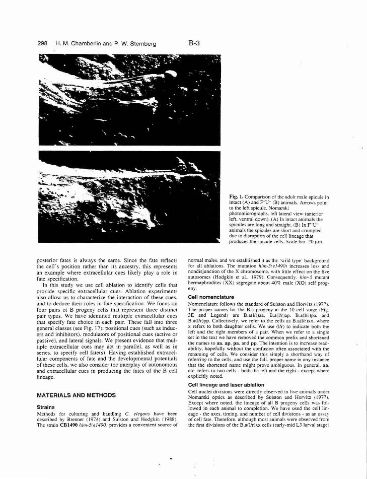
298 H. M. Chamberlin and P. W. Sternberg
posterior fates is always the same. Since the fate reflects the celrs position rather than its ancestry. this represents an example where extracellular cues likely play a role in fate specification.
In this study we use cell ablation to identify cells that pro\'ide specific extracellular cues. Ablation experiments also allow us to characterize the interaction of these cues. and to deduce their roles in fate specification. We focus on four pairs of B progeny cells that represent three distinct pair types. We have identified multiple extracellular cues that specify fate choice in each pair. These fall into three general classes (see Fig. 17): positional cues (such as inducers and inhibi tors). modulators of positional cues (active or passive). and lateral signals. We present evidence that multiple extracellular cues may act in parallel. as well as in series. to specify cell fate(s). Having established extracellular components of fate and the developmental potentials of these cells. we also consider the interplay of autonomous and extracellular cues in producing the fates of the B cell lineage.
MATERIALS AND METHODS
Strains Methods for culturing and handling C. elegans have been described by Brenner (1974) and Sulston and Hodgkin (1988). The strain CB1490 him-5(el490) provides a convenient source of
B-3
Fig. 1. Comparison of the adult male spicule in intact (A) and p-u- (B) animals. Arrows point to the left spicule. Nomarski photomicrographs. left lateral view (anterior left. ventral down). (A) In intact animals the spicules are long and straight. <B ) In F-uanimals the spicules are shon and crumpled due to disruption of the cell lineage that produces the spicule cell~. Scale bar. 20 Jlm.
normal males. and we established it as the ·wild type· background for all ablations. The mutation him-5(el490! increases los~ and nondisjunction of the X chromosome. with little effect on the fi,·e autosomes (Hodgkin et al.. 1979). Con~equently. him-5 mutant hermaphrodi tes <XX) segregate about 40'k male (XO) self progeny.
Cell nomenclature Nomenclature follows the standard of Suls10n and Horvitz ( 1977 ). The proper names for the B.a progeny at the I 0 cell stage <Fig. 3E and Legend) are B.a(J/r)aa. B.a(l/r)ap. B.a(l/r)pa. and B.a(l/r)pp. Collectively. we refer 10 the cells as B.a(l/r)xx. where x refers 10 both daughter cells. We use (1/r) to indicate both the left and the right members of a pair. When we refer 10 a single set in the text we have removed the common prefix and shonened the names 10 aa. ap. pa. and pp. The intention is to increase readability. hopefully without the confusion often associated with the renaming of cells. We consider this simply a shonhand way of referring 10 the cells. and use the full. proper name in any instance that the shonened name might prove ambiguous. In general. aa. etc. refers to two cells - both the left and the right -except where explicitly noted.
Cell lineage and laser ablation Cell nuclei divisions were directly observed in live animals under Nomarski optics as described by Sulston and Horvitz (I 977 ). Except where noted. the lineage of all B progeny cells was followed in each animal to completion. We have used the cell lineage - the axes. timing. and number of cell divisions - as an assay of cell fate. Therefore. although most animals were observed from the first divisions of the B.a(llr)xx cells (early-mid L3 larval stage)
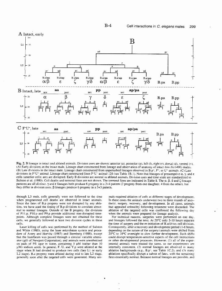
B-4 Cell interactions in C. elegans males 299
A Intact, early hr~
Ll 10 B
ad P'' 15
L2 "' ad
:!;,
B.alaa B.alap B.alpa B.alpp B.araa B.arap B.arpa B.arpp
all) £ ~ y/8 all) £ ~ ylo B.pa B.pp ----------------------------- -----------------------------------------------------------------
ant B Intact, late ant ~~----------~po~s~t-
aa pp ap/pa post post ant
L3l :~ 35
y
l B.pa
3tlr B.pp
·~ C F-u-, late aa pp
ant ant post 25
post ap/pa
post B.pa
ant
B.pp
L3 3(l
Jj
·r4'· ~ Fig. 2. B lineage in intact and ab!a1ed animals. Division axes are shown: anterior (a). posterior (pl. left <IJ. right (r). dorsal <d). ventral (v). (AI Early divisions in !he intact male. Lineage chan cons1ructed from lineage and observation of anatomy of intact him-5(e/490) males. (BJ Late di,·isions in the intact male. Lineage chan constructed from unperturbed lineages observed in B.p-. F-. or u - animals. (CJ Late divisions in F-u - animal. Lineage chan construc1ed from F-u- animal 128 (see Table lB. I). Note that lineages of presumptive a. y. and E
cells (anterior cells: ant) are disrupted. Early B divisions are normal in ablated animals. Division axes and time scale are standardized to Sulston et al. ( 1980). Cell deaths and terminal fates are not shown. The terminal fates are indicated in Table 8. The a. [3. 5 and ~lineage panerns are all distinct. y and E lineages both produce 6 progeny in a 2+4 panern (2 progeny from one daughter. 4 from the other). but they differ in division axes. [31ineages produce 6 progeny in a 3+3 panern.
through L3 molt. cells generally were not followed to the time when programmed cell deaths are observed in intact animals. Since the fates of B.p progeny were not disrupted by any ablation. we have used the timing of B.p divisions to correlate abnormal 10 normal lineages. Outside of the B progeny. the divisions of P !J.p. PI O.p and P9.p provide additional non-disrupted time· points. Although complete lineages were not obtained for these cells. we generally followed at least two division cycles in these cells.
Laser killing of cells was performed by the method of Sulston and White ( 1980). using the laser microbeam system and procedure of Avery and Horvitz (1987) and Sternberg (1988). except that the laserbcam was passed through a circular variable attenuator (gift of Newport Corporation). and animals were anesthetized on pads of 5'7c agar in water. containing 5 !lM (rather than I 0 !!MJ sodium azide. In general. F. U. and Y.p were ablated at the stage when B had divided to produce two cells (late L1 or early L2 stage). B.a progeny were ablated during mid to late L2 stage. generally soon after the targeted cells were generated. Many ani-
mals required ablation of cells at different stages of development. In these cases the animals underwent two to three rounds of anes· thetic. surgery. recovery. and development. In all cases. animals that appeared unhealthy following treatment were discarded. The ablation of the targeted cells was confirmed the following day when the animals were prepared for lineage analysis.
For technical reasons. surgeries were performed on one day. and lineages followed the next. At 20°C only 5-10 hours separate the time of surgery and the re-initiation of B.a(l/rlxx cell division. Consequently. after a recovery and development period ( 1-6 hours. depending on the nature of the surgery) animals were shifted from 20°C to I 0°C overnight to slow further development. Such shifts could disrupt temperature-sensitive aspects of the cell interactions or other developmental processes. However. (I) all of the experimental animals were treated the same. so our experiments are internally consistent. (2) normal lineages are observed in many ablation backgrounds (e.g .. B.p-. see Table IC.2). and (3) many ablations specifically disrupt a subset of fates. with the remaining fates essentially normal. Because normal lineages are possible. and
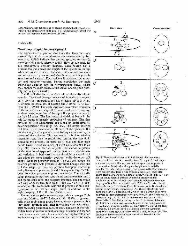
300 H. M. Chamberlin and P. W. Sternberg B-5
abnormal lineages are specific ro certain ablation backgrounds. we believe the tempermure shifl does not fundamemally affect our A resuhs . All lineages were observed at 20°C.
Side view Cross section
RESULTS
Summary of spicule development The spicules are a pair of structures that flank the male cloaca (Fig. I). Electron microscope reconstruction by Sulston et al. ( 1980) indicate that the two spicules are sensilla covered with a hard. sclerotic cuticle. Each spicule includes two presumptive sensory neurons. Each neuron has a process that runs down the length of the spicule to the tip. "·here it is open to the environment. The neuronal processes are surrounded by socket and sheath cells. which provide structure and support. Each spicule is anchored by extensor and retractor muscles. During copulation the male inserts his spicules into the hermaphrodite vulva. where they anchor the male cloaca at the vulval opening and possibly aid in sperm transfer.
The B cell divides to produce all of the cells of the spicules. The B cell lineage consists of three distinct stages: early divisions. migration. and late divisions (Figs 2. 3 and 4: original observations of Sulston and Horvitz. 1977: Sulston et a!.. 1980). The early divisions take place primarily in the second larval stage (L:Z). and result in 10 progeny. A short range migration of the eight B.a progeny occurs in the late L:Z stage. The late round of divisions begin in the mid-L3 stage. ultimately producing 47 progeny. The first division of B is asymmetric and along an approximately anterior/posterior axis (Figs 3A. 4A). The larger anterior cell (B.a) is the precursor of all cells of the spicules. B.a divides along a left/right axis. establishing the bilateral symmetry of the spicules. This symmetry is broken (during migration) and then re-established (during the late divisions) in the progeny of these cells. B.al and B.ar each divide twice to produce a ring of eight cells. one cell thick (Fig. 3D). These cells then migrate. The medial migration of the two dorsal (pp) and ventral (aa) cells exhibits natural variation. In both cases. either the right or the left cell can adopt the more anterior position. while the other cell adopts the more posterior position. The cell that adopts the anterior position will produce a different lineage than the cell that adopts the posterior position (compare anterior a and y lineages to posterior 13 and o lineages in Fig. 2). The other four B.a progeny migrate invariantly. The ap cells adopt the anterior position (one on the left. one on the right). and the pa cells adopt the posterior position. The end result is a rim! of cells. two cells thick (Figs 3E. 4B). It is convenient-to refer to animals with the B progeny in this configuration as the ·I 0 cell stage.· since in addition to the eight progeny of B.a. B.p has divided once.
The aa and pp pairs represent equivalence groups (Sulston and Horvitz. 1977: Sulston et a!.. 1980). In general. cells in an equivalence group have equivalent potential. but they adopt different fates after interacting with each other. after receiving positional cues. or both. Because of the variability from animal to animal. one must distinguish between lineal ancestry and fate choice when referring to cells in an equivalence group. Within the aa pair. the fate of the ante-
B B
~ c B
~ D B
~ E
~ ~
y 0 0
0
Fig. 3. The early divisions of B. Left lateral view and cross section of B.a at one (A). two <B J. four <CJ. ei(!ht (D) cell sta2e. and after migration (E). Arrows indicate appr;ximate plane of cross section. B.a divides along a lefl-right axis to initially establish the bilateral symmetry of the spicules <B 1. B.a produces eight progeny that form a ring of cells. a single cell thick (D). These cells migrate to form a ring of cells. two cells thick (E). It is convenient to refer to animals with the B progeny in this configuration as the ·10 cell stage· since in addition to the eight progeny of B.a. B.p has divided once. F. U. and Y.p also divide during the early B divisions. F and U lie anterior to B. dorsal and ventral to the rectum. respectively <AJ. These cells divide once during the early B lineage. so that when the B.a(l/r)xx cells have migrated to their anterior/posterior positions. F.J and F.r lie next to presumptive y. and U.J and U.r lie next to presumptive a <E). These cells funher divide during the late B divisions ( Sulsron et al.. 1980). Y divides asymmetrically prior to the first division of B. producing a neuron and the Y.p blast cell (A). During the early B lineage. Y .p divides once to establish left/right symmetry. Funher divisions resuh in a cluster of five cells on each side. The position of these clusters is more dorsal and lateral than the original position of Y <E).
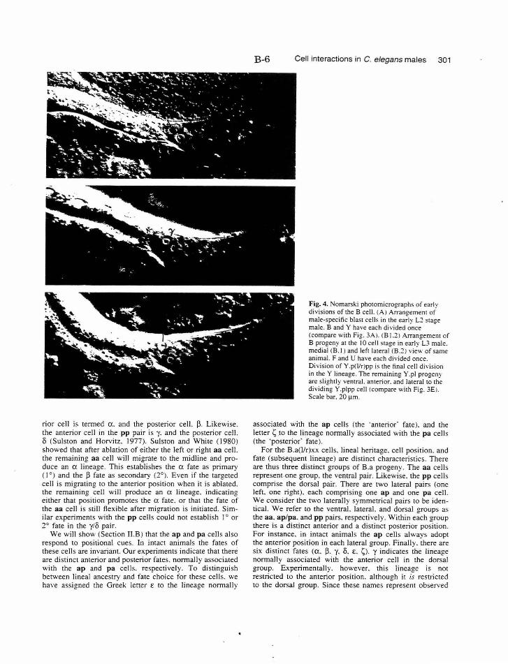
rior cell is tenned a. and the posterior cell. ~- Likewise. the anterior cell in the pp pair is y. and the posterior cell. 8 (Sulston and Horvitz. I 977). Sulston and White ( 1980) showed that after ablation of either the left or right aa cell. the remaining aa cell will migrate to the midline and produce an a lineage. This establishes the a fate as primary (I 0 ) and the ~ fate as secondary (2° ). Even if the targeted cell is migrating to the anterior position when it is ablated. the remaining cell will produce an a lineage. indicating either that position promotes the a fate. or that the fate of the aa cell is still flexible after migration is initiated. Similar experiments with the pp cells could not establish I 0 or 2° fate in the y/8 pair.
We will show (Section II.B) that the ap and pa cells also respond to positional cues. In intact animals the fates of these cells are invariant. Our experiments indicate that there are distinct anterior and posterior fates. nonnally associated with the ap and pa cells. respectively . To distinguish between lineal ancestry and fate choice for these cell s. we have assigned the Greek letter E to the lineage nonnally
B-6 Cell interactions in C. elegans males 301
Fig. 4. Nomarski photomicrographs of early divisions of the 8 cell. (A) Arran!!ement of male-specific blast cells in the ea;ly L2 stage male. 8 and Y have each divided once (compare with Fig. 3A ). (8 1.2) Arrangement of B progeny at the I 0 cell stage in early L3 male. medial (8.1) and left lateral (8.2) view of same animal. F and U have each divided once. Division of Y.p(l!r)pp is the final cell d ivision in the Y lineage. The remaining Y .pi progeny are slightly ventral. anterior. and lateral to the dividing Y.plpp cell (compare with Fig. 3E). Scale bar. 20 1-1m.
associated with the ap cells (the ·anterior' fate). and the letter ~.to the lineage nonnally associated with the pa cells (the 'posterior· fate).
For the B.a(llr)xx cells. lineal heritage. cell position. and fate (subsequent lineage) are distinct characteristics. There are thus three distinct groups of B.a progeny. The aa cells represent one group. the ventral pair. Likewise. the pp cells comprise the dorsal pair. There are two lateral pairs (one left. one right). each comprising one ap and one pa cell. We consider the two laterally symmetrical pairs to be identical. We refer to the ventral. lateral. and dorsal groups as the aa. ap/pa. and pp pairs. respecti vely. Within each group there is a distinct anterior and a distinct posterior position. For instance. in intact animals the ap cells always adopt the anterior position in each lateral group. Finally. there are six distinct fates (a. ~- y. 8. E. ~). y indicates the lineage nonnally associated with the anterior cell in the dorsal group. Experimentally. however. this lineage is not restricted to the anterior position. although it is restricted to the dorsal group. Since these names represent observed
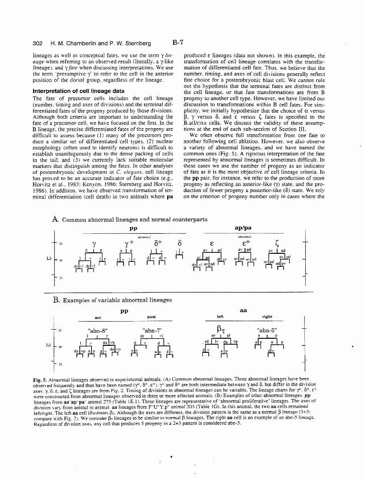
302 H. M. Chamberlin and P. W. Sternberg B-7
lineages as v.-·ell as conceptual fates. we use the term y lineage when referring to an observed result (literally. a y-like lineage). andy fare when discussing interpretations. We use the term ·presumptive y' to refer to the cell in the anterior position of the dorsal group. regardless of the lineage.
Interpretation of cell lineage data The fate of precursor cells includes the cell lineage (number. timin2 and axes of divisions) and the terminal differentiated fate; of the progeny produced by those divisions. Although both criteria are important to understanding the fate of a precursor cell. we have focused on the first. In the B lineage. the precise differentiated fates of the progeny are difficult to assess because ( I ) many of the precursors produce a similar set of differentiated cell types. (2) nuclear morphology (often used to identify neurons) is difficult to establish unambiguously due to the dense packing of cells in the tail. and (3) we currently lack suitable molecular markers that distinguish among the fates. In other analyses of postembryonic development in C. elegans. cell lineage has proved to be an accurate indicator of fate choice (e.g .. Horvitz et al.. 1983: Kenvon. 1986: Stembenz and Horvi tz. 1986). In addition. we ha~·e observed transfo~ation of terminal differentiation (cell death) in two animals where pa
produced E lineages (data not shown). In this example. the transformation of cell linea2e correlates with the transformation of differentiated celt fate. Thus. we believe that the number. timing. and axes of cell divisions generally reflect fate choice for a postembryonic blast cell. We cannot rule out the hypothesis that the terminal fates are distinct from the cell lineage. or that fate transformations are from B progeny to another cell type. However. we have limited our discussion to transformations within B cell fates. For simplicity. we initially hypothesize that the choice of a versus !3. y versus 8. and E versus 1;, fates is specified in the B.a(l/r)xx cells. We discuss the validity of these assumptions at the end of each sub-section of Section III.
We often observe full transformation from one fate to another following cell ablation. However. we also observe a variety of abnormal lineages. and we have named the common ones (fig. 5). A rigorous interpretation of the fate represented by abnormal lineages is sometimes difficult. In these cases we use the number of progeny as an indicator of fate as it is the most objective of cell lineage criteria. In the pp pair. for instance. we refer to the production of more progeny as reflecting an anterior-like (y) state. and the production of fewer progeny a posterior-like (8) state. We rely on the criterion of progeny number only in cases where the
A. Common abnormal lineages and normal counterparts
pp ap/pa
L3
L3
abnormal
:!:0 y y* o*
~ rttri, :~o
,. r
3:i
B. Examples ofvariable abnormal lineages
30
35
ant
"abn-8" I
pp post
"abn-7"
::~.bnorm:~ol
0 E E* s tLr ~ a pel
aa left right
nabn-5 11
Fig. 5. Abnormal lineages observed in experimental animals. (A) Common abnormal lineages. Three abnormal lineages have been observed frequently and thus have been named (y*. 8*. E* ). y* and 8* are both mtermedtate between y and 8. but dtffer m the dtvtston axes. y. 8. E. and ~ lineages are from Fig. 1. Timing of divisions in abnormal lineages can be variable. The lineage chartS for y<·. O*. E''
were constructed from abnormal lineaees observed in three or more affected animals. (B) Examples of other abnormal lineages. PP linea~es from aa-ap-pa- animal275 <Table IE. I). These lineages are representative of ·abnormal proliferative· lineage;. The axes of division varv from animal to animal. aa lineaees from F- u -y .p- anima1203 (Table IGJ. In this animal. the two aa cells remained left/rieht. The left aa cell illustrates 13,. Altho~gh the axes are different. the division pattern is the same as a normal 13 lineage <3+3: compire with Fig. 1 ). We consider 13, lineages to be similar to normal 13 lineages. The right aa cell is an example of an abn-5 lineage. Regardless of division axes. any cell that produces 5 progeny in a 2+3 pattern is considered abn-5.
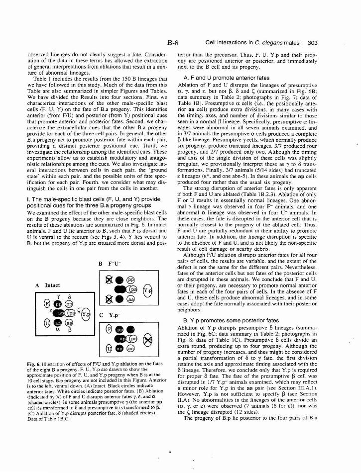
B-8 Cell interactions in C. elegans males 303
observed lineages do not clearly suggest a fate . Consideration of the data in these terms has allowed the extraction of general interpretations from ablations that result in a mixture of abnormal lineages.
Table I includes the results from the !50 B Iinea!!es that we have followed in this study. Much of the data fr"Om this Table are also summarized in simpler Figures and Tables. We have divided the Results into four sections. First. we characterize interactions of the other male-specific blast cells (F. U. Y) on the fate of B.a progeny. This identifies anterior (from FlU) and posterior (from Y) positional cues that promote anterior and posterior fates. Second. we characterize the extracellular cues that the other B.a progeny provide for each of the three cell pairs. In general. the other B.a progeny act to promote posterior fate wi thin each pair. providing a distinct posterior positional cue. Third. we investigate the relationship among the identified cues. These experiments allow us to establish modulatory and antagonistic relationships among the cues. We also investigate lateral interactions between cells in each pair. the ·ground state' within each pair. and the possible units of fate specification for each pair. Fourth. we consider what may distinguish the cells in one pair from the cells in another.
I. The male-specific blast cells (F, U, andY) provide positional cues for the three B.a progeny groups We examined the effect of the other male-specific blast cells on the B progeny because they are close neighbors. The results of these ablations are summarized in Fig. 6. In intact animals. F and U lie anterior to B. such that F is dorsal and U is ventral to the rectum (see Figs 3. 4). Y lies ventral to B. but the progeny of Y.p are situated more dorsal and pos-
B F-u-
A Intact ®e®
8 e:@ y 0
~-~ @O@ £~~8 c Y.p-
@8~ @oe a p ~ 8 @·~
Fig. 6. Illustration of effects of FlU and Y .p ablation on the fates of the eight B.a progeny. F. U. Y.p are drawn to show the approximate position of F. U. and Y.p progeny when B is at the 10 cell stage. B.p progeny are not included in this Figure. Anterior is to the left. ventral down. (A l Intact. Black circles indicate anterior fates. White circles indicate posterior fates. (B) Ablation <indicated by X) ofF and U disrupts anterior fate~ y. E. and a (shaded circles). In some animals presumptive y (the anterior pp cell) is transformed too and presumptive a is transformed to [3. <CI Ablation of Y .p disrupts posterior fate. o (shaded circles). Data of Table I B.C.
terior than the precursor. Thus. F. U. Y.p and their progeny are positioned anterior or posterior. and immediately next to the B cell and its progeny.
A. F and U promote anterior fates Ablation of F and U disrupts the lineages of presumptive a. y. and £. but not p. 8 and 1; (summarized in Fig. 6B: data summary in Table 2: photographs in Fig. 7; data of Table I B). Presumptive a cells (i.e .. the positionally anterior aa cell) produce extra divisions. in many cases with the timing. axes, and number of divisions similar to those seen in a normal P lineage. Specifically. presumptive a lineages were abnormal in all seven animals examined. and in 3/7 animals the presumptive a cells produced a complete P-Iike lineage. Presumpti ve y cells. which normally produce six progeny. produce truncated lineages. 3/7 produced four progeny. and 2/7 produced only two. Although the timing and axis of the single division of these cells was slightly irregular. we provisionally interpret these as y to 8 transformations. Finally. 3/7 animals (5/14 sides) had truncated £lineages(£*. and one abn-5). In these animals the ap cells produced four rather than the usual six progeny.
The strong disruption of anterior fates is only apparent if both F and U are ablated (Table I B.2.3 ). Ablation of only F or U results in essentially normal lineages. One abnormal y lineage was observed in four F- animals. and one abnormal a lineage was observed in four u- animals. In these cases. the fate is disrupted in the anterior cell that is normally closest to the progeny of the ablated cell. Thus. F and U are partially redundant in the ir ability to promote anterior fate . In addition. the lineage disruption is specific to the absence ofF and U. and is not likely the non-specific result of cell damage or nearby debris.
Although FlU ablation disrupts anterior fates for all four pairs of cells. the results are variable. and the extent of the defect is not the same for the different pairs. Nevertheless. fates of the anterior cells but not fates of the posterior cells are disrupted in these animal s. We conclude that F and U. or their progeny. are necessary to promote normal anterior fates in each of the four pairs of cells. In the absence of F and U. these cells produce abnormal lineages. and in some cases adopt the fate normally associated with their posterior neighbors.
B. Y.p promotes some posterior fates Ablation of Y.p disrupts presumptive 8 lineages (summarized in Fig. 6C: data summary in Table 2: photographs in Fig. 8: data of Table I C). Presumptive o cells divide an extra round. producing up to four progeny. Although the number of progeny increases. and thus might be considered a partial transformation of 8 to y fate . the first division retains the axis and approximate timing associated with the o lineage. Therefore. we conclude only that Y.p is required for proper 8 fate . The fate of the presumptive P cell was disrupted in 1/7 Y.p- animals examined. which may reflect a minor role for Y.p in the aa pair (see Section III.A.l i. However. Y.p is not sufficient to specify P (see Section II.A ). No abnormalities in the lineages of the anterior cells (a. y. or £) were observed (7 animals (6 for £)). nor was the 1; lineage disrupted (12 sides).
The progeny of B.p lie posterior to the four pairs of B.a
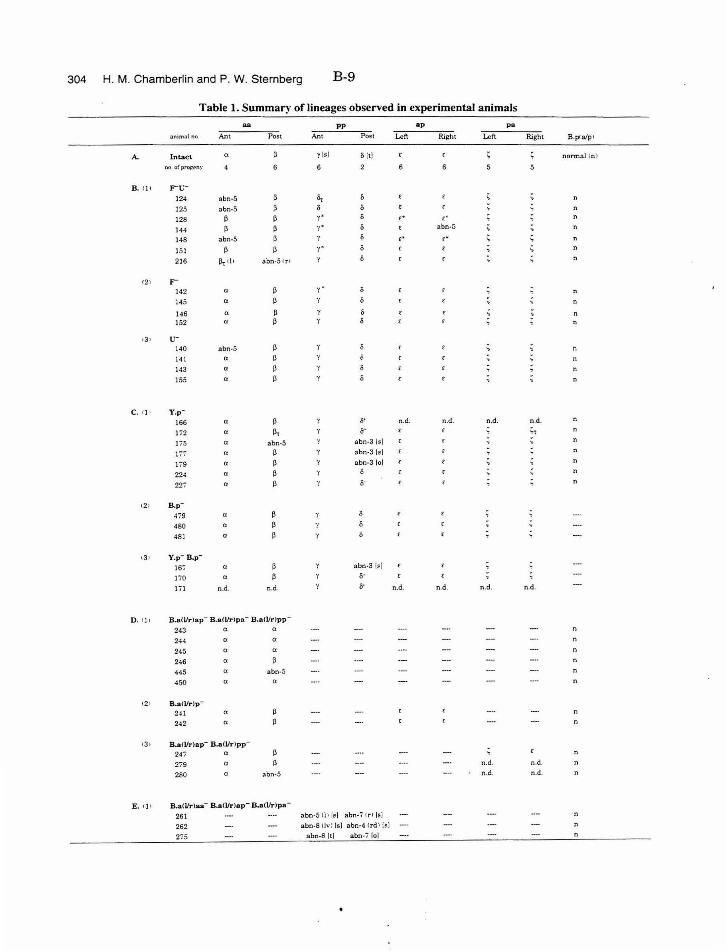
304 H. M. Chamberlin and P. W. Sternberg B-9
Table 1. Summary of lineages observed in experimental animals
"" PP ap pa
3n1m:dno. Ant Post Ant Po~t Left Right Left Right B .p<a/p l
A. Intact a y 1•1 0 ltl normal In I
no o(p~~ny 6
B. 111 rti-124 abn·S "' 125 ;:abn-5 128 ~ y • ,. c'
144 ~ "(~ abn-5
148 abn-5 ,. ,. 151 y · ' 216 ll, tll abn-5c r '
12 1 r 142 a y.
145 a
146 a ~ 0
152 0 ~ 0
13• u-140 abn-5 n
141 a 143 a
155 a n
c. I} I Y.p·
166 a ~ o- n.d . n.d. n.d . n.d.
172 a flt ii" " 175 a 3bn-5 abn-3 lsi
177 a ~ abn-31s\
179 a ~ abn-3 lol
22< n ~ 227 " ~ o· n
12 1 B.p·
~79 a
480 a
481 a
131 Y.p· B.p·
167 " abn-3 l!l]
170 a o· lil n .d. n.d. o· n.d. n.d. n.d. n .d.
D. •Jj B.a(l/r lap- B.a(l/rlpa- B .a<Vrlpp -
243 a a n
244 a a
245 a a
246 a ~
445 a abn-5
450 a a n
12> B.a<l/r)p-
24 1 a
242 a
t3 • B.aO/r)ap- B.a(l/rlpp-
247 a
279 n.d. n.d.
260 n abn-5 n.d . n.d.
E. c]1 B.aCl/r)aa- B.a(l/r)ap- B.a(l/r)pa-
261 abn-5 Il l /~] abn· i 1 rl Is]
262 abn-SIIv l Is] abn-4 Crdl {!t) n
275 abn-Sitl abn-7 (of
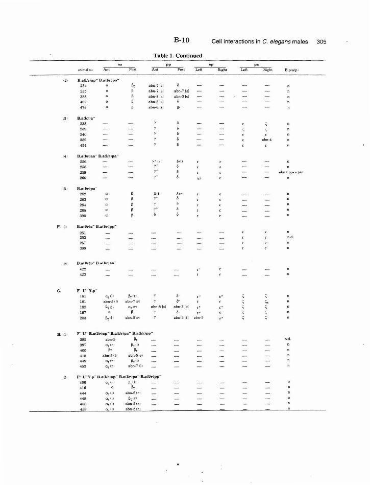
B-10 Cell interactions in C. elegans males 305
Table 1. Continued
aa pp ap p a :mimal nu. Ant Post Ant Post Left Right Left Right B .p< a/p<
t2 1 B.a(l/r)ap- B .aCl/r )pa-
234 flt abn-i lol s 235 " ~ abn-i [s j a bn-i lsl
388 ~ abn·S lsi abn·31ui
402 ll ~ abn-8 lsi s 4i8 " p abn-6 lsi s·
<31 B.aCVrla-238 239
240 ' 389 abn-4
454
i41 B.aU/rJaa- B.aU/r )pa-
256 ·r"' trl St l l n
258 '!'" 0
259 y • abn t .pp->.pa t
260 •("' {( I
15 1 B.alllrlpa-282 " Otl l Otr' n
283 " '! ... 0
284 " 285 "
._, .. 390 a
F . 11 1 B.a<l/rla- B.a(llr)p p -
251 n
252 n .d.
25i
399
12 1 B.a(l/r)p - B.a(l/r)aa-
422 ,. 423
G. r-u- v .p-
161 ~1)1 ~IT I o· E"' , .. 18 1 abn-5 c! J abn-i trl &'
" n
182 lltn , o::...tr t abn-5 lsi abn-3 [o] ,. ,. lSi ll ~ ,. n
203 P,.tl l abn-5 t r t abn-3 lt i abn-5 ,. n
lL t l l F"" v- B.a(l/r)a p - B.a lllr)p a- B.a(l/r )p p -
395 abn -5 llt n.d.
397 Cl.c-ITI ll, li l n
400 p, II, 415 abn-5t l l abn-5 tr l
449 U-r 'T' 11,<11
453 O-r i T I abn-i <It
<2 • r u -y.p - B .a<l/rlap- B.a(llrJpa- B.a!Vrlpp-
406 ~tT l ll,<l l
416 a [;,
444 a, th abn-6 tr l
448 a.,<l t e.r tr l
455 a., <I< abn -5 u·J
458 a.. tl• abn-5 tr l
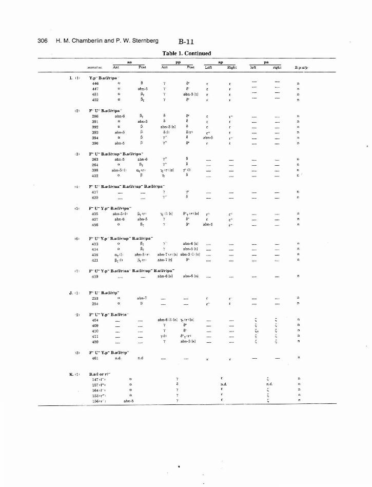
306 H. M. Chamberlin and P. W. Sternberg B-11
Table 1. Continued
aa p p ap pa
anim:~ l no. Ant Post Ant Post Left ffight lei\ right B.palp•
I . t l • Y.p- B.aflfr}pa -
446 (l o• 441 (}. abn-5 s·· 4 5 1 (}. 1>, abn-5 1tl
452 a 1>, 0"'
t2 • F u- B.a(l/r lp a-
286 abn-6 ~, 0"' ,. 391 a abn-5
392 a ~ abn-3 1~1
393 abn-5 ~ li t It Sir • £ .,
394 a ~ ., .. li abn-5 ,. 396 abn-5 ~ y " o•
•3• F u- B.a(l/rlap - B.alllrlpa-
263 abn-5 abn-6 y • n
264 a ll, '( '
395 abn-5tl t D..r ~ T! 'fr: IT ) lsj )" "' n
412 a p Yt li
... F"'" u- B.a{l/r)aa- B.a(l/ r )ap- B.a(l/r )p a -
4li' 1 -r 420 y" 0
t5 • F u- Y.p- B.a(l/r lpa-
405 abn-5 t i t Pt t rt 'lr;ll llt l s .. t tr t lol , .. ,. 40i abn-6 abn-5 o· r £'• n
456 ll, o• abn-5 ,.
16• F"" u-v.p- B.a(l/r)ap- B.a <llrJp a-
41 3 " p, y ~· abn-6 Is!
41 4 ll, a bn-5lt l
416 a..;. t i t abn-5 t r • abn-7 u· t Is) abn-5 tl • lol n
421 j), tl l f.\.r ITI abn-7 ltl o"
ti l r u- Y.p- B.aU/rJaa- B..atllrJa p - B.a<Vr )pa-
419 abn-6 !•I abn-6 !oi n
J , l} l F u- B.a(l/rlp -
253 a abn -i ,. 254 " ,.
•2• F u- Y.p - B.a lllrla-
404 abn-6 tl 1 loJ 'Yo: tr• (o) ; n
409 1 0"' n
410 o· " 411 yt ) l S .. , t rl
459 abn-3 lsi
•3 • F u- Y.p- B .aCI/rlp-
461 n.d. n .d
K.. t] l B.a<l orr)-1471}-1 Ct
lSi n- , a n,d. n.d.
16-t t )- 1 a 153tr- , a 156tr-, abn·5
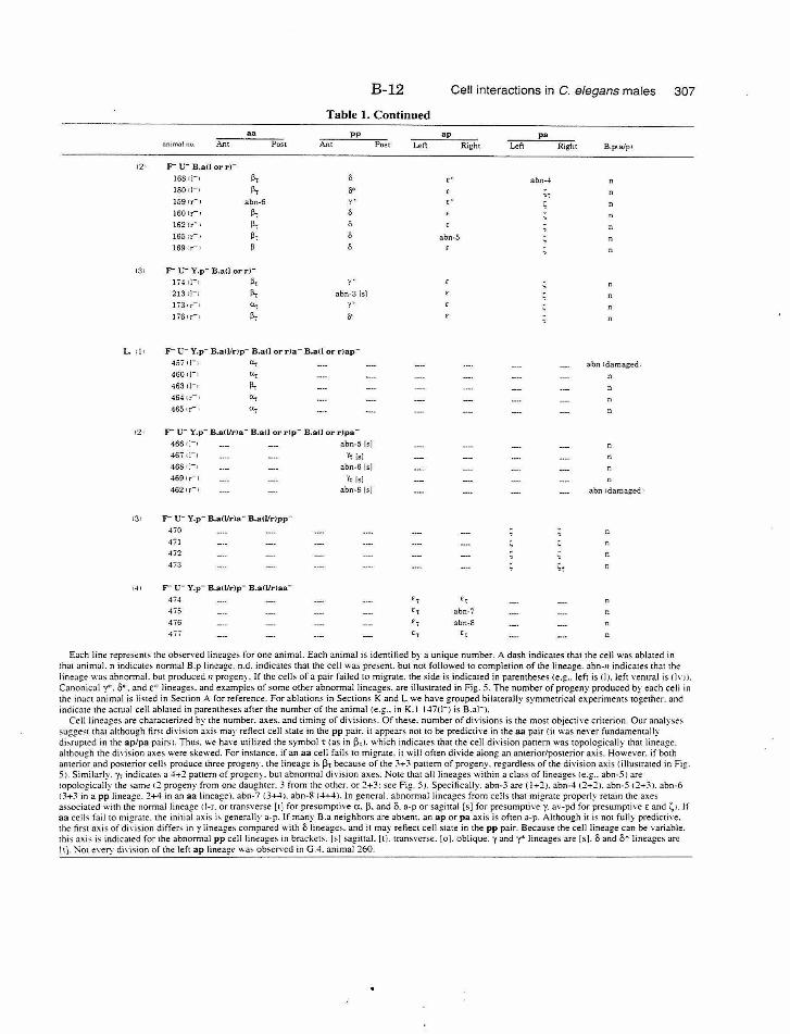
B-12 Cell interactions in C. elegans males 307
Table 1. Continued aa pp ap pa
.1nim3! no Ant Post Ant Post Left !tight Left Right B.p<a/p1
(21 r u- B.aCl or rl-1ss n-~ 1>, ,. abn-4 180 !)-r lit s· ,, 159 t r- ) abn-6 y• ,. <; n 160 tr- ~ 1>, s 162 tr- t 1>, 165tr- , 1>, abn-5 169 tr - t ~
131 r u- Y.p- B.aCl or rl-174 t)- ) 1>, y•
213 tl- · 1>, abn-3 lsi )";'3 IT-I a, '("'
l'i6 tr-+ 1>, s·
L t l • r t;- Y.p- B.a(l/rJp- B.a(l or r )a- B.a<l or r)ap-457 1)- t a, abn 1 dam aged t 460 .,-. a, 463 tl-· 1>, n 464 tr- • a, 465 tr- • a,
121 r u- Y.p- B.aO/r)a- B.a(l or r )p- B.a(l or r)pa-466tl-· abn-5 15.]
467 d-· r, lsi 468 rJ-, abn-61sl 469tr-t <t lsi 462tr- t abn-S 1•1 abn tdamaged 1
131 r u- Y.p- B.all/rla - B.aWrlpp-
470
471
472 n 473 ;,
•4, r u- Y.p- B.a(lfr)p- B.aCI/r)aa-
474 r , r, 475 ,, abn-7
4 76 r, abn-8 n 471 r, r,
Each line represents the observed lineages for one animal. Each animal is identified by a unique number. A dash indicates that the cell was ablated in that animal. n indicate; normal B.p lineage. n.d. indicates that the cell was present. but not followed to completion of the lineage. abn-n indicates that the lineage was abnormal. but produced n progen' . If the cell s of a pair failed to migrate. the side is indicated in parentheses !e.g .. left is (]). left ,·entral is (],· l 1. Canonical y*. 8" . and£" lineages. and examples of some other abnormal lineages. are illustrated in Fig . 5. The number of progeny produced by each cell in the inact animal is listed in Section A for reference. For ablations in Sections K and L we have grouped bilaterally symmetrical experiments together. and indicate the actual cell abla ted in parentheses after the number of the animal (e.g .. in K. l 1~7(1-] is B.a]-).
Cell lineages are characterized by the number. axes. and timing of divisions. Of these. number of divisions is the most objecti\'e criterion. Our analyse~ suggest that although firs t division axis may reflect cell state in the pp pair. it appears not to be predictiYe in the aa pair <it was ne \·er fundamentally disrupted in the ap/pa pairs). Thus. we have utilized the symbol t Cas in (3;1. which indicates that the cell di,·ision pattern was topologically that lineage. although the di\'ision axes were skewed. For instance. if an aa cell fails to migrate. it will often divide along an amerior/posterior axis. However. if both antl!rior and posterior cells produce three progeny. the lineage is ~t because of the 3+3 pattern of progeny. regardless of the di\'ision axis (illustrated in Fig. SJ. Similarly. Y-:. indicates a 4+2 panem of progeny. but abnormal division axes. Note that all lineages within a class of lineages I e .g .. abn-5) are topologically the samt! 12 progeny from one daughter. 3 from the other. or 2+3: see Fig. 5}. Specifically. abn-3 are ( 1+2}. abn-4 12+2). abn-5 12+3). abn-6 13+3 in a pp lint!agc. 2+4 in an aa lineage). abn-7 (3+4). abn-8 (4+4). In general. abnormal lineages from cells that migrate properly retain the axes associated with the normal lineage ct-r. or transverse [t] for presumpti\'e a. p. and 8. a-p or sagiual [s] for presumptive y. av-pd for presumptive E and ~) . If aa cells fail to migrate. the initial ax is i~ generally a-p. If many B.a neighbors are absent. an ap or pa axis is often a-p. Although it is not fully predictiYe. the first axis of di\'ision differ=-- in y lineages compared with 0 lineages. and it may reflect cell state in the pp pair. Because the cell lineage can be variable. this axis is indicated for the abnormal pp cell lineages in brackets . [>) sagittal. [t). transver>e. [o). oblique. -y and-y* lineages are [s]. 8 and 8• lineages arc [t]. Not every di"ision of the \eft ap lineage wa' obserYed in G.4. animal 260.
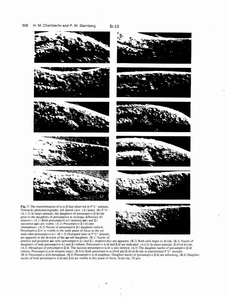
308 H. M. Chamberlin and P. W. Sternberg
Fig. 7. The transformation of a to (l fate observed in F-u- animals. Nomarski photomicrographs. left lateral view. (A) Intact. (B) F-u-. (A.I -3) In intact animals. the daughters of presumptive (l divide prior to the daughters of presumptive a (average difference 40 minute~). (A. I ) Both presumptive a .l (anterior aa l) and (l.J !posterior aal ) are visible. (A.2) Presumptive (l.J divides (metaphase). (A.3) Nuclei of presumptive (l.J daughters reform. Presumptive (l.lv is visible in the same plane of focus as the yet undivided presumptive a.l. (B.l-3) Disrupted fates in F-u- animals are apparent in the division of the aa cell daughters. (B. I ) Nuclei of
B-13
. ... . ... ... . ~ . . -. ~· ~. , ;. -'~ .. -- . . . ' -. '-
~ - .. .. ~ ~ - -; . ·'...."'' ·.""· ~ - ..... - .
~ ............ ·. .... .· . . . -.
anterior and posterior aal cells (presumptive a .l and (l.l. respectively) are apparent. (B.2) Both cells begin to divide. (B.3) Nuclei of daughters of both presumpti ve a.l and (l.J reform. Presumptive a.ld and (l.ld are indicated. (A.4.5) In intact animals.(l.(l/r)d divide. (A.4) Metaphase of presumptive (l.ld. The nucleus presumptive a.ld is also labeled. (A.S ) The daughter nuclei of presumptive (l.Jd reform. Presumptive a.ld remains intact. (B.4-6) Both presumptive a.(Vr)d and (l.(Vr)d divide in transformed F-u- animals. CB.4J Presumptive (l.Jd metaphase. (B.S) Presumptive a .ld anaphase. Daughter nuclei of presumptive (l. ld are reforming. CB.6) Daughter nuclei of both presumptive a.ld and ~.ld are visible in this plane of focus. Scale bar. 20 J.!m.
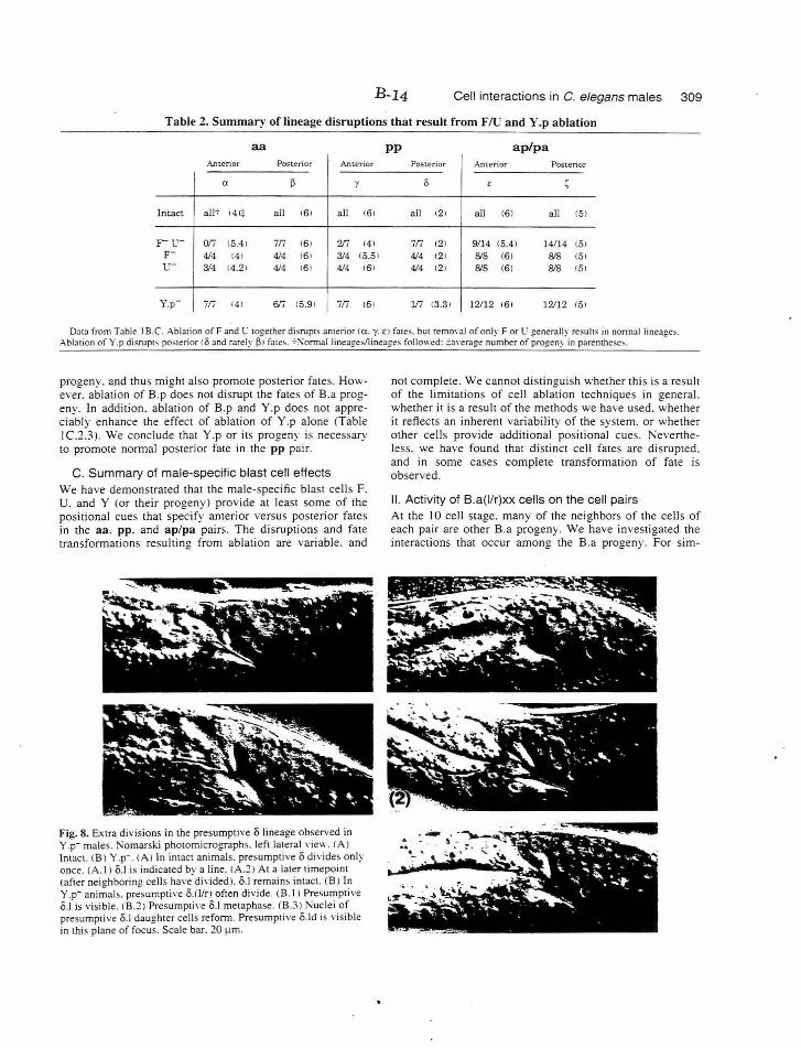
B-14 Cell interactions in C. elegans males 309
Table 2. Summary· of lineage disruptions that result from FlU and Y .p ablation
aa pp ap/pa A.nterior Posterior Anterior Posterior Anterior Posterior
a 13 v 0 £ ~
Intact all~ 141;: all 16 1 all 16 1 all 12 1 all (61 all 151
r v- or; 15.4 1 iri 161 Z/i 141 7n 121 9/14 (5.41 14/14 (51 F- 4/4 t4 1 414 16 1 3/4 (5.5 1 4/4 12 1 &'8 (6) 818 (51 v- 314 t4.2 J 4/4 t6 J 4/4 t6 1 4/4 12 1 818 (6 ) &'8 t5J
Y.p- 717 141 6fi 15.9 1 717 16 1 117 13.3 1 12112 t61 12112 (5 1
Da1a from Table I B.C. Ablation ofF and C together disrupts anterior to. y. £l fa tes. but removal of only F or C generally results in nonnal lineages. Abl:nion of Y .p disrupr... poslerior C 0 and rarely J31 fate~. 7!\onnal lineages/ lineages followed: ::;a.,·erage number of progen~ in parenthese:-..
progeny. and thus might also promote posterior fates. However. ablat ion of B.p does not disrupt the fates of B.a progeny. In addition. ablation of B.p and Y.p does not appreciably enhance the effect of ablation of Y.p alone (Table I C.2.3 ). We conclude that Y .p or its progeny is necessary to promote normal posterior fate in the pp pair.
C. Summary of male-specific blast cell effects We have demonstrated that the male-specific blast cell s F. U. and Y (or their progeny) provide at least some of the positional cues that specify anterior versus posterior fates in the aa. pp. and ap/pa pairs. The di sruptions and fate transformations resulting from ablation are variable. and
Fig. 8. Extra divisions in the presumptive o lineage observed in Y.p- males. Nomarski photomicrographs. left lateral view. CA) Intact. (B 1 Y .p- . (A) In intact animals. presumptive o divides only once. (A. l) o.l is indicated by a line. CA.:?) At a later timepoint (after neighboring cells have divided). o.l remains intact. (B J In Y.p- ani~al s. pr.;sumpti,·e o.(l/ rJ often divide. (B.l I Presumptive o.l is visible. (B.::? I Presumpti,·e o.l metaphase. (8.3 ) Nuclei of presumptive o.l daughter ce lls reform. Presumptive o.ld is visible in thi s plane of focus. Scale bar. 20 ).lm.
not complete. We cannot distinguish whether this is a result of the limitations of cell ablation techniques in general. whe ther it is a result of the methods we have used. whether it reflects an inherent variability of the system. or whether other cells provide additional positional cues. Nevertheless. we have found that distinct cell fate s are disrupted. and in some cases complete transformation of fate is observed.
II. Activity of B.a(l/ r)xx cells on the cell pairs At the 10 cell stage. many of the neighbors of the cells of each pair are other B.a progeny. We have investigated the interactions that occur among the B.a progeny. For sim-
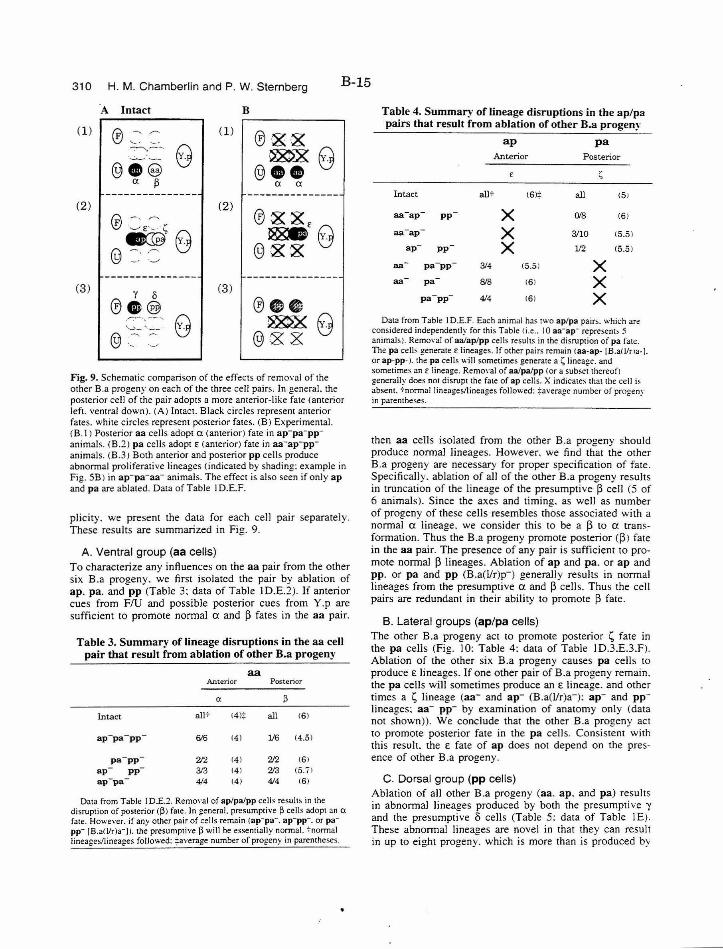
310 H. M. Chamberlin and P. W. Sternberg B-15
·A In tact B
(1) (1) .-:--....,--.
(2) (2)
(3) (3) y 0
® G ®
Fig. 9. Schematic comparison of the effects of removal of the other B.a progeny on each of the three cell pairs. In general. the posterior cell of the pair adopts a more anterior-like fate (anterior left. ventral down). (A) Intact. Black circles represent anterior fates. white circles represent posterior fates. (B) Experimental. (B. I ) Posterior aa cells adopt a (anterior) fate in ap-p a- ppanimals. CB.2) pa cells adopt E (anterior) fate in aa-ap-ppanimals. (B.3) Both anterior and posterior pp cells produce abnormal proli ferative lineages (indicated by shading : example in Fig. SB) in ap- pa- aa- animals. The effect is also seen if only ap and pa are ablated. Data of Table ID.E.F.
plici ty. we present the data for each cell pair separately. These results are summarized in Fig. 9.
A. Ventral group (aa cells) To characterize any influences on the aa pair from the other six B.a progeny. we first isolated the pair by ablation of ap . pa. and pp (Table 3: data of Table I D.E.2). If anterior cues from FlU and possible posterior cues from Y.p are sufficient to promote normal a and ~ fates in the aa pair.
Table 3. Summary of lineage disruptions in the aa cell pair that result from a blation of other B.a progeny
aa Anterior Posterior
a J3
Intact all+ 14 )~ all 16)
ap- pa-pp 616 14) 116 14.51
p a-pp 'l/2 141 'l/2 161 ap - -pp 313 141 213 (5.71 ap-pa - 414 141 414 161
Data from Table I D.E.~ . Removal of ap/palpp cells result, in the disruption of posterior (J31 fate. In general. presumptive J3 cells adopt an a fate. However. if any other pair of cells remain (ap-pa- . ap- pp-. or papp- [B.aiVr)a-)1. the presumptive J3 will be essentially normal. 7normal lineages/lineages followed: ~average number of progeny in parentheses.
Table 4. Summary of lineage disruptions in the ap/pa pairs that result from ablation of other B.a progeny
ap p a Anterior Posterior
~
Intact alit (6)~ all (51
aa- ap p p X 018 (6)
aa- a p X 3/10 15.5) -ap PP X 112 (5.51
-aa -pa-pp 3/4 (5.51 X aa pa &'8 (61 X
p a-pp- 414 161 X Data from Table I D.E.F. Each animal has two ap/pa pairs. which are
considered independently for this Table ti.e .. 10 aa-ap- represent> S animals). Remo,·al of aalap/pp cells resul" in the disruption of pa fate. The pa cells generate E lineages. If other pairs remain taa-ap- [B.a!llr>a· ). or ap-pp-1. the pa cells will sometimes generate a~ lineage. and sometimes an E lineage. RemO\·aJ of aa/pa/pp (or a subset thereof! generally does not disrupt the fate of ap cells. X indicates that the cell is ~bsem. 7normal lineages/lineages followed: :f:average number of progeny m parenthe~es.
then a a cells isolated from the other B.a progeny should produce normal lineages. However. we find that the other B.a progeny are necessary for proper specification of fate. Specifically. ablation of all of the olher B.a progeny resulls in truncation of the lineage of the presumptive ~ cell (5 of 6 animals). Since the axes and timing. as well as number of progeny of these cells -resembles those associated with a normal a lineage. we consider this to be a ~ to a transformation. Thus the B.a progeny promote posterior ( j3 ) fate in the aa pair. The presence of any pair is sufficient to promote normal ~ lineages. Ablation of ap and pa. or ap and pp. or pa and pp (B.a(Vr)p-) generally resulls in normal lineages from the presumptive a and ~ cells. Thus the cell pairs are redundant in their ability to promote ~ fate.
B. Lateral groups (ap/pa cells) The other B.a progeny act to promote posterior 1;, fate in the pa cells (Fig. 10: Table 4: data of Table ID.3.E.3.F). Ablation of the other six B.a progeny causes pa cells to
produce £lineages. If one other pair of B.a progeny remain. the pa cells will sometimes produce an £ lineage. and other times a t;, lineage (aa- and ap- (B.a(l/r)a-): ap- and pplineages: aa- pp- by examination of anatomy only (data not shown)). We conclude that the other B.a progeny act to promote posterior fate in the pa cells. Consistent with this resull. the £ fate of ap does not depend on the presence of olher B.a progeny.
C. Dorsal group (pp cells) Ablation of all other B.a progeny (aa. ap. and pa) results in abnormal lineages produced by both the presumptive y and the presumptive 8 cells (Table 5: data of Table I E). These abnormal lineages are novel in that they can resull in up to eight progeny. which is more than is produced by
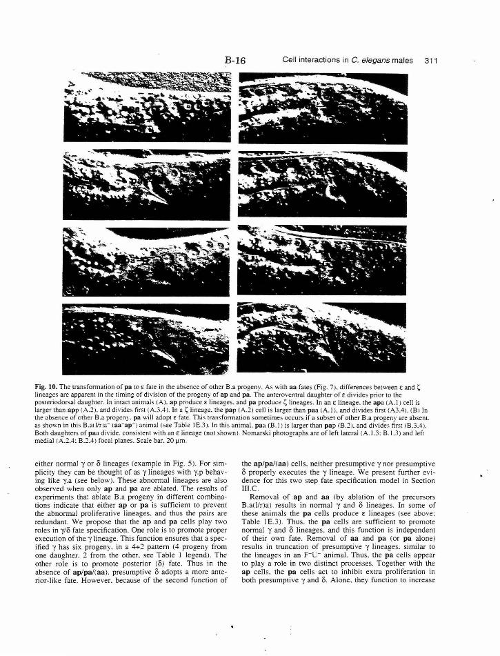
B-16 Cell interactions in C. elegans males 311
~ ' .. -~ 'tt.: " .. . . . { -- ... -~ --- .. "\. ,.... ~ .. '• . . ~ ~ -.W... "'
. .. .. ..... .. .. ~ Ctf. . - -
Fig. 10. The transformation of pa to f fa te in the absence of other B.a progeny. As with aa fates (Fig. 7 ). differences between E and ~ lineages are apparent in the timing of division of the progeny of ap and pa. The anteroventral daughter of E divides prior to the posteriodorsal daughter. In intact animals (A). ap produce E lineages. and pa produce ~ lineages. InanE lineage. the apa (A.I) cell is larger than app <A.2). and divides first (A.3.4). In a~ lineage. the pap (A.2) cell is larger than paa (A. I). and divides first (A3.4). (B) In the absence of other B.a progeny. pa will adopt f fate. This transformation sometimes occurs if a subset of other B.a progeny are absent. as shown in this B.al 1/r)a- laa-ap-l animal <see Table I E.3 l. In this animal. paa (B.I) is larger than pap (B.2). and divides first (B.3.4). Both daughters of paa divide. consistent with an E lineage (not shown). Nomarski photographs are of left lateral (A.I.3: B.l.3) and left medial (A.2.4: B.2.4) focal planes. Scale bar. 20 J.lm.
either normal y or 8 lineages (example in Fig. 5). For simplicity they can be thought of as y lineages with y.p behaving like y.a (see below). These abnormal lineages are also observed when only ap and pa are ablated. The results of experiments that ablate B.a progeny in different combinations indicate that either ap or pa is sufficient to prevent the abnormal proliferative lineages. and thus the pairs are redundant . We propose that the ap and pa cells play two roles in y/8 fate specification. One role is to promote proper execution of the -y lineage. This function ensures that a specified y has six progeny. in a 4+::! pattern (4 progeny from one daughter. 2 from the other. see Table I legend). The other role is to promote posterior (8) fate. Thus in the absence of ap/pa/(aa). presumptive 8 adopts a more anterior-like fate. However. because of the second function of
the ap/pa/(aa) cells. neither presumpti ve y nor presumptive 8 properly executes the y lineage. We present further evidence for this two step fate specification model in Section Ill.C.
Removal of ap and aa (by ablation of the precursors B .a{llr)a) results in normal -y and 8 lineages. In some of these animals the pa cells produce f. lineages (see above: Table I E.3 ). Thus. the pa cells are sufficient to promote normal y and 8 lineages. and this function is independent of their own fate. Removal of aa and pa (or pa alone) results in truncation of presumptive y lineages. similar to the lineages in an F-u- animal. Thus. the pa cells appear to play a role in two distinct processes. Together with the ap cells. the pa cells act to inhibit extra proliferation in both presumptive y and 8. Alone. they function to increase
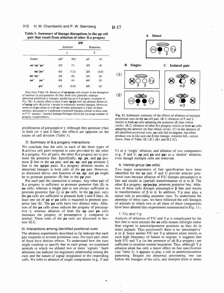
312 H. M. Chamberlin and P. W. Sternberg
Table 5. Su m m ary of lineage d isruptions in the pp cell pair that result from ablation of other B.a progeny
p p . .L\nterior Posterior
y 0
Intact a!l7 <61:j: all (21
aa-ap- pa - 0/3 <71 0/3 <61
ap-pa 0/5 (7.21 2/5 (3.61
aa- ap 515 161 5/5 (21
aa pa 0/4 (4) 414 ( 2 )
pa l/5 <3 .61 5/5 <21
Data from Table I E. Remo,·aJ of aa/ap/pa cells resuhs in the disrup1ion of anterior 1y1 and posterior eli! fate. B01h cells generally undergo abnormal proliferati\·e lineages. producing up to 8 progeny l ex ample in Fig. 58 ). A similar effec1 is seen if only ap/pa celb are ablated. Removal of aa/ap celb (B.aWna-1 resuhs in essentiallv normal linea~es. However. remm·al of pa !alone or with aa 1 disrup1s pre~umptive y f3le. In these
· anima b . presumpti\'e y undergoes truncated lineages similar to those seen in F-u- animals. -:-normal linea£!es/linea1!es followed: ~a\'erat!e number of progeny in parenrheses. - - ..
proliferation of presumptive y. Although they promote y fate in both (or y and o fate). the effects are opposite on the extent of cell division (Table 5 ).
D. Summary of B.a progeny interactions We conclude that the cells in each of the three types of B.a(l/r)xx cell pairs respond to cues provided by the other B.a progeny. For all pairs. the other B.a progeny act to promote the posterior fate. Specifically. ap. pa. and pp promote 13 fate in the aa pair. and aa. a p . and pp promote t; fate in the ap/pa pairs. B.a progeny ablation results in abnormal lineages in both cells of the pp pair. However. as discussed above. one function of aa. ap. and pa might be to promote posterior (o) fate in the pp pair.
For each pair the interaction is uniqut:. Any other pair of B.a progeny is sufficient to promote posterior fate (l3) in aa cells. whereas a single pair is not always sufficient to promote posterior fate (s) in pa cells. In the pp pair. only the p a cells are sufficient to promote both y and o fates. At least one set of ap or pa cells is required to promote posterior fate (o). The pa cells have two distinct roles. Ablation of the pa cells alone reduces the progeny of presumptive y. whereas ablation of both the ap and pa cells increases the progeny of presumptive y. compared to normal. These roles of the pa cells are discussed in Section III.C.
Ill. Interactions among identified positional cues The ablation experiments described so far indicate that each pair responds to several positional cues. For each pair. most of these have distinct effects. To understand how the cues might combine to specify fate in each group. we examined animals in which we ablated two or more ·signals.· Such ablations can indicate the regulatory relationship among the cues and the nature of signal integration in the responding cells. We refer to ablation of single components (e.g .. F and
B-17
A Intact
B Singles C Isolated pair
(1) ~ @OO@
13 13
(2) ~ 8 ••8 a a
Fig. 11. Schematic summary of the effects of ablation of multiple positional cues on the aa cell pair. (B.I) Ablation ofF and U results in both aa cells adopting the posterior <13J fa te (white circle) <B.2) Ablation of other B.a progeny results in both aa cells adopting the anterior (a) fate (black circle). !CJ In the absence of all identified positional cues. aa cells fail to migrate. but often produce one a-like and one 13-like lineage. Anterior left. ventral down. Data of Table IB.I :D. I (B). and H.2 CC).
U) as a ·single· ablation. and ablation of two components (e.g .. F and U. ap and pa and pp) as a 'double· ablation. even though multiple cells are removed.
A. Ventral group (aa cells) Two major components of fate specification have been identified for the aa pair. F and U provide anterior positional cues because ablation of FlU disrupts presumptive a fate and results in (partial) transformation of a to 13. The other B.a progeny. ap/palpp. promote posterior fate. Ablation of these cells disrupts presumptive 13 fate and results in transformation of 13 to a . In addition. Y.p may play a minor role in providing posterior cues. To understand the interplay of these cues. we have followed the cell lineages of animals in which two or all three of these components have been ablated (key experiments summarized in Fig. I I).
1. F/U and Y.p Analysis of ablations of FlU and Y.p is complicated by the fact that in most animals the aa cells remain left/right rather than migrate to anterior/posterior positions as they do in intact animal s. Thus positionally there is no ·presumptive· a or~· Since neither FlU nor Y.p ablation alone results in such high frequency of failure to migrate. it suggests that both FlU and Y .p (in the presence of all B.a progeny) are sufficient to promote normal migration. Thus. although Y.p ablation alone has only a minor effect on fate specification (see Table 2), it appears to play a role in anterior/posterior patterning. Despite the abnormal positioning. one can follow the lineages of the cells. and interpret them in terms
B-18 Cell interactions in C. elegans males 313
of the lineages that those cells would normally produce. However. the division axes of the abnormally positioned cells are usually abnormal.
The aa cells in F-u-Y.p- animals can produce both aand .(3-like lineages. although some abnormal lineages are also observed (2/5 animals abnormal. Table I G). In each of the three animals without abnormal lineages. one of the two aa cells produced an a-like lineage. and the other produced a I)-like lineage. Since a lineages were observed in 315 F-u-Y.p- animals while no a lineages were observed in 7 F-u- animals. the removal of Y.p may partially counteract the absence of F and U in specification of fate. Thus although single ablation of Y.p results in only a minor disruption of fate in the aa cells. ablation of Y.p together with FlU indicates that the aa cells can respond to Y.p cues.
2. F/U , and ap/pa/pp Ablation of the two primary components of aa fate specification. FlU and ap/pa/pp. does not clearly resemble one or the other single ablation (Table I H.l ). As in the F-u-y.p- double ablation. the aa cells tend to remain side by side. and some abnormal lineages are observed. In contrast to the single ablation of ap/pa/pp. we observe many I)-like lineages. Since the I) to a transformations that occur upon ap/pa/pp ablation are dependent on the presence of F and U. one of the roles of the other B.a progeny may be to modulate (inhibit. regulate. or otherwise localize) the FlU activity. However. the B.a progeny must also have an active role of their own. antagonistic to FlU. since the double ablation does not simply ;esemble the single ablation of FlU.
3. F/U Y.p. and ap/pa/pp Removal of all three components (flU. Y.p. ap/pa/pp) isolates the aa pair from all characterized components of fate specification (Table IH.:?.J. The majority of the cells remain left/right <and thus first division axes are a-p rather than 1-r). but both a-like and I)-like lineages are observed. In each animal. one cell produces four progeny suggesting it is alike. and the other cell produces six (or sometimes five) progeny. often in the 3+3 pattern associated with the I) fate. Thus patterning of the two cells is apparent. although the cells do not migrate properly and the extracellular cues that we have identified are absent. There might be other factors that interact with the aa cells to promote their fates. However. since the aa cells remain side by side. these factors are unlikely to be providing anterior-posterior positional cues. A more satisfactory explanation is that the two aa cells interact with each other. In the presence of the cues provided by the other cells this lateral interaction may act to reinforce the positional information. to ensure the result of one cell with each fate. However. in the absence of those cues the cells can interact to establish one cell adopting an a-like fate. and one a I)-like fate.
4. Interaction between aa cells a. B.al- or B.ar- background
To further characterize the possible role of interaction between the two aa cells in promoting fate. we have carried out a series of experiments in which a single aa cell remains in different ablation backgrounds (Table I K). We eliminated one of the aa cells by ablating either the pre-
cursor B.al or B.ar. These ablations also eliminate all of the other B.a progeny on one side. Although we have evidence that the other B.a progeny can play a role in <XII) fate specification. in pairwise ablations their activity is redundant. In addition. all B.aJ- or B.ar animals have the same ablation background. so comparison among these experiments can provide some information about the contribution of interaction between aa cells in the specification of fate.
Ablation of B.al or B.ar leaves one aa daughter. but the other positional cues (from FlU. Y.p. and potentially from the remaining B.a progeny) are intact. Four of five of the aa cells in this case produce an a lineage (one abnormal). This result is consistent with the results of Sulston and White ( 1980). who found that if a single aa cell is ablated. the remaining cell will produce an a lineage. We find that ablation of F and U in a B.aJ- or B.ar- animal results in the single aa cell producing a I) lineage. Therefore. it is F and U that promotes a fate in the single aa cell. In the absence of F and U. the remaining B.a progeny and Y.p promote the I) fate . If both FlU and Y.p are ablated in a B.aJ- or B.ar- background. the remaining aa cell still usually adopts the posterior fate (Table I K.3 ). although the presence of the remaining B.a progeny is apparently not always sufficient to promote. I) fate. We have not distinguished whether this is because Y.p is absent. or because the full set of B.a progeny is not present. Nevertheless. in general. single aa cells can respond to positional cues.
b. Isolated B.a(l or r)aa We isolated single aa cells in five animals by removing FlU. Y .p. and all of the B.a progeny except for one of the aa cells (Table IL.l ). In four cases this cell underwent an a-like lineage. and in one case it underwent a I)-like lineage. Thus in the absence of all identified cues. aa cells generally adopt a fate . However. because of the variable results. we have not established a as the ·ground state·. Another possibility is that the 'isolated' aa cell chooses between a and I) fate stochastically.
5. Summary of experiments in the ventral group Multiple cell interactions play a role in fate specification in the aa pair. FlU. ap/pa/pp. and Y.p contribute external cues to distinguish anterior and posterior fates. as well as to promote proper migration of the cells to an anterior/posterior orientation. This role in promotion of migration is apparent only if more than one component is removed. and is therefore redundant. The presence of any two of the three components is generally sufficient to promote normal migration. whereas the presence of any one is not. Double ablation experiments suggest that one role of the ap/pa/pp cells may be to localize the activity ofF and U. Isolation of the aa pair suggests that the two aa cells interact to specify fate or ensure that the cells each adopt a different fate. Experiments that leave a single aa cell indicate that single cells can respond to the identified positional cues. and that interaction between the aa cells is not required for the adoption or execution of a or I) fate. Isolation experiments. however. did not identify a ·ground state· for aa cells.
Our observations may be consistent with the hypothesis that a and I) lineages represent distinct precursor cell fates. The first division axis may not be critical since essentially
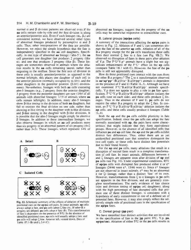
314 H. M. Chamberlin and P. W. Sternberg B-19
normal a and ~ division patterns are observed even if the aa cells remain side-by-side and the first division is along an anterior/posterior axis. Even if such lineage~ (a,. ~.l are considered normal. we have observed several other types of abnormal lineages produced by presumptive a and ~ cells. Thus. other interpretations of the data are possible. However. we reject the simple hypothesis that the fate is independently specified in the aa cell daughters. Specifically. in one type of abnormal aa lineage ( ·abn-5' ). the aa cell generates one daughter that produces 2 progeny (like a). and one that produces 3 progeny (like ~~- These lineages are sometimes observed in animals where the ablation results in the aa cells remaining lateral. rather than migrating to the midline. Since the first axis of division in these cells is usually anterior/posterior. as opposed to the normal left/right. this places one daughter of each cell in the anterior position (normally occupied by a.WrJ). and the other daughters in the posterior position (~.(1/r) environment). Nevertheless. lineages with both aa cell~ executing abn-5 lineages (e.g .. 2 progeny from the anterior daughter. 3 progeny from the posterior daughter) are rare (I /64 of all animals with abnormal lineages: 1/19 of animals where aa cells remain left/right). In addition. many abn-5 lineages show ~-like timing in the division of both aa daughters. but fail to execute the final di vision on one side. rather than showing a-like timing in the daughter with a-like divisions and ~-like timing in -the daught;r with ~-like divisions. It is possible that the abn-5 lineages might simply be abortive ~ lineages. In addition to these intermediate lineages. we al so observe lineages in which an aa cell produces more than six progeny or an abnom1al pattern of six progeny (4+2 rather than 3+3). These lineages. which represent 149c of
A Intact
@ - . £~s0
@ - .. 'V
C Isolated Cells
B
@XX ~£0
@XX v
Fig. 12. Schematic summary of the effects of ablation of multiple positional cues on the ap/pa cell pairs. In intact animals. ap cells alway '< adopt £ fate. and pa cells adopts fate (A). If other B.a progeny are ablated. pa cells adopt£ fate (B). This transformation of fate is dependent on the presence of FlU . In the absence of identi fied positional cues. ap cells will usually adopt £ fate. and pa cells will adopt s fate. Anterior left. ventral down. Data of Table IF. \ CBJ and L.3.41CJ.
abnormal aa lineages. suggest that the progeny of the aa cells may be somewhat responsive to extracellular cues.
B. Lateral groups (ap/pa cells} A summary of the interactions affecting the ap/pa pairs is shown in Fig. 12. Ablation ofF and U can sometimes disrupt the fate of the anterior ap cells. Ablation of all of the B.a progeny except for the pa cells transforms these cells from their normal 1;, fate to E fate (see Fig. 9B.2). We observed no disruption of fates in these pairs after ablation of Y.p. The F-u-Y.p- animals have a slight but not significant enhancement of the F-u- effect on the ap cells (compare Table IB.I with G). However. the fate of the pa cells is still not appreciably disrupted.
How do these positional cues interact with the cues from the other B.a progeny'? The 1; to E transformation observed in aa-ap-pp- (B.a(l!r)a- B.a(l/r)pp-) animals is dependent on the presence ofF and U (Table I L.3 ). Although we have not examined F-u-B.a(l/r)a-B.a(llr)pp- animals specifically. Y.p does not appear to play a role in pa fate specification. F- u-Y.p-B.a(Ur)a-B.a()!r)pp- ablation isolates the pa cells (one on each side). and these cells produce 1; lineages. Thus in the absence of F and U. pa cells do not require the other B.a progeny to adopt the L; fate. In contrast. an F-u- Y.p-B.a(llr)p-B.a()/r)aa- ablation isolates the ap cells. and these cells usually produce £ lineages (Table IL.4).
Both the ap and the pa cells exhibit plasticity in fate specification. Indeed. since the pa cells can adopt the fate normally associated with ap. the pairs have some characteristics that might indicate that they form equivalence groups. However. in the absence of all identified cells that influence pa and ap cell fate. the ap and the pa cells exhibit distinct fate differences. Thus. either there are as yet unidentified positional cues that somehow distinguish the two cell types. or these cells have distinct fate potentials due to their lineal history.
For the ap and pa cells. many ablations that result in a disruption of normal fates result in a complete transformation of cell fate . In intact animals. differences between E
and 1; lineages are apparent soon after division of ap and pa cells (see Fig. I 0). Under experimental conditions. 85'7c of ap/pa cells with disrupted fate produced either E or £" lineages (24/46 were E*. I 5/46 were £). Since £" lineages are not observed in intact animals. E" may be an abnormal E (or 1;1 lineage. rather than a distinct ·fate· of its own. However. transformations from L; to E lineages in pa cells are apparent in the first division. and are generally complete transformations. The early evidence of fate choice (size and division timing of ap/pa cell daughters). along with the high percentage of fate disrupted cells that produce one of three distinct lineages. is consistent with a hypothesis of early commitment to one of a defined set of potential fates. However. it may also simply reflect the rel atively simple role of positional cues in the specification of the ap/pa fates .
C. Dorsal group (pp pair) We have identified four distinct activities that are involved in the specification of fate in the pp pairs: FlU. Y.p. pa. ap/pa/(aa). Ablation of either FlU or the pa cells results in
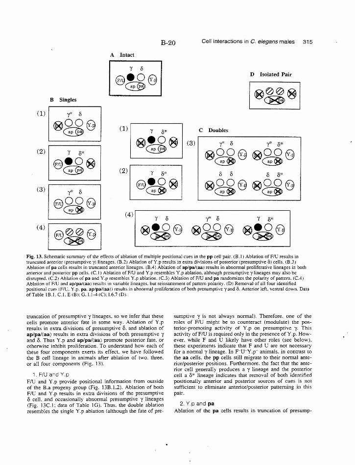
B-20 Cell interactions in C. elegans males 315
(1)
(2)
(3)
(4)
B Singles
~ ~
A Intact
D Isolated Pair
(1) C Doubles
(2) 8 8 8 8*
~8£8 ®8£8 (4)
Fig. 13. Schematic summary of the effects of ablation of multiple positional cues in the pp cell pair. (B.I) Ablation of FlU results in truncated anterior !presumptive y) lineages. <B.2) Ablation ofY.p results in extra divisions of posterior (presumptive O) cells. (B .3J Ablation of pa cells results in truncated anterior lineages. (B.4) Ablation of ap/pa/(aa) results in abnormal proliferative lineages in both anterior and posterior pp cells. (C. I) Ablation of FlU and Y.p resembles Y.p ablation. although presumptive y lineages may also be disrupted. (C.2J Ablation of pa and Y.p resembles Y .p ablation. (C.3) Ablation of FlU and pa randomizes the polarity of paltem. (C.4) Ablation of FlU and ap/pal(aa) results in variable lineages. but reinstatement of paHem polarity. (D) Removal of all four identified positional cues (FlU. Y.p. pa. ap/pal(aa)) results in abnormal proliferation of both presumptive yand 8. Anterior left. ventral down. Data of Table IB.L C. I.E (B): G.l.l-4 (C): 1.6.7 (D).
truncation of presumptive y lineages. so we infer that these cells promote anterior fate in some way. Ablation of Y.p results in extra divisions of presumptive 8. and ablation of ap/pa/(aa) results in extra divisions of both presumptive y and 8. Thus Y.p and ap/pa/(aa) promote posterior fate. or otherwise inhibit proliferation. To understand how each of these four components exerts its effect. we have followed the B cell lineage in animals after ablation of two. three. or all four components (Fig. 13).
1. F/U and Y.p FlU and Y.p provide positional information from outside of the B.a progeny group (Fig. 13B.I.2). Ablation of both FlU and Y.p results in extra divisions of the presumptive 8 cell. and occasionally abnormal presumptive y lineages (Fig. 13C.l: data of Table I G). Thus. the double ablation resembles the single Y.p ablation (although the fate of pre-
sumptive y is not always normal). Therefore. one of the roles of FlU might be to counteract (modulate) the posterior-promoting activity of Y.p on presumptive y. This activity of FlU is required only in the presence ofY.p. However. while F and U likely have other roles (see below). these experiments indicate that F and U are not necessary for a normal y lineage. In F-u-y .p- animals. in contrast to the aa cells. the pp cells still migrate to their normal anterior/posterior positions. Furthermore. the fact that the anterior cell generally produces a y lineage and the posterior cell a 8* lineage indicates that removal of both identified positionally anterior and posterior sources of cues is not sufficient to eliminate anterior/posterior patterning in this pair.
2. Y p and pa Ablation of the pa cells results in truncation of presump-
316 H. M. Chamberlin and P. W. Sternberg B-21
tive y lineages. and ablation of Y .p results in extra divisions of the presumpti ve 8 cell (Fig. 138.2.3). In the pp pair. the double Y.p- pa- animals resemble Y .p- animals (Fig. 13C.2: data of Table l 1.1 ). Thus. one of the roles of the pa cells is to inhibit Y.p activity from influencing the fate of the presumptive y cell. Thi s fu nction is only required if Y.p is present. In addition. thi s experiment suggests how the positionally posterior pa cells can act to promote an anterior fate. The position of the pa cells (see Figs 3E. 48.2) suggests this role may be ·passive.' Specifically. the pa cells may physically block the activity of Y.p from reaching the presumptive y cell. This proposed mechanism does not require that the pa cells be biochemically distinct from the other 8 .a progeny. although it also does not rule out this possibility.
3. F/U and pa Single ablations of either FlU or pa result in intermediate and variable disruption of presumptive y lineages (Fig. 138.1.3 ). To establish whether complete transformations from the y to the 8 fate could be achieved by removal of both components. we characterized F-u -pa- animals (Fig. l3C.3: data of Table I l.2J. While this double ablation does not result in an increased frequency of complete y to 8 transformations. the normal pattern of anterior/posterior polarity is disrupted. In either F-u- or pa- animals. the fate of presumptive 8 is not disrupted. whereas presumptive y lineages are often truncated. Thus. two patterns are observed: normal polarity and apolar. In contrast. double ablation of FlU and pa can result in a fate disruption of both presumptive y and 8 cells. In addition. we observe all possible classes of pattern: normal polarity. apolar. and reversed polarity.
4. F/U and ap/pa/(aa) Single ablation of ap/pa/(aa ) results in novel lineages and abnormal proliferation of presumptive y and 8. whereas ablation of FlU results in truncated lineages produced by the presumptive y cell (Fig. 138.1.4). The double ablation of FlU and ap/pa or ap/pa/aa (considered together as ap/pa/(aa)) has allowed us to determine whether the excessive proliferation results in part from inappropriate modulation of FlU information. In F-u-ap- pa- (aa-) animals. lineages of presumptive y and 8 do not closely resemble those seen in either the single F-u- or ap-pa-(aa- ) animals (Fig. 13C.4: data of Table l 1.3.4). Proliferati ve lineages are not observed. Although many of the lineages are slightly abnormal. we only observe the more common lineages: normal y and 8. and y"'/8*. However. the presence of Y.p is not sufficient to ensure 8 fate in the posterior cell. Disruption of both anterior and posterior fates is observed. although not in the same animal. In Section II.C we proposed that the ap/pa/(aa ) cell s have two function s: promotion of posterior fate (0) and promotion of proper execution of the y lineage. The double ablation result indicates that FlU may likewise act at two steps: promotion of anterior fate (YJ and execution of y lineage. We hypothesize that one role of the FlU activity for y fate execution is to promote pp proliferation. The ap/pa/(aa) cells counteract thi s activity. thus localizing it to y.a (or y.a(l!r)). Since removal of both FlU and ap/pa/(aa) results in more normal lineages than ei ther
single ablation. we conclude that FlU and ap/pa/(aa) act antagonistically and in parallel on the same process(es).
One consistent characteristic among F-u- ap-pa-(aa- J animals is that the fate of the positionally anterior pp cell is relatively more ·anterior· than the fate of the positionally posterior pp cell : the pair exhibits polarity of pattern. In other words. anterior/posterior polarity. which is lost in F-u-pa- animals. is regained by the additional ablation of the ap (compare Fig. 13C.3 with C.4). Y.p. the posteriorpromoting cell. can promote polarity in the pp pair in the absence of FlU and ap/pa/(aa). Since polarity can be disrupted in F-u- pa- animals. the presence of ap must somehow counteract this activity of Y.p.
5. F/U, Y.p , and ap/pa/(aa) We have removed all of the identified components that specify fate in the pp pair (Fig. 130). As with the aa pair. this isolation may allow identification of potential interaction between pp cells. The normal lineages and patterning observed in F-u-ap-pa-(aa-) animals are lost with the removal of Y.p (Table 1 1.6.7). A variety of proliferative lineages are observed from both presumptive y and 8. Although there is no appreciable difference between presumpti ve y and presumptive 8. proliferation may be less than in ap- pa-(aa-) animals. Thus. without the identified cues. no polarity or evidence for interaction between the pp cells is readily apparent. However. the lineages are abnormal and variable enough that they are not easily interpreted.
A difference between the aa and pp cells is that the pp cells generally migrate to anterior/posterior positions even in the absence of the identified extracellular cues. Althou!!h this may indicate that anterior/posterior patterning cues for the pp cells may still be present. it may also reflect a difference in the physical environments within which the two pairs reside. The ventral area (aa environment) is relatively smaller and more crowded than the dorsal area (pp environment). Thus. in the absence of positional cues the aa cells might not be able to migrate medially. whereas the pp cells can. We cannot. however. rule out the possibili ty that additional cues exist for pp cells.
6. Interaction between pp cells a. B.al- or B.ar background
Isolation of both pp cells did not provide direct evidence that they interact with each other to specify their fates. However. we have also characterized the lineages of single pp cells obtained after ablation of the precursor 8 .al or 8 .ar (Table I K). Four of fi ve single pp cells obtained by ablation of 8 .al or B.ar produced a y lineage. whereas one produced a 8 lineage. Thus. interaction between pp cells is not essential to produce either normal y or 8 fates. In these animals. it is possible that choice between the two fates is related to the relative anterior-posterior positioning of the single pp cell in the normal pp environment. Adoption of the y fate. however. is dependent on the presence of F and U. Remaining pp cells in F-u- [8.al- or 8.ar-] animals produce 8 lineages (5/7) or y"'/8* lineages (2/7 ). Likewise. the 8 fate is dependent on the presence of Y.p. Single pp cells in F- u - Y.p- [B.al- or B.ar-] animals produce y"'/8* (3/4 y"'/8*: 1/4 abn-3) lineages. We do not believe this
B-22 Cell interactions in C. e/egans males 317
effect is merely the result of fewer neighbors (or more debris). because even single isolated pp cells can produce y lineages (see below) . These observations are consistent wi th the proposed roles of FlU and Y.p in promoting y and 8 fates. respectively.
Loss-of-function mutation~ in lin-12. a gene known to
play a role in the interactions between cells in other equivalence groups. result in 8 to y fate transformations (Greenwald et al.. 1983 ). This observation implies that the pp cells do interact. by analogy to other /in-12-dependent equivalence groups. Although isolation of the two pp cells did not offer evidence to suppon this hypothesis. comparison of F-u-Y.p- to F-u-Y.p-[B.al- or B.ar-] animals is suggestive. Specifically. the single pp cells in F-u-Y.p-[B.aJ- or B.ar-] animals do not produce y lineages (0/4 are y). whereas in F-u-y.p- animals. where both pp cells are present. the anterior cell generally will produce a y lineage (415). Although these animals also differ in the total number of B.a progeny present. we suggest that the y fate in the F-u-Y.p- animals may result from interaction between the two pp cells. If the pp cells do interact. it is possible that the interaction is not apparent in ap-pa-(aa- ) animals because the ap/pa/(aa) cells are necessary either for this interaction to occur. or for all aspects of the fates to be properly executed.
b. Isolated B.a(l or r)pp After ablation of FlU. Y.p. and all B.a progeny except a single pp cell. we observed y-like lineages in two of five animals. and abnormal proliferative lineages in three of five animals (Table IL.2). We conclude that a pp cell requires positional cues to adopt the correct fate. Although the isolated fate can be y-like. extracellular cues are apparently required to reliably ensure that the fate is properly executed. As is the case for isolated aa cells. there is no clear ·ground state· for the pp cells.
7. Summary of experiments in the dorsal group The results of our removal of identified components of fate specification for the pp pair suggest how some of the cues may interact. The pa cells likely promote anterior fate by inhibiting or localizing the activity of Y.p. and the ap cells likewise may inhibit or localize the activity of FlU. One of the roles ofF and U is to counteract Y.p. However. F and U have additional roles in the promotion of fate. For instance. comparison of ap-pa-(aa-) to F-u-ap-pa-(aa-) animals. both of which have Y.p intact. suggests that F and U play a role in producing the abnormal. proliferative lineages. Likewise. although F and U are not required for the production of y lineages in F- u-Y.p- animals. in a [B.alor B.ar] background. y lineages are only seen if F and U are present.
In most experimental animals the cells move roughly anterior/posterior relative to each other. as in intact animals. This movement allows the additional analysis of patterning and polarity within the pp pair. Removal of the positionally anterior and posterior FlU and Y.p cells still results in normal polarity within the pp pair. Thus either additional. unidentified cues can establish this polarity. or polarity information can come from the other B.a progeny . Analysis of F-u-pa- animals suggests that the latter explanation
may indeed be the case. since the pattern appears random in these animals. However. polarity is restored after removal of the ap cells in this background. Therefore positional cues are not absent in the F-u-pa- animals. but they are either inhibited or somehow equally balanced. There is no obvious polarity in F-u-Y.p-ap-pa-(aa-) animals. However. the abnormality of the resultant lineages makes it difficult to score polarity.
Although there is some evidence for lateral interactions between the pp cells. the ablation results are less conclusive in the pp pair than in the aa pair. Experiments that include a single pp cell suggest that interaction between the pp cells is not necessary for the specification of y and 8 fates. and isolation of the pp pair does not provide evidence that the homologues interact. However. in F-u-Y.p- animals y lineages are observed if both pp cells remain. but not if only one remains. suggesting that the two pp cells may have the potential to interact.
In contrast to our initial hypothesis. we believe that specification of pp lineages involves two steps based on (I) the nature of the '('/8* lineages. and (2) the existence of the abnormal proliferative lineages. The aspects of pp cell fate that we have used in our analysis are the axis of pp cell division (sagittal versus transverse: see Table I legend ) and the number of progeny produced by each pp cell. Considering these criteria. 8* lineages include characteristics of both y and 8 lineages: the initial division is transverse (8-like). but each cell divides again. Thus the pp division axis can be uncoupled from the number of progeny produced. Similarly. Y lineages have an initial sagittal division (like normal y lineages). but only two rounds of division. In abnormal proliferative lineages the pp cell produces up to eight progeny. Although the first division of an abnormal proliferative lineage is often sagittal (y-like), the axes and timing of divisions and the placement of progeny are highly variable. We propose that these abnormal types of lineages reflect two steps in lineage specification: an earlier step (possibly reflected in division axis) and a later step (reflected in proliferation). This .two step process model of lineage specification distinguishes the pp pair because it suggests that y and 8 fates do not result from the simple choice and execution of distinct sublineages. but rather a series of decisions influenced by extracellular cues. Thus although y and 8 are distinct pp fates. they do not represent distinct sublineages.
IV. Differences between B.a(l/r)a and B.a(l/r)p Our ablation results suggest that there is a fundamental difference in fate potential between the aa pair and the pp pair. For instance. single isolated aa cells generally produce a like lineages (average number of progeny=4.4). whereas single isolated pp cells generally produce more proliferative and variable lineages (av. prog.=6.2). The cells also respond differently in the same ablation backgrounds. In addition. although the siblings of the aa cells (ap) and the siblings of the pp cells (pa) can respond to extracellular cues. the behavior of these cells when isolated from the identified cues suggests that they may also be inherently different from one another. Specifically. isolated pa cells produce ~ lineages. and isolated ap cells generally produce £ lineages. the fates associated with these cells in intact animals. Since we now
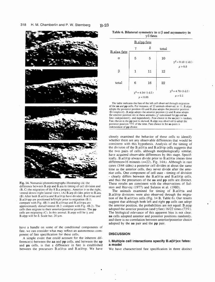
318 H. M. Chamberlin and P. W. Sternberg B-23
Fig. 14. Nomarski photomicrographs illustrating (AI the difference between B.arp and B.ara in timing of cell division and <B. C) the migration of the 8 B.a progeny. Anterior is to the right. ventral down <right lateral view). IAI B.arp di,·ides prior to B.ara. <B! After both B.a(!lna and B.a(!/rJp ha,·e divided. B.a<l/r)aa and B.a(!lrJpp are positioned left/right prior to migration cB. I: compare with Fig . 4B. I l and B.a<l/rJap and B.a<l/r)pa are approximately dorsal/ventral <B.2: compare with Fig. 48.2). The cells then migrate to their anterior/posterior positions. The p p cells are migrating <Cl. In this animal. B.arpp will bey. and B.alpp w ill be o. Scale bar. 20 J.lm.
have a handle on some of the conditional components of fate. we can consider what may reflect an autonomous component of fate specification for these cells.
A simple event that could account for the inherent difference(s) between the aa and pp cells. and between the ap and pa cells. is that a difference in fate is established between the precursors B.a(l/r)a and B.a(]/r)p. We have
Table 6. Bilateral symmetry in a/~ and asymmetry in y/8 fates
B.alppfate
y 8 total B.alaa fate
a. 5 5 10 ;.:2 = o.1s n d.f.>
p > 0.5
(3 1 11 12
total 6 16 22
x2 = 4.54 c 1 d.f.J x2 =4.78<3d.f. J
p < 0.05 p > 0.1
The table indicat"' the fate of the left cell observed throu2h mioration of the aa and pp cells. For instance. of 22 animals observed~ in I J. B.alpp adopts the posterior position (OJ and B.alaa adopts the posterior position !J3 J !implicitly. B.arpp adopts the anterior position (YI and B.araa adopts the anterior position caJ in these animals). x= calculated for pp and aa fates independently. and dependently . Fate choice in the aa pair is random. Fate choice in the pp pair is skewed. B.alpp was obsef\·ed to adopt the posterior position 7?tf7r of the time. Fale choice in the aa pairs i ~ independent of pp choice.
closely examined the behavior of these cells to identifv whether there are any observable differences that would b~ consistent with this hypothesis. Analysis of the timing of the diYision of the B.a(l!r)a and B.a(l/r)p cells suggests that the two pairs of cells. although morphologically similar. have acquired observable differences by this stage. Specifically. B.a(l/r)p always div ide prior to B.a(Ur)a (mean time difference=l6 minutes (n=22). Fig. 14A). Although in rare cases (3/44 sides) a posterior cell divides at about the same time as the anterior cells. they never div ide after the anterior cells. One component of cell state - tim ing of division - clearly differs between the B.a(l/r)a and B.a(l/r)p cells. and thus the precursors of the aa and pp cells are distinct. These results are consistent with the observations of Sulston and Horvitz (I 977) and Sulston et al. (1980).
The animal s examined for timing of B.a(l/r)a and B.a(l!r)p divisions were also observed through the migration of the B.a(l/r)xx cells (Fig. 14 B. Table 6). Our results suggest that although both left and right pp cells can adopt the anterior position. the probabilities are not equal: B.arpp adopted the anterior position (andy fate) 16/22 times (73'7<- ). The biological relevance of this apparent bias is not clear. aa cells adopted anterior and posterior positions randomly. and there is no correlation between anterior/posterior choice adopted by the aa pair and the pp pair.
DISCUSSION
I. Multiple cell interact ions spec ify B.a(l/r)xx fates: a model We have characterized fate specification in three distinct
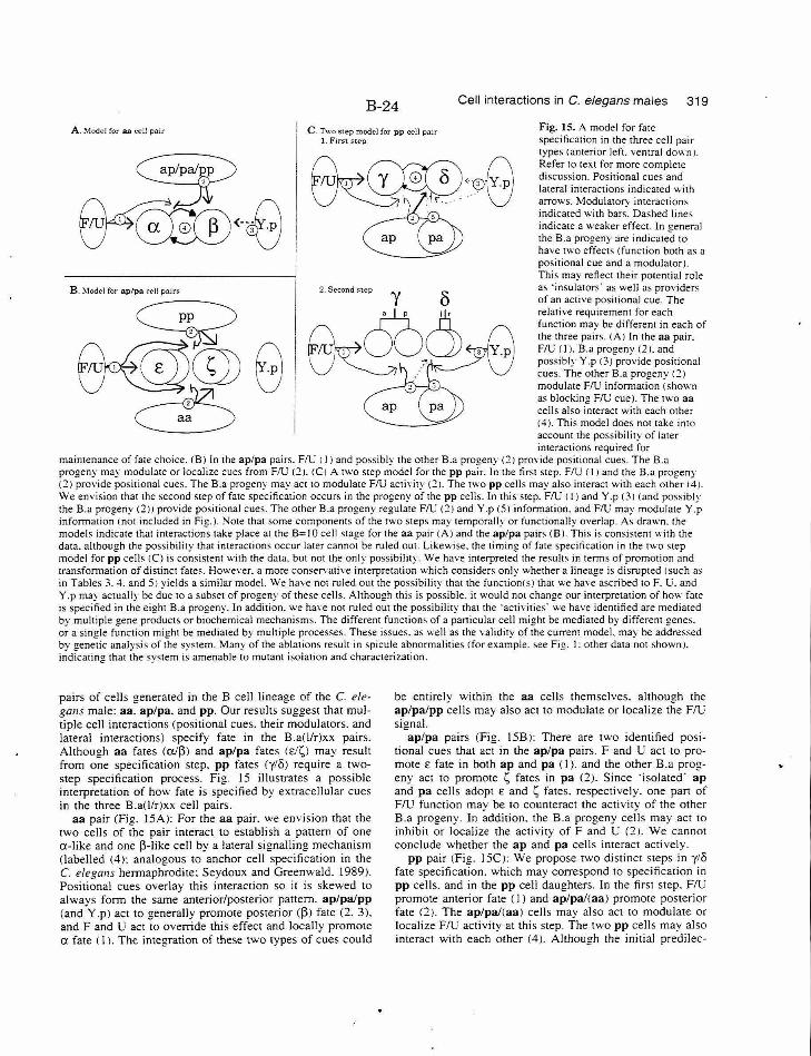
B-24 Cell interactions in C. elegans males 319
A . .!\1odel for aa cell pair C. 1\.l.·o step model for pp cell p<Jir 1. Fin;t step
Fig. IS. A model for fate specification in the three cell pair types (anterior left. ventral down). Refer to text for more complete discussion. Positional cues and lateral interactions indicated with arrows. Modulator• interactions indicated with bar;. Dashed lines indicate a weaker effect. In general the B.a progeny are indicated to have two effects (function both as a positional cue and a modulator). This may reflect their potential role as "insulators· as well as providers of an active positional cue. The relative requirement for each function may be different in each of the three pairs. IAJ In the aa pair. FlU I I J. B.a progeny (2 1. and possibly Y.p 13) provide positional cues. The other B.a progeny <2J modulate FlU information 1 shown as blocking F/lJ cue). The two aa cells also interact with each other {4). Thi s model does not take into account the possibility of later
B . :\1odel for aplpa <'ell pairs
~~<~0 0~ v
,
a
2. Second l'tep Y ()
9;? Ali ~,/7'\\ 0 G~Ou~O~ ~vv~;?V
interactions required for maintenance of fate choice. IB J In the ap/pa pairs. FlU ( I 1 and possibly the other B.a progeny (2) provide positional cues. The B.a progeny may modulate or localize cues from FlU <2 ). I C) A two step model for the pp pair. In the first step. FlU (I 1 and the B.a progeny (2) provide positional cues. The B.a progeny may act to modulate FlU activity 121. The two pp cells may also interact with each other 14). We envision that the second step of fate specification occurs in the progeny of the pp cells. In this step. FlU I I) and Y.p 131 land possibly the B.a progeny 12)) provide positional cues. The other B.a progeny regulate F/l! <21 and Y.p 151 information. and F/U may modulate Y.p information I not included in Fig.). Note that some components of the two steps may temporally or functionally overlap. As drawn. the models indicate that interactions take place at the B= I 0 cell stage for the aa pair (A) and the ap/pa pairs IB ). This is consistent with the data. although the possibi lity that interactions occur later cannot be ruled out. Likewise. the timing of fate specification in the two step model for pp cells iCl is consistent with the data. but not the only possibility. We have interpreted the results in terms of promotion and transformation of distinct fates. However. a more conservative interpretation which considers only whether a lineage is disrupted (such as in Tables 3. 4. and 51 yields a similar model. We have not ruled out the possibility that the fu nctionlsJ that we have ascribed to F. U. and Y .p may actually be due to a subset of progeny of these cells. Although this is possible. it would not change our interpretation of how fate is specified in the eight B.a progeny. In addition. we ha,·e not ruled out the possibility that the ·activities· we have identified are mediated by multiple gene products or biochemical mechanisms. The different functions of a panicular cell might be mediated by different genes. or a single function might be mediated by multiple processes. These issues. as well as the validity of the current model. may be addressed by genetic analysis of the system. Many of the ablations result in spicule abnormalities <for example. see Fig. l: other data not shown 1. indicating that the system is amenable to mutant isolation and characterization.
pairs of cells generated in the B cell lineage of the C. e/egallS male: aa. ap/pa. and pp. Our results suggest that multiple cell interactions (positional cues. their modulators. and lateral interactions) specify fate in the B.a(JJr)xx pairs. Although aa fates (a/~) and ap/pa fates (Ei /;) may result from one specification step. pp fates (y/8) require a twostep specification process. Fig. 15 illustrates a possible interpretation of how fate is specified by extracellular cues in the three B.a(l/r)xx cell pairs.
aa pair (Fig. 15A): For the aa pair. we envision that the two cells of the pair interact to establish a pattern of one a-like and one ~-like cell by a late ral signalling mechanism (labelled (4): analogous to anchor cell specification in the C. elegans hermaphrodite: Seydoux and Greenwald. 1989). Positional cues overlay this interaction so it is skewed to always form the same anterior/posterior pattern. ap/pa/pp (and Y.p) act to generally promote posterior (~) fate (2. 3). and F and U act to override this effect and locally promote a fate (I). The integration of these two types of cues could
be entirely within the aa cells themselves. although the ap/palpp cells may also act to modulate or localize the FlU signal.
ap/pa pairs (Fig. 15B ): There are two identified positional cues that act in the ap/pa pairs. F and U act to promote E fate in both ap and pa (I). and the other B.a progeny act to promote 1; fates in pa (2). Since "isolated· ap and pa cells adopt E and 1; fates. respectively. one part of FlU function may be to counteract the activity of the other B.a progeny. In addition. the B.a progeny cells may act to inhibit or localize the activity ofF and U (2). We cannot conclude whether the ap and pa cells interact actively.
pp pair (Fig. 15C): We propose two distinct steps in y/8 fate specification. which may correspond to specification in pp cells. and in the pp cell daughters. In the first step. FlU promote anterior fate (I ) and ap/pa/(aa) promote posterior fate (2). The ap/pal(aa) cells may also act to modulate or localize FlU activity at this step. The two pp cells may also interact with each other (4). Altho ugh the initial predilec-
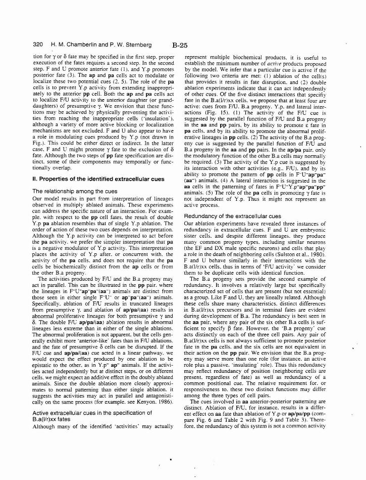
320 H. M. Chamberlin and P. W. Sternberg B-25
tion for y or o fate may be specified in the first step. proper execution of the fates requires a second step. In the second step. F and U promote anterior fate ( I ). and Y .p promotes posterior fate (3). The ap and pa cells act to modulate or localize these two potential cues (2. 5 ). The role of the pa cells is to prevent Y.p activity from extending inappropriately to the anterior pp cell. Both the ap and pa cells act to localize FlU activity to the anterior daughter (or granddaughters) of presumptive y. We envision that these functions may be achieved by physically preventing the acti\·ities from reaching the inappropriate cells (' insulation·). although a variety of more active blocking or localization mechanisms are not excluded. F and U also appear to have a role in modulating cues produced by Y .p (not drawn in Fig.). This could be either direct or indirect. In the latter case. F and U might promote y fate to the exclusion of o fate . Although the two steps of pp fate specification are distinct. some of their components may temporally or functionally overlap.
II. Properties of the identified extracellular cues
The relationship among the cues Our model results in part from interpretation of lineages observed in multiply ablated animals. These experiments can address the specific nature of an interaction. For example. with respect to the pp cell fates. the resull of double Y.p pa ablation resembles that of single Y.p ablation. The order of action of these two cues depends on interpretation. Although the Y.p activity can be interpreted to act before the pa acti vity. we prefer the simpler interpretation that pa is a negative modulator of Y.p activity. This interpretation places the activity of Y.p after. or concurrent with. the activity of the pa cells. and does not require that the pa cells be biochemically distinct from the ap cells or from the other B.a progeny.
The activities produced by FlU and the B.a progeny may act in parallel. This can be illustrated in the pp pair. where the lineages in F-u-ap-pa-(aa-) animals are distinct from those seen in either single F-u- or ap-pa-(aa-) animals. Specifically. ablation of FlU results in truncated lineages from presumptive y. and ablation of ap/pa/(aa) results in abnormal proliferative lineages for both presumptive y and o. The double FlU ap/pa/(aa) ablation results in abnormal lineages less extreme than in either of the single ablations. The abnormal proliferation is not apparent. but the cells generally exhibit more ·anterior-like· fates than in FlU ablations. and the fate of presumptive 0 cells can be disrupted. If the FlU cue and ap/pa/(aa) cue acted in a linear pathway. we would expect the effect produced by one ablation to be epistatic to the other. as in Y.p- ap- animals. If the activities acted independently but at distinct steps. or on different cells. we might expect an additive effect in the doubly ablated animals. Since the double ablation more closely approximates to normal patterning than either single ablation. it suggests the activities may act in parallel and antagonistically on the same process (for example. see Kenyon. 1986).
Active extracellular cues in the specification of B.a(l/r)xx fates Although many of the identified ·activities· may actually
represent multiple biochemical products. it is useful to
establish the minimum number of aoil·e products proposed by the model. We infer that a particular cue is active if the following two criteria are met : ( I ) ablation of the cell(s ) that provides it results in fate disruption. and (2) double ablation experiments indicate that it can act independently of other cues. Of the five distinct interactions that specifiy fate in the B.a(l/r)xx cells. we propose that at least four are active: cues from FlU. B.a progeny. Y.p. and lateral interactions (fig. 15 ). (I) The activity of the FlU cue is suggested by the parallel function of FlU and B.a progeny in the aa and pp pairs. by its ability to promote E fate in pa cells. and by its ability to promote the abnormal proliferative lineages in pp cells. (2) The activity of the B.a progeny cue is suggested by the parallel function of FlU and B.a progeny in the aa and pp pairs. In the ap/pa pair. only the modulatory function of the other B.a cells may normally be required. (3) The activity ·of the Y.p cue is suggested by its interaction with other activities (e.g .. flU ). and by its ability to promote the pattern of pp cells in F-u-ap-pa(aa-) animals. (4) A lateral interaction is su22ested in the aa cells in the patterning of fates in F-u-y~p-ap-pa-ppanimals. (5) The role of the pa cells in promoting y fate is not independent of Y.p. Thus it might not represent an active process.
Redundancy of the extracellular cues Our ablation experiments have revealed three instances of redundancy in extracellular cues. F and U are embrvonic sister cells. and despite different lineages. they pr~duce many common progeny types. including similar neurons (the EF and DX male specific neurons) and cells that play a role in the death of nei!!:hbori n!! cells (Sulston et al.. 1980). F and U behave simil;rly in -their interactions with the B.a(]!r)xx cells. thus in terms of 'FlU activity" we consider them to be duplicate cells with identical function.
The B.a progeny sets provide the second example of redundancy. It involves a relatively large but specifically characterized set of cells that are present (but not essential) as a group. Like F and U. they are lineally related. Although these cells share many characteristics. distinct differences in B.a(l!r)xx precursors and in terminal fates are evident during development of B.a. The redundancy is best seen in the aa pair. where any pair of the six other B.a cells is sufficient to specify l3 fate . However. the 'B .a progeny· cue acts distinctly on each of the three cell pairs. Any pair of B.a(l/r)xx cells is not always sufficient to promote posterior fate in the pa cells. and the six cells are not equivalent in their action on the pp pair. We envision that the B.a progeny may serve more than one role (for instance. an active role plus a passive. 'insulating· role). Thus this redundancy may reflect redundancy of position (neighboring cells are present. regardless of fate) as well as redundancy of a common positional cue. The relative requirement for. or responsiveness to. these two distinct functions may differ among the three types of cell pairs.
The cues involved in aa anterior-posterior patterning are distinct. Ablation of FlU. for instance. results in a different effect on aa fate than ablation of Y.p or ap/pa/pp (compare Fig. 6 and Table 2 with Fig. 9 and Table 3). Therefore. the redundancy of this system is not a common activity
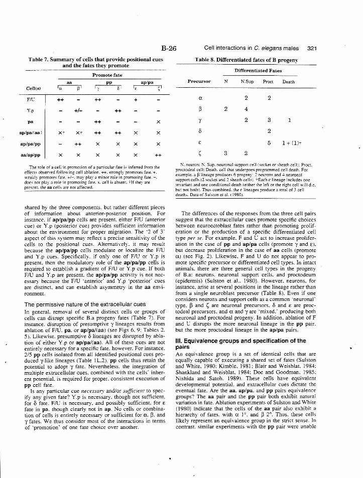
B-26 Cell interactions in C. elegans males 321
Table 7. Summary of cells that provide positional cues and the fates they promote
Promote fate
aa pp ap/pa Cell(s) Ia pi 'I 8
F/C ++ ++ +
Y.p +I- ++
pa ++ X
ap/pa/laal X-i X-i ++ ++ X X
ap/palpp ++ X X X X
aalaplpp X X X X X ++
The role of a cell in prommion of a panicular fate is inferred from the effect:io obsen·ed following cell ablation.++. strongly promote:- fate.+. weakly promotes fate. +1-. may play a minor role in promoting fate.-. doeS not play a role in promoting fate . x. cell is absent. 7Jf they are present. the aa celb are no1 affec1ed.
shared by the three componems. but rather different pieces of information about anterior-posterior position. For instance. if ap/pa/pp cells are present. either FlU (anterior cue) or Y.p (posterior cue) provides sufficient information about the environment for proper migration. The ·2 of 3· aspect of this system may reflect a precise sensitivity of the cells to the positional cues. Alternatively. it may result because the ap/palpp cells modulate or localize the FlU and Y.p cues. Specifically. if only one of FlU or Y.p is presem. then the modulatory role of the ap/palpp cells is required to establish a gradient of FlU or Y.p cue. If both FlU and Y.p are presem. the ap/palpp activity is not necessary because the FlU ·anterior" and Y.p ·posterior· cues are distinct. and can establish asymmetry in the aa environment.
The permissive nature of the extracellular cues In general. removal of several distinct cells or groups of cells can disrupt specific B.a progeny fates (Table 7). For instance. disruption of presumptive y lineages results from ablation of FlU. pa. or ap/pa/(aa) (see Figs 6. 9: Tables 2. 5 ). Likewise. presumptive 8 lineages are disrupted by ablation of ei ther Y.p or ap/pa/(aa). All of these cues are not emirely necessary for a specific fate. however. For instance. 2/5 pp cells isolated from all identified positional cues produced y-like lineages (Table I L.2). pp cells thus retain the potential to adopt y fate. Nevertheless. the integration of multiple extracellular cues. combined with the cells· inherent potential. is required for proper. consistent execution of pp cell fate.
Is any particular cue necessary and/or sufficie111 to specify any given fate? Y.p is necessary. though not sufficient. for 8 fate. FlU is necessary. and possibly sufficient. for £ fate in pa. though clearly not in ap. No cells or combination of cells is entirely necessary or sufficient for a. ~- and y fates. We thu s consider most of the interactions in terms of ·promotion· of one fate choice over another.
Table 8. Differentiated fates of B progen~·
Differentiated Fates
Precursor N N.Sup Proct Death
a 2 2
~ 2 4
y 2 3 1
0 2
£ 5 1 + (l)-i
s 3 2
:-<.neuron::\. Sup. neuronal suppon cell I socket or sheath cell): Procl. proc10deal cell: Demh. cell that undergoes programmed cell dea1h. For example. a ~ lineage produces 6 progeny: 2 neurons and-" neuronal support cells (2 socket and 2 sheath cells). '"i"Each £lineage includes one in,·ariam and one conditional death I either lhe left or the rieh1 cell will d ie. bul no1 both ). Thus combined. the£ lineages produce a 101;1 of 3 ce ll death,. Da1a of Su!Sion et al. i 1980J.
The differences of the responses from the three cell pairs suggest that the extracellular cues promote specific choices between neuroectoblast fates rather than promoting proliferation or the production of a specific differentiated cell type per se. For example. F and U act to increase proliferation in the case of pp and ap/pa cells (promote y and E).
but decrease proliferation in the case of aa cells (promote a) (see Fig. 2). Likewise. F and U do not appear to promote specific precursor or differentiated cell types. In intact animals. there are three general cell types in the progeny of B.a: neurons. neuronal suppon cells. and proctodeum (epidermis) (Sulston et al.. 1980). However. neurons. for instance. arise at several positions in the lineage rather than from a single neuroblast precursor (Table 8). Even if one considers neurons and suppon cells as a common ·neuronal" type. ~ and s are neuronal precursors. 8 and £ are proctodea! precursors. and a and y are ·mixed." producing both neuronal and proctodea! progeny. In addition. ablation ofF and U disrupts the more neuronal lineage in the pp pair. but the more proctodea! lineage in the ap/pa pairs.
Ill. Equivalence groups and specification of the pairs An equivalence group is a set of identical cells that are equally capable of executing a shared set of fates (Sulston and White. I 980: Kimble. 1981 : Blair and Weisblat. I 984: Shankland and Weisblat. 1984: Doe and Goodman. 1985: Nishida and Satoh. 1989). These cells have equivalent developmental potential. and extracellular cues dictate the eventual fate. Are the aa. ap/pa. and pp pairs equivalence groups? The aa pair and the pp pair both exhibit natural variation in fate. Ablation experiments of Sulston and White ( 1980) indicate that the cells of the aa pair also exhibit a hierarchy of fates. with a I o. and ~ 2°. Thus. these cells likely represent an equivalence group in the strict sense. In contrast. similar experiments with the pp pair were unable
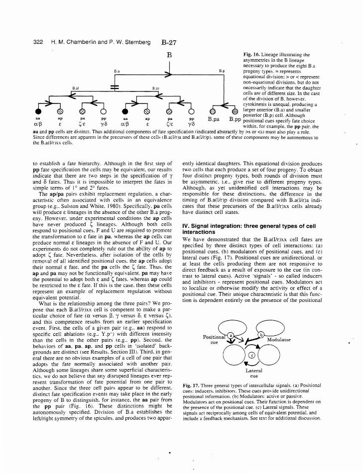
322 H. M. Chamberlin and P. W. Sternberg
B.a
ap pa a a ap
B-27
B
pa
B.p
B.pa
Fig. 16. Lineage illustrating the asymmetries in the B lineage necessary to produce the eight B.a progeny types. = represents equational di,·ision: > or < represent non-equational divisions. but do not necessarily indicate that the daughter cells are of different size. In the case of the division of B. however. cytokinesis is unequal. producing a larger anterior (B.a) and smaller posterior (B.p) cell. Although
B.pp positional cues specify fate choice within. for example. the pp pair. the
aa and pp cell> are distinct. Thus additional components of fate specification (indicated abstractly by >s or < S) must also play a role. Since differences are apparent in the precursors of these cells tB.a(llr)a and B.a<l!r)p). some of these components may be autonomous to the B.a(llnxx celb.
pp
y/0 SfE pp
y/0
to establish a fate hierarchy. Although in the first step of pp fate specification the cells may be equivalent. our results indicate that there are two steps in the specification of y and 8 fates . Thus it is impossible to interpret the fates in simple terms of I 0 and :?0 fates.
The ap/pa pairs exhibit replacement regulation. a characteristic often associated with cells in an equh·aJence group (e.g .. Sulston and White. I 980>. Specifically. pa cells will produce£ lineages in the absence of the other B.a progeny. However. under experimental conditions the ap cells have never produced ~ lineages. Although both cells respond to positional cues. F and U are required to promote the transformation to £ fate in pa. whereas the ap cells can produce normal E lineages in the absence ofF and U. Our experiments do not completely rule out the ability of ap to adopt ~ fate. Nevertheless. after isolation of the cells by removal of all identified positional cues. the ap cells adopt their normal E fate. and the pa cells the ~ fate . Thus. the ap and pa may not be functionally equivalent. pa may have the potential to adopt both E and ~ fates. whereas ap could be restricted to the E fate. If thi s is the case. then these cells represent an example of replacement regulation without equivalent potential.
What is the relationship among the three pairs? We propose that each B.a(l/r)xx cell is competent to make a particular choice of fate (a versus ~- y versus 8. £ versus ~). and this competence results from an earlier specification event. First. the cells of a given pair (e.g .. aa) respond to specific cell ablations (e.g .. Y.p-J with different intensity than the cells in the other pairs <e.g .. pp). Second. the behaviors of aa. pa. ap. and pp cells in 'isolated· back!!rounds are distinct (see Results. Section III ). Third. in !!enera! there are no obvious examples of a cell of one pair -that adopts the fate normall y associated with another pair. Although some lineages share some superficial characteristics. we do not believe that any disrupted lineages ever represent transformation of fate potential from one pair to another. Since the three cell pairs appear to be different. distinct fate specification events may take place in the early progeny of B to distinguish. for instance. the aa pair from the pp pair (Fig. I 6). These dist inctions might be autonomously specified. Division of B.a establishes the left/right symmetry of the spicules. a nd produces two appar-
ently identical daughters. This equational division produces two cells that each produce a set of four progeny. To obtain four distinct progeny types. both rounds of division must be asymmetric. i.e .. give rise to different progeny types. Although. as yet unidentified cell interactions may be responsible for these distinctions. the difference in the timing of B.a(l/r)p division compared with B.a(l!r)a indicates that these precursors of the B.a(l!r)xx cells already have distinct cell states.
IV. Signal integration: three general types of cell interactions We have demonstrated that the B.aO/r)xx cell fates are specified by three distinct types of cell interactions: (a) positional cues. (b) modulators of positional cues. and (c)
lateral cues (Fig. 17). Positional cues are unidirectional. or at least the cell s producing them are not responsive to direct feedback as a result of exposure to the cue (in contrast to lateral cues). Active 'signals' - so called inducers and inhibitors - represent positional cues. Modulators act to localize or otherwise modify the activity or effect of a positional cue. Their unique characteristic is that this function is dependent entirely on the presence of the positional
p··Q~ osmona Modulator
'"' 03:) Lateral
cue
Fig. 17. Three general types of intercellular signals. (a) Positional cues: inducers. inhibitors. These cues pro,·ide unidirectional positional information. (b) Modulators: active or passive. Modulators act on positional cues. Their function is dependent on the presence of the positional cue. (c) Lateral signals. These signals act reciprocally among cells of equivalent potential. and include a feedback mechanism. See text for additional discussion.
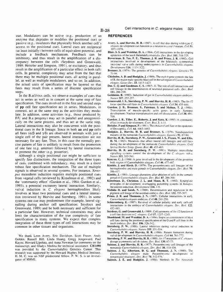
B-28 Cell interactions in C. elegans males 323
cue. Modulators can be active (e.g .. producti on of an enzyme that degrades or modifies the positional cue) or passi,·e (e.g .. insulators that physically block another celr s access to the positional cue). Lateral cues are reciprocal (at least initially! between cells of equivalent potential. and include a feedback mechanism. Feedback can be inhibitory. and thus result in the amplification of a discrepancy between the cells ( Seydoux and Greenwald. 1989: Heitzler and Simpson. 1991 ). or excitatory. and thus result in the amplification of a particular effect in both (all) cells. In general. complexity may arise from the fact that there may be multiple positional cues. all acting in parallel. as well as multiple modulators. and so on . In addition. the actual units of specification may be layered so that fa tes may result from a series of discrete specification steps.
In the B.a(l/r)xx cells. " ·e obsen·e examples of cues that act in series as well as in concert at the same step of fate specification. The cues involved in the first and second step~ of pp cell fate specification act in series. Modulators. in contrast. act at the same step as the acti vities they modulate. In addition. some activities (e.g .. those produced by F/Li and B.a progeny) may act in parallel and antagonistically on the same process. and thus act at the same step. Lateral interactions may also be concurrent with the positional cues in the B lineage. Since in both aa and pp cell s all fates (a/~ and y/o) are observed in animab with just a single cell of the pair present. lateral interactions are not necessary for any fate . Thus. in thi s case the normally precise pattern of fate is unlikely to result from the promotion of one fa te (e.g. anterior) followed by lateral interaction~ to promote the other (e.g. posterior).
Although a single positional cue might be sufficient to specify fate distinctions. the integration of the three types of cues. combined with redundancy. may result in a more robust fate specification mechanism. The use of multiple signals is observed in several systems. For instance. Xenopus mesoderm induction requires mult iple positional cues from vegetal cells (reviewed by Kimelman et al.. 1992) and the ·community effect' (Gurdon et al.. 198-l: Gurdon et al.. 1993). a potential excitatory lateral interaction. Similarly. vulval induction in C. elegans hermaphrodites likely involves at least two positional cues and a lateral interaction (reviewed by Horvitz and Sternberg. 199 1 ). In some systems one cue may predominate (for example. lateral signalling during anchor cell specification: Seydoux and Greenwald. 1989 ) and be both necessary and sufficient for a particular fate. However. technical constraints may also limit the characterization of the true complexity of fate specification in many systems. We expect that complex integration of these three types of cell interactions may be common in other tissues and organisms.
W e thank Leon A very. Eric Da,·id son. Scott Fraser. Andy G o lden. Russell Hill. Linda Huang. Greg g Jongeward. Paul Kay ne. How ard Lipshitz. and Anna Newman for comments on the manuscript. and Gladys Medina for technical assistance . CBI490 was provided by the Caennrhabdiris Genetics Ce nter. T hi s re search was supponed by the Howard Hug hes Medical Institute. H . M . C. was an NSF pred octoral fellow. P . W . S . is a n im·estigator of HHM I.
REFERENCES
Anry. L. and Hon·itz. H. R. ( 1987 >. A ce ll thot die' durin~ wild-type C. t'll•gans de,·clopmcnt can function a~ a neuron in a n·d-3 mutant. Cd/ 51. 1071-1078.
Blair. S. S. and Weisblat. D . . .\. f 1 98~ 1. Ce ll interactions in the de' elop in~ ep idermi:-. ofth~ leech Helobd!!lla trh eriali:-.. DC'L Bioi. 101. ~ 1 S- ~.:!5.
Bowerman. B. Tax. F . E .• Thomas. J. H. and Priess. j. R. 11'1'12 1. Cell intcmction:-. inYoh ·cd in development of the bilateralh svmmetrical intestinal , ·ah·e cell:- during e mbryogene:-. i:-. in Ccu•norhaiuli;;_, t'lel.!al1.\. D l' r l'lopmem 116. I I 1.'- I I::!.:!. .
Brenner. S. < 197-+J. T he genetic~ of Cac•norlw/J(/iri., t•legan.\. Gt'nerio 77. 7 1 -9~ .
Chisholm. A. D. and Hod~kin. j. f 198'1 1. Tho IIUtl>-'1 eene control' the fate of B. the major male-:-.pe<:i tic bla:-.t ce ll in the: tail regi~n of Caenorlwbditi., elt•gwB". Geue_,. Dl'l·. 33. l-l1 3-1 -C.~.
Doe. C. Q. and Goodman. C. S. t 1985 >. II. The role of cell intcroction, and cell lineage in the dt!termina ticm of nl!uron.:tl precur:-.or ct!ll ~ . Del·. Bioi. Ill. 206-219.
Goldstein. B. ( 1992 ). Induction of !!UI in Coenorftahcliti., dl' ~.tcm ... ernhr,·o :-. . Nmurc 357 . .:!55-257. ~ . ·
G reenwald. I. S .. Sternberg. P. \\'. and Horvitz. H. R. 1 198:11. The li11 -l:? locu:-. :-.pecitie:-. cell f::ue:-. in Cacnorlwl>diTi.' C'i<'gcm., . Cd/ 34. -435--4-l .
Gurdon. J. B .• Brennan. S .. Fairman. S. and Mohun. T. J. < 1 '18~>. Tr::m:-.cript ion of musc le-:-.pe1.:irk al.·tin gene-:-. in carl~ Xenopu:-. dt!,·e lopment: ~ucll!:..~r tran~plantation and '-·e ll di~:-.o<.:iation . Cdl 38. 691· 700.
Gurdon. J. B .. Tiller. E •. Roberts. j . and Kato. K 1199:11 .-'. communi!\ c! ffect in mu:-.c l ~ dc!\"Ciopment. Carr. Bioi. 3. 1-11. ·
Heitzler. P and Simpson. P. I 1991 >. T he choi,·c of cell fa te in the cp idermi> of Drosophila . C<-11. 64. I 08.'-1 092.
Hodj!kin. J .. Horvitz. H. R. and Brenner. S. 11979>. !'onJi,j unction mutan~:- of th e.~ nematode CuenorlwluliTi., f!h·:..:an.'i. G('lletic.\ 91. 67-9~.
Hon·itz. H. R .. Sternberj!. P. W .. G reenwald. I. S .. Fixsen. W. and Ellis. H . !\I. I 198.' L Mutat ion:- that affe<.:t neur.Jl cell linc:-a!:!e:- and cell fat~:during the de,·elopmem of the nematode Caenorlwbciir;, t'lt·guu.,. Cold Spring Hurbor Symp. Quam. B ioi. 48. -45 ~--+6~.
Hon·itz. H. R. a nd Sternberg. P. W. 1199 11. ~1u l tiple intercell ular s ign-.~ ll i n~ ' ystem:-. contro l the d('\Ciopment of tht: C. degan' ,·uh a . Nm 11 re 351. 535-5-1 I.
Kenyon. C. J. 11 9861 A gene im ·o h ·c!d in the de\·elopmc!nt 1) f the po, terior bod~ region of Caenorhahditi :-. elegan,. Cel/ 46. -4 77--lSi'.
Kimble. J. and Hirsh. D. ( 1979 >. The postembryonic coli lineage, of the hermaphrodite! :..~nd male gonad:-. in Cae norhabdit i:-. cleg:.~n:-.. D('L B ioi. 70 . 396-~ 17.
Kimble. J . I 19811. Linea!:!C' alteration~ after abbtion of cclb in the ,omatic !!Onad of Coeuorlwhditf., c•h·wm.\. D e1·. B ioi. 87. ~R6-~00 .
Ki;:,elman. D .• Christian. J.' L. and !\loon. R. T . f 19921. Synergi>t ic principle, of d e'"elopmem: o,·erl.:..~pping p;mern ing s~ ~tern, in Xenopus ml!~oderm induction. D e ,·d oJ,memll6. 1-9.
Nishida H. and Satoh. !\. ( J9g9 >. Determinat ion and reoulotion in the pigment cc!ll li nege of the ascidian embryo. Del·. B ioi. 13£ ~55- :;6 7.
Priess. J. R. and Thomson. j. !'\. t 19871. Cellular interaction, in earh CtwnorlwbdiTiJ d~s,:ans embrYOS. Ce/1 48 . .:!-+ 1-250. .
Schierenberg. E. 11987 1. Re\·c.rsal of cellular polarity and earl~ ce lk e ll intcr.s.ction:-. in the embryo of Ca~norlwbdiris el<:gan:.. D('l". Bioi. 12.2. 452-~63.
Seydoux. G. and Greenwald. I. t 1989 >. Cell autonom~ of lill·/2 function in a cell fate deci sion in C. elt·gm1s. Cdl 57. 1 :!~7- J ::!~ 5.
Shankland~ I\ I. and \\"eisblat. D.:\..~ I YS...t l. Stcpwi :-.~ commitmcm of b i:J:--1 c ell fates during the positi onal spcci ri..:ati on o f tht! 0 and P ~.:ell line :- in the leech embryo. De•\·. Bini. 106. 326<;-12.
S ternberg. P. W. f 19881. Lateral inh ibition duri ng \'Uh al induction in CuenorhahdiTi.\ ele,f.!.CIIIS. Ntrtur(' 335. 55 1-55-+.
S ternberg. P. W. and Horvitz. H. R. (1 98!\i. Pattern formation during \"U) \"a) de,·elopment in Caenorlw h(liTi., de.s.:m 1.\. C d/ 44. 761-772.
Sternberg. P. W.and Hon·itz. H. R. t 1988 >. /in-/7 mutation> of C. <1<-gall.\ disrupt asymmetric cell di,·is ion~. Det·. Bioi. 130. 67-7;..
Sulston. J. and Horvitz. H. R. f 19i7>. Postembryonic cell li neages of the ncmatod~ Caenorlw hdiri., e/egt1m . De L Bioi. 56. 110- 156.
Sulston. J. E .. Albertson. D. G. and Thomson. j. !\. f 1980 >. The Cat•tJo rlwhdiTi., e/egans male: Po.ste mbr:onic de,·elopment of nongonadal ~tructure,. Dt' l". Bioi. 78. 5-+.:!-576.
Sulston. J. E . and Hod~kin. j. 11988>. Method> In Tit<· A'ent<uode
324 H. M. Chamberlin and P. W. Sternberg
Caenorlwbdiri.' eic·.~mrs. red. W. Wood!. pp. 587-606. Cold Spring Harbor Laboratory: Cold Spring Harbor. !\ew York.
Sulston. J. E .. Schierenberg. E .. White. J. G. and Thomson. J. ~- ( 19831. The embryonic cell lineage of the nematod~ Caenorhahdiris elegan.'i. Ocr. Bioi. 100. 6-l- I 19.
Sulston. J. E. and White. J. G. ( 19801. Regulation and cell autonomy
B-29 during postembryonic de\·elopmem of Cat•norhabdiriJ elegans. De1·. Bioi. 78.577-597.
Wood. W. B. ( I 9911. Evidence from re,·ersal of handedn.s' in C. eiegmrs for early ceiJ interactions determining celt fates. Nature 349. 536-538.
(Accepted 1 0 March 1993)

B-30
Table 1 enlargement. These are the data in Table 1 pages B-9 - B-12.
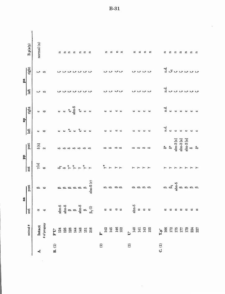
a a
pp
ap
p
a
anim
al#
an
t po
st
ant
post
le
ft
righ
t le
ft
rig
ht
B.p
(alp
)
A.
Inta
ct
a p
y[s]
8
[t]
E
E
~ ~
norm
al (
n)
#o
f pro
geny
4
6 6
2 6
6 5
5
B.
(1)
F"U"
12
4 ab
n-5
p 8'
t 8
E
E
~ ~
n
125
abn-
5 p
8 8
E
E
~ ~
n
128
p p
y*
8 £*
£*
~
~ n
144
p p
y*
8
£ ab
n-5
~ ~
n
148
abn-
5 p
'Y 8
£*
£*
~ ~
n
151
p p
y*
8
£ £
~ ~
n
216
P-r (I
) ab
n-5
(r)
'Y 8
£ £
~ ~
n
(2)
F" 14
2 a
p y*
8
£ £
~ ~
n
145
a p
'Y 8
£ £
~ ~
n to
I
146
a p
'Y 8
£ £
~ ~
n Co
:l J-
0 15
2 a
p 'Y
8 £
E
~ ~
n
(3)
u· 14
0 ab
n-5
p y
8 £
£ ~
~ n
141
a p
'Y 8
£ £
~ ~
n
143
a p
'Y 8
£ £
~ ~
n
155
a p
y 8
£ £
~ ~
n
c. (1
) Y
.p·
166
a p
y 8*
n.
d.
n.d
. n.
d.
n.d.
n
172
a p't
y
8*
£ £
~ ~t
n
175
a ab
n-5
y ab
n-3
[s]
£ £
~ ~
n
177
a p
'Y ab
n-3
[s]
E
£ ~
~ n
179
a p
y ab
n-3
[o]
£ £
~ ~
n
224
a p
y I)
£
£ ~
~ n
227
a p
y 8*
£
£ ~
~ n
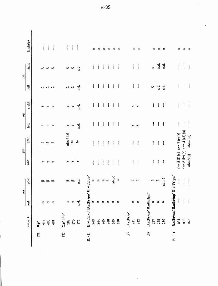
a a
pp
ap
p
a
anim
al#
an
t po
st
ant
post
le
ft
rig
ht
left
ri
gh
t B
.p(a
/p)
(2)
B.p
·
479
a. ~
'Y 8
€ €
~ ~
480
a. ~
'Y 8
€ €
~ ~
481
a. ~
'Y 8
€ €
~ ~
(3)
Y.p
· B.p
·
167
a. ~
'Y ab
n-3
[s]
€ €
~ ~
170
a. ~
'Y 8*
€
€ ~
~ 17
1 n.
d.
n.d.
'Y
8*
n.d.
n.
d.
n.d.
n
. d.
D.
(1)
B.a
(l/r
)ap
· B.a
(l/r
)pa"
B.a
(l/r
)pp"
243
a. a.
----
----
........
....
----
----
----
n 24
4 a.
a. ----
----
----
----
.........
.. ...
... n
245
a. a.
----
.........
----
----
----
----
n td
I
246
a. ~
----
----
........
---
----
-.....
....
CJ.:)
n
1:-.:>
44
5 a.
abn-
5 .....
.... ---
----
----
----
----
-n
450
a. a.
----
----
----
----
--------
n
(2)
B.a
(l!r
)p"
241
a. ~
.........
----
€ €
----
........
n
242
a. ~
----
----
€ €
----
----
n
(3)
B.a
(l/r
)ap"
B.a
(l!r
)pp"
247
a. ~
----
----
----
----
~ €
n
279
a. ~
.........
-------
-----
n.d
. n
.d.
n
280
a. ab
n-5
.......
.......
--·-
----
n.d.
n
.d.
n
E.
(1)
B.a
(l/r
)aa·
B.a
(llr
)ap"
B.a
(l/r
)pa"
261
......
---
-ab
n-5
(I)
[s]
abn-
7 (r
) [s
] ... ...
.... .. ..
.. .. ..
.. .. ....
.... n
262
----
----
abn-
8 (l
v) [
s] a
bn-4
(rd
) [s
] ---
-.....
.. ... ..
...
.. ....
n
275
----
.......
abn-
8 (t
] ab
n-7
[o]
----
.........
. ......
----
n
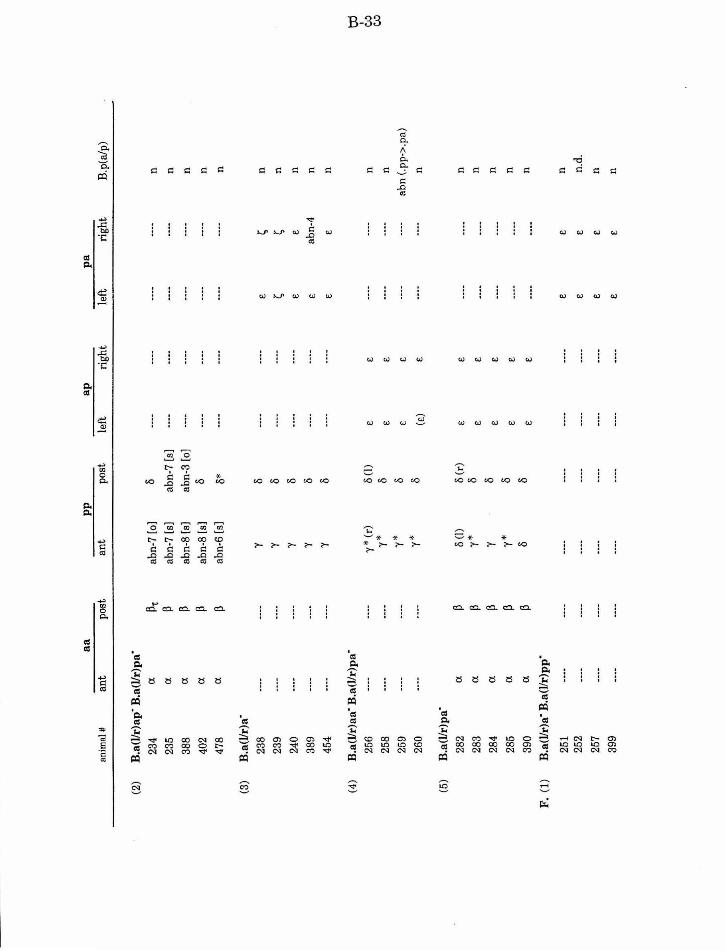
a a
pp
ap
p
a
anim
al#
ant
po
st
ant
po
st
left
ri
gh
t le
ft
rig
ht
B.p
(a/p
)
(2)
B.a
(l/r
)ap·
B.a
(l/r
)pa·
234
(l
p't
abn-
7 [o
) li
......
---
-.. ..
... .....
.....
n
235
(l
p ab
n-7
[s]
abn-
7 [s
] ---
----
-....
....
.. ......
n
388
(l
p ab
n-8
[s)
abn-
3 [o
] .....
.... ---
----
----
-n
402
(l
p ab
n-8
[s]
li ....
... ....
.... .....
.. .. ..
.....
n
478
(l
p ab
n-6
[s)
li*
----
......
----
----
n
(3)
B.a
(l/r
)a·
238
--·-
----
"(
li ---
----
-e
~ n
239
........
----
"(
li ---
----
-~
~ n
240
......
----
"( li
........
......
e
e n
389
----
---·
"(
li ---
-...
...
e ab
n-4
n
454
..........
---
-"(
li
----
---·
e e
n
(4)
B.a
(l/r
)aa·
B.a
(l/r
)pa·
td
I
256
----
----
'Y *
(r)
li (!
) e
e --·
----
-n
co
y*
li co
25
8 .....
... ....
... e
e ---
-....
.. n
259
----
----
y*
li E
E
---
----
-ab
n (.
pp·>
.pa)
260
......
.......
y*
li (e
) e
.....
----
n
(5)
B.a
(l/r
)pa·
282
(l
p li
(l)
li (r
) e
e ---
-.....
.. n
283
(l
p y*
li
e e
n .....
.....
.. ........
284
(l
p "(
li
E
e n
----
----
285
(l
p y*
li
E
e n
........
----
390
(l
p li
I)
e e
n
F.
(1)
B.a
(l/r
)a"
B.a
(l/r
)pp"
251
......
.......
----
----
----
........
e E
n
252
.......
.......
.. ......
----
----
----
E
e n
.d.
257
----
----
........
........
.......
.. .....
e e
n
399
........
........
. .....
----
----
........
e E
n
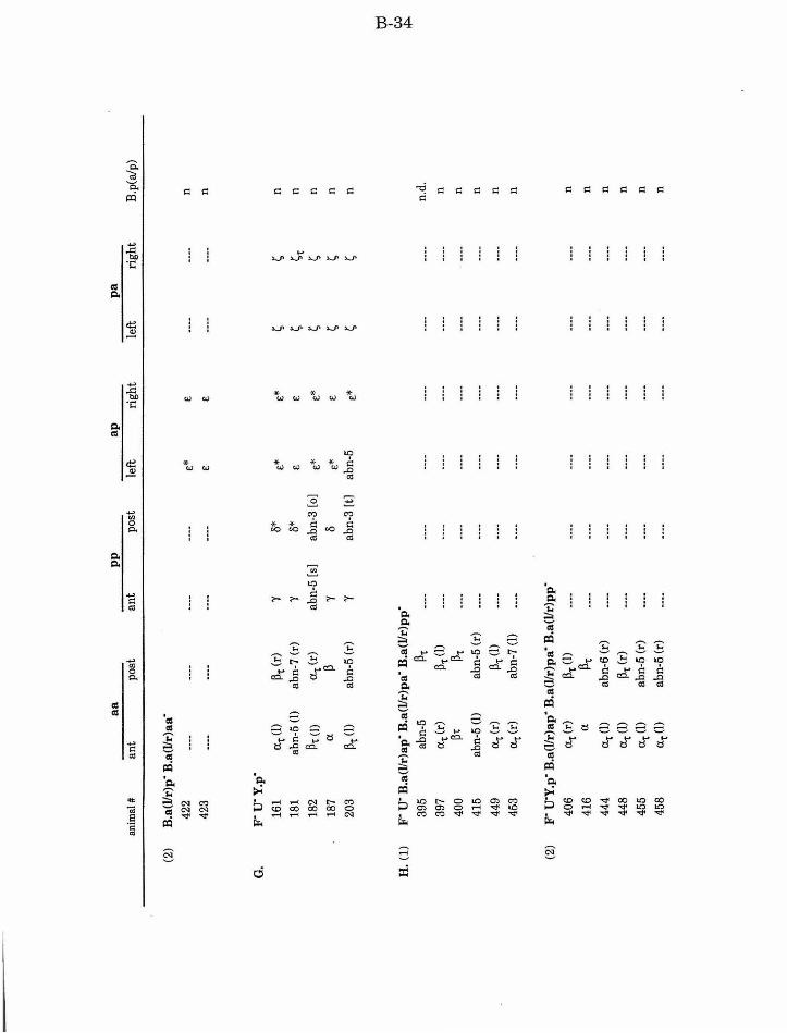
a a
pp
a
p
pa
anim
al#
an
t po
st
ant
post
le
ft
righ
t le
ft
righ
t B
.p(a
/p)
(2)
B.a
(Jir
)p"
B.a
(Jir
)aa·
422
..........
.. ...
... ---
-.....
.... e*
e
........
.. ......
n
423
........
.. ......
.....
... .....
... e
e ... ...
....
----
n
G.
F" U
"Y.p
"
161
a't
(l)
~'t
(r)
'Y 8*
e*
e*
~
~ n
181
abn-
5 (l
) ab
n-7
(r)
'Y 8*
e
e ~
~'t
n
182
~'t (l
) a,;
(r)
abn-
5 [s
] ab
n-3
[o]
e*
e*
~ 1;,
n
187
a ~
'Y 0
e*
e 1;,
~ n
203
~'t (
l) ab
n-5
(r)
'Y ab
n-3
[t]
abn-
5 e*
~
~ n
ttl
I
H.
(1)
F" u
· B.a
(l/r
)ap
· B.a
(Jir
)pa·
B.a
(l/r
)pp
" c,.
.J ~
395
abn-
5 ~'t
---
----
-....
....
........
.. ...
... .. ...
.. n.
d.
397
a,
(r)
~'t (
l)
----
----
----
........
... .......
.. ..
... n
400
~'t
~'t
........
----
----
.........
.. ......
..
.......
n
415
abn-
5 (l
) ab
n-5
(r)
........
·---
---·
----
----
----
n
449
a,; (r
) ~'
t (l)
---
-.....
... .....
... .. ...
... ---
-.....
.... n
453
a,; (r
) ab
n-7
(l)
........
.. ......
... ......
.. ......
.. .....
----
n
(2)
F" U
"Y.p
" B
.a(J
ir)a
p"
B.a
(Jir
)pa"
B.a
(Jir
)pp
"
406
a,; (r
) ~'
t (l)
----
........
........
... ...
... ... ...
... ---
-n
416
a ~'t
.....
... ---
----
-.....
....
.. ......
.. .......
n
444
a,; (l
) ab
n-6
(r)
.........
... .......
. ---
-.....
... .. ...
.. .. ..
.....
n
448
a,;(l
) ~'
t (r
) ....
....
.. ......
----.. ...
... .....
... .. ...
... n
455
a,;(l
) ab
n-5
(r)
.........
.. ......
... ..
....
.. .....
. ......
----
n
458
a't
(l)
abn-
5 (r
) .....
... .....
... .. ...
... .. ..
....
.. ......
.. ......
n
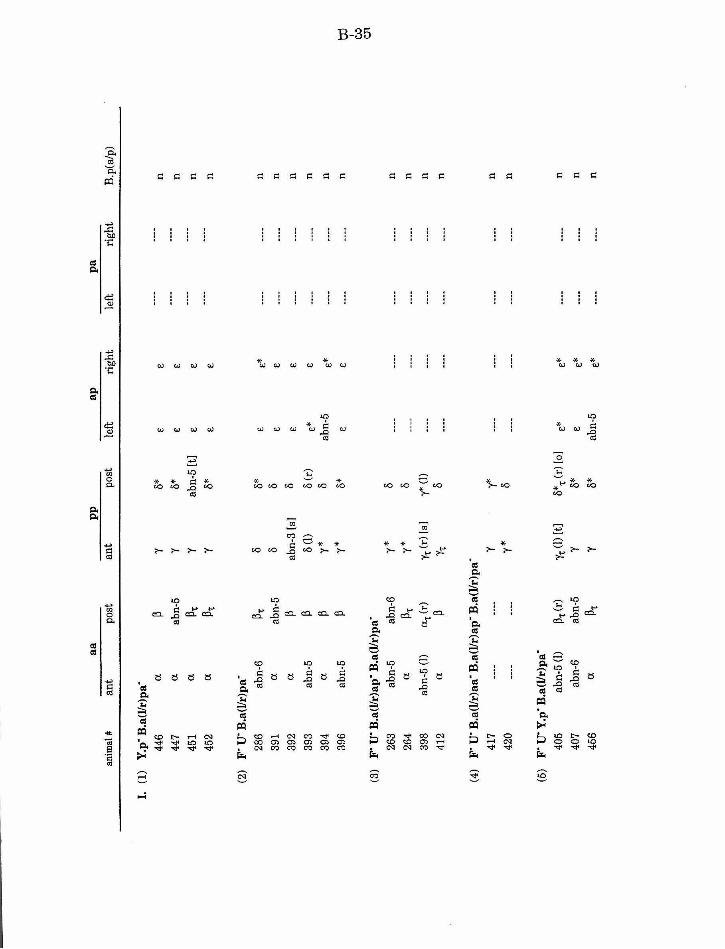
a a
pp
ap
p
a an
imal
#
ant
post
an
t po
st
left
ri
gh
t le
ft
rig
ht
B.p
(a/p
)
I.
(1)
Y.p
· B.a
(l!r
)pa·
446
a ~
'Y 8*
e
e n
447
a ab
n-5
'Y 8*
....
.......
..........
. e
e n
451
a ~.
'Y
abn-
5 [t
) .....
.... .. ..
.....
e e
n
452
a ~.
'Y
8*
.........
----
e e
n
(2)
F' u
· B.a
(l/r
)pa·
286
abn-
6 ~.
0
8*
£ e*
.....
... ---
-n
391
a ab
n-5
0 0
E
!: ....
....
----
n
392
a ~
abn-
3 [s
) 0
!: £
----
----
n
393
abn-
5 ~
ll (I
) ll
(r)
e*
£ ----
----
n
394
a ~
y*
ll ab
n-5
e*
........
........
n
396
abn-
5 ~
y*
ll*
e
e ---
----
-n
td
I ~
(3)
F' u
· B.a
(l!r
)ap·
B.a
(l/r
)pa·
0
1
263
abn-
5 ab
n-6
y*
ll
----
........
----
----
n
264
a ~.
y
*
ll --
----
-----
-....
....
n
398
abn-
5 (I
) C
X.(r
) r,
(r)[
s)
y* (1
) ....
....
.. ......
.. ......
----
n
412
a ~
r, 8
----
----
........
----
n
(4)
F' u
· B.a
(l/r
)aa·
B.a
(l/r
)ap
· B.a
(l/r
)pa·
417
........
.......
'Y y*
---
-.....
... .....
... .. ...
... n
420
........
---
-y
*
0 ---
-.....
.. .. ...
... .. ....
... n
(5)
F' u
· Y.p
· B.a
(l/r
)pa·
405
abn-
5 (I
) ~.
(r)
r,
(!) [t
l 8*
, (r
) [o
) e*
e*
.....
... .. ...
... n
407
abn-
6 ab
n-5
'Y
ll*
E
e*
........
.. ......
n
456
a p,
'Y
8*
abn-
5 e*
.....
... .. ...
... n
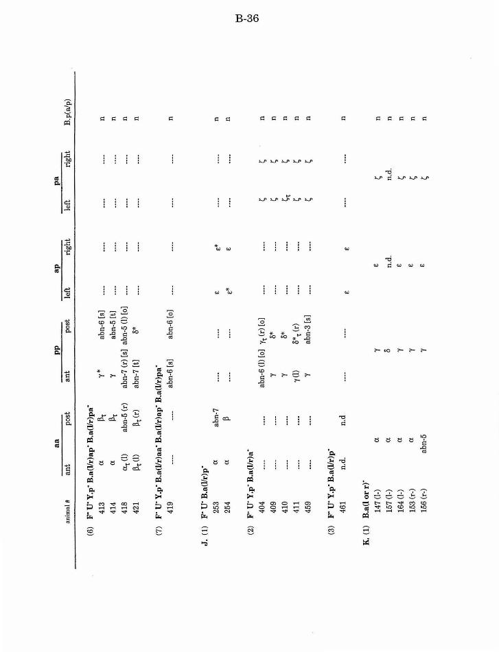
a a
pp
ap
p
a
anim
al#
an
t po
st
ant
post
le
ft
rig
ht
left
ri
gh
t B
.p(a
/p)
(6)
F" U
" Y
.p· B
.a(l
/r)a
p· B
.a(l
/r)p
a·
413
(J.
p't
y*
abn-
6 [s
] .....
.. .....
.... .. ...
... .. ...
... n
414
(J.
p't
y ab
n-5
[t]
----
........
----
........
n
418
~(1)
abn-
5 (r
) ab
n-7
(r)
[s]
abn-
5 (!
) [o
) .....
... .. ...
... .. ..
....
----n
421
p't (!
) p't
(r)
abn-
7 [t
) c'i*
.....
... .. ...
... ---
-.. ...
... n
(7)
F" u
· Y.p
· B.a
(l/r
)aa·
B.a
(l/r
)ap
· B.a
(l/r
)pa·
419
----
.......
abn-
6 [s
) ab
n-6
[o]
.. ......
.. ...
... .. ...
.... .. ...
... n
J. (
1)
F' u
· B.a
(l/r
)p"
253
a ab
n-7
........
.. ..
....
E
e*
.. ......
----
n
254
(J.
p .....
... ... ..
... E*
E
.. ...
... .. ....
... n
td
I (2
) F"
u· Y
.p· B
.a(l
lr)a
" c..:
> m
404
----
----
abn-
6 (!
) (o
] y 1
(r)
[o)
----
........
I;
~ n
409
----
----
y c'i*
---
-.....
... I;
I; n
410
----
.......
y c'i*
.....
... .....
... i;'t
I;
n
411
----
........
y(l)
c'i*
't (r)
---
-.....
... I;
~ n
459
........
----
y ab
n-3
[s]
----
----
I; ~
n
(3)
F' u
· Y.p
· B.a
(l/r
)p"
461
n.d.
n.
d ---
-.....
... E
E
---
-.....
.... n
K.
(1)
B.a
(l o
rr)'
14
7 (!
-)
a y
E
~ n
157
(1-)
(J
. c'i
n.d
. n.
d.
n
164
{1-)
a y
E
~ n
153
(r-)
(J
. y
E
~ n
156
(r-)
ab
n-5
y E
~
n
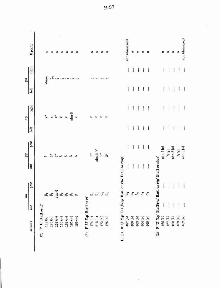
a a
pp
ap
p
a an
imal
# an
t po
st
ant
post
le
ft
rig
ht
left
ri
gh
t B
.p(a
/p)
(2)
F" u
· B.a
(l o
rr)"
16
8 (1
.) ~t
8
E*
abn-
4 n
180
(1.)
~'t
8*
E
~t
n
159
(r-)
ab
n-6
r*
E*
~
n
160
(r-)
~t
8
E
~ n
162
(r-)
~'t
8
E
~ n
165
(r-)
~'t
8
abn-
5 ~
n
169
(r-)
~
8 E
~
n
(3)
F" u
· Y.p
· B.a
(l o
rr)"
174
(1-)
~'t
r*
E
~
n
213
(1-)
~'t
ab
n-3
[s)
E
~ n
173
(r-)
CX
t r*
E
~
n
17
6(r
-)
~'t
B*
E
~ n
tO
I ~
.....:!
L.
(1)
F" u
· Y.p
· B.a
(l/r
)p"
B.a
(l o
r r)
a" B
.a(l
or
r)ap
·
457
{1-)
a,
---
-....
....
----
........
.. ......
.. ......
abn
(dam
aged
)
460
(1-)
CX
t ---
----
-.....
... .. ...
... .. ...
... .. ...
... n
463
(1-)
~'t
----
........
---
-....
....
.. ......
----
n
464
(r-)
CX
t ---
----
----
-.....
... .. ...
... .. ...
... n
465
(r-)
CX
t ---
----
-....
....
... ......
---
-....
....
n
(2)
F" u
· Y.p
· B.a
(l/r
)a· B
.a(l
or
r)p"
B.a
(l o
r r)
pa·
466
(1-)
.....
... .. ...
... ab
n-5
[s)
.. ......
.. ......
.. ......
.. ......
n
467
(1-)
....
....
----
'Yt (
s]
........
.. ......
.. ......
----n
468
(1-)
.....
... .. ...
.. ab
n-6
(s)
.. ......
.. ......
... ......
.. ...
... n
469
(r-)
.....
.... ---
-'Y
t (s]
---
----
-----
........
n
462
(r-)
.....
... .. ...
... ab
n-8
(s]
----
........
----
----
abn
(dam
aged
)
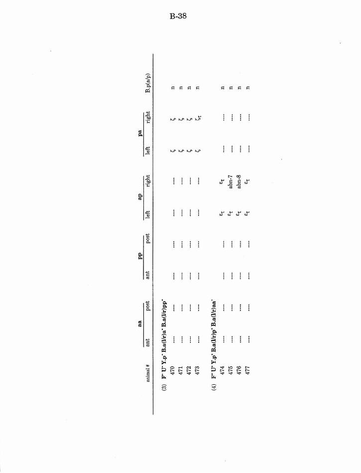
a a
pp
a
p
pa
anim
al#
an
t po
st
ant
post
le
ft
rig
ht
left
ri
gh
t B
.p(a
/p)
(3)
F" u
· Y.p
· B.a
(l/r
)a"
B.a
(l/r
)pp"
470
----
----....
.. ......
.. ---
----
-I;
I; n
td
I
471
........
---
----
-.....
... ---
----
-I;
I; n
~
00
47
2 ---
-.....
... .. ..
....
.. ......
----
----
I; I;
n
473
.........
----
........
........
---
-.....
... I;
1;1
n
(4)
F" u
· Y.p
· B.a
(l/r
)p"
B.a
(l/r
)aa·
474
----
----
........
.. ...
... E.r
E
't .....
.... ---
-n
475
........
----
........
........
E-r
abn-
7 .....
... .. ...
... n
476
.........
........
----
. ......
E.r
abn-
8 ---
----
-n
477
.........
........
........
.. ......
E.r
E.r
----
.. ......
n
C-1
Chapter3
The lin-3/let-23 pathway mediates inductive signalling during male
spicule development in Caenorhabditis elegans
Helen M. Chamberlin and Paul W. Sternberg
C-2
SUMMARY
During Caenorhabditis elegans male spicule development four pairs
of precursor cells establish a pattern of fates that correlates with
relative anterior-posterior cell position. The F and U cells provide
one of the extracellular cues that establish this pattern by promoting
anterior fates. We show that the genes in the lin-3/let-23 signalling
pathway required for hermaphrodite vulval induction also mediate
this FlU signal. Reduction-of-function mutations in lin-3, let-28, sem-
5, let-60 or lin-45 disrupt the fate of anterior cells. Likewise,
activation of the pathway with ectopically produced signal results in
posterior cells inappropriately adopting the anterior fate even in the
absence ofF and U. We have further used this genetic pathway to
begin to understand how multiple positional cues are integrated to
specify cell fate. One negative regulatory pathway is represented by
the lin-15locus. lin-15 acts in spicule development as it does in
vulval induction: as a negative regulator of let-23 receptor activity.
A second extracellular cue, from Y.p, also acts antagonistically to the
lin-8/let-28 pathway. However, this signal is integrated into the lin-
8/let-28 pathway at some step after lin-45 raf, and is thus functionally
distinct from lin-15. We have investigated the role of lin-12 in
forming the anterior/posterior pattern of fates. A lin-12 gain-of
function defect is masked by redundant positional information from.
F and U.
C-3
INTRODUCTION
Cell interactions play a central role in the development of many
organisms. As the specific proteins and cellular processes involved in classic
vertebrate inductions become identified, the underlying complexities of
multiple signals that act in parallel are striking (reviewed in Kimelman, et
al. , 1992; Davidson, 1993). A key issue in understanding how fate is specified
by multiple signals is understanding how the information of such signals is
integrated in the responding cells. In C. elegans , the essentially invariant
and precisely described cell lineage provides a reproducible background to
study cell interactions. The set of characterized interactions required during
the development of the male B cell indicate that it may serve as a model that
combines complex signal integration with the precision of single cell analysis
(Chamberlin and Sternberg, 1993).
During C. elegans male postembryonic development several precursor
cells divide in a sex-specific manner to produce the cells of the copulatory tail.
One such cell, B, is the precursor of all of the cells of the male spicules (Fig.
LA). During male development the anterior daughter of B (B.a) divides to
produce eight progeny (Fig. 2). These eight progeny make up four pairs:
ventral (aa), dorsal (pp), and two identical lateral pairs ( ap/pa). For each
pair there is an anterior and a posterior fate that differ in the subsequent cell
lineage produced by each cell, as well as the differentiated fates of the
progeny produced by that lineage (Sulston and Horvitz, 1977; Sulston, et al.,
1980). The choice of fate within each pair is responsive to extracellular cues
provided by neighboring cells. Cell ablation experiments suggest that the
other male-specific blast cells, or their progeny, provide distinct positional
C-4
cues, and that the eight B.a greatgrandprogeny may also interact
(Chamberlin and Sternberg, 1993) (Fig. 3). For example, ablation of two
other male specific blast cells called F and U disrupts the fates of positionally
anterior cells. In some cases they produce the cell lineage normally
associated with their posterior neighbors. In contrast, ablation of the blast
cell Y. p disrupts the fate of positionally posterior cells.
To identify the genes that mediate the cell interactions required for
proper development of the B cell, we have begun to characterize the role of
genes required for other cell interactions in C. elegans. In this study we have
focused on a subset of the genes in the lin-3 I let-23 signalling pathway that
mediates C. elegans vulval induction since some mutations in these genes
result in an abnormal spicule phenotype (e.g., see Aroian and Sternberg,
1991). C. elegans hermaphrodite vulval development requires a signal from
the anchor cell (AC) in the gonad that acts on three of six epidermal blast
cells termed vulval precursor cells (VPCs) (Kimble, 1981). In normal
development, the three proximal VPCs produce vulval tissue, whereas the
three distal VPCs produce nonspecific hypodermis. The AC signal (an
epidermal growth factor (EGF)-like protein encoded by the gene lin-3 (Hill
and Sternberg, 1992)) is both necessary and sufficient to promote the VPCs to
initiate vulval development. Genes that are necessary for the response to lin-
3 include let-23 (receptor) (Aroian, et al., 1990), sem-5 (adaptor) (Clark, et al.,
1992), let-60 (ras) (Han and Sternberg, 1990), and lin-45 (raf) (Han, et al.,
1993). Reduction-of-function mutations in any of these genes result in a
Vulvaless (Vul) phenotype where all six VPCs may produce hypodermis at the
expense of vulval tissue. Gain-of-function mutations in let-60 (Beitel, et al.,
1990) and over-production ofLIN-3 (Hill and Sternberg, 1992) result in a
C-5
Multivulva (Muv) phenotype in which all six VPCs may produce vulval tissue.
Loss-of-function mutations at another locus, lin-15, also result in a Muv
phenotype. In vulval development, genetic analysis suggests that lin-15 acts
as a negative regulator of let-23, in parallel to lin-3 (Ferguson, et al., 1987;
Huang, et al., 1994).
MATERIALS AND METHODS
Strains
Nematode strains were cultured according to standard techniques
(Brenner, 1974; Sulston and Hodgkin, 1988). Loss-of-function mutations in
lin-3, let-23, sem-5, let-60, and lin-45 are known or believed to be lethal.
However, we believe the genotypes in Table LB. represent a reduction of
normal gene function based on two criteria. First, in the male tail each allele
is recessive to a wild type copy of the gene (except for let-60(sy95dn) and let-
60(sy100dn); data not shown). Second, more stringent genetic tests indicate
that the alleles represent a reduction of normal function of the gene for vulval
development (Aroian and Sternberg, 1991; Clark, et al., 1992; Ferguson and
Horvitz, 1985; Han, et al., 1990; Han, et al., 1993). For lin-3 , let-23, and let-
60 we have tested several alleles or allelic combinations in order to verify that
the observed phenotypes represent the common phenotypes associated with
mutation, as well as to identify genotypes that may represent severe
reduction of gene function for the male tail. The extent of the lineage defects
can be variable both between and within given genotypes. This may be due to
the alleles retaining partial gene activity.
G-6
Mutations used are described by Brenner (1974), Hodgkin, et al.
(1988), and as noted below.
Linkage Group (LG) II: clr-l(e1745). let-23(sy97, sy278, n2020)
(Aroian and Sternberg, 1991; H.M.C. and P.W.S., unpublished; S. Clark and
R. Horvitz, unpublished). unc-4(e120).
LG III: lin-12(n137; n137n760) (Greenwald, et al., 1983).
LG IV: unc-24(e138). mec-3(e1338) . lin-3(n378, n1058, n1059, sy53)
(Ferguson and Horvitz, 1985; Hill and Sternberg, 1992). lin-45(sy96) (Han, et
al., 1993). let-60(n2021, sy95dn, sylOOdn, n1046gf, sy103gfJ (Beitel, et al.,
1990; Han and Sternberg, 1991). dpy-20(el282). unc-22(s7). nTl[unc(n754)
let] (=DnTl balancer; Ferguson and Horvitz, 1985).
LG V: him-5(e1467; e1490) (Hodgkin, et al., 1979).
LG X: sem-5(n1619) (Clark, et al. , 1992). lin-15(e1763, n309, n377)
(Ferguson and Horvitz, 1985).
Extrachromasomal arrays: syEx21(hsp::lin-3), syEx23(hsp::lin-3) (Hill,
et al. , 1994).
Full genotypes of animals in Tables 1, 2 and 3 are as follows: mut-x;
him-5(e1490) doubles were constructed for let-23(sy97), let-23(sy278), let-
60(n2021), lin-45(sy96), let-60(n1046gf), let-60(sy103gf), lin-15(e1763), lin-
15(n309) , lin-15(n377), and lin-12(n137). Data originally summarized in
Greenwald, et al. (1983) (Tables 2 and 3) are from lin-12(n137); him-5(e1467)
and lin-12(n137n760); him-5(e1467) animals. let-23(n2020) is let-23(n2020)
unc-4(e120); him-5(e1490) . A male strain was maintained for syEx21 and
syEx23.
Other genotypes and construction:
C-7
lin-3(sy53ln1058): unc-24(e138) mec-3(e1338) lin-3(sy53) dpy-20(e1282)
I lin-3(n1058). Construction: unc-24(e138) mec-3(e1338) dpy-20(e1282) I+
males were crossed with lin-3(n1058) I DnTl hermaphrodites. Single non-Unc
cross males (genotype: unc-24(e138) mec-3(e1338) dpy-20(e1282) I lin-3(n1058)
or+ I lin-3(n1058)) were crossed with unc-24(e138) mec-3(e1338) lin-3(sy53)
dpy-20(e1282) I DnTl hermaphrodites. non-Dpy non-Unc animals from
crosses that yield Dpy Unc progeny (indicating paternal genotype ofunc-
24(e138) mec-3(e1338) dpy-20(e1282) I lin-3(n1058)) are desired genotype.
lin-3(n378ln1059): lin-3(n378) I unc-24(e138) lin-3(n1059) dpy-
20(e1282); him-5(e1490) I+. Construction: lin-3(n378); him-5(e1490) males
were crossed with lin-3(n1058) I DnTl hermaphrodites. non-Unc male cross
progeny are desired genotype.
sem-5(n1619): clr-l(e1745) I+; sem-5(n1619). Construction: N2 males
were crossed with clr-l(e1745); sem-5(n1619). clr-l(e1745) suppresses the
lethality associated with sem-5(n1619) and maternally rescues for lethality.
However, the male tail defect is still observed.
let-60(dn): let-60(sy95) I dpy-20(e1282); him-5(e1490) or unc-24(e138)
let-60(sy100) dpy-20(e1282) lunc-22(s7); him-5(e1490).
Double mutant strains of let-23 or lin-45 and lin-15 were constructed
according to standard methods (Ferguson, et al., 1987). In addition to the two
mutations, double mutant strains include him-5(e1490). let-23(sy278); lin-
15(e1763) is let-23(sy278) unc-4(e120); him-5(e1490); lin-15(e1763).
Cell lineage and ablation
Cell nuclei divisions in living animals were directly observed using
Nomarski differential interference contrast optics as described by Sulston
C-8
and Horvitz (1977). Nomenclature follows the standard ofSulston and
Horvitz (1977), with modifications of Chamberlin and Sternberg (1993). All
lineages were followed from the first divisions of the B.a(llr)xx cells (early
mid L3) through the L3 molt. Cell nuclei were destroyed by a laser
microbeam as described by Avery and Horvitz (1987). F, U, and Y.p were
ablated at the stage when B had divided to produce two progeny (early L2),
according to the procedures in Chamberlin and Sternberg (1993). Ablation of
B.a progeny was during mid to late L2 stage, soon after the targeted cells
were generated. Ablation of the "B.a positional cue" for the aa cells in Table
2 represents the ablation ofB.a(l/r)p, followed by the ablation ofB.a(llr)ap.
Heat shock induction of lin-3 transgenes
Heat shock was carried out according to the method of Hill, et al.
(1994). All animals received heat shock at late L2 larval stage. Individual
animals were anaesthetized on pads of 5% agar in water containing 5 1-LM
sodium azide, staged, and allowed to recover one hour on a standard 5 em
NGM agar petri plate seeded with OP50. Plates were then sealed with
parafilm and floated in a 33°C water bath for 90 minutes.
RESULTS
The lin-3/let-23 signalling pathway mediates the FlU signal during
male spicule development
We have examined the effects of increasing and decreasing the activity
of the lin-3 I let-23 pathway using chromosomal mutations and
extrachromasomal transgenes. Taken together the results indicate that
C-9
activation of the pathway is both necessary and sufficient to promote anterior
fates, and it likely mediates the positional cue provided by F and U.
Mutations in some genes in the lin-3/let-23 pathway disrupt fates of anterior
cells in the male B lineage
Some mutations in a subset of genes in the lin-3 I let-23 signalling
pathway result in an abnormal male spicule phenotype (see Fig. 1; also
Aroian and Sternberg, 1991). These genes include lin-3, let-23, sem-5, let-60,
and lin-45. In the B lineage, reduction-of-function mutations in any of these
genes disrupt the lineages of the anterior cells (Table 1, B.2-10). For
instance, of seven lin-3(sy53 /n1058) animals followed, none had a normal a
or a normal "(lineage, and only one had normal E lineages (Table 1, B.2). The
phenotypes are similar to the abnormalities that result from ablation of both
F and U in wild type males (Table 1, B.1). In some cases the anterior cells
produce a lineage similar to their posterior neighbors. For instance, in all
seven lin-3(sy53 /n1058) animals both aa cells produced~ lineages.
Although the mutant phenotypes resemble the effects of FlU ablation,
the F and U cells appear normal in the mutants. First, the F and U lineages
are normal in mutant animals (2/2lin-3(sy53 /n1058) and 2/2 lin-45(sy96)
animals followed). Second, the linker cell in the gonad dies as in wild type
animals (10/10 lin-3(sy53 /n1058) and 8/8 lin-45(sy96) animals). Since
U.(llr)p "murder" the linker cell in intact animals (Sulston and White, 1980),
these U progeny are still capable of one of their normal functions.
The lineage defect in mutants is enhanced by ablation ofF and U
(Table 1, B.11, 12). However, ablation ofF and U in mutant animals still did
not result in a consistent transformation of all anterior cells to posterior fate.
C-10
In particular, although the lineages of anterior ap cells were disrupted, the
ap cells did not produce s lineages.
Ectopic expression of the ligand, LIN-3, disrupts the fates of posterior cells
To further test the role of this pathway in the B lineage, we used a
transgenic construct that includes the EGF-coding domain of lin-3 under
control of a tissue-general heat shock promoter (Hill, et al., 1994). In the B
lineage, heat shock treatment of transgenic animals results in disruption of
the lineage of the posterior cells (Table 1, C.1,2). In the aa and ap/pa pairs
the posterior cells produce lineages normally associated with their anterior
neighbors. For example, in five of six heat-shocked syEx21 animals followed,
both aa cells produced a lineages and both ap and pa cells produced £
lineages (Fig. 4).
In the pp pair, the posterior cells produce more anterior-like lineages.
However, lineages of both anterior and posterior cells in this pair can be
disrupted to produce up to eight progeny. Although abnormal, these lineages
are consistent with our hypothesis that all four pp daughters can produce up
to four progeny like the normal anterior daughter of"( (see Fig. 2) if the
positional cue from F and U is not localized or "modulated" (Chamberlin and
Sternberg, 1993). Such abnormal lineages are observed following ablation of
the ap/pa cells in wild type (Chamberlin and Sternberg, 1993) as well as in
transgenic animals.
Transgenic ubiquitous LIN-3 is also sufficient to compensate for the
absence ofF and U (Table 1, C.4), suggesting that activation of the lin-3 I let-
23 pathway is sufficient to promote anterior fates. Gain-of-function let-60
C-11
mutations that result in an activated protein also disrupt posterior fate, but
to a lesser extent than ubiquitous LIN-3 (Table 1, C.3).
Two activities that act antagonistically to lin-3/let-23 are integrated
at functionally distinct steps in the pathway
Mutations in lin-15 disrupt the fate of posterior pp cells
In the hermaphrodite vulva, reduction-of-function mutations in the lin-
15locus result in a phenotype opposite from reduction-of-function mutations
in the lin-3 I let-23 genes required for vulval fates. In the male tail, mutations
in lin-15 disrupt posterior fates (Table 1, D.1-3). However, even in lin-15 null
mutants the lineage defect is only observed in some animals, and only the
fate of the posterior pp cell is usually disrupted: it divides to produce up to
four progeny instead of the normal two. Thus mutations in lin-15 result in a
phenotype opposite from those seen in lin-3 I let-23 mutants, but the effect is
weak. 31/63 (49%) lin-15(n309), 23/55 (42%) lin-15(n377), and 23/57 (40%)
lin-15(e1763) adult males have abnormal spicule morphology, consistent with
the observation that the B lineage is abnormal in only 5/13 lin-15 mutant
animals followed.
lin-15 likely acts as a negative regulator oflet-23 in the male B lineage
We followed the B lineage in a key subset of double mutants to confirm
the position of lin-15 in the genetic pathway. Mutations in both let-23 and
lin-45 block the requirement for functionallin-15 (Table 1, D.4-6). This
result is consistent with lin-15 acting in the B lineage as it does in vulval
development, where it is integrated genetically upstream of let-23.

C-12
The positional cue from Y.p represents a signalling pathway distinct from the
lin-3/let-23 pathway
Y.p, or its progeny, produces a positional cue that promotes posterior
fate, especially in the pp pair (Chamberlin and Sternberg, 1993). Ablation of
Y. p disrupts the fate of the posterior pp cell similar to the defect observed in
lin-15 mutants (Table 1, E.1), suggesting that Y.p may be the source of a
signal mediated by lin-15. Ablation ofF, U, and Y.p together results in a
disruption of both anterior and posterior pp cell lineages (Table 1, E.2).
Ablation ofY.p in lin-3, let-23, let-60, or lin-45 mutants resembles F-U-Y.p
animals (Table 1, E.3-6). The similar results obtained for lin-3, let-23, let-60,
and lin-45 mutants following Y.p ablation indicate that the positional
information from Y.p is not integrated into the lin-3 I let-23 signalling
pathway upstream of lin-45 raf. Thus, lin-15 and the Y.p signal are
functionally distinct.
lin-12 can mediate a lateral interaction between the pp cells
Reduction-of-function mutations in lin-12 (lin-12(0)) result in both pp
cells producing y-like lineages. In contrast, noB lineage defects are observed
in animals bearing lin-12 gain-of-function mutations (lin-12(d)) (Greenwald,
et al., 1983). To better understand the role of lin-12 in pp fate specification,
we have carried out cell ablation experiments in lin-12( d) mutants. Our
results suggest that lin-12(d) mutations result in the opposite transformation
from lin-12(0) mutations ifF and U are ablated (Table 2). The effect is best
seen when F, U, and Y.p are removed. In wild type animals, this ablation
C-13
results in y and y*/8* lineages from both pp cells, whereas in lin-12(d)
animals both pp cells produce 8 lineages (compare Table 2. f to g). The
presence ofF and U is sufficient to override the effect of the lin-12( d)
mutation. An additional defect in lin-12(0) animals is that the presumptive Y
cell is transformed to a neuronal fate similar to its lineal homolog DA9
(Greenwald, et al., 1983). Since lin-12(0) animals are also missing the
positional cue from Y.p, at present we cannot establish if the presence ofY.p
is likewise sufficient to compensate for the absence of lin-12 .
Ablation experiments suggested that the aa cells may also interact
with each other (Chamberlin and Sternberg, 1993). However, ablation
experiments in lin-12(d) mutants carried out for the aa pair did not produce
conclusive results (Table 3).
DISCUSSION
The genes in the lin-3/let-23 signalling pathway mediate the FlU
signal defined by cell ablation
Cell ablation experiments suggest that four distinct cell interactions
are essential for normal anterior/posterior patterning of fates for four pairs of
cells in the C. elegans male B lineage (Chamberlin and Sternberg, 1993). The
male specific blast cells F and U mediate one of these signals that is
n ecessary for normal anterior fates . The lin-3 I let-23 pathway is both
necessary and sufficient to promote anterior fates, and thus likely mediates
the FlU signal.
C-14
The lineage defect in lin-3 I let-23 mutants is enhanced by ablation ofF
and U. This result suggests that not only might the analyzed mutations not
represent null mutations for male tail function, but that all signalling activity
may not be eliminated from animals in which F and U have been ablated.
Such residual activity may come from the debris of the ablated cells, or may
come from other, unidentified sources. However, if such other sources exist
they are not sufficient to promote the normal anterior fates in the absence of
F and U, and likely do not play a significant role in normal development.
The role of lin-3/let-23 in fate specification
What specific role in fate specification does the lin-3 I let-23 pathway
play in C. elegans spicule development? Our results suggest that the same
genes mediate the FlU signal for all four pairs of B.a progeny. Since
mutations in a single gene of the lin-3 I let-23 pathway can result in all of the
defects observed in animals with F and U ablated we can exclude the
possibility that F and U produce three different signals, with one for the aa
pair, one for the pp pair, and one for the ap/pa pairs. Nevertheless, the
cellular response is distinct for each pair. In response to ectopic LIN-3, the
pp cells produce more progeny than normal while aa cells produce fewer
progeny. LIN-3 also does not appear to induce a specific differentiated cell
type (Chamberlin and Sternberg, 1993), since neuronal and hypodermal
progeny arise from both anterior and posterior blast cells (Sulston, et al.,
1980). These differences among the three pairs suggest that the lin-3 I let-23
pathway promotes a particular choice among possible responses. The final
outcome depends upon functional differences among the responding cells.
C-15
The role of lin-15 in the B lineage
In the C. elegans vulva, loss-of-function, molecular null, mutations in
lin-15 result in all VPCs adopting vulval fates. The defect in the B lineage, in
contrast, is less extreme, and less than 50% penetrant. Both vulval
development and spicule development have a necessary requirement for the
positive acting genes of the lin-3 I let-23 pathway. One possible reason for
this difference in the requirement for lin-15 is that development of the B cell
includes additional specification mechanisms, such as positional cues from
Y.p and the other B.a progeny, that act antagonistically to the lin-3 I let-23
pathway. Thus although lin-15 may function biochemically the same in both
developmental processes, its role is diminished in the B lineage as it is
partially redundant with other activities.
Integration of multiple signals
Two activities that act antagonistically to the lin-3 I let-23 pathway are
required for normal fate specification in the dorsal pp pair. If lin-15 acts in
the B lineage as it does in vulval induction and is required in cells other than
the responding cells (Herman and Hedgecock, 1990), then both lin-15 and the
Y.p positional cue represent extracellular cues that must be integrated by the
responding cells. We have characterized the integration of both these
activities relative to the lin-3 I let-23 pathway. If the function of a gene (like
lin-15) or a signal (like the Y.p cue) is to negatively regulate the activity of
the r eceptor let-23, mutations in the receptor or any downstream gene such
C-16
as lin-45 should render the pathway insensitive to the removal of the
regulator. Consequently, the lineage defect observed in let-23 or lin-45
mutants with the activity removed should be the same as when the activity is
intact. This is the case with lin-15, as let-23; lin-15 and lin-45; lin-15 double
mutants resemble the single let-23 and lin-45 mutants, and the posterior pp
cell always produces a normal cS lineage. However, ablation ofY.p in let-23,
let-60, or lin-45 mutants results in abnormal lineages similar to F-u-Y.p- or
lin-3 Y.p- animals. In all cases, the posterior pp cell produces abnormal
lineages rather than the normal cS lineage. Therefore, the Y. p cue does not act
by negatively regulating the lin-3 I let-23 pathway. We propose that it
represents a distinct, parallel signalling pathway that acts at the same time
or later than the lin-3 I let-23 pathway.
The positional cues from FlU and Y.p represent two of the four active
cell interactions identified by cell ablation experiments (see Fig. 3.B). F and
U provide an anterior positional cue (labeled 1 in Fig. 3) that is mediated by
the lin-3 I let-23 signalling pathway. Our results and the results of
Greenwald, et al., (1983) suggest that, at least in the pp pair, the lateral
interaction (4 in Fig. 3) is mediated by the gene lin-12. Our results
specifically indicate that lin-15 does not mediate the Y.p cue (3 in Fig. 3).
However, our data do not rule out the possibility that lin-15 plays a role in
mediating the positional cue from the other B.a progeny (2 in Fig. 3). Testing
of this possibility, as well as identifying the point of integration of the lateral
cue and the B.a progeny cue into the lin-3 I let-23 pathway, awaits further
analysis.
C-17
ACKNOWLEDGMENTS
We thank T.R. Clandinin, A. Golden, R.J. Hill, L.S. Huang, G.D. Jongeward,
W. Katz, H.D. Lipshitz, J. Liu, and R. Palmer for comments on the
manuscript, and G. Medina andY. Hajdu-Cronin for technical assistance. We
also thank R.J. Hill for providing syEx21 and syEx23 and S. Clark for
providing let-23(n2020) prior to publication. Some of the strains used in this
study were provided by the Caenorhabditis Genetics Center, which is funded
by the NIH National Center for Research Resources (NCRR). This research
was supported by the Howard Hughes Medical Institute and an NSF
Presidental Young Investigator Award toP. W. S. H. M. C. was an NSF
predoctoral fellow. P. W. S. is an investigator of the HHMI.
C-18
Table 1. Effects of disruption of cell interactions on the B lineage. Each line
represents all of the animals observed under a specific experimental
condition. Each of the pairs ofB.a progeny cells (aa, pp, ap/pa) are
indicated, along with the lineage produced by the positionally anterior and
the positionally posterior member of the pair. The numbers represent the
number of animals followed that produced the indicated lineage. For
instance, in all seven lin-3(sy53 /nl058) mutant animals (line B.2), both aa
cells produced a B lineage. In some animals the left and right cells of a pair
fail to migrate to anterior and posterior positions. Superscript numbers
indicate the lineage associated with cells that fail to migrate, and the number
of animals. Thus the aa cells failed to migrate in two lin-3(sy53 I nl 058)
animals, and both cells produced B-like lineages. In cases where the two cells
that fail to migrate produce dissimilar lineages, they are aligned in the table
to most closely approximate wild type. Cases where a cell produced a lineage
in which the timing and axes are consistent with a normal lineage but the
axes are skewed are included as a normal lineage. "."indicates no animals
produced the lineage. Data for B.1, E.1, and E .2 are from Chamberlin and
Sternberg (1993). y*/8* and E *are commonly observed abnormal lineages.
Each results in four progeny. "abn" indicates any other abnormal lineage.
"abn>4" and "ahn<4" indicate abnormal with more than, or less than four
progeny, respectively. The nature ofthe abnormal lineages is further
discussed in Chamberlin and Sternberg (1993). In some cases the numbers of
aa , pp, and ap/pa cells do not follow a 1:1:2 ratio because all cells were not
followed to the completion of the lineage for some animals.
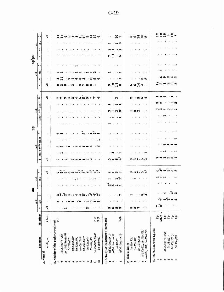
a a
ant
post
ge
noty
pe
,...-
----
.. ..--
abla
tion
s a
abn
P ~---
------
-----
a nb
n p I'
A. N
orm
al
wild
typ
e in
tact
a
ll
all
B. A
ctiv
ity
of th
e pa
thw
ay r
edu
ced
1
F-U
· 4
3 7
2 li
n-3(
sy53
/n10
58)
72
72
3 li
n-3(
n378
/nl0
59J
1 21
31
4
let-
23(s
y97)
1
4 5
5 le
t-23
(n20
20J
1 1
2 6
/et-2
3(sy
278J
4
1 3
7 se
m-6
(nl6
19)
11
4 51
8
let-
60(n
2021
) 51
51
9
/et-
60(d
n)/ +
3
4 1
8 10
lin
-45(
sy96
) 3
41
1 61
II
li
n-3(
sy5
3/ n
105
8)
F-U
-1
41
1 41
12
lin
-45(
sy96
1 F-
U·
1 21
31
C. A
ctiv
ity
of t
he
pat
hway
Inc
reas
ed
l sy
Ex2
3(hs
p::l
in-3
) 51
2
1 1
2 2
syE
x2l!
hsp:
:lin-
3J
6 5
1 3
let-
60!g
{)
81
1 1
6 4
syE
x21(
hsp:
:lin
-3J
F-U
-3
2 1
11
3
D.
Rol
e o
( lln
-16
I lin
-16(
n309
) 5
1 6
2 lin
-16(
n377
) 3
1 2
3 li
n-16
(e17
631
7 7
4 le
t-23
(sy9
7); l
in-1
5(n3
09)
5 5
5 /e
t-23
(sy27
81;
lin-1
5(e1
763J
43
4
3
E. I
nte
ract
ion
wit
h Y
.p c
ue
I Y
.p-
7 22
1
6 2
F-U
-Y.p
-3
2 11
43
3
lin·
3(sy
63/n
1058
) Y
.p·
1 4
1 4
4 le
t-23
(sy9
7)
Y.p
· 32
3
2 5
/et-
60(n
2021
) Y
.p-
1 41
5
1 6
lin-4
5(sy
96)
Y.p
-2
2 1
3
pp
an
t po
st
l nb
n>4
yol6
• ab
n<4
6 ""
' ""'
0
bn>
4 y>
/6•
abn<
4 6
all
al
l
2 3
2 7
1 5
1 7
3 3
3 1
4 2
2 2
2 4
1 4
33
5 1
1 5
3 4
1 5
2 4
1 7
74
74
2 1
1 4
5 1
3 1
1 4
4 1
8 3
2 3
41
1 1
31
1
3 1
2 3
2 1
7 2
5 5
5 3
1 4
7 3
3 1
4 1
2 2
1 1
1 3
1 3
1 2
1 3
3 1
1 3
1 1
1 1
1 1
2 2
ant
-£
• nb
""
'
all 9
4 1
2 1
1
1 6 1
7 3
1 6 1
1 3
6 1
7 3
8 1
3 1
0
1 1
9 2
6
9 1
12
11
8
1 1
5 6 14
4 6 8
12
3
6 1
1 9
2 2
6 4
3 5
ap/p
a oo
st
··-··
7 2
1 1
1
1 1
5 1
3
all
14
1
4
6 8 4 8 10
1
0
9 14
1
2
6 10
1 5 6 14
10
8 1
2
10
1
0
4 10
8
0 1-'
<
0
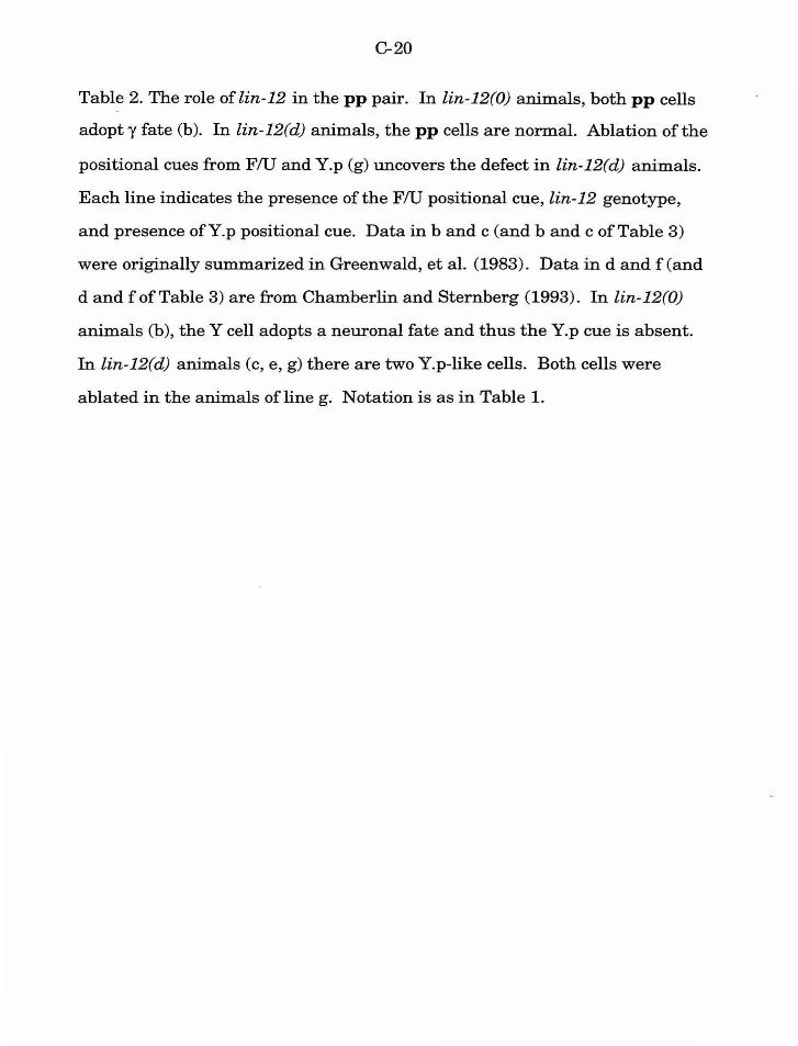
C-20
Table 2. The role of lin-12 in the pp pair. In lin-12(0) animals, both pp cells
adopty fate (b). In lin-12(d) animals, the pp cells are normal. Ablation ofthe
positional cues from FlU and Y.p (g) uncovers the defect in lin-12(d) animals.
Each line indicates the presence of the FlU positional cue, lin-12 genotype,
and presence ofY.p positional cue. Data in band c (and band c ofTable 3)
were originally summarized in Greenwald, et al. (1983). Data in d and f (and
d and f of Table 3) are from Chamberlin and Sternberg (1993). In lin-12(0)
animals (b), theY cell adopts a neuronal fate and thus the Y.p cue is absent.
In lin-12(d) animals (c, e, g) there are two Y.p-like cells. Both cells were
ablated in the animals of line g. Notation is as in Table 1.
pp
an
teri
or
Eos
teri
or
FlU
li
n-12
Y
. "(
ab
n 0
"(
abn
0
a.
+
+
+
all
all
b.
+
--
32
22
1 c.
+
d
+
3 1
4 <;1
d.
-
+
+
2 3
2 7
tv
1-'
e.
-d
+
31
31
f. -
+
-4
1 4
1 g.
-
d -
53
53
C-22
Table 3. The role of lin-12 in the aa pair. aa lineages are normal in both lin-
12(0) and lin-12(d) mutants. Removal ofF/U, Y.p and B.a positional cues
uncovers a possible lateral interaction between aa cells (Chamberlin and
Sternberg, 1993). Removal ofF/U, Y.p and B.a positional cues in lin-12(d)
mutants indicates that lin-12 does not appear to play an essential role in the
specification of aa fates even in the absence of positional cues. Notation is as
in Tables 1 and 2.
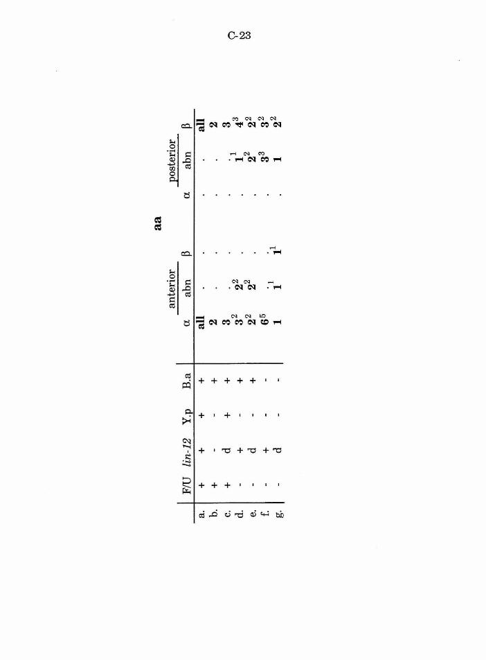
a a
ante
rior
Eo
ster
ior
FlU
li
n-12
Y
.p
B.a
a
abn
~
a ab
n
~ a.
+
+
+
+
al
l al
l b.
+
-
-+
2
2 c.
+
d
+
+
3 .
1 3
0 d.
3
2 2
2 43
-
+
-+
1
t...:>
e.
d +
22
22
22
2
2 C
/j
--
f. -
+
--
65
. 1
33
32
g.
-d
--
1 1
11
1 2
2
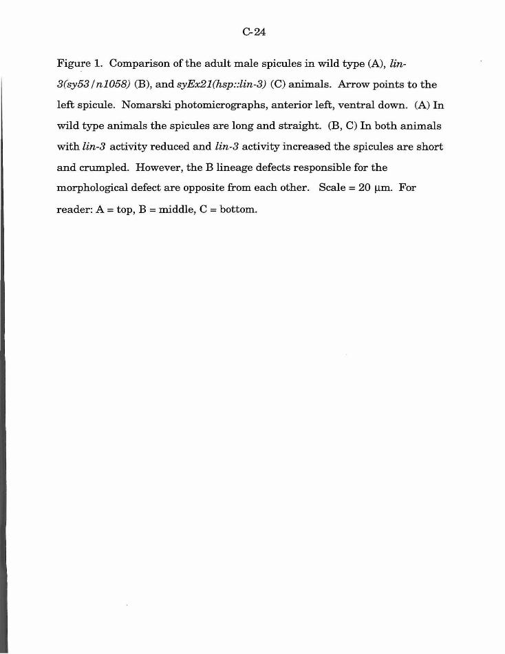
C-24
Figure 1. Comparison of the adult male spicules in wild type (A), lin-
3(sy53 /n1058) (B), and syEx21(hsp::lin-3) (C) animals. Arrow points to the
left spicule. Nomarski photomicrographs, anterior left, ventral down. (A) In
wild type animals the spicules are long and straight. (B, C) In both animals
with lin-3 activity reduced and lin-3 activity increased the spicules are short
and crumpled. However, the B lineage defects responsible for the
morphological defect are opposite from each other. Scale= 20 f.Lm. For
reader: A = top, B = middle, C = bottom.
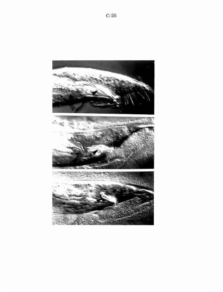
C-25
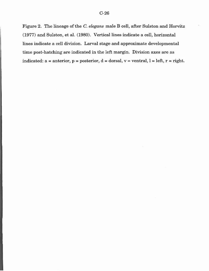
C-26
Figure 2. The lineage of the C. elegans male B cell, after Sulston and Horvitz
(1977) and Sulston, et al. (1980). Vertical lines indicate a cell, horizontal
lines indicate a cell division. Larval stage and approximate developmental
time post-hatching are indicated in the left margin. Division axes are as
indicated: a= anterior, p =posterior, d =dorsal, v =ventral, l =left, r =right.
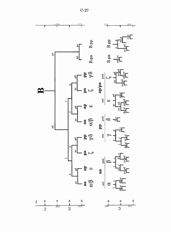
hrs
B
Ll
1-
to
ad
pv
L2 r 20
I ,~
I
-··
I _ _,
'"
' I
nrl
-··
I
-.:1
!l
V
I n
rl ad
I
I f !
25
I I
I I
I I
I I
I 0
a a
ap
pa
p
p
a a
ap
p
a
pp
tv
a/~
~ y/
8 a/
~ ~
y/8
..:)
£ £
B.p
a B
.pp
a a
pp
ap
/pa
ant
post
an
t po
st
ant
post
~~.
~ 25
a
~ 'Y
0 £
~ B
.pa
B.p
p
L3
L 3
0
~
d r ~ ft
r t4
~
au
d
arlr
35
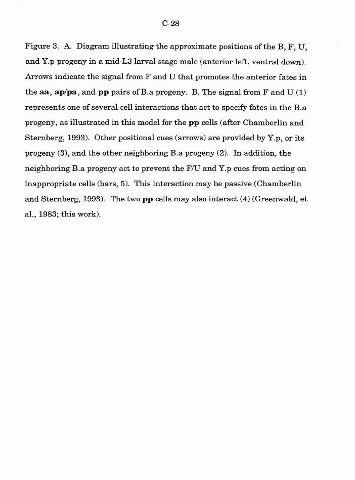
C-28
Figure 3. A. Diagram illustrating the approximate positions of the B, F, U,
and Y.p progeny in a mid-L3larval stage male (anterior left, ventral down).
Arrows indicate the signal from F and U that promotes the anterior fates in
the aa, ap/pa, and pp pairs ofB.a progeny. B. The signal from F and U (1)
represents one of several cell interactions that act to specify fates in the B.a
progeny, as illustrated in this model for the pp cells (after Chamberlin and
Stemberg, 1993). Other positional cues (arrows) are provided by Y.p, or its
progeny (3), and the other neighboring B.a progeny (2). In addition, the
neighboring B.a progeny act to prevent the FlU and Y.p cues from acting on
inappropriate cells (bars, 5). This interaction may be passive (Chamberlin
and Sternberg, 1993). The two pp cells may also interact (4) (Greenwald, et
al., 1983; this work).
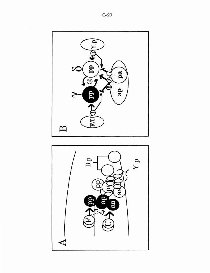
C-29
-~-.(®
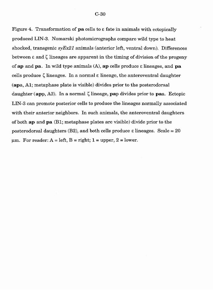
C-30
Figure 4. Transformation of pa cells to E fate in animals with ectopically
produced LIN-3. Nomarski photomicrographs compare wild type to heat
shocked, transgenic syEx21 animals (anterior left, ventral down). Differences
between E and s lineages are apparent in the timing of division of the progeny
of ap and pa. In wild type animals (A), ap cells produce E lineages, and pa
cells produce s lineages. In a normal E lineage, the anteroventral daughter
(apa, Al; metaphase plate is visible) divides prior to the posterodorsal
daughter ( app, A2). In a normal s lineage, pap divides prior to paa. Ectopic
LIN -3 can promote posterior cells to produce the lineages normally associated
with their anterior neighbors. In such animals, the anteroventral daughters
of both ap and pa (Bl; metaphase plates are visible) divide prior to the
posterodorsal daughters (B2), and both cells produce E lineages. Scale= 20
J..Lm. For reader: A= left, B =right; 1 =upper, 2 =lower.
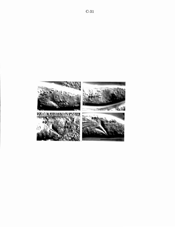
C-31
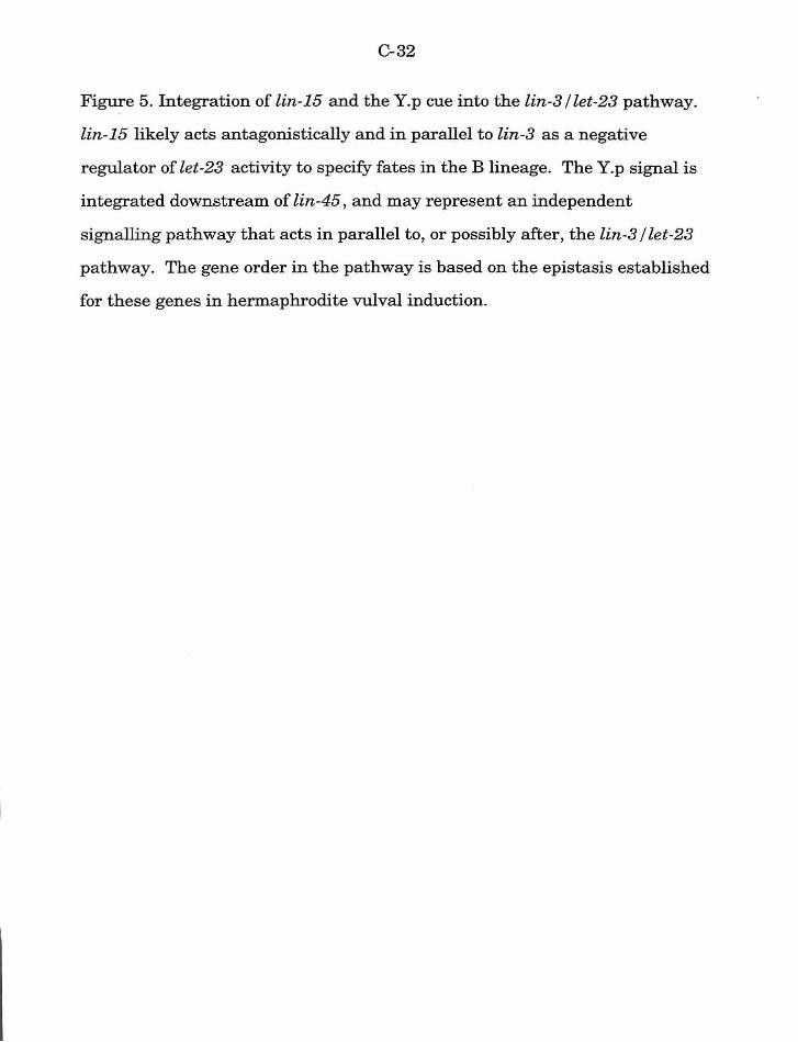
C-32
Figure 5. Integration of lin-15 and the Y.p cue into the lin-3 I let-23 pathway.
lin-15 likely acts antagonistically and in parallel to lin-3 as a negative
regulator of let-23 activity to specify fates in the B lineage. The Y.p signal is
integrated downstream of lin-45 , and may represent an independent
signalling pathway that acts in parallel to, or possibly after, the lin-3 I let-23
pathway. The gene order in the pathway is based on the epistasis established
for these genes in hermaphrodite vulval induction.
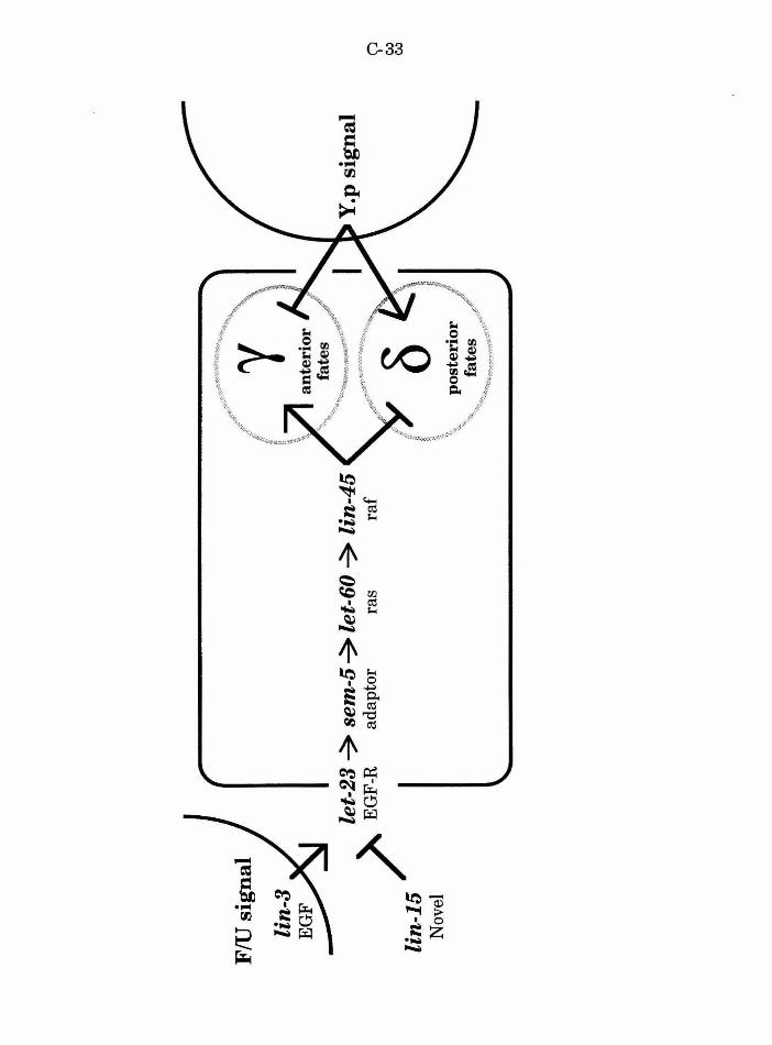
FlU
sig
nal
let-
23 ~ s
em-5
~ le
t-60
~ l
in-4
5
)' E
GF-
R
adap
tor
ras
raf
lin
-15
N
ovel
I; '\
() ;)
-~ J
'\..
po
ster
ior
/ ,"'
<~,
fate
s .. ,/-
" ~~~~~.~X~~~~·<,~· ...
~
0 w
w
C-34
References
Aroian, R. V., Koga, M., Mendel, J. E., Ohshima, Y. and Sternberg, P.
W. (1990). The let-23 gene necessary for Caenorhabditis elegans vulval
induction encodes a tyrosine kinase of the EGF receptor subfamily. Nature
348' 693-699.
Aroian, R. V. and Sternberg, P. W. (1991). Multiple functions of let-23, a C.
elegans receptor tyrosine kinase gene required for vulval induction. Genetics
128, 251-267.
Avery, L. and Horvitz, H. R. (1987). A cell that dies during wild-type C.
elegans development can function as a neuron in a ced-3 mutant. Cell 51,
1071-1078.
Beitel, G., Clark, S. and Horvitz, H. R. (1990). The Caenorhabditis elegans
ras gene let-60 acts as a switch in the pathway of vulval induction. Nature
348, 503-509.
Brenner, S. (1974). The genetics ofCaenorhabditis elegans. Genetics 77,71-
94.
Chamberlin, H. M. and Sternberg, P. W. (1993). Multiple cell interactions
are required for fate specification during male spicule development in
Caenorhabditis elegans. Development 118, 297-323.
Clark, S. G., Stern, M. J. and Horvitz, H. R. (1992). C. elegans cell
signalling gene sem-5 encodes a protein with SH2 and SH3 domains. Nature
356' 340-344.
Davidson, E. H. (1993). Later embryogenesis: regulatory circuitry in
morphogenetic fields. Development 118, 665-690.
C-35
Ferguson, E. and Horvitz, H. R. (1985). Identification and characterization
of 22 genes that affect the vulval cell lineages of Caenorhabditis elegans.
Genetics 110, 17-72.
Ferguson, E. L., Sternberg, P. W. and Horvitz, H. R. (1987). A genetic
pathway for the specification of the vulval cell lineages of Caenorhabditis
elegans. Nature 326, 259-267.
Greenwald, I. S., Sternberg, P. W. and Horvitz, H. R. (1983). The lin-12
locus specifies cell fates in Caenorhabditis elegans . Cell 34, 435-444.
Han, M., Aroian, R. and Sternberg, P. W. (1990). The let-60 locus controls
the switch between vulval and non-vulval cell types in C. elegans. Genetics
126, 899-913.
Han, M., Golden, A, Han, Y. and Sternberg, P. W. (1993). C. elegans lin-
45 raf gene participates in let-60 ras stimulated vulval differentiation. Nature
363, 133-140.
Han, M. and Sternberg, P. W. (1990). let-60, a gene that specifies cell fates
during C. elegans vulval induction, encodes a ras protein. Cell 63 , 921-931.
Han, M. and Sternberg, P. W. (1991). Analysis of dominant negative
mutations of the Caenorhabditis elegans let-60 ras gene. Genes & Deuel. 5 ,
2188-2198.
Herman, R. K. and Hedgecock, E. M. (1990). The size ofthe C. elegans
vulval primordium is limited by lin-15 expression in surrounding
hypodermis. Nature 348, 169-171.
Hill, R. J., Katz, W. S. and Sternberg, P. W. (1994). The EGF domain of
Lin-3 is sufficient to induce vulval development. submitted
Hill, R. J. and Sternberg, P. W. (1992). The lin-3 gene encodes an inductive
signal for vulval development in C. elegans. Nature 358, 470-476.
C-36
Hodgkin, J., Edgley, M., Riddle, D. L. and Albertson, D. G. (1988).
Appendix 4, Genetics In The Nematode Caenorhabditis elegans (ed. W. B.
Wood), pp. 491-584. Cold Spring Harbor, N.Y.: Cold Spring Harbor
Laboratory.
Hodgkin, J., Horvitz, H. R. and Brenner, S. (1979). Nondisjunction
mutants ofthe nematode Caenorhabditis elegans. Genetics 91,67-94.
Huang, L. S., Tzou, P. and Sternberg, P. W. (1994). The lin-15 locus
encodes two negative regulators of C. elegans vulval development. MoZee.
Biol. Cell 5, -------.
Kim.ble, J. (1981). Lineage alterations after ablation of cells in the somatic
gonad of Caenorhabditis elegans. Dev. Biol. 87, 286-300.
Kimelman, D., Christian, J. L. and Moon, R. T. (1992). Synergistic
principles of development: overlapping patterning systems in Xenopus
mesoderm induction. Development 116, 1-9.
Sulston, J. and Horvitz, H. R. (1977). Postembryonic cell lineages of the
nematode Caenorhabditis elegans. Dev. Biol. 56, 110-156.
Sulston, J. E., Albertson, D. G. and Thomson, J. N. (1980). The
Caenorhabditis elegans male: Postembryonic development of nongonadal
structures. Dev. Biol. 78, 542-576.
Sulston, J. E. and Hodgkin, J. (1988). Methods In The nematode
Caenorhabditis elegans (ed. W. Wood), pp. 587-606. Cold Spring Harbor, New
York: Cold Spring Harbor Laboratory.
Sulston, J. E. and White, J. G. (1980). Regulation and cell autonomy
during postembryonic development of Caenorhabditis elegans. Deuel. Biol. 78,
577-597.
D-1
Chapter4
A screen for Caenorhabditis elegans mutants defective in lineages of
male-specific blast cells
D-2
ABSTRACT
The specialized structures of the adult C. elegans male tail result
from sex-specific cell division of blast cells during larval
development. To identify genes required for the normal
development of specific postembryonic lineages I screened over 9000
mutagenized gametes for mutations that result in morphologically
abnormal male tails. Mutations that disrupt gross tail morphology
and others that specifically disrupt the copulatory spicules were
recovered. Candidate isolates were further screened to identify
mutants in which the morphological defect results from abnormal
cell lineages. 22 mutations identified (likely) 20 genes, ofwhich at
least seven are previously unknown to play a role in a specific
developmental process. Although the screen focused on mutations
that disrupt the male B lineage (the precursor to the spicules),
mutations that confer F, U, Y, and V lineage defects were also
recovered. These mutations provide the genetic tools to begin
understanding the mechanisms involved in the postembryonic
development of the male tail.
D-3
INTRODUCTION
At hatching male and hermaphrodite C. elegans are morphologically
similar. The specialized mating structures of the adult animals-- such as the
hermaphrodite vulva and the male tail -- result from the sex-specific division
of postembryonic blast cells (Sulston and Horvitz, 1977; Sulston, et al., 1980).
In the male tail, additional divisions oflateral epidermal blast cells V1-V6
and T, and ventral epidermal blast cells P10 and P11 produce the cells of the
eighteen ray sensilla and the hook sensillum, respectively. Cells of the
spicules, the postcloacal sensillum, and other male specific neurons and
hypodermal cells derive from four male-specific blast cells: B, U, F, andY.
These cells divide in the male, but not in the hermaphrodite. The B cell is the
precursor of all of the cells of the spicules. The B lineage produces 4 7 progeny
that include hypodermal, neuronal and neuronal support cells, and cell death
fates.
To identify genes that mediate fate specification during male tail
development I have screened for mutations that result in morphologically
abnormal male tails. Animals from candidate mutant lines were further
screened for lineage defects. I have focused on the B lineage because (1) at
least some mutations that disrupt fate specification in the B lineage result in
deformed spicules or male tail (Sternberg and Horvitz, 1988; Chisholm and
Hodgkin, 1989; Chisholm, 1991; Herman and Horvitz, 1994; Chamberlin and
Sternberg, in preparation), (2) the B lineage is complex, with multiple
specification steps, and (3) some steps in the lineage utilize asymmetric cell
divisions (Sternberg and Horvitz, 1988; Herman, 1991) whereas others
require cell interactions (Sulston and White, 1980; Chamberlin and
D-4
Sternberg, 1993) to properly specify fate. Thus mutations that disrupt this
lineage can be recovered, and they provide a genetic handle to understand a
variety of developmental processes.
In this chapter I describe the isolation and initial characterization of a
set ofEMS induced mutations that disrupt the B lineage and other lineages
in the male tail. Some of these mutations disrupt genes already known to
play a role in male tail development. Others define novel genes, or at least
genes without previously characterized developmental functions. In all, 22
mutations defining at least 20 genes were isolated from a screen of over 9000
chromosome sets (Table 1), suggesting both that fate specification in the C
elegans male tail is genetically tractable, and that many genes are required
for normal development.
MATERIALS AND METHODS
General Methods
Nematode strains were cultured according to standard techniques
(Brenner, 1974; Sulston and Hodgkin, 1988).
Mutations used
Mutations described in Hodgkin, et al. (1988).
Linkage Group (LG) 1: dpy-5(e61). hlnl(unc-54(h1040)). sy258. sy316.
LG II: dpy-10(e128). let-23(sy97, sy278). lin-31(n301, bx31). rol-
6(e187). unc-4(e120). unc-52(e444). mnCl. mnDf61.
D-5
LG III: dpy-l(el). dpy-17(e164). dpy-18(e1096). egl-5(n945, sy279).
lin-48(sy234) . mab-5(e1293, sy173). unc-25(e156). unc-32(e189). unc-
45(r450). unc-79(e1068). eDf2. nDfll. eTl.
LG IV: dpy-20(e1282). fem(sy240). lin-49(sy238). mec-3(e1338). unc-
24(e138). unc-31(e169). DnTl. eDflB. eDf19. sy287.
LG V: dpy-11(e224). him-5(e1467, e1490). lin-25(e1446, sy29). eTl.
DnTl.
LG X: let-l(mn119). let-4(mn105). let-5(mn106). let-9(mn107). lin-
2(e1309). lin-15(sy307). lin-47(sy32). lon-2(e678). mab-27(sy202). unc-
3(e131) . vab-3(?sy66, sy281). mnDpl.
Unknown linkage: sy282. sy285. sy285. sy294. sy295. K.33.3.
Q.29.3.
Cell lineage analysis
Cell nuclei divisions in living animals were directly observed using
Nomarski differential interference contrast optics as described by Sulston
and Horvitz (1977). Nomenclature follows the standard ofSulston and
Horvitz, with modifications of Chamberlin and Stemberg (1993).
Isolation of mutations
Mutant isolation consisted of a positive and a negative selection
scheme. First, mutant lines that display a male tail defect (male abnormal,
or Mab) were selected. Second, animals from these candidate strains were
screened for the presence of a cell lineage defect (Lin). Mab non-Lin strains
were excluded from further study.
D-6
Selection of two general defects identified Mab candidates: male
mating defects and male morphology defects. Mutageneses were carried out
according to standard conditions (Sulston and Hodgkin, 1988): worms were
suspended in a solution of 50 mM Ethylmethanesulfonate (EMS) in M9 buffer
for four hours at 20°C. The animals were then washed and allowed to recover
for four hours. F2 screens for mutations that confer a male mating defect
were carried out by Leslie Barber (Table 2, screen 1) and Yvonne Hajdu and
Katherine Liu (screen 2), using the protocol of Hodgkin (1983). Briefly, ten
F2 hermaphrodites were cloned from the F1 progeny of mutagenized him-
5(e1490) animals. Six F3 males from the cloned hermaphrodites were tested
together for ability to sire cross progeny with unc-52(e444) or unc-31(e169)
hermaphrodites. Strains from which males sired no cross progeny were
tested a second time. Strains which failed the mating test twice are
candidate Copulation Defective (Cod) strains. Adult males from these lines
were then observed under Nomarski optics at 1250x magnification for any
tail morphology defects associated with the mating defect (Mab candidates).
Additional screens (screens 3 and 4) were carried out directly for male
tail morphology defects using a Wild Makroskop. Four to five F1 progeny
were cloned from mutagenized Po him-5(e1490) hermaphrodites. The
population ofF2 males from F1 clones was screened at 200x-400x
magnification for the presence of males with deformed tails. If the F1 clone
harbored a mutation that confers a male tail defect, 1/4 of self progeny males
are expected to be abnormal (assuming a recessive, viable mutation with no
maternal rescue). Sibling hermaphrodites were selected from strains with
abnormal males in order to recover the homozygous mutation. The Mab
phenotype was verified by observing adult males under Nomarski optics at
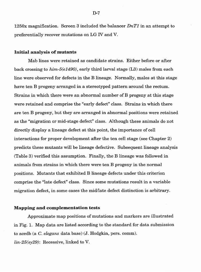
D-7
1250x magnification. Screen 3 included the balancer DnTl in an attempt to
preferentially recover mutations on LG IV and V.
Initial analysis of mutants
Mab lines were retained as candidate strains. Either before or after
back crossing to him-5(e1490), early third larval stage (L3) males from each
line were observed for defects in the B lineage. Normally, males at this stage
have ten B progeny arranged in a stereotyped pattern around the rectum.
Strains in which there were an abnormal number of B progeny at this stage
were retained and comprise the "early defect" class. Strains in which there
are ten B progeny, but they are arranged in abnormal positions were retained
as the "migration or mid-stage defect" class. Although these animals do not
directly display a lineage defect at this point, the importance of cell
interactions for proper development after the ten cell stage (see Chapter 2)
predicts these mutants will be lineage defective. Subsequent lineage analysis
(Table 3) verified this assumption. Finally, the B lineage was followed in
animals from strains in which there were ten B progeny in the normal
positions. Mutants that exhibited B lineage defects under this criterion
comprise the "late defect" class. Since some mutations result in a variable
migration defect, in some cases the mid/late defect distinction is arbitrary.
Mapping and complementation tests
Approximate map positions of mutations and markers are illustrated
in Fig. 1. Map data are listed according to the standard for data submission
to acedb (a C. elegans data base) (J. Hodgkin, pers. comm).
lin-25(sy29): Recessive, linked to V.
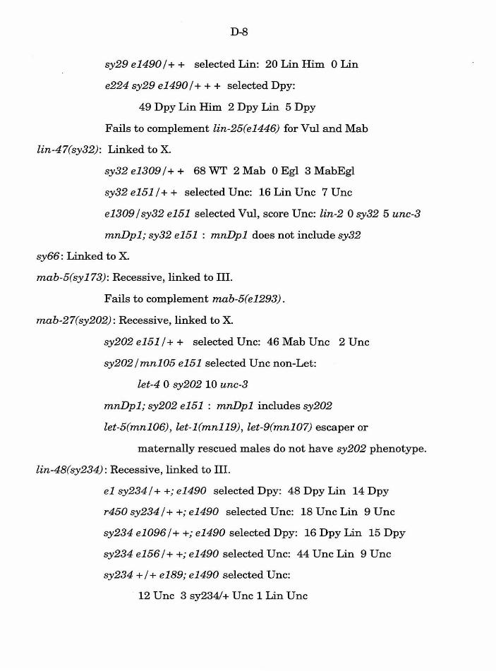
D-8
sy29 e14901+ + selected Lin: 20 Lin Him 0 Lin
e224 sy29 e14901+ + + selected Dpy:
49 Dpy Lin Him 2 Dpy Lin 5 Dpy
Fails to complement lin-25(e1446) for Vul and Mab
lin-47(sy32): Linked to X.
sy32 e1309 I+ + 68 WT 2 Mab 0 Egl 3 MabEgl
sy32 e151 I+ + selected Unc: 16 Lin Unc 7 Unc
e1309 I sy32 e151 selected Vul, score Unc: lin-2 0 sy32 5 unc-3
mnDpl; sy32 e151 : mnDpl does not include sy32
sy66: Linked to X
mab-5(sy173): Recessive, linked to III.
Fails to complement mab-5(e1293).
mab-27(sy202): Recessive, linked to X.
sy202 e151 I+ + selected Unc: 46 Mab Unc 2 Unc
sy202 I mn105 e151 selected Unc non-Let:
let-4 0 sy202 10 unc-3
mnDpl; sy202 e151 : mnDpl includes sy202
let-5(mn106), let-l(mn119), let-9(mn107) escaper or
maternally rescued males do not have sy202 phenotype.
lin-48(sy234): Recessive, linked to III.
el sy234 I+ +; e1490 selected Dpy: 48 Dpy Lin 14 Dpy
r450 sy234 I++; e1490 selected Unc: 18 Unc Lin 9 Unc
sy234 e10961+ +; e1490 selected Dpy: 16 Dpy Lin 15 Dpy
sy234 e156 I+ +; e1490 selected Unc: 44 Unc Lin 9 Unc
sy234 +I+ e189; el490 selected Unc:
12 Unc 3 sy2341+ Unc 1 Lin Unc
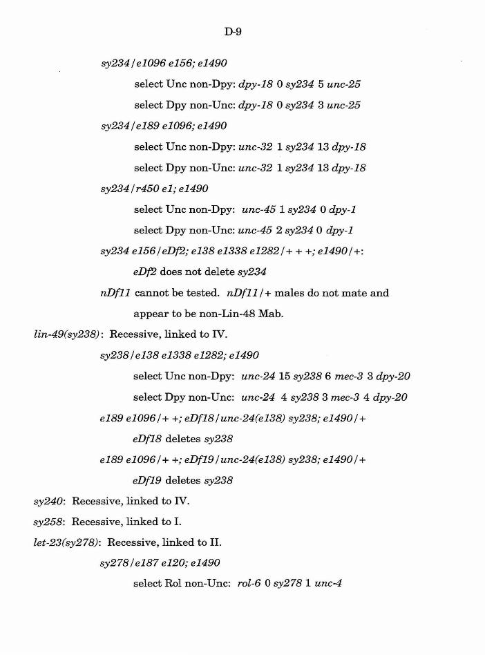
D-9
sy234le1096 e156; e1490
select Unc non-Dpy: dpy-18 0 sy234 5 unc-25
select Dpy non-Unc: dpy-18 0 sy234 3 unc-25
sy234 I e189 e1096; e1490
select Unc non-Dpy: unc-32 1 sy234 13 dpy-18
select Dpy non-Unc: unc-32 1 sy234 13 dpy-18
sy234lr450 e1; e1490
select Unc non-Dpy: unc-451 sy234 0 dpy-1
select Dpy non-Unc: unc-45 2 sy234 0 dpy-1
sy234 e156 I eDf2; e138 e1338 e1282 I+ + +; e1490 I+:
eDf2 does not delete sy234
nDf11 cannot be tested. nDf11 I+ males do not mate and
appear to be non-Lin-48 Mab.
lin-49(sy238): Recessive, linked to IV.
sy238 I e138 e1338 e1282; e1490
select Unc non-Dpy: unc-24 15 sy238 6 mec-3 3 dpy-20
select Dpy non-Unc: unc-24 4 sy238 3 mec-3 4 dpy-20
e189 e1096 I++; eDf18 I unc-24(e138) sy238; e1490 I+
eDf18 deletes sy238
e189 e1096 I ++; eDf19 lunc-24(e138) sy238; e1490 I+
eDf19 deletes sy238
sy240: Recessive, linked to IV.
sy258: Recessive, linked to I.
let-23(sy278): Recessive, linked to II.
sy278 I e187 e120; e1490
select Rol non-Unc: rol-6 0 sy278 1 unc-4
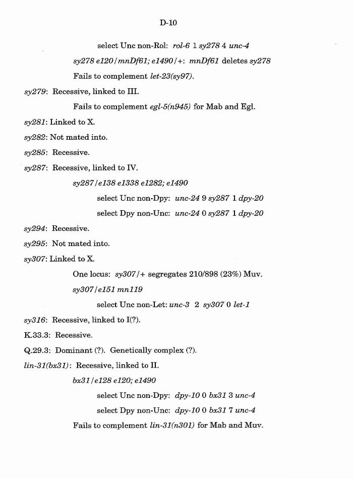
D-10
select Unc non-Rol: rol-6 1 sy278 4 unc-4
sy278 e120imnDf61; e14901+: mnDf61 deletes sy278
Fails to complement let-23(sy97).
sy279: Recessive, linked to III.
Fails to complement egl-5(n945) for Mab and Egl.
sy281 : Linked to X
sy282: Not mated into.
sy285: Recessive.
sy287: Recessive, linked to IV.
sy287 I e138 e1338 e1282; e1490
select Unc non-Dpy: unc-24 9 sy287 1 dpy-20
select Dpy non-Unc: unc-24 0 sy287 1 dpy-20
sy294: Recessive.
sy295: Not mated into.
sy307: Linked to X.
One locus: sy307 I+ segregates 210/898 (23%) Muv.
sy307 I e151 mn119
select Unc non-Let: unc-3 2 sy307 0 let-1
sy316: Recessive, linked to I(?).
K.33.3: Recessive.
Q.29.3: Dominant (?). Genetically complex(?).
lin-31 (bx31): Recessive, linked to II.
bx31 I e128 e120; e1490
select Unc non-Dpy: dpy-10 0 bx31 3 unc-4
select Dpy non-Unc: dpy-10 0 bx31 7 unc-4
Fails to complement lin-31(n301) for Mab and Muv.
D-11
RESULTS
Although some of the other lineages of male specific blast cells are
abnormal in the mutants, candidates were initially classified according to
identified B lineage defects.
Mutants with early B lineage defects
Mutants with early B lineage defects generally displayed fewer than
normal B progeny at the L3 stage. These lineage defects are commonly
associated with grossly abnormal tails ("club tails") and structural instability
of the tail. Often, these mutants die before reaching adulthood because they
explode as larvae, or at the L4 molt into the adult stage.
sy66, sy281 [vab-3 ?]. sy66 and sy281 are linked to X and have a
distinctive B lineage defect with six B progeny rather than the normal ten in
the early L3 (see Chapter 5). TheY lineage is also abnormal in that Y.p
appears to divide only a single time leaving two large cells of indeterminate
differentiation. Both mutations occasionally confer a notched head phenotype
in both males and hermaphrodites, although this phenotype is rare in sy281
animals. The lineage defect and head morphology defect is similar to
phenotypes associated with viable mutations in vab-3 (A. Chisholm, pers.
comm.). The B lineages followed in two sy66 animals are illustrated in
Chapter 5, Fig. 1.
sy279 [egl-5]. sy279 males have four B cells in the L3 stage. This
mutation is linked to LG III, and fails to complement egl-5(n945) for both the
male tail defect and the egg laying defect. egl-5 is part of the C. elegans
D-12
homeotic cluster. Mutations in egl-5 disrupt a specific subset of cells in the
posterior body region, including B. The B lineage defect in egl-5 mutants has
been described by Chisholm (1991).
sy282 and K.33.3. sy282 hermaphrodites have a knobby protruding
vulva, do not lay eggs, and are often sterile. The sy282 strain was not
successfully mated into after over 200 attempts. It is not temperature
sensitive, nor can hermaphrodites lay eggs after passing through the dauer
stage. K.33.3 hermaphrodites are Egl and difficult to mate into. They have
small brood sizes, many worms die early in adulthood, and they occasionally
have protruding vulvae. No chromosomal linkage for K33.3 has been
identified, although a marker for the cluster for each linkage group, as well
as balancers for many of the arms, have been tested.
Mutants with migration or mid-stage defects in the B lineage
Mutations that disrupt proper specification ofF and U
sy238 [lin-49] . sy238 animals are sickly, and some larvae as well as
adults display an abnormal "scarring'' or refractile material at the junction of
the intestine and the rectum. sy238 I Df is lethal: animals arrest at or before
the L4 stage (based on gonad morphology), but they are significantly smaller
than wild type. They display heavy scarring at the rectum such that it is
blocked. It is likely that this morphological defect blocks proper digestion
and contributes to the lethality in sy238 I Df animals. The male B lineage of
sy238 homozygotes can be disrupted in the specification of proper anterior
and posterior positioning and fates of the aa and pp pairs (Table 3 B.l). The
F and U cells are abnormal in sy238 homozygous males. One or the other cell
D-13
can appear to be missing in individual males, and the lineage of the cell(s)
that remains can be abnormal (Fig. 2 B). The fate of the "missing" F and U
cells has not been established. However, it is reasonable that lin-49 is
required for proper specification ofF and U. F and U (and their progeny in
males) make up part of the rectal epithelium, and are essential for forming a
connection between the intestine and the rectum (Sulston, et al., 1983;
Herman, 1991). Thus sy238/Dfis likely lethal because F and U are absent
and do not perform this function. It is not yet clear if the B lineage defect
associated with sy238 results directly from the lin-49 mutation, or if it is a
secondary effect of the abnormal F and U lineages.
sy294. sy294 mutants are sickly and can die at different stages. sy294
hermaphrodites display an abnormal "notch" anterior to the anus. In sy294
males the B cell produces the normal ten progeny by the L3 stage. However,
all of the B.a cells remain dorsal to the rectum rather than organized around
it in two rings of four cells. The U cell in sy294 males is abnormal. It
produces a lineage with the timing, axes, number of progeny, and apparent
differentiated fates ofY.p (or possibly Y, Fig. 2 C). It is not clear if the B
lineage defect in sy294 males is due directly to the mutation or a secondary
effect of the U lineage defect.
Q.29.3. The mutation in the Q.29.3 strain has not been successfully
outcrossed. The strain appears to be genetically complex and includes at
least one dominant component. Although the lineage defect in the strain has
not been characterized, the fact that the strain exhibits the sickly phenotype
associated with scarring at the rectum and a migration defect in the B
lineage suggests that it may be another member of the class of mutations
that disrupt F and U specification.
D-14
Other mutations that confer a mid-stage defect
sy285. sy285 males exhibit a variable migration defect among the
progeny of the B cell. In general, when the cells migrate to the proper
positions they produce the normal lineage, but cells that migrate incorrectly
can be abnormal (Table 3 B.2). In many animals all B progeny remain dorsal
and posterior to the rectum. The variable lineage defect is consistent with
the observation that about 20% of sy285 males are morphologically wild type.
Mutants with late defects in the B lineage
Mutations in genes also required for normal hermaphrodite vulval
development and egg laying
Egglaying defective (Egl) and Vulvaless (Vul)
sy278 [let-23]. In sy278 males the lineages of the anterior aa, pp, and
ap/pa cells are disrupted (Table 3 A.6). These cells can produce the lineage
normally associated with their posterior neighbors. This is the phenotype
associated with mutations in genes in the lin-3 I let-23 pathway also required
for vulval induction (see Chapter 3 ). sy278 maps between rol-6 and unc-4 on
LG II, and fails to complement let-23(sy97) for the male tail defect (Table 4).
However, sy278 complements sy97 for the defect in vulval development, and
for lethality. Furthermore, sy278 I Df males are Mab, but sy278 complements
for the other let-23 functions. sy278 I Df hermaphrodites have normal vulval
development and are fertile, and sy278 I Df animals are normal in Pll/P12
specification. There is also no appreciable lethality associated with sy278
homozygotes or hemizygotes. sy278 likely represents a mutation in let-23
D-15
that disrupts male tail function but retains significant activity for the other
let-23 functions.
sy29 [lin-25]. In sy29 males the lineages of the anterior B.a progeny
cells are sometimes disrupted (Table 3 A.1). Hermaphrodites are also Egl
and Vul. sy29 maps between dpy-11 and him-5 on LG V, and fails to
complement lin-25(e1446). sy29 is temperature sensitive (Simon Tuck, pers.
comm.), and likely represents a hypomorphic mutation in lin-25 .
sy258 and sy316. In sy258 and sy316 males the lineages of the anterior
B.a progeny cells are sometimes disrupted (Table 3 A.5, A.9).
Hermaphrodites are Egl and weakly Vul, although the reduction in vulval
development is not sufficient to account for the extent of the egg laying defect.
Both mutations are on LG I, and they may represent alleles of sur-2, a locus
defined originally because mutations are suppressors of gain-of-function
mutations in let-60 (N. Singh and M. Han, pers. comm.).
sy295. A lineage defect in the anterior aa cell is associated with sy295.
sy295 hermaphrodites are Egl but not Vul. Nevertheless, attempts to cross in
to the strain failed with over 200 hermaphrodites. sy295 is not temperature
sensitive, and it is not suppressed by passing worms through dauer stage.
Multivulva (Muv)
sy307 [lin-15]. sy307 males have a weak spicule defect, but also have
hook abnormalities, pseudovulvae, and a gonad migration defect. sy307
hermaphrodites have excessive vulval development (multivulva or Muv).
These are phenotypes associated with mutations in lin-15 and other genes in
that class (Ferguson and Horvitz, 1985; 1989). sy307 maps to the right of
D-16
unc-3 on LG X, and segregates as a single mutation. sy307 likely represents
a mutation in the lin-15 locus.
bx31 [lin-31 ]. bx31 males have a variable late B lineage defect similar
to lin-31(n301) males (Table 3 A.10, A.11). The hermaphrodites are variably
Muv. bx31 maps left of dpy-10 on LG II, and fails to complement lin-31(n301)
for male tail and vulval defects.
Other mutations that disrupt development ofF and U
sy234 [lin-48] . The cells in the aa pair, and occasionally the anterior
pp cell are disrupted in sy234 males (Table 3 A.4). F and U lineages are also
disrupted: both F and U divide earlier and produce more progeny than
normal (Fig. 2 E). There are no obvious pleiotropies associated with sy234.
Lineages in sy234 animals are normal for the following cells: (1) lateral
hypodermis: hermaphrodite V cell through the L2 stage (postdeirid),
hermaphrodite T cell, male T and V ray sublineages (2) ventral hypodermis:
hermaphrodite early P cells (Pn.a), male and hermaphrodite Pn.p cells (vulva
and hook) (3) gonad: male early Z1, Z4 cells, hermaphrodite ventral uterine
cells (VU), partial hermaphrodite dorsal uterine cells (DU) ( 4) early
hermaphrodite M cell (5) hermaphrodite K cell and (6) early divisions and
migration of hermaphrodite Q cells.
sy234 defines the gene lin-48, and maps between dpy-1 and unc-32 on
LG III. It has not been established whether the B lineage and F and U
lineage defects are independent effects of sy234, or if the B lineage defect is a
secondary effect of the F and U lineage defect, or vice versa. However,
neither lineage defect is the defect associated with simply removal of the
other cell(s) (Chisholm and Hodgkin, 1989; Chamberlin and Sternberg, 1993).
D-17
sy287. sy287 hermaphrodites are essentially normal and fertile. In the
B lineage sy287 males exhibit a defect in the pp pair: the cells fail to migrate
to their normal anterior/posterior positions, and both cells behave like a
normal posterior pp cell, producing a b lineage (Table 3 A. 7). The F lineage
can also be abnormal, producing more progeny than normal (Fig. 2 D). The F
lineage defect may be similar to that associated with sy234, but the B lineage
defect is clearly distinct between the two.
Other mutations that confer a late defect
sy32 [lin-47]. Several late B lineage defects are observed in sy32
males. The fates ofthe anterior cells in the aa and ap/pa pairs and the
posterior cell in the pp pair can be disrupted (Table 3 A.2). In a lineage of
the F and U cells the lineages were esentially wild type, although the polarity
of one of the asymmetric divisions ofF (F.r) was reversed. This is the lineage
defect observed when B is ablated, so the defect may reflect a secondary effect
of the B lineage defect, although this has not been verified. There are no
overt pleiotropies associated with sy32, although it appears to result in
reduced viability (not quantified). sy32 defines the gene lin-47, and is linked
to X, probably between the right breakpoint of stDp2 and the left breakpoint
ofmnDpl.
sy202 [mab-27]. sy202 males have only a weak B lineage defect,
although the timing of all cell divisions can be abnormal (Table 3 A.3). sy202
animals are weakly Unc. sy202 defines the gene mab-27, and maps to the X
LG between the left breakpoint ofmnDpl and unc-3. Rare escapers or
maternally rescued males mutant for essential genes in the region (let-
D-18
l(mn119), let-5(mn106), let-9(mn107)) do not have the mab-27 phenotype, and
thus sy32 is not likely a hypomorphic mutation in one of those genes.
Mutants with other lineage defects
syl73 [mab-5]. sy173 males display a distinctive phenotype where the
normal V rays are missing and the lateral cuticular lines (alae) extend
posteriorly. sy173 maps to LG III and fails to complement mab-5(e1293).
syl75 males have abnormal spicules, but mosaic analysis (Kenyon, 1986)
suggests that this defect is due to abnormal M lineage in mab-5 mutants
rather than abnormal B lineage (M progeny are required for proper
morphogenesis of the spicules).
sy240 [fem ]. sy240 males have rudimentary tails, and some have
oocytes in the gonad rather than sperm. sy240 hermaphrodites are self
sterile, but can produce cross-progeny with N2 males. sy240 hermaphrodites
also lack sperm, suggesting that the self-sterility results from feminization.
sy240 maps to LG IV, the location of two genes involved in sex determination:
fem-1 and fem-3. Although complementation was not tested, sy240 likely
represents an allele of one of these two genes. Defects in the male tail
lineages of sy240 males presumably arise from (partial) sexual
transformation rather than from disrupted fate specification per se.
DISCUSSION
A screen for mutations that disrupt cell lineages in the C. elegans male
tail recovered 22 mutations in over 9000 mutagenized gametes screened, with
a frequency of about one in 450. Of these 22 mutations, at most two loci are
D-19
represented by two mutations, and the other eighteen are distinct either by
map location, phenotype, or both. The relatively small number of
chromosomes screened, combined with the high number of loci represented by
only a single mutation, indicate that this screen does not represent saturation
for the Mab Lin phenotype. Nevertheless, it is clear that many genes can
mutate to produce a Mab Lin phenotype. Many of the mutations are
pleiotropic, and some represent alleles of genes previously identified for their
role in other developmental processes. However, this study has identified
and allowed the characterization of (1) additional functions of already known
genes, (2) a tissue preferential allele of a gene (let-23) known to function in
several tissues, and (3) possibly new genes required for development.
Several genes have previously been identified to play a role in the
development of the male tail, and as expected this screen recovered
mutations in some of these genes. Mutations in genes involved in sex
determination can partially feminize XO animals, and sy240 represents this
class of mutation. sy 173 and sy279 represent the genes in the C. elegans
homeotic cluster required for proper fate specification in the posterior body
region (mab-5 and egl-5). Alleles of other genes known to have abnormal
male tails associated with mutations (Hodgkin, 1983) were also expected
(e.g., vab-3: sy66 and sy281). Mutations in let-23, lin-15, lin-25, lin-31 , and
likely sur-2 were recovered. Genes involved in vulval development are
required for F and U promotion of anterior fates in the B.a progeny (see
Chapter 3). Other mutations were recovered that concomitantly disrupt male
tail development and vulval function (sy282, K.33.3, sy295). However, these
mutations do not appear to disrupt vulval induction or development, and thus
represent a different class of gene.
D-20
Among recovered mutations in genes required for vulval development,
sy278 is of particular interest because it represents a mutation that
preferentially disrupts the male tail function of let-23 but not any of the other
known functions . Other screens for let-23 alleles have required that the
mutation either be lethal (Herman, 1978) or that the vulval development
function be reduced (Aroian and Sternberg, 1991). sy278 shows no
appreciable lethality and complements a deficiency of let-23 for vulval
development. It represents a unique allele that could only be recovered by
specifically screening for mutations that disrupt male tail development.
Several mutations likely represent novel genes, or novel alleles and
functions for known genes (such as essential genes). Most detailed
characterization so far has been for sy32, sy202, sy234 , and sy238, which
define the genes lin-47 , mab-27, lin-48 , and lin-49, respectively. Because of
the novel lineage defects, additional genetic mapping will likely indicate that
sy285, sy287, and sy294 also represent new genes, or define new functions for
existing genes. Several of these mutations are associated with "sickness" and
lethality, so some ofthese mutations may represent hypomorphic mutations
in genes with a loss-of-function lethal phenotype. For example, sy238 I Df
animals arrest as larvae, with a phenotype that likely represents an
enhancement of the sy238 I sy238 phenotype. When tested, mutations such as
sy294 may behave likewise.
Many of the isolated mutants have weak male tails that are
structurally unstable and prone to explosion. Of these, a novel class of
mutations recovered in this screen result in a rectal "scarring" phenotype.
These mutations result in misspecification ofF and U cells. In addition to
providing a signal for proper B development, these cells make up part of the
D-21
rectal epithelium and are required for the formation of the junction between
the rectum and the intestine. Other mutations that disrupt the development
ofF, U, orB can be detrimental to the survival of mutant males but not
hermaphrodites. This is likely because among the progeny ofF, U, and B are
cells that must function as the parent cell does in the young male larva or in
the hermaphrodite. If the precursor is properly specified but the lineage is
abnormal, these fates may be misspecified, resulting in males with damaged
tails, but not hermaphrodites.
In summary, this screen for mutations that disrupt male tail
development specifically associated with a lineage defect is an effective way
to identify a class of developmental mutants in C. elegans. The direct screen
for F2 morphological mutants followed by selection of mutant lines with
lineage defects prior to backcross and genetic mapping (the protocol followed
in screen 4) is particularly efficient in identifying mutants in this class.
These mutations represent a wide range of functions in male tail
development.
D-22
Table 1. Mutations recovered in the screen for Mab Lin mutants. The
mutations are subdivided according to the B lineage defect observed in
mutants. Male morphology defect: spicule = only spicules are abnormal;
blowout= male tails tend to be delicate, and often become damaged or
explode; club tail= male tails are extremely deformed, affecting all
structures. Other phenotypes: Egl = egglaying defective; p-Vul =protruding
vulva; Ste = sterile; Let = lethal; Vul = vulvaless; Muv = multivulva. Link
= linkage group. dom/rec = dominance or recessiveness of the mutant allele
in trans to a wild type allele. comp/gene = Results of complementation or
other tests assign the mutation to the indicated gene. Genes followed by (? )
indicate that the mutation is likely in the indicated gene, but has not yet
been formally tested.
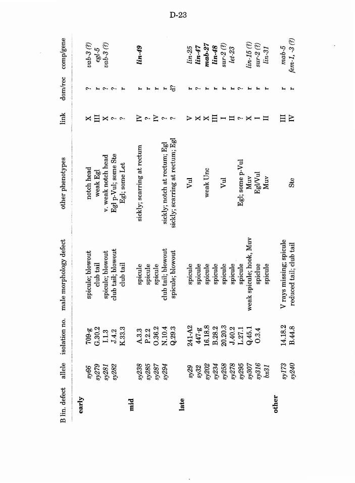
B l
in.
defe
ct
alle
le
isol
atio
n no
. m
ale
mor
phol
ogy
defe
ct
oth
er p
heno
type
s li
nk
do
m/r
ec
com
p/ge
ne
----
----
-··----~---
-------
----
-·-··
-..
.•. --
·····-
·--···
····
-----
··-··
----
------
---
-. -
----
----
----
----
----~----
------·--
---
·····
----
------
--
----
---·
-··-
-·
earl
y
sy66
70
9-g
spic
ule;
blo
wou
t no
tch
hea
d
X
? va
b-3
(?)
sy27
9 G
.30.
2 cl
ub t
ail
wea
k E
gl
III
r eg
l-5
sy28
1 1.
1.3
spic
ule;
blo
wou
t v
. wea
k n
otch
hea
d
X
? va
b-3
(?)
sy28
2 J.
4.2
club
tai
l; b
low
out
Egl
p-V
ul;
som
e S
te
? ?
K.3
3.3
cl
ub t
ail
Egl
; so
me
Let
?
r m
id
sy23
8 A
.3.3
sp
icul
e si
ckly
; sc
arri
ng
at
rect
um
IV
r li
n-4
9
sy28
5 P
.2.2
sp
icul
e ?
r sy
287
0.36
.2
spic
ule
IV
r sy
294
N.1
0.4
club
tai
l; b
low
out
sick
ly; n
otch
at
rect
um;
Eg
l ?
r Q
.29.
3 sp
icul
e; b
low
out
sick
ly;
scar
ring
at
rect
um; E
gl
? d?
t:J
' la
te
l'V
VJ
sy29
24
1-A
2 sp
icul
e V
ul
v r
lin-
25
sy32
44
7-g
spic
ule
X
? li
n-4
7
sy20
2 16
.18.
8 sp
icul
e w
eak
Unc
X
r
mab
-27
sy23
4 B
.28.
2 sp
icul
e II
I r
lin
-48
sy
258
20.2
0.3
spic
ule
Vul
I
r su
r-2
(?)
sy27
8 J.
40.2
sp
icul
e II
r
let-
23
sy
295
L.2
7.1
spic
ule
Egl
; som
e p-
Vul
?
? sy
307
Q.4
5.1
wea
k s
picu
le; h
ook,
Muv
M
uv
X
r li
n-15
(?)
sy31
6 0
.3.4
sp
iclu
e E
gW
ul
I r
sur-
2 (?
)
bx31
sp
icul
e M
uv
II
r li
n-31
o
ther
sy
173
14.1
8.2
V r
ays
mis
sing
; sp
icul
e II
I r
mab
-5
sy24
0 B
.44.
8 re
duce
d ta
il;
club
tail
S
te
IV
r fe
m-1
, -3
(?)
D-24
Table 2. Screens used to isolate Mab Lin mutants. The Table indicates the
source of mutations and the methods used to recover the mutations.
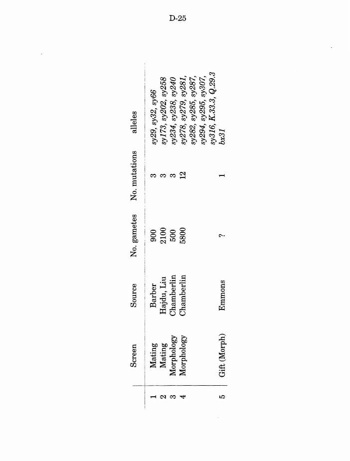
Scr
een
Sou
rce
No.
gam
etes
N
o. m
uta
tio
ns
alle
les
. ~--
·-
.-
-----
·-·-·
··-· -
1 M
atin
g B
arb
er
900
3 sy
29,
sy32
, sy
66
2 M
atin
g H
ajdu
, Liu
21
00
3 sy
173,
sy2
02,
sy25
8 3
Mor
phol
ogy
Cha
mbe
rlin
50
0 3
sy23
4,sy
238,
sy2
40
t:1
I
4 M
orph
olog
y C
ham
berl
in
5800
12
sy
278,
sy2
79,
sy28
1,
tv
01
sy28
2, s
y285
, sy
287,
sy
294,
sy2
95,
sy30
7,
sy31
6, K
.33.
3, Q
.29.
3 5
I G
ift (
Mor
ph)
Em
mon
s ?
1 bx
31
D-26
Table 3. B lineage defects observed in male mutants. abn-n indicates the
lineage was abnormal, producing n progeny. n.d. =not determined (lineage
not followed to completion for that cell). Other notation as in Chapter 2,
Table 1. Animals #602, #603, and #612 are from non-backcrossed strains.
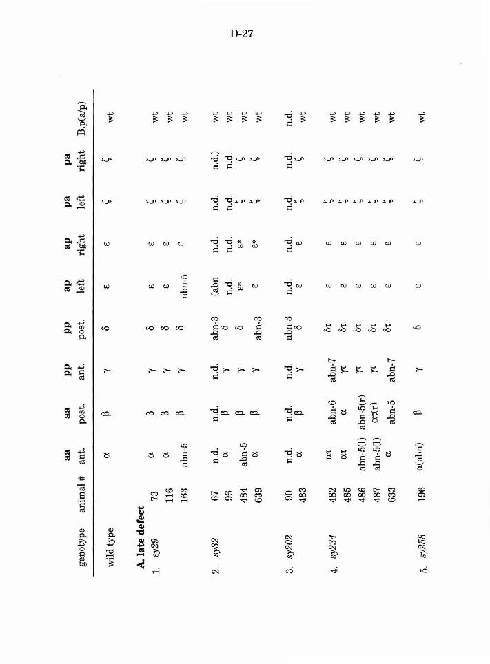
a a
a a
pp
p
p
ap
ap
pa
pa
geno
type
an
imal
#
ant.
po
st.
ant.
po
st.
left
ri
ght
left
ri
ght
B.p
(a/p
)
wild
typ
e a
p y
0 £
£ s
s w
t
A. l
ate
def
ect
1.
sy29
73
a
p y
0 £
£ s
s w
t
116
a p
y 0
£ £
s s
wt
163
abn-
5 p
y 0
abn-
5 £
s s
wt
2.
sy32
67
n.
d.
n.d.
n.
d.
abn-
3 (a
bn
n.d
. n.
d.
n.d
.)
wt
96
a p
y 0
n.d.
n.
d.
n.d
. n
.d.
wt
p y
0 £
*
£*
s
s t:l
48
4 ab
n-5
wt
I 1.\J
a p
y £
£*
s
s 'I
63
9 ab
n-3
wt
3.
sy20
2 90
n.
d.
n.d.
n.
d.
abn-
3 n.
d.
n.d.
n.
d.
n.d.
n
.d.
483
a p
y 0
£ £
s s
wt
4.
sy23
4 48
2 a'
t ab
n-6
abn-
7 ot
£
£ s
s w
t
485
at
a Y'
t ot
£
£ s
s w
t
486
abn-
5(1)
ab
n-5(
r)
Y't
ot
£ £
s s
wt
487
abn-
5(1)
a'
t(r)
Y'
t ot
£
£ s
s w
t
633
a ab
n-5
abn-
7 ot
£
£ s
s w
t
5.
sy25
8 19
6 a(
abn)
p
y 0
£ £
s s
wt
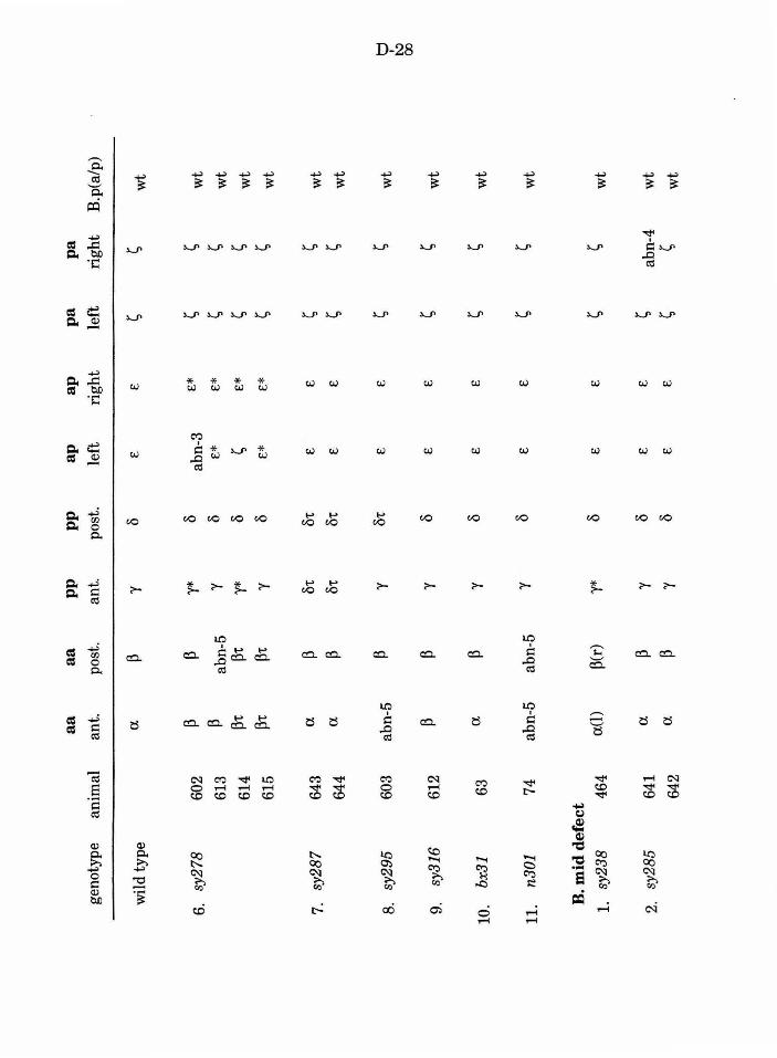
a a
a a
pp
p
p
ap
ap
pa
p
a ge
noty
pe
anim
al
ant.
post
. an
t.
post
. le
ft
rig
ht
left
ri
ght
B.p
(a/p
)
wild
typ
e a.
p "{
0
£ £
s s
wt
6.
sy27
8 60
2 p
p "{*
0
abn-
3 £
* s
s w
t
613
p ab
n-5
"{
0 £*
£
* s
s w
t
614
p't
p't
'Y*
0 s
£*
s s
wt
615
p't
p't
"{
0 £
* £
* s
s w
t
7.
sy28
7 64
3 a.
p 01
: 01
: £
£ s
s w
t
644
a. p
01:
01:
£ £
s s
wt
8.
sy29
5 60
3 ab
n-5
p "{
01
: £
£ s
s w
t t:
l I 1:\:)
0
0
9.
sy31
6 61
2 p
p "{
0
£ £
s s
wt
10.
bx31
63
a.
p "{
0
£ £
s s
wt
11.
n301
74
ab
n-5
abn-
5 "{
0
£ £
s s
wt
B. m
id d
efec
t 1.
sy
238
464
a.(l
) p(
r) "{
* 0
£ £
s s
wt
2.
sy28
5 64
1 a
p y
0 £
£ s
abn-
4 w
t
642
a. p
y 0
£ £
s s
wt

D-29
Table 4. Phenotypes associated with let-23 mutations. sy97 I sy97 data are
from Aroian and Sternberg (1991). Numbers indicate the number of wild
type/number of animals screened. wt = wild type.
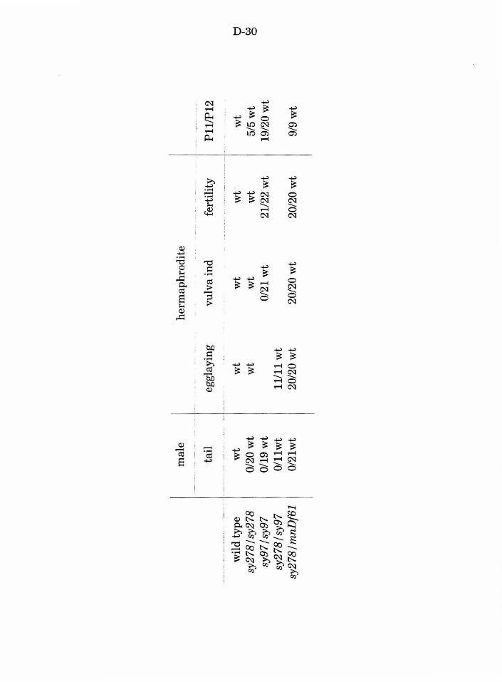
mal
e he
rmap
hrod
ite
--··-
····
....
·-.
---.
. --
· --
...
---.
,---p~~/P~2
tail
eg
glay
ing
vulv
a in
d fe
rtil
ity
.. --
-·-·
····-
----
···-·
· --
-----
. ··-
---·
· ---
·-··
--·-
.
wil
d ty
pe
wt
wt
wt
wt
wt
tJ
I
sy27
8 I s
y278
0/
20 w
t w
t w
t w
t 5/
5 w
t C
lj
0
sy97
lsy9
7
0/19
wt
0/21
wt
21/2
2 w
t 19
/20
wt
sy27
Bis
y97
0/11
wt
11/1
1 w
t sy
278 I m
nDf6
1 0/
21w
t 20
/20
wt
20/2
0 w
t 20
/20
wt
I 9/
9 w
t
D-31
Fig. 1. Genetic maps showing approximate positions of mutations and
markers used in this study. Left is up, right is down. Mutations identified in
this study or the genes they represent are indicated in bold.
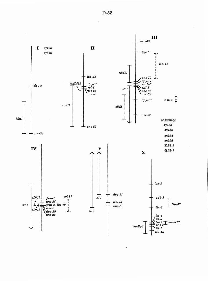
hl n 1
I
dpy-5
unc-54
IV
eDf19 )
nT1 I,} eDf18
sy258
ay316
m nC1
sy287 fem·l ,-unc-24 1
fem-3, lin -49 1
mec-3 1
dpy-20 - 1-
u nc-22
n
unc-52
v
eT1
nT1
D-32
eDf2
dpy-11
lin-25 h im-5
m unc-45
dpy-1
unc-79 dpy-17 IIUib-5 egl-5 un c-36 unc-32
dpy-18
unc-25
X
,-
lin-48
..J.
S m .u. f
n11 liDka~
sy282
sy285
sy2 94
sy295
K.33.3
Q.29.3
vab·3 ,. I I lin-47
lin-2 - 1-
let-4 let -S let-9 T mab-27 unc-~ let-1
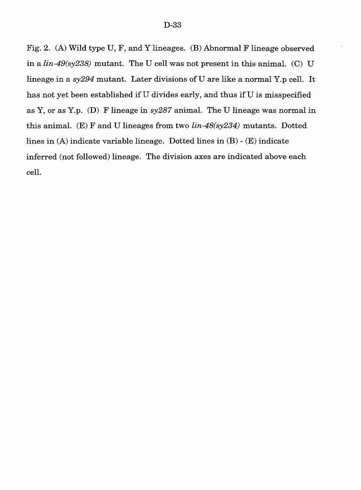
D-33
Fig. 2. (A) Wild type U, F, andY lineages. (B) Abnormal F lineage observed
in a lin-49(sy238) mutant. The U cell was not present in this animal. (C) U
lineage in a sy294 mutant. Later divisions ofU are like a normal Y.p cell. It
has not yet been established ifU divides early, and thus ifU is misspecified
as Y, or as Y.p. (D) F lineage in sy287 animal. The U lineage was normal in
this animal. (E) F and U lineages from two lin-48(sy234) mutants. Dotted
lines in (A) indicate variable lineage. Dotted lines in (B)- (E) indicate
inferred (not followed) lineage. The division axes are indicated above each
cell.

D-34
• e 8 !:3 0
< -= "" I
..... ~~·.:i C'l
...:1 ...:1
"' "" I
~'§}]
Q..----i:L-----1
"0 c.
c:::•_---
c.r·-· ~'---~ eo •. • • •
0 ., "' "'
<=':> ~~
...:1
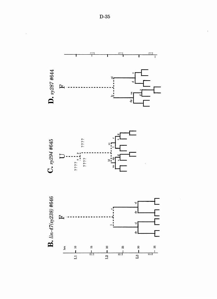
B. l
in-4
7(sy
238)
#64
6
hrs
Ll
1-
10
20
25
30
-35
dr ...
F
I I
---
.J.
--·
vi
dr
vi
C. s
y294
#64
5
u I
? ?
? ?
I ....
a I
P '_
.. ?
???
???
?
ld
•
~~
D. s
y287
#6
44
F
l,r _
_ _
; __
_ r
d
dp
1
va
t:l
I ~
en
E. l
in-4
8(sy
234)
#48
7
hrs
10
20
25
L3
t-
30
p ' -35
nv
,....
u
1_ •
•• \.
••• r
av ,...
F
! • •
.J.
• .r
vi
lin
-48(
sy23
4) #
633
nv ....
u F
IS
" ·~=t
t1
I C.:>
0)
D-37
References
Aroian, R. V. and Sternberg, P. W. (1991). Multiple functions of let-23, a C.
elegans receptor tyrosine kinase gene required for vulval induction. Genetics
128, 251-267.
Brenner, S. (1974). The genetics ofCaenorhabditis elegans. Genetics 77,71-
94.
Chamberlin, H. and Sternberg, P. W. (in preparation). The lin-3 I let-23
signaling pathway mediates induction during male spicule development in
Caenorhabditis elegans.
Chamberlin, H. M. and Sternberg, P. W. (1993). Multiple cell interactions
are required for fate specification during male spicule development in
Caenorhabditis elegans. Development 118, 297-323.
Chisholm, A. (1991). Control of cell fate in the tail region of C. elegans by the
gene egl-5. Genes and Development 111, 921-932.
Chisholm, A. D. and Hodgkin, J. (1989). The mab-9 gene controls the fate
of B, the major male-specific blast cell in the tail region of Caenorhabditis
elegans. Genes & Deuel. 33, 1413-1423.
Ferguson, E. and Horvitz, H. R. (1985). Identification and characterization
of 22 genes that affect the vulval cell lineages of Caenorhabditis elegans.
Genetics 110, 17-72.
Ferguson, E. and Horvitz, H. R. (1989). The multivulva phenotype of
certain C. elegans mutants results from defects in two functionally-redundant
pathways. Genetics 123, 109-121.
D-38
Herman, M. A (1991). Cell interactions and the polarity of asymmetric cell
divisions during Caenorhabditis elegans development. Ph.D. Thesis.
Massachusetts Institute of Technology, Cambridge MA.
Herman, M. A and Horvitz, H. R. (1994). The Caenorhabditis elegans gene
lin-44 controls the polarity of asymmetric cell divisions. Dev. in press.
Herman, R. K. (1978). Crossover suppressors and balanced recessive lethals
in Caenorhabditis elegans. Genetics 88, 49-65.
Hodgkin, J. (1983). Male phenotypes and mating efficiency in
Caenorhabditis elegans. Genetics 103, 43-64.
Hodgkin, J., Edgley, M., Riddle, D. L. and Albertson, D. G. (1988).
Appendix 4, Genetics In The Nematode Caenorhabditis elegans (ed. W. B.
Wood), pp. 491-584. Cold Spring Harbor, N.Y.: Cold Spring Harbor
Laboratory.
Kenyon, C. J. (1986). A gene involved in the development of the posterior
body region of Caenorhabditis elegans. Cell 46, 477-487.
Sternberg, P. W. and Horvitz, H. R. (1988).lin-17 mutations of C. elegans
disrupt asymmetric cell divisions. Developmental Biology 130, 67-73.
Sulston, J. and Horvitz, H. R. (1977). Postembryonic cell lineages of the
nematode Caenorhabditis elegans. Dev. Biol. 56, 110-156.
Sulston, J. E., Albertson, D. G. and Thomson, J. N. (1980). The
Caenorhabditis elegans male: Postembryonic development of nongonadal
structures. Dev. Biol. 78, 542-576.
Sulston, J. E., Schierenberg, E., White, J. G., and Thomson, J. N.
(1983). The embryonic cell lineage ofthe nematode Caenorhabditis elegans.
Dev. Biol. 100, 64-119.
D-39
Sulston, J. E. and Hodgkin, J. (1988). Methods In The nematode
Caenorhabditis elegans (ed. W. Wood), pp. 587-606. Cold Spring Harbor, New
York: Cold Spring Harbor Laboratory.
Sulston, J. E. and White, J. G. (1980). Regulation and cell autonomy
during postembryonic development of Caenorhabditis elegans. Deuel. Biol. 78,
577-597.
E-1
ChapterS
Asymmetric cell divisions and the segregation of fate potential: The
role of lin-17 and vab-3 in the Caenorhabditis elegans male B lineage
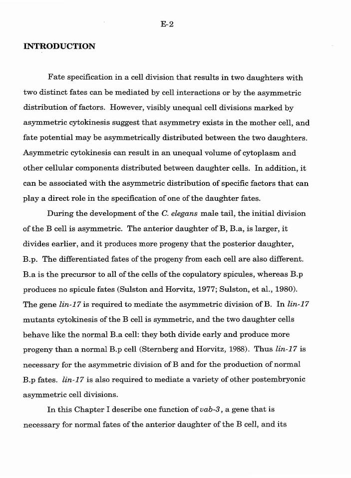
E-2
INTRODUCTION
Fate specification in a cell division that results in two daughters with
two distinct fates can be mediated by cell interactions or by the asymmetric
distribution of factors. However, visibly unequal cell divisions marked by
asymmetric cytokinesis suggest that asymmetry exists in the mother cell, and
fate potential may be asymmetrically distributed between the two daughters.
Asymmetric cytokinesis can result in an unequal volume of cytoplasm and
other cellular components distributed between daughter cells. In addition, it
can be associated with the asymmetric distribution of specific factors that can
play a direct role in the specification of one of the daughter fates.
During the development of the C. elegans male tail, the initial division
of the B cell is asymmetric. The anterior daughter ofB, B.a, is larger, it
divides earlier, and it produces more progeny that the posterior daughter,
B .p . The differentiated fates of the progeny from each cell are also different.
B.a is the precursor to all of the cells of the copulatory spicules, whereas B.p
produces no spicule fates (Sulston and Horvitz, 1977; Sulston, et al. , 1980).
The gene lin-17 is required to mediate the asymmetric division of B. In lin-17
mutants cytokinesis of the B cell is symmetric, and the two daughter cells
behave like the normal B.a cell: they both divide early and produce more
progeny than a normal B.p cell (Sternberg and Horvitz, 1988). Thus lin-17 is
necessary for the asymmetric division of B and for the production of normal
B.p fates. lin-17 is also required to mediate a variety of other postembryonic
asymmetric cell divisions.
In this Chapter I describe one function of vab-3, a gene that is
necessary for normal fates of the anterior daughter of the B cell, and its
E-3
interaction with lin-17. I also describe additional functions of lin-17 in the B
lineage subsequent to the first division.
MATERIALS AND :METHODS
Nematode strains and handling
Nematode strains were cultured according to standard techniques
(Brenner, 1974; Sulston and Hodgkin, 1988). In general, mutant genotypes
include him-5(e1490) in the background. The following mutations were used
in this study: lin-17(n671, n677) I (Ferguson and Horvitz, 1985). him-
5(e1490) V (Hodgkin, et al., 1979). vab-3(e648, e1176, sy66) X.
Cell lineage and ablation
Cell nuclei divisions in living animals were directly observed using
Nomarski differential interference contrast optics as described by Sulston
and Horvitz ( 1977) . Nomenclature follows the standard of Sulston and
Horvitz (1977), with modifications of Chamberlin and Sternberg (1993). Cell
nuclei were destroyed by a laser microbeam as described by Avery and
Horvitz (1987).
RESULTS
vab-3 is required for normal B.a fates
Candidate mutations in vab-3 were recovered in the screen for male
tail mutants (sy66, sy281; Chapter 4). These mutations result in a variable
E-4
notched-head phenotype, and a characteristic pattern of six B progeny in
males (rather than the normal ten) at the L3 stage, phenotypes also
associated with mutations in the gene vab-3 (A. Chisholm, pers. comm.).
Lineage analysis of vab-3 mutants indicates that vab-3 is necessary for
the normal B. a lineage. The initial division of B is asymmetric as in wild
type, and the B.a cell divides in early L2 larval stage along a left/right axis.
However, B.al and B.ar fail to divide until about 5-6 hours later, often when
the B.p cell divides. These cell divisions produce the six progeny observed at
the L3 stage. The observed abnormal lineages of these cells are consistent
with both B.al and B.ar behaving like a normal B.p cell (Figs 1, 2). Although
the more anterior daughters ofB.a(l/r) may divide more than once (in
contrast to B.pa), overall the cells divide with the normal timing and
appropriate axes of the B.p cell. In addition, these cells produce at least one
ofthe differentiated fates ofthe B.p lineage. The B.ppaa cell normally
undergoes a programmed cell death, and in vab-3 mutants the corresponding
cells from B.a(l/r) lineages can likewise die. Although cell death is a fate also
observed in the normal B.a lineage, the specific pattern of which cell dies is
distinctly B.p-like.
The vab-3 and lin-17 mutant defects are additive
To establish the relationship between vab-3 and lin-17 in specification
ofB.a and B.p fates, I constructed lin-17; vab-3 double mutants and
compared them to lin-17 and vab-3 single mutants (Table 1). At the L3 stage,
the B cell can divide to produce sixteen progeny in lin-17 mutants, whereas it
produces six progeny in vab-3 mutants. In the lin-17; vab-3 double mutants
E-5
the B cell can produce eight progeny. Thus lin-17 is necessary for the
asymmetric division ofB and mutations make B.a and B.p similar. However,
vab-3 is necessary for B.a fates. Both B.a and B.p behave like B.a in vab-3
single mutants, and produce only four progeny at this stage.
I have followed the B lineage in two lin-17; vab-3 animals to determine
if both B.a and B.p behave like the B.a cell in vab-3 mutants and produce two
B.p lineages (Fig. 3). Although the lineages bear some similarity to normal
B.p lineages, in general they were abnormal. The asymmetric timing of
different cell divisions (e.g., B.ppp normally divides about 2 hours prior to
B. ppa) was generally lost, with most of the cells of a given generation dividing
within 10-15 minutes of each other. This defect was observed both in the
double mutant with eight early L3 B progeny and the one with six. Since
mutations in lin-17 can disrupt the B.p-like lineages in a vab-3 mutant lin-17
may play additional roles in the B lineage, specifically in mediating normal
asymmetric cell divisions in the B. p lineage.
lin-17 has additional functions in both the B.a and B.p lineages
Although in many lin-17 mutant animals both the B.a and B. p cells
behave like B.a, some animals escape the lineage defect at the first division
and produce the normal ten B progeny at the L3 stage. To corroborate the
B.p lineage defect observed in lin-17; vab-3 double mutants, I have followed
the B cell lineage in these escaper animals. In these animals, mutations in
lin-17 disrupt the normal B.p lineage, as both daughters ofB.pp are of equal
size and divide at the same time and produce only two progeny (Fig. 4).
E-6
However, the B.pa cell divides later than the B.pp cell, and produces only two
progeny as it does in wild type.
A side effect of the abnormal duplication of the B.a lineage in lin-17
mutants is that the eight progeny of each B daughter end up in abnormal
positions with abnormal neighbors. Since proper fate specification for these
cells requires a variety of cell interactions (see Chapter 2), it is difficult in
intact animals with sixteen L3 B progeny to identify if a lineage defect results
from the abnormal environment of a cell, or is a direct result of mutant lin-
17. I have used two methods to identify additional requirements for lin-17 in
the B.a lineage. First, I also followed the B.a lineage in the escaper animals
with ten L3 B progeny. Second, I ablated the B.p cell in two lin-17 mutants,
and followed the lineage of the B.a cell. In wild type animals, this ablation
results in a normal lineage from the B.a cell (Chamberlin and Sternberg,
1993). Thus any defects observed following ablation in lin-17 mutants reflect
a requirement for lin-17.
Lineage analysis ofboth escapers and B.p ablated animals suggest that
lin-17 mediates additional asymmetric cell divisions in the B.a lineage (Table
2). 7/12 E lineages were disrupted, with 4/4 disrupted in the animals in which
B. p was ablated. The E lineage is normally produced by the anterior cell of
the lateral pairs (B.a(l/r )ap ). The first division of an E lineage is asymmetric:
the anteroventral daughter is larger, divides earlier, and produces more
progeny than the posterodorsal daughter. In lin-17 mutants the cell division
is symmetric, and both cells behave like the normal anteroventral daughter.
Other lineages with asymmetric divisions such as the s lineage and they
lineage are also occasionally disrupted. Thus lin-17 is required for both the

E-7
initial division of the B cell and specific asymmetric divisions later in the B
lineage.
DISCUSSION
lin-17 and vab-3 act to specify fate in the asynunetric division of the
Bcell
The gene lin-17 is necessary for the asymmetric division of the B cell,
and in lin-17 mutants both B.a and B.p behave like B.a cells. Although the
B.p cells are not normal in lin-17 mutants, lin-17 is not specifically necessary
for B.p fate, but rather the asymmetric distribution ofB.a and B.p fate
potential. The gene vab-3 is necessary for B.a fate; in vab-3 mutants both
daughters ofB.a behave like B.p cells. Unlike lin-17, mutations in vab-3
uncouple fate specification from the asymmetric cell division. vab-3 may
represent a gene thats product is asymmetrically distributed between B.a and
B.p, or a gene that is activated by an asymmetrically distributed factor in
B.a. The gene lin-44 is also required for the normal division of the B cell.
Mutations in lin-44 result in a reversal of the polarity of the B cell: B. a
behaves like a normal B.p, and B.p behaves like a normal B.a (Herman and
Horvitz, 1994). lin-17 is epistatic to lin-44, as lin-17lin-44 double mutants
have the lin-17 phenotype. lin-17, lin-44, and vab-3 thus represent three
major functions in an asymmetric cell division. lin-17 is necessary to
establish the asymmetric division, lin-44 is necessary to establish the proper
orientation of the division, and vab-3 activity is asymmetrically distributed
by the division (Fig. 5).
E-8
lin-17 acts at several distinct steps in the B lineage to mediate
specific asymmetric cell divisions
Lineage analysis of lin-17; vab-3 double mutants, lin-17 animals with
ten L3 B progeny, and lin-17 animals with B.p ablated suggest that lin-17
functions both at the initial division of the B cell as well as specific
asymmetric cell divisions later in the lineage. In the B.p lineage lin-17 is
required for the asymmetric division of the B.pp cell and possibly the B.p cell
itself. The B.p lineage in lin-17 escaper animals retains the asymmetry in
division timing between B.pa and B.pp, whereas both anterior and posterior
cells in the lin-17; vab-3 double mutants generally divide at the same time.
This discrepancy may be a side effect of selecting animals with ten L3 B
progeny, as it is possible that polarity in the division of B.p is not
independent from polarity in the division of B.
There are many asymmetric cell divisions -- divisions that result in two
daughters that are different from each other -- in the B lineage. Lineage
analysis suggest that lin-17 acts at some, but notably not all of these cell
divisions. lin-17 appears to play a role in visibly unequal cell divisions. For
instance, the division ofB and many ofthe cells in the B.p lineage result in
daughters that are morphologically distinct. In contrast, the B.a(llr) cells
each divide asymmetrically to produce four cells with distinct potentials that
respond differently to the same extracellular cues (see Chapters 2 and 3).
Although there is a subtle timing difference in the division ofB.a(llr)
daughters (Sulston and Horvitz, 1977; Chamberlin and Sternberg, 1993), the
four cells are morphologically similar. Interestingly, mutations in lin-17 do
not disrupt either of the two asymmetric cell divisions that would be required
to generate these four progeny types. For instance, the aa cells produce
E-9
generally normal aa fates in the lin-17 mutants. They do not produce the
fates normally associated with their sisters (ap cells and e fate) or their lineal
homologs (pa cells and s fate) that would be expected if these divisions were
disrupted. The late lineage defects in lin-17 mutants do not represent a
transformation from one intermediate blast fate to another. The disrupted e
lineages are not like any other normal B.a progeny fate. Although the rare
disrupted s lineages superficially produce six progeny in a 3+3 pattern like a
normal ~ lineage, the timing of divisions and appearance of the progeny cells
are more consistent with both cells producing a lineage like the normal pap
cell. Cytokinesis is unequal in both the e and the s lineages, and the
anteroventral and posterodorsal daughters are distinct from each other in the
timing of division and in the number of progeny. Thus in the B lineage lin-17
appears to mediate a subset of asymmetric cell divisions in which unequal
division correlates with the fates of the daughter cells.
Do lin-17, lin-44, and vab-3 represent a genetic pathway that functions
generally as a unit at unequal cell divisions? This does not appear to be the
case. Although in the B lineage lin-17 is required for specific unequal
divisions, it also functions in other postembryonic cell divisions that are not
visibly asymmetric (Sternberg and Horvitz, 1988). Other unequal divisions,
such as the first division of the Pn cells or the male Y cell, are not disrupted
in lin-17 mutants. Thus, although lin-17 is required for unequal cell division
in certain cells, its function is not one required generally for asymmetric
cytokinesis.
The three genes also do not always act together as a genetic "module."
Without the use of mosaics, we cannot test whether vab-3 acts in the later B
lineage cell divisions that require lin-17. However, although lin-17, lin-44 ,
E-10
and vab-3 are required in several other postembryonic lineages, they are not
required in the same lineages. For example, vab-3 is required for a normal Y
lineage but not male Pn.p lineages (data not shown). Mutations in lin-17. in
contrast, disrupt the Pn.p lineages but not Y (Sternberg and Horvitz, 1988).
lin-44 is required in a subset of lin-17 -mediated cell divisions, but not all
(Herman and Horvitz, 1994). Thus these genes function together in
mediating the division ofB and the specification of its progeny, but they can
also function independently.
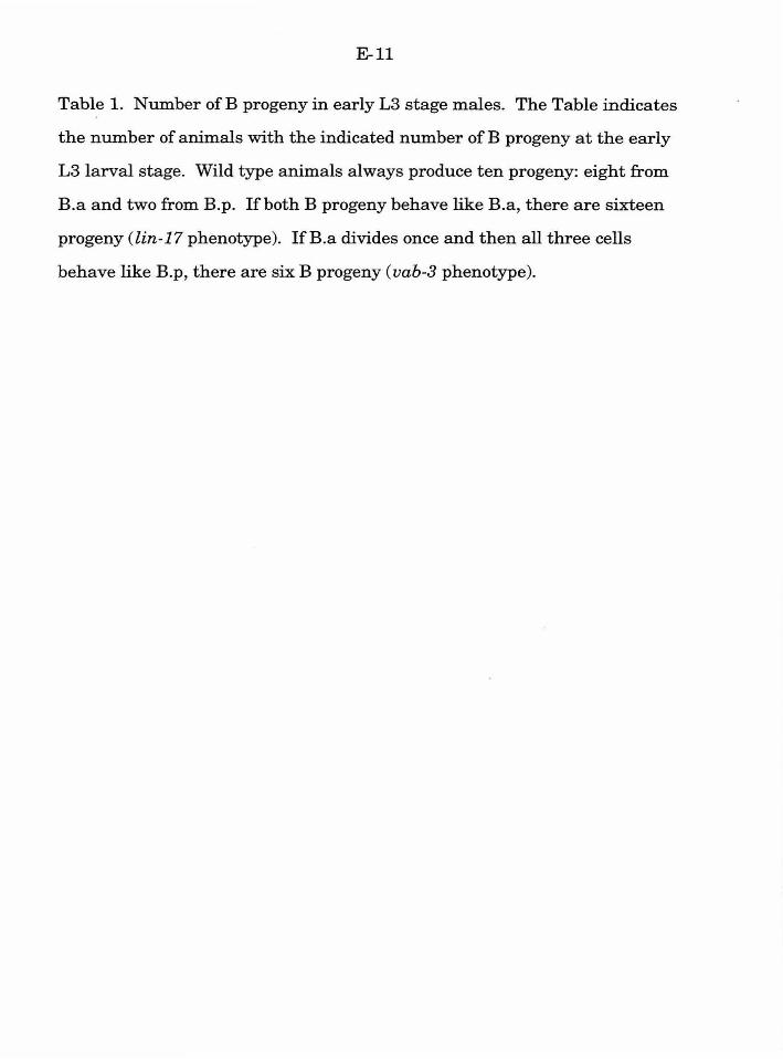
E-ll
Table 1. Number ofB progeny in early L3 stage males. The Table indicates
the number of animals with the indicated number of B progeny at the early
L3 larval stage. Wild type animals always produce ten progeny: eight from
B.a and two from B.p. If both B progeny behave like B.a, there are sixteen
progeny (lin-17 phenotype). lfB.a divides once and then all three cells
behave like B.p, there are six B progeny (vab-3 phenotype).
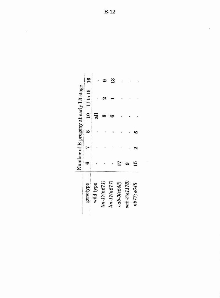
____
_____
_ , ~~
~~er
?!.~
J>!~~~ny
_ at ~~
_rl;y _
_ ~3 _s_~~
-~~
____
_ ~~!
l~t~
pe __
wil
d ty
pe
lin-
17(n
671)
6 7
8 10
11
to
15
16
lin
-17 (
n677
)
vab-
3(e6
48)
I 17
vab-
3(e1
178)
9
n677
; e6
48
15
2 5
all
8 6
2 1
9 13
~
1-'
t\
J
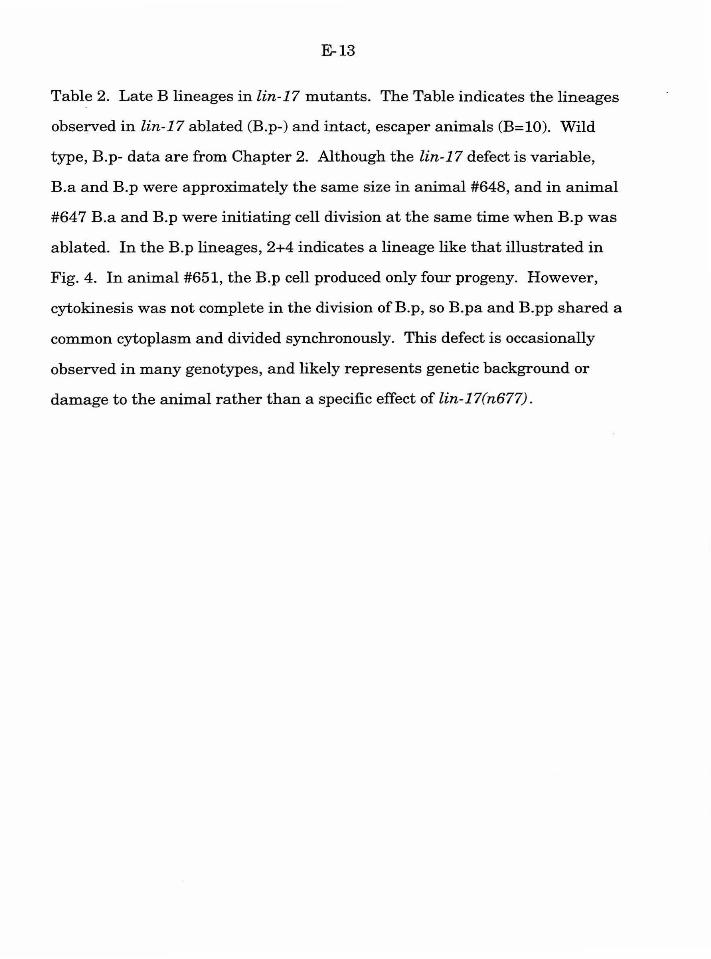
E-13
Table 2. Late B lineages in lin-17 mutants. The Table indicates the lineages
observed in lin-17 ablated (B.p-) and intact, escaper animals (B=10). Wild
type, B.p- data are from Chapter 2. Although the lin-17 defect is variable,
B.a and B.p were approximately the same size in animal #648, and in animal
#647 B.a and B.p were initiating cell division at the same time when B.p was
ablated. In the B.p lineages, 2+4 indicates a lineage like that illustrated in
Fig. 4. In animal #651, the B.p cell produced only four progeny. However,
cytokinesis was not complete in the division ofB.p, so B.pa and B.pp shared a
common cytoplasm and divided synchronously. This defect is occasionally
observed in many genotypes, and likely represents genetic background or
damage to the animal rather than a specific effect of lin-17(n677).

a a
a a
pp
p
p
ap
ap
pa
pa
geno
type
an
imal
an
t.
post
. an
t.
post
. le
ft
rig
ht
left
ri
ght
B.p
(a/p
) . .
----
----
... -
------
-----
----
----
----
---
-· --
---
----
----
---
----
-··-.
-·
----
----
-.-
----
. ·-
wild
typ
e a
p y
0 E
E
~
~ 2+
5 w
ild t
ype
B.p
-a
p y
0 E
E
~
~ X
---
----
----
----
-----
--------
-------
----
---
----
---
-.....
--
....
----
--.
. --
---
····-
···
.. -
---
.. --
----
-----
----
-------
-----
----
----
. -··
-· --
-~
n671
B
.p-
647
p ab
n-8
0 ab
n-8
abn-
8 ~
~ ,....
. a
X
~
B.p
-64
8 a
p y
t 0
abn-
7 ab
n-8
~ ~'t
X
n671
B
=lO
64
9 a
p y
0 E
ab
n-7
abn-
6 ~
2+4
B=
lO
650
a p
abn-
5 0
E
E
~ ~
2+4
B=l
O
652
abn-
5 p
y 0
abn-
7 E
ab
n-6
~ 2+
4 n6
77
B=l
O
651
a Pt
y 0
abn-
8 E
~
~ 2+
2
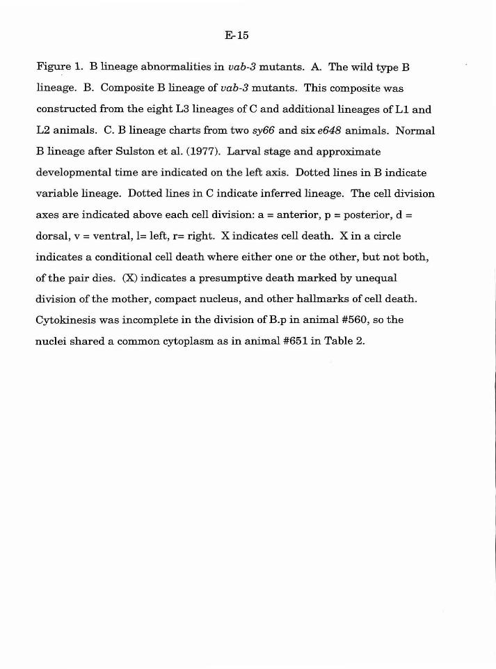
E-15
Figure 1. B lineage abnormalities in vab-3 mutants. A. The wild type B
lineage. B. Composite B lineage of vab-3 mutants. This composite was
constructed from the eight L3 lineages of C and additional lineages of L1 and
L2 animals. C. B lineage charts from two sy66 and six e648 animals. Normal
B lineage after Sulston et al. (1977). Larval stage and approximate
developmental time are indicated on the left axis. Dotted lines in B indicate
variable lineage. Dotted lines in C indicate inferred lineage. The cell division
axes are indicated above each cell division: a= anterior, p =posterior, d =
dorsal, v = ventral, l= left, r= right. X indicates cell death. X in a circle
indicates a conditional cell death where either one or the other, but not both,
of the pair dies. (X) indicates a presumptive death marked by unequal
division of the mother, compact nucleus, and other hallmarks of cell death.
Cytokinesis was incomplete in the division ofB.p in animal #560, so the
nuclei shared a common cytoplasm as in animal #651 in Table 2.
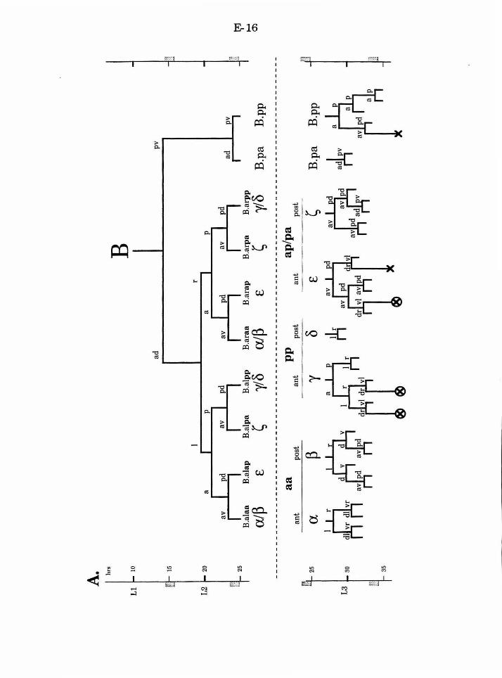
A. hr
s
B
10
ad
pv
15
I r
20
a I
r
~
~ av
I
pd
av I
pd
25
I I
I I
~
B.a
laa
B.a
lap
B.a
lpa
B.a
lpp
B.a
raa
B.a
rap
B. a
rpa
B.a
rpp
.....
alP
£
~ yl
8 a
lP
£ ~
yl8
B.p
a B
.pp
0')
a a
pp
ap
/pa
ant
post
an
t po
st
ant
post
p 8
~ n
a 'Y
£ B
.pa
B.p
p 25
30
~
d r ~ Jtl
r
t4 ~
nu
d
arlr
35
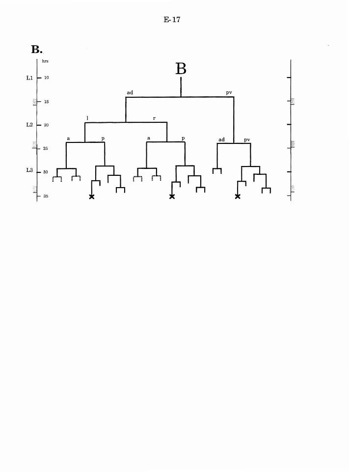
E-17
B.
B ad pv
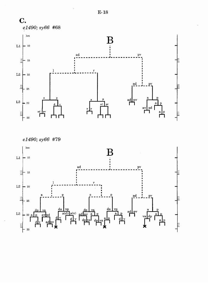
E-18
c. el490; sy66 #68
hrs
B 10
I
ad 1 pv
·-------------~------------15
1 1 r
---------~--------20
25
a
30
ad pv
35
e1490; sy66 #79
hrs
B 10
I
ad 1 pv
r····-····---~------------15
1 r 20 i---------~---------.
a ____ ~ ____ P a_. • • ~.- _P
25
35
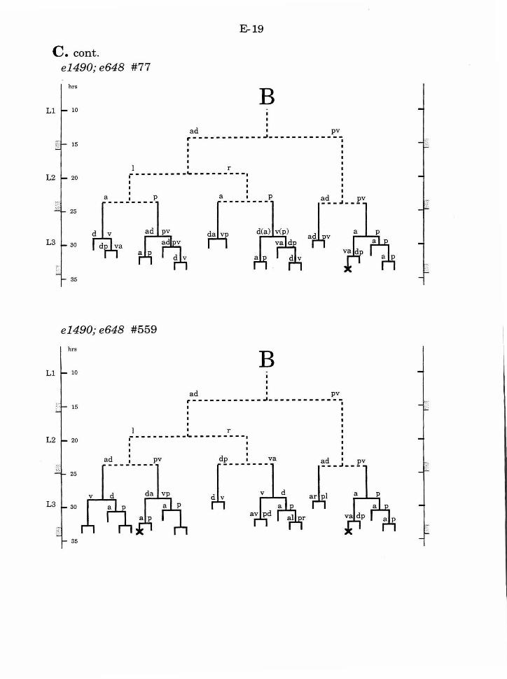
E-19
C. cont. e1490; e648 #77
hrs
B 10
I
ad 1 pv
r------------~------------15
I r 20 .---------_._-------- -. 25
30
35
e 1490; e648 #559
hrs
B 10
I ad 1 pv
r------------~------------15
I r
20 .---------_._-------- -. a~ ___
1 ____ pv
25
30
35
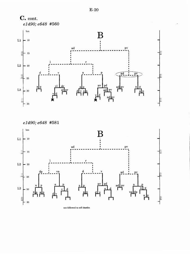
C. cont. e1490;e648 #560
hrs
10
E-20
B ad pv
r·-----------~-------------1 15 I
l r 20 i---------~---------. I
I I
25
30
(1·-~<!, --~-=-~pv:) -----
ad pv a p
a P
35
e1490;e648 #581
hrs
B 10
I
ad 1 pv
r------------~------------15
l r
20 r··-------~---------~ I I
d I V -------- -- pv 25
a p
30
35
not followed to cell deaths
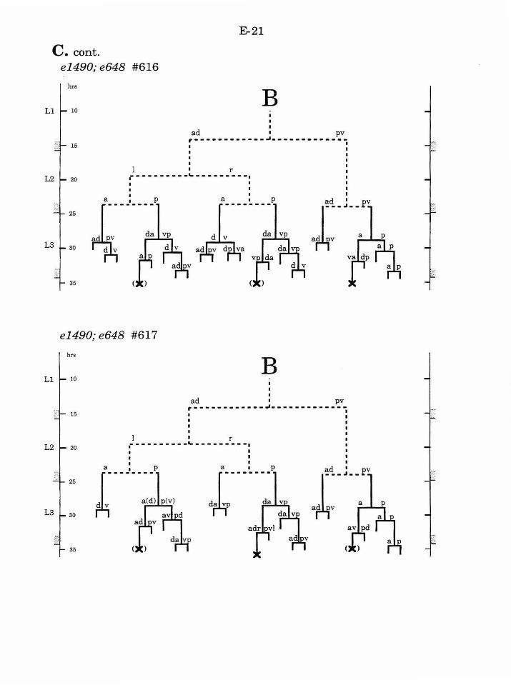
E-21
C. cont. e1490; e648 #616
hrs
10
15
20
25
30
35
B I I
ad 1 pv
r------------~------------
I r i---------~---------. I I
~-~-- -..---~d~'~ a----~---p ad
.~~-da vp
e1490;e648 #617
hrs
B I
I ad 1 pv
r------------~------------
I r .--------- _ ... -------- -. I
-- pv
- _pv
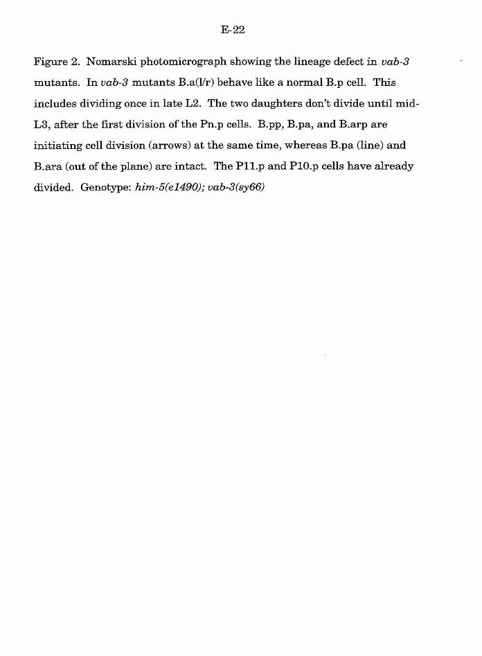
E-22
Figure 2. Nomarski photomicrograph showing the lineage defect in vab-3
mutants. In vab-3 mutants B.a(llr) behave like a normal B.p cell. This
includes dividing once in late L2. The two daughters don't divide until mid
L3, after the first division of the Pn.p cells. B.pp, B.pa, and B.arp are
initiating cell division (arrows) at the same time, whereas B.pa (line) and
B.ara (out of the plane) are intact. The Pll.p and PlO.p cells have already
divided. Genotype: him-5(e1490); vab-3(sy66)

E-23
• ~~ ~ I
l.-. .
E-24
Figure 3. B lineage in lin-17; vab-3 double mutants. Notation as in Fig. 1.
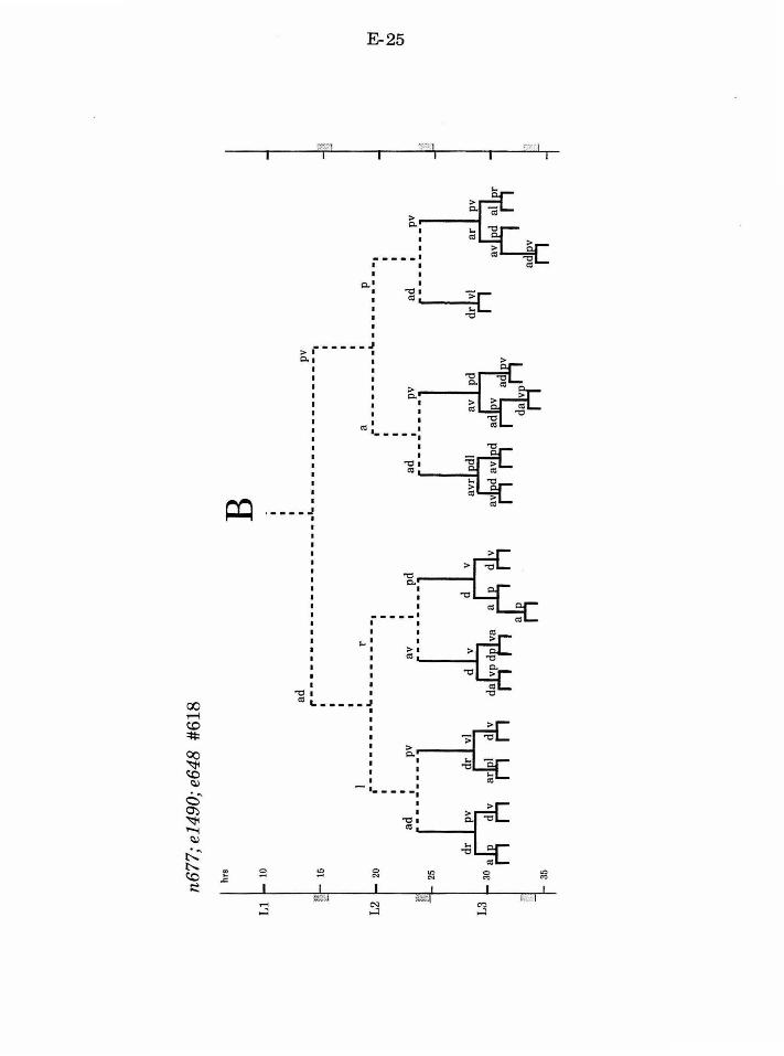
n677
;el4
90;e
648
#618
hrs
10
15
20
25
35
B
~
I ~
r---
----
----
----
----
--L
----
----
----
----
-,
I •
r .--
------
-.... -
------
-.,
a~ _
__
1 _
__
_ pv
a
v ad
I I
a I
p r-
---
----
-"---
----
---~
I
ad
1 pv
dr
I pv
d
r I
vi
d I
v dL:
-----~
: r-
~
t\:)
0
1
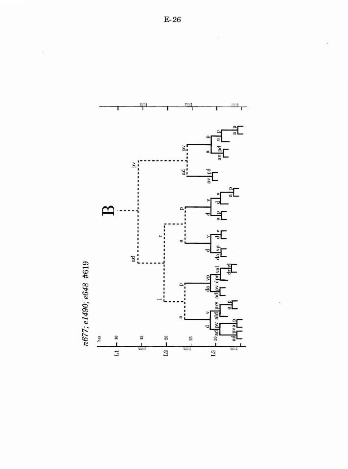
m ,..-j
t.O ::;;:
~
~ ~ . ~
c ~ ~ ~ ~
t:...~ ~ e c:.c ..0:: s::
E-26
I .-- ----- ----, ~I I
I "t::l <lS I
I ·---- ...
~
...... ....J
-- ---· I I
,_I I
I
I I
"'~II>: "t:: >
"' "t::
-----J I
I - ._--- -:
0
"' "' "' C'l
I ;.,;..q ....J
E-27
Figure 4. B lineage defect in B. p of lin-17 mutants that escape the lineage
defect of the initial B division. A . Normally the B.pp cell division is
a symmetric, and the posterior daughter is larger, divides earlier, and
produces more progeny than the anterior daughter. B. In lin-17 mutants the
B.pp cell division is equal, and both B.ppa and B.ppp divide only once.
Notation as in Fig. 1.
E-28
I
~ ~ • ~
t--~ • . s ..... • ~ = ~ .
~ I
I
Q;l ~ ~ ~
c • ~
"'C """" •1'"1
~
< = ~ . ~
I
E-29
Figure 5. Model for the action of lin-17, lin-44, and vab-3 . lin-17 is necessary
to establish the asymmetric division ofB, lin-44 is necessary to establish the
proper orientation of the division, and vab-3 activity is asymmetrically
distributed by the division.
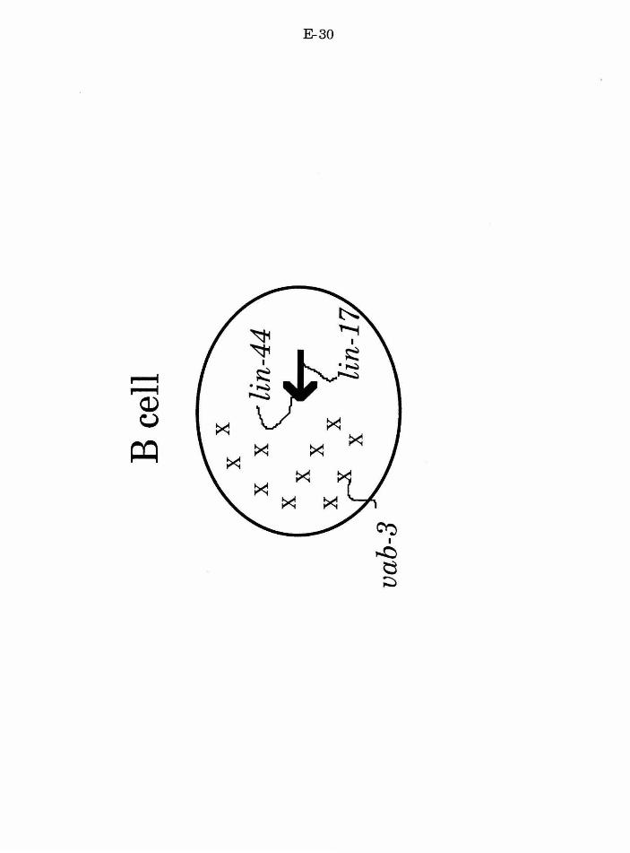
E-30
~ ~
I
r--4 ~ . .....-:. ~
........t 0) \,.)
~ ~
~ ~ ~ ~
~
~ ~
~
~ I
....0 tJ ~
E-31
References
Avery, L. and Horvitz, H. R. (1987). A cell that dies during wild-type C.
elegans development can function as a neuron in a ced-3 mutant. Cell 51,
1071-1078.
Brenner, S. (1974). The genetics ofCaenorhabditis elegans. Genetics 77,71-
94.
Chamberlin, H. M. and Sternberg, P. W. (1993). Multiple cell interactions
are required for fate specification during male spicule development in
Caenorhabditis elegans. Development 118, 297-323.
Ferguson, E. and Horvitz, H. R. (1985). Identification and characterization
of 22 genes that affect the vulval cell lineages of Caenorhabditis elegans.
Genetics 110, 17-72.
Herman, M. A. and Horvitz, H. R. (1994). The Caenorhabditis elegans gene
lin-44 controls the polarity of asymmetric cell divisions. Dev. in press.
Hodgkin, J., Horvitz, H. R. and Brenner, S. (1979). Nondisjunction
mutants ofthe nematode Caenorhabditis elegans. Genetics 91, 67-94.
Sternberg, P. W. and Horvitz, H. R. (1988). lin-17 mutations of C. elegans
disrupt asymmetric cell divisions. Developmental Biology 130, 67-73.
Sulston, J. and Horvitz, H. R. (1977). Postembryonic cell lineages ofthe
nematode Caenorhabditis elegans. Dev. Biol. 56 , 110-156.
Sulston, J. E., Albertson, D. G. and Thomson, J. N. (1980). The
Caenorhabditis elegans male: Postembryonic development of nongonadal
structures. Dev. Biol. 78, 542-576.
E-32
Sulston, J. E. and Hodgkin, J. (1988). Methods In The nematode
Caenorhabditis elegans (ed. W. Wood), pp. 587-606. Cold Spring Harbor, New
York: Cold Spring Harbor Laboratory.
F-1
ChapterS
Summary
F-2
Several related observations contributed to the initiation of the
experiments described in this thesis. Three results hinted at the cell
interactions that play a role in fate specification in the B lineage: (1) the
correlation of alj3 and y/'fJ fates with cell position but not cell lineage (Sulston
and Horvitz, 1977; Sulston, et al., 1980; Sulston and White, 1980), (2) the
lineage defect in the y/'fJ pair in lin-12(0) mutants (Greenwald, et al., 1983),
and (3) the pleiotropic defects in mutants of other genes required for vulval
induction (Ferguson and Horvitz, 1985; Aroian and Sternberg, 1991). An
early model was that lin-12 might mediate fate specification in the y/'fJ pair
whereas the lin-3 I let-23 pathway might act in the alj3 pair. In addition to the
question of cell interactions, the role of lin-17 in the first division of the B cell
(Sternberg and Horvitz, 1988) suggested that mutations that disrupt different
steps in the development of the B cell could be recovered, and the function(s)
of the corresponding gene could be interpreted using cell lineage analysis.
Here, I review the results in the context of stepwise fate specification and
multiple signal integration.
The integral relationship between autonomous and conditional fate
specification
The initial division of the B cell is asymmetric and unequal. The genes
lin-17, lin-44, and vab-3 act at this division to mediate the asymmetric
distribution offate potential and the specification offate (Chapter 5). In both
lin-17 and lin-44 mutants the defect is observed in the cytokinesis of the B
cell itself, suggesting a mechanism of autonomous fate specification. The
vab-3 phenotype is consistent with this model. In vab-3 mutants the initial
F-3
division is normal, but B.a fates are misspecified, and B.al and B.ar behave
like B.p. One difference between B.a progeny and B.p progeny is that B.a
progeny are responsive to a set of specific cell interactions, and require the
genes of the lin-3 I let-23 signalling pathway for nonnal fate specification
(Chapters 2 and 3). All eight intennediate B.a progeny utilize these genes,
and thus inducing the expression of the let-23 receptor, for instance, may be a
function (direct or indirect) of vab-3. Cell interactions are likely not absent
from the B.p progeny, as mutations in lin-12 can disrupt the fates ofB.pa and
B.pp (Greenwald, et al., 1983). However, none ofthe cell ablations or lin-
3 I let-23 class of mutations that disrupt B.a progeny fates disrupt B. p
progeny fates. Furthennore, ectopically placed cells with B.p fates (as in the
vab-3 mutants) can be essentially nonnal. Thus the progeny ofB.a and B.p
are different in their ability to respond to positional cues, and this difference
is specified, at least indirectly, by the asymmetric distribution of vab-3
activity.
Following the cell interactions that establish anterior and posterior
fates among the eight progeny of B.a, the cells must execute the fate
appropriate to their position. Both cell interactions and cell divisions that
asymmetrically distribute fate potential may play a role at this later step. In
the ap/pa pairs, lin-17 is required for the asymmetric division associated
with£ and s fates (although both defects are variable; Chapter 5). In
contrast, cell ablation experiments (Chapter 2) and ectopic expression of lin-3
(Chapter 3) suggest that additional cell interactions may play a role in the
execution of the 'Y lineage. Nonnally a 'Y lineage comprises an initial
anterior/posterior division, with y.p producing two progeny and y.a producing
four. However, ectopic expression of lin-3, or a failure to localize the FlU
F-4
positional cue, can result in both daughters behaving like y.a, such that there
are a total of eight progeny (the abn-8lineage) rather than the normal six.
Although these experiments at least begin to identify the genes and
processes involved at different steps of fate specification throughout the B
lineage, some key steps are still poorly understood. Specifically, what
mechanisms are involved in establishing the differences between the cells of
the different pairs of intermediate B. a progeny? The aa cells, for instance,
respond to ectopic LIN-3 differently from the ap/pa cells or the pp cells. Cell
ablation experiments did not identify any examples where cells in one pair
adopted the fates normally associated with cells in another pair (Chapter 2).
Although the negative result cannot completely rule out the possibility of cell
interactions, there is no evidence that they play a role. Analysis of the
defects associated with one gene required for a variety of asymmetric cell
divisions, lin-17, suggests that the fates of the different precursors are
specified by a lin-17-independent mechanism (Chapter 5). It is possible that
a mutant that will be useful to understand these steps in B lineage
development exists among the mutants with a mid-stage class of defect
recovered in the genetic screen (Chapter 4). In a mutant where intermediate
B.a progeny fates are abnormal, the cells may not migrate to their normal
dorsal/ventral positions, but to the position associated with their misspecified
fate. Thus, sy285, possibly sy294, or mutations like them represent candidate
mutations that disrupt specification of intermediate B.a progeny fate.
Further lineage analysis and cell ablation experiments may be able to verify
if this is the case.
It is interesting to note the parallels between the development of the
male B lineage and early C. elegans embryogenesis. Both involve an initial
F-5
asymmetric division that produces a larger anterior and a smaller posterior
daughter. The posterior daughter divides with a modified stem-cell-like
lineage, where one progeny retains the function of the mother cell. The first
division of the anterior daughter is equivalent, and a complex set of cell
interactions specify different fates among the progeny. One difference is that
in the embryo, all mechanisms offate specification must come from "within"
the lineage; maternal factors are localized in the ooplasm, and cell
interactions are restricted to occurring among the progeny of PO. In contrast,
the B cell develops within the context of a multicellular organism, and
positional cues can arise from neighboring cells. In terms of fate specification
the two processes may share some mechanisms, whereas others may be
unique. Presently, however, several genes have been identified that are
required for early embryogenesis and others required for B cell development,
but no gene has been demonstrated to function in both processes. This may
reflect the differences in selection protocols for mutations that disrupt each
process, as well as the likelihood that all of the functions may not be
understood for a given gene.
Fate specification and the integration of multiple signals
Cell ablation experiments have identified four active cell interactions
that establish anterior/posterior fates in each pair of intermediate B. a
progeny. How do the responding cells integrate these positional cues? The
lin-3 I let-23 signalling pathway mediates the FlU positional cue that
promotes anterior fates. Double mutant analysis indicates that lin-15 likely
acts antagonistic and in parallel to lin-3 as a negative regulator of let-23
F-6
activity as it does in vulval development (Chapter 3). Thus both lin-3 and lin-
15 are integrated at the let-23 receptor.
The disruption of posterior pp fate following ablation ofY.p is not
blocked by any mutation in genes of the lin-3 I let-23 pathway, so the Y.p cue
is integrated at some point after lin-45. The Y.p cue does not represent the
activity mediated by lin-15, and it may represent an independent signalling
pathway that acts in parallel to, or after, the lin-3 I let-23 pathway. The pa
cells are required for normal anterior fates in the pp cells (Chapter 2).
However, this function is likely to localize (modulate) the Y.p positional cue
so that it does not act inappropriately on the anterior pp cells. Thus the pa
and Y.p information is integrated at the level of the Y.p signal. There are
presently no candidate mutations that disrupt the Y.p signalling pathway.
The lateral interaction, at least in the pp cells, is mediated by lin-12,
and the integration of lin-12 information may parallel the relationship
between lin-3 I let-23 and lin-12 in vulval development. AE, in vulval
development, the lin-12(d) defect can be overridden by lin-3 (or at least the
presence ofF and U). In relation to the Y.p cue, lin-12(d) is sufficient to
produce o fates even in the absence ofY.p, whereas Y.p is necessary when lin-
12 is not mutant.
The information from the other B .a progeny likely represents two
functions: a posterior positional cue and an insulating function that localizes
or modulates the positional cues from FlU and Y.p (Chapter 2). Because both
functions are eliminated when the cells are ablated, it is difficult to predict
what the lineage defect would be if just the positional cue is eliminated, as it
would be in a genetic mutant. It is possible that this positional cue would be
redundant, or partially redundant, with the modulatory function and the
F-7
other positional cues from FlU and Y.p. If this is the case, then we cannot
rule out the possibility that lin-15 represents a component of this signal.
Alternatively, this interaction may be disrupted in lin-47(sy234) animals.
F-8
References
Aroian, R. V. and Sternberg, P. W. (1991). Multiple functions of let-23, a C.
elegans receptor tyrosine kinase gene required for vulval induction Genetics
128, 251-267.
Ferguson, E. and Horvitz, H. R. (1985). Identification and characterization
of 22 genes that affect the vulval cell lineages of Caenorhabditis elegans
Genetics 110, 17-72.
Greenwald, I. S., Sternberg, P. W. and Horvitz, H. R. (1983). The lin-12
locus specifies cell fates in Caenorhabditis elegans Cell 34, 435-444.
Sternberg, P. W. and Horvitz, H. R. (1988).lin-17 mutations of C. elegans
disrupt asymmetric cell divisions Developmental Biology 130, 67-73.
Sulston, J. and Horvitz, H. R. (1977). Postembryonic cell lineages of the
nematode Caenorhabditis elegans Dev. Biol. 56, 110-156.
Sulston, J. E., Albertson, D. G. and Thomson, J. N. (1980). The
Caenorhabditis elegans male: Postembryonic development of nongonadal
structures Dev. Biol. 78, 542-576.
Sulston, J. E. and White, J. G. (1980). Regulation and cell autonomy
during postembryonic development of Caenorhabditis elegans Deuel. Biol. 78,
577-597.


