Embed Size (px)
Citation preview
ARTICLES
Dissecting a circuit for olfactory behaviourin Caenorhabditis elegansSreekanth H. Chalasani1, Nikos Chronis1, Makoto Tsunozaki1, Jesse M. Gray1, Daniel Ramot2, Miriam B. Goodman2
& Cornelia I. Bargmann1
Although many properties of the nervous system are shared among animals and systems, it is not known whether differentneuronal circuits use common strategies to guide behaviour. Here we characterize information processing by Caenorhabditiselegans olfactory neurons (AWC) and interneurons (AIB and AIY) that control food- and odour-evoked behaviours. Usingcalcium imaging and mutations that affect specific neuronal connections, we show that AWC neurons are activated by odourremoval and activate the AIB interneurons through AMPA-type glutamate receptors. The level of calcium in AIBinterneurons is elevated for several minutes after odour removal, a neuronal correlate to the prolonged behavioural responseto odour withdrawal. The AWC neuron inhibits AIY interneurons through glutamate-gated chloride channels; odourpresentation relieves this inhibition and results in activation of AIY interneurons. The opposite regulation of AIY and AIBinterneurons generates a coordinated behavioural response. Information processing by this circuit resembles informationflow from vertebrate photoreceptors to ‘OFF’ bipolar and ‘ON’ bipolar neurons, indicating a conserved or convergentstrategy for sensory information processing.
Neural circuits actively transform sensory signals: they extract themost relevant sensory information from the environment, determinewhether stimuli are increasing or decreasing, and use this informa-tion to regulate behaviours on timescales from seconds to hours. Ithas been suggested that a few connected neurons in statistically over-represented synaptic ‘motifs’ could perform simple circuit computa-tions1. The potential to analyse circuit function at single-cellresolution exists in the nematode C. elegans, the nervous system ofwhich contains just 302 neurons with known synaptic connections2–4.Functions for many individual C. elegans neurons have been inferredfrom cell ablation studies, chronic activation or recording of neur-onal activity5–10. However, little has been done to study the dynamicflow of information between neurons—an essential link betweencircuits and behaviour.
In some C. elegans behaviours, sensory inputs lead to long-lastingand complex behavioural sequences. During chemotaxis to foododours or tastes, animals reach an attractant source by regulatingturns over many minutes5,11. In another food-related behaviourcalled local search or area-restricted search, animals that haverecently been removed from food spend about 15 min exploring arestricted area by interrupting long forward movements with stoch-astic turns, and then disperse by suppressing turning12–15. A pair ofolfactory neurons called AWC is important in both of these food-seeking behaviours: the AWC neurons direct chemotaxis to manyattractive odours5, and also increase turning probability during localsearch14. The AWC neurons synapse onto several interneuronsincluding AIB and AIY, which enhance and suppress turning,respectively13,14 (Fig. 1a). Although the sensory signalling of theAWC neuron has been extensively characterized at a genetic level,nothing is known about the effects of odours on AWC neuron acti-vity, or the mechanisms by which the AWC neuron communicateswith downstream neurons. Here we analyse the functional connec-tivity between the AWC, AIB and AIY neurons that initiates thetransformation of chemosensory cues into behaviour.
AWC neurons respond to odour removal
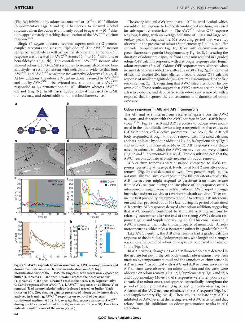
To monitor the odour response of AWC neurons, we used AWC-selective promoters to express G-CaMP, a genetically encoded cal-cium sensor, the fluorescence intensity of which increases on calciumbinding in the physiological range16,17. C. elegans neurons haverapidly activating voltage-gated calcium channels, suggesting thatcalcium transients should correlate with strong neuronal depolariza-tion9,18. A custom-designed microfluidic device fabricated from thetransparent polymer polydimethylsiloxane was used to trap andstimulate animals expressing G-CaMP in one of the two AWC neu-rons, AWCON (Fig. 1b–d)19. The device restrained z-plane movementof the animal so that the AWC neuron remained in focus for imaging,while allowing limited movement in the x–y plane. Fluid streamsunder laminar flow were used to deliver the buffer or odour stimulito the animal’s nose. Buffer exchange controls (n 5 18) showed thatshort-motion transients, photobleaching and other measurementerrors contributed DF/F (fluorescence change/baseline fluorescenceintensity) signals ,25% to individual traces, and ,10% to averagedtraces (Supplementary Figs 1–3); averaged traces were used for quan-tification and statistical analysis (Supplementary Fig. 2).
Individual animals were followed in a paired odour addition–odour removal sequence (Fig. 1e, f). Presentation of the attractiveodour isoamyl alcohol diminished G-CaMP fluorescence in theAWCON cell body; although slight, the decrease was significant whenaveraged over multiple trials (Fig. 1e, h and Supplementary Figs 1–3).Large increases in G-CaMP fluorescence were reliably observedwithin one second when isoamyl alcohol was replaced with buffer,suggesting an increase in AWCON calcium on odour removal (anodour-OFF response) (Fig. 1f, i, Supplementary Fig. 1b andSupplementary Movie 1). Increases in G-CaMP fluorescence inAWCON were also observed when bacteria-conditioned mediumwas removed from the animal’s nose (Fig. 1g and SupplementaryFig. 1h). The AWCON odour-OFF response was observed at isoamylalcohol dilutions of 1027 to 1023, with a maximal response at 1024
1Howard Hughes Medical Institute, Laboratory of Neural Circuits and Behaviour, The Rockefeller University, New York, New York 10065, USA. 2Program in Neurobiology andDepartment of Molecular and Cellular Physiology, Stanford University, Stanford, California 94305, USA.
Vol 450 | 1 November 2007 | doi:10.1038/nature06292
63Nature ©2007 Publishing Group
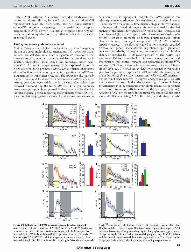
(Fig. 2a); inhibition by odour was maximal at 1025 to 1024 dilution(Supplementary Figs 2 and 3). Chemotaxis to isoamyl alcoholsaturates when the odour is uniformly added to agar at ,1024 dilu-tion, approximately matching the saturation of the AWCON calciumresponse20,21.
Single C. elegans olfactory neurons express multiple G-protein-coupled receptors and sense multiple odours5. The AWCON neuronsenses benzaldehyde as well as isoamyl alcohol, and an odour-OFFresponse was observed in AWCON across 1026 to 1024 dilutions ofbenzaldehyde (Fig. 2b). The contralateral AWCOFF neuron alsoshowed odour-OFF G-CaMP responses to isoamyl alcohol and ben-zaldehyde—a result consistent with behavioural evidence that bothAWCON and AWCOFF sense these two attractive odours22 (Fig. 2c, d).At low dilutions, the odour 2,3-pentanedione is sensed by AWCOFF
and not by AWCON in behavioural assays, and similarly AWCOFF
responded to 2,3-pentanedione at 1027 dilution whereas AWCON
did not (Fig. 2e). In all cases, odour removal increased G-CaMPfluorescence, and odour addition diminished fluorescence.
The strong bilateral AWC response to 1024 isoamyl alcohol, whichresembled the response to bacterial-conditioned medium, was usedfor subsequent characterization. The AWCON odour-OFF responsewas long-lasting, with an average half-time of ,20 s and large sec-ondary peaks throughout the 50 s recording period that were notobserved in the presence of odour (Supplementary Fig. 1a), in buffercontrols (Supplementary Fig. 1c, d) or with calcium-insensitivegreen-fluorescent protein (Supplementary Fig. 1e, f). Increasing theduration of odour pre-exposure from 1 to 5 min resulted in a gradedodour-OFF calcium response, with a stronger response after longerodour exposure (Fig. 2f). Odour-OFF responses were silenced whenisoamyl alcohol was added back after 10 s or 30 s (Fig. 2g, h). Removalof isoamyl alcohol 20 s later elicited a second odour-OFF calciumresponse of smaller magnitude (42–46% 6 14% compared to the firstresponse, Fig. 2g, h), suggesting that AWC integrates odour historyover .20 s. These results suggest that AWC neurons are inhibited byattractive odours, and depolarize when odours are removed, with aresponse that integrates the concentration and duration of odourexposure.
Odour responses in AIB and AIY interneurons
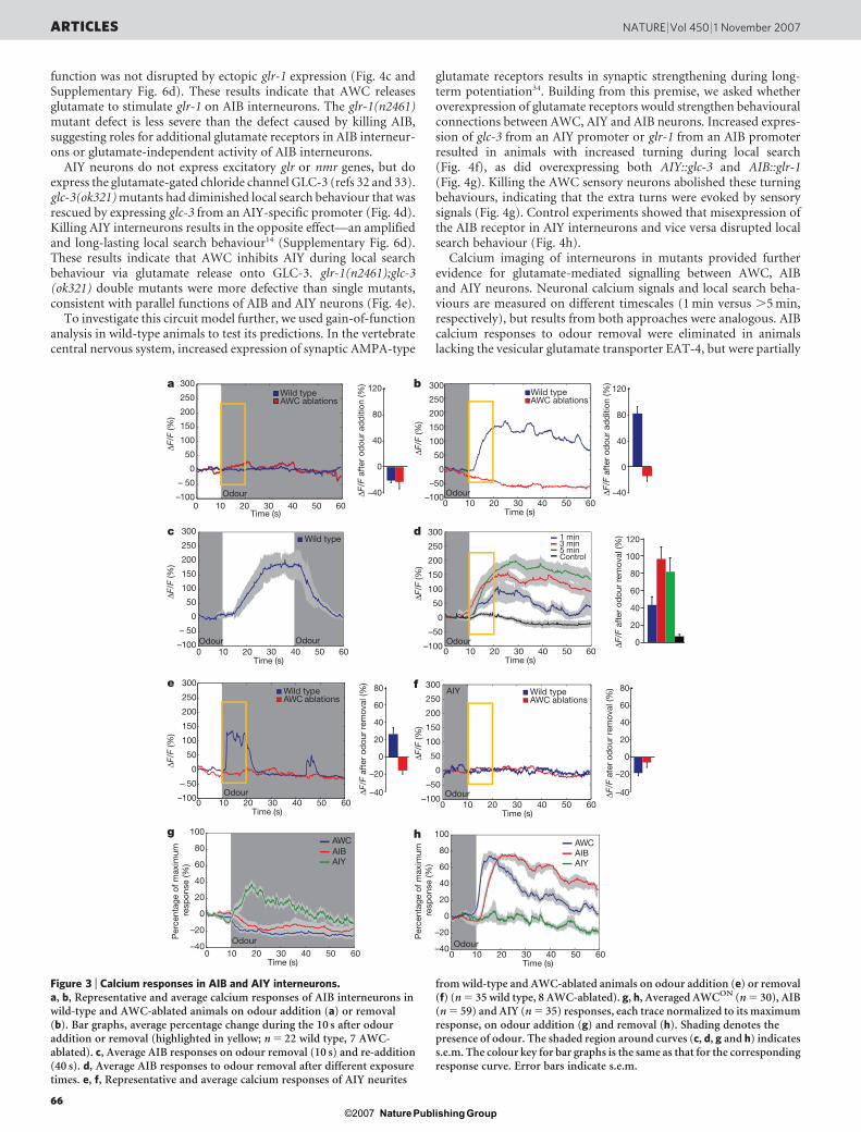
The AIB and AIY interneurons receive synapses from the AWCneurons, and function with the AWC neurons in local search beha-viour3,13,14 (Fig. 1a). AIB and AIY responses to odours were moni-tored in the microfluidic device using transgenic lines that expressedG-CaMP under cell-selective promoters. Like AWC, the AIB cellbody responded strongly to odour removal with increased calcium,and was inhibited by odour addition (Fig. 3a, b, Supplementary Figs 3and 4a, b and Supplementary Movie 2). AIB responses were elimi-nated in animals in which the AWC sensory neurons were ablated(Fig. 3b and Supplementary Fig. 4c, d). These results indicate that theAWC neurons activate AIB interneurons on odour removal.
AIB calcium responses were sustained compared to AWC res-ponses, persisting at near-peak levels for at least 2 min after odourremoval (Fig. 3b and data not shown). Two possible explanations,not mutually exclusive, could account for this persistent activity: theAIB interneurons might respond to persistent transmitter releasefrom AWC neurons during the late phase of the response, or AIBinterneurons might remain active without AWC input throughintrinsic persistent activity or reverberant circuit activity23. To exam-ine the first possibility, we removed odour to activate AIB interneur-ons and then provided odour 30 s later during the period of sustainedAIB activity. AIB responses decayed after odour addition, suggestingthat AWC neurons continued to stimulate AIB interneurons byreleasing transmitter after the end of the strong AWC calcium res-ponse (Fig. 3c and Supplementary Fig. 4e, f). This conclusion aboutAWC is consistent with the known property of nematode (Ascaris)motor neurons, which release neurotransmitter in a graded fashion24.
Like AWC neurons, the AIB interneurons had a graded calciumresponse to the duration of odour exposure, with longer and strongerresponses after 5 min of odour pre-exposure compared to 3 min or1 min (Fig. 3d).
In AIY neurons, changes in G-CaMP fluorescence were detected inthe neurite but not in the cell body; similar observations have beenmade using temperature stimuli and the cameleon calcium sensor inAIY neurons25. In contrast with AWC and AIB neurons, increases inAIY calcium were observed on odour addition and decreases wereobserved on odour removal (Fig. 3e, f, Supplementary Figs 3 and 5a, band Supplementary Movie 3). AIY responses were brief, poorly syn-chronized to odour onset, and appeared sporadically throughout theperiod of odour presentation (Fig. 3e and Supplementary Fig. 5a).Ablation of the AWC neurons eliminated the AIY response (Fig. 3e, fand Supplementary Fig. 5c, d). These results indicate that AIY isinhibited by AWC, even at the resting level of AWC activity, and thatrelease from this inhibition on odour presentation results in AIYactivation.
1
2 3
4OFF
ON
Wormnose
Wormnose
1
2 3
4
150 µm
OFFON
Odour
OdourControl
a b
c
g
AWC
AIB AIYAIA AIZ
h i
–100
0
50403020100 60
f
Time (s)
–50
50
100
150
200
250
300
∆F/F
(%)
0Bacterial- conditioned medium
50403020100 60Time (s)
∆F/F
(%)
–100
–50
50
100
150
200
250
300
C. elegans
1
2 3
4
1 mm
0
50403020100 60Odour
OdourControl
e
Time (s)
–50
–100
50
100
150
200
250
300
∆F/F
(%)
d
OdourControl
OdourControl
–20
0
20
40
60
80
100
∆F/F
aft
er o
dou
r re
mov
al (%
) 120
–20
0
20
40
60
80
100
∆F/F
aft
er o
dou
r re
mov
al (%
) 120
Figure 1 | AWC responds to odour removal. a, AWC sensory neurons anddownstream interneurons. b, Low magnification and c, d, highmagnification view of the PDMS imaging chip, with worm nose exposed tobuffer (c, streams 1–3 are open; stream 2 reaches the nose) or odour(d, streams 2–4 are open; stream 3 reaches the nose). e–g, RepresentativeG-CaMP responses from AWCON. e, f, AWCON responses on addition (e) orremoval (f) of isoamyl alcohol odour (coloured traces) or buffer (blacktraces) at 10 s. Grey shading denotes presence of odour; yellow intervals areanalysed in h and i. g, AWCON responses on removal of bacterial-conditioned medium at 10 s. h, i, Average fluorescence change in AWCON
during the 10 s after odour addition (h) or removal (i) (n 5 30). Error barsindicate standard error of the mean (s.e.m.).
ARTICLES NATURE | Vol 450 | 1 November 2007
64Nature ©2007 Publishing Group
Thus, AWC, AIB and AIY neurons have distinct dynamic res-ponses to odours (Fig. 3g, h). AWC has a transient odour-OFFresponse that peaks and then decays, and AIB has a sustainedodour-OFF response, suggesting that it performs a temporalintegration of AWC activity. AIY has an irregular odour-ON res-ponse, with short asynchronous events that are not well-representedin averaged traces.
AWC synapses are glutamate-mediated
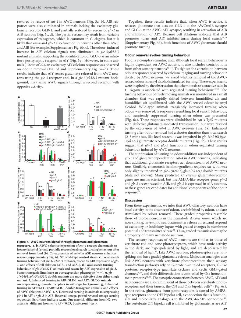
AWC neurons have small clear vesicles at their synapses, suggestingthe use of a small-molecule neurotransmitter3. C. elegans eat-4(ky5)mutants are defective in a vesicular glutamate transporter thatconcentrates glutamate into synaptic vesicles, and are defective inolfactory chemotaxis, local search and numerous other beha-viours12,26. An eat-4 complementary DNA expressed from theAWC-selective odr-3 promoter (AWC::eat-4) rescued chemotaxisof eat-4(ky5) mutants in odour gradients, suggesting that AWC usesglutamate as its transmitter (Fig. 4a). The transgene also partiallyrestored eat-4(ky5) local search behaviour—the AWC-dependentturning behaviour observed in the first 15 min after animals areremoved from food (Fig. 4b). In the AWC::eat-4 transgenic animals,turns were appropriately suppressed in the presence of food and inthe later dispersal period, indicating that glutamate from AWC neu-rons stimulates appropriate local search and not continuous turning
behaviour6. These experiments indicate that AWC neurons canrelease glutamate to stimulate olfactory chemotaxis and local search.
Local search behaviour is a time-dependent, quantitative responseto the removal of food odours, so this assay was used for detailedanalysis of the circuit downstream of AWC neurons. C. elegans hasfour classes of glutamate receptors: AMPA (a-amino-3-hydroxy-5-methyl-4-isoxazole propionic acid)-type glutamate-gated cationchannels (encoded by eight glr genes); NMDA (N-methyl-D-aspartate receptor)-type glutamate-gated cation channels (encodedby two nmr genes); metabotropic G-protein-coupled glutamatereceptors (encoded by two mgl genes); and glutamate-gated chloridechannels (encoded by 10–20 glc/avr genes)27–30. The AMPA-typereceptor GLR-1 is expressed by AIB interneurons as well as commandinterneurons that control forward and backward locomotion27,28,and glr-1(n2461) mutant animals have diminished local search beha-viour12 (Fig. 4c). The local search defect was rescued by expressingglr-1 from a promoter expressed in AIB and AIZ interneurons, butnot in the bulk of glr-1-expressing neurons31 (Fig. 4c). AIZ interneur-ons have not been reported to express endogenous glr-1, so AIBinterneurons are probably the relevant site of glr-1 rescue. Ablatingthe AIB neurons in the transgenic strain eliminated rescue, consistentwith reconstitution of AIB function by the transgene (Fig. 4c).Ablation of AIZ interneurons in the transgenic strain had the samemoderate effect as ablating AIZ in the wild type, indicating that AIZ
c
e
g
f
a b
0
20
40
60
80
100
120
Time (s)
∆F/F
(%)
AWC ON
AWC OFF
10–4
10–5
10–3
10–6
10–7
–40
0
5040302010IAA
0 60
40
80
120
160
–40
0
40
80
120
20100 30 40 50 60
10–4
10–4
10–5
10–6
10–7
0
20
40
60
80
100
120
∆F/F
aft
er o
dou
r re
mov
al (%
)
∆F/F
aft
er o
dou
r re
mov
al (%
)
Time (s)
BZ
d
0
0
20
40
60
80
100
120
Time (s)
∆F/F
(%)
–40
0
0
50403020100 60
40
80
120
160
Time (s)
∆F/F
(%)
–4050403020100 60
Time (s)50403020100 60
Time (s)50403020100 60
40
80
120
160
0
∆F/F
(%)
–40
40
80
120
160
Time (s)50403020100 807060 90
∆F/F
(%)
–40
0
40
80
120
160
0
∆F/F
(%)
∆F/F
(%)
∆F/F
(%)
–40
40
80
120
160
–40
0
40
80
120
20100 30 40 50 600
20
40
60
80
100
120∆F
/F a
fter
od
our
rem
oval
(%)
0
20
40
60
80
100
120
140
∆F/F
aft
er o
dou
r re
mov
al (%
)
∆F/F
aft
er o
dou
r re
mov
al (%
)
20
40
60
80
100
120
∆F/F
aft
er o
dou
r re
mov
al (%
)
Time (s)
BZ
10–4
BZ
Odour
1 min3 min5 minControl
Odour
h
Odour Odour Odour Odour
IAA
Figure 2 | Both classes of AWC neurons respond to odour removal.a–d, G-CaMP calcium responses of AWCON (a, b) or AWCOFF (c, d) afterremoval from different concentrations of isoamyl alcohol (IAA; a, c) orbenzaldehyde (BZ; b, d). e, Removal of 2,3-pentanedione activates AWCOFF,but not AWCON. f, AWCON calcium responses in animals removed fromisoamyl alcohol after different times of exposure. g, h, Secondary responses in
AWCON after isoamyl alcohol was removed at 10 s, added back at 20 s (g) or40 s (h), and then removed again 20 s later. Traces represent averages of 7–30individual recordings (Supplementary Fig. 2). Bar graphs, average percentagechange during the 10 s after odour removal (highlighted in yellow). Error barsand the shaded region around the curves represent s.e.m. The colour key forbar graphs is the same as that for the corresponding response curve.
NATURE | Vol 450 | 1 November 2007 ARTICLES
65Nature ©2007 Publishing Group
function was not disrupted by ectopic glr-1 expression (Fig. 4c andSupplementary Fig. 6d). These results indicate that AWC releasesglutamate to stimulate glr-1 on AIB interneurons. The glr-1(n2461)mutant defect is less severe than the defect caused by killing AIB,suggesting roles for additional glutamate receptors in AIB interneur-ons or glutamate-independent activity of AIB interneurons.
AIY neurons do not express excitatory glr or nmr genes, but doexpress the glutamate-gated chloride channel GLC-3 (refs 32 and 33).glc-3(ok321) mutants had diminished local search behaviour that wasrescued by expressing glc-3 from an AIY-specific promoter (Fig. 4d).Killing AIY interneurons results in the opposite effect—an amplifiedand long-lasting local search behaviour14 (Supplementary Fig. 6d).These results indicate that AWC inhibits AIY during local searchbehaviour via glutamate release onto GLC-3. glr-1(n2461);glc-3(ok321) double mutants were more defective than single mutants,consistent with parallel functions of AIB and AIY neurons (Fig. 4e).
To investigate this circuit model further, we used gain-of-functionanalysis in wild-type animals to test its predictions. In the vertebratecentral nervous system, increased expression of synaptic AMPA-type
glutamate receptors results in synaptic strengthening during long-term potentiation34. Building from this premise, we asked whetheroverexpression of glutamate receptors would strengthen behaviouralconnections between AWC, AIY and AIB neurons. Increased expres-sion of glc-3 from an AIY promoter or glr-1 from an AIB promoterresulted in animals with increased turning during local search(Fig. 4f), as did overexpressing both AIY::glc-3 and AIB::glr-1(Fig. 4g). Killing the AWC sensory neurons abolished these turningbehaviours, indicating that the extra turns were evoked by sensorysignals (Fig. 4g). Control experiments showed that misexpression ofthe AIB receptor in AIY interneurons and vice versa disrupted localsearch behaviour (Fig. 4h).
Calcium imaging of interneurons in mutants provided furtherevidence for glutamate-mediated signalling between AWC, AIBand AIY neurons. Neuronal calcium signals and local search beha-viours are measured on different timescales (1 min versus .5 min,respectively), but results from both approaches were analogous. AIBcalcium responses to odour removal were eliminated in animalslacking the vesicular glutamate transporter EAT-4, but were partially
0 10 20 30 40 50 60Time (s)
0 10 20 30 40 50 60Time (s)
0 10 20 30 40 50 60Time (s)
c
–100
– 50
0
50
100
150
200
250
300
∆F/F
(%)
e
–100
– 50
0
50
100
150
200
250
300
∆F/F
(%)
a
0 10 20 30 40 50 60–100
– 50
0
50
100
150
200
250
300
Time (s)
Wild typeAWC ablations
∆F/F
(%)
Odour
g h
0
80
60
40
20
0
–20
–40
100
80
60
40
20
0
–20
–40
100
10 20 30 40 50 60Time (s)
0 10 20 30 40 50 60
0
b
∆F/F
(%)
–100
–50
50
100
150
200
250
300
Time (s)
Wild typeAWC ablations
Odour
∆F/F
aft
er o
dou
r re
mov
al (%
)
–40
60
40
20
0
–20
80
–40
60
40
20
0
–20
80f
∆F/F
(%)
–100
–50
50
0
100
150
200
250
300
0 10 20 30 40 50 60Time (s)
AIY
Odour
Wild typeAWC ablations
Wild typeAWC ablations
Odour
Wild type
Odour Odour
d
∆F/F
(%)
–100
–50
50
100
150
200
250
300
0
0 10 20 30 40 50 60Time (s)
1 min3 min5 minControl
Odour
AWCAIBAIY
Odour
AIY
Odour
∆F/F
aft
er o
dou
r ad
diti
on (%
)
–40 –40
0
40
80
120
∆F/F
aft
er o
dou
r ad
diti
on (%
)
0
40
80
120
∆F/F
ate
r od
our
rem
oval
(%)
∆F/F
aft
er o
dou
r re
mov
al (%
)
0
100
80
60
40
20
120
AWCAIB
Per
cent
age
of m
axim
um
resp
onse
(%)
Per
cent
age
of m
axim
um
resp
onse
(%)
Figure 3 | Calcium responses in AIB and AIY interneurons.a, b, Representative and average calcium responses of AIB interneurons inwild-type and AWC-ablated animals on odour addition (a) or removal(b). Bar graphs, average percentage change during the 10 s after odouraddition or removal (highlighted in yellow; n 5 22 wild type, 7 AWC-ablated). c, Average AIB responses on odour removal (10 s) and re-addition(40 s). d, Average AIB responses to odour removal after different exposuretimes. e, f, Representative and average calcium responses of AIY neurites
from wild-type and AWC-ablated animals on odour addition (e) or removal(f) (n 5 35 wild type, 8 AWC-ablated). g, h, Averaged AWCON (n 5 30), AIB(n 5 59) and AIY (n 5 35) responses, each trace normalized to its maximumresponse, on odour addition (g) and removal (h). Shading denotes thepresence of odour. The shaded region around curves (c, d, g and h) indicatess.e.m. The colour key for bar graphs is the same as that for the correspondingresponse curve. Error bars indicate s.e.m.
ARTICLES NATURE | Vol 450 | 1 November 2007
66Nature ©2007 Publishing Group
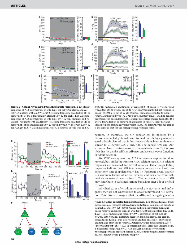
restored by rescue of eat-4 in AWC neurons (Fig. 5a, b). AIB res-ponses were also eliminated in animals lacking the excitatory glu-tamate receptor GLR-1, and partially restored by rescue of glr-1 inAIB neurons (Fig. 5c, d). The partial rescue may result from variableexpression of transgenes, which is common in C. elegans, but it islikely that eat-4 and glr-1 also function in neurons other than AWCand AIB (for example, Supplementary Fig. 6b, c). The odour-inducedincrease in AIY calcium signals was eliminated in glc-3(ok321)mutant animals, supporting the identification of GLC-3 as an inhib-itory postsynaptic receptor in AIY (Fig. 5e). However, in some ani-mals (10 out of 22), an excitatory AIY calcium response was observedon odour removal (Fig. 5f and Supplementary Fig. 5e–h). Theseresults indicate that AIY senses glutamate released from AWC neu-rons using the glc-3 receptor and, in a glc-3(ok321) mutant back-ground, may sense AWC signals through a second receptor withopposite activity.
Together, these results indicate that, when AWC is active, itreleases glutamate that acts on GLR-1 at the AWC::AIB synapseand GLC-3 at the AWC::AIY synapse, resulting in activation of AIBand inhibition of AIY. Because cell ablations indicate that AIBpromotes turns and AIY inhibits turns during local search13,14
(Supplementary Fig. 6d), both functions of AWC glutamate shouldpromote turning.
Odour removal evokes turning behaviour
Food is a complex stimulus, and, although local search behaviour ishighly dependent on AWC activity, it also includes contributionsfrom other sensory neurons12–14. To tighten the correlation betweenodour responses observed by calcium imaging and turning behaviourelicited by AWC neurons, we asked whether removal of the AWC-sensed odour isoamyl alcohol stimulated turning. These experimentswere inspired by the observation that chemotaxis to attractive salts inC. elegans is associated with regulated turning behaviour11,35. Theturning behaviour of freely moving animals was monitored in a smallchamber that was rapidly shifted between humidified air andhumidified air equilibrated with the AWC-sensed odour isoamylalcohol. Wild-type animals transiently increased turning whenodour was removed, a response resembling local search behaviour,and transiently suppressed turning when odour was presented(Fig. 6a). These responses were diminished in eat-4(ky5) mutantswith defective glutamate-mediated transmission, but were rescuedby the expression of eat-4 in AWC neurons (Fig. 6a). Enhancedturning after odour removal had a shorter duration than local search(2–5 min) but, like local search, it was impaired in glr-1(n2461);glc-3(ok321) glutamate receptor double mutants (Fig. 6b). These resultssuggest that glr-1 and glc-3 function in odour-regulated turningbehaviour induced by AWC neurons.
The suppression of turning on odour addition was independent ofglr-1 and glc-3, yet dependent on eat-4 in AWC neurons, indicatingthat additional glutamate receptors act downstream of AWC neu-rons. Similarly, chemotaxis in odour gradients requires eat-4, but wasonly slightly impaired in glr-1(n2461);glc-3(ok321) double mutants(data not shown). Many predicted C. elegans glutamate-receptorgenes are uncharacterized, but the AMPA-like receptor genes glr-2and glr-5 are expressed in AIB, and glr-2 is expressed in AIA neurons,so these genes are candidates for additional components of the odourresponse36.
Discussion
From these experiments, we infer that AWC olfactory neurons havebasal activity in the absence of odour, are inhibited by odour, and arestimulated by odour removal. These graded properties resemblethose of motor neurons in the nematode Ascaris suum, which arenon-spiking, have tonic neurotransmitter release at rest, and respondto excitatory or inhibitory inputs with graded changes in membranepotential and transmitter release24. Thus, graded transmission may bea property of many nematode neurons.
The sensory responses of AWC neurons are similar to those ofvertebrate rod and cone photoreceptors, which have tonic activityin the dark, are hyperpolarized by light, and are depolarized bythe removal of light37. Like AWC neurons, photoreceptors are non-spiking and have graded glutamate release. Molecular analogies alsolink AWC neurons with vertebrate photoreceptors: their sensorytransduction pathways rely on G-protein-coupled receptors, Gi-likeproteins, receptor-type guanylate cyclases and cyclic GMP-gatedchannels5,37, and their differentiation is controlled by Otx homeodo-main proteins38,39. The synaptic connections between AWC, AIY andAIB neurons are also reminiscent of those between vertebrate photo-receptors and their targets, the ON and OFF bipolar cells40 (Fig. 6c).In the retina, glutamate from photoreceptors is sensed by AMPA-type receptors on the OFF bipolar cell, a connection that is function-ally and molecularly analogous to the AWC-to-AIB connection41.The vertebrate ON bipolar cell is inhibited by glutamate, as are AIY
a
0
0.2
0.4
0.6
0.8
1.0
N2 eat-4 eat-4;AWC::eat-4
*
**
Che
mot
axis
ind
ex
Rev
ersa
l-om
egas
per
min
Rev
ersa
l-om
egas
per
min
Rev
ersa
l-om
egas
per
min
Rev
ersa
l-om
egas
per
min
Rev
ersa
l-om
egas
per
min
Rev
ersa
l-om
egas
per
min
Rev
ersa
l-om
egas
per
min
00.40.81.21.62.02.42.8
On food 1–6min
7–12min
35–40min
On food 1–6min
7–12min
35–40min
On food 1–6min
7–12min
35–40min
On food 1–6min
7–12min
35–40min
On food 1–6min
7–12min
35–40min
N2
eat-4eat-4;AWC::eat-4
*** *
**
b
00.40.81.21.62.02.42.8
On food 1–6 min
7–12 min
35–40min
On food 1–6 min
7–12 min
35–40min
N2glr-1
glc-3glc-3;AIY::glc-3+
glc-3;AIY::glc-3++
glr-1;AIB::glr-1glr-1;AIB::glr-1 (AIB–)glr-1;AIB::glr-1 (AIZ–)
*** *
*
*
c
00.40.81.21.62.0
2.82.4
N2
glr-1glc-3glr-1;glc-3
N2
*
*
*
*
* *
d
00.40.81.21.62.02.42.8
***
***
e
00.40.81.21.62.02.42.8 N2
AIY::glc-3
* * * *
*
f
00.40.81.21.62.02.42.8
*
* *
*
**
g
00.40.81.21.62.02.42.8
*** *
* *
h
AIB::glr-1
N2AIY::glr-1AIB::glc-3
N2AIY::glc-3;AIB::glr-1 (AWC+)AIY::glc-3;AIB::glr-1 (AWC–)
Figure 4 | AWC neurons signal through glutamate and glutamatereceptors. a, b, AWC-selective expression of eat-4 rescues chemotaxis toisoamyl alcohol (a) and partially rescues local search turning behaviour afterremoval from food (b). Co-expression of eat-4 in ASK neurons enhancesrescue (Supplementary Fig. 6). N2, wild-type control strain. c, Local searchturning behaviour of glr-1(n2461) mutants, rescue by AIB expression of glr-1, and effects of cell ablation (AIB– and AIZ–). d, Local search turningbehaviour of glc-3(ok321) animals and rescue by AIY expression of glc-3.Some transgenic lines have an overexpression phenotype (11). e, glr-1(n2461);glc-3(ok321) double mutants are more defective than either singlemutant. f, Enhanced turning in AIB::GLR-1 and AIY::GLC-3 animalsoverexpressing glutamate receptors in wild-type background. g, Enhancedturning in AIY::GLC-3;AIB::GLR-1 double-transgenic animals, and effectsof AWC ablations (AWC–). h, Decreased turning in animals misexpressingglr-1 in AIY or glc-3 in AIB. Reversal-omega, paired reversal-omega turningsequences. Error bars indicate s.e.m. One asterisk, different from N2; twoasterisks, different from eat-4 (P , 0.05, Bonferroni t-test).
NATURE | Vol 450 | 1 November 2007 ARTICLES
67Nature ©2007 Publishing Group
neurons. In mammals, the ON bipolar cell is inhibited by aG-protein-coupled glutamate receptor and, in fish, by a glutamate-gated chloride channel that is functionally although not molecularlysimilar to C. elegans GLC-3 (ref. 42). The parallel ON and OFFstreams enhance contrast sensitivity in vertebrate vision43; it is pos-sible that the parallel AIY and AIB neurons have analogous functionsin odour detection.
Like AWC sensory neurons, AIB interneurons respond to odourremoval, but, unlike the transient AWC calcium signals, AIB calciumresponses are sustained for several minutes. These longer-lastingresponses indicate that AIB interneurons integrate the AWC res-ponse over time (Supplementary Fig. 7). Persistent neural activityis a common feature of neural circuits, and can arise from cell-intrinsic or network mechanisms23. The persistent activity of AIBmay contribute to sustained turning behaviour after odour or foodremoval.
Individual turns after odour removal are stochastic and infre-quent; they are not synchronized to odour removal and AIB activa-tion. This mismatch suggests that the AIB could be a gating neuron
d
ba
Odour
Wild typeeat-4eat-4;AWC::eat-4
–20
0
20
40
80
100
120
60
Odour Wild typeeat-4eat-4;AWC::eat-4
∆F/F
(%)
∆F/F
aft
er o
dou
r ad
diti
on (%
)
–20
0
20
40
80
100
120
60
∆F/F
aft
er o
dou
r ad
diti
on (%
)
–20
–40
0
20
40
80
100
60
–50
350
0
50
100
150
200
250
300
Time (s)20100 30 40 50 60
c
∆F/F
(%)
–50
350
0
50
100
150
200
250
300
e
–40
–20
0
20
40
60
80
100
–40
–20
0
20
40
60
80
100Time (s)
20100 30 40 50 60
Time (s)20100 30 40 50 60
∆F/F
(%)
–50
350
0
50
100
150
200
250
300
Time (s)20100 30 40 50 60
∆F/F
(%)
–50
350
0
50
100
150
200
250
300
f
Per
cent
age
of m
axim
um r
esp
onse
Per
cent
age
of m
axim
um r
esp
onse
Per
cent
age
of m
axim
um r
esp
onse
Per
cent
age
of m
axim
um r
esp
onse
Time (s)20100 30 40 50 60
Time (s)20100 30 40 50 60
1
Odourglr-1Wild type
glr-1;AIB::glr-1
Odour
glc-3 #1glc-3 #2
Odour
Wild typeglc-3
Odour
–20∆F/F
aft
er o
dou
r re
mov
al (%
)
0
20
40
80
100
120
60
–20∆F/F
aft
er o
dou
r re
mov
al (%
)
0
20
40
80
100
120
60
–20
–40
0
20
40
80
100
60
glr-1Wild type
Wild type
glr-1;AIB::glr-1
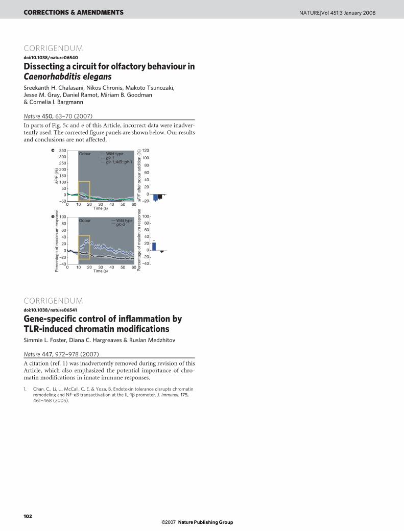
Figure 5 | AIB and AIY require different glutamate receptors. a, b, Calciumresponses of AIB interneurons in wild type, eat-4(ky5) mutants, and eat-4(ky-5) mutants with an AWC::eat-4 rescuing transgene on addition (a) orremoval (b) of the odour isoamyl alcohol (n 5 12 for each). c, d, Calciumresponses of AIB interneurons in wild type, glr-1(n2461) mutants, and glr-1(n2461) mutants with an AIB::glr-1 rescuing transgene on addition (c) orremoval (d) of isoamyl alcohol (n 5 27 for wild type, n 5 25 for glr-1, n 5 12for AIB::glr-1). e, f, Calcium responses of AIY neurites in wild type and glc-
3(ok321) mutants on addition (e) or removal (f) of odour (n 5 35 for wildtype, 22 for glc-3). Twelve out of 22 glc-3(ok321) mutants did not respond toodour (glc-3#1); 10 out of 22 glc-3(ok321) mutants responded to odourremoval, unlike wild type (glc-3#2) (Supplementary Fig. 5). Shading denotesthe presence of odour. Bar graphs, average percentage change during the 10 safter odour addition or removal (highlighted in yellow). Error bars andshaded regions around curves represent s.e.m. The colour key for bar graphsis the same as that for the corresponding response curve.
Odour
a
b
AWC
Odour
AIB
GLR-1
AIY
GLC-3
PR
Light
OFFbipolar
ONbipolar
mGluR1/5/6iGLR-1–7
Odour
0
0.4
0.8
1.2
1.6
2.0
2.4
0 60 120 180 240 300 360 420 480 540
N2eat-4AWC::eat-4
Om
egas
per
min
Om
egas
per
min
Time (s)
Om
egas
per
min
Baseline
N2eat-4AWC::eat-4
Odour Odourremoval
**
**1.8
1.5
1.2
0.9
0.6
0.3
0
c
N2glr-1;glc-3
0 60 120 180 240 300 360 420 480 540Time (s)
0
0.4
0.8
1.2
1.6
2.0
2.4
Om
egas
per
min
Baseline Odour Odourremoval
N2 glr-1;glc-31.8
1.5
1.2
0.9
0.6
0.3
0
*
*
Figure 6 | Odour-regulated turning behaviours. a, b, Omega turns of freelymoving animals recorded before, during and after a 3-min pulse of the odourisoamyl alcohol (t 5 120–300 s). Odour addition suppressed turns andodour removal enhanced turns in wild type N2 (Supplementary Fig. 6e, f).a, eat-4(ky5) mutants and rescue by AWC expression of eat-4. b, glr-1(n2461);glc-3(ok321) glutamate receptor double mutants. Bar graphs,omega turns during 1 min before odour addition (baseline), after odouraddition and after odour removal. Asterisks, values different from eat-4(a) or wild type (b) at P , 0.01 (Bonferroni t-test). Error bars indicate s.e.m.c, Schematic comparing AWC, AIB and AIY neurons to vertebratephotoreceptors and bipolar neurons. iGluR, ionotropic glutamate receptor;mGluR, metabotropic glutamate receptor.
ARTICLES NATURE | Vol 450 | 1 November 2007
68Nature ©2007 Publishing Group
that facilitates turns, but does not trigger individual turning events44.The circuitry downstream of AIB interneurons may generate turnsonly when AIB is active and other unknown conditions are met.Possible sources for the turning signal are the secondary calciumtransients in AWC neurons and the irregular, transient activity ofAIY interneurons. Additional AWC target neurons such as AIA inter-neurons may also regulate turning; so far, the mechanism by whichthe AWC neuron suppresses turns on odour addition is unexplained,suggesting the existence of other target cells or receptors.
These experiments provide the beginnings of a circuit-levelexplanation for food-seeking behaviour in C. elegans. The regulationof turning by AWC neurons has features in common with the biasedrandom walk, first described in bacterial chemotaxis and subse-quently applied to C. elegans salt chemotaxis11,45,46. In this strategy,the animal senses temporal changes in attractant concentrations as itmoves, resulting in long runs towards the attractant, short runs awayfrom the attractant, and eventual accumulation at the point source.For AWC neurons, turning regulation can generate either chemo-taxis or undirected local search; in both cases, turns are probabilisticand are not coupled tightly to stimuli. Our results and those of otherssuggest that the probabilistic turning circuit described here is bothgeneral and flexible. For example, temperature and salt stimuli affectC. elegans behaviour in a manner consistent with the biased randomwalk model35,46–48, and the temperature-sensing and salt-sensing neu-rons synapse onto AIB, AIY, AIA and/or AIZ interneurons3,25,49. Aserotonin receptor essential for olfactory learning acts in AIY, AIBand AIZ interneurons, suggesting that the circuit is subject to plas-ticity50. Further studies of the circuit should shed light on the mech-anism for stochastic behaviours, the generation of persistentbehavioural responses to transient stimuli, and temporal integrationin the nervous system.
METHODS SUMMARYNeuronal calcium responses were measured by detecting changes in fluorescence
of G-CaMP, a non-ratiometric calcium indicator16; control experiments indi-
cated that G-CaMP performed comparably to the ratiometric indicator came-
leon in this system19 (Supplementary Methods). Custom-designed microfluidic
devices for trapping, stimulating and imaging calcium transients in C. elegans
were fabricated from PDMS19. Fluid streams under laminar flow were used to
deliver buffer or odour stimuli to the animal’s nose, using flow by means of two
alternative side-streams to control the delivery of stimuli without changing fluid
pressure. Images were captured using a Coolsnap HQ camera and were analysedusing Metamorph software.
Exploratory behaviours were scored essentially as described14. Animals were
removed from food to a covered, food-free nematode growth medium (NGM)
plate. Their behaviour was observed directly, and turning events were recorded
using a Perl script. At least eight animals were scored blind for each condition.
Odour-induced turning responses were monitored in a custom-fabricated
Plexiglass chamber over an agar surface, with an inlet, an outlet and a central
imaging arena. Humidified air with or without odour was delivered to .20 freely
moving animals using an electronic valve. Movies of these animals were analysedusing automated tracking software, which is distributed at http://wormsense.
stanford.edu/tracker. The software automatically identifies tens of animals, com-
putes and tracks the centroid of each animal, and identifies turns on the basis of
changes in angular velocity (see Supplementary Methods).
Full Methods and any associated references are available in the online version ofthe paper at www.nature.com/nature.
Received 14 August; accepted 24 September 2007.
1. Milo, R. et al. Network motifs: simple building blocks of complex networks. Science298, 824–827 (2002).
2. Chalfie, M. et al. The neural circuit for touch sensitivity in Caenorhabditis elegans.J. Neurosci. 5, 956–964 (1985).
3. White, J. G., Southgate, E., Thomson, J. N. & Brenner, S. The structure of thenervous system of the nematode Caenorhabditis elegans. Phil. Trans. R. Soc. Lond. B314, 1–340 (1986).
4. de Bono, M. & Maricq, A. V. Neuronal substrates of complex behaviors in C.elegans. Annu. Rev. Neurosci. 28, 451–501 (2005).
5. Bargmann, C. I. in Wormbook (ed. The C. elegans Research Community)WormBook doi/10.1895/wormbook.1.123. 1 Æhttp://www.wormbook.orgæ(2006).
6. Zheng, Y., Brockie, P. J., Mellem, J. E., Madsen, D. M. & Maricq, A. V. Neuronalcontrol of locomotion in C. elegans is modified by a dominant mutation in the GLR-1 ionotropic glutamate receptor. Neuron 24, 347–361 (1999).
7. Tobin, D. et al. Combinatorial expression of TRPV channel proteins defines theirsensory functions and subcellular localization in C. elegans neurons. Neuron 35,307–318 (2002).
8. Goodman, M. B., Hall, D. H., Avery, L. & Lockery, S. R. Active currents regulatesensitivity and dynamic range in C. elegans neurons. Neuron 20, 763–772 (1998).
9. Kerr, R. et al. Optical imaging of calcium transients in neurons and pharyngealmuscle of C. elegans. Neuron 26, 583–594 (2000).
10. Mellem, J. E., Brockie, P. J., Zheng, Y., Madsen, D. M. & Maricq, A. V. Decoding ofpolymodal sensory stimuli by postsynaptic glutamate receptors in C. elegans.Neuron 36, 933–944 (2002).
11. Pierce-Shimomura, J. T., Morse, T. M. & Lockery, S. R. The fundamental role ofpirouettes in Caenorhabditis elegans chemotaxis. J. Neurosci. 19, 9557–9569(1999).
12. Hills, T., Brockie, P. J. & Maricq, A. V. Dopamine and glutamate control area-restricted search behavior in Caenorhabditis elegans. J. Neurosci. 24, 1217–1225(2004).
13. Wakabayashi, T., Kitagawa, I. & Shingai, R. Neurons regulating the duration offorward locomotion in Caenorhabditis elegans. Neurosci. Res. 50, 103–111 (2004).
14. Gray, J. M., Hill, J. J. & Bargmann, C. I. A circuit for navigation in Caenorhabditiselegans. Proc. Natl Acad. Sci. USA 102, 3184–3191 (2005).
15. Zhao, B., Khare, P., Feldman, L. & Dent, J. A. Reversal frequency in Caenorhabditiselegans represents an integrated response to the state of the animal and itsenvironment. J. Neurosci. 23, 5319–5328 (2003).
16. Nakai, J., Ohkura, M. & Imoto, K. A high signal-to-noise Ca21 probe composed of asingle green fluorescent protein. Nature Biotechnol. 19, 137–141 (2001).
17. Pologruto, T. A., Yasuda, R. & Svoboda, K. Monitoring neural activity and [Ca21]with genetically encoded Ca21 indicators. J. Neurosci. 24, 9572–9579 (2004).
18. Jospin, M., Jacquemond, V., Mariol, M. C., Segalat, L. & Allard, B. The L-typevoltage-dependent Ca21 channel EGL-19 controls body wall muscle function inCaenorhabditis elegans. J. Cell Biol. 159, 337–348 (2002).
19. Chronis, N., Zimmer, M. & Bargmann, C. I. Microfluidics for in vivo imaging ofneuronal and behavioral activity in Caenorhabditis elegans. Nature Methods 4,727–731 (2007).
20. Bargmann, C. I., Hartwieg, E. & Horvitz, H. R. Odorant-selective genes andneurons mediate olfaction in C. elegans. Cell 74, 515–527 (1993).
21. Colbert, H. A. & Bargmann, C. I. Environmental signals modulate olfactory acuity,discrimination, and memory in Caenorhabditis elegans. Learn. Mem. 4, 179–191(1997).
22. Wes, P. D. & Bargmann, C. I. C. elegans odour discrimination requires asymmetricdiversity in olfactory neurons. Nature 410, 698–701 (2001).
23. Major, G. & Tank, D. Persistent neural activity: prevalence and mechanisms. Curr.Opin. Neurobiol. 14, 675–684 (2004).
24. Davis, R. E. & Stretton, A. O. W. Signalling properties of Ascaris motor neurons:graded synaptic transmission and tonic transmitter release. J. Neurosci. 9,415–425 (1989).
25. Clark, D. A., Biron, D., Sengupta, P. & Samuel, A. D. The AFD sensory neuronsencode multiple functions underlying thermotactic behavior in Caenorhabditiselegans. J. Neurosci. 26, 7444–7451 (2006).
26. Lee, R. Y., Sawin, E. R., Chalfie, M., Horvitz, H. R. & Avery, L. EAT-4, a homolog of amammalian sodium-dependent inorganic phosphate cotransporter, is necessaryfor glutamatergic neurotransmission in Caenorhabditis elegans. J. Neurosci. 19,159–167 (1999).
27. Hart, A. C., Sims, S. & Kaplan, J. M. Synaptic code for sensory modalities revealedby C. elegans GLR-1 glutamate receptor. Nature 378, 82–85 (1995).
28. Maricq, A. V., Peckol, E., Driscoll, M. & Bargmann, C. I. Mechanosensory signallingin C. elegans mediated by the GLR-1 glutamate receptor. Nature 378, 78–81(1995).
29. Cully, D. F. et al. Cloning of an avermectin-sensitive glutamate-gated chloridechannel from Caenorhabditis elegans. Nature 371, 707–711 (1994).
30. Dillon, J., Hopper, N. A., Holden-Dye, L. & O’Connor, V. Molecularcharacterization of the metabotropic glutamate receptor family in Caenorhabditiselegans. Biochem. Soc. Trans. 34, 942–948 (2006).
31. Chou, J. H., Bargmann, C. I. & Sengupta, P. The Caenorhabditis elegans odr-2 geneencodes a novel Ly-6-related protein required for olfaction. Genetics 157, 211–224(2001).
32. Horoszok, L., Raymond, V., Sattelle, D. B. & Wolstenholme, A. J. GLC-3: a novelfipronil and BIDN-sensitive, but picrotoxin-insensitive, L-glutamate-gatedchloride channel subunit from Caenorhabditis elegans. Br. J. Pharmacol. 132,1247–1254 (2001).
33. Wenick, A. S. & Hobert, O. Genomic cis-regulatory architecture and trans-actingregulators of a single interneuron-specific gene battery in C. elegans. Dev. Cell 6,757–770 (2004).
34. Malinow, R. & Malenka, R. C. AMPA receptor trafficking and synaptic plasticity.Annu. Rev. Neurosci. 25, 103–126 (2002).
35. Miller, A. C., Thiele, T. R., Faumont, S., Moravec, M. L. & Lockery, S. R. Step-response analysis of chemotaxis in Caenorhabditis elegans. J. Neurosci. 25,3369–3378 (2005).
36. Brockie, P. J., Madsen, D. M., Zheng, Y., Mellem, J. & Maricq, A. V. Differentialexpression of glutamate receptor subunits in the nervous system of
NATURE | Vol 450 | 1 November 2007 ARTICLES
69Nature ©2007 Publishing Group
Caenorhabditis elegans and their regulation by the homeodomain protein UNC-42.J. Neurosci. 21, 1510–1522 (2001).
37. Zhang, X. & Cote, R. H. cGMP signaling in vertebrate retinal photoreceptor cells.Front. Biosci. 10, 1191–1204 (2005).
38. Furukawa, T., Morrow, E. M. & Cepko, C. L. Crx, a novel otx-like homeobox gene,shows photoreceptor-specific expression and regulates photoreceptordifferentiation. Cell 91, 531–541 (1997).
39. Lanjuin, A., VanHoven, M. K., Bargmann, C. I., Thompson, J. K. & Sengupta, P.Otx/otd homeobox genes specify distinct sensory neuron identities in C. elegans.Dev. Cell 5, 621–633 (2003).
40. Yang, X.-L. Characterization of receptors for glutamate and GABA in retinalneurons. Prog. Neurobiol. 73, 127–150 (2004).
41. Wassle, H. Parallel processing in the mammalian retina. Nature Rev. Neurosci. 5,747–757 (2004).
42. Grant, G. B. & Dowling, J. E. A glutamate-activated chloride current in cone-drivenON bipolar cells of the white Perch retina. J. Neurosci. 15, 3852–3862 (1995).
43. Schiller, P. H., Sandell, J. H. & Maunsell, J. H. Functions of the ON and OFFchannels of the visual system. Nature 322, 824–825 (1986).
44. Taha, S. A. & Fields, H. L. Inhibitions of nucleus accumbens neurons encode agating signal for reward-directed behavior. J. Neurosci. 26, 217–222 (2006).
45. Berg, H. C. & Brown, D. A. Chemotaxis in Escherichia coli analysed by three-dimensional tracking. Nature 239, 500–504 (1972).
46. Dusenbery, D. B. Responses of the nematode Caenorhabditis elegans to controlledchemical stimulation. J. Comp. Physiol. 136, 327–331 (1980).
47. Ryu, W. S. & Samuel, A. D. Thermotaxis in Caenorhabditis elegans analyzed bymeasuring responses to defined thermal stimuli. J. Neurosci. 22, 5727–5733(2002).
48. Zariwala, H. A., Miller, A. C., Faumont, S. & Lockery, S. R. Step responseanalysis of thermotaxis in Caenorhabditis elegans. J. Neurosci. 23, 4369–4377(2003).
49. Mori, I. & Ohshima, Y. Neural regulation of thermotaxis in Caenorhabditis elegans.Nature 376, 344–348 (1995).
50. Zhang, Y., Lu, H. & Bargmann, C. I. Pathogenic bacteria induce aversive olfactorylearning in Caenorhabditis elegans. Nature 438, 179–184 (2005).
Supplementary Information is linked to the online version of the paper atwww.nature.com/nature.
Acknowledgements We thank the C. elegans Knockout Consortium and theCaenorhabditis Genetic Center (CGC) for strains, A. Wolstenholme for the glc-3cDNA, P. Sengupta for the srsx-3 promoter, and M. Meister, M. Zimmer, B. Snyder,G. Lee, D. Albrecht, M. Hilliard and other Bargmann laboratory members for criticalhelp, insights and advice. S.H.C. was supported by the Damon Runyon CancerResearch Foundation and C.I.B. is an Investigator of the Howard Hughes MedicalInstitute. This work was supported by the Howard Hughes Medical Institute(C.I.B.) and the Klingenstein Fund for Neuroscience (M.B.G.).
Author Contributions S.H.C. designed and performed experiments, analysed dataand wrote the paper; N.C., M.T. and J.M.G. designed and performed experiments;D.R. and M.B.G. developed analytical tools; and C.I.B. designed experiments,analysed data and wrote the paper.
Author Information Reprints and permissions information is available atwww.nature.com/reprints. Correspondence and requests for materials should beaddressed to C.I.B. ([email protected]).
ARTICLES NATURE | Vol 450 | 1 November 2007
70Nature ©2007 Publishing Group
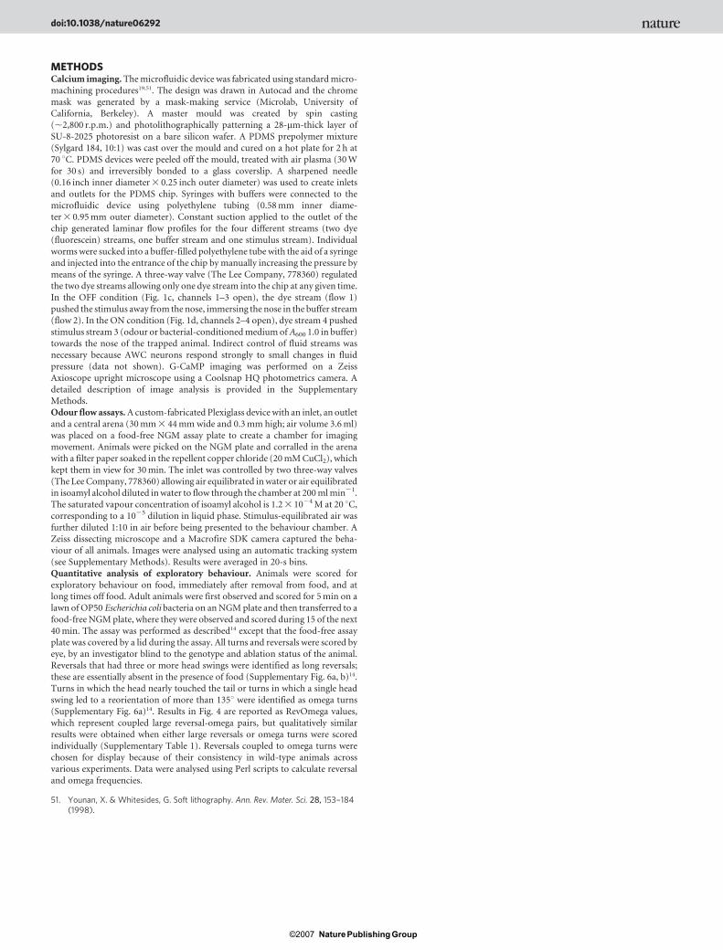
METHODSCalcium imaging. The microfluidic device was fabricated using standard micro-
machining procedures19,51. The design was drawn in Autocad and the chrome
mask was generated by a mask-making service (Microlab, University of
California, Berkeley). A master mould was created by spin casting
(,2,800 r.p.m.) and photolithographically patterning a 28-mm-thick layer of
SU-8-2025 photoresist on a bare silicon wafer. A PDMS prepolymer mixture
(Sylgard 184, 10:1) was cast over the mould and cured on a hot plate for 2 h at
70 uC. PDMS devices were peeled off the mould, treated with air plasma (30 W
for 30 s) and irreversibly bonded to a glass coverslip. A sharpened needle(0.16 inch inner diameter 3 0.25 inch outer diameter) was used to create inlets
and outlets for the PDMS chip. Syringes with buffers were connected to the
microfluidic device using polyethylene tubing (0.58 mm inner diame-
ter 3 0.95 mm outer diameter). Constant suction applied to the outlet of the
chip generated laminar flow profiles for the four different streams (two dye
(fluorescein) streams, one buffer stream and one stimulus stream). Individual
worms were sucked into a buffer-filled polyethylene tube with the aid of a syringe
and injected into the entrance of the chip by manually increasing the pressure by
means of the syringe. A three-way valve (The Lee Company, 778360) regulated
the two dye streams allowing only one dye stream into the chip at any given time.
In the OFF condition (Fig. 1c, channels 1–3 open), the dye stream (flow 1)
pushed the stimulus away from the nose, immersing the nose in the buffer stream
(flow 2). In the ON condition (Fig. 1d, channels 2–4 open), dye stream 4 pushed
stimulus stream 3 (odour or bacterial-conditioned medium of A600 1.0 in buffer)
towards the nose of the trapped animal. Indirect control of fluid streams was
necessary because AWC neurons respond strongly to small changes in fluid
pressure (data not shown). G-CaMP imaging was performed on a Zeiss
Axioscope upright microscope using a Coolsnap HQ photometrics camera. Adetailed description of image analysis is provided in the Supplementary
Methods.
Odour flow assays. A custom-fabricated Plexiglass device with an inlet, an outlet
and a central arena (30 mm 3 44 mm wide and 0.3 mm high; air volume 3.6 ml)
was placed on a food-free NGM assay plate to create a chamber for imaging
movement. Animals were picked on the NGM plate and corralled in the arena
with a filter paper soaked in the repellent copper chloride (20 mM CuCl2), which
kept them in view for 30 min. The inlet was controlled by two three-way valves
(The Lee Company, 778360) allowing air equilibrated in water or air equilibrated
in isoamyl alcohol diluted in water to flow through the chamber at 200 ml min21.
The saturated vapour concentration of isoamyl alcohol is 1.2 3 1024 M at 20 uC,
corresponding to a 1025 dilution in liquid phase. Stimulus-equilibrated air was
further diluted 1:10 in air before being presented to the behaviour chamber. A
Zeiss dissecting microscope and a Macrofire SDK camera captured the beha-
viour of all animals. Images were analysed using an automatic tracking system
(see Supplementary Methods). Results were averaged in 20-s bins.
Quantitative analysis of exploratory behaviour. Animals were scored for
exploratory behaviour on food, immediately after removal from food, and atlong times off food. Adult animals were first observed and scored for 5 min on a
lawn of OP50 Escherichia coli bacteria on an NGM plate and then transferred to a
food-free NGM plate, where they were observed and scored during 15 of the next
40 min. The assay was performed as described14 except that the food-free assay
plate was covered by a lid during the assay. All turns and reversals were scored by
eye, by an investigator blind to the genotype and ablation status of the animal.
Reversals that had three or more head swings were identified as long reversals;
these are essentially absent in the presence of food (Supplementary Fig. 6a, b)14.
Turns in which the head nearly touched the tail or turns in which a single head
swing led to a reorientation of more than 135u were identified as omega turns
(Supplementary Fig. 6a)14. Results in Fig. 4 are reported as RevOmega values,
which represent coupled large reversal-omega pairs, but qualitatively similar
results were obtained when either large reversals or omega turns were scored
individually (Supplementary Table 1). Reversals coupled to omega turns were
chosen for display because of their consistency in wild-type animals across
various experiments. Data were analysed using Perl scripts to calculate reversal
and omega frequencies.
51. Younan, X. & Whitesides, G. Soft lithography. Ann. Rev. Mater. Sci. 28, 153–184(1998).
doi:10.1038/nature06292
Nature ©2007 Publishing Group
CORRIGENDUMdoi:10.1038/nature06540
Dissecting a circuit for olfactory behaviour inCaenorhabditis elegansSreekanth H. Chalasani, Nikos Chronis, Makoto Tsunozaki,Jesse M. Gray, Daniel Ramot, Miriam B. Goodman& Cornelia I. Bargmann
Nature 450, 63–70 (2007)
In parts of Fig. 5c and e of this Article, incorrect data were inadver-tently used. The corrected figure panels are shown below. Our resultsand conclusions are not affected.
CORRIGENDUMdoi:10.1038/nature06541
Gene-specific control of inflammation byTLR-induced chromatin modificationsSimmie L. Foster, Diana C. Hargreaves & Ruslan Medzhitov
Nature 447, 972–978 (2007)
A citation (ref. 1) was inadvertently removed during revision of thisArticle, which also emphasized the potential importance of chro-matin modifications in innate immune responses.
1. Chan, C., Li, L., McCall, C. E. & Yoza, B. Endotoxin tolerance disrupts chromatinremodeling and NF-kB transactivation at the IL-1b promoter. J. Immunol. 175,461–468 (2005).
c
1
0 10 30 50 60
glr-1glr-1;AIB::glr-1
350
0
50
100
150
200
250
300
Time (s)20 40
0
20
40
80
100
60
–20
120Odour Wild type
e
Time (s)0 10 20 30 40 50 60
100
80
60
40
20
0
–20
–40
glc-3
–50
100
80
60
40
20
0
–20
–40
∆F/F
(%)
Per
cent
age
of m
axim
um r
esp
onse
Odour Wild type
∆F/F
aft
er o
dou
r ad
diti
on (%
)P
erce
ntag
e of
max
imum
res
pon
se
CORRECTIONS & AMENDMENTS NATUREjVol 451j3 January 2008
102Nature ©2007 Publishing Group


