Embed Size (px)
Citation preview

OSM-11 Facilitates LIN-12 Notch Signalingduring Caenorhabditis elegansVulval DevelopmentHidetoshi Komatsu
1,2[¤a, Michael Y. Chao
3[, Jonah Larkins-Ford
1¤b, Mark E. Corkins
1, Gerard A. Somers
1,
Tim Tucey1¤c
, Heather M. Dionne1¤d
, Jamie Q. White1,2¤e
, Khursheed Wani1¤f
, Mike Boxem4,5
, Anne C. Hart1,2*
1 Massachusetts General Hospital, Center for Cancer Research, Charlestown, Massachusetts, United States of America, 2 Department of Pathology, Harvard Medical School,
Boston, Massachusetts, United States of America, 3 Department of Biology, California State University San Bernardino, San Bernardino, California, United States of America,
4 Center for Cancer Systems Biology (CCSB) and Department of Cancer Biology, Dana-Farber Cancer Institute, Boston, Massachusetts, United States of America,
5 Department of Genetics, Harvard Medical School, Boston, Massachusetts, United States of America
Notch signaling is critical for cell fate decisions during development. Caenorhabditis elegans and vertebrate Notchligands are more diverse than classical Drosophila Notch ligands, suggesting possible functional complexities. Here, wedescribe a developmental role in Notch signaling for OSM-11, which has been previously implicated in defecation andosmotic resistance in C. elegans. We find that complete loss of OSM-11 causes defects in vulval precursor cell (VPC) fatespecification during vulval development consistent with decreased Notch signaling. OSM-11 is a secreted, diffusibleprotein that, like previously described C. elegans Delta, Serrate, and LAG-2 (DSL) ligands, can interact with the lineagedefective-12 (LIN-12) Notch receptor extracellular domain. Additionally, OSM-11 and similar C. elegans proteins share acommon motif with Notch ligands from other species in a sequence defined here as the Delta and OSM-11 (DOS) motif.osm-11 loss-of-function defects in vulval development are exacerbated by loss of other DOS-motif genes or by loss ofthe Notch ligand DSL-1, suggesting that DOS-motif and DSL proteins act together to activate Notch signaling in vivo.The mammalian DOS-motif protein Deltalike1 (DLK1) can substitute for OSM-11 in C. elegans development, suggestingthat DOS-motif function is conserved across species. We hypothesize that C. elegans OSM-11 and homologous proteinsact as coactivators for Notch receptors, allowing precise regulation of Notch receptor signaling in developmentalprograms in both vertebrates and invertebrates.
Citation: Komatsu H, Chao MY, Larkins-Ford J, Corkins ME, Somers GA, et al. (2008) OSM-11 facilitates LIN-12 Notch signaling during Caenorhabditis elegans vulvaldevelopment. PLoS Biol 6(8): e196. doi:10.1371/journal.pbio.0060196
Introduction
The Notch signaling pathway is essential for cell fatedetermination during embryogenesis and postembryonicdevelopment in multicellular organisms. Classical Notchsignaling begins with activation of the Notch receptor bytransmembrane DSL ligands (Delta and Serrate in Drosophilaor LAG-2 [Lin and Glp-2] in C. elegans [1–3]) expressed onadjacent cells, resulting in proteolytic cleavage of the Notchreceptor, internalization of the ligand-receptor complex, andnuclear translocation of the Notch IC (intracellular) domain[4–8]. In the nucleus, the Notch IC domain acts as atranscriptional regulator together with a conserved tran-scription factor called Su(H) (Suppressor of Hairless) inDrosophila and LAG-1 [Lin and Glp-1] in C. elegans [9,10]. Themolecular mechanisms of Notch signaling are highly con-served. Vertebrate homologs exist for each of these compo-nents in the Notch signaling pathway, and mutations in Notchsignaling have been implicated in various developmentaldisorders, including Alagille and CADASIL [11–14]
In C. elegans, the Notch receptor LIN-12 (Lineage defective-12) plays critical roles in cell fate specification in multipletissues. The roles of LIN-12 in two steps of vulval develop-ment have been particularly well studied. First, LIN-12 isrequired for cell fate specification of an anchor cell (AC) anda vulval uterine (VU) cell from the descendents of equipotentprecursor cells Z1 and Z4 during the L1 larval stage [15–18].
Academic Editor: Julie Ahringer, University of Cambridge, United Kingdom
Received April 21, 2008; Accepted June 26, 2008; Published August 12, 2008
Copyright: � 2008 Komatsu et al. This is an open-access article distributed underthe terms of the Creative Commons Attribution License, which permits unrestricteduse, distribution, and reproduction in any medium, provided the original authorand source are credited.
Abbreviations: AC, anchor cell; AD, activation domain; APX, anterior pharynxdefective; DLK, Deltalike; DLL, Delta like; DOS, Delta and OSM-11; DSL, Delta,Serrate, and LAG-2; EGF, epidermal growth factor; EGFL, EGFlike; FA1, fetal antigen1; GFP, green fluorescent protein; LAG, Lin and Glp; LIN, lineage defective; Muv,multivulva; RNAi, RNA interference; VPC, vulval precursor cell; VU, vulval uterine
* To whom correspondence should be addressed. E-mail: [email protected]
[ These authors contributed equally to this work.
¤a Current address: Takeda Pharmaceutical Company Limited, Osaka, Japan
¤b Current address: Department of Molecular Biology, Massachusetts GeneralHospital, Boston, Massachusetts, United States of America
¤c Current address: University of California, San Diego, La Jolla, California, UnitedStates of America
¤d Current address: State University of New York, Buffalo, New York, United Statesof America
¤e Current address: University of Utah, Salt Lake City, Utah, United States ofAmerica
¤f Current address: University of Massachusetts, Amherst, Massachusetts, UnitedStates of America
PLoS Biology | www.plosbiology.org August 2008 | Volume 6 | Issue 8 | e1961730
PLoS BIOLOGY

Loss of lin-12 signaling generally results in the specification oftwo ACs, whereas increased lin-12 signaling results in two VUcells. The AC produces a diffusible epidermal growth factor(EGF) signal that induces the primary (18) cell fate in P6.p, oneof six equipotent vulval precursor cells (VPCs) (reviewed in[19]). Additionally, LIN-12 specifies secondary (28) cell fates ofP5.p and P7.p, two VPCs adjacent to P6.p, by antagonizingEGF signaling via lateral inhibition [17,20]. Loss of lin-12signaling generally causes VPCs to take on 18 and tertiary (38)fates, whereas strong lin-12 gain-of-function alleles causeVPCs to take on 28 fates with consequent changes in the fatesof descendent cells that contribute to the adult vulva.
Canonical Notch receptor ligands are exemplified byDrosophila Delta, which contains a conserved N-terminalDSL domain originally found in Delta, Serrate, and LAG-2proteins [2,3,7,21,22]. The DSL domain is followed by a seriesof EGF repeats and a transmembrane domain. The DSLdomain is critical for Notch receptor activation based ontissue culture studies and genetic analysis [23,24], but Notchligand EGF repeats are also required for Notch receptoractivation [25,26]. Numerous Notch ligands containing DSLdomains have been identified in various organisms [23,27–32].C. elegans LAG-2 is a classical Notch ligand containing acanonical DSL domain and transmembrane domain and isessential for LIN-12 activation in vivo in many contexts[21,22].
Although key components in the Notch pathway wereidentified decades ago in classical genetic studies in Drosophilaand C. elegans [33,34], additional proteins that play importantor redundant roles in Notch signaling have been identifiedmore recently. C. elegans anterior pharynx defective-1 (APX-1)and DSL-1 are DSL domain–containing soluble proteins thatfunction redundantly with LAG-2 during vulval development[35]. Noncanonical ligands for vertebrate Notch receptorshave been identified, including Delta/notch-like EGF repeatcontaining protein (DNER), F3/contactin, and MAGP proteins[36–40], but functional C. elegans homologs of these non-canonical ligands have not been identified. Deltalike 1 (a.k.a.,
DLK1, fetal antigen 1 [FA1], ZOG, pG2, Preadipocyte Factor 1[PREF1]) also encodes a putative soluble Notch ligand thatlacks a DSL domain [41–43,44]. DLK1 is a paternallyimprinted gene with diverse developmental roles. DLK1knockout mice are growth retarded and obese with eye andskeletal defects [45]. Overexpression of DLK1 due to polaroverdominance results in callipyge sheep with muscle over-proliferation and decreased adipogenesis [45–47]. AlthoughDrosophila lacks a DLK1 homolog, ectopic expression ofmammalian DLK1 in Drosophila inhibits Notch signaling [48].DLK1 has multiple mRNA isoforms; some transcripts aretranslated as membrane-bound proteins with subsequentproteolytic release of the EGF-repeat–containing extracellu-lar domain, while others encode soluble secreted proteins[42,43,49]. DLK1 EGF repeats bind Notch1 EGF repeats inbacterial two-hybrid assays and inhibit activity of a Notch-dependent reporter gene. However, DLK1 inhibits Notchactivation by previously described DSL Notch ligands in thesesame studies [50]. Therefore, a role for DLK1 as a Notchligand is controversial, given the lack of a canonical DSLdomain and the inability of DLK1 to activate vertebrateNotch receptors.Here, we examine the secreted C. elegans protein, OSM-11.
A role for OSM-11 in osmotic sensitivity and defecation wasrecently described, but the molecular function of these geneswas not elucidated in previous studies, and no homologousproteins outside of nematodes were identified [51,52]. Wefound that OSM-11 and related C. elegans proteins contain amotif found only in known and putative Notch ligands,including Serrate and DLK1. We examined the functionalrole of osm-11 in development. We find that osm-11 increaseslin-12 Notch receptor signaling during vulval cell fatespecification. Our results suggest a model in which OSM-11normally acts with C. elegans DSL ligands to activate Notchreceptor signaling in vivo.
Results
OSM-11 Is Required for Cell Fate Specification duringVulval DevelopmentWe identified a deletion allele of osm-11 that removes all of
the predicted mature protein, osm-11(rt142). The majority ofanimals lacking osm-11 had visibly misshapen vulva ordefective vulva based on retention of eggs (Figure 1A–1C).A smaller fraction had an additional protrusion near thenormal position of the vulva. Vulval development was alsomodestly perturbed by RNA interference (RNAi) knockdownof osm-11 (16% defective, n ¼ 82), suggesting that osm-11defects in vulval developmental were caused by loss of osm-11function. Consistent with this hypothesis, osm-11 defects wererescued by reintroduction of either genomic DNA containingthe entire osm-11 gene or the osm-11 cDNA expressed underthe control of 3.4 kb of upstream genomic DNA sequences 59
to the predicted osm-11 initiator methionine (describedbelow) and the unc-54 39 UTR. osm-11 loss of function alsocaused non-vulval developmental defects, including mis-shapen heads and anal protrusions (Figure 1D and 1E)reminiscent of animals with decreased Notch signaling orincreased EGF signaling [9]. To determine the biochemicalrole of OSM-11, a molecular and cellular analysis was firstundertaken.
PLoS Biology | www.plosbiology.org August 2008 | Volume 6 | Issue 8 | e1961731
OSM-11 and LIN-12 Notch in Development
Author Summary
The classic view of Notch receptor activation involves receptorbinding to transmembrane Notch ligands that contain a conservedDSL (Delta, Serrate, and LAG-2) domain. Here, we find that theCaenorhabditis elegans OSM-11 protein is a novel ligand of the well-characterized Notch signal transduction pathway and plays a role incell fate specification during development. OSM-11 is a secreted,diffusible protein whose loss decreases Notch signaling in vivo.OSM-11 and related C. elegans proteins do not contain a DSLdomain, but contain a conserved motif we have named DOS (Deltaand OSM-11) that is also found in the extracellular domain of knownNotch ligands in organisms other than C. elegans. The functionalmammalian homolog of OSM-11 is the secreted protein Deltalike1(Dlk1), also known as Preadipocyte Factor 1 (PREF1), which plays apoorly defined role in Notch signaling regulating obesity and otherdevelopmental decisions. This suggests that Notch ligands are splitinto two complementary coligand families that act together toregulate Notch signaling in developmental contexts. In addition toregulating development, DOS ligands play roles in osmotic stressand C. elegans behavior, suggesting previously unsuspected rolesfor Notch signaling across species.

osm-11 Encodes a Novel Protein with Similarity to NotchLigands
osm-11 corresponds to the C. elegans gene designated asF11C7.5 at the National Center for Biotechnology (NCBI).F11C7.5 is predicted to have two exons and one splice form,which was confirmed by cDNA sequencing (unpublisheddata). OSM-11 and four similar predicted C. elegans proteins,OSM-7 (T05D4.4), ZK507.4, K10G6.2, and K02F3.7, contain asignal peptide for secretion and a potential cEGF-1 domain[53] that is part of a conserved motif described below (Figure2A). cEGF-1 domains contain a small amino acid and sixcysteine residues with characteristic spacing that forms threedisulfide bonds, and are found in extracellular proteinsincluding Notch receptors and ligands.
As standard similarity searching programs (i.e., BLAST)failed to identify additional proteins similar to OSM-11outside of helminthes, we undertook further bioinformaticanalysis, which revealed similarity between OSM-11 andpreviously described Notch ligands. First, the predictedsequences of C. elegans OSM-11, OSM-7, K10G6.2, ZK507.4,and K02F3.7 proteins were aligned, which revealed conserved
amino acids in a common motif containing the putativecEGF-1 domain and additional amino acids: C-X(3)-C-X(3,8)-C-X(2,5)-C-[KVER]-C-X(10,12)-C-X(1,3)-P-X(6,9)-C-X(1,4)-W-X(1,4)-C. Motif-based database searches revealed that allproteins containing the new motif in Drosophila, zebrafish,mouse, and humans are either DSL-containing Notch ligandsor suspected Notch ligands. We named the motif DOSbecause it is found in Delta and OSM-11-like proteins(shaded in Figure 2) and designated the C. elegans genesZK507.4, K10G6.2, and K02F3.7 as dos-1, dos-2, and dos-3,respectively. All five C. elegans DOS-motif proteins are likelysecreted based on the presence of a predicted N-terminalsignal peptide. However, OSM-11 and DOS-3 also have aconsensus proprotein convertase protease cleavage site and aC-terminal transmembrane domain, suggesting that they maybe translated as transmembrane preproproteins prior toproteolytic processing and release of a soluble DOS protein.In known Notch ligands from Drosophila and vertebrates,
the DOS motif is always located immediately following theDSL domain and overlapping the first two EGF repeats. Thefirst two EGF repeats of most Notch ligands differ from theremaining EGF repeats [27] (this study, Figure 2C). The roleof these EGF repeats remains unclear, but several previousstudies suggest that these EGF repeats play roles in Notchactivation: they are required for the DSL domain of Jagged1to bind to the mammalian Notch2 receptor in biochemicalstudies [54]; perturbation of the second EGF repeat interfereswith Notch signaling in Drosophila [25]; and mutations in theseEGF repeats of human Jagged1 are associated with Alagillesyndrome [55]. The DOS motif may define a unique group ofEGF repeats and EGF-like repeats that have a distinctfunctional role in Notch signaling.Outside of helminthes, only three proteins were identified
with DOS motifs that are not canonical Notch ligands: C901,DLK1, and EGFL9 (DLK2). These proteins have a signalpeptide sequence, and the DOS motif is located in the firsttwo EGF repeats (Figure 2B). C901 is a predicted Drosophilaprotein of unknown function containing a DSL domain andmultiple EGF repeats [56]; it is unclear whether C901 is atransmembrane DSL domain protein. DLK1 and EGFlike 9(EGFL9) are vertebrate proteins that contain EGF domains,but lack DSL domains. EGFL9 is poorly characterized [57].DLK1 has membrane-bound and secreted isoforms, and playsdiverse roles in normal development. Altered DLK1 expres-sion causes developmental defects in mammals [42,45–47].DLK1 EGF repeats containing the DOS motif bind to specificNotch1 receptor EGF repeats in two-hybrid studies and intissue culture [50], but the role of DLK1 in Notch signalingremains controversial because DLK1 lacks a DSL domain anddoes not activate mammalian Notch receptors [42,45–47].Given this controversy and given the limited homologyobserved between OSM-11 and previously described canon-ical Notch ligands, we turned to cellular, genetic, andmolecular tools in C. elegans to elucidate the role of OSM-11in developmental signaling pathways.
Loss of osm-11 Perturbs Cell Fate Specification duringVulval DevelopmentWe first examined the role of osm-11 in specification of the
AC. LIN-12 Notch function is required for cell fatespecification of an AC and a VU cell from the equipotentprecursor cells Z1 and Z4 during the L1 larval stage [15–18].
Figure 1. OSM-11 Is Required for Normal Development
(A) Thirty-one percent of osm-11(lf) adult animals had overtly normalvulva and did not retain eggs resembling control animals. (B) Fifty-sevenpercent of osm-11(lf) animals inappropriately retained eggs and/or had asingle misshapen or protruding vulva (15% and 42%, respectively). (C)Twelve percent of osm-11(lf) animals had an extra protrusion near thenormally positioned vulva. (D) osm-11(lf) animals had defects in headmorphology at low frequency (arrowhead). (E) Two thirds of osm-11(lf)animals had a ventral protrusion behind the anus (arrowhead). n . 100animals were scored.doi:10.1371/journal.pbio.0060196.g001
PLoS Biology | www.plosbiology.org August 2008 | Volume 6 | Issue 8 | e1961732
OSM-11 and LIN-12 Notch in Development

PLoS Biology | www.plosbiology.org August 2008 | Volume 6 | Issue 8 | e1961733
OSM-11 and LIN-12 Notch in Development
Loss of lin-12 signaling results in the specification of two ACs,whereas increased lin-12 signaling results in two VU cells. ACcells are readily quantified by examining expression of a lin-3p::gfp reporter construct [58]. No alterations in lin-3p::gfpwere observed in osm-11(lf) animals compared to osm-11(þ)animals (unpublished data; n¼92), suggesting that loss of osm-11 does not alter AC cell fate specification in otherwisenormal animals.
We next examined VPC specification. After AC specifica-tion, the AC produces the diffusible EGF protein LIN-3 that isrequired for induction of the 18 cell fate in P6.p, one of sixequipotent VPCs (reviewed in [19]). LIN-3 EGF acts via thewell-characterized Ras/MAPK (mitogen activated proteinkinase) pathway in VPCs. LIN-12 Notch function is requiredto specify 28 cell fates of P5.p and P7.p, two VPCs adjacent toP6.p, by antagonizing EGF signaling via lateral inhibition[17,59]. Loss of EGF signaling eliminates 18 and 28 cell fates,whereas aberrantly increased EGF/Ras/MAP kinase signalingcan cause all VPCs to adopt the 18 cell fate. By contrast, loss oflin-12 Notch signaling causes all VPCs to take on 18 or 38 fates,whereas strong Notch gain-of-function alleles cause all sixVPCs to take on 28 fates (Figure 3A). These VPC fate decisionswere assessed in osm-11(lf) animals and control animals atspecific larval stages using the previously described greenfluorescent protein (GFP) reporter constructs egl-17p::gfp, lin-11p::gfp, and lip-1p::gfp [60].
In L3 animals, egl-17p::gfp expression in P6.p is directlydependent on EGF/Ras signaling, and egl-17 expression isrepressed in P5.p or P7.p by lateral inhibition via LIN-12Notch signaling [60]. At the Pn.p stage, when cell fates are firstestablished, egl-17p::gfp is only expressed in P6.p in wild-typeanimals. We found appropriate egl-17p::gfp expression in theP6.p cell (Figure 3B and 3C) of animals lacking osm-11, butectopic egl-17p::gfp expression in P5.p or P7.p in approx-imately 10% of osm-11(lf) L3 animals. This ectopic egl-17p::gfpexpression suggests that in osm-11(lf) animals, P5.p and P7.psecondary cell fates are not correctly established whereas the18 cell fate choice of P6.p is unaffected. Later, at the L4 larvalstage, egl-17 expression normally is lost in wild-type animalsfrom P6.p descendents and observed only in 28 cell lineages,i.e., in P5.p and P7.p descendants. In 71% of osm-11(lf) L4animals, egl-17p::gfp expression in P5.p and/or P7.p descend-ants was lost, consistent with loss of 28 cell fates (unpublisheddata; n¼ 63). The aberrant egl-17p::gfp expression observed inosm-11(lf) animals suggests that 18 and 28 cell fates are not
correctly specified in a fraction of osm-11(lf) animals,consistent with decreased Notch signaling.To determine whether secondary cell fates are lost in osm-
11(lf) animals, cell fate specification was examined using lin-11p::gfp and lip-1p::gfp reporter genes. lin-11p::gfp is expressedexclusively in P5.p and P7.p vulval secondary lineages duringdevelopment [61,62] (98% of control wild-type late-L3larvae), but lin-11p::gfp expression is lost in P5.p and/or P7.pdescendents in 67% of osm-11(lf) animals (Figure 3B and 3D).Strikingly, 69% of osm-11(lf) adult animals had an overtlydefective vulva or retained eggs (Figure 1), which correlatesquantitatively with the loss of secondary cell fates observedwith altered lin-11p::gfp expression. Loss of lin-11 expressionat this stage suggests that secondary cell fates are either notproperly specified or not maintained in osm-11(lf) animals.Secondary cell fate specification can be more directly
assessed using lip-1p::gfp. In normal L3 animals, lip-1p::gfpexpression is up-regulated in P5.p and P7.p upon assumptionof secondary cell fate [63]. This up-regulation is directlydependent on lin-12 Notch receptor signaling. However, in35% of osm-11(lf) L3 animals, lip-1p::gfp was not up-regulatedin P5.p and/or P7.p (Figure 3B and 3E; vs. up-regulation in98% of control animals). The loss of lip-1p::gfp and lin-11p::gfpexpression observed in osm-11(lf) animals is reminiscent ofchanges observed when LIN-12 Notch signaling is decreasedand is not consistent with decreased EGF/Ras signaling.
osm-11 Is Expressed in VPCs and Hypodermal CellsThe functional significance of the similarity of OSM-11 to
classic Notch ligands was unclear, particularly as OSM-11lacks a DSL domain. To address the role of osm-11 indevelopment, the cellular and temporal pattern of osm-11expression was examined to delineate its potential roles inVPC fate specification. A transcriptional GFP reporter (osm-11p::gfp) was generated using the same upstream sequencesused for osm-11 cDNA rescue. In animals harboring thistransgene, GFP expression was observed in numerousunidentified cells during embryonic development from thecomma stage onward (unpublished data). GFP expression wasobserved in the VPCs during larval development, as well asvarious hypodermal cells during larval stages (Figure 4). Usingpolyclonal antisera raised against OSM-11 to stain wild-typeanimals, we found that OSM-11 was expressed in the VPCs ofL3 larvae prior to and during cell fate specification (Figure4B). OSM-11 immunoreactivity was also observed in the seamcells of L1 larvae and adult animals (Figure 4A and 4D). In
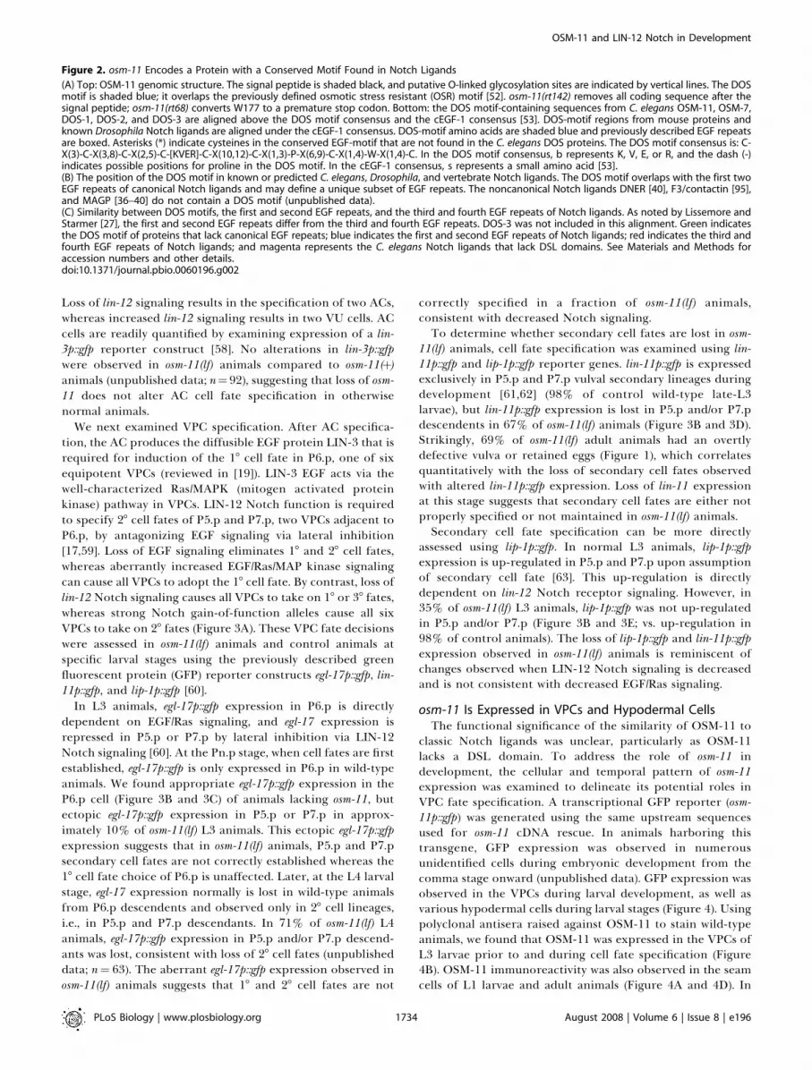
Figure 2. osm-11 Encodes a Protein with a Conserved Motif Found in Notch Ligands
(A) Top: OSM-11 genomic structure. The signal peptide is shaded black, and putative O-linked glycosylation sites are indicated by vertical lines. The DOSmotif is shaded blue; it overlaps the previously defined osmotic stress resistant (OSR) motif [52]. osm-11(rt142) removes all coding sequence after thesignal peptide; osm-11(rt68) converts W177 to a premature stop codon. Bottom: the DOS motif-containing sequences from C. elegans OSM-11, OSM-7,DOS-1, DOS-2, and DOS-3 are aligned above the DOS motif consensus and the cEGF-1 consensus [53]. DOS-motif regions from mouse proteins andknown Drosophila Notch ligands are aligned under the cEGF-1 consensus. DOS-motif amino acids are shaded blue and previously described EGF repeatsare boxed. Asterisks (*) indicate cysteines in the conserved EGF-motif that are not found in the C. elegans DOS proteins. The DOS motif consensus is: C-X(3)-C-X(3,8)-C-X(2,5)-C-[KVER]-C-X(10,12)-C-X(1,3)-P-X(6,9)-C-X(1,4)-W-X(1,4)-C. In the DOS motif consensus, b represents K, V, E, or R, and the dash (-)indicates possible positions for proline in the DOS motif. In the cEGF-1 consensus, s represents a small amino acid [53].(B) The position of the DOS motif in known or predicted C. elegans, Drosophila, and vertebrate Notch ligands. The DOS motif overlaps with the first twoEGF repeats of canonical Notch ligands and may define a unique subset of EGF repeats. The noncanonical Notch ligands DNER [40], F3/contactin [95],and MAGP [36–40] do not contain a DOS motif (unpublished data).(C) Similarity between DOS motifs, the first and second EGF repeats, and the third and fourth EGF repeats of Notch ligands. As noted by Lissemore andStarmer [27], the first and second EGF repeats differ from the third and fourth EGF repeats. DOS-3 was not included in this alignment. Green indicatesthe DOS motif of proteins that lack canonical EGF repeats; blue indicates the first and second EGF repeats of Notch ligands; red indicates the third andfourth EGF repeats of Notch ligands; and magenta represents the C. elegans Notch ligands that lack DSL domains. See Materials and Methods foraccession numbers and other details.doi:10.1371/journal.pbio.0060196.g002
PLoS Biology | www.plosbiology.org August 2008 | Volume 6 | Issue 8 | e1961734
OSM-11 and LIN-12 Notch in Development

adult animals, osm-11p::gfp was expressed only in hypodermalseam cells in adult animals; hypodermal seam cell expressionin adult animals was also confirmed with staining with OSM-11 antisera (Figure 4D). The larval hypodermal expressionpattern of osm-11p::gfp is reminiscent of the osm-7p::gfpexpression pattern described previously, but osm-7p::gfpexpression in seam cells was not reported [52]. OSM-11protein was also expressed in the developing uterus of L4larvae (Figure 4B) and in the spermatheca (Figure 4D); theLIN-12 Notch receptor plays a developmental role in these
tissues as well [64], but only OSM-11 expression in VPCs wascharacterized further.Initially, OSM-11 protein is detected at uniform levels in all
six equivalent VPCs. OSM-11 disappears from P5.p, P6.p, andP7.p after 18 and 28 vulval cell fates are specified (based on up-regulation of lip-1p::gfp; Figure 4B). OSM-11 was not detectedin VPC descendents. Previously described C. elegans DSL-containing Notch ligands also have temporally regulatedexpression patterns in the VPCs [35]. For example, based onreporter construct analysis, soluble DSL-1 is only expressed
Figure 3. OSM-11 Loss Results in Cell Fate Specification Defects
(A) A simplified diagram of cell fate GFP marker expression in P5.p, P6.p, and P7.p. GFP expression is schematically shown in green. Note thatequivalence group members P3.p, P4.p, and P8.p are not shown. In wild-type animals, primary (18) cell fate markers are expressed in P6.p (top left),whereas secondary (28) cell fate markers are normally expressed in P5.p and P7.p (top right). The first Pn.p division (by P5.p, P6.p, and P7.p) in mid-L3larvae gives rise to Pn.px cells; the next divisions give rise to Pn.pxx cells in late-L3 larvae. Loss of Notch signaling does not stop 18 cell fate assumptionby P6.p, but results in inappropriate adoption of 18 cell fates by P5.p, P7.p, and their descendents. Loss of EGF/Ras signaling results in adoption of thetertiary fate by P5.p and P7.p, and in some cases, P6.p, depending on the severity of the defect [96].(B) Quantification of data from (C–E). p , 0.05 based on v2 for each marker.(C) Ten percent of L3 osm-11(lf) animals (right) ectopically express the 18 cell fate marker egl-17p::gfp in P5.p or P7.p, which normally adapt the 28 fate(left).(D) Sixty-seven percent of L3 osm-11(lf) animals lack expression of the 28 cell fate marker lin-11p::gfp in descendants of P5.p and/or P7.p.(E) Thirty-five percent of L3 osm-11(lf) animals do not up-regulate expression of the 28 fate marker lip-1p::gfp in P5.p and/or P7.p. p , 0.05 based on v2
for each marker. These data suggest osm-11(lf) animals have a loss of 28 cell fate specification consistent with loss of LIN-12 Notch signaling.In (C–E), arrowheads indicate the positions of P5.p, P6.p, and P7.p.doi:10.1371/journal.pbio.0060196.g003
PLoS Biology | www.plosbiology.org August 2008 | Volume 6 | Issue 8 | e1961735
OSM-11 and LIN-12 Notch in Development

Figure 4. osm-11 Is Expressed in VPCs and Other Tissues
(A) OSM-11 expression in seam cells of L1 larvae detected using a-OSM-11 antisera. The seam cells on the right side of an L1 animal are in focus; theseam cells on the left side are visible and slightly out of focus. OSM-11 was not expressed in seam cells or hypoderm at other larval stages.(B) OSM-11 expression in the developing uterus of L4 larvae. Left, a-OSM-11 antisera staining; right, visible light image.(C) OSM-11 expression in vulval precursor cells (VPCs; arrowheads) in L3 larvae. The top panels show a-OSM-11 antisera staining of VPCs prior (top left)and immediately after (top right) cell fate specification as assessed by lip-1p::gfp expression. An overlay of a-OSM-11 staining and lip-1p::gfp expressionshows that OSM-11 is concentrated on the apical surface of the VPCs (bottom right); this was confirmed using an ajm-1::gfp fusion (unpublished data).(D) OSM-11 expression in seam cells and spermatheca in adult animals. An osm-11p::gfp reporter gene containing unc-54 39 UTR sequences is expressed
PLoS Biology | www.plosbiology.org August 2008 | Volume 6 | Issue 8 | e1961736
OSM-11 and LIN-12 Notch in Development

in P6.p and its descendents. OSM-11 expression in Pn.p cellsis consistent with a role for OSM-11 in initial cell fatespecification.
Like LIN-12 Notch receptors, OSM-11 is primarily localizedto the apical side of VPCs (Figure 4B, inset). VPCs arepolarized epithelial cells; EGF and Notch signaling normallyoccurs in separate cellular compartments. Lethal-23 (LET-23)EGF receptors are localized to the basolateral surface of theVPCs in close proximity to the AC [65], which is the source ofLIN-3 EGF. In contrast, LIN-12 receptors are primarilylocalized to the apical surface of the VPCs. The apicallocalization of OSM-11 in VPCs during cell fate specificationsuggests that OSM-11 is available to bind to LIN-12 receptorsin VPCs at the time of cell fate specification.
Osmotic Stress Response Does Not Alter Vulval Cell FateSpecification
osm-11 and osm-7 were previously implicated in osmoticstress resistance [51,52]. Pre-exposure of wild-type C. elegansto high external osmolarity is sufficient to induce osmoticresistance. Loss of either osm-7 or osm-11 allows animals tosurvive high external osmolarity without pre-exposure. Thecellular and molecular mechanisms underlying osmotic stressresistance in either scenario are poorly understood, but up-regulation of gpdh-1 and increased levels of the osmolyteglycerol have been implicated [51,52]. As loss of osm-11increases glycerol levels and increased osmolyte levels canalter protein folding, osm-11 could act indirectly to decreaseNotch receptor signaling in VPC fate specification. Alter-natively, OSM-11 might act directly upon Notch receptorsinvolved in VPC fate specification. Our experimental resultsbelow favor the latter model; the role of OSM-11 in vulval cellfate specification is distinct from the role of OSM-11 inosmotic stress.
If osmotic stress indirectly decreases Notch receptorsignaling, then vulval development should be altered by
osmotic stress and altered by genetic backgrounds withincreased osmotic stress resistance. We first tested thishypothesis by raising wild-type animals under previouslydefined osmotic stress conditions: 200 and 400 mM NaCl.Rearing under osmotic stress conditions did not alter vulvalmorphology, and the cellular expression patterns of vulvalcell lineage markers (lip-1p::gfp, egl-17p::gfp, or lin-11p::gfp) inVPCs were unchanged (unpublished data). We also examinedgenetic backgrounds previously implicated in osmotic stressresistance; neither osr-1 nor daf-2 animals have altered vulvalmorphology [66–68]. In addition, we considered the possi-bility that OSM-11 expression in the vulval cell precursorsmight be altered by osmotic stress. We found that rearingunder osmotic stress conditions (400 mM NaCl) did not alterOSM-11 protein levels in VPCs (unpublished data). Com-bined, all of these data suggest that osmotic stress does notitself regulate vulval development. Instead, these data suggestthat the roles of osm-11 in vulval development and osmoticstress resistance are independent.
OSM-11 Is a Secreted ProteinBecause the predicted peptide sequence of OSM-11
contains a signal peptide, we tested whether OSM-11 is asecreted protein. When an osm-11 cDNA was expressed inDrosophila S2 tissue culture cells, OSM-11 protein accumulatesin the media and not in cells (Figure 5A), consistent withOSM-11 acting in vivo as a soluble protein in the extracellularmilieu. The ability of OSM-11 to diffuse and act as a solublefactor in vivo was tested by ectopically expressing OSM-11 innon-VPC cells in osm-11(lf) animals. osm-11 cDNA was fused toosm-10 or glr-1 promoter fragments that drive expression innonoverlapping subsets of neurons throughout larval devel-opment. The osm-10 promoter drives expression in fourclasses of sensory neurons located exclusively in the head andtail [69]. The glr-1 promoter drives expression in 17 otherclasses of neurons (distinct from osm-10–expressing neurons)
Figure 5. osm-11 Encodes a Secreted Protein Required for Vulval Development
(A) Western blot of conditioned media from Drosophila S2 cells containing an OSM-11 cDNA expression construct or empty vector. OSM-11 was notdetected in cell lysates (unpublished data). The molecular weight of mature OSM-11 was predicted at 18.9 kDa; the detected protein migrated at 20.7kDa (arrowhead). OSM-11 may be O-linked glycosylated (see Figure 2).(B) Transgenic rescue of osm-11(lf) vulval defects. osm-11(lf) animals harboring transgenes with empty expression vectors were indistinguishable fromnontransgenic osm-11(lf) animals (n¼ 129 animals, 5 transgenic lines) and were used as controls. Multiple transgenic lines were scored for all rescueexperiments; data are reported as mean 6 standard error of the mean (S.E.M.) In addition to a genomic osm-11 construct, expression of the osm-11cDNA using the following promoters also significantly rescued osm-11(lf) vulval defects: osm-11p, hsp-16p (ubiquitous expression; 79% normal vulval;unpublished data), wrt-6p (hypodermal), osm-10p (sensory neurons), and glr-1p (nonoverlapping set of neurons vs. osm-10p). In addition, heterologousexpression of mammalian DLK1 driven by the hsp-16 promoter also significantly rescued osm-11(lf) vulval phenotypes. n . 52 animals for eachtransgene, p , 0.05 by v2.doi:10.1371/journal.pbio.0060196.g005
in adult seam cells (left); a-OSM-11 antisera was used to confirm seam cell and spermatheca expression (right). No OSM-11 was detected in neurons oflarvae or adult animals (unpublished data); embryonic expression was not characterized.In (A–D), the scale bar represents 10 lm.doi:10.1371/journal.pbio.0060196.g004
PLoS Biology | www.plosbiology.org August 2008 | Volume 6 | Issue 8 | e1961737
OSM-11 and LIN-12 Notch in Development

located in the head and tail [70,71]. Some of the glr-1–expressing neurons have processes in the ventral nerve cordnear the VPCs. We found that neuronal expression of the osm-11 cDNA significantly rescued osm-11 vulval defects to levelscomparable with osm-11 promoter-driven cDNA rescue(Figure 5B). Consistent with these results, hypodermalexpression of osm-11 cDNA using the wrt-6 promoter [72]also rescued osm-11 defects, albeit at a lower level. Weconclude that osm-11 can act nonautonomously and thatsoluble OSM-11 can diffuse in vivo. Although OSM-11expressed in VPCs may be sufficient for normal vulvaldevelopment, OSM-11 can probably function at a distancein some contexts like soluble DSL ligands in C. elegans [35].
osm-11 Acts Upstream of lin-12 Notch Receptor Activationto Increase Signaling
The phenotypic defects caused by loss of osm-11 might bedue to OSM-11 action upon previously identified molecularpathways that regulate cell fate specification in vulvaldevelopment. We tested the sensitivity of the EGF, Notch,and synthetic multivulva (SynMuv) VPC fate specificationpathways to osm-11 levels by RNAi knockdown of osm-11 inmutants that have been previously used as sensitized back-grounds for each pathway: lin-12(n137n460csgf) Notch (seebelow), let-23(sa62gf) EGF receptor, let-60(n1046gf) Ras, or lin-15(n765tslf) SynMuv [73–78]. osm-11(RNAi) had the most effectin animals with compromised lin-12 Notch signaling (27%change in multivulva (Muv) of lin-12(n137n460);osm-11(RNAi)at 20 8C versus less than 9% change in other backgrounds, p ,
0.05, n . 50 each). Although it is difficult to assess the relativesensitivity of these various genetic backgrounds, these resultssuggested that Notch signaling might be particularly sensitiveto OSM-11 levels and that osm-11 might modulate lin-12signaling during vulval development.
To more accurately assess the possible role of osm-11 in lin-12 Notch signaling in vivo, we undertook genetic studies
using the osm-11(lf) null allele and previously described lin-12alleles. lin-12(n137) is a ligand-independent dominant gain offunction (gf) allele, whereas lin-12(n137n460) is a recessive,cold-sensitive gain-of-function allele (csgf). Both cause multi-ple ectopic vulvae (Muv) due to secondary cell specificationdefects [79,80]. If OSM-11 normally functions to increaseNotch signaling, then loss of OSM-11 should decrease LIN-12Notch signaling. We found that osm-11(lf) partially suppressedthe Muv defect of lin-12(csgf) at the restrictive temperature,consistent with OSM-11 normally increasing lin-12 signaling(Figure 6C and 6D). However, osm-11(lf) did not suppress thestronger lin-12(gf) allele (Figure 6E and 6F). Since lin-12(n137gf) is thought to activate lin-12 signaling in a ligand-independent manner, the inability of osm-11(lf) to suppresslin-12(gf) is consistent with osm-11 acting before or duringligand activation of LIN-12.If osm-11 normally acts before or during ligand activation
of LIN-12, then lin-12(lf) should be epistatic to osm-11(lf). lin-12(lf) animals are sterile and have a single large protrudingvulva [80], a phenotype that is easily distinguishable from themisshapen vulva of osm-11(lf) animals (compare Figure 1B and1C with Figure 6B). lin-12(lf);osm-11(lf) double-mutant animalswere indistinguishable from lin-12(lf) animals, suggesting thatosm-11 acts upstream of lin-12 Notch (Figure 6A and 6B).Combined, these results suggest that OSM-11 normallyincreases LIN-12 Notch signaling in vivo and acts before orduring receptor activation.
OSM-11 Functions with Other DOS Proteins inDevelopmentFive C. elegans genes encode putative secreted DOS-motif
proteins: osm-11, osm-7, dos-1, dos-2 [51,52], and dos-3. Loss-of-function alleles are not currently available for dos-2 and dos-3,but osm-7(tm2256) and dos-1(ok2398) are deletion allelesgenerated by the C. elegans gene knockout consortia and arelikely strong loss-of-function (lf) or null alleles. osm-
Figure 6. osm-11 Normally Increases Notch Signaling during Vulval Development
(A and B) lin-12(lf) is epistatic to osm-11(lf). lin-12(lf) is the null allele n941; animals carrying this allele have a protruding vulva (pVul; [A]) that is distinctfrom the defective vulva seen in osm-11(lf) animals (see Figure 1). lin-12(lf);osm-11(lf) animals were indistinguishable from lin-12(lf) animals (B).(C and D) osm-11(lf) suppresses lin-12(csgf) at 15 8C. lin-12(csgf) is n137n460, a recessive cold-sensitive gain-of-function allele; animals carrying thismutation have multiple pseudovulvae (Muv; [C]). lin-12(csgf);osm-11(lf) animals were significantly less Muv (nonMuv) than lin-12(csgf) animals ([D]; p ,0.05).(E and F) osm-11(lf) does not suppress lin-12(gf). lin-12(gf) is n137, a dominant gain-of-function allele that is ligand independent; animals carrying thismutation are Muv (E). lin-12(gf);osm-11(lf) animals were indistinguishable from lin-12(gf) animals (F). n . 50 animals were scored for each genotype.doi:10.1371/journal.pbio.0060196.g006
PLoS Biology | www.plosbiology.org August 2008 | Volume 6 | Issue 8 | e1961738
OSM-11 and LIN-12 Notch in Development
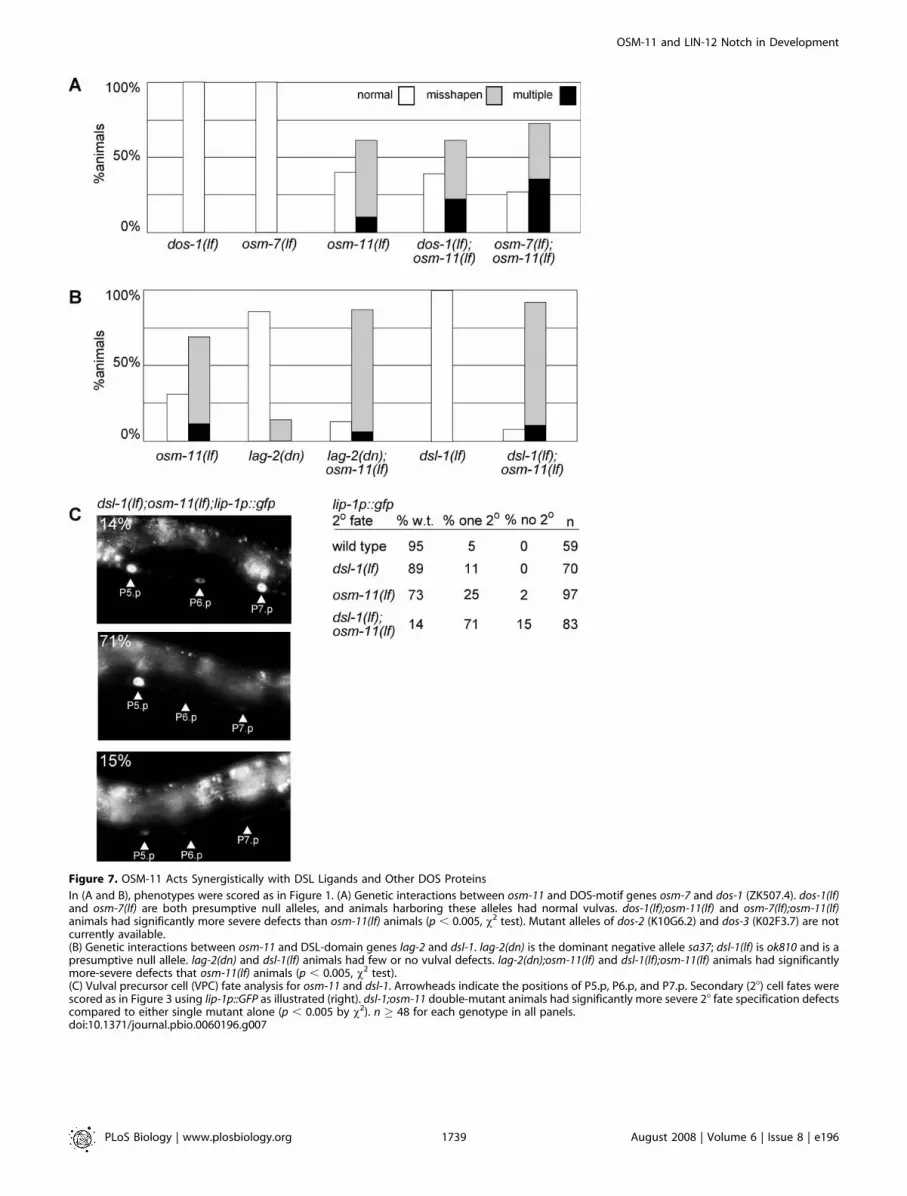
Figure 7. OSM-11 Acts Synergistically with DSL Ligands and Other DOS Proteins
In (A and B), phenotypes were scored as in Figure 1. (A) Genetic interactions between osm-11 and DOS-motif genes osm-7 and dos-1 (ZK507.4). dos-1(lf)and osm-7(lf) are both presumptive null alleles, and animals harboring these alleles had normal vulvas. dos-1(lf);osm-11(lf) and osm-7(lf);osm-11(lf)animals had significantly more severe defects than osm-11(lf) animals (p , 0.005, v2 test). Mutant alleles of dos-2 (K10G6.2) and dos-3 (K02F3.7) are notcurrently available.(B) Genetic interactions between osm-11 and DSL-domain genes lag-2 and dsl-1. lag-2(dn) is the dominant negative allele sa37; dsl-1(lf) is ok810 and is apresumptive null allele. lag-2(dn) and dsl-1(lf) animals had few or no vulval defects. lag-2(dn);osm-11(lf) and dsl-1(lf);osm-11(lf) animals had significantlymore-severe defects that osm-11(lf) animals (p , 0.005, v2 test).(C) Vulval precursor cell (VPC) fate analysis for osm-11 and dsl-1. Arrowheads indicate the positions of P5.p, P6.p, and P7.p. Secondary (28) cell fates werescored as in Figure 3 using lip-1p::GFP as illustrated (right). dsl-1;osm-11 double-mutant animals had significantly more severe 28 fate specification defectscompared to either single mutant alone (p , 0.005 by v2). n � 48 for each genotype in all panels.doi:10.1371/journal.pbio.0060196.g007
PLoS Biology | www.plosbiology.org August 2008 | Volume 6 | Issue 8 | e1961739
OSM-11 and LIN-12 Notch in Development

7(tm2256lf) animals are resistant to osmotic stress and fail toavoid high osmolarity, similar to previously published osm-7alleles [51,52].
To determine whether DOS-motif proteins have over-lapping functions, we tested whether mutants defective inmore than one DOS-motif protein had stronger vulvaldefects. Loss of either osm-7 or dos-1 alone had little or noovert effect on vulval morphology. However, loss of dos-1 orosm-7 increased the percentage of osm-11(lf) animals withmultiple vulval protrusions (Figure 7A). This result isconsistent with multiple DOS-motif proteins acting in vulvaldevelopment.
OSM-11 Functions with DSL Ligands to Increase NotchSignaling
Classical C. elegans Notch DSL ligands are expressed inVPCs and function redundantly during cell specification [35].
Accordingly, DOS-motif proteins may also function redun-dantly in VPC specification. One might expect that DSL-domain proteins and DOS-motif proteins would act togetherto activate Notch signaling. Therefore, loss of a DSL proteinshould exacerbate osm-11 developmental defects. dsl-1 enc-odes a DSL domain-containing ligand which acts redundantlywith two other DSL proteins to activate LIN-12 Notchsignaling during vulval development [35]. Because of thisredundancy, the dsl-1(ok810lf) null allele does not itself causevulval defects [35]. However, dsl-1(lf);osm-11(lf) double mutantshad modestly increased phenotypic defects in vulval mor-phology compared to osm-11 single mutants. osm-11(lf) vulvaldefects were similarly enhanced by lag-2(lf), which encodes aDSL ligand (Figure 7B). To more precisely assess interactionsbetween osm-11 and dsl-1, the expression of lip-1p::gfp in dsl-1(lf);osm-11(lf) animals was assessed during VPC fate specifi-cation. Eighty-six percent of the double-mutant animalslacked lip-1p::gfp up-regulation in either one or bothpresumptive secondary VPCs, indicating a substantial syner-gistic loss of secondary fate specification (Figure 7C). Thisresult is consistent with DOS-motif (i.e., OSM-11) and DSL-domain proteins working together to increase LIN-12 Notchsignaling.
OSM-11 Interacts with LIN-12 Extracellular EGF Repeats ina Yeast Two-Hybrid AssayThe cellular nonautonomy of osm-11, the similarity of OSM-
11 to Notch ligands, the expression pattern of OSM-11, andthe genetic implication that osm-11 functions before orduring LIN-12 Notch activation in VPC fate specificationcollectively suggest that OSM-11 may function as a LIN-12Notch ligand. We tested the hypothesis that OSM-11 directlyinteracts with the LIN-12 extracellular domain. Previousstudies demonstrated that Drosophila and vertebrate DSLligands bind to the extracellular EGF repeats of Notchreceptors. In preliminary studies, we were unable todemonstrate direct binding between OSM-11 and LIN-12biochemically using a heterologous expression system (un-published data). Therefore, we turned to the yeast two-hybridassay to test whether OSM-11 can interact with LIN-12extracellular EGF repeats. Conventional wisdom suggests thatthe yeast two-hybrid system is not suitable for testingextracellular protein–protein interactions, especially fordomains rich in disulfide bridges (e.g., EGF repeats). However,two-hybrid interactions have been demonstrated betweenNotch receptors and ligand pairs in other species for whichbiochemical interactions have been previously validated[38,39,50], as well as for numerous other extracellularproteins [81–86].To validate our yeast two-hybrid approach, we first
confirmed that the extracellular domain of LAG-2 [23] andthe soluble DSL-domain LIN-12 ligand DSL-1 [35] interactwith LIN-12 extracellular EGF repeats 1 through 6 in the two-hybrid assay (Figure 8). To the best of our knowledge, this isthe first in vitro evidence that C. elegans DSL ligands may binddirectly to LIN-12 Notch. As a negative control and toconfirm specificity of the two-hybrid assay, we showed thatthe unrelated C. elegans ligands LIN-3 (an EGF homolog) andegg laying defective-17 (EGL-17) (an FGF homolog) do notinteract with LIN-12 extracellular EGF repeats (Figure 8). TheLAG-2 and DSL-1 interactions with LIN-12 in the two-hybridassay are consistent with previous genetic studies in C. elegans
Figure 8. OSM-11 and C. elegans DSL Ligands Interact with LIN-12 Notch
Extracellular Domain EGF Repeats in the Two-Hybrid System
DSL-1, OSM-11, LAG-2 extracellular domain (LAG-2Ex), EGL-17, or LIN-3was fused to the GAL4 DNA binding domain (DB); the first six LIN-12 EGFrepeats were fused to the GAL4 activation domain (AD). Pairwiseinteractions were tested with the yeast two-hybrid assay; positiveinteractions are indicated by blue staining. Both Notch DSL ligands andOSM-11 interacted with LIN-12 EGF repeats, whereas no interaction ofLIN-3 EGF or EGL-17 FGF with LIN-12 Notch receptor EGF repeats wasdetected. LIN-12::DB fusion proteins exhibited strong self-activation(unpublished data); therefore, reciprocal fusions were not tested.Interaction controls are: (1) empty vectors; (2) DB-pRb and AD-E2F; (3)DB-Fos and AD-Jun; (4) Gal4p and pPC86; and (5) DB-DP1 and AD-E2F1.doi:10.1371/journal.pbio.0060196.g008
PLoS Biology | www.plosbiology.org August 2008 | Volume 6 | Issue 8 | e1961740
OSM-11 and LIN-12 Notch in Development

and with biochemical analyses of Notch ligand/receptorinteractions in other systems. Ligand-receptor interactionswere only assayed using LIN-12 fused to the GAL4 activationdomain (AD) as LIN-12 EGF fusion to the DNA-bindingdomain resulted in strong self-activation in the presence ofAD empty vector (unpublished data).
We found that OSM-11 also interacted with LIN-12extracellular EGF repeats 1 through 6 (Figure 8); OSM-11did not interact with DSL-1 or LAG-2 ligands. We alsoconfirmed previous studies [50] in which murine DLK1 EGFrepeats 1 and 2 containing the DOS motif interactedspecifically with murine Notch1 EGF repeats 12 and 13 inthe same two-hybrid assay format (unpublished data). Thetwo-hybrid interaction does not necessarily demonstrate thatOSM-11 and LIN-12 interact in vivo; however, combined withthe genetic interactions, the apical expression pattern ofOSM-11 in VPCs, and previous studies of DLK1/Notchinteractions, we favor a simple model in which OSM-11binds directly to LIN-12 Notch EGF repeats. Furtherbiochemical studies will be required to demonstrate DOS-motif protein direct interactions with Notch receptors.
The Mammalian DOS-Motif Protein DLK1 Can Substitutefor OSM-11
Our results suggest that the DOS-motif protein OSM-11may act as a soluble LIN-12 ligand in C. elegans. This raises theissue of whether other DOS-motif proteins such as DLK1,which has been implicated in Notch signaling in mammaliancells, also acts as soluble Notch ligands. The precise role ofDLK1 in mammalian Notch signaling is controversial. Toaddress the function of mammalian DLK1 in Notch signaling,we tested the ability of DLK1 to functionally substitute forOSM-11 in vivo in C. elegans. We found that expression of asoluble, mature DLK1 protein isoform named FA1 [43] in osm-
11(lf) animals significantly rescued vulval development,consistent with DLK1 protein increasing LIN-12 signaling(Figure 5D). This result suggests that the function of C. elegansDOS-motif proteins is to increase Notch receptor signalingand that the molecular mechanism may be conserved acrossspecies.
Discussion
The data presented herein demonstrate that osm-11 isrequired for normal vulval development in C. elegans. osm-11encodes a novel cEGF-1 protein that is similar to, but distinctfrom, previously characterized Notch ligands in vertebrates[53]. OSM-11 contains a previously unidentified protein motifthat we have named DOS (Delta and OSM-11) overlapping theEGF motifs. The DOS motif is conserved across species andfound in canonical Notch ligands. OSM-11 is a secretedprotein that is expressed in VPCs during cell fate specifica-tion. Genetic analysis suggests that OSM-11 acts upstream ofLIN-12 and that OSM-11 normally increases LIN-12 Notchsignaling in vivo. Two-hybrid data and expression on the VPCapical surfaces suggest that OSM-11 may directly bind to theLIN-12 extracellular domain, although additional biochem-ical studies will be required to further confirm this. Finally,we demonstrated that the mammalian DOS-motif proteinDLK1 can partially substitute for OSM-11 in C. elegans vulvaldevelopment, suggesting that DOS-motif protein function isconserved across species.Our data suggest a model wherein OSM-11 and C. elegans
DSL ligands act together to activate Notch receptors,potentially as a C. elegans bipartite ligand that is functionallyequivalent to Drosophila Delta or mammalian Jagged1 (Figure9). Previously described C. elegans DSL ligands such as LAG-2lack a DOS motif; C. elegans DSL ligands, such as LAG-2, and
Figure 9. Model: C. elegans DSL and DOS Proteins May Act as Ligands for Notch Receptors
Canonical Notch ligands in Drosophila contain both DSL domains and DOS motifs as do some vertebrate Notch ligands (e.g., Delta). However, classicalNotch ligands from C. elegans and several vertebrate Notch ligands contain a DSL domain, but lack DOS-motif EGF repeats (e.g., LAG-2 or DLL3). The C.elegans proteins characterized in this study (e.g., OSM-11) and the two presumptive vertebrate ligands DLK1 and EGFL9/DLK2 lack DSL domains, butcontain DOS motifs. In the simplest model, both a DOS motif and DSL domain are required for coordinated Notch receptor activation. These could act incis in canonical Notch receptors like Drosophila Delta or in trans in the case of LAG-2 and OSM-11. Overexpression of a ‘‘DOS-only’’ or a ‘‘DSL-only’’ligand may inhibit Notch receptor activation by competition with canonical ligands containing both a DSL domain and a DOS motif, such as Jagged1 orDelta. This model is consistent with osm-11(lf) animals having phenotypic defects usually associated with Notch loss of function. We do not excludeother possible scenarios; see Discussion for details.doi:10.1371/journal.pbio.0060196.g009
PLoS Biology | www.plosbiology.org August 2008 | Volume 6 | Issue 8 | e1961741
OSM-11 and LIN-12 Notch in Development

DOS-motif proteins, such as OSM-11, may both be requiredto activate LIN-12 Notch receptor signaling in vivo. Classicalstudies in C. elegans have shown that expression of the APX-1N-terminus (which contains the DSL domain) is sufficient toactivate Notch signaling; however, this is not inconsistentwith our model because endogenous DOS-motif proteinswere present [23]. Our model is also consistent with previousbiochemical and genetic studies that showed the first two EGFrepeats of Jagged1 and Delta are critical for high-affinityDSL-domain binding to mammalian Notch receptors andNotch receptor activation [25,54].
Bipartite or heteromeric ligands are relatively rarecompared to heteromeric receptors. To our knowledge,bipartite ligands have only been described previously in theimmune system. The binding of antigen to complementfragment creates, in effect, a bipartite ligand for antigenreceptor as does the binding of an antigenic peptide to acompatible major histocompatibility complex (MHC) subunit.Additionally, and perhaps more pertinently, heterodimericcytokines have been described in the immune system thatbind to cytokine receptors [87]; for example, the interleukin-12 (IL-12) cytokine is composed of p40 and p35, whereas theIL-23 is composed of p40 and p19. Although bipartite ligandsare unusual, they are not unprecedented.
Previous studies have shown that the mammalian DOS-motif protein DLK1 acts as a competitive antagonist of ligandJagged1, a canonical ligand that contains both a DSL domainand DOS motif [50]. Therefore, a plausible alternative model(which takes into account DLK1 antagonism of Jagged1) isthat DOS-motif proteins bind to Notch receptors, butfunction as antagonists of DSL-domain Notch ligands in allspecies. DOS proteins such as OSM-11 might play a role inmaintaining C. elegans Notch receptor levels or localization,although LIN-12 Notch expression is unaltered in animalslacking osm-11. Based on our data, we instead favor thesimpler model of DOS-motif proteins as activators of Notchreceptors acting with DSL proteins. In an independentbehavioral analysis (M. Chao, J. Larkins-Ford, T. Tucey, H.Komatsu, and H. Dionne, et al., unpublished data), we alsofound that OSM-11 activates both LIN-12 and germlineproliferation defective-1 (GLP-1) in the adult nervous systemto regulate behavior. We speculate that if DLK1 wascoexpressed in mammalian systems with a C. elegans DSL-only ligand, then Notch signaling might be increased.Mammalian Delta like 3 (DLL3) and DLL4 ligands containDSL domains, but not DOS motifs. Biochemical studies haveshown that DLL3 inhibits Notch signaling and DLL4 increasesNotch signaling in various contexts. It would be useful toexamine Notch activation when DLK1 and DLL3 are coex-pressed. Clearly, biochemical analyses addressing the role ofDOS motifs and DSL domains in Notch receptor activationwill be required to discriminate between these two modelsand to determine the relative contributions of DSL and DOS-motif proteins to Notch signaling.
C. elegans DSL ligands function redundantly, activating LIN-12 Notch during vulval development; loss of any one DSLligand gene causes mild or no overt defects [35]. Similarly,loss of osm-11 alone caused only mild defects in vulvalmorphogenesis, whereas loss of more than one DOS-motifgene resulted in more-severe vulval defects. Like DSL ligands,DOS-motif proteins function semiredundantly to increaseNotch signaling in vivo. In addition, genetic analysis suggests
that DOS-motif proteins and DSL proteins may act togetherto regulate Notch receptors. It is possible that Notch receptoractivation by ligands during VPC development is robust dueto this redundancy. This multifactorial system for regulationof Notch receptors might allow use of individual soluble DOSor DSL proteins in other cell–cell signaling events in othertissues simultaneously.Defining a role herein for osm-7 and osm-11 in Notch
signaling suggests that this pathway also plays a previouslyunsuspected role in osmotic stress response. C. elegans canadapt to increased environmental osmolarity; animals ex-posed to moderate osmotic stress increase internal osmolytelevels and have altered behavior reminiscent of animalslacking osm-11 or osm-7 [51,52]. A role for Notch signaling inosmotic stress has not been reported in any species. Thedevelopmental role of Notch signaling in vulval cell fatespecification is distinct from the role in osmotic stressresponse based on data presented here. Further studies willbe required to determine whether diffusible DOS proteins actas humoral factors to regulate Notch signaling in multipletissues to coordinate physiological and behavioral adaptationto osmotic stress.The diversity of Notch receptors and ligands is remarkable.
C. elegans has two Notch receptors (lin-12 and glp-1), ten DSLdomain proteins that lack DOS motifs [35] and five DOS-motif proteins without DSL domains (this study). Mammalshave four Notch receptors, multiple DSL ligands, and twopresumptive DOS-motif–only ligands: DLK1 and EGFL9/DLK2. Additional proteins have been suggested to act asNotch ligands in vertebrates [36–40], but invertebratehomologs have not been identified. At least one DSL domainNotch ligand in each vertebrate species we examined (zebra-fish, humans, and mice) lacks the conserved DOS motif; theseproteins are potentially analogous to C. elegans DSL domainligands (e.g., LAG-2) that also lack DOS motifs. Soluble Notchligands are now predicted in all of these species based on thisand previous studies. In contrast, Drosophila has only oneNotch receptor, and the two previously characterized trans-membrane Drosophila Notch ligands contain both DSLdomains and DOS motifs. This heterogeneity of Notchligands and receptors indicates that the functional relation-ship between Notch receptors and ligands is highly complex,allowing precise regulation of signaling.
Materials and Methods
Characterization of osm-11. The osm-11(rt68) mutant allele wasidentified in a classical genetic screen based on defective chemo-sensory response and temporarily designated sel-14 (suppressor/enhancerof lin-12-14). The rt68mutation was mapped to the predicted C. elegansgene F11C7.5 and mutates W177 to a premature stop codon, resultingin premature truncation of translation near the end of the DOSmotif. Recent published studies and our analysis herein confirmedthat sel-14(rt68) is an allele of osm-11 and has the same amino acidchange as the previously identified allele osm-11(n1604) [52,88];therefore, we refer to this gene as osm-11. The deletion allele osm-11(rt142) was identified by PCR-based screening of a frozen ethyl-methane sulfonate (EMS)-mutagenized library of C. elegans strains[89]. The rt68 and rt142 alleles had similar phenotypic defects, but thert142 deletion allele was more severe. Both osm-11 alleles are recessive.osm-11(rt142) is likely a complete loss-of-function (lf) allele and wasused exclusively herein. RNAi of osm-11 was performed by raising N2animals on a lawn of bacteria expressing osm-11 double-stranded(dsRNA). Other than morphological defects, vulva perturbations, andconsequent egg-laying defects, osm-11(rt142lf) animals are overtlynormal in locomotion, male mating, and reproduction, although
PLoS Biology | www.plosbiology.org August 2008 | Volume 6 | Issue 8 | e1961742
OSM-11 and LIN-12 Notch in Development

their growth rate is slightly slower and they are slightly smaller thanwild-type animals. osm-11 animals frequently had ventral protrusionsposterior to the anus. Postanal swelling is frequently associated withbacterial infections, but swelling occurs in uncontaminated osm-11animals raised on standard OP50 bacteria. Gonad morphology wassubtly altered in osm-11 animals but was not further characterizedhere. osm-11 loss of function alters glp-1 germline proliferationdefects (M. Chao, J. Larkins-Ford, T. Tucey, H. Komatsu, and H.Dionne, et al, unpublished data). osm-11(rt142) animals are osmoticstress resistant and are motile on 500 mM NaCl NGM plates,consistent with previously published phenotypes of osm-11(n1604)[52].
Strains and genetics. Gain-of-function lin-12 alleles used hereinincluded the constitutive dominant allele lin-12(n137gf) and the cold-sensitive recessive gain-of-function allele lin-12(n137n460gfcs). Resultsfrom homozygous lin-12(n137) and heterozygote lin-12(n137)/þ ani-mals were pooled in Figure 6. The lin-12(n941) null allele wasmaintained by balancing over either qC1 containing qIs26 [rol-6(d), lag-2::gfp] or over unc-32(e189). Genetic epistasis of osm-11 with lin-12(n941) was assessed using homozygous lin-12(lf) progeny of lin-12(lf)/unc-32(e189) animals. A fraction of animals were singled as larvae andsubsequently scored for vulval morphology and genotype. lin-12(n941)animals were always sterile regardless of osm-11 status; lin-12(n941)/unc-32 animals lacked protruding vulva and yielded unc-32 progenyregardless of osm-11 status.
The deletion alleles osm-7(tm2256) and dos-1(ok2398) were generatedby the C. elegans gene knockout consortia. The osm-7(tm2256) deletionremoves the first part of the DOS motif and eliminates an exon splicesite, resulting in a predicted frame shift after amino acid 200 withpremature truncation after translation of 21 amino acids. The dos-1(ok2398) allele is a 1.7-kb deletion that removes the initiatormethionine and the first five exons, including the DOS motif. Alldeletion alleles were backcrossed at least four times prior to analysis.Double-mutant analysis in Figure 7 was performed in an ayIs4 geneticbackground. Other alleles used in this study include lag-2(sa37) anddsl-1(ok810).
Analysis of VPC fate specification. Pn.p cells and descendents wereidentified by differential interference contrast (DIC) imaging on aZeiss Axioskop2. The transgenic arrays used for VPC fate analysiswere: ayIs4 [egl-17p::gfp], syIs107 [lin-3p::gfp], oyIs31 [lin-11p::gfp], andzhIs4 [lip-1p::gfp] [58,60,63,90]. Animals were scored at the Pn.p andPn.px stages for egl-17p::gfp and lip-1p::gfp, but only at the Pn.pxx stagefor lin-11p::gfp. Rearing on 400 mM NaCl NGM plates dramaticallyslows growth and results in partially penetrant embryonic and larvallethality. In less than 10% of all animals raised under theseconditions, Pn.p cells/descendents could not be identified by DIC;these animals were excluded from the analysis. oyIs31 animals werenonviable on 400 mM NaCl NGM plates
Immunohistochemistry. Polyclonal antisera specific to OSM-11were raised in rabbits using the C-terminal peptide YSKCTMFTPV-QY (Sigma-Genosys) and was used as a 1:200 dilution of unpurifiedsera. OSM-11 immunoreactivity was detected in larval and adultanimals in paraformaldehyde-fixed wild-type animals, but not in osm-11(rt142lf) animals (unpublished data). Eggs were not examined, andno immunoreactivity in germ cells was observed. OSM-11 wasdetected at the junction of the presumptive vulva and uterus of L4larvae and in the spermatheca of late-larval and adult animals. OSM-11 mRNA localization by in situ hybridization is consistent withexpression in VPCs and hypoderm in young larvae and in seam cellsin adult animals (see NEXTDB, http://nematode.lab.nig.ac.jp/db2/ShowCloneInfo.php?clone¼59g10; Y. Kohara, personal correspond-ence).
Molecular biology. Plasmids and cloning details are available uponrequest. Transgenic strains were generated by microinjection withplasmids of interest at 20 to 50 ng/ll. Transgenesis coinjectionmarkers were pJM#67 elt-2::gfp [91], pPD48.33 myo-2::gfp [92], orphenotypic rescue of pha-1(e2123) using pBX#1 [93]. The osm-11 cDNAclone was obtained by PCR from the Vidal laboratory ORFeomecDNA library [94] and agrees exactly with the predicted sequence inWormBase and at NCBI. osm-11 cDNA constructs used herein forrescue contained the unc-54 39 UTR, whereas genomic rescue clonescontained the osm-11 39 UTR. Although osm-11 vulval defects aresubstantially rescued by both types of constructs, we cannot rule outtranscriptional regulation by the osm-11 39 UTR. Multiple transgeniclines were scored for each transgenic experiment; results weresubstantially equal for each transgenic line and were pooled byconstruct. The soluble lag-2 construct was previously described andfully rescues a lag-2mutant [23]. Mammalian DLK1 has multiple spliceforms yielding soluble and membrane-bound isoforms. Proteolysis ofmembrane-bound DLK1 yields the soluble protein originally known
as fetal antigen 1 (FA1). A murine DLK1 cDNA fragment that encodesthe DLK1 FA1 protein isoform was used in C. elegans rescueexperiments and was expressed ubiquitously using the hsp-16promoter.
Bioinformatics. C. elegans and C. briggsae homologs of OSM-11 wereidentified by BLAST analysis against genomic sequences andpredicted genes at NCBI and WormBase. A short, common motifwas identified manually and used to search for similar proteins usingPattern Search at the Swiss Institute for Experimental CancerResearch (ISREC) (http://myhits.isb-sib.ch/cgi-bin/pattern_search). Asubset of Notch ligands was identified. DLK1 and Drosophila Deltaproteins were manually compared to C. elegans and C. briggsaehomologs of OSM-11 and used to generate the final DOS-motifconsensus (Figure 2). Proteins were aligned using ClustalW at ISREC(http://myhits.isb-sib.ch/cgi-bin/clustalw). The proteins identified areknown Notch ligands except for mouse DLK1, Drosophila C901, andhuman EGFL9. Drosophila C901 contains a signal peptide, a DSLdomain, and EGF repeats, but has not been well characterized [56].DLK1 and EGFL9 do not contain DSL domains, but do contain signalpeptides and EGF repeats. Given that all previously identified DSLdomains are located between the signal peptide sequence and theEGF repeats, we conclude that DLK1 and EGFL9 do not contain DSLdomains. It is interesting to note that many classical Notch ligandgenes contain an intron immediately after the DSL domain.
T05D4.4 and ZK507.4 (OSM-7 and DOS-1, respectively) predictedC. elegans proteins are partially confirmed by existing cDNAs and areconserved in C. briggsae. A cDNA fragment containing predicted C.elegans K10G6.2 (dos-2) exons was successfully amplified from a cDNAlibrary by the Vidal ORFeome project; the K10G6.2 predicted proteinis also conserved in C. briggsae. The C. briggsae homologs of C. elegansproteins are CBG18238 for T05D4.4, CBG18440 for K10G6.2,CBG06935 for ZK507.4, and CBG15929 for F11C7.5. The newprediction for K02F3.7/DOS-3 has been submitted to WormBase;the C. briggsae homolog is CBP19746. All of these C. briggsae proteinsare predicted to have signal peptide sequences. Proteins in D.melanogaster, C. elegans, C. briggsae, Homo sapiens, Danio rerio, and Musmusculus that contain the DOS motif are (amino acids): tr:A1L1P2_-DANRE/224–274, tr:A1C3M9_DANRE/228–278, sw:DLL1_HUMAN/226–276, sw:DLL1_MOUSE/225–275, tr:A4V346_DROME/231–279,sw:DLLB_DANRE/208–258, tr:Q9VZ44_DROME/212–262,tr:Q925U3_MOUSE/26–76, NP_003827/26–76, sw:EGFL9_HU-MAN/29–79, sw:Q8K1E3 EGFL9/29–79, tr:A1A3Y8_DANRE/235–285, tr:A1A3Y7_DANRE/231–281, sw:JAG1_HUMAN/234–284,sw:JAG1_MOUSE/234–284, sw:JAG2_MOUSE/245–295, sw:JA-G2_HUMAN/245–295, tr:Q90Y55_DANRE/237–287, sw:SERR_-DROME/284–335, tr:O45750_CAEEL/205–253, tr:Q60YH7_CAEBR/205–253, tr:Q21149_CAEEL/127–175, tr:Q60JE9_CAEBR/385–433,sw:YOO4_CAEEL/130–179, tr:Q614N0_CAEBR/135–180,tr:O45346_CAEEL/136–181, and tr:Q60Y06_CAEBR/130–177.
DOS-motif proteins were clustered using CLUSTALW in theMegAlign package (Lasergene) with an identity matrix and thefollowing default parameters: gap penalty 20.0, gap length penalty 0.2,delay divergent sequences 30%, and DNA transition weight 0.5. TheN- and C-terminal boundaries of the amino acid sequences used forthe alignment began at the first cysteine residue of the first EGFrepeat, and ended at the cysteine residue immediately preceding theconserved CXC motif of the second EGF repeat. The only exceptionsto this were the sequences used for MmJagged1 and HsJagged1; inthese proteins, a gap between EGF repeats 1 and 2 contained cysteineand tryptophan residues that followed the spacing of the DOS motifconsensus sequence but were clearly not part of EGF repeat 2. Aminoacid sequence from the gap instead of from EGF repeat 2 was used forthese two proteins. Two outgroups were used in the alignment: EGFrepeats 1 and 2 from CeAPX-1 and CeLAG-2, which lack the SELCTmotif and have been previously shown to be phylogenetically distinctfrom EGF repeats 1 and 2 of other DSL ligands [27]; and EGF repeats3 and 4 (EGF3–4) of selected DOS motif–containing proteins (usingthe same N- and C-terminal boundaries as above), as examples ofcanonical EGF repeats. DmSerrate EGF repeat 4 contains aphylogenetically unique insertion; for the purposes of sequencealignment, amino acids 407–470 were deleted [27]. Accessionnumbers used are: CeT05D4.4, O45750; Cezk507.4, P34636; CeSEL-14, O45346; CeK10G6.2, O16627; CeAPX-1, P41990; CeLAG-2, P45442;DrDeltaA, AAC41249; DrDeltaB, AAH76414; DrDeltaD, Q8UWJ4;DrJagged1, Q90Y57; DrJagged2, CAH69088; DrSerrateB, AAC98354;DmDelta, P10041; DmSerrate, P18168; DmC901, CAA72010; HsDll1,O00548; HsEgfl9, Q6UY11; HsJagged1, P78504; HsJagged2, Q9Y219;MmDlk1, NP_034182; MmDll1, Q61483; MmJagged1, Q9QXX0; andMmJagged2, Q9QYE5. Species designations are: Ce, Caenorhabditis
PLoS Biology | www.plosbiology.org August 2008 | Volume 6 | Issue 8 | e1961743
OSM-11 and LIN-12 Notch in Development
elegans; Dr, Danio rerio; Dm, Drosophila melanogaster; Hs, Homo sapiens;Mm, Mus musculus.
Acknowledgments
We are grateful for advice and/or reagents from members of the Hart,Vidal, van den Heuvel, and Artavanis-Tsakonas laboratories, StevenBlacklow, and numerous generous members of the C. eleganscommunity. We thank Yuji Kohara for sharing NEXTDB in situhybridization results, Victoriano Baladron and Jorge Laborda fortwo-hybrid reagents, and Jonathan Whetstine, Ketu Mishra-Gorur,and Spyros Artavanis-Tsakonas for help with tissue culture studies.Assistance and advice from Li Na, Adriana Jones, and John Satterleewas deeply appreciated.
Author contributions. HK, MYC, JLF, MEC, GAS, TT, HMD, JQW,KW, MB, and ACH conceived and designed the experiments,
performed the experiments, and analyzed the data. MYC and ACHwrote the paper.
Funding. Some nematode strains used in this work were providedby the Caenorhabditis Genetics Center, which is funded by the NationalInstitutes of Health (NIH) National Center for Research Resources(NCRR), by the C. elegans Gene Knockout Project at the OklahomaMedical Research Foundation funded by the NIH, and by theNational Bioresource Project at the Tokyo Women9s MedicalUniversity School of Medicine funded by the Ministry of Education.This work was supported by funding from NIH National Institute ofGeneral Medical Sciences (NIGMS), the Ellison Medical Foundation(ACH), the Massachusetts Biomedical Research Foundation (MYC),Japan Society for the Promotion of Science (HK), and an NIHpostdoctoral fellowship (JQW).
Competing interests. The authors have declared that no competinginterests exist.
References1. Fleming RJ, Scottgale TN, Diederich RJ, Artavanis-Tsakonas S (1990) The
gene Serrate encodes a putative EGF-like transmembrane protein essentialfor proper ectodermal development in Drosophila melanogaster. Genes Dev 4:2188–2201.
2. Kopczynski CC, Alton AK, Fechtel K, Kooh PJ, Muskavitch MA (1988) Delta,a Drosophila neurogenic gene, is transcriptionally complex and encodes aprotein related to blood coagulation factors and epidermal growth factorof vertebrates. Genes Dev 2: 1723–1735.
3. Thomas U, Speicher SA, Knust E (1991) The Drosophila gene Serrateencodes an EGF-like transmembrane protein with a complex expressionpattern in embryos and wing discs. Development 111: 749–761.
4. Eastman DS, Slee R, Skoufos E, Bangalore L, Bray S, et al. (1997) Synergybetween suppressor of Hairless and Notch in regulation of Enhancer ofsplit m gamma and m delta expression. Mol Cell Biol 17: 5620–5628.
5. Hsieh JJ, Henkel T, Salmon P, Robey E, Peterson MG, et al. (1996)Truncated mammalian Notch1 activates CBF1/RBPJk-repressed genes by amechanism resembling that of Epstein-Barr virus EBNA2. Mol Cell Biol 16:952–959.
6. Kidd S, Lieber T, Young MW (1998) Ligand-induced cleavage andregulation of nuclear entry of Notch in Drosophila melanogaster embryos.Genes Dev 12: 3728–3740.
7. Rebay I, Fleming RJ, Fehon RG, Cherbas L, Cherbas P, et al. (1991) SpecificEGF repeats of Notch mediate interactions with Delta and Serrate:implications for Notch as a multifunctional receptor. Cell 67: 687–699.
8. Tamura K, Taniguchi Y, Minoguchi S, Sakai T, Tun T, et al. (1995) Physicalinteraction between a novel domain of the receptor Notch and thetranscription factor RBP-J kappa/Su(H). Curr Biol 5: 1416–1423.
9. Lambie EJ, Kimble J (1991) Two homologous regulatory genes, lin-12 andglp-1, have overlapping functions. Development 112: 231–240.
10. Christensen S, Kodoyianni V, Bosenberg M, Friedman L, Kimble J (1996)lag-1, a gene required for lin-12 and glp-1 signaling in Caenorhabditis elegans,is homologous to human CBF1 and Drosophila Su(H). Development 122:1373–1383.
11. Joutel A, Corpechot C, Ducros A, Vahedi K, Chabriat H, et al. (1996) Notch3mutations in CADASIL, a hereditary adult-onset condition causing strokeand dementia. Nature 383: 707–710.
12. Li L, Krantz ID, Deng Y, Genin A, Banta AB, et al. (1997) Alagille syndromeis caused by mutations in human Jagged1, which encodes a ligand forNotch1. Nat Genet 16: 243–251.
13. Oda T, Elkahloun AG, Pike BL, Okajima K, Krantz ID, et al. (1997)Mutations in the human Jagged1 gene are responsible for Alagillesyndrome. Nat Genet 16: 235–242.
14. McDaniell R, Warthen DM, Sanchez-Lara PA, Pai A, Krantz ID, et al. (2006)NOTCH2 mutations cause Alagille syndrome, a heterogeneous disorder ofthe notch signaling pathway. Am J Hum Genet 79: 169–173.
15. Greenwald I, Seydoux G (1990) Analysis of gain-of-function mutations ofthe lin-12 gene of Caenorhabditis elegans. Nature 346: 197–199.
16. Seydoux G, Greenwald I (1989) Cell autonomy of lin-12 function in a cellfate decision in C. elegans. Cell 57: 1237–1245.
17. Sternberg PW (1988) Lateral inhibition during vulval induction inCaenorhabditis elegans. Nature 335: 551–554.
18. Wilkinson HA, Fitzgerald K, Greenwald I (1994) Reciprocal changes inexpression of the receptor lin-12 and its ligand lag-2 prior to commitmentin a C. elegans cell fate decision. Cell 79: 1187–1198.
19. Chang C, Sternberg PW (1999) C. elegans vulval development as a modelsystem to study the cancer biology of EGFR signaling. Cancer MetastasisRev 18: 203–213.
20. Newman AP, Acton GZ, Hartwieg E, Horvitz HR, Sternberg PW (1999) Thelin-11 LIM domain transcription factor is necessary for morphogenesis ofC. elegans uterine cells. Development 126: 5319–5326.
21. Henderson ST, Gao D, Lambie EJ, Kimble J (1994) lag-2 may encode asignaling ligand for the GLP-1 and LIN-12 receptors of C. elegans.Development 120: 2913–2924.
22. Tax FE, Yeargers JJ, Thomas JH (1994) Sequence of C. elegans lag-2 reveals acell-signalling domain shared with Delta and Serrate of Drosophila. Nature368: 150–154.
23. Fitzgerald K, Greenwald I (1995) Interchangeability of Caenorhabditiselegans DSL proteins and intrinsic signalling activity of their extracellulardomains in vivo. Development 121: 4275–4282.
24. Gu Y, Hukriede NA, Fleming RJ (1995) Serrate expression can functionallyreplace Delta activity during neuroblast segregation in the Drosophilaembryo. Development 121: 855–865.
25. Parks AL, Stout JR, Shepard SB, Klueg KM, Dos Santos AA, et al. (2006)Structure-function analysis of delta trafficking, receptor binding andsignaling in Drosophila. Genetics 174: 1947–1961.
26. Shimizu K, Chiba S, Saito T, Kumano K, Hirai H (2000) Physical interactionof Delta1, Jagged1, and Jagged2 with Notch1 and Notch3 receptors.Biochem Biophys Res Commun 276: 385–389.
27. Lissemore JL, Starmer WT (1999) Phylogenetic analysis of vertebrate andinvertebrate Delta/Serrate/LAG-2 (DSL) proteins. Mol Phylogenet Evol 11:308–319.
28. Vargesson N, Patel K, Lewis J, Tickle C (1998) Expression patterns ofNotch1, Serrate1, Serrate2 and Delta1 in tissues of the developing chicklimb. Mech Dev 77: 197–199.
29. Jarriault S, Le Bail O, Hirsinger E, Pourquie O, Logeat F, et al. (1998) Delta-1 activation of notch-1 signaling results in HES-1 transactivation. Mol CellBiol 18: 7423–7431.
30. Dunwoodie SL, Henrique D, Harrison SM, Beddington RS (1997) MouseDll3: a novel divergent Delta gene which may complement the function ofother Delta homologues during early pattern formation in the mouseembryo. Development 124: 3065–3076.
31. Parks AL, Turner FR, Muskavitch MA (1995) Relationships betweencomplex Delta expression and the specification of retinal cell fates duringDrosophila eye development. Mech Dev 50: 201–216.
32. Muskavitch MA, Hoffmann FM (1990) Homologs of vertebrate growthfactors in Drosophila melanogaster and other invertebrates. Curr Top Dev Biol24: 289–328.
33. Greenwald I (1998) LIN-12/Notch signaling: lessons from worms and flies.Genes Dev 12: 1751–1762.
34. Artavanis-Tsakonas S, Rand MD, Lake RJ (1999) Notch signaling: cell fatecontrol and signal integration in development. Science 284: 770–776.
35. Chen N, Greenwald I (2004) The lateral signal for LIN-12/Notch in C. elegansvulval development comprises redundant secreted and transmembraneDSL proteins. Dev Cell 6: 183–192.
36. Lai EC, Bodner R, Posakony JW (2000) The enhancer of split complex ofDrosophila includes four Notch-regulated members of the bearded genefamily. Development 127: 3441–3455.
37. Brennan K, Gardner P (2002) Notching up another pathway. Bioessays 24:405–410.
38. Miyamoto A, Lau R, Hein PW, Shipley JM, Weinmaster G (2006) Micro-fibrillar proteins MAGP-1 and MAGP-2 induce Notch1 extracellulardomain dissociation and receptor activation. J Biol Chem 281: 10089–10097.
39. Nehring LC, Miyamoto A, Hein PW, Weinmaster G, Shipley JM (2005) Theextracellular matrix protein MAGP-2 interacts with Jagged1 and induces itsshedding from the cell surface. J Biol Chem 280: 20349–20355.
40. Eiraku M, Tohgo A, Ono K, Kaneko M, Fujishima K, et al. (2005) DNER actsas a neuron-specific Notch ligand during Bergmann glial development. NatNeurosci 8: 873–880.
41. Jensen CH, Schroder HD, Teisner B, Laursen I, Brandrup F, et al. (1999)Fetal antigen 1, a member of the epidermal growth factor superfamily, inneurofibromas and serum from patients with neurofibromatosis type 1. Br JDermatol 140: 1054–1059.
42. Lee YL, Helman L, Hoffman T, Laborda J (1995) dlk, pG2 and Pref-1mRNAs encode similar proteins belonging to the EGF-like superfamily.Identification of polymorphic variants of this RNA. Biochim Biophys Acta1261: 223–232.
PLoS Biology | www.plosbiology.org August 2008 | Volume 6 | Issue 8 | e1961744
OSM-11 and LIN-12 Notch in Development

43. Jensen CH, Krogh TN, Hojrup P, Clausen PP, Skjodt K, et al. (1994) Proteinstructure of fetal antigen 1 (FA1). A novel circulating human epidermal-growth-factor-like protein expressed in neuroendocrine tumors and itsrelation to the gene products of dlk and pG2. Eur J Biochem 225: 83–92.
44. Laborda J, Sausville EA, Hoffman T, Notario V (1993) dlk, a putativemammalian homeotic gene differentially expressed in small cell lungcarcinoma and neuroendocrine tumor cell line. J Biol Chem 268: 3817–3820.
45. Moon YS, Smas CM, Lee K, Villena JA, Kim KH, et al. (2002) Mice lackingpaternally expressed Pref-1/Dlk1 display growth retardation and acceler-ated adiposity. Mol Cell Biol 22: 5585–5592.
46. Kim KS, Kim JJ, Dekkers JC, Rothschild MF (2004) Polar overdominantinheritance of a DLK1 polymorphism is associated with growth and fatnessin pigs. Mamm Genome 15: 552–559.
47. Murphy SK, Freking BA, Smith TP, Leymaster K, Nolan CM, et al. (2005)Abnormal postnatal maintenance of elevated DLK1 transcript levels incallipyge sheep. Mamm Genome 16: 171–183.
48. Bray SJ, Takada S, Harrison E, Shen SC, Ferguson-Smith A (2008) Theatypical mammalian ligand Delta-like1 (Dlk1) can regulate Notch signallingin Drosophila. BMC Dev Biol 8: 11.
49. Mei B, Zhao L, Chen L, Sul HS (2002) Only the large soluble form ofpreadipocyte factor-1 (Pref-1), but not the small soluble and membraneforms, inhibits adipocyte differentiation: role of alternative splicing.Biochem J 364: 137–144.
50. Baladron V, Ruiz-Hidalgo MJ, Nueda ML, Diaz-Guerra MJ, Garcia-RamirezJJ, et al. (2005) dlk acts as a negative regulator of Notch1 activation throughinteractions with specific EGF-like repeats. Exp Cell Res 303: 343–359.
51. Lamitina T, Huang CG, Strange K (2006) Genome-wide RNAi screeningidentifies protein damage as a regulator of osmoprotective gene expres-sion. Proc Natl Acad Sci U S A 103: 12173–12178.
52. Wheeler JM, Thomas JH (2006) Identification of a novel gene familyinvolved in osmotic stress resistance in C. elegans. Genetics 174: 1327–1336.
53. Wouters MA, Rigoutsos I, Chu CK, Feng LL, Sparrow DB, et al. (2005)Evolution of distinct EGF domains with specific functions. Protein Sci 14:1091–1103.
54. Shimizu K, Chiba S, Kumano K, Hosoya N, Takahashi T, et al. (1999) Mousejagged1 physically interacts with notch2 and other notch receptors.Assessment by quantitative methods. J Biol Chem 274: 32961–32969.
55. Warthen DM, Moore EC, Kamath BM, Morrissette JJ, Sanchez P, et al. (2006)Jagged1 (JAG1) mutations in Alagille syndrome: increasing the mutationdetection rate. Hum Mutat 27: 436–443.
56. Kozlova T, Zhimulev IF, Kafatos FC (1997) Molecular organization of anindividual Drosophila polytene chromomere: transcribed sequences in the10A1–2 band. Mol Gen Genet 257: 55–61.
57. Nueda ML, Baladron V, Garcia-Ramirez JJ, Sanchez-Solana B, Ruvira MD,et al. (2007) The novel gene EGFL9/Dlk2, highly homologous to Dlk1,functions as a modulator of adipogenesis. J Mol Biol 367: 1270–1280.
58. Hwang BJ, Sternberg PW (2004) A cell-specific enhancer that specifies lin-3expression in the C. elegans anchor cell for vulval development. Develop-ment 131: 143–151.
59. Newman AP, Sternberg PW (1996) Coordinated morphogenesis of epitheliaduring development of the Caenorhabditis elegans uterine-vulval connection.Proc Natl Acad Sci U S A 93: 9329–9333.
60. Burdine RD, Branda CS, Stern MJ (1998) EGL-17(FGF) expressioncoordinates the attraction of the migrating sex myoblasts with vulvalinduction in C. elegans. Development 125: 1083–1093.
61. Gupta BP, Sternberg PW (2002) Tissue-specific regulation of the LIMhomeobox gene lin-11 during development of the Caenorhabditis elegans egg-laying system. Dev Biol 247: 102–115.
62. Sarafi-Reinach TR, Melkman T, Hobert O, Sengupta P (2001) The lin-11LIM homeobox gene specifies olfactory and chemosensory neuron fates inC. elegans. Development 128: 3269–3281.
63. Berset T, Hoier EF, Battu G, Canevascini S, Hajnal A (2001) Notchinhibition of RAS signaling through MAP kinase phosphatase LIP-1 duringC. elegans vulval development. Science 291: 1055–1058.
64. Wilkinson HA, Greenwald I (1995) Spatial and temporal patterns of lin-12expression during C. elegans hermaphrodite development. Genetics 141:513–526.
65. Levitan D, Greenwald I (1998) LIN-12 protein expression and localizationduring vulval development in C. elegans. Development 125: 3101–3109.
66. Kenyon C, Chang J, Gensch E, Rudner A, Tabtiang R (1993) A C. elegansmutant that lives twice as long as wild type. Nature 366: 461–464.
67. Vowels JJ, Thomas JH (1992) Genetic analysis of chemosensory control ofdauer formation in Caenorhabditis elegans. Genetics 130: 105–123.
68. Lamitina ST, Strange K (2005) Transcriptional targets of DAF-16 insulinsignaling pathway protect C. elegans from extreme hypertonic stress. Am JPhysiol Cell Physiol 288: C467–474.
69. Hart A, Kass J, Shapiro J, Kaplan J (1999) Distinct signaling pathways
mediate touch and osmosensory responses in a polymodal sensory neuron.J Neurosci 19: 1952–1958.
70. Hart A, Simms S, Kaplan J (1995) Synaptic code for sensory modalitiesrevealed by C. elegans GLR-1 glutamate receptor. Nature 378: 82–85.
71. Maricq AV, Peckol E, Driscoll M, Bargmann CI (1995) Mechanosensorysignalling in C. elegans mediated by the GLR-1 glutamate receptor. Nature378: 78–81.
72. Hao L, Aspock G, Burglin TR (2006) The hedgehog-related gene wrt-5 isessential for hypodermal development in Caenorhabditis elegans. Dev Biol290: 323–336.
73. Beitel GJ, Clark SG, Horvitz HR (1990) Caenorhabditis elegans ras gene let-60acts as a switch in the pathway of vulval induction. Nature 348: 503–509.
74. Clark SG, Lu X, Horvitz HR (1994) The Caenorhabditis elegans locus lin-15, anegative regulator of a tyrosine kinase signaling pathway, encodes twodifferent proteins. Genetics 137: 987–997.
75. Ferguson EL, Horvitz HR (1985) Identification and characterization of 22genes that affect the vulval cell lineages of the nematode Caenorhabditiselegans. Genetics 110: 17–72.
76. Huang LS, Tzou P, Sternberg PW (1994) The lin-15 locus encodes twonegative regulators of Caenorhabditis elegans vulval development. Mol BiolCell 5: 395–411.
77. Katz WS, Lesa GM, Yannoukakos D, Clandinin TR, Schlessinger J, et al.(1996) A point mutation in the extracellular domain activates LET-23, theCaenorhabditis elegans epidermal growth factor receptor homolog. Mol CellBiol 16: 529–537.
78. Moghal N, Sternberg PW (2003) A component of the transcriptionalmediator complex inhibits RAS-dependent vulval fate specification in C.elegans. Development 130: 57–69.
79. Ambros V (1999) Cell cycle-dependent sequencing of cell fate decisions inCaenorhabditis elegans vulva precursor cells. Development 126: 1947–1956.
80. Greenwald IS, Sternberg PW, Horvitz HR (1983) The lin-12 locus specifiescell fates in Caenorhabditis elegans. Cell 34: 435–444.
81. Amano T, Kwak O, Fu L, Marshak A, Shi YB (2005) The matrixmetalloproteinase stromelysin-3 cleaves laminin receptor at two distinctsites between the transmembrane domain and laminin binding sequencewithin the extracellular domain. Cell Res 15: 150–159.
82. Fujimoto N, Terlizzi J, Brittingham R, Fertala A, McGrath JA, et al. (2005)Extracellular matrix protein 1 interacts with the domain III of fibulin-1Cand 1D variants through its central tandem repeat 2. Biochem Biophys ResCommun 333: 1327–1333.
83. Lee NV, Rodriguez-Manzaneque JC, Thai SN, Twal WO, Luque A, et al.(2005) Fibulin-1 acts as a cofactor for the matrix metalloproteaseADAMTS-1. J Biol Chem 280: 34796–34804.
84. Paraoanu LE, Layer PG (2005) Mouse AChE binds in vivo to domain IV oflaminin-1beta. Chem Biol Interact 157–158: 411–413.
85. Torres-Collado AX, Kisiel W, Iruela-Arispe ML, Rodriguez-Manzaneque JC(2006) ADAMTS1 interacts with, cleaves, and modifies the extracellularlocation of the matrix inhibitor tissue factor pathway inhibitor-2. J BiolChem 281: 17827–17837.
86. Liu CJ, Kong W, Ilalov K, Yu S, Xu K, et al. (2006) ADAMTS-7: ametalloproteinase that directly binds to and degrades cartilage oligomericmatrix protein. FASEB J 20: 988–990.
87. Trinchieri G, Pflanz S, Kastelein RA (2003) The IL-12 family ofheterodimeric cytokines: new players in the regulation of T cell responses.Immunity 19: 641–644.
88. Bargmann CI, Thomas JH, Horvitz HR (1990) Chemosensory cell functionin the behavior and development of Caenorhabditis elegans. Cold SpringHarbor Symp Quant Biol 55: 529–538.
89. Jansen G, Hazendonk E, Thijssen KL, Plasterk RH (1997) Reverse geneticsby chemical mutagenesis in Caenorhabditis elegans. Nat Genet 17: 119–121.
90. Inoue T, Sherwood DR, Aspock G, Butler JA, Gupta BP, et al. (2002) Geneexpression markers for Caenorhabditis elegans vulval cells. Mech Dev 119:S203–209.
91. Fukushige T, Hawkins MG, McGhee JD (1998) The GATA-factor elt-2 isessential for formation of the Caenorhabditis elegans intestine. Dev Biol 198:286–302.
92. Fire A, Harrison SW, Dixon D (1990) A modular set of lacZ fusion vectorsfor studying gene expression in Caenorhabditis elegans. Gene 93: 189–198.
93. Granato M, Schnabel H, Schnabel R (1994) pha-1, a selectable marker forgene transfer in C. elegans. Nucleic Acids Res 22: 1762–1763.
94. Vaglio P, Lamesch P, Reboul J, Rual JF, Martinez M, et al. (2003) WorfDB:the Caenorhabditis elegans ORFeome Database. Nucleic Acids Res 31: 237–240.
95. Hu QD, Ang BT, Karsak M, Hu WP, Cui XY, et al. (2003) F3/contactin acts asa functional ligand for Notch during oligodendrocyte maturation. Cell 115:163–175.
96. Sternberg PW (2005) Vulval development. WormBook: 1–28.
PLoS Biology | www.plosbiology.org August 2008 | Volume 6 | Issue 8 | e1961745
OSM-11 and LIN-12 Notch in Development


