Embed Size (px)
Citation preview
Genetic modifiers of abnormal organelle biogenesisin a Drosophila model of BLOC-1 deficiency
Veronica T. Cheli1, Richard W. Daniels4, Ruth Godoy5, Diego J. Hoyle1, Vasundhara Kandachar6,
Marta Starcevic1, Julian A. Martinez-Agosto1,2, Stephen Poole7, Aaron DiAntonio4,
Vett K. Lloyd5, Henry C. Chang6, David E. Krantz3 and Esteban C. Dell’Angelica1,�
1Department of Human Genetics, 2Department of Pediatrics and 3Department of Psychiatry and Biobehavioral
Sciences, David Geffen School of Medicine, University of California, Los Angeles, CA 90095, USA, 4Department of
Developmental Biology, Washington University, St Louis, MO 63110, USA, 5Department of Biology, Mount Allison
University, Sackville, New Brunswick E4L 1G7, Canada, 6Department of Biological Sciences, Purdue University, West
Lafayette, IN 47907, USA and 7Department of Molecular, Cellular and Developmental Biology, University of California,
Santa Barbara, CA 93106, USA
Received November 5, 2009; Revised and Accepted December 14, 2009
Biogenesis of lysosome-related organelles complex 1 (BLOC-1) is a protein complex formed by the productsof eight distinct genes. Loss-of-function mutations in two of these genes, DTNBP1 and BLOC1S3, causeHermansky–Pudlak syndrome, a human disorder characterized by defective biogenesis of lysosome-relatedorganelles. In addition, haplotype variants within the same two genes have been postulated to increase therisk of developing schizophrenia. However, the molecular function of BLOC-1 remains unknown. Here, wehave generated a fly model of BLOC-1 deficiency. Mutant flies lacking the conserved Blos1 subunit displayedeye pigmentation defects due to abnormal pigment granules, which are lysosome-related organelles, as wellas abnormal glutamatergic transmission and behavior. Epistatic analyses revealed that BLOC-1 function inpigment granule biogenesis requires the activities of BLOC-2 and a putative Rab guanine-nucleotide-exchange factor named Claret. The eye pigmentation phenotype was modified by misexpression of proteinsinvolved in intracellular protein trafficking; in particular, the phenotype was partially ameliorated by Rab11and strongly enhanced by the clathrin-disassembly factor, Auxilin. These observations validate Drosophilamelanogaster as a powerful model for the study of BLOC-1 function and its interactions with modifier genes.
INTRODUCTION
Endosomes comprise a series of dynamic intracellular com-partments that serve as major sorting stations for a widevariety of proteins; in turn, this active sorting has profoundeffects on key cellular functions such as signaling and mor-phogenesis (1). Several components of the endosomalprotein sorting machinery were described first through basiccell biological studies and later found associated withgenetic defects that cause human disease; conversely, otherswere first identified through their association with genetic dis-orders in humans or mice (2,3). To the first group belongsadaptor protein (AP)-3 (4), and to the second biogenesis oflysosome-related organelles complex (BLOC)-1 and
BLOC-2 (5). The three are biochemically stable, multimericprotein complexes that contain as subunits the products ofgenes mutated in various forms of Hermansky–Pudlak syn-drome (HPS), an autosomal recessive disorder in which defec-tive biogenesis of melanosomes and platelet dense granulesresults in the combined clinical manifestations of oculocuta-neous albinism and platelet storage pool deficiency (6,7).AP-3 is a hetero-tetramer containing d, b3, m3 and s3 sub-units; mutations in the AP3B1 gene encoding one of twoalternative isoforms of the b3 subunit cause HPS-2 disease(8). BLOC-1 appears to exist as an octamer formed by onemolecule each of pallidin, muted, dysbindin, cappuccino,snapin and BLOC subunit (BLOS)1, BLOS2 and BLOS3;mutations in the DTNBP1 gene encoding dysbindin and the
�To whom correspondence should be addressed. Tel: þ1 3102063749; Fax: þ1 3107945446; Email: [email protected]
# The Author 2009. Published by Oxford University Press. All rights reserved.For Permissions, please email: [email protected]
Human Molecular Genetics, 2010, Vol. 19, No. 5 861–878doi:10.1093/hmg/ddp555Advance Access published on December 16, 2009
BLOC1S3 gene encoding BLOS3 underlie HPS-7 and -8diseases, respectively (9,10). BLOC-2 is considered a hetero-trimer containing the HPS3, HPS5 and HPS6 proteins, whichare encoded by the genes mutated in HPS-3, -5 and -6 diseases(11,12). The three protein complexes are ubiquitouslyexpressed, and can be found in the cytoplasm in solubleform as well as associated to endosomal membranes (4–6).
The cellular mechanisms by which mutations causingdeficiency in AP-3, BLOC-1 or BLOC-2 lead to hypopigmen-tation in HPS patients and mouse models of the disease havebegun to be unraveled. Melanosomes are specialized compart-ments of the endosomal–lysosomal system, and despite theirunique morphology and function they are consideredlysosome-related organelles (LROs), at least in what pertainsto the key role of endosomes in the biogenesis of both typesof organelles (reviewed in 6, see also 13). In most celltypes, AP-3 is involved in membrane budding and cargo rec-ognition events required for vesicle-mediated trafficking ofintegral membrane proteins (4). In melanocytes, AP-3 isknown to mediate the trafficking of the key melanin-synthesizing enzyme, tyrosinase, from early endosomes tomaturing melanosomes (14,15). Abnormal trafficking ofvarious melanosomal membrane proteins through endosomeshas been observed in melanocytes deficient in BLOC-1 or -2(16–20). These observations support the idea that AP-3 andBLOC-1 and -2 are all components of a molecular machinerythat mediates protein targeting to melanosomes. Although notformally demonstrated, it is likely that analogous functions forthese complexes in platelet-producing megakaryocytes mayaccount for the fact that mutations in subunits of these com-plexes also result in defective platelet dense granules, whichlike melanosomes are LROs (6,7). However, the molecularfunctions of BLOC-1 and -2 remain obscure. Accumulatingevidence suggests that AP-3 and BLOC-2 may function inde-pendently of each other (16–18,21). Whether BLOC-1 func-tions only with BLOC-2 in an AP-3-independent pathway(18) or also in concert with AP-3 (17,22,23) remains to bedetermined.
It has long been recognized that AP-3 is physiologicallyimportant in the brain (24,25), and accumulating evidenceargues for the same to be the case of BLOC-1 (26). In themammalian brain, expression of alternative isoforms of theb3 and m3 subunits results in the assembly of at least twotypes of AP-3 complexes; one of them is thought to regulateprotein trafficking to lysosomes and the other to synaptic ves-icles (27–29). Consistent with this idea, neurological pheno-types such as locomotor hyperactivity and spontaneousseizures, as well as abnormal synaptic transmission, havebeen documented for mice deficient in the unique d subunit(common to all forms of AP-3) or upon targeted disruptionof the brain-specific isoforms of b3 and m3 (reviewed in27,28). However, no genetic association between AP-3 andany human neurological or psychiatric disorder has beendemonstrated to date (30,31). In contrast, allelic variations inthe DTNBP1 gene encoding the dysbindin subunit ofBLOC-1 have been proposed to increase the genetic risk ofdeveloping schizophrenia (32,33), which is a geneticallycomplex, common psychiatric disorder with poorly understoodpathophysiology (34). The potential association betweenDTNBP1 haplotypes and schizophrenia stemmed from family-
based analyses on a region of chromosome 6p where geneticlinkage to the disease had been noted (32). This initial workwas followed by a large number of genetic associationstudies (33), and predated demonstration that the geneproduct is a subunit of BLOC-1 in several tissues (9), includ-ing brain (35), and that a mouse strain carrying a spontaneousmutation in Dtnbp1 displays, besides the typical manifes-tations of HPS (9), abnormal glutamatergic transmission(36,37) and various behavioral phenotypes (37–42). Inaddition, reduced levels of dysbindin protein were observedin postmortem brain samples from schizophrenic patients(43,44). A second gene encoding a BLOC-1 subunit,BLOC1S3, was also associated with schizophrenia in acase–control study (45). While the potential roles ofDTNBP1 and BLOC1S3 as genetic risk factors of schizo-phrenia, like those of virtually every other candidate suscepti-bility gene for this disease, have yet to be confirmed (46), aprovocative pathogenesis model has been put forth wherebyendosomes could serve as a common cellular ‘arena’ for epi-static interactions between multiple susceptibility genes totranslate into biological effects of relevance to brain develop-ment and function (26).
The fruit fly, Drosophila melanogaster, is widely used as arelatively simple yet powerful model organism for the study ofmany aspects of human biology and disease, among otherthings because of its suitability for forward genetic screening.Relevant to the above-mentioned disorder of LROs, HPS, isthe fact that mutations in fly orthologues of the human genesdefective in HPS-2 and -5 resulted in eye pigmentationdefects (47–50). The cellular basis for these eye pigmentationdefects appears to mirror that for the hypopigmentation associ-ated with HPS: while fly eye pigments are chemically unre-lated to mammalian melanins, the pigment granules wherethey are synthesized and stored are, like mammalian melano-somes, LROs (51). Hence, the endosome-associated proteinsorting machinery may be important for efficient traffickingof integral membrane proteins involved in pigmentation,such as the ABC transporter encoded by the white gene(52), to maturing pigment granules, and at least some com-ponents of this machinery are likely to be conservedbetween flies and mammals.
The fruit fly has also been successfully used to model someaspects of more complex human disorders, including Parkin-son’s and other neurodegenerative diseases (53), and recentwork has raised the possibility of generating fly models of rel-evance to schizophrenia (54). Here, one major strategy is toobtain a visible phenotype that would allow exploiting thepower of fly genetics to identify novel modifier genes of phys-iological or medical relevance; this strategy has alreadyyielded significant new insights into the mechanisms ofvarious neurodegenerative diseases (reviewed in 53).
In this work, we sought to generate a fly model of BLOC-1deficiency with which to test for genetic interactions withmodifier genes. Our long-term goals are 2-fold: first, the phe-notypic and genetic interaction data could potentially help usto understand the molecular function of this proteincomplex, which as mentioned above remains obscure (5–7).Second, the genetic interactions observed in flies couldguide the formulation of hypotheses for the analysis ofepistasis in association studies of schizophrenia, which
862 Human Molecular Genetics, 2010, Vol. 19, No. 5
owing to the multiple-test problem can become overwhelmingif attempted indiscriminately without any prior biologicalinformation (55). We herein report the generation of mutantflies carrying null alleles in the most conserved BLOC-1subunit, Blos1, and eye pigmentation phenotypes that areconsistent with a role of fly BLOC-1 in pigment granule bio-genesis. We also report abnormal electrophysiology of gluta-matergic terminals and behavioral phenotypes, thus implyingneuronal functions for fly BLOC-1. Using the eye pigmenta-tion phenotype as a biological readout, we provide geneticevidence suggesting that the function of BLOC-1 in fliesrequires the activities of BLOC-2 and a putative Rabguanine-nucleotide-exchange factor named Claret (56), andthat the phenotype elicited by BLOC-1 deficiency can bemodified by altered expression of various proteins involvedin endosomal protein trafficking, such as Rab11 and theclathrin-disassembly factor, Auxilin (57–59).
RESULTS
Homologues of BLOC-1 subunits in Drosophilamelanogaster
Previous work had suggested the existence of fly homologuesfor several subunits of mammalian BLOC-1, including dysbin-din but notably excluding BLOS3 (5,49). In contrast, a recentstudy concluded that the dysbindin-encoding gene originatedin chordates after divergence from invertebrates (60). Toaddress the question of whether a full complement of genesencoding BLOC-1 subunits exists in D. melanogaster, wecarried out systematic homology searches using the sensitivePSI-BLAST algorithm (61). Starting from the sequences ofhuman BLOC-1 subunits as ‘queries,’ a single homologueencoded by the genome of D. melanogaster was identifiedfor each of BLOS1, BLOS2, cappuccino, dysbindin, pallidinand snapin upon the first PSI-BLAST iteration, which is equiv-alent to a standard gapped-BLASTP search (61). The resultingE-values of 0.001 or less (Table 1) were considered significantgiven the large effective search space, which ranged from6.3 � 1011 to 9.7 � 1012. A second PSI-BLAST iterationwas required to identify one homologue for each of theremaining two subunits, BLOS3 and muted, again with signifi-cant E-values (Table 1). As judged from pairwise sequencealignment, homology between each pair of human and fly pro-teins extended through most of the molecules, although someof the proteins had amino- or carboxyl-terminal extensionsthat were absent from their counterparts (Fig. 1A and Sup-plementary Material, Fig. S1). Additional PSI-BLASTsearches, this time using each fly protein as the query,yielded the corresponding BLOC-1 subunit as the onlyhuman homologue with significant E-value—upon a singleiteration for six of the subunits and two iterations forBLOS3 and muted. Although the BCAS4 gene is considereda paralogue of human cappuccino (62), the product ofCG14149 from D. melanogaster is clearly more similar tothe latter, such that using the fly protein as the query only cap-puccino was identified with significant E-value (3 � 1026)upon a standard BLASTP search. In addition, the proteinsencoded by the human DBNDD1 and DBNDD2 genes beara discrete region with homology to dysbindin; however, only
the similarity between human dysbindin and the fly CG6856gene product was high enough to allow their identificationas homologues of each other by means of standard BLASTPsearches. Based on these analyses, we propose that the
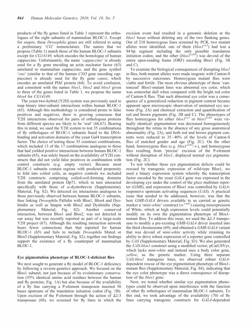
Table 1. Homologues of human BLOC-1 subunits encoded by the fruit flygenome
Humansubunit
Drosophila gene E-valuea Amino acididentityb (%)Current
nameProposedname
BLOS1 CG30077 blos1 6 � 10231 (1) 55BLOS2 CG14145 blos2 7 � 10216 (1) 40BLOS3 CG34255 blos3 9 � 1026 (2) 22Cappuccino CG14149 blos4 2 � 1024 (1) 25Dysbindin CG6856 dysbindin 6 � 10215 (1) 38Muted CG34131 muted 2 � 1027 (2) 18Pallidin CG14133 pallidin 1 � 1023 (1) 24Snapin snapin snapin 3 � 10213 (1) 33
aHomology searches were carried out using the PSI-BLAST algorithm and theamino acid sequences of human BLOC-1 subunits as ‘queries.’ Numbersbetween parentheses indicate iteration number; the first iteration ofPSI-BLAST is equivalent to a standard BLASTP search.bCalculated from the alignments shown in Fig. 1A and SupplementaryMaterial, Fig. S1.
Figure 1. Sequence analysis and mutant alleles of the fly orthologue of humanBLOS1. (A) Alignment of the primary sequences of BLOS1 from Homosapiens (Hs) and the product of the blos1 gene from Drosophila melanogaster(Dm). Identical amino acid residues are highlighted. (B) Schematic represen-tation of the organization of the blos1 gene, indicating the two exonic regionsthat compose the ORF, the insertion site of a P-element in the fly lineEY06269 and the segments deleted (dotted lines) in the blos1ex2 andblos1ex65 mutant alleles. (C) Analysis by agarose gel electrophoresis of PCRproducts obtained by amplification of genomic DNA from the indicated flylines, using primers derived from sequences about 206 bp upstream and1156 bp downstream of the P-element insertion site. Molecular size markerswere run on both sides of the gel. Notice the reduced sizes of the PCR productsobtained from homozygous blos1ex2 and blos1ex65 flies. Due to the large sizeof the inserted P-element (�10 kb), no PCR product was obtained under theseconditions for the EY06269 line.
Human Molecular Genetics, 2010, Vol. 19, No. 5 863
products of the fly genes listed in Table 1 represent the ortho-logues of the eight subunits of mammalian BLOC-1. Exceptfor snapin, these Drosophila genes are still referred to usinga preliminary ‘CG’ nomenclature. The names that wepropose (Table 1) match those of the human BLOC-1 subunitsexcept for CG14149, which encodes the homologue of humancappuccino. Unfortunately, the name ‘cappuccino’ is alreadyused for a fly gene encoding an actin nucleator factor (63)unrelated to mammalian cappuccino, and the gene symbol‘cno’ (similar to that of the human CNO gene encoding cap-puccino) is already used for the fly gene canoe, whichencodes an unrelated PDZ protein (64). To avoid confusion,and consistent with the names blos1, blos2 and blos3 givento three of the genes listed in Table 1, we propose the nameblos4 for CG14149.
The yeast-two-hybrid (Y2H) system was previously used tomap binary inter-subunit interactions within human BLOC-1(65). Although this methodology is considered prone to falsepositives and negatives, there is growing consensus thatY2H interactions observed for pairs of orthologous proteinsfrom different species are likely to be ‘real’ (66,67). Bearingthis in mind, we used the Y2H system to test 35 combinationsof fly orthologues of BLOC-1 subunits fused to the DNA-binding and activation domains of the yeast Gal4 transcriptionfactor. The choice of testing these 35 construct combinations,which included 13 of the 17 combinations analogous to thosethat had yielded positive interactions between human BLOC-1subunits (65), was solely based on the availability of Y2H con-structs that did not yield false positives in combination withcontrol constructs (e.g. empty vector). Because mostBLOC-1 subunits contain regions with predicted propensityto fold into coiled coils, as negative controls we includedY2H constructs comprising coiled-coil-forming domainsfrom the unrelated protein Dp71, which in turn interactedspecifically with those of a-dystrobrevin (SupplementaryMaterial, Fig. S2). We detected six interactions analogous tothose previously observed between human BLOC-1 subunits,thus linking Drosophila Pallidin with Blos1, Blos4 and Dys-bindin as well as Snapin with Blos2 and Dysbindin (Sup-plementary Material, Fig. S2). Another conservedinteraction, between Blos1 and Blos2, was not detected inour assay but was recently reported as part of a large-scaleY2H project (67). Although the resulting interaction networkbears fewer connections than that reported for humanBLOC-1 (65) and fails to include Drosophila Muted orBlos3 (Supplementary Material, Fig. S2), together our findingssupport the existence of a fly counterpart of mammalianBLOC-1.
Eye pigmentation phenotype of BLOC-1-deficient flies
We next sought to generate a fly model of BLOC-1 deficiencyby following a reverse-genetics approach. We focused on theBlos1 subunit, not just because of its evolutionary conserva-tion (55% identical amino acid residues between the humanand fly proteins; Fig. 1A) but also because of the availabilityof a fly line carrying a P-element transposon inserted 56bases upstream of the translation initiation codon (Fig. 1B).Upon excision of the P-element through the action of D2-3transposase (68), we screened for fly lines in which the
excision event had resulted in a genomic deletion at theblos1 locus without deleting any of the two flanking genes.Out of 210 homozygous lines screened by PCR, two mutantalleles were identified: one of them (blos1ex2) had lost a94 bp segment including the only possible translationinitiation codon, and the other (blos1ex65) was devoid of theentire open-reading frame (ORF) encoding Blos1 (Fig. 1Band C).
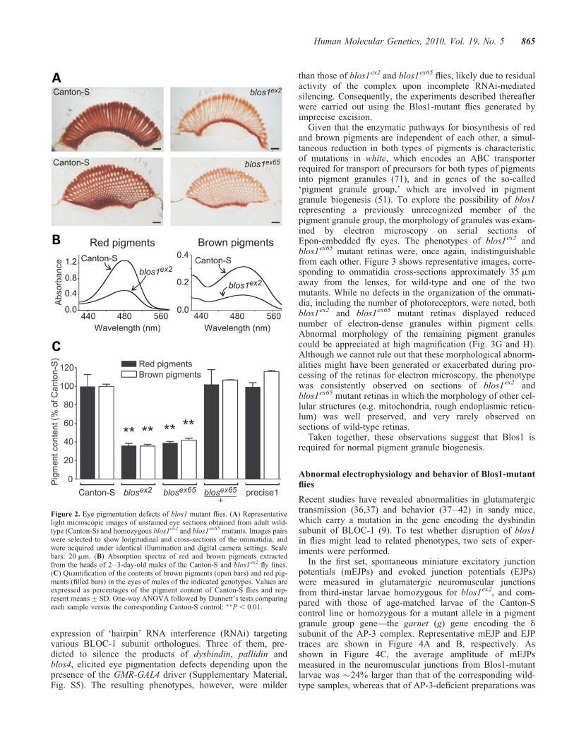
To examine the biological consequences of disrupting blos1in flies, both mutant alleles were made isogenic with Canton-Sby successive outcrosses. Homozygous mutant flies wereviable and fertile. The most obvious phenotype of these ‘can-tonized’ Blos1-mutant lines was abnormal eye color, whichwas somewhat dull when compared with the bright red colorof Canton-S flies. That such abnormal eye color was a conse-quence of a generalized reduction in pigment content becameapparent upon microscopic observation of unstained eye sec-tions (Fig. 2A) and chemical extraction and quantification ofred and brown pigments (Fig. 2B and C). The phenotypes offlies homozygous for either blos1ex2 or blos1ex65 were vir-tually identical: pigmentation was decreased homogeneouslythroughout the retina in the absence of any gross anatomicalabnormality (Fig. 2A), and both red and brown pigment con-tents were reduced to 35–40% of the levels of Canton-Sflies of matched gender and age (Fig. 2C). On the otherhand, heterozygous flies (e.g. blos1ex65/þ), and homozygousflies resulting from ‘precise’ excision of the P-elementwithout disruption of blos1, displayed normal eye pigmenta-tion (Fig. 2C).
To test whether these eye pigmentation defects could berescued by transgenic expression of wild-type Blos1, weused a binary expression system whereby the transcriptionfactor encoded by the yeast GAL4 gene was expressed in thedeveloping eye under the control of the glass multimer repor-ter (GMR), and expression of Blos1 was controlled by GAL4-responsive upstream activating sequences (UAS). A practicalissue that needed to be addressed, however, was that thebest GMR-GAL4 drivers available to us carried as geneticmarker a ‘mini-white’ construct (wþmC) causing misexpressionof White, which at high expression levels could potentiallymodify on its own the pigmentation phenotype of Blos1-mutant flies. To address this issue, we used the D2-3 transpo-sase to modify a pre-existing GMR-GAL4 driver inserted intothe third chromosome (69), and obtained a GMR-GAL4 variantthat was devoid of mini-white activity while retaining itsability to drive robust expression of a reporter gene controlledby UAS (Supplementary Material, Fig. S3). We also generatedthe UAS-blos1 construct using a modified vector, pCarUSVyr,which lacks mini-white and instead uses a body color gene,yellow, as the genetic marker. Using three separateUAS-blos1 transgenic lines, we observed robust GAL4-dependent rescue of the eye pigmentation phenotype of Blos1-mutant flies (Supplementary Material, Fig. S4), indicating thatthe eye color phenotype was a direct consequence of disrup-tion of the blos1 gene.
Next, we tested whether similar eye pigmentation pheno-types could be observed upon interference with the functionof other fly orthologues of mammalian BLOC-1 subunits. Tothis end, we took advantage of the availability (70) of flylines carrying transgenic constructs for GAL4-dependent
864 Human Molecular Genetics, 2010, Vol. 19, No. 5
expression of ‘hairpin’ RNA interference (RNAi) targetingvarious BLOC-1 subunit orthologues. Three of them, pre-dicted to silence the products of dysbindin, pallidin andblos4, elicited eye pigmentation defects depending upon thepresence of the GMR-GAL4 driver (Supplementary Material,Fig. S5). The resulting phenotypes, however, were milder
than those of blos1ex2 and blos1ex65 flies, likely due to residualactivity of the complex upon incomplete RNAi-mediatedsilencing. Consequently, the experiments described thereafterwere carried out using the Blos1-mutant flies generated byimprecise excision.
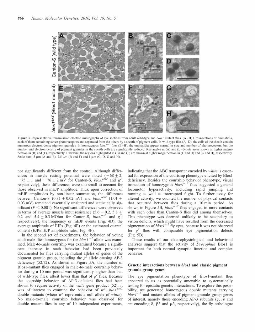
Given that the enzymatic pathways for biosynthesis of redand brown pigments are independent of each other, a simul-taneous reduction in both types of pigments is characteristicof mutations in white, which encodes an ABC transporterrequired for transport of precursors for both types of pigmentsinto pigment granules (71), and in genes of the so-called‘pigment granule group,’ which are involved in pigmentgranule biogenesis (51). To explore the possibility of blos1representing a previously unrecognized member of thepigment granule group, the morphology of granules was exam-ined by electron microscopy on serial sections ofEpon-embedded fly eyes. The phenotypes of blos1ex2 andblos1ex65 mutant retinas were, once again, indistinguishablefrom each other. Figure 3 shows representative images, corre-sponding to ommatidia cross-sections approximately 35 mmaway from the lenses, for wild-type and one of the twomutants. While no defects in the organization of the ommati-dia, including the number of photoreceptors, were noted, bothblos1ex2 and blos1ex65 mutant retinas displayed reducednumber of electron-dense granules within pigment cells.Abnormal morphology of the remaining pigment granulescould be appreciated at high magnification (Fig. 3G and H).Although we cannot rule out that these morphological abnorm-alities might have been generated or exacerbated during pro-cessing of the retinas for electron microscopy, the phenotypewas consistently observed on sections of blos1ex2 andblos1ex65 mutant retinas in which the morphology of other cel-lular structures (e.g. mitochondria, rough endoplasmic reticu-lum) was well preserved, and very rarely observed onsections of wild-type retinas.
Taken together, these observations suggest that Blos1 isrequired for normal pigment granule biogenesis.
Abnormal electrophysiology and behavior of Blos1-mutantflies
Recent studies have revealed abnormalities in glutamatergictransmission (36,37) and behavior (37–42) in sandy mice,which carry a mutation in the gene encoding the dysbindinsubunit of BLOC-1 (9). To test whether disruption of blos1in flies might lead to related phenotypes, two sets of exper-iments were performed.
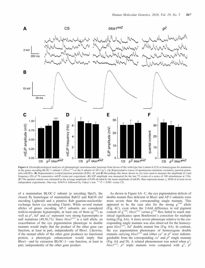
In the first set, spontaneous miniature excitatory junctionpotentials (mEJPs) and evoked junction potentials (EJPs)were measured in glutamatergic neuromuscular junctionsfrom third-instar larvae homozygous for blos1ex2, and com-pared with those of age-matched larvae of the Canton-Scontrol line or homozygous for a mutant allele in a pigmentgranule group gene—the garnet (g) gene encoding the dsubunit of the AP-3 complex. Representative mEJP and EJPtraces are shown in Figure 4A and B, respectively. Asshown in Figure 4C, the average amplitude of mEJPsmeasured in the neuromuscular junctions from Blos1-mutantlarvae was �24% larger than that of the corresponding wild-type samples, whereas that of AP-3-deficient preparations was
Figure 2. Eye pigmentation defects of blos1 mutant flies. (A) Representativelight microscopic images of unstained eye sections obtained from adult wild-type (Canton-S) and homozygous blos1ex2 and blos1ex65 mutants. Images pairswere selected to show longitudinal and cross-sections of the ommatidia, andwere acquired under identical illumination and digital camera settings. Scalebars: 20 mm. (B) Absorption spectra of red and brown pigments extractedfrom the heads of 2–3-day-old males of the Canton-S and blos1ex2 fly lines.(C) Quantification of the contents of brown pigments (open bars) and red pig-ments (filled bars) in the eyes of males of the indicated genotypes. Values areexpressed as percentages of the pigment content of Canton-S flies and rep-resent means+SD. One-way ANOVA followed by Dunnett’s tests comparingeach sample versus the corresponding Canton-S control: ��P , 0.01.
Human Molecular Genetics, 2010, Vol. 19, No. 5 865
not significantly different from the control. Although differ-ences in muscle resting potential were noted (268+ 2,275+ 1 and 276+ 2 mV for Canton-S, blos1ex2 and g2,respectively), these differences were too small to account forthose observed in mEJP amplitude. Thus, upon correction ofmEJP amplitudes by non-linear summation, the differencebetween Canton-S (0.81+ 0.02 mV) and blos1ex2 (1.01+0.03 mV) remained essentially unaltered and statistically sig-nificant (P , 0.001). No significant differences were observedin terms of average muscle input resistance (5.6+ 0.2, 5.8+0.2 and 5.4+ 0.3 MOhm for Canton-S, blos1ex2 and g2,respectively), the frequency or mEJP events (Fig. 4D), theaverage amplitude of EJPs (Fig. 4E) or the estimated quantalcontent (EJP/mEJP amplitude ratio; Fig. 4F).
In the second set of experiments, the behavior of youngadult male flies homozygous for the blos1ex2 allele was exam-ined. Male-to-male courtship was examined because a signifi-cant increase in such behavior had been previouslydocumented for flies carrying mutant alleles of genes of thepigment granule group, including the g2 allele causing AP-3deficiency (52,72). As shown in Figure 5A, the number ofBlos1-mutant flies engaged in male-to-male courtship behav-ior during a 10 min period was significantly higher than thatof wild-type files, albeit lower than that of g2 flies. Becausethe courtship behavior of AP-3-deficient flies had beenshown to require activity of the white gene product (52), itwas of interest to examine the behavior of w�; blos1ex2
double mutants (where w� represents a null allele of white).No male-to-male courtship behavior was observed fordouble mutant flies in any of 10 independent experiments,
indicating that the ABC transporter encoded by white is essen-tial for expression of the courtship phenotype elicited by Blos1deficiency. Besides the courtship behavior phenotype, visualinspection of homozygous blos1ex2 flies suggested a generallocomotor hyperactivity, including rapid jumping andrunning as well as interrupted flight. To further assay foraltered activity, we counted the number of physical contactsthat occurred between flies during a 10 min period. Asshown in Figure 5B, blos1ex2 flies engaged in more contactswith each other than Canton-S flies did among themselves.This phenotype was deemed unlikely to be secondary tovision deficits, which might have resulted from the decreasedpigmentation of blos1ex2 fly eyes, because it was not observedfor g2 flies with comparable eye pigmentation defects(Fig. 5B).
These results of our electrophysiological and behavioralanalyses suggest that the activity of Drosophila Blos1 isimportant for both normal synaptic function and complexbehavior.
Genetic interactions between blos1 and classic pigmentgranule group genes
The eye pigmentation phenotype of Blos1-mutant fliesappeared to us as potentially amenable to systematicallytesting for epistatic genetic interactions. To explore this possi-bility, we generated homozygous double mutants carryingblos1ex2 and mutant alleles of pigment granule group genesof interest, namely those encoding AP-3 subunits (g, rb andcm encoding d, b3 and m3, respectively), the fly orthologue
Figure 3. Representative transmission electron micrographs of eye sections from adult wild-type and blos1 mutant flies. (A–H) Cross-sections of ommatidia,each of them containing seven photoreceptors and separated from the others by a sheath of pigment cells. In wild-type flies (A–D), the cells of the sheath containnumerous electron-dense pigment granules. In homozygous blos1ex2 flies (E–H), the ommatidia appear normal in size and number of photoreceptors, but thenumber and electron density of pigment granules in the sheath cells are significantly reduced. Rectangles in (A) and (E) denote areas shown at higher magni-fication in (B) and (F), respectively. Likewise, the regions highlighted in (B) and (F) are shown at higher magnification in (C and D) and (G and H), respectively.Scale bars: 5 mm (A and E), 2.5 mm (B and F) and 1 mm (C, D, G and H).
866 Human Molecular Genetics, 2010, Vol. 19, No. 5
of a mammalian BLOC-2 subunit (p encoding Hps5), theclosest fly homologue of mammalian Rab32 and Rab38 (ltdencoding Lightoid) and a putative Rab guanine-nucleotide-exchange factor (ca encoding Claret). While several mutantalleles of genes encoding AP-3 subunits are consideredmild-to-moderate hypomorphs, at least one of them (g53d) aswell as pp, ltd1 and ca1 represent very strong hypomorphs ornull mutations (49,56,73). Since blos1ex2 is a null allele, anexacerbation of the eye pigmentation phenotype in doublemutants would imply that the product of the other gene canfunction, at least in part, independently of Blos1. Likewise,if the mutant allele of the other gene produces no functionalprotein, a phenotypic enhancement would imply thatBlos1—and by extension BLOC-1—can function, at least inpart, independently of the other gene product.
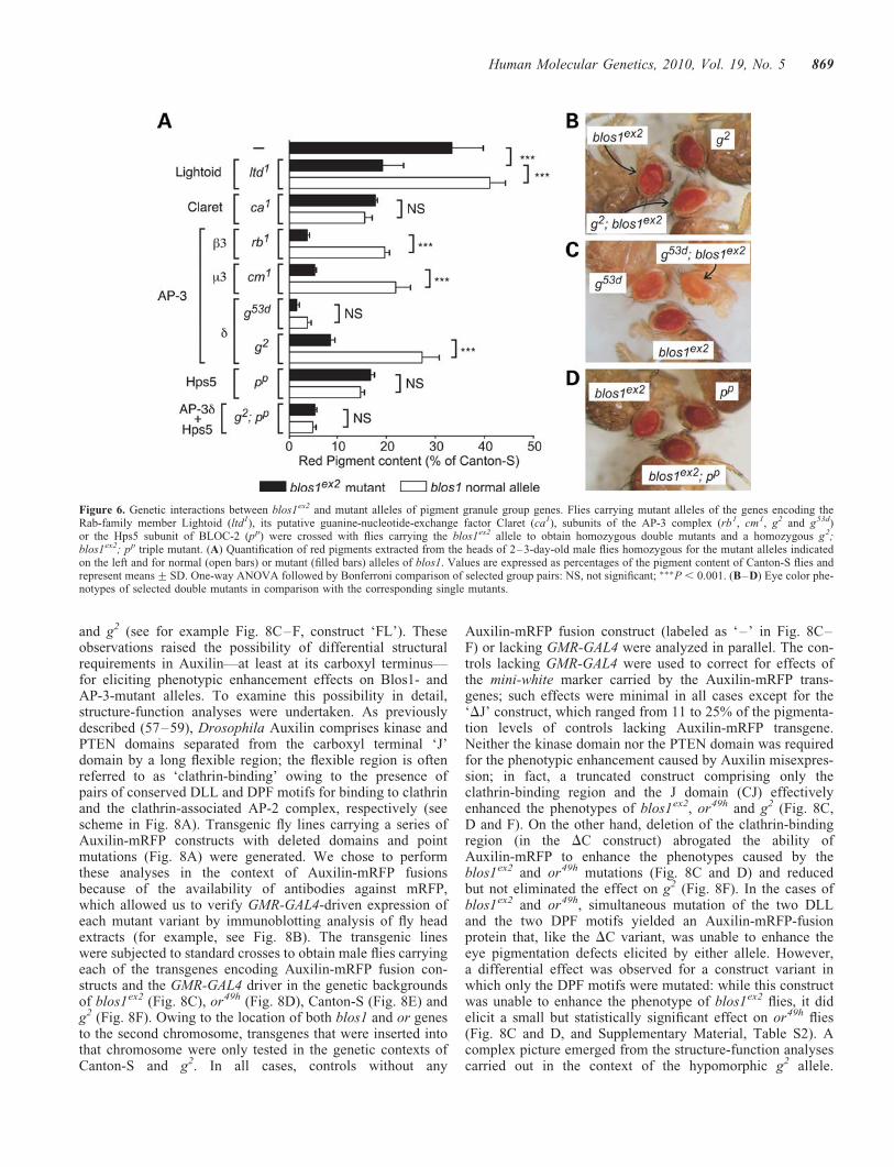
As shown in Figure 6A–C, the eye pigmentation defects ofdouble mutant flies deficient in Blos1 and AP-3 subunits weremore severe than the corresponding single mutants. Thisappeared to be the case also for the strong g53d allele(Fig. 6C), even when the 2-fold difference in red pigmentcontent of g53d; blos1ex2 versus g53d flies failed to reach stat-istical significance upon Bonferroni’s correction for multipletesting (Fig. 6A). A more severe phenotype relative to the cor-responding single mutants was also observed for the homozy-gous blos1ex2, ltd1 double mutant line (Fig. 6A). In contrast,the eye pigmentation phenotypes of homozygous doublemutants carrying blos1ex2 and either ca1 or pp were indistin-guishable from the corresponding ca1 and pp single mutants(Fig. 6A and D). A related phenomenon was noted when g2;blos1ex2; pp triple mutants were compared with g2; pp
Figure 4. Electrophysiological analyses of glutamatergic neuromuscular junctions from larvae of the wild-type line Canton-S (CS) or homozygous for mutationsin the genes encoding BLOC-1 subunit 1 (blos1ex2) or the d subunit of AP-3 (g2). (A) Representative traces of spontaneous miniature excitatory junction poten-tials (mEJPs). (B) Representative evoked junction potentials (EJPs). (C and D) Recordings like those shown in (A) were used to measure the amplitude (C) andfrequency (D) of 70 consecutive mEJP events per experiment. (E) EJP amplitude was measured for the last 75 events of a series of 100 stimulations at 2 Hz.(F) The quantal content was estimated as the average amplitude of EJPs divided by the mean amplitude of mEJPs. Bars represent means+SEM of at least sevenindependent experiments. One-way ANOVA followed by Tukey’s test: ���P , 0.001 versus CS.
Human Molecular Genetics, 2010, Vol. 19, No. 5 867
double mutants: although blos1ex2 was able to exacerbate thepigmentation phenotype elicited by the g2 allele alone, itfailed to modify that of g2; pp double mutants (Fig. 6A).These observations suggest that Blos1 is able to function, atleast in part, independently of AP-3 and Lightoid, and thatits role in pigment granule biogenesis requires the activitiesof Claret and Hps5.
Misexpression of Auxilin enhances the eye pigmentationphenotype of Blos1-mutant flies
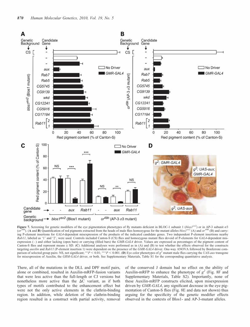
Encouraged by the results described in the previous section,we embarked upon the search for novel modifiers of Blos1function. A GAL4-dependent misexpression strategy wasappealing given our success in using a modified GMR-GAL4driver to rescue the pigmentation phenotype of Blos1-mutantflies (Supplementary Material, Fig. S4) and the availabilityof a wide variety of fly lines carrying P-element transposonsengineered for misexpression of either cloned ORFs or theproducts of endogenous genes located in the vicinity of theinsertion sites (74). One practical limitation, however, wasthe need to perform two successive fly crosses per candidate,owing to the behavior of blos1ex2 as a recessive allele. Hence,we chose to restrict our initial screening to fly lines predictedto misexpress candidate gene products implicated in proteintrafficking within the endosomal–lysosomal system—the pro-posed site of action of mammalian BLOC-1 (17,18,20,23,29).Figure 7A shows quantitative results obtained in a secondaryanalysis of a subset of these candidates, which includedseveral Rab GTPases (Rab5, Rab7 and Rab11), a putative
Rab guanine-nucleotide-exchange factor homologous tomammalian Rabex-5 (CG9139), four putative Rab GTPase-activator proteins (wkd, CG5745, CG5916 and CG12241), aputative effector of Rho and Arf GTPases (CG17184), and aclathrin-disassembly factor (aux). The same candidates werealso tested in parallel for their ability to modify the eyepigmentation phenotype of the or49h allele of the AP-3s3-encoding gene, which like blos1 resides on the flysecond chromosome (Fig. 7B). A striking phenotypic enhance-ment effect on both blos1ex2 and or49h was observed upon mis-expression of Drosophila Auxilin (aux gene product). Incontrast, misexpression of the other candidate genes elicitedpartial amelioration of eye pigmentation defects or no effectat all (Fig. 7A and B). Additional control experiments verifiedthat the phenotypic enhancement effect of aux was strictlydependent upon the presence of the GMR-GAL4 driver; GAL4-dependence was also verified for the partial suppression effectof Rab11 on the phenotype of blos1ex2 flies but not for that ofthe same candidate on the phenotype of or49h flies (Fig. 7C).
The phenotypic enhancement effect of Auxilin was unex-pected, and hence it was further investigated. Using a trans-genic UAS-aux construct for expression of the full-lengthprotein without any epitope tags, strong GAL4-dependenteffects were observed not only for blos1ex2 and or49h butalso for mutant alleles of genes encoding other AP-3 subunits(Fig. 7D and Supplementary Material, Table S1). In compari-son, the GAL4-dependent effect of the UAS-aux transgene oneye pigmentation was less pronounced in the context of theltd1 mutation and significantly milder in the genetic back-ground of Canton-S flies (Supplementary Material,Table S1). We considered the possibility of the effects ofAuxilin misexpression resulting from interference with theactivity of the endogenous Auxilin protein, for instance, bycompetition with binding partners (clathrin, Hsc70) that mayneed to interact in a concerted fashion. While null mutationsin aux had been previously shown to result in early lethality,various hypomorphic alleles had been characterized andshown to elicit abnormal eye morphology, e.g. rough eye, inpart due to the role of Auxilin in endocytic events that arecritical for Delta/Notch signaling (57–59). In contrast, weobserved neither rough eye phenotype nor any other obviousalteration in eye morphology upon GMR-GAL4-driven misex-pression of Auxilin in Canton-S or mutant flies. Conversely,when three hypomorphic alleles, auxG257E, auxL78H andauxI670K (57,58), were transferred by standard crosses intothe genetic backgrounds of Canton-S and g2 (the latter beinga background in which the enhancement effect of misex-pressed Auxilin was most severe; Fig. 7D and SupplementaryMaterial, Table S1), for each of them we observed rough eyephenotype but no significant effect on eye pigmentation (datanot shown). Hence, the phenotypic enhancement effect causedby Auxilin misexpression was deemed unlikely to arise from adominant-negative mechanism.
Compared with the effects of misexpressing untaggedAuxilin (Fig. 7 and Supplementary Materials, Table S1),those elicited by full-length Auxilin fused at its carboxyl ter-minus to monomeric red fluorescence protein (mRFP) wereless dramatic in the context of the blos1ex2 mutation andminimal in Canton-S flies; however, they were essentiallyunaltered in the context of the AP-3 subunit mutations or49h
Figure 5. Abnormal behavior of blos1 mutant flies. (A and B) Adult male flies(5+2 days after eclosion) of the Canton-S (CS) control line, or carryingmutant alleles that render them deficient in BLOC-1 subunit 1 (blos1ex2),the AP-3 complex subunit d (g2) or the ABC transporter White (w�), wereplaced in groups of 25 in plastic Petri dishes, under conditions of high humid-ity and controlled temperature, and visually monitored for a 10 min period. (A)The male-to-male courtship index was calculated as the number of males dis-playing courting behaviors in ‘courtship chains,’ not counting the first male ineach chain. (B) Physical contacts were assessed by counting the number oftimes that a male fly touched another during the 10 min period, regardlessof the type of behavior displayed. See Materials and Methods for furtherexperimental details. Bars represent means+SEM of 10 independent exper-iments. One-way ANOVA followed by Bonferroni comparison of selectedgroup pairs: NS, not significant; �P , 0.05; ��P , 0.01.
868 Human Molecular Genetics, 2010, Vol. 19, No. 5
and g2 (see for example Fig. 8C–F, construct ‘FL’). Theseobservations raised the possibility of differential structuralrequirements in Auxilin—at least at its carboxyl terminus—for eliciting phenotypic enhancement effects on Blos1- andAP-3-mutant alleles. To examine this possibility in detail,structure-function analyses were undertaken. As previouslydescribed (57–59), Drosophila Auxilin comprises kinase andPTEN domains separated from the carboxyl terminal ‘J’domain by a long flexible region; the flexible region is oftenreferred to as ‘clathrin-binding’ owing to the presence ofpairs of conserved DLL and DPF motifs for binding to clathrinand the clathrin-associated AP-2 complex, respectively (seescheme in Fig. 8A). Transgenic fly lines carrying a series ofAuxilin-mRFP constructs with deleted domains and pointmutations (Fig. 8A) were generated. We chose to performthese analyses in the context of Auxilin-mRFP fusionsbecause of the availability of antibodies against mRFP,which allowed us to verify GMR-GAL4-driven expression ofeach mutant variant by immunoblotting analysis of fly headextracts (for example, see Fig. 8B). The transgenic lineswere subjected to standard crosses to obtain male flies carryingeach of the transgenes encoding Auxilin-mRFP fusion con-structs and the GMR-GAL4 driver in the genetic backgroundsof blos1ex2 (Fig. 8C), or49h (Fig. 8D), Canton-S (Fig. 8E) andg2 (Fig. 8F). Owing to the location of both blos1 and or genesto the second chromosome, transgenes that were inserted intothat chromosome were only tested in the genetic contexts ofCanton-S and g2. In all cases, controls without any
Auxilin-mRFP fusion construct (labeled as ‘–’ in Fig. 8C–F) or lacking GMR-GAL4 were analyzed in parallel. The con-trols lacking GMR-GAL4 were used to correct for effects ofthe mini-white marker carried by the Auxilin-mRFP trans-genes; such effects were minimal in all cases except for the‘DJ’ construct, which ranged from 11 to 25% of the pigmenta-tion levels of controls lacking Auxilin-mRFP transgene.Neither the kinase domain nor the PTEN domain was requiredfor the phenotypic enhancement caused by Auxilin misexpres-sion; in fact, a truncated construct comprising only theclathrin-binding region and the J domain (CJ) effectivelyenhanced the phenotypes of blos1ex2, or49h and g2 (Fig. 8C,D and F). On the other hand, deletion of the clathrin-bindingregion (in the DC construct) abrogated the ability ofAuxilin-mRFP to enhance the phenotypes caused by theblos1ex2 and or49h mutations (Fig. 8C and D) and reducedbut not eliminated the effect on g2 (Fig. 8F). In the cases ofblos1ex2 and or49h, simultaneous mutation of the two DLLand the two DPF motifs yielded an Auxilin-mRFP-fusionprotein that, like the DC variant, was unable to enhance theeye pigmentation defects elicited by either allele. However,a differential effect was observed for a construct variant inwhich only the DPF motifs were mutated: while this constructwas unable to enhance the phenotype of blos1ex2 flies, it didelicit a small but statistically significant effect on or49h flies(Fig. 8C and D, and Supplementary Material, Table S2). Acomplex picture emerged from the structure-function analysescarried out in the context of the hypomorphic g2 allele.
Figure 6. Genetic interactions between blos1es2 and mutant alleles of pigment granule group genes. Flies carrying mutant alleles of the genes encoding theRab-family member Lightoid (ltd1), its putative guanine-nucleotide-exchange factor Claret (ca1), subunits of the AP-3 complex (rb1, cm1, g2 and g53d)or the Hps5 subunit of BLOC-2 (pp) were crossed with flies carrying the blos1es2 allele to obtain homozygous double mutants and a homozygous g2;blos1ex2; pp triple mutant. (A) Quantification of red pigments extracted from the heads of 2–3-day-old male flies homozygous for the mutant alleles indicatedon the left and for normal (open bars) or mutant (filled bars) alleles of blos1. Values are expressed as percentages of the pigment content of Canton-S flies andrepresent means+SD. One-way ANOVA followed by Bonferroni comparison of selected group pairs: NS, not significant; ���P , 0.001. (B–D) Eye color phe-notypes of selected double mutants in comparison with the corresponding single mutants.
Human Molecular Genetics, 2010, Vol. 19, No. 5 869
There, all of the mutations in the DLL and DPF motif pairs,alone or combined, resulted in Auxilin-mRFP-fusion variantsthat were less active than the full-length or CJ versions butnonetheless more active than the DC variant, as if bothtypes of motifs contributed to the enhancement effect butwere not the only active elements in the clathrin-bindingregion. In addition, while deletion of the clathrin-bindingregion resulted in a construct with partial activity, removal
of the conserved J domain had no effect on the ability ofAuxilin-mRFP to enhance the phenotype of g2 (Fig. 8F andSupplementary Materials, Table S2). Importantly, none ofthese Auxilin-mRFP constructs elicited, upon misexpressiondriven by GMR-GAL4, any significant decrease in the eye pig-mentation of Canton-S flies (Fig. 8E and data not shown) thusarguing for the specificity of the genetic modifier effectsobserved in the contexts of Blos1- and AP-3-mutant alleles.
Figure 7. Screening for genetic modifiers of the eye pigmentation phenotypes of fly mutants deficient in BLOC-1 subunit 1 (blos1ex2) or in AP-3 subunit s3(or49h). (A and B) Quantification of red pigments extracted from the heads of male flies homozygous for the mutant alleles blos1ex2 (A) and or49h (B) and carry-ing P-element insertions for GAL4-dependent misexpression of the products of the indicated candidate genes. Two independent P-element insertions nearbyRab11, labeled as ‘1’ and ‘2’, were used. Controls included Canton-S (CS) flies and homozygous mutant flies devoid of P-elements for GAL4-dependent mis-expression (–) and either lacking (open bars) or carrying (filled bars) the GMR-GAL4 driver. Values are expressed as percentages of the pigment content ofCanton-S flies and represent means+SD. (C) Additional analyses were performed as in (A) and (B) to test whether the effects observed for the constructstargeting auxilin and Rab11 (P-element insertion 1) were dependent on the presence of the GMR-GAL4 driver. One-way ANOVA followed by Bonferroni com-parison of selected group pairs: NS, not significant; ��P , 0.01; ���P , 0.001. (D) Eye color phenotypes of g2 mutant male flies carrying the UAS-aux transgenefor misexpression of Auxilin, the GRM-GAL4 driver, or both. See Supplementary Materials, Table S1 for the corresponding quantitative analysis.
870 Human Molecular Genetics, 2010, Vol. 19, No. 5
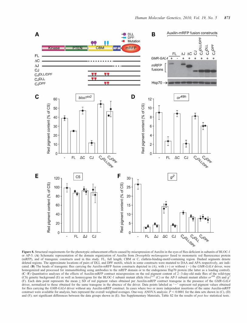
Figure 8. Structural requirements for the phenotypic enhancement effects caused by misexpression of Auxilin in the eyes of flies deficient in subunits of BLOC-1or AP-3. (A) Schematic representation of the domain organization of Auxilin from Drosophila melanogaster fused to monomeric red fluorescence protein(mRFP), and of transgenic constructs used in this study. FL, full length; CBM or C, clathrin-binding-motif-containing region. Dashed segments denotedeleted regions. The approximate locations of pairs of DLL and DPF motifs, which in some constructs were mutated to DAA and APA respectively, are indi-cated. (B) The heads of transgenic flies carrying the Auxilin-mRFP fusion constructs depicted in (A), with (þ) or without (2) the GMR-GAL4 driver, werehomogenized and processed for immunoblotting using antibodies to the mRFP domain or to the endogenous Hsp70 protein (the latter as a loading control).(C–F) Quantitative analyses of the effects of Auxilin-mRFP contruct misexpression on the red pigment content of 2–3-day-old male flies of the wild-type(CS) genetic background (E) as well as homozygous for the BLOC-1 subunit mutant allele blos1ex2 (C) or the AP-3 subunit mutant alleles or49h (D) and g2
(F). Each data point represents the mean+SD of red pigment values obtained per Auxilin-mRFP contruct transgene in the presence of the GMR-GAL4driver, normalized to those obtained for the same transgene in the absence of the driver. Data points labeled as ‘2’ represent red pigment values obtainedfor flies carrying the GMR-GAL4 driver without any Auxilin-mRFP construct. In cases where two or more independent insertions of the same Auxilin-mRFPconstruct were available for analysis, bars represent the overall weighted averages. One-way ANOVA analysis: P , 0.0001 for the data sets shown in (C), (D)and (F); not significant differences between the data groups shown in (E). See Supplementary Materials, Table S2 for the results of post hoc statistical tests.
Human Molecular Genetics, 2010, Vol. 19, No. 5 871
DISCUSSION
As stated in the Introduction section, recessive mutations intwo of the eight genes encoding BLOC-1 subunits areknown to cause HPS-7 and -8 disease in humans. However,only one HPS-7 patient (9) and six HPS-8 patients from asingle family (10) have been described in the literature. Con-sequently, most of what we know so far about the function ofBLOC-1 has resulted from the study of mutant mouse linesand derived cell cultures (6,7). In this work, we describe thefirst non-mammalian model of BLOC-1 deficiency. Flieshomozygous for null mutations in blos1, the best conservedof the genes encoding BLOC-1 subunits, displayed phenotypesthat relate to those documented for BLOC-1-deficient mice,namely: reduced pigmentation caused by impaired biogenesisof LROs, altered glutamatergic transmission and abnormal be-havior. Furthermore, we describe the use of Blos1-mutant fliesto uncover genetic modifiers of BLOC-1 function.
Among the several mouse models of HPS, those expressinglittle or no BLOC-1 activity display the most severe coat pig-mentation defects (5,21). These phenotypes, consistent withultrastructural analyses showing accumulation of pre-melanosomes with little or no melanin (75,76), have led tothe notion that BLOC-1 plays a key role in protein traffickingto melanosomes (and presumably other LROs), with this rolebeing relatively more important than those of otherHPS-associated complexes such as AP-3 and BLOC-2(5,21,76). On the other hand, an alternative view hasstemmed from the findings that a significant pool of tyrosi-nase, a major determinant of pigmentation in mammals,reaches melanosomes despite the lack of BLOC-1 function(18), and that poor melanogenesis in BLOC-1-deficient cellscan be explained instead by specific missorting of a coppertransporter required for tyrosinase activation (20). Becausepigment synthesis in the fly eye is independent of tyrosinaseactivity (and, to our knowledge, of copper transport), ourresults offer quite another perspective on how criticalBLOC-1 function may be for LRO biogenesis. We foundthat Blos1-mutant fly eyes display a significant decrease incontent of both red and brown pigments, which togetherwith the observed defects in number and morphology ofpigment granules imply that the function of BLOC-1 in fliesis, like in mammals, required for normal LRO biogenesis.However, the eye color phenotype of these null mutant flieswas clearly less severe than those of flies deficient in AP-3(carrying strong alleles such as g53d and or49h) or BLOC-2(null pp allele), thus arguing against the notion that BLOC-1would be more important than these two complexes for LRObiogenesis—at least in the fly eye. Similarly to what hadbeen reported for mice (17,21), the color phenotype ofdouble mutant flies simultaneously deficient in BLOC-1 andAP-3 was more severe than those of the correspondingsingle mutants, further supporting the idea that these two com-plexes can function, at least in part, independently of eachother. It should be noted, however, that these results do notexclude the possibility of BLOC-1 and AP-3 acting togetherunder some physiological conditions, nor they imply thattheir independent functions must occur in distinct organellesor trafficking routes. With regards to the functional relation-ship between BLOC-1 and -2, contrasting views have arisen
from interpretation of the phenotypes of double mutantmice. One view, based on the semi-dominant exacerbationof the phenotype of homozygous Bloc1s3rp mice by a nullallele of the gene encoding a subunit of BLOC-2, is thatBLOC-1 and -2 can function independently of each other(77). A caveat of this interpretation, however, is thatBloc1s3rp mice express residual BLOC-1 activity due to theexistence of a small pool of assembled complex (65). On theother hand, the phenotypes of double mutant mice carryingthe Pldnpa mutation (thought to express no BLOC-1 activity)and null mutations in either of two BLOC-2 subunit geneswere virtually identical to that of homozygous Pldnpa mice(17,21). These observations led to the alternative interpretationthat BLOC-1 and -2 function together in the same pathway,although the severe pigmentation phenotype of Pldnpa miceand the relatively milder phenotype of BLOC-2-deficientmice represent a potential confounding factor. Again, our ana-lyses using flies offer a different perspective, among otherthings because none of the eye color phenotypes of singlemutant flies deficient in BLOC-1 or -2 is severe enough toraise a similar concern. While our results are consistent withthe idea that BLOC-1 and -2 function in a commonpathway, surprisingly the observed epistatic effect is thereverse of that observed in mice. Here, the pigmentation phe-notype of double mutants flies carrying null alleles of blos1(affecting BLOC-1) and p (BLOC-2) was indistinguishablefrom that of BLOC-2 single mutants, and so was that oftriple mutants affecting BLOC-1, BLOC-2 and AP-3 fromthat of double mutants affecting BLOC-2 and AP-3. Theseresults underscore the need of investigating more than onemodel system before drawing general conclusions about thefunctions of these protein complexes.
In addition to the eye pigmentation phenotype, we reportthat Blos1-mutant flies display abnormal glutamatergic trans-mission and complex behavior. Specifically, the averageamplitude of mEJPs was increased in neuromuscular junctionsof Blos1-mutant larvae, and adult mutant flies behaved abnor-mally in two assays that examined interactions between flies,including male-to-male courtship. Abnormal glutamatergicsynaptic transmission has been documented for hippocampal(36) and prefrontal cortical neurons (37) from sandy mice,which carry an in-frame mutation in the dysbindin-encodinggene, as well as for isolated cortical neurons from miceobtained by targeted disruption of the snapin-encoding gene(78). In addition, a number of complex behavioral phenotypeshave been described for sandy mice (37–42). Granted,drawing analogies between these phenotypes in flies andmice is not straightforward. Nevertheless, our results indicatethat, like its mammalian counterpart, fly BLOC-1 is required,directly or indirectly, for normal synaptic function in glutama-tergic terminals and is physiologically important for complexbehavior. It is plausible that effects of BLOC-1 deficiency onglutamatergic transmission in the central nervous system,similar to those observed in the neuromuscular junction,could relate mechanistically to the behavioral phenotypes.Along these lines, it is noteworthy that misexpression of thefly counterpart of vesicular glutamate transporter 1, which inmammals was tentatively linked to dysbindin/BLOC-1 func-tion (43), was reported to increase the amplitude of mEJPsin larval neuromuscular junctions (79) as well as male-to-male
872 Human Molecular Genetics, 2010, Vol. 19, No. 5
courtship in adult flies (80), two phenotypes that we hereinreport for BLOC-1-deficient flies. However, alternative mech-anisms must be considered. For example, given that misex-pression of White is sufficient to exacerbate male-to-malecourtship behavior (72,81) and that of Blos1-mutants was sup-pressed by lack of White function, one possible scenario is thatmale-to-male courtship, like reduced eye pigmentation, was aconsequence of mislocalization of the White protein inBLOC-1-deficient flies. Future research exploiting thegenetic tools available for D. melanogaster should help toelucidate the mechanism(s) of neuronal BLOC-1 function.
In addition to having used Blos1-mutant flies to examinegenetic interactions between BLOC-1 and AP-3 or BLOC-2,which as discussed above had been first studied inmammals, we have taken advantage of this genetic tool touncover novel functional interactions, such as those withClaret, Rab11 and Auxilin.
Claret is encoded by a classic eye color gene of the pigmentgranule group, and predicted to act as a guanine-nucleotide-exchange factor to activate GTPases of the Rabfamily (56). A likely substrate of Claret is Lightoid, a Rabthat is encoded by another pigment granule group gene andcapable of binding to Claret in vitro (56). However, the eyecolor phenotype of flies devoid of Lightoid function (nullltd1 allele) is milder than that of flies lacking Claret (nullca1 allele) (Fig. 6A, open bars) (56), thus arguing for the exist-ence of one or more additional Rabs that can act downstreamof Claret—at the very least as a compensatory mechanism inLightoid-mutant flies. Our results from epistatic analysessuggest that BLOC-1 functions in a Claret-dependentpathway, and that BLOC-1 and Lightoid can act—at least inpart—independently of each other. While the identity of analternative Rab for this pathway, with which BLOC-1 couldinteract functionally, remains to be ascertained, our screeningfor genetic modifiers of BLOC-1 function revealed thatRab11, but not Rab5 or Rab7, can partially suppress the phe-notype of Blos1-mutant flies upon misexpression. So far, ourattempts to demonstrate a direct functional relationshipbetween Rab11 and Lightoid have been unsuccessful; forexample, misexpression of a dominant-negative form ofRab11 under the control of GMR-GAL4 resulted in flies withvery small eyes, likely as a consequence of the role ofRab11 in tissue development (82), and attempts to manipulatethe expression levels of endogenous Rab11 in Lightoid-mutantflies resulted in early lethality (data not shown, see also 83).Nevertheless, even if fly Rab11 turns out not to overlap func-tionally with Lightoid, the finding that it can act as a modifierof BLOC-1 function is significant and deserves future investi-gation, particularly in light of recent work describing humanRab11 as a potential binding partner of the dysbindinsubunit of BLOC-1 (83,84) and demonstrating a direct rolefor Rab11-positive endosomes in protein trafficking to mam-malian melanosomes (85).
We have observed a strong phenotypic enhancement effectupon misexpression of Auxilin in the eye of Blos1-mutantflies. This effect was not unique to BLOC-1; Auxilin misex-pression also enhanced the eye pigmentation defect of fliesdeficient in AP-3 and, to a lesser extent, of those devoid ofLightoid function. Auxilin is the only fly counterpart of twohuman proteins named auxilin and cyclin G associated
kinase (also known as auxilin 2). Mammalian auxilin wasinitially isolated as a factor that can promote in vitro theassembly of clathrin, a conserved scaffolding protein that iscritical for multiple protein trafficking routes originating atthe plasma membrane, trans-Golgi network and endosomes.It was later demonstrated, however, that the main (albeit prob-ably not the only) biological role of the auxilins is to promoteclathrin disassembly and recycling (reviewed in 86). Becausethis recycling process is critical to ensure the availability ofenough molecules for additional rounds of clathrin-coatedvesicle formation, strong depletion or complete removal ofauxilin function in vivo has profound effects on clathrin-dependent trafficking and causes early lethality in variousanimals (86), including flies (57). We have considered thepossibility of Auxilin misexpression eliciting an incompletedominant-negative effect on the endogenous protein;however, the effects of Auxilin misexpression (enhancementof hypopigmentation phenotypes in the absence of grossdefects in eye morphology) were in marked contrast withthose of hypomorphic mutations (rough eye phenotype withno detectable enhancement of pigmentation defects). At leasttwo alternative mechanisms should be considered. Onepossibility is that the phenotypic enhancement effect was aconsequence of excessive Auxilin function, e.g. clathrin disas-sembly, in a sensitized background where trafficking to thepigment granule was already compromised due to deficienciesin BLOC-1 or AP-3. Another possibility is that the effect wasa consequence of interference with the function of one or moreAuxilin-binding partners. These two alternatives are notmutually exclusive. Furthermore, our results using Auxilinmutant variants revealed instances in which the structuraldeterminants for Auxilin to enhance the phenotypes causedby BLOC-1 and AP-3 deficiency were not identical, implyingthe existence of more than one mechanism and/or site ofaction. Thus, a free carboxyl terminus and the two DPFmotifs seemed to be more critical for the effects elicited inBLOC-1-deficient flies than for those in AP-3-deficient flies.Although mutations of the DPF motifs are predicted toimpair interaction with the AP-2 complex involved in clathrin-mediated endocytosis (87), it should be noted that in mammalsone of the DPF motifs in auxilin 2 overlaps with a binding sitefor the related AP-1 complex (88), which like AP-3 has beenimplicated in LRO biogenesis (15,85). Hence, a conservativeconclusion from our results is that the ability of Auxilin tointeract with one or more AP complexes may be requiredfor its function as a modifier of BLOC-1 function.
Could any of these genetic interactions involving BLOC-1in flies be of relevance to its postulated role in schizophrenia?While it is not yet clear which human protein represents theorthologue of Claret (56), there are two human genes(RAB11A and RAB11B) encoding counterparts of fly Rab11and two (DNAJC6 and GAK) encoding counterparts of flyAuxilin. To our knowledge, none of these genes has beeninvestigated specifically as a genetic risk factor for thedisease. However, a recent proteomics analysis of post-mortem dorsolateral prefrontal cortex reported elevatedlevels of brain auxilin (DNAJC6 gene product) in samplesfrom schizophrenic patients (89), which we find intriguingconsidering another recent report of reduced dysbindinprotein in the same region (44) and our finding of phenotypic
Human Molecular Genetics, 2010, Vol. 19, No. 5 873
enhancement elicited by increased Auxilin levels in BLOC-1-deficient flies. In principle, the functions reported forRab11 and mammalian auxilin in brain would be compatiblewith some of the roles postulated for BLOC-1/dysbindin.For example, Rab11 has been implicated in brain developmentthrough its role in polarized neurite outgrowth (90), a processin which mammalian BLOC-1 has been implicated as well(35). In addition, brain auxilin is best known for its role inclathrin-dependent recycling of synaptic vesicles (91), andaccumulating evidence argues for a role of mammalianBLOC-1 in synaptic vesicle biogenesis and/or dynamics(29,36). Given these considerations, it seems clear that thesegenetic interactions should deserve further research.
MATERIALS AND METHODS
Protein sequence analyses
Homology searches were performed by means of the PSI-BLAST algorithm (61) available at the National Center forBiotechnology Information server (http://blast.ncbi.nlm.nih.gov/Blast.cgi), using the non-redundant protein sequencesdatabase and default parameters, except for the thresholdE-value required to include new sequences in subsequent iter-ations, which was conservatively lowered to 9 � 1024. Initial‘queries’ consisted in full-length amino acid sequences of thefollowing human BLOC-1 subunits (RefSeq accessionnumbers provided in parentheses): BLOS1 (NP_001478),BLOS2 isoform 1 (NP_776170), BLOS3 (NP_997715), cap-puccino (NP_060836), dysbindin isoform a (NP_115498),muted (NP_958437), pallidin (NP_036520) and snapin(NP_036569). Potential orthologues encoded by the genomeof Drosophila melanogaster were subsequently used asqueries in PSI-BLAST searches aimed at verifying that theinitial query sequence represented the closest human proteinsequence. Amino acid sequence alignment was carried outusing the MULTIALIN algorithm (92) as available at theNetwork Protein Sequence Analysis server of the PoleBioinformatique Lyonnais (http://npsa-pbil.ibcp.fr/cgi-bin/npsa_automat.pl?page=/NPSA/npsa_multalin.html).
DNA constructs
The pCarUSVyr vector was derived from a modified Carnegie4 vector (kindly provided by Pamela K. Geyer) that carriesintronless yellow as the genetic marker (93). The majority ofthe polylinker of the modified Carnegie 4 vector wasremoved and replaced with a BamHI fragment containingthe UAS-MCS-SV40 module of pUAST (94), which wasadded by blunt-end ligation. For transgenic expression ofBlos1 in flies, the complete ORF of Blos1 from Drosophilamelanogaster (GenBank NM_166060) plus the five nucleo-tides upstream of the translation initiation codon were ampli-fied by reverse transcriptase–PCR (RT–PCR) from total RNAisolated from Canton-S flies, and engineered for cloning intothe NotI–XbaI sites of the pCarUSVyr vector. For Y2H ana-lyses, the resulting plasmid was used as a template for PCRengineering of the ORF (without upstream nucleotides) andcloning into the EcoRI–SalI sites of the pGBT9 andpGAD424 vectors (Clontech, Mountain View, CA, USA).
Other Y2H constructs generated in both pGBT9 andpGAD424 comprised the complete ORFs of the followingDrosophila melanogaster proteins: Blos2 (GenBankNM_140170; PCR from Canton-S genomic DNA; clonedinto EcoRI–BamHI), Pallidin (GenBank NM_140237; RT–PCR from S2 cell total RNA; cloned into EcoRI–SalI),Snapin (GenBank NM_164499; PCR from Canton-Sgenomic DNA; cloned into EcoRI–SalI), Blos4 (GenBankNM_140157; engineered from BDGP Gold cDNA RE17115;cloned into EcoRI–SalI), Dysbindin (GenBank NM_140807;engineered from BDGP Gold cDNA RE09163; cloned intoSalI–BamHI) and Muted (GenBank NM_001043279; engin-eered from BDGP Gold cDNA RE40914; cloned intoEcoRI–SalI). The control Y2H constructs Dp71(CC) anda-DBN(CC) in both pGBT9 and pGAD424 vectors weredescribed previously (95). For the generation of constructsencoding mutant forms of the Drosophila Auxilin CJ frag-ment, the CJ fragment fused to mRFP (58) was first clonedinto the pGEX-6p-1 vector (GE Healthcare Bio-SciencesCorp., Piscataway, NJ, USA) and subjected to multiplerounds of mutagenesis using the QuikChangew II Site-Directed Mutagenesis Kit (Stratagene, La Jolla, CA, USA)to replace two DLL motifs (comprising residues 802–804and 831–833) to DAA and/or two DPF motifs (comprisingresidues 868–870 and 920–922) to APA. Each of themutated fragments was then subcloned as EcoRI–NotI frag-ments into pUAST (94). All constructs were verified byDNA sequencing.
Yeast-two-hybrid assay
Co-transformation of Saccharomyces cerevisiae strain AH109with pairs of Y2H constructs, selection of double transfor-mants and growth assay were carried out as described else-where (96).
Fly stocks
The following fly stocks were obtained from the BloomingtonDrosophila Stock Center (Bloomington, IN, USA): cm1
(AP-3 m3 mutant), rb1 (AP-3 b3 mutant), ca1 (Claretmutant), y1 w67c23; PfEPgy2gEY06269 (referred herein to as‘EY06269’), y1 w67c23; PfEPgy2gEY16920 (for potential mis-expression of the product of CG5745), w1118;PfEPgCG9139EP681a PfEPgslmbEP681b (for potential mis-expression of the product of CG9139). y1 w67c23;PfEPgy2gEY00704 (for potential misexpression of theproduct of CG12241), y1 w67c23; PfEPgy2gCG5903EY11938
(for potential misexpression of the product of CG5916) andy1 w67c23; PfEPgy2gCG17184EY11874 (for potential misexpres-sion of the product of CG17184). The following stocks wereobtained from the Exelixis Collection at Harvard MedicalSchool (Boston, MA, USA): PBacfWHgCG5342f05247 (forpotential misexpression of the product of wkd),PfXPgRab11d01994 (for potential misexpression of Rab11;referred herein to as ‘P-element insertion 1’) andPfXPgRab11d04643 (for potential misexpression of Rab11;referred herein to as ‘P-element insertion 2’). Transgenicflies carrying UAS-w were kindly provided by Paul T. Tarrand Peter A. Edwards (71). Flies carrying the GMR-GAL4
874 Human Molecular Genetics, 2010, Vol. 19, No. 5
driver on the third chromosome were kindly provided byGeorge R. Jackson (69). The following transgenic flies carry-ing RNAi constructs under the control of UAS (70) wereobtained from the Vienna Drosophila RNAi Center (Vienna,Austria): Transformant ID 34354 (to target CG6856), Trans-formant ID 23322 (to target CG14133) and Transformant ID24851 (to target CG14149). The sources of all other flystocks were described elsewhere (49,57,58,73,83).
Generation of transgenic flies
Transgenic flies were generated by P-element-mediated trans-formation into w1118 fly embryos; microinjection of embryoswith UAS-blos1 and UAS-aux constructs was performed atthe Duke University Model System Genomics facility(Durham, NC, USA) and Rainbow Transgenic Flies(Newbury Park, CA, USA), respectively.
Imprecise excision mutagenesis
To generate blos1 mutant alleles, the P-element carried by thefly line EY06269 was excised with the aid of D2-3 transposase(68) as previously described (49). Homozygous fly linesresulting from independent excision events were screened byPCR to identify those in which imprecise removal of theP-element resulted in genomic deletions at the blos1 locus.Prior to phenotypic characterization, the resulting blos1mutant lines were partially ‘cantonized’ by 5–10 consecutiveoutcrosses into the genetic background of Canton-S. A vari-ation of this strategy was used to generate a fly line carryingthe GMR-GAL4 driver without of the ‘mini-white’ (wþmC)marker. In brief, flies carrying GMR-GAL4 along with mini-white on the third chromosome (69) were crossed with fliescarrying D2-3, and the F1 progeny was crossed with flies car-rying third-chromosome balancers to select individualslacking D2-3 and carrying modified third chromosomes inwhich excision had resulted in loss of mini-white activity (asjudged by lack of eye color on a w-null mutant background);those flies were then subjected to test crosses with femaleflies carrying UAS-w on a w-null background, to select thosethat had retained GMR-GAL4 activity (as judged by comple-mentation of eye-color phenotype through expression ofWhite from the UAS-w transgene). Two lines carrying modi-fied GMR-GAL4 drivers devoid of mini-white were obtained.One of them elicited a ‘patchy’ expression pattern in theretina and was discarded; the other line was able to drive amore homogeneous expression of a reporter gene in the flyeye (for example, see Supplementary Material, Fig. S3) andwas chosen for the experiments described herein.
Double mutant flies
Homozygous double mutant D. melanogaster lines were gen-erated by standard genetic crosses using appropriate balancerchromosomes, except for the blos1ex2, ltd1 line, whichrequired a recombination event owing to the location of bothblos1 and ltd on the second chromosome. Briefly, theprogeny of heterozygous blos1ex2/ltd1 females was screenedby test crosses for recombinants that failed to complement
the eye color phenotypes of both blos1ex2 and ltd1 singlemutants.
Quantification of eye pigments
Red (pteridines) and brown (ommochromes) pigments wereextracted from pools of heads of adult male flies, collected2–3 days after eclosion and quantified as previously described(49). Results were expressed as percentages of the pigmentcontent of Canton-S flies, which in each experiment were ana-lyzed in parallel.
Light and electron microscopy
Light microscopy of unstained fly head sections was carriedout as described (49). For electron microscopy, dissected flyheads were first fixed overnight at room temperature in 3.5%(w/v) glutaraldehyde and 4% (w/v) paraformaldehyde solutionin 0.1 M phosphate buffer (pH 7.4), washed with phosphatebuffer, incubated for 1 h in 2% (w/v) OsO4 solution in phos-phate buffer, washed with deionized H2O and stained enbloc with 2% (w/v) aqueous uranyl acetate, overnight at48C. Stained samples were subsequently washed in deionizedH2O, dehydrated through a series of aqueous solutions ofincreasing ethanol concentration, treated with propyleneoxide and finally embedded in Eponate 12 resin (Ted Pella,Redding, CA, USA). Approximately 60–70 nm thick sectionswere cut on a Reichert-Jung Ultracut E ultramicrotome andpicked up on formvar coated copper grids. Sections were col-lected at 1 mm intervals starting 5 mm below the surface of theeye. The sections were stained with 2% (w/v) uranyl acetateand Reynolds lead citrate and examined on a JEOL 100CXelectron microscope (JEOL, Tokyo, Japan) at 80 kV.
Electrophysiology
Electrophysiology was performed as previously described(79). Briefly, wandering third-instar larvae were dissectedunder HL-3 saline solution (115 mM sucrose, 70 mM NaCl,20 mM MgCl2, 10 mM NaHCO3, 5 mM HEPES, 5 mM KCl,5 mM trehalose, pH 7.2) containing 0.25 mM CaCl2, and thenwashed and recorded in the same solution containing 0.6 mM
CaCl2. Intracellular recordings were made from muscle 6 insegments A3 and A4, using sharp electrodes with tip resist-ances of 17–27 MOhms when filled with 3 M KCl. Cellswere selected for analysis if the resting membrane potentialwas below 260 mV and the muscle input resistance was atleast 5 MOhms. Spontaneous mEJP events were measuredusing MiniAnal software (Synaptosoft, Decatur, GA, USA).Seventy consecutive events were measured, eliminatingevents with slow rise times that originated from the neighbor-ing muscles. For EJP events, the cut segmental nerve wasstimulated 100 times at 2 Hz and the last 75 events were aver-aged for each cell.
Fly behavioral assays
Fly behavior was examined as described elsewhere (72).Briefly, groups of 25 young adult male flies (5+ 2 days old)were placed inside plastic Petri dishes (60 � 15 mm),
Human Molecular Genetics, 2010, Vol. 19, No. 5 875
incubated at 378C for 60 min and then at 228C for 2–4 h underhigh humidity conditions, and then monitored visually for10 min. The courtship index was calculated as the number ofmales engaged in ‘courtship chains’ (not counting the chainleader), whereby males pursuing other males displayed atleast one of the following: a wing at 90 degrees from thebody, sampling of genitalia with the proboscis and attemptedcopulation. The total number of times that a male approachedanother male for any reason—including aggression—was alsocounted.
Immunoblotting
Dissected fly heads were homogenized in Laemmli samplebuffer as previously described (49) and analyzed by immuno-blotting using a DsRed polyclonal antibody (Clontech) todetect mRFP-fusion proteins and a monoclonal antibodyanti-HSP70, clone BRM-22 (SigmaAldrich) to detectendogenous Hsp70 as a loading control.
Statistical analyses
Statistical tests were performed using GraphPad Prism 4.0(GraphPad Software, San Diego, CA, USA) except for electro-physiological data, which were analyzed using Origin (Origi-nLab, Northampton, MA, USA).
SUPPLEMENTARY MATERIAL
Supplementary Material is available at HMG online.
ACKNOWLEDGEMENTS
We are indebted to Marianne Cilluffo and colleagues at theUCLA Brain Research Institute Microscopic Techniques andElectron Microscopy core facilities for their help with thepreparation of histological samples for light and electronmicroscopy. We also thank Pamela K. Geyer, Paul T. Tarr,Peter A. Edwards and George R. Jackson for kindly providingreagents.
Conflict of Interest statement: None declared.
FUNDING
This work was supported by the American Heart Association(0855092F to E.C.D.), the National Institutes of Health(NS043171 to A.D.), the Natural Sciences and EngineeringResearch Council (to V.K.L.) and the American CancerSociety (RSG-07-175-01-CSM to H.C.C.).
REFERENCES
1. Perret, E., Lakkaraju, A., Deborde, S., Schreiner, R. andRodriguez-Boulan, E. (2005) Evolving endosomes: how many varietiesand why? Curr. Opin. Cell Biol., 17, 423–434.
2. Olkkonen, V.M. and Ikonen, E. (2006) When intracellular logistics fails—genetic defects in membrane trafficking. J. Cell Sci., 119, 5031–5045.
3. Saksena, S. and Emr, S.D. (2009) ESCRTs and human disease. Biochem.Soc. Trans., 37, 167–172.
4. Dell’Angelica, E.C. (2009) AP-3-dependent trafficking and disease: thefirst decade. Curr. Opin. Cell Biol., 21, 552–559.
5. Dell’Angelica, E.C. (2004) The building BLOC(k)s of lysosomes andrelated organelles. Curr. Opin. Cell Biol., 16, 458–464.
6. Raposo, G. and Marks, M.S. (2007) Melanosomes—dark organellesenlighten endosomal membrane transport. Nat. Rev. Mol. Cell Biol., 8,786–797.
7. Huizing, M., Helip-Wooley, A., Westbroek, W., Gunay-Aygun, M. andGahl, W.A. (2009) Disorders of lysosome-related organelle biogenesis:clinical and molecular genetics. Annu. Rev. Genomics Hum. Genet., 9,359–386.
8. Dell’Angelica, E.C., Shotelersuk, V., Aguilar, R.C., Gahl, W.A. andBonifacino, J.S. (1999) Altered trafficking of lysosomal proteins inHermansky–Pudlak syndrome due to mutations in the b3A subunit of theAP-3 adaptor. Mol. Cell, 3, 11–21.
9. Li, W., Zhang, Q., Oiso, N., Novak, E.K., Gautam, R., O’Brien, E.P.,Tinsley, C.L., Blake, D.J., Spritz, R.A., Copeland, N.G. et al. (2003)Hermansky–Pudlak syndrome type 7 (HPS-7) results from mutantdysbindin, a member of the biogenesis of lysosome-related organellescomplex 1 (BLOC-1). Nat. Genet., 35, 84–89.
10. Morgan, N.V., Pasha, S., Johnson, C.A., Ainsworth, J.R., Eady, R.A.,Dawood, B., McKeown, C., Trembath, R.C., Wilde, J., Watson, S.P. et al.(2006) A germline mutation in BLOC1S3/reduced pigmentation causes anovel variant of Hermansky–Pudlak syndrome (HPS8). Am. J. Hum.Genet., 78, 160–166.
11. Anikster, Y., Huizing, M., White, J., Shevchenko, Y.O., Fitzpatrick, D.L.,Touchman, J.W., Compton, J.G., Bale, S.J., Swank, R.T., Gahl, W.A.et al. (2001) Mutation of a new gene causes a unique form ofHermansky–Pudlak syndrome in a genetic isolate of central Puerto Rico.Nat. Genet., 28, 376–380.
12. Zhang, Q., Zhao, B., Li, W., Oiso, N., Novak, E.K., Rusiniak, M.E.,Gautam, R., Chintala, S., O’Brien, E.P., Zhang, Y. et al. (2003) Ru2 andRu encode mouse orthologs of the genes mutated in human Hermansky–Pudlak syndrome types 5 and 6. Nat. Genet., 33, 145–153.
13. Hearing, V.J. (2005) Biogenesis of pigment granules: a sensitive way toregulate melanocyte function. J. Dermatol. Sci., 37, 3–14.
14. Huizing, M., Sarangarajan, R., Strovel, E., Zhao, Y., Gahl, W.A. andBoissy, R.E. (2001) AP-3 mediates tyrosinase but not TRP-1 trafficking inhuman melanocytes. Mol. Biol. Cell, 12, 2075–2085.
15. Theos, A.C., Tenza, D., Martina, J.A., Hurbain, I., Peden, A.A.,Sviderskaya, E.V., Stewart, A., Robinson, M.S., Bennett, D.C., Cutler,D.F. et al. (2005) Functions of adaptor protein (AP)-3 and AP-1 intyrosinase sorting from endosomes to melanosomes. Mol. Biol. Cell, 16,5356–5372.
16. Richmond, B., Huizing, M., Knapp, J., Koshoffer, A., Zhao, Y., Gahl,W.A. and Boissy, R.E. (2005) Melanocytes derived from patients withHermansky–Pudlak Syndrome types 1, 2 and 3 have distinct defects incargo trafficking. J. Invest. Dermatol., 124, 420–427.
17. Di Pietro, S.M., Falcon-Perez, J.M., Tenza, D., Setty, S.R., Marks, M.S.,Raposo, G. and Dell’Angelica, E.C. (2006) BLOC-1 interacts withBLOC-2 and the AP-3 complex to facilitate protein trafficking onendosomes. Mol. Biol. Cell, 17, 4027–4038.
18. Setty, S.R., Tenza, D., Truschel, S.T., Chou, E., Sviderskaya, E.V., Theos,A.C., Lamoreux, M.L., Di Pietro, S.M., Starcevic, M., Bennett, D.C. et al.(2007) BLOC-1 is required for cargo-specific sorting from vacuolar earlyendosomes toward lysosome-related organelles. Mol. Biol. Cell, 18, 768–780.
19. Helip-Wooley, A., Westbroek, W., Dorward, H.M., Koshoffer, A.,Huizing, M., Boissy, R.E. and Gahl, W.A. (2007) Improper trafficking ofmelanocyte-specific proteins in Hermansky–Pudlak syndrome type-5.J. Invest. Dermatol., 127, 1471–1478.
20. Setty, S.R., Tenza, D., Sviderskaya, E.V., Bennett, D.C., Raposo, G. andMarks, M.S. (2008) Cell-specific ATP7A transport sustains copper-dependent tyrosinase activity in melanosomes. Nature, 454, 1142–1146.
21. Gautam, R., Novak, E.K., Tan, J., Wakamatsu, K., Ito, S. and Swank, R.T.(2006) Interaction of Hermansky–Pudlak Syndrome genes in theregulation of lysosome-related organelles. Traffic, 7, 779–792.
22. Salazar, G., Zlatic, S., Craige, B., Peden, A.A., Pohl, J. and Faundez, V.(2009) Hermansky–Pudlak syndrome protein complexes associate withphosphatidylinositol 4-kinase type II a in neuronal and non-neuronalcells. J. Biol. Chem., 284, 1790–1802.
23. Salazar, G., Craige, B., Styers, M.L., Newell-Litwa, K.A., Doucette,M.M., Wainer, B.H., Falcon-Perez, J.M., Dell’Angelica, E.C., Peden,
876 Human Molecular Genetics, 2010, Vol. 19, No. 5
A.A., Werner, E. et al. (2006) BLOC-1 complex deficiency alters thetargeting of adaptor protein complex-3 cargoes. Mol. Biol. Cell, 17,4014–4026.
24. Faundez, V., Horng, J.-T. and Kelly, R.B. (1998) A function for the AP3coat complex in synaptic vesicle formation from endosomes. Cell, 93,423–432.
25. Kantheti, P., Qiao, X., Diaz, M.E., Peden, A.A., Meyer, G.E., Carskadon,S.L., Kapfhamer, D., Sufalko, D., Robinson, M.S., Noebels, J.L. et al.
(1998) Mutation in AP-3 d in the mocha mouse links endosomal transportto storage deficiency in platelets, melanosomes, and synaptic vesicles.Neuron, 21, 111–122.
26. Ryder, P.V. and Faundez, V. (2009) Schizophrenia: the ‘BLOC’ may be inthe endosomes. Sci. Signal., 2, e66.
27. Newell-Litwa, K., Seong, E., Burmeister, M. and Faundez, V. (2007)Neuronal and non-neuronal functions of the AP-3 sorting machinery.J. Cell Sci., 120, 531–541.
28. Danglot, L. and Galli, T. (2007) What is the function of neuronal AP-3?Biol. Cell, 99, 349–361.
29. Newell-Litwa, K., Salazar, G., Smith, Y. and Faundez, V. (2009) Roles ofBLOC-1 and adaptor protein-3 complexes in cargo sorting to synapticvesicles. Mol. Biol. Cell, 20, 1441–1453.
30. Huang, M.C., Okada, M., Nakatsu, F., Oguni, H., Ito, M., Morita, K.,Nagafuji, H., Hirose, S., Sakaki, Y., Kaneko, S. et al. (2007) Mutationscreening of AP3M2 in Japanese epilepsy patients. Brain Dev., 29, 462–467.
31. Hashimoto, R., Ohi, K., Okada, T., Yasuda, Y., Yamamori, H., Hori, H.,Hikita, T., Taya, S., Saitoh, O., Kosuga, A. et al. (2009) Associationanalysis between schizophrenia and the AP-3 complex genes. Neurosci.
Res., 65, 113–115.32. Straub, R.E., Jiang, Y., MacLean, C.J., Ma, Y., Webb, B.T., Myakishev,
M.V., Harris-Kerr, C., Wormley, B., Sadek, H., Kadambi, B. et al. (2002)Genetic variation in the 6p22.3 gene DTNBP1, the human ortholog of themouse dysbindin gene, is associated with schizophrenia. Am. J. Hum.
Genet., 71, 337–348.33. Allen, N.C., Bagade, S., McQueen, M.B., Ioannidis, J.P., Kavvoura, F.K.,
Khoury, M.J., Tanzi, R.E. and Bertram, L. (2008) Systematicmeta-analyses and field synopsis of genetic association studies inschizophrenia: the SzGene database. Nat. Genet., 40, 827–834.
34. Tandon, R., Keshavan, M.S. and Nasrallah, H.A. (2008) Schizophrenia,‘just the facts’ what we know in 2008. 2. Epidemiology and etiology.Schizophr. Res., 102, 1–18.
35. Ghiani, C.A., Starcevic, M., Rodriguez-Fernandez, I.A., Nazarian, R.,Cheli, V.T., Chan, L.N., Malvar, J.S., de Vellis, J., Sabatti, C. andDell’Angelica, E.C. (2009) The dysbindin-containing complex (BLOC-1)in brain: developmental regulation, interaction with SNARE proteins androle in neurite outgrowth. Mol. Psychiatry, doi:10.1038/mp.2009.58.
36. Chen, X.W., Feng, Y.Q., Hao, C.J., Guo, X.L., He, X., Zhou, Z.Y., Guo,N., Huang, H.P., Xiong, W., Zheng, H. et al. (2008) DTNBP1, aschizophrenia susceptibility gene, affects kinetics of transmitter release.J. Cell Biol., 181, 791–801.
37. Jentsch, J.D., Trantham-Davidson, H., Jairl, C., Tinsley, M., Cannon, T.D.and Lavin, A. (2009) Dysbindin modulates prefrontal corticalglutamatergic circuits and working memory function in mice.Neuropsychopharmacology, 34, 2601–2608.
38. Hattori, S., Murotani, T., Matsuzaki, S., Ishizuka, T., Kumamoto, N.,Takeda, M., Tohyama, M., Yamatodani, A., Kunugi, H. and Hashimoto,R. (2008) Behavioral abnormalities and dopamine reductions in sdy
mutant mice with a deletion in Dtnbp1, a susceptibility gene forschizophrenia. Biochem. Biophys. Res. Commun., 373, 298–302.
39. Feng, Y.Q., Zhou, Z.Y., He, X., Wang, H., Guo, X.L., Hao, C.J., Guo, Y.,Zhen, X.C. and Li, W. (2008) Dysbindin deficiency in sandy mice causesreduction of snapin and displays behaviors related to schizophrenia.Schizophr. Res., 106, 218–228.
40. Takao, K., Toyama, K., Nakanishi, K., Hattori, S., Takamura, H., Takeda,M., Miyakawa, T. and Hashimoto, R. (2008) Impaired long-term memoryretention and working memory in sdy mutant mice with a deletion inDtnbp1, a susceptibility gene for schizophrenia. Mol. Brain, 1, 11.
41. Bhardwaj, S.K., Baharnoori, M., Sharif-Askari, B., Kamath, A., Williams,S. and Srivastava, L.K. (2009) Behavioral characterization of dysbindin-1deficient sandy mice. Behav. Brain Res., 197, 435–441.
42. Cox, M.M., Tucker, A.M., Tang, J., Talbot, K., Richer, D.C., Yeh, L. andArnold, S.E. (2009) Neurobehavioral abnormalities in the dysbindin-1
mutant, sandy, on a C57BL/6J genetic background. Genes Brain Behav.,8, 390–397.
43. Talbot, K., Eidem, W.L., Tinsley, C.L., Benson, M.A., Thompson, E.W.,Smith, R.J., Hahn, C.G., Siegel, S.J., Trojanowski, J.Q., Gur, R.E. et al.(2004) Dysbindin-1 is reduced in intrinsic, glutamatergic terminals of thehippocampal formation in schizophrenia. J. Clin. Invest., 113, 1353–1363.
44. Tang, J., Legros, R.P., Louneva, N., Yeh, L., Cohen, J.W., Hahn, C.G.,Blake, D.J., Arnold, S.E. and Talbot, K. (2009) Dysbindin-1 indorsolateral prefrontal cortex of schizophrenia cases is reduced in anisoform-specific manner unrelated to dysbindin-1 mRNA expression.Hum. Mol. Genet., 18, 3851–3863.
45. Morris, D.W., Murphy, K., Kenny, N., Purcell, S.M., McGhee, K.A.,Schwaiger, S., Nangle, J.M., Donohoe, G., Clarke, S., Scully, P. et al.(2008) Dysbindin (DTNBP1) and the biogenesis of lysosome-relatedorganelles complex 1 (BLOC-1): main and epistatic gene effects arepotential contributors to schizophrenia susceptibility. Biol. Psychiatry, 63,24–31.
46. Shi, J., Levinson, D.F., Duan, J., Sanders, A.R., Zheng, Y., Pe’er, I.,Dudbridge, F., Holmans, P.A., Whittemore, A.S., Mowry, B.J. et al.(2009) Common variants on chromosome 6p22.1 are associated withschizophrenia. Nature, 460, 753–757.
47. Kretzschmar, D., Poeck, B., Roth, H., Ernst, R., Keller, A., Porsch, M.,Strauss, R. and Pflugfelder, G.O. (2000) Defective pigment granulebiogenesis and aberrant behavior caused by mutations in the DrosophilaAP-3b adaptin gene ruby. Genetics, 155, 213–223.
48. Mullins, C., Hartnell, L.M. and Bonifacino, J.S. (2000) Distinctrequirements for the AP-3 adaptor complex in pigment granule andsynaptic vesicle biogenesis in Drosophila melanogaster. Mol. Gen.
Genet., 263, 1003–1014.49. Falcon-Perez, J.M., Romero-Calderon, R., Brooks, E.S., Krantz, D.E. and
Dell’Angelica, E.C. (2007) The Drosophila pigmentation gene pink (p)encodes a homolog of human Hermansky–Pudlak syndrome 5 (HPS5).Traffic, 8, 154–158.
50. Syrzycka, M., McEachern, L.A., Kinneard, J., Prabhu, K., Fitzpatrick, K.,Schulze, S., Rawls, J.M., Lloyd, V.K., Sinclair, D.A. and Honda, B.M.(2007) The pink gene encodes the Drosophila orthologue of the humanHermansky–Pudlak syndrome 5 (HPS5) gene. Genome, 50, 548–556.
51. Lloyd, V., Ramaswami, M. and Kramer, H. (1998) Not just pretty eyes:Drosophila eye-colour mutations and lysosomal delivery. Trends CellBiol., 8, 257–259.
52. Lloyd, V.K., Sinclair, D.A., Alperyn, M. and Grigliatti, T.A. (2002)Enhancer of garnet/dAP-3 is a cryptic allele of the white gene andidentifies the intracellular transport system for the white protein. Genome,45, 296–312.
53. Lu, B. (2009) Recent advances in using Drosophila to modelneurodegenerative diseases. Apoptosis, 14, 1008–1020.
54. Sawamura, N., Ando, T., Maruyama, Y., Fujimuro, M., Mochizuki, H.,Honjo, K., Shimoda, M., Toda, H., Sawamura-Yamamoto, T., Makuch,L.A. et al. (2008) Nuclear DISC1 regulates CRE-mediated genetranscription and sleep homeostasis in the fruit fly. Mol. Psychiatry, 13,1138–1148.
55. Moore, J.H. and Williams, S.M. (2009) Epistasis and its implications forpersonal genetics. Am. J. Hum. Genet., 85, 309–320.
56. Ma, J., Plesken, H., Treisman, J.E., Edelman-Novemsky, I. and Ren, M.(2004) Lightoid and Claret: a rab GTPase and its putative guaninenucleotide exchange factor in biogenesis of Drosophila eye pigmentgranules. Proc. Natl Acad. Sci. USA, 101, 11652–11657.
57. Hagedorn, E.J., Bayraktar, J.L., Kandachar, V.R., Bai, T., Englert, D.M.and Chang, H.C. (2006) Drosophila melanogaster auxilin regulates theinternalization of Delta to control activity of the Notch signaling pathway.J. Cell Biol., 173, 443–452.
58. Kandachar, V., Bai, T. and Chang, H.C. (2008) The clathrin-binding motifand the J-domain of Drosophila Auxilin are essential for facilitating Notchligand endocytosis. BMC Dev. Biol., 8, 50.
59. Eun, S.H., Banks, S.M. and Fischer, J.A. (2008) Auxilin is essential forDelta signaling. Development, 135, 1089–1095.
60. Guo, A.Y., Sun, J., Riley, B.P., Thiselton, D.L., Kendler, K.S. and Zhao,Z. (2008) The dystrobrevin-binding protein 1 gene: features and networks.Mol. Psychiatry, 14, 18–29.
61. Altschul, S.F., Madden, T.L., Schaffer, A.A., Zhang, J., Zhang, Z., Miller,W. and Lipman, D.J. (1997) Gapped and PSI-BLAST: a new generation ofprotein database search programs. Nucleic Acids Res., 25, 3389–3402.
Human Molecular Genetics, 2010, Vol. 19, No. 5 877
62. Ciciotte, S.L., Gwynn, B., Moriyama, K., Huizing, M., Gahl, W.A.,Bonifacino, J.S. and Peters, L.L. (2003) Cappuccino, a mouse model ofHermansky–Pudlak syndrome, encodes a novel protein that is part of thepallidin-muted complex (BLOC-1). Blood, 101, 4402–4407.
63. Manseau, L.J. and Schupbach, T. (1989) cappuccino and spire: twounique maternal-effect loci required for both the anteroposterior anddorsoventral patterns of the Drosophila embryo. Genes Dev., 3, 1437–1452.
64. Tepass, U. and Harris, K.P. (2007) Adherens junctions in Drosophilaretinal morphogenesis. Trends Cell Biol., 17, 26–35.
65. Starcevic, M. and Dell’Angelica, E.C. (2004) Identification of snapin andthree novel proteins (BLOS1, BLOS2 and BLOS3/reduced pigmentation)as subunits of biogenesis of lysosome-related organelles proteincomplex-1 (BLOC-1). J. Biol. Chem., 279, 28393–28401.
66. Gandhi, T.K., Zhong, J., Mathivanan, S., Karthick, L., Chandrika, K.N.,Mohan, S.S., Sharma, S., Pinkert, S., Nagaraju, S., Periaswamy, B. et al.(2006) Analysis of the human protein interactome and comparison withyeast, worm and fly interaction datasets. Nat. Genet., 38, 285–293.
67. Schwartz, A.S., Yu, J., Gardenour, K.R., Finley, R.L. Jr and Ideker, T.(2009) Cost-effective strategies for completing the interactome. Nat.Methods, 6, 55–61.
68. Robertson, H.M., Preston, C.R., Phillis, R.W., Johnson-Schlitz, D.M.,Benz, W.K. and Engels, W.R. (1988) A stable genomic source ofP element transposase in Drosophila melanogaster. Genetics, 118,461–470.
69. Sang, T.K., Li, C., Liu, W., Rodriguez, A., Abrams, J.M., Zipursky, S.L.and Jackson, G.R. (2005) Inactivation of Drosophila Apaf-1 related killersuppresses formation of polyglutamine aggregates and blockspolyglutamine pathogenesis. Hum. Mol. Genet., 14, 357–372.
70. Dietzl, G., Chen, D., Schnorrer, F., Su, K.C., Barinova, Y., Fellner, M.,Gasser, B., Kinsey, K., Oppel, S., Scheiblauer, S. et al. (2007) Agenome-wide transgenic RNAi library for conditional gene inactivationin Drosophila. Nature, 448, 151–156.
71. Tarr, P.T., Tarling, E.J., Bojanic, D.D., Edwards, P.A. and Baldan, A.(2009) Emerging new paradigms for ABCG transporters. Biochim.Biophys. Acta, 1791, 584–593.
72. Anaka, M., MacDonald, C.D., Barkova, E., Simon, K., Rostom, R.,Godoy, R.A., Haigh, A.J., Meinertzhagen, I.A. and Lloyd, V. (2008) Thewhite gene of Drosophila melanogaster encodes a protein with a role incourtship behavior. J. Neurogenet., 22, 243–276.
73. Lloyd, V.K., Sinclair, D.A., Wennberg, R., Warner, T.S., Honda, B.M.and Grigliatti, T.A. (1999) A genetic and molecular characterization of thegarnet gene of Drosophila melanogaster. Genome, 42, 1183–1193.
74. Zhong, J. and Yedvobnick, B. (2009) Targeted gain-of-function screeningin Drosophila using GAL4-UAS and random transposon insertions. Genet.Res., 91, 243–258.
75. Theriault, L.L. and Hurley, L.S. (1970) Ultrastructure of developingmelanosomes in C57 black and pallid mice. Dev. Biol., 23, 261–275.
76. Nguyen, T., Novak, E.K., Kermani, M., Fluhr, J., Peters, L.L., Swank,R.T. and Wei, M.L. (2002) Melanosome morphologies in murine modelsof Hermansky–Pudlak syndrome reflect blocks in organelle development.J. Invest. Dermatol., 119, 1156–1164.
77. Gwynn, B., Martina, J.A., Bonifacino, J.S., Sviderskaya, E.V., Lamoreux,M.L., Bennett, D.C., Moriyama, K., Huizing, M., Helip-Wooley, A., Gahl,W.A. et al. (2004) Reduced pigmentation (rp), a mouse model ofHermansky–Pudlak syndrome, encodes a novel component of theBLOC-1 complex. Blood, 104, 3181–3189.
78. Pan, P.-Y., Tian, J.-H. and Sheng, H. (2009) Snapin facilitates thesynchronization of synaptic vesicle fusion. Neuron, 61, 412–424.
79. Daniels, R.W., Collins, C.A., Gelfand, M.V., Dant, J., Brooks, E.S.,Krantz, D.E. and DiAntonio, A. (2004) Increased expression of the
Drosophila vesicular glutamate transporter leads to excess glutamaterelease and a compensatory decrease in quantal content. J. Neurosci., 24,10466–10474.
80. Grosjean, Y., Grillet, M., Augustin, H., Ferveur, J.F. and Featherstone,D.E. (2008) A glial amino-acid transporter controls synapse strength andcourtship in Drosophila. Nat. Neurosci., 11, 54–61.
81. Zhang, S.D. and Odenwald, W.F. (1995) Misexpression of the white (w)gene triggers male-male courtship in Drosophila. Proc. Natl Acad. Sci.USA, 92, 5525–5529.
82. Alone, D.P., Tiwari, A.K., Mandal, L., Li, M., Mechler, B.M. and Roy,J.K. (2005) Rab11 is required during Drosophila eye development.Int. J. Dev. Biol., 49, 873–879.
83. Rodriguez-Fernandez, I.A. and Dell’Angelica, E.C. (2009) A data-miningapproach to rank candidate protein-binding partners—The case ofbiogenesis of lysosome-related organelles complex-1 (BLOC-1).J. Inherit. Metab. Dis., 32, 190–203.
84. Camargo, L.M., Collura, V., Rain, J.C., Mizuguchi, K., Hermjakob, H.,Kerrien, S., Bonnert, T.P., Whiting, P.J. and Brandon, N.J. (2007)Disrupted in Schizophrenia 1 Interactome: evidence for the closeconnectivity of risk genes and a potential synaptic basis for schizophrenia.Mol. Psychiatry, 12, 74–86.
85. Delevoye, C., Hurbain, I., Tenza, D., Sibarita, J.B., Uzan-Gafsou, S.,Ohno, H., Geerts, W.J., Verkleij, A.J., Salamero, J., Marks, M.S. et al.(2009) AP-1 and KIF13A coordinate endosomal sorting and positioningduring melanosome biogenesis. J. Cell Biol., 187, 247–264.
86. Eisenberg, E. and Greene, L.E. (2007) Multiple roles of auxilin and hsc70in clathrin-mediated endocytosis. Traffic, 8, 640–646.
87. Owen, D.J., Vallis, Y., Noble, M.E., Hunter, J.B., Dafforn, T.R., Evans,P.R. and McMahon, H.T. (1999) A structural explanation for thebinding of multiple ligands by the a-adaptin appendage domain. Cell, 97,805–815.
88. Kametaka, S., Moriyama, K., Burgos, P.V., Eisenberg, E., Greene, L.E.,Mattera, R. and Bonifacino, J.S. (2007) Canonical interaction of cyclin Gassociated kinase with adaptor protein 1 regulates lysosomal enzymesorting. Mol. Biol. Cell, 18, 2991–3001.
89. Martins-de-Souza, D., Gattaz, W.F., Schmitt, A., Rewerts, C.,Maccarrone, G., Dias-Neto, E. and Turck, C.W. (2009) Prefrontal cortexshotgun proteome analysis reveals altered calcium homeostasis andimmune system imbalance in schizophrenia. Eur. Arch. Psychiatry Clin.Neurosci., 259, 151–163.
90. Ng, E.L. and Tang, B.L. (2008) Rab GTPases and their roles in brainneurons and glia. Brain Res. Rev., 58, 236–246.
91. Morgan, J.R., Prasad, K., Jin, S., Augustine, G.J. and Lafer, E.M. (2001)Uncoating of clathrin-coated vesicles in presynaptic terminals: roles forHsc70 and auxilin. Neuron, 32, 289–300.
92. Corpet, F. (1988) Multiple sequence alignment with hierarchicalclustering. Nucleic Acids Res., 16, 10881–10890.
93. Patton, J.S., Gomes, X.V. and Geyer, P.K. (1992) Position-independentgermline transformation in Drosophila using a cuticle pigmentation geneas a selectable marker. Nucleic Acids Res., 20, 5859–5860.
94. Brand, A.H. and Perrimon, N. (1993) Targeted gene expression as ameans of altering cell fates and generating dominant phenotypes.Development, 118, 401–415.
95. Nazarian, R., Starcevic, M., Spencer, M.J. and Dell’Angelica, E.C. (2006)Reinvestigation of the dysbindin subunit of BLOC-1 (biogenesis oflysosome-related organelles complex-1) as a dystrobrevin-binding protein.Biochem. J., 395, 587–598.
96. Falcon-Perez, J.M., Starcevic, M., Gautam, R. and Dell’Angelica, E.C.(2002) BLOC-1, a novel complex containing the pallidin and mutedproteins involved in the biogenesis of melanosomes and platelet densegranules. J. Biol. Chem., 277, 28191–28199.
878 Human Molecular Genetics, 2010, Vol. 19, No. 5


