Embed Size (px)
Citation preview
OPEN
Disruption of cellular homeostasis induces organellestress and triggers apoptosis like cell-death pathwaysin malaria parasite
S Rathore1,3, G Datta2,3, I Kaur2, P Malhotra2 and A Mohmmed*,2
A regulated protein turnover machinery in the cell is essential for effective cellular homeostasis; any interference with this systeminduces cellular stress and alters the normal functioning of proteins important for cell survival. In this study, we show thatpersistent cellular stress and organelle dysfunction because of disruption of cellular homeostasis in human malaria parasitePlasmodium falciparum, leads to apoptosis-like cell death. Quantitative global proteomic analysis of the stressed parasites beforeonset of cell death, showed upregulation of a number of proteins involved in cellular homeostasis; protein network analysesidentified upregulated metabolic pathways that may be associated with stress tolerance and pro-survival mechanism. However,persistent stress on parasites cause structural abnormalities in endoplasmic reticulum and mitochondria, subsequently a cascadeof reactions are initiated in parasites including rise in cytosolic calcium levels, loss of mitochondrial membrane potential andactivation of VAD-FMK-binding proteases. We further show that activation of VAD-FMK-binding proteases in the parasites leads todegradation of phylogenetically conserved protein, TSN (Tudor staphylococcal nuclease), a known target of metacaspases, as wellas degradation of other components of spliceosomal complex. Loss of spliceosomal machinery impairs the mRNA splicing,leading to accumulation of unprocessed RNAs in the parasite and thus dysregulate vital cellular functions, which in turn leads toexecution of apoptosis-like cell death. Our results establish one of the possible mechanisms of instigation of cell death byorganelle stress in Plasmodium.Cell Death and Disease (2015) 6, e1803; doi:10.1038/cddis.2015.142; published online 2 July 2015
Malaria is a major healthcare problem worldwide resulting inan estimated 0.65 million deaths every year. Present strategyof malaria control is totally dependent on pharmacologicaltreatments and there is a constant need to identify new drugtargets involved in important metabolic pathways in theparasite.1 The cellular machinery responsible for proteinquality control and folding is essential for cellular homeostasisand survival of eukaryotic cells. The protein quality control isparticularly important for malaria parasites because of its highreplication rate, high temperature stress and high load onendoplasmic reticulum (ER) because of large amount ofproteins that are to be secreted or exported to the host cytosol.In eukaryotic cells, inhibition of 26 S proteasome is one of themajor causes for low clearance of unfolded proteins from ERand therefore leads to ER stress. ER stress response mayhelp the cell to survive through the stress, it can also triggerapoptosis when high levels of unfolded proteins persist for alonger time.2 We have earlier shown that disruption of animportant metabolic pathway of the parasite can incite theparasite to undergo apoptosis-like cell death.3 A number ofother studies have suggested that apoptosis-like cell deathcan be induced in Plasmodium falciparum by different anti-malarial drugs, antibiotics and other small molecules.4,5
However, the mode of induction of cell death and differentcascade of molecular/cellular events leading to apoptosis-likecell death in the parasite are not clearly understood.In this study, we have assessed cellular stress induced
by proteasome inhibition on asexual stage P. falciparumparasites. Global quantitative proteomic analyses identifiedputative pro-survival pathways in the parasites under cellularstress. We further show that persistent proteasome inhibitioncause parasite cell death, which is mediated by a cascade ofmolecular and cellular events. Overall, our results highlight aprobable mechanism of cell death and survival in Plasmodiumunder cellular stress.
Results
Proteasome inhibition inP. falciparum leads to apoptotic-likecell death. We assessed the effect of the 26 S proteasomeinhibitor, MG132, on asexual blood stage P. falciparum.MG132 can inhibit parasite growth in a dose-dependentmanner (EC50 ~ 50 nM) (Supplementary Figure S1A). In thetreated cultures, majority of parasites showed developmentalabnormalities, delayed growth and were not able to growbeyond healthy trophozoites (Supplementary Figure S1C);
1Department of Biotechnology, All India Institute of Medical Sciences, New Delhi, India and 2International Centre for Genetic Engineering and Biotechnology, New Delhi,India*Corresponding author: A Mohmmed, Malaria, International Centre for Genetic Engineering and Biotechnology, Aruna Asaf Ali Marg, New Delhi, 110067, India.Tel: +91 11 2674 1358; Fax: +91 11 2674 2316; E-mail: [email protected] authors contributed equally to this work.
Received 15.10.14; revised 06.4.15; accepted 07.4.15; Edited by L Galluzzi
Abbreviations: PCD, programmed cell death; MG132, Z-Leu-Leu-Leu-al; FITC-VAD-FMK, fluorescein isothiocyanate-valyl-alanyl-aspartyl-[O-methyl]-fluoromethyl-ketone; ΔΨm, mitochondrial membrane potential; TUNEL, terminal deoxynucleotidyl transferase dUTP nick end labeling
Citation: Cell Death and Disease (2015) 6, e1803; doi:10.1038/cddis.2015.142& 2015 Macmillan Publishers Limited All rights reserved 2041-4889/15
www.nature.com/cddis
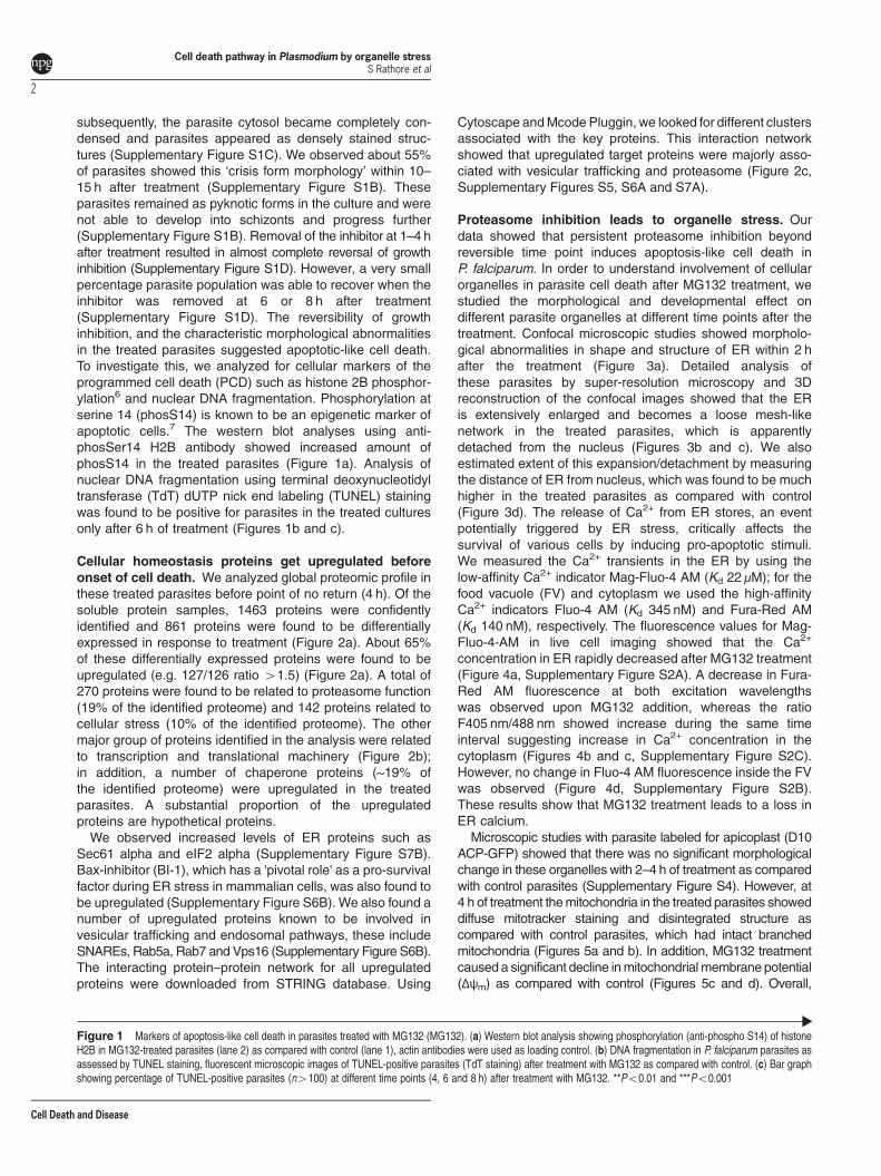
subsequently, the parasite cytosol became completely con-densed and parasites appeared as densely stained struc-tures (Supplementary Figure S1C). We observed about 55%of parasites showed this ‘crisis form morphology’ within 10–15 h after treatment (Supplementary Figure S1B). Theseparasites remained as pyknotic forms in the culture and werenot able to develop into schizonts and progress further(Supplementary Figure S1B). Removal of the inhibitor at 1–4 hafter treatment resulted in almost complete reversal of growthinhibition (Supplementary Figure S1D). However, a very smallpercentage parasite population was able to recover when theinhibitor was removed at 6 or 8 h after treatment(Supplementary Figure S1D). The reversibility of growthinhibition, and the characteristic morphological abnormalitiesin the treated parasites suggested apoptotic-like cell death.To investigate this, we analyzed for cellular markers of theprogrammed cell death (PCD) such as histone 2B phosphor-ylation6 and nuclear DNA fragmentation. Phosphorylation atserine 14 (phosS14) is known to be an epigenetic marker ofapoptotic cells.7 The western blot analyses using anti-phosSer14 H2B antibody showed increased amount ofphosS14 in the treated parasites (Figure 1a). Analysis ofnuclear DNA fragmentation using terminal deoxynucleotidyltransferase (TdT) dUTP nick end labeling (TUNEL) stainingwas found to be positive for parasites in the treated culturesonly after 6 h of treatment (Figures 1b and c).
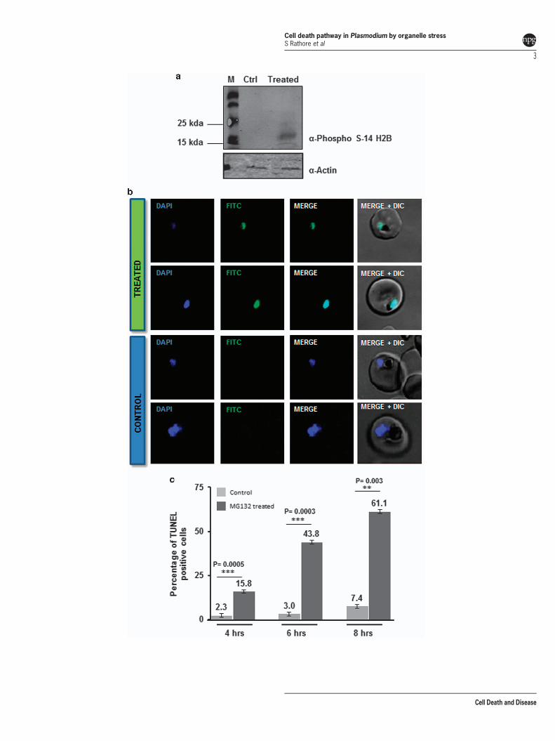
Cellular homeostasis proteins get upregulated beforeonset of cell death. We analyzed global proteomic profile inthese treated parasites before point of no return (4 h). Of thesoluble protein samples, 1463 proteins were confidentlyidentified and 861 proteins were found to be differentiallyexpressed in response to treatment (Figure 2a). About 65%of these differentially expressed proteins were found to beupregulated (e.g. 127/126 ratio 41.5) (Figure 2a). A total of270 proteins were found to be related to proteasome function(19% of the identified proteome) and 142 proteins related tocellular stress (10% of the identified proteome). The othermajor group of proteins identified in the analysis were relatedto transcription and translational machinery (Figure 2b);in addition, a number of chaperone proteins (~19% ofthe identified proteome) were upregulated in the treatedparasites. A substantial proportion of the upregulatedproteins are hypothetical proteins.We observed increased levels of ER proteins such as
Sec61 alpha and eIF2 alpha (Supplementary Figure S7B).Bax-inhibitor (BI-1), which has a 'pivotal role' as a pro-survivalfactor during ER stress in mammalian cells, was also found tobe upregulated (Supplementary Figure S6B). We also found anumber of upregulated proteins known to be involved invesicular trafficking and endosomal pathways, these includeSNAREs, Rab5a, Rab7 and Vps16 (Supplementary Figure S6B).The interacting protein–protein network for all upregulatedproteins were downloaded from STRING database. Using
Cytoscape andMcode Pluggin, we looked for different clustersassociated with the key proteins. This interaction networkshowed that upregulated target proteins were majorly asso-ciated with vesicular trafficking and proteasome (Figure 2c,Supplementary Figures S5, S6A and S7A).
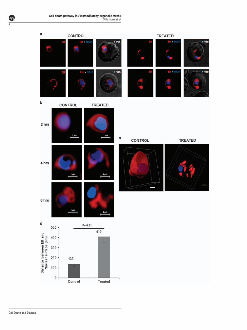
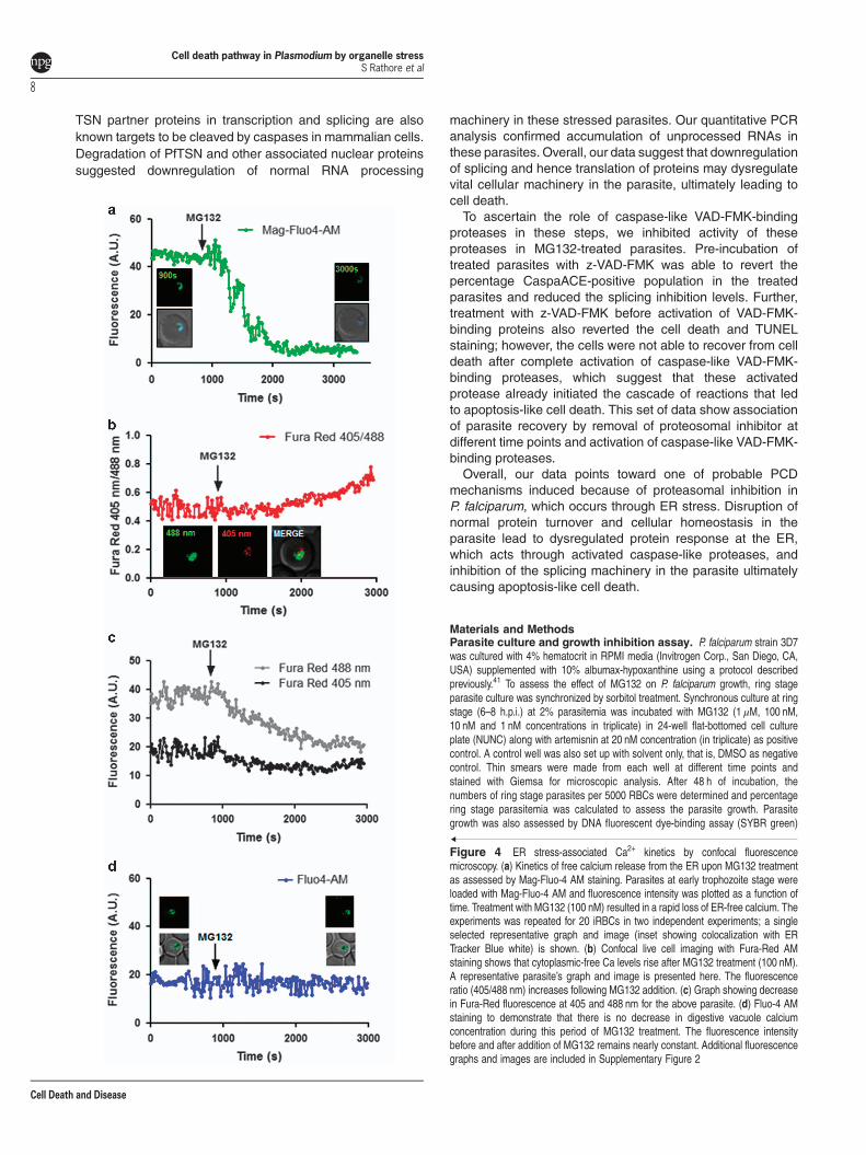
Proteasome inhibition leads to organelle stress. Ourdata showed that persistent proteasome inhibition beyondreversible time point induces apoptosis-like cell death inP. falciparum. In order to understand involvement of cellularorganelles in parasite cell death after MG132 treatment, westudied the morphological and developmental effect ondifferent parasite organelles at different time points after thetreatment. Confocal microscopic studies showed morpholo-gical abnormalities in shape and structure of ER within 2 hafter the treatment (Figure 3a). Detailed analysis ofthese parasites by super-resolution microscopy and 3Dreconstruction of the confocal images showed that the ERis extensively enlarged and becomes a loose mesh-likenetwork in the treated parasites, which is apparentlydetached from the nucleus (Figures 3b and c). We alsoestimated extent of this expansion/detachment by measuringthe distance of ER from nucleus, which was found to be muchhigher in the treated parasites as compared with control(Figure 3d). The release of Ca2+ from ER stores, an eventpotentially triggered by ER stress, critically affects thesurvival of various cells by inducing pro-apoptotic stimuli.We measured the Ca2+ transients in the ER by using thelow-affinity Ca2+ indicator Mag-Fluo-4 AM (Kd 22 μM); for thefood vacuole (FV) and cytoplasm we used the high-affinityCa2+ indicators Fluo-4 AM (Kd 345 nM) and Fura-Red AM(Kd 140 nM), respectively. The fluorescence values for Mag-Fluo-4-AM in live cell imaging showed that the Ca2+
concentration in ER rapidly decreased after MG132 treatment(Figure 4a, Supplementary Figure S2A). A decrease in Fura-Red AM fluorescence at both excitation wavelengthswas observed upon MG132 addition, whereas the ratioF405 nm/488 nm showed increase during the same timeinterval suggesting increase in Ca2+ concentration in thecytoplasm (Figures 4b and c, Supplementary Figure S2C).However, no change in Fluo-4 AM fluorescence inside the FVwas observed (Figure 4d, Supplementary Figure S2B).These results show that MG132 treatment leads to a loss inER calcium.Microscopic studies with parasite labeled for apicoplast (D10
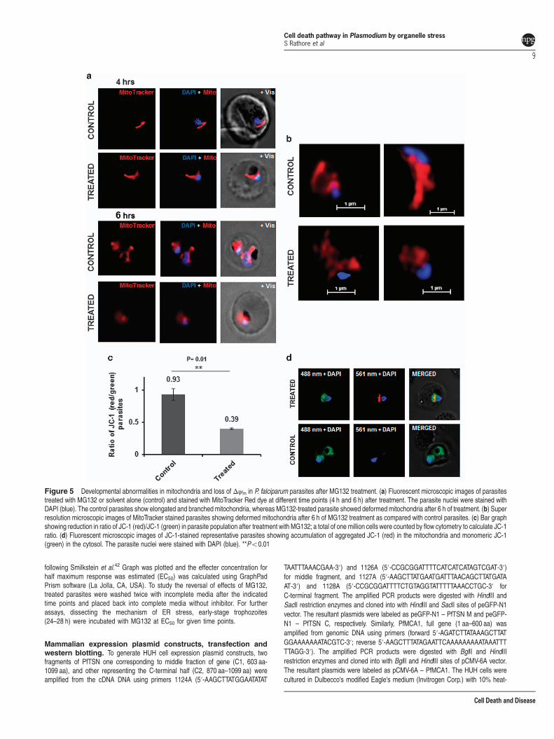
ACP-GFP) showed that there was no significant morphologicalchange in these organelles with 2–4 h of treatment as comparedwith control parasites (Supplementary Figure S4). However, at4 h of treatment themitochondria in the treated parasites showeddiffuse mitotracker staining and disintegrated structure ascompared with control parasites, which had intact branchedmitochondria (Figures 5a and b). In addition, MG132 treatmentcaused a significant decline inmitochondrial membrane potential(Δψm) as compared with control (Figures 5c and d). Overall,
Figure 1 Markers of apoptosis-like cell death in parasites treated with MG132 (MG132). (a) Western blot analysis showing phosphorylation (anti-phospho S14) of histoneH2B in MG132-treated parasites (lane 2) as compared with control (lane 1), actin antibodies were used as loading control. (b) DNA fragmentation in P. falciparum parasites asassessed by TUNEL staining, fluorescent microscopic images of TUNEL-positive parasites (TdT staining) after treatment with MG132 as compared with control. (c) Bar graphshowing percentage of TUNEL-positive parasites (n4100) at different time points (4, 6 and 8 h) after treatment with MG132. **Po0.01 and ***Po0.001
Cell death pathway in Plasmodium by organelle stressS Rathore et al
2
Cell Death and Disease
Cell death pathway in Plasmodium by organelle stressS Rathore et al
3
Cell Death and Disease
0
coun
ts
0.1 0.4 0.7 1 1.3 1.6 1.9
Ratio 127/126
2.2 2.5 2.8 3.1 3.4 3.7 4 4.3 4.6 4.9 5.2 5.5 5.8
0
20
40
60
80
100
Figure 2 Quantitative proteomic analysis reveals increase in levels of cellular homeostasis proteins at 4 h of proteosomal inhibition. (a) Histogram and table (inset) showingthe distribution of number of proteins having different peptide ratios (127/126; MG132 treated/control) as estimated by isobaric tagging based MS quantification. (b) Pie chartshowing the percentage of upregulated proteins belonging to different functional classes based upon selected gene ontology (GO) terms from KEGG pathway. (c) Functionalassociation network of selected clusters of proteins that are upregulated after MG132 exposure. The network shows probable linkage between the vesicle-transport, signaling andmetabolic clusters
Cell death pathway in Plasmodium by organelle stressS Rathore et al
4
Cell Death and Disease
these studies show that two cell death-associated organellesdevelop morphological abnormalities before the cell death isinitiated in the treated parasites.
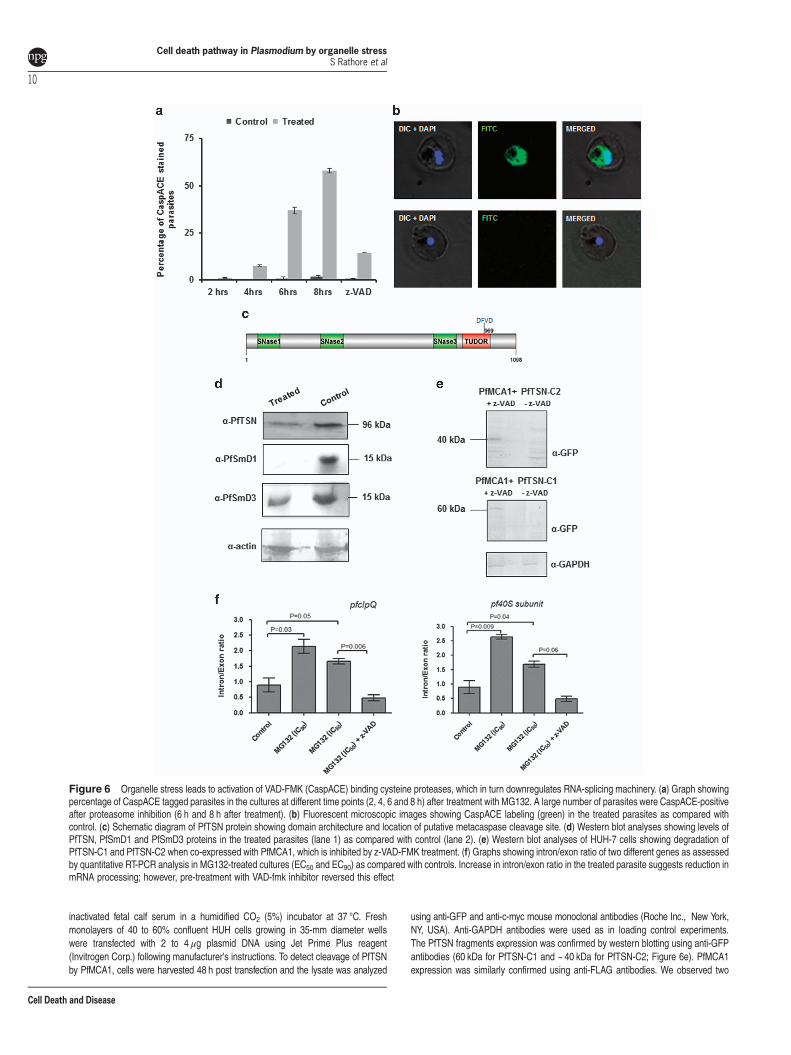
Organelle stress in parasite activates caspase-likecysteine protease. Our results with parasite morphology,development and proteomic studies showed that the protea-somal inhibition causes ER stress-like phenotype in theparasites. We further examined downstream pathways thatled to apoptosis-like cell death in the treated parasites. TheMG132-treated parasites (50 nM ~EC50) showed activationof a small population of CaspACE-positive cells at 4 h oftreatment; after 4 h the percentage of parasites showingCaspACE labeling increased significantly reaching ~35% at6 h and ~ 60% at 8 h after treatment (Figures 6a and b,Supplementary Figure S3A). However, at time point earlierthan 4 h there was no CaspACE-stained parasite populationin the treated cultures as compared with the control,suggesting that activation of VAD-FMK-binding proteasesoccur only 44 h after the treatment.
Activation of VAD-FMK-binding cysteine proteases leadsto downregulation of RNA-splicing machinery. Caspasesare known to be the major proteins in the pathway of PCD byacting on various important proteins needed for cell survival,which include several components of transcription andsplicing machineries.8 P. falciparum harbors caspase-related cysteine proteases, metacaspases.9 Recently, TudorStaphylococcus Nuclease (TSN) has been identified as oneof the substrates for these activated metacaspases.8 Weassessed TSN protein levels in these parasites with activatedVAD-FMK-binding caspase-like cysteine proteases. Levels ofPfTSN protein are drastically reduced in MG132-treatedparasites after activation of VAD-FMK-binding proteases ascompared with control parasites. In addition, we alsoassessed levels of two other important components ofsplicing machinery in Plasmodium, that is, PfSmD1 andPfSmD3 also found to be reduced in these parasites cells(Figure 6d). Overall, components of the splicing machineryare downregulated in stressed parasites probably due tocleavage by activated cysteine proteases.To demonstrate direct interaction between PfTSN and
PfMCA1, we expressed two fragments of PfTSN (PfTSN-C1and PfTSN-C2) along with full-length PfMCA1 in Huh-7hepatoblastoma cell lines (Supplementary Figure S3B). Weco-expressed PfMCA1 with PfTSN-C1 fragment and PfMCA1with PfTSN-C2 in Huh-7 cell line and analyzed by westernblotting. We observed a number of bands in lanes correspond-ing to degraded fragments of PfTSN-C1 and PfTSN-C2. Theintact PfTSN-C1 and PfTSN-C2 (i.e. 60kDa and 40 kDabands) were observed when the CaspACE inhibitor z-VAD-FMK was added (10 μM); this shows inhibition of PfMCA1activity by z-VAD-FMK (Figure 6e).
Unprocessed mRNA levels accumulates in stressedparasites. We further assessed changes in the levels ofunprocessed and processed mRNA in the treated parasites.Quantitative RT-PCR analyses was carried out to estimateintron/exon ratio for a given gene. The pfclpQ RNA in thetreated parasite (oEC50 ~ 50 nM) showed a intron/exon ratio
nearly double as compared with the control parasites(Figure 6f); similarly, RNA of 40 S ribosomal subunit alsoshowed a similar effect having intron/exon ratio nearly twiceas compared with control (Figure 6f). These effect on RNAprocessing in stressed parasites can be reverted when theparasites were treated simultaneous with caspase inhibitor(z-VAD-FMK); in these parasite the intron/exon ratio wasrestored to nearly normal (Figure 6f), clearly indicating therole of caspase-like proteases in downregulation of RNA-splicing machinery. However, the effect on RNA processing instressed parasites were not ubiquitous; there was no changein intro/exon ratio for two other genes that we analyzed,pfhrp2 and pfhsp90 (data not shown).
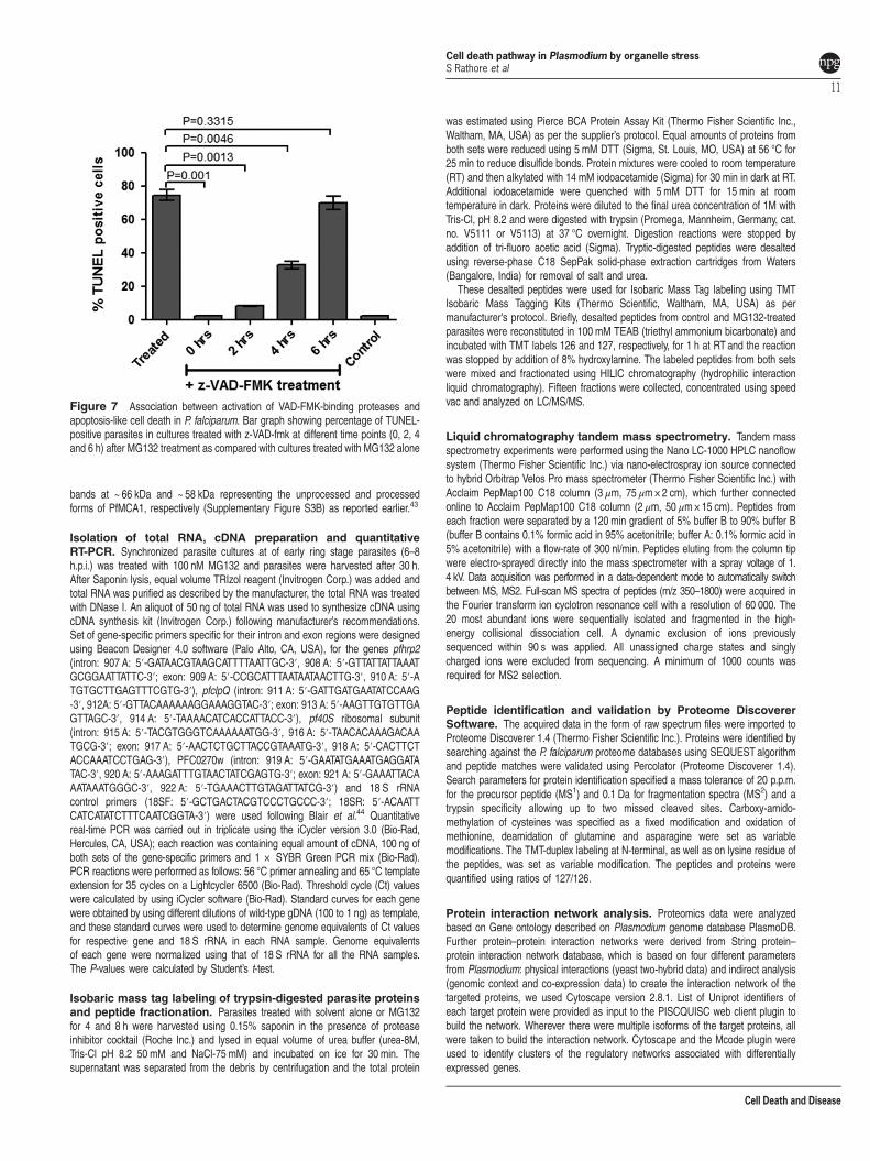
Activation and reversal of VAD-FMK-binding cysteineproteases associates with parasite cell death.Pre-incubation of treated parasites with z-VAD-FMK wasable to revert the percentage CaspACE-positive population inthe treated parasite to normal levels as in the control set(Figure 6a); further, z-VAD-fmk was able to reduce thesplicing inhibition levels in MG132-treated parasites(Figure 6f). Therefore, z-VAD-FMK was used to treatparasites at different time points after proteasome inhibitionand reversal of cell death was assessed using TUNEL assay.The MG132-treated parasites were able to overcome the celldeath by Z-VAD-FMK treatment, which support role ofcaspase-like VAD-FMK-binding proteases in the parasite celldeath after organelle stress because of proteasomal inhibi-tion. However, when caspase inhibitor was added at 6 h aftertreatment, a large percentage of parasite population was notable to recover from cell death (Figure 7).
Discussion
Maintenance of cellular homeostasis and integrity of cellularproteome depends upon protein turnover including stability,refolding and degradation of the proteins. During asexualblood stage cycles of malaria parasites, there is high load onprotein synthesis and turnover because of rapid multiplicationrate of the parasite; therefore, proper functioning of proteinquality control machinery is essential for maintenance ofcellular homeostasis. In this study, we analyzedmolecular andcellular events in the P. falciparum parasite after induction oforganelle stress by proteosomal inhibition, MG132. Develop-mental arrest at the trophozoite stage and the appearance ofpyknotic forms in MG132-treated parasite cultures, pointstoward apoptosis-like cell death as described earlier.3
Plasmodium cell death remains a topic of discussion sincerecent past; a number of studies have tried to understand themode of apoptosis-like cell death and its machinery in theparasite.10,11 One of the main characteristic of apoptosis-likecell death is a programmed cascade of committed events witha point of no return.11,12 Our data from wash-off experimentsshowed that proteasomal inhibition induced parasite deathcan be reversed by removal of the inhibitor within 4 h oftreatment; however, after that time point the cells arecommitted to die, this may suggest that it is also programmedevent. In eukaryotic cells, the hallmarks of apoptosis-like celldeath include the fragmentation of nuclear DNA and histoneH2B phosS14.13,14 Our results with TUNEL assay and histone
Cell death pathway in Plasmodium by organelle stressS Rathore et al
5
Cell Death and Disease
Cell death pathway in Plasmodium by organelle stressS Rathore et al
6
Cell Death and Disease
modifications markers analyses indicate an apoptotic-like celldeath in MG132-treated parasites.In eukaryotic cells, the interplay between these survival and
cell death responses ultimately determines the fate of thestressed cell.15 We first tried to assess the molecular events inparasite at early stages of stress because of proteasomalinhibition. Our results of global quantitative proteomic analysissuggest that P. falciparum tries to increase number of stress-related proteins after proteasome inhibition. Our data showedincrease in levels of various chaperones, such as Der-1,cpn60 and Hsp70, along with various translation machineryproteins, which points toward Plasmodium’s efforts to combatstress. The other major group of proteins, which were found tobe increased in levels were proteins of Rab family, that is,Rab1a, Rab2, Rab7 and Rab18, which are known to beassociated with different vesicular trafficking pathways inPlasmodium.16,17 Recently it was shown that in stressedP. falciparum parasites, the Rab7 associates with ATG8coated vesicles.18 ATG8 is an important protein for formationof autophagy vesicles, auto-phagosomes, and thus ATG8 isan important component of autophagy. Autophagy in malariaparasites remains to be a topic of debate and it is not clear iftrue autophagy occurs in Plasmodium.19,20 Rab7 is known tobe involved in trafficking of endosomal material to lysosomalsystem in eukaryotic cells21 and toward the FV inP. falciparum.18 We have also observed increased levels ofother components of autophagy machinery, ATG18, in thetreated samples as compared with controls. Increase in levelsof these proteins in our data points toward increased vesiculartrafficking by parasite to combat stress as shown in case ofother organisms.22
Another important protein involved in UPR signaling, that is,eIF2α was found to be upregulated after treatment withMG132. eIF2α is known to be linked between UPR signalingand stimulation of autophagy-like events; under stress theeIF2α gets phosphorylated after activation of PERK pathway.Although a clear UPR signaling pathway is not present inP. falciparum, a putative homolog of PERK is present in theparasite.23,24 Another set of interesting proteins, which arefound to be upregulated in the treated parasites includeER-associated Sec61 and Bax-inhibitor. Earlier studies haveprovided evidence that the Sec61 channel mediates passivecalcium efflux from the intact ER6,25 and also during ERstress.26 Signal peptide peptidase (SPP) was found to bequntitatively reduced in the treated parasite. Eukaryotic SPPsare multi-pass integral membrane proteins from the aspartylprotease family that cleave transmembrane substrates.27,28
PfSPP inhibition has been shown to reduce the parasitesability to cope up with ER stress, and could have a significantrole in deciding the survival of parasite.29 Taken together, ourresults showing increased eIF2a α along with increasedvesicular trafficking components, may also be one of the likesurvival strategy in the parasite under stress.
It has been reported that proteasome inhibition may induceautophagy via UPR-dependent pathways.30 However, themachinery required for UPR pathways is absent in Plasmo-dium.24 Upregulation of autophagy-related proteins in MG132-treated parasitesmay suggest that autophagy-likemechanismmay be a survival mechanism in Plasmodium under stress in aUPR-independent manner. A recent study also suggests thatthe parasite directs vesicles/organelle fragments to the FV instarved parasites.18 Overall, quantitative proteomics studies,at 4 h, the time point at which parasite retain the ability to revertback to normal growth, suggest that the parasite experiencingstress are able to device a survival strategy. These strategiesmay help the parasites to survive initial stress and revive if thestress is removed; however, if the stress continues further thecell undergoes apoptosis-like PCD.As our results pointed out the relation between organeller
stress and cell death in the parasite, therefore in this study wealso tried to analyze effect of cellular stress on variousorganelle. Recent study from Chaubey et al. have shown thatinduction of ER stress in Plasmodium parasite by exposure toa reducing agent for short period, leads to gametocytogenesisin the next growth cycle. However, in our study, we showed ifthe ER stress persists beyond point of no return, parasite caninitiate cell death processes. The ER is also an important sitefor calcium storage within the cell, which is coupled with itsquality control machinery to produce correctly folded protein.31
We found the induction of ER stress simultaneously causedrelease of stored Ca2+ in the parasite cytosol. Subsequently,another important organelle, mitochondria in the parasite, alsoget stressed, which loses its membrane potential and ability todivide further. Eventually, we observed activation of caspase-like VAD-FMK-binding proteases in the stressed parasite. TheP. falciparum genome does not contain any classical caspase-like protein, however, caspase-like activity is also describedduring apoptosis in other organisms that lack classicalcaspases.32–35 P. falciparum genome harbors threemetacaspase-like proteases that are closely related toeukaryotic caspases, these proteases contains C14 domainwith a catalytic dyad of cysteine an histidine as in case ofcaspases.36 Metacaspases are known to have essential rolein during PCD in plants;37,38 the TSN is shown to be asubstrate of activated metacaspases in plants.8 The TSN is aconserved proteins that is known to be involved in regulationdifferent transcription and translation steps.39 The TSNhomolog in P. falciparum, PfTSN, is shown to be an importantfunctional component of spliceosome Sm core complex.40
PfTSN harbors the DFVD motif near its C-terminus, which is aprobable cleavage site for caspase-like enzymes. Weobserved reduction of PfTSN levels in parasites afteractivation of caspase-like z-VAD-FMK-binding proteases;along with PfTSN, other associated nuclear proteins likePfSmD1 and PfSmD3, which are part of nuclear splicingmachinery, were also found to be reduced. Indeed, most of the
Figure 3 Altered morphology of ER and release of stored calcium in P. falciparum parasites under prolonged proteosomal inhibition. (a) Fluorescent microscopic images ofP. falciparum parasites stained with ER Tracker Red at 6 h after treatment with MG132. The parasite nuclei were stained with DAPI (blue). (b) Super resolution microscopic imagesat different time points (2, 4, 6 h) after MG132 treatment. The parasite showed a disrupted ER network at 4 and 6 h as compared with control that showed a continuous ER aroundthe nucleus. (c) 3D images reconstructed using Z-stacks of ER Tracker stained control and treated parasites. (d) Bar graph showing increase in distance between nuclear and ERsurface as assessed by super-resolution microscopic images in MG132-treated parasites as compared with control
Cell death pathway in Plasmodium by organelle stressS Rathore et al
7
Cell Death and Disease
TSN partner proteins in transcription and splicing are alsoknown targets to be cleaved by caspases in mammalian cells.Degradation of PfTSN and other associated nuclear proteinssuggested downregulation of normal RNA processing
machinery in these stressed parasites. Our quantitative PCRanalysis confirmed accumulation of unprocessed RNAs inthese parasites. Overall, our data suggest that downregulationof splicing and hence translation of proteins may dysregulatevital cellular machinery in the parasite, ultimately leading tocell death.To ascertain the role of caspase-like VAD-FMK-binding
proteases in these steps, we inhibited activity of theseproteases in MG132-treated parasites. Pre-incubation oftreated parasites with z-VAD-FMK was able to revert thepercentage CaspaACE-positive population in the treatedparasites and reduced the splicing inhibition levels. Further,treatment with z-VAD-FMK before activation of VAD-FMK-binding proteins also reverted the cell death and TUNELstaining; however, the cells were not able to recover from celldeath after complete activation of caspase-like VAD-FMK-binding proteases, which suggest that these activatedprotease already initiated the cascade of reactions that ledto apoptosis-like cell death. This set of data show associationof parasite recovery by removal of proteosomal inhibitor atdifferent time points and activation of caspase-like VAD-FMK-binding proteases.Overall, our data points toward one of probable PCD
mechanisms induced because of proteasomal inhibition inP. falciparum, which occurs through ER stress. Disruption ofnormal protein turnover and cellular homeostasis in theparasite lead to dysregulated protein response at the ER,which acts through activated caspase-like proteases, andinhibition of the splicing machinery in the parasite ultimatelycausing apoptosis-like cell death.
Materials and MethodsParasite culture and growth inhibition assay. P. falciparum strain 3D7was cultured with 4% hematocrit in RPMI media (Invitrogen Corp., San Diego, CA,USA) supplemented with 10% albumax-hypoxanthine using a protocol describedpreviously.41 To assess the effect of MG132 on P. falciparum growth, ring stageparasite culture was synchronized by sorbitol treatment. Synchronous culture at ringstage (6–8 h.p.i.) at 2% parasitemia was incubated with MG132 (1 μM, 100 nM,10 nM and 1 nM concentrations in triplicate) in 24-well flat-bottomed cell cultureplate (NUNC) along with artemisnin at 20 nM concentration (in triplicate) as positivecontrol. A control well was also set up with solvent only, that is, DMSO as negativecontrol. Thin smears were made from each well at different time points andstained with Giemsa for microscopic analysis. After 48 h of incubation, thenumbers of ring stage parasites per 5000 RBCs were determined and percentagering stage parasitemia was calculated to assess the parasite growth. Parasitegrowth was also assessed by DNA fluorescent dye-binding assay (SYBR green)
Figure 4 ER stress-associated Ca2+ kinetics by confocal fluorescencemicroscopy. (a) Kinetics of free calcium release from the ER upon MG132 treatmentas assessed by Mag-Fluo-4 AM staining. Parasites at early trophozoite stage wereloaded with Mag-Fluo-4 AM and fluorescence intensity was plotted as a function oftime. Treatment with MG132 (100 nM) resulted in a rapid loss of ER-free calcium. Theexperiments was repeated for 20 iRBCs in two independent experiments; a singleselected representative graph and image (inset showing colocalization with ERTracker Blue white) is shown. (b) Confocal live cell imaging with Fura-Red AMstaining shows that cytoplasmic-free Ca levels rise after MG132 treatment (100 nM).A representative parasite’s graph and image is presented here. The fluorescenceratio (405/488 nm) increases following MG132 addition. (c) Graph showing decreasein Fura-Red fluorescence at 405 and 488 nm for the above parasite. (d) Fluo-4 AMstaining to demonstrate that there is no decrease in digestive vacuole calciumconcentration during this period of MG132 treatment. The fluorescence intensitybefore and after addition of MG132 remains nearly constant. Additional fluorescencegraphs and images are included in Supplementary Figure 2
Cell death pathway in Plasmodium by organelle stressS Rathore et al
8
Cell Death and Disease
following Smilkstein et al.42 Graph was plotted and the effecter concentration forhalf maximum response was estimated (EC50) was calculated using GraphPadPrism software (La Jolla, CA, USA). To study the reversal of effects of MG132,treated parasites were washed twice with incomplete media after the indicatedtime points and placed back into complete media without inhibitor. For furtherassays, dissecting the mechanism of ER stress, early-stage trophozoites(24–28 h) were incubated with MG132 at EC50 for given time points.
Mammalian expression plasmid constructs, transfection andwestern blotting. To generate HUH cell expression plasmid constructs, twofragments of PfTSN one corresponding to middle fraction of gene (C1, 603 aa-1099 aa), and other representing the C-terminal half (C2, 870 aa–1099 aa) wereamplified from the cDNA DNA using primers 1124A (5′-AAGCTTATGGAATATAT
TAATTTAAACGAA-3′) and 1126A (5′-CCGCGGATTTTCATCATCATAGTCGAT-3′)for middle fragment, and 1127A (5′-AAGCTTATGAATGATTTAACAGCTTATGATAAT-3′) and 1128A (5′-CCGCGGATTTTCTGTAGGTATTTTTAAACCTGC-3′ forC-terminal fragment. The amplified PCR products were digested with HindIII andSacII restriction enzymes and cloned into with HindIII and SacII sites of peGFP-N1vector. The resultant plasmids were labeled as peGFP-N1 – PfTSN M and peGFP-N1 – PfTSN C, respectively. Similarly, PfMCA1, full gene (1 aa–600 aa) wasamplified from genomic DNA using primers (forward 5′-AGATCTTATAAAGCTTATGGAAAAAAATACGTC-3′; reverse 5′-AAGCTTTATAGAATTCAAAAAAAAATAAATTTTTAGG-3′). The amplified PCR products were digested with BglII and HindIIIrestriction enzymes and cloned into with BglII and HindIII sites of pCMV-6A vector.The resultant plasmids were labeled as pCMV-6A – PfMCA1. The HUH cells werecultured in Dulbecco's modified Eagle's medium (Invitrogen Corp.) with 10% heat-
Figure 5 Developmental abnormalities in mitochondria and loss of Δψm in P. falciparum parasites after MG132 treatment. (a) Fluorescent microscopic images of parasitestreated with MG132 or solvent alone (control) and stained with MitoTracker Red dye at different time points (4 h and 6 h) after treatment. The parasite nuclei were stained withDAPI (blue). The control parasites show elongated and branched mitochondria, whereas MG132-treated parasite showed deformed mitochondria after 6 h of treatment. (b) Superresolution microscopic images of MitoTracker stained parasites showing deformed mitochondria after 6 h of MG132 treatment as compared with control parasites. (c) Bar graphshowing reduction in ratio of JC-1 (red)/JC-1 (green) in parasite population after treatment with MG132; a total of one million cells were counted by flow cytometry to calculate JC-1ratio. (d) Fluorescent microscopic images of JC-1-stained representative parasites showing accumulation of aggregated JC-1 (red) in the mitochondria and monomeric JC-1(green) in the cytosol. The parasite nuclei were stained with DAPI (blue). **Po0.01
Cell death pathway in Plasmodium by organelle stressS Rathore et al
9
Cell Death and Disease
inactivated fetal calf serum in a humidified CO2 (5%) incubator at 37 °C. Freshmonolayers of 40 to 60% confluent HUH cells growing in 35-mm diameter wellswere transfected with 2 to 4 μg plasmid DNA using Jet Prime Plus reagent(Invitrogen Corp.) following manufacturer's instructions. To detect cleavage of PfTSNby PfMCA1, cells were harvested 48 h post transfection and the lysate was analyzed
using anti-GFP and anti-c-myc mouse monoclonal antibodies (Roche Inc., New York,NY, USA). Anti-GAPDH antibodies were used as in loading control experiments.The PfTSN fragments expression was confirmed by western blotting using anti-GFPantibodies (60 kDa for PfTSN-C1 and ~ 40 kDa for PfTSN-C2; Figure 6e). PfMCA1expression was similarly confirmed using anti-FLAG antibodies. We observed two
Figure 6 Organelle stress leads to activation of VAD-FMK (CaspACE) binding cysteine proteases, which in turn downregulates RNA-splicing machinery. (a) Graph showingpercentage of CaspACE tagged parasites in the cultures at different time points (2, 4, 6 and 8 h) after treatment with MG132. A large number of parasites were CaspACE-positiveafter proteasome inhibition (6 h and 8 h after treatment). (b) Fluorescent microscopic images showing CaspACE labeling (green) in the treated parasites as compared withcontrol. (c) Schematic diagram of PfTSN protein showing domain architecture and location of putative metacaspase cleavage site. (d) Western blot analyses showing levels ofPfTSN, PfSmD1 and PfSmD3 proteins in the treated parasites (lane 1) as compared with control (lane 2). (e) Western blot analyses of HUH-7 cells showing degradation ofPfTSN-C1 and PfTSN-C2 when co-expressed with PfMCA1, which is inhibited by z-VAD-FMK treatment. (f) Graphs showing intron/exon ratio of two different genes as assessedby quantitative RT-PCR analysis in MG132-treated cultures (EC50 and EC90) as compared with controls. Increase in intron/exon ratio in the treated parasite suggests reduction inmRNA processing; however, pre-treatment with VAD-fmk inhibitor reversed this effect
Cell death pathway in Plasmodium by organelle stressS Rathore et al
10
Cell Death and Disease
bands at ~ 66 kDa and ~ 58 kDa representing the unprocessed and processedforms of PfMCA1, respectively (Supplementary Figure S3B) as reported earlier.43
Isolation of total RNA, cDNA preparation and quantitativeRT-PCR. Synchronized parasite cultures at of early ring stage parasites (6–8h.p.i.) was treated with 100 nM MG132 and parasites were harvested after 30 h.After Saponin lysis, equal volume TRIzol reagent (Invitrogen Corp.) was added andtotal RNA was purified as described by the manufacturer, the total RNA was treatedwith DNase I. An aliquot of 50 ng of total RNA was used to synthesize cDNA usingcDNA synthesis kit (Invitrogen Corp.) following manufacturer’s recommendations.Set of gene-specific primers specific for their intron and exon regions were designedusing Beacon Designer 4.0 software (Palo Alto, CA, USA), for the genes pfhrp2(intron: 907 A: 5′-GATAACGTAAGCATTTTAATTGC-3′, 908 A: 5′-GTTATTATTAAATGCGGAATTATTC-3′; exon: 909 A: 5′-CCGCATTTAATAATAACTTG-3′, 910 A: 5′-ATGTGCTTGAGTTTCGTG-3′), pfclpQ (intron: 911 A: 5′-GATTGATGAATATCCAAG-3′, 912A: 5′-GTTACAAAAAAGGAAAGGTAC-3′; exon: 913 A: 5′-AAGTTGTGTTGAGTTAGC-3′, 914 A: 5′-TAAAACATCACCATTACC-3′), pf40S ribosomal subunit(intron: 915 A: 5′-TACGTGGGTCAAAAAATGG-3′, 916 A: 5′-TAACACAAAGACAATGCG-3′; exon: 917 A: 5′-AACTCTGCTTACCGTAAATG-3′, 918 A: 5′-CACTTCTACCAAATCCTGAG-3′), PFC0270w (intron: 919 A: 5′-GAATATGAAATGAGGATATAC-3′, 920 A: 5′-AAAGATTTGTAACTATCGAGTG-3′; exon: 921 A: 5′-GAAATTACAAATAAATGGGC-3′, 922 A: 5′-TGAAACTTGTAGATTATCG-3′) and 18 S rRNAcontrol primers (18SF: 5′-GCTGACTACGTCCCTGCCC-3′; 18SR: 5′-ACAATTCATCATATCTTTCAATCGGTA-3′) were used following Blair et al.44 Quantitativereal-time PCR was carried out in triplicate using the iCycler version 3.0 (Bio-Rad,Hercules, CA, USA); each reaction was containing equal amount of cDNA, 100 ng ofboth sets of the gene-specific primers and 1 × SYBR Green PCR mix (Bio-Rad).PCR reactions were performed as follows: 56 °C primer annealing and 65 °C templateextension for 35 cycles on a Lightcycler 6500 (Bio-Rad). Threshold cycle (Ct) valueswere calculated by using iCycler software (Bio-Rad). Standard curves for each genewere obtained by using different dilutions of wild-type gDNA (100 to 1 ng) as template,and these standard curves were used to determine genome equivalents of Ct valuesfor respective gene and 18 S rRNA in each RNA sample. Genome equivalentsof each gene were normalized using that of 18 S rRNA for all the RNA samples.The P-values were calculated by Student’s t-test.
Isobaric mass tag labeling of trypsin-digested parasite proteinsand peptide fractionation. Parasites treated with solvent alone or MG132for 4 and 8 h were harvested using 0.15% saponin in the presence of proteaseinhibitor cocktail (Roche Inc.) and lysed in equal volume of urea buffer (urea-8M,Tris-Cl pH 8.2 50 mM and NaCl-75 mM) and incubated on ice for 30 min. Thesupernatant was separated from the debris by centrifugation and the total protein
was estimated using Pierce BCA Protein Assay Kit (Thermo Fisher Scientific Inc.,Waltham, MA, USA) as per the supplier’s protocol. Equal amounts of proteins fromboth sets were reduced using 5 mM DTT (Sigma, St. Louis, MO, USA) at 56 °C for25 min to reduce disulfide bonds. Protein mixtures were cooled to room temperature(RT) and then alkylated with 14 mM iodoacetamide (Sigma) for 30 min in dark at RT.Additional iodoacetamide were quenched with 5 mM DTT for 15 min at roomtemperature in dark. Proteins were diluted to the final urea concentration of 1M withTris-Cl, pH 8.2 and were digested with trypsin (Promega, Mannheim, Germany, cat.no. V5111 or V5113) at 37 °C overnight. Digestion reactions were stopped byaddition of tri-fluoro acetic acid (Sigma). Tryptic-digested peptides were desaltedusing reverse-phase C18 SepPak solid-phase extraction cartridges from Waters(Bangalore, India) for removal of salt and urea.
These desalted peptides were used for Isobaric Mass Tag labeling using TMTIsobaric Mass Tagging Kits (Thermo Scientific, Waltham, MA, USA) as permanufacturer's protocol. Briefly, desalted peptides from control and MG132-treatedparasites were reconstituted in 100 mM TEAB (triethyl ammonium bicarbonate) andincubated with TMT labels 126 and 127, respectively, for 1 h at RT and the reactionwas stopped by addition of 8% hydroxylamine. The labeled peptides from both setswere mixed and fractionated using HILIC chromatography (hydrophilic interactionliquid chromatography). Fifteen fractions were collected, concentrated using speedvac and analyzed on LC/MS/MS.
Liquid chromatography tandem mass spectrometry. Tandem massspectrometry experiments were performed using the Nano LC-1000 HPLC nanoflowsystem (Thermo Fisher Scientific Inc.) via nano-electrospray ion source connectedto hybrid Orbitrap Velos Pro mass spectrometer (Thermo Fisher Scientific Inc.) withAcclaim PepMap100 C18 column (3 μm, 75 μm× 2 cm), which further connectedonline to Acclaim PepMap100 C18 column (2 μm, 50 μm× 15 cm). Peptides fromeach fraction were separated by a 120 min gradient of 5% buffer B to 90% buffer B(buffer B contains 0.1% formic acid in 95% acetonitrile; buffer A: 0.1% formic acid in5% acetonitrile) with a flow-rate of 300 nl/min. Peptides eluting from the column tipwere electro-sprayed directly into the mass spectrometer with a spray voltage of 1.4 kV. Data acquisition was performed in a data-dependent mode to automatically switchbetween MS, MS2. Full-scan MS spectra of peptides (m/z 350–1800) were acquired inthe Fourier transform ion cyclotron resonance cell with a resolution of 60 000. The20 most abundant ions were sequentially isolated and fragmented in the high-energy collisional dissociation cell. A dynamic exclusion of ions previouslysequenced within 90 s was applied. All unassigned charge states and singlycharged ions were excluded from sequencing. A minimum of 1000 counts wasrequired for MS2 selection.
Peptide identification and validation by Proteome DiscovererSoftware. The acquired data in the form of raw spectrum files were imported toProteome Discoverer 1.4 (Thermo Fisher Scientific Inc.). Proteins were identified bysearching against the P. falciparum proteome databases using SEQUEST algorithmand peptide matches were validated using Percolator (Proteome Discoverer 1.4).Search parameters for protein identification specified a mass tolerance of 20 p.p.m.for the precursor peptide (MS1) and 0.1 Da for fragmentation spectra (MS2) and atrypsin specificity allowing up to two missed cleaved sites. Carboxy-amido-methylation of cysteines was specified as a fixed modification and oxidation ofmethionine, deamidation of glutamine and asparagine were set as variablemodifications. The TMT-duplex labeling at N-terminal, as well as on lysine residue ofthe peptides, was set as variable modification. The peptides and proteins werequantified using ratios of 127/126.
Protein interaction network analysis. Proteomics data were analyzedbased on Gene ontology described on Plasmodium genome database PlasmoDB.Further protein–protein interaction networks were derived from String protein–protein interaction network database, which is based on four different parametersfrom Plasmodium: physical interactions (yeast two-hybrid data) and indirect analysis(genomic context and co-expression data) to create the interaction network of thetargeted proteins, we used Cytoscape version 2.8.1. List of Uniprot identifiers ofeach target protein were provided as input to the PISCQUISC web client plugin tobuild the network. Wherever there were multiple isoforms of the target proteins, allwere taken to build the interaction network. Cytoscape and the Mcode plugin wereused to identify clusters of the regulatory networks associated with differentiallyexpressed genes.
Figure 7 Association between activation of VAD-FMK-binding proteases andapoptosis-like cell death in P. falciparum. Bar graph showing percentage of TUNEL-positive parasites in cultures treated with z-VAD-fmk at different time points (0, 2, 4and 6 h) after MG132 treatment as compared with cultures treated with MG132 alone
Cell death pathway in Plasmodium by organelle stressS Rathore et al
11
Cell Death and Disease
Isolation of parasites and western immunoblotting. Total parasitelysate were separated by SDS-PAGE under reducing conditions before proteinswere transferred to PVDF membrane using a Transblot Wet transfer system(Bio-Rad) according to manufacturer's instructions. The membranes were blocked inblocking buffer (1 × PBS, 0.1% Tween-20, 5% milk powder) for 2 h. The blots werewashed and incubated for 1 h with a primary antibody (rabbit anti-PfTSN; rat anti-PfSmD1; and rat anti-PfSmD3 (in-house made)) each used at 1 : 1000 dilutions;anti-phospho S14 H2B, rabbit polyclonal (Santa Cruz Biotechnology, Dallas, TX,USA), respectively, and mice monoclonal anti-H2B (Santa Cruz Biotechnology). Thesecondary anti-mouse-HRP conjugate or anti-rat-HRP conjugate (Promega)antibodies were used at 1 : 3000 dilutions for the respective blots. The proteinbands reacting with the antibodies were detected using the Amersham ECLdetection kit (Piscataway, NJ, USA) and visualized by exposing blots toautoradiography films (Kodak, Rochester, NY, USA).
Organelle staining and fluorescence microscopy. To visualize ERmorphology, ER Tracker Red (Life Technologies, Grand Island, NY, USA) was added ata final concentration of 200 nM directly to parasite suspensions in complete culturemedium and incubated with shaking at 37 °C for 20 min after which 0.1 ng/μl, 6-diamidino-2-phenylindole (DAPI, Sigma) was added for further 10 min. Following threewashes with 1 × PBS (pH 7.4), samples were mounted on glass slides and observedeither on a Nikon A1 Confocal Microscope (Nikon Corporation, Tokyo, Japan) or NikonA1 microscope with N-SIM (Structured Illumination Microscopy, for super-resolution). Tovisualize mitochondria, the parasites were stained with MitoTracker Red CMXRos(Invitrogen Corp.) as described earlier (Rathore et al.45) and the parasite was visualizedusing confocal fluorescence microscope.For confocal microscopy, images were acquired with Plan Apochromat 100 × 1.40
NA oil immersion objective lens (Nikon Corporation) in NIS Elements and Z-stackswere taken for 21 steps at 200 nm intervals. For 3D-SIM images, Ti-sapphire solidstate lasers (405, 488, 561 nm) provided wide field illumination and multi-channelimages were captured simultaneously using three cameras Andor Technology iXonDU897 EMCCD (Andor Technology, Belfast, UK). Data were captured using a CFIApo TIRF 100x oil (NA 1.49) lens (Nikon Corporation) and sectioned using a 200 nmZ-step size. Raw 3-phase images were reconstructed as described elsewhere. Allimages were captured and analyzed using NIS Elements C/NIS Elements AR.Individual Z-stacks were exported as 8-bit RGB TIFF formats and selected confocalZ-stacks were further reconstructed in Imaris (Bitplane, Zurich, Switzerland) as 3Dmodels.
Measurement of intracellular calcium by confocal microscopyand live cell imaging. Confocal fluorescence microscopy and live cell imagingwas performed using a Nikon A1 microscope as mentioned above. To detect ERstress-associated Ca2+ dysregulation, we used a three Ca2+ fluorophores to staindifferent subcellular compartments. The ER Ca2+ was measured using Mag-Fluo-4AM. Parasites were stained with Mag-Fluo-4 AM (10 μM) and ER Tracker BlueWhite DPX (2 μM) for 30 min at 37 C, followed by controlled saponin treatment(0.01% for 2 min) to trap the dye within ER. This sample was then transferred to aglass bottomed Petri dish kept in a stage top incubator (Tokai Hit, GM-8000, TokaiHit Corporation, Gendoji-cho, Japan). Physiological conditions were maintained by acontinuous supply of mixed gas (5% CO2, 5% O2 and balanced nitrogen). ER Ca2+
transients using Mag-Fluo-4 AM was then monitored in these parasites by excitingthe sample at 488 nm and detecting the emission signals at 561 nm and sequentialER Tracker Blue White DPX signals were detected by excitation at 364 nm andemission at 640 nm. To detect changes in cytoplasmic Ca levels, cells were labeledby the ratiometric dye Fura-Red AM.46 Fura-Red is a ratiometric dye with dualexcitation (488 nm for free Ca2+ and 405 nm for bound Ca2+) and single emission(640 nm). For Fura-Red, a decrease in fluorescence corresponds to an increase inCa2+ concentration for excitation at both wavelengths, but the ratio F405 nm/488 nmrises with an increase in Ca2+ concentration.47–49 Parasites were loaded with Fura-Red and confocal images were acquired every 15 s by alternate excitation with 405and 488 nm lasers. Fura-Red was alternately excited with the 405 and 488 nmlasers, and fluorescence signals were separated from excitation wavelengths usinga quad-band dichroic mirror and emission filter set (405/488/561/640, Semrock Inc.,Rochester, NY, USA). Images were acquired using an Andor iXon DU897 EMCCDcamera every 15 s keeping the pinhole at 1.0 μm. This resulted in a pair of imagesand the cytoplasm was marked by region of interest (ROI), which was tracked forchanges in mean fluorescence intensity over time. Changes in digestive vacuole-free calcium was detected by staining with Fluo-4 AM (10 μM) and imaged using the488 nm laser and marking the DV area as ROI. All image analysis in these ROIs
were carried out in the NIS Elements AR Analysis software (ver 4.13.04,Nikon Corporation) and fluorescence values were exported to MS Excel and plotusing GraphPad Prism (ver 5.00).
Measurement of caspase-like cysteine protease activation. Toassess the activation of caspase-like cysteine proteases, cells were stained withCaspACE FITC-VAD-FMK in Situ Marker (Promega) as per manufacturer’sinstructions. Briefly, parasites were collected from experimental and control sets atdifferent time points and incubated with 10 μM CaspACE FITC-VAD-FMK(fluorescein isothiocyanate-valyl-alanyl-aspartyl-[O-methyl]-fluoromethylketone) incomplete media for 30 min at 37 °C followed by washing with 1 × PBS. Thestained samples were analyzed by flow cytometry using CellQuestPro software onFACS Calibur (Becton Dickinson, San Jose, CA, USA) to assess fluorescencestaining (Em-525 nm/Ex-488 nm) of infected RBCs.
Mitochondria membrane potential assay. The ΔΨm was assessed inthe parasite using MitoProbe JC-1 Assay Kit for Flow Cytometry (Molecular Probe,Eugene, OR, USA) as described earlier this kit uses a cationic dye, JC-1 (5,50,6,60-tetrachloro-1,10,3,30-tetraethylbenzimidazolylcarbocyanine iodide), which remainsin monomeric form in the cytoplasm and has a green fluorescence (525 nm).However, the membrane potential of functional mitochondria establishes a negativecharge that allows the lipophilic dye to accumulate and form aggregates in themitochondria, which have red fluorescence (590 nm). Infected RBCs were collectedfrom parasite cultures in control and experimental sets and incubated with JC-1 dye(at a final concentration. of 10 mM) for 30 min at 37 °C. Cells were washed with PBSand analyzed by flow cytometry using FACS Calibur flow cytometer andCellQuestPro software (Becton Dickinson). The infected RBCs were analyzedusing green (488 nm) and red (635 nm) filters. Ratio of JC-1 (red)/JC-1 (green) werecalculated to assess the loss of ΔΨm. The JC-1-stained uninfected RBCs wereused as background controls.
TdT-mediated dUTP nick end labeling. Parasites at early trophozoitestages were treated with either MG132 or solvent alone and incubated for differenttime points. The DNA fragmentation in the treated and untreated samples wasassessed by TUNEL using In Situ Cell Death Detection Kit, TMR Red(Roche Applied Science, Mannheim, Germany), as per manufacturer’s instruc-tions. Briefly, samples were fixed with paraformaldehyde and glutaraldehyde,washed with PBS and permeabilized by treating with 0.01% Triton-X 100.Subsequently, RBCs were incubated with a mix of TdT enzyme and TMR Redlabeled dUTP for 1 h at 37 °C and washed thrice with 1 × PBS. The labeledparasites were observed using Nikon A1 Confocal Microscope and the percentageof TUNEL-positive cells were calculated.
Conflict of InterestThe authors declare no conflict of interest.
Acknowledgements. We thank Rotary blood bank, New Delhi for providing thehuman RBCs. GD is supported by research fellowships from CSIR, Govt. of India. SRis supported by DST INSA INSPIRE Faculty Fellowship. We thank Shaifali for help inproteomic analysis. The research work in AM’s laboratory is supported by ProgramSupport Grant (BT/01/CEIB/11/V/01) from Department of Biotechnology, Govt. ofIndia, and ISJRP grant (DST/INT/SWISS/P-45/2012) from Department of Scienceand Technology, Ministry of Science and Technology, Govt. of India. AM is a recipientof National Bioscience Award for Career Development (BT/HRD/NBA/34/01/2011(v))from Department of Biotechnology, Govt. of India.
1. Schwartz L, Brown GV, Genton B, Moorthy VS. A review of malaria vaccine clinical projectsbased on the WHO rainbow table. Malar J 2012; 11: 11.
2. Tan Y, Wu C, De Veyra T, Greer PA. Ubiquitous calpains promote both apoptosis andsurvival signals in response to different cell death stimuli. J Biol Chem 2006; 281:17689–17698.
3. Rathore S, Jain S, Sinha D, Gupta M, Asad M, Srivastava A et al. Disruption of amitochondrial protease machinery in Plasmodium falciparum is an intrinsic signal forparasite cell death. Cell Death Dis 2011; 2: e231.
4. Kreidenweiss A, Kremsner PG, Mordmuller B. Comprehensive study of proteasomeinhibitors against Plasmodium falciparum laboratory strains and field isolates from Gabon.Malar J 2008; 7: 187.
Cell death pathway in Plasmodium by organelle stressS Rathore et al
12
Cell Death and Disease
5. Pradel G, Schlitzer M. Antibiotics in malaria therapy and their effect on the parasiteapicoplast. Curr Mol Med 2010; 10: 335–349.
6. Van Coppenolle F, Vanden Abeele F, Slomianny C, Flourakis M, Hesketh J, Dewailly E et al.Ribosome-translocon complex mediates calcium leakage from endoplasmicreticulum stores. J Cell Sci 2004; 117: 4135–4142.
7. Cheung WL, Ajiro K, Samejima K, Kloc M, Cheung P, Mizzen CA et al. Apoptoticphosphorylation of histone H2B is mediated by mammalian sterile twenty kinase. Cell 2003;113: 507–517.
8. Sundstrom JF, Vaculova A, Smertenko AP, Savenkov EI, Golovko A, Minina E et al. Tudorstaphylococcal nuclease is an evolutionarily conserved component of the programmed celldeath degradome. Nat Cell Biol 2009; 11: 1347–1354.
9. Le Chat L, Sinden RE, Dessens JT. The role of metacaspase 1 in Plasmodium bergheidevelopment and apoptosis. Mol Biochem Parasitol 2007; 153: 41–47.
10. Ch'ng JH, Kotturi SR, Chong AG, Lear MJ, Tan KS. A programmed cell death pathway in themalaria parasite Plasmodium falciparum has general features of mammalian apoptosis but ismediated by clan CA cysteine proteases. Cell Death Dis 2010; 1: e26.
11. Milella M, Estrov Z, Kornblau SM, Carter BZ, Konopleva M, Tari A et al. Synergistic inductionof apoptosis by simultaneous disruption of the Bcl-2 and MEK/MAPK pathways in acutemyelogenous leukemia. Blood 2002; 99: 3461–3464.
12. Wlodkowic D, Skommer J, Pelkonen J. Multiparametric analysis of HA14-1-inducedapoptosis in follicular lymphoma cells. Leuk Res 2006; 30: 1187–1192.
13. Ajiro K, Scoltock AB, Smith LK, Ashasima M, Cidlowski JA. Reciprocal epigeneticmodification of histone H2B occurs in chromatin during apoptosis in vitro and in vivo. CellDeath Differ 2010; 17: 984–993.
14. Fullgrabe J, Hajji N, Joseph B. Cracking the death code: apoptosis-related histonemodifications. Cell Death Differ 2010; 17: 1238–1243.
15. Galluzzi L, Morselli E, Vicencio JM, Kepp O, Joza N, Tajeddine N et al. Life, death and burial:multifaceted impact of autophagy. Biochem Soc Trans 2008; 36: 786–790.
16. Attal G, Langsley G A. Plasmodium falciparum homologue of a rab specific GDP dissociationinhibitor. Mol Biochem Parasitol 1996; 79: 91–95.
17. Howe R, Kelly M, Jimah J, Hodge D, Odom AR. Isoprenoid biosynthesis inhibition disrupts Rab5localization and food vacuolar integrity in Plasmodium falciparum. Eukaryot Cell 2013; 12: 215–223.
18. Tomlins AM, Ben-Rached F, Williams RA, Proto WR, Coppens I, Ruch U et al. Plasmodiumfalciparum ATG8 implicated in both autophagy and apicoplast formation. Autophagy 2013; 9:1540–1552.
19. Sinai AP, Roepe PD. Autophagy in Apicomplexa: a life sustaining death mechanism? TrendsParasitol 2012; 28: 358–364.
20. Hain AU, Bosch J. Autophagy in Plasmodium, a multifunctional pathway? Comput StructBiotechnol J 2013; 8: e201308002.
21. Jager S, Bucci C, Tanida I, Ueno T, Kominami E, Saftig P et al. Role for Rab7 in maturation oflate autophagic vacuoles. J Cell Sci 2004; 117: 4837–4848.
22. Cavalli V, Vilbois F, Corti M, Marcote MJ, Tamura K, Karin M et al. The stress-induced MAPkinase p38 regulates endocytic trafficking via the GDI:Rab5 complex.Mol Cell 2001; 7: 421–432.
23. Rzymski T, Milani M, Singleton DC, Harris AL. Role of ATF4 in regulation of autophagy andresistance to drugs and hypoxia. Cell Cycle 2009; 8: 3838–3847.
24. Chaubey S, Grover M, Tatu U. Endoplasmic reticulum stress triggers gametocytogenesis inthe malaria parasite. J Biol Chem 2014; 289: 16662–16674.
25. Flourakis M, Van Coppenolle F, Lehen'kyi V, Beck B, Skryma R, Prevarskaya N. Passivecalcium leak via translocon is a first step for iPLA2-pathway regulated store operatedchannels activation. FASEB J 2006; 20: 1215–1217.
26. Hammadi M, Oulidi A, Gackiere F, Katsogiannou M, Slomianny C, Roudbaraki M et al.Modulation of ER stress and apoptosis by endoplasmic reticulum calcium leak via transloconduring unfolded protein response: involvement of GRP78. FASEB J 2013; 27: 1600–1609.
27. Weihofen A, Martoglio B. Intramembrane-cleaving proteases: controlled liberation of proteinsand bioactive peptides. Trends Cell Biol 2003; 13: 71–78.
28. Weihofen A, Lemberg MK, Ploegh HL, Bogyo M, Martoglio B. Release of signal peptidefragments into the cytosol requires cleavage in the transmembrane region by a proteaseactivity that is specifically blocked by a novel cysteine protease inhibitor. J Biol Chem 2000;275: 30951–30956.
29. Harbut MB, Patel BA, Yeung BK, McNamara CW, Bright AT, Ballard J et al. Targeting theERAD pathway via inhibition of signal peptide peptidase for antiparasitic therapeutic design.Proc Natl Acad Sci USA 2012; 109: 21486–21491.
30. Ding WX, Ni HM, Yin XM. Absence of Bax switched MG132-induced apoptosis to non-apoptotic cell death that could be suppressed by transcriptional or translational inhibition.Apoptosis 2007; 12: 2233–2244.
31. Malhotra JD, Kaufman RJ. The endoplasmic reticulum and the unfolded protein response.Semin Cell Dev Biol 2007; 18: 716–731.
32. Segovia JC, Guenechea G, Gallego JM, Almendral JM, Bueren JA. Parvovirus infectionsuppresses long-term repopulating hematopoietic stem cells. J Virol 2003; 77: 8495–8503.
33. Lee N, Bertholet S, Debrabant A, Muller J, Duncan R, Nakhasi HL. Programmed cell death inthe unicellular protozoan parasite Leishmania. Cell Death Differ 2002; 9: 53–64.
34. Madeo F, Herker E, Maldener C, Wissing S, Lachelt S, Herlan M et al. A caspase-relatedprotease regulates apoptosis in yeast. Mol Cell 2002; 9: 911–917.
35. Al-Olayan EM, Williams GT, Hurd H. Apoptosis in the malaria protozoan, Plasmodiumberghei: a possible mechanism for limiting intensity of infection in the mosquito. Int J Parasitol2002; 32: 1133–1143.
36. Meslin B, Zalila H, Fasel N, Picot S, Bienvenu AL. Are protozoan metacaspases potentialparasite killers? Parasit Vectors 2011; 4: 26.
37. Suarez MF, Filonova LH, Smertenko A, Savenkov EI, Clapham DH, von Arnold et al.Metacaspase-dependent programmed cell death is essential for plant embryogenesis. CurrBiol 2004; 14: R339–R340.
38. Bozhkov PV, Suarez MF, Filonova LH, Daniel G, Zamyatnin Jr AA, Rodriguez-Nieto et al.Cysteine protease mcII-Pa executes programmed cell death during plant embryogenesis.Proc Natl Acad Sci USA 2005; 102: 14463–14468.
39. Caudy AA, Ketting RF, Hammond SM, Denli AM, Bathoorn AM, Tops BB et al. A micrococcalnuclease homologue in RNAi effector complexes. Nature 2003; 425: 411–414.
40. Hossain MJ, Korde R, Singh S, Mohmmed A, Dasaradhi PV, Chauhan VS et al. Tudordomain proteins in protozoan parasites and characterization of Plasmodium falciparum tudorstaphylococcal nuclease. Int J Parasitol 2008; 38: 513–526.
41. Trager W, Jensen JB. Human malaria parasites in continuous culture. Science 1976; 193:673–675.
42. Smilkstein M, Sriwilaijaroen N, Kelly JX, Wilairat P, Riscoe M. Simple and inexpensivefluorescence-based technique for high-throughput antimalarial drug screening. AntimicrobAgents Chemother 2004; 48: 1803–1806.
43. Meslin B, Beavogui AH, Fasel N, Picot S. Plasmodium falciparum metacaspasePfMCA-1 triggers a z-VAD-fmk inhibitable protease to promote cell death. PLoS One2011; 6: e23867.
44. Blair PL, Witney A, Haynes JD, Moch JK, Carucci DJ, Adams JH. Transcripts ofdevelopmentally regulated Plasmodium falciparum genes quantified by realtime RT-PCR.Nucleic Acids Res 2002; 30: 2224–2231.
45. Rathore S, Sinha D, Asad M, Böttcher T, Afreen F, Chauhan VS et al. A cyanobacterialserine protease of Plasmodium falciparum is targeted to the apicoplast and plays importantrole in its growth and development. Mol Microbiol 2010; 77: 873–890.
46. Glushakova S, Lizunov V, Blank PS, Melikov K, Humphrey G, Zimmerberg J. Cytoplasmicfree Ca2+ is essential for multiple steps in malaria parasite egress from infectederythrocytes. Malar J 2013; 12: 41.
47. Lohr C. Monitoring neuronal calcium signalling using a new method for ratiometric confocalcalcium imaging. Cell Calcium 2003; 34: 295–303.
48. Takahashi A, Camacho P, Lechleiter JD, Herman B. Measurement of intracellular calcium.Physiol Rev 1999; 79: 1089–1125.
49. Kurebayashi N, Harkins AB, Baylor SM. Use of fura red as an intracellular calcium indicatorin frog skeletal muscle fibers. Biophys J 1993; 64: 1934–1960.
Cell Death and Disease is an open-access journalpublished by Nature Publishing Group. This work is
licensed under a Creative Commons Attribution 4.0 InternationalLicense. The images or other third party material in this article areincluded in the article’s Creative Commons license, unless indicatedotherwise in the credit line; if the material is not included under theCreative Commons license, users will need to obtain permission fromthe license holder to reproduce the material. To view a copy of thislicense, visit http://creativecommons.org/licenses/by/4.0/
Supplementary Information accompanies this paper on Cell Death and Disease website (http://www.nature.com/cddis)
Cell death pathway in Plasmodium by organelle stressS Rathore et al
13
Cell Death and Disease