Embed Size (px)
Citation preview
Saturday, 17 July 2010
doi:10.1093/cvr/cvq174
Vascular development and angiogenesis
34Notch inhibition destroys vessel stability and induces intussusception by recruitingmononuclear cells
I. Dimova ; R. Hlushchuk ; A. Makanya ; V. DjonovUniversity of Fribourg, Department of Medicine, Vascular Biology, Fribourg, Switzerland
The formation of blood vessels during angiogenesis is a result of tight coordination of cell proliferation,differentiation, migration, matrix adhesion and cell-cell interplays. Notch-signalling is an intercellularpathway, which plays a central role in the establishment of patterns of gene expression, definingcell fate during development and angiogenesis. Because of ubiquitous role of the Notch receptor,we assumed that it interferes with the intussusceptive mechanism of angiogenesis. Here we havestudied the effects of inhibiting Notch by the highly potent g-secretase inhibitor (GSI) on vasculardevelopment using the chick area vasculosa (CAV), a model with rapidly growing vasculature.Our results indicated that Notch inhibition leads to formation of an immature capillary network by i)activating intussusceptive angiogenesis through robust induction of pillar formation which results inincreased capillary density by more than 50%; ii) detachment of pericytes from endothelium followedby extravasation of mononuclear cells. The latter are recruited most likely to the growing translumi-naly tissue pillars, i.e contribute to intussusceptive angiogenesis; iii) relatively retardation in arterial treeformation corresponding to reduction of arterial length and diameter in a range of 30-40%. Theobserved effects were dependant on developmental stage, applied dosage and the treatment protocol.The morphologic alterations were associated with marked downregulation of EphrinB2 transcriptionaland aSMA protein levels 24 hr after GSI treatment. On transcription levels VEGFR1 demonstratedsignificant elevation in spite of the expression profiles of VEGFR2, bFGF and PCNA, which increasedof about 2 times on average but without reaching statistically significant level.Notch inhibition caused dramatically extravasation of mononuclear cells into the perivascular space.In order to investigate the recruitment pattern, injection of fluorescently traced mouse bone-marrow-derived cells after Notch inhibition revealed spectacular induction of intussusception 4hafter injection by utilisation of traced cells.All our observations assumed that Notch inhibition destroy vessel stability by disturbing maturityand alignment of pericytes. This effect is followed by invasion of progenitor cells in perivascularspace, participating actively in the process of intussusceptive vascular growth.Our study is considerable progress in understanding of cellular and molecular events in intussuscep-tive angiogenesis. It also provide with data for molecular modulation of angiogenesis with medicalrelevance.
35Catestatin induces angiogenesis by a basic fibroblast growth factor dependentmechanism
M. Theurl 1; W. Schgoer 1; K. Albrecht 1; A. Beer 1; JR. Patsch 1; P. Schratzberger 1; SK. Mahata 2;R. Kirchmair 1
1Innsbruck Medical University, Department of Internal Medicine I, Innsbruck, Austria; 2University ofCalifornia, San Diego, United States of America
Introduction: Catestatin (Cat), a biologically active fragment of Chromogranin A was described asa nicotinic antagonist inhibiting catecholamine release. Recently, other biological effects for thispeptide were described like release of histamine or activation during cutaneous wounds. Wefound that Cat induces chemotaxis on a variety of cells including endothelial cells (EC) and there-fore hypothesized that Cat might act as a novel angiogenic cytokine.Results: To investigate the effect of Cat on EC differentiation into vascular structures in vitro, weperformed a tube formation assay using different concentrations of Cat. Cat at 1nM was most effec-tive in promoting tube formation (rel. tube form. 1.8+/20.08 vs. ctr;n ¼ 4, P , 0.01). This effectcould be blocked by a Cat antibody (Ab) (0.87 +/20.1 vs. ctr;n ¼ 4, P , 0.01 vs. Cat) as well as bya basic fibroblast growth factor (bFGF) Ab (1.16+/20.09 vs. ctr;n ¼ 4, P , 0.01 vs. Cat) suggestingthat Cat induced angiogenesis is mediated by bFGF. Consistent with this finding we could detectelevated levels of bFGF in supernatants of EC incubated with Cat 1nM (ctr 28.62+/22.25 pg/ml;Cat 53.81+/23.4 pg/ml;n ¼ 3; P , 0.01).Additionally, Cat induced chemotaxis of EC (rel. chemotact. index 1nM: 1.67+/20.03 vs. ctr; n ¼ 6,P , 0.01), EPC (rel. chemotact. index 1nM: 1.61+/20.08, P , 0.05 vs. ctr; n ¼ 3) and smoothmuscle cells (rel. chemotact. index 1nM: 1.64 + 0.05, P , 0.05 vs. ctr; n ¼ 3). Cat-induced EC che-motaxis was completely abolished by inhibition of G-protein coupled receptors (GPCR) by pertus-sis toxin (PTX) indicating Cat signaling via GPCR (Cat 1nM 1.74+/20.06, PTX 1.0+/20.03,Cat+PTX 1.0+/20.04; P , 0.01;n ¼ 3). Western blot analysis revealed stimulation of ERK andAKT by Cat.We also tested for angiogenic effects in vivo by using different mouse models. In the cornea neo-vascularization model Cat induced significant growth of new blood vessels. In the unilateral limbischemia model injection of Cat (10 mg every other day for 2 weeks) into adductor musclesincreased capillary (475+/231 vs. 303+/228/mm2;n ¼ 7, P , 0.01) and arteriole (10.1+/20.8vs. 5.2+/21.0/mm2; n ¼ 7, P , 0.01) density, and accelerated perfusion recovery as shown byLDPI (LDPI ratio ischemic/control leg after 28 days of ischemia) 0.94 vs. 0.74;n ¼ 10, P , 0.01.Moreover, Cat treated mice showed more incorporated EPC in ischemic limbs using the GFPbone marrow transplant model (21.7+/22.2 vs. 9.6+/22.1; n ¼ 6, P , 0.05).Conclusion: In summary our observations demonstrate that Cat induces angiogenesis, arteriogen-esis and vasculogenesis in the hind-limb ischemia model suggesting that Cat might be a promisingagent for therapeutic angiogenesis.
New opportunities in stem cell research
39Pluripotent parthenogenetic stem cells with a cardiogenic potential can begenerated to express heterozygous and homozygous major histocompatibilitycomplexes
M. Didie 1; P. Christalla 1; T. Rau 2; T. Eschenhagen 2; U. Schumacher 3; Q. Lin 4; M. Zenke 4;WH. Zimmmermann 1
1University Medical Center Gottingen, Department of Pharmacology, Gottingen, Germany; 2Univ. MedicalCenter Hamburg-Eppendorf, Institute of Exp. and Clin. Pharmacology and Toxicology, Hamburg, Germany;3Univ. Medical Center Hamburg-Eppendorf, Institute of Anatomy II, Hamburg, Germany; 4HelmholtzInstitute for Biomedical Engineering, Rheinisch Westfalische Technische Hochschule, RWTH, Aachen,Germany
Introduction: Cardiomyocytes can be generated from pluripotent cells and may be applicable incell-based cardiac repair. Mismatch of major histocompatibility complexes (MHC) between cellsource and recipient would, however, limit their application due to adverse immune responses.Parthenogenetic stem cells (PSC) are pluripotent and can reliably give rise to cardiomyocytes.Here we hypothesized that uniparental PSCs with haploidentical and heterozygote MHC-compo-sition can be generated from the same source for potential applications in allogenic and autologousrepair, respectively.Methods: Oocytes of heterozygous mice (C57BL/6 × DBA/2) were parthenogenetically activatedwith SrCl2 (10 mM) in the presence of cytochalasine B (5 mg/ml). PSC lines were generated fromthe inner cell masses of the resulting parthenogenetic blastocysts. Expression of stemness markersand pluripotency factors (Oct3/4, Nanog, Sox2, Klf4, c-Myc, Lin28, Rex1) of PSC and ESC werecompared by gene array analysis and quantitative PCR. Pluripotency was assessed after subcu-taneous PSC injection in SCID-mice and subsequent histological examination. Cardiac differen-tiation of PSC was analyzed by in vitro differentiation using hanging drop cultures andimmunfluorescence labelling of cardiac specific structural proteins, cell-to-cell contacts and tran-scription factors. MHC-haplotypes of 4 PSC-lines were determined by PCR amplification of micro-satellite markers on chromosome 17.Results: PSC showed similar expression of Oct3/4, Nanog, Sox2, Klf4 and Lin28 compared to ESCwhile c-Myc and Rex 1 were expressed at lower levels. Teratomas formed after subcutaneous PSCtransplantation. Immunofluorescence labeling of PSC-derived cardiomyocytes revealed the pres-ence of (1) cardiac-specific structural proteins organized as registered sarcomerers (a-sarcomericactinin, myosin, troponin-I, f-actin), (2) junctional complexes containing connexin43 and pan-cad-herin, and (3) cardiac transcription-factors Nkx2.5 and GATA4. One PSC-line showed a donor-matched heterozygous MHC-haplotype (C57BL/6 × DBA/2) caused by recombination eventswhile 3 lines showed homozygous MHC-haplotypes (DBA/2 × DBA/2).Conclusion: Pluripotent PSC with a cardiogenic potential could represent a new cell source for (1)MHC-heterozygous donor matched cardiomyocytes for autologous cardiac repair applications and(2) MHC-homozygous cardiomyocytes with a reduced MHC-complexity for immune-matched allo-geneous cell based therapy.
40Modulation of the CCL2/CCR2 recpetor system by erythropoietin affects angiogenicdifferentiation of cardiac progenitor cells
M. Hoch 1; P. Fischer 1; B. Stapel 1; E. Missol-Kolka 1; S. Erschow 1; M. Scherr 2; H. Drexler 1;D. Hilfiker-Kleiner 1
1Hannover Medical School, Department of Cardiology and Angiology, Hannover, Germany; 2HannoverMedical School, Department of Hematology, Hemostasis, Oncology & Stem Cell Transplantation, Hannover,Germany
Purpose: Mice with a cardiomyocyte-restricted knockout of STAT3 (aMHC-Cretg/+; STAT3flox/flox, CKO) display a continuous decrease of cardiac capillary density and a spontaneous develop-ment of heart failure. Sca-1+ cardiac progenitor cells (CPC) participate in myocardial regenerationby differentiating into multiple cell types. The present work evaluated the angiogenic regenerationpotential of Sca-1+ CPC in CKO mice (CKO-CPC).Methods and Results: The total number of immunomagnetically isolated Sca-1+ CPC from wild-type (WT) and CKO hearts were similar. Both CPC revealed identical STAT3 expression confirm-ing the cardiomyocyte-restricted deletion of STAT3 in CKO mice. However, CKO-CPC displayedimpaired angiogenic differentiation capacity in terms of endothelial net formation compared to WT-CPC.Freshly isolated Sca-1+ CPC expressed 29% erythropoietin receptor (EpoR) and of those 99% alsothe chemokine receptor-2 (CCR2) while the VEGF receptor FLK-1 could not be detected (FACSand immunohistochemistry (IHC)). Furthermore IHC revealed CCL2 expression on freshly isolatedCPC. In vitro cultivation of the EpoR+/CCR2+ CPC subpopulation exerted a high endothelial netformation capacity. Blockade of CCR2 with RS-102895 attenuated angiogenic differentiation of WT-CPC, a feature that could not be corrected by EpoR stimulation. Realtime PCR showed up-regu-lated CCR2, CCL2 and MMP12 expression in CKO-CPC compared to WT-CPC. MMP12 isknown to cleave CCL2 into an antagonist of CCR2.Western blot, IHC, ELISA and erythropoesis assay revealed expression of active Epo in adult mousehearts and cardiomyocytes, which was depressed in CKO hearts. Treatment of CKO mice with theEpoR agonist CERA (Roche; low dose 3 mg/kg/week, no effect on hematocrit or hematopoietic cellrecruitment) increased capillary density (CKO control 1.52 + 0.074 capillaries/cardiomyocyte vsCKO Epo 1.76 + 0.096, p , 0.005) and delayed onset of heart failure (CKO control 22 +9%FS vs. CKO Epo 33 + 5%FS, p , 0.05), associated with markedly improved angiogenic differ-entiation of CKO-CPC. In contrast to CKO-CPC, EpoR activation in vivo or in vitro had noeffect on WT-CPC indicating that EpoR activation alone does not augment the angiogenic differen-tiation of CPC.Conclusion: Our data reveal the presence of endogenous Epo/EpoR and CCL2/CCR2 receptorsystems in the adult heart. Disturbance in these receptor systems seem largely responsible for
Abstracts S45
Cardiovascular Research Supplements
at Colum
bia University H
ealth Sciences Library on M
arch 4, 2013http://cardiovascres.oxfordjournals.org/
Dow
nloaded from
impaired angiogenic differentiation potential of CPC in CKO hearts. Epo is able to revert impairedangiogenic differentiation of CPC in vivo and in vitro by switching the antagonistic CCL2/CCR2system into a pro-angiogenic system.
Hypoxia sensing
44Urotensin II promotes angiogenesis via NADPH oxidases and HIF
I. Diebold 1; A. Petry 1; P. Kennel 1; T. Djordjevic 1; J. Hess 1; A. Goerlach 1
1German Heart Center, Clinic at the Technical University of Munich, Munich, Germany
Human urotensin II (hU-II) binds to its receptor, GPR14, thereby acting as a potent regulator of thevascular tone. In addition, U-II has been associated with remodeling processes in the systemic andpulmonary vasculature. Since we could detect GPR14 primarily in the endothelial layer of pulmon-ary vessels, we investigated the endothelial response to U-II and its receptor.U-II increased the generation of reactive oxygen species (ROS) in endothelial cells in a GPR14-dependent manner. This response involved upregulation of the NOX2-containing NADPHoxidase. Subsequently, stimulation with U-II increased the formation of capillaries, and this responsewas prevented not only by pharmacological inhibition or depletion of GPR14 using shRNA, but alsoby antioxidants and downregulation of NOX2. Furthermore, U-II enhanced angiogenesis in vivo, andthis response was abrogated in NOX22/2 mice. Furthermore, U-II was able to increase theexpression of the hypoxia-inducible transcription factor HIF-1a, a major regulator of angiogenicresponses, involving again ROS and NOX2. Downregulation of HIF-1a by shRNA, however, pre-vented U-II-stimulated angiogenesis.These results show that hU-II is a potent pro-angiogenic peptide acting via its receptor GPR14 toinduce NOX2 and ROS generation and to activate HIF-1-dependent signaling cascades. Since thereis evidence that HIF-1a and U-II are upregulated in pulmonary hypertension also independently ofhypoxia, this novel pathway may contribute to the enhanced formation of small vessels in thecourse of pulmonary vascular remodeling frequently observed in this disease.
45Hypoxia stimulates cholesteryl ester accumulation in human vascular smoothmuscle cells through HIF-1alpha dependent expression of LRP1
J. Castellano 1; R. Aledo 1; J. Sendra 1; P. Costales 1; L. Badimon 1; V. Llorente-Cortes 1
1Cardiovascular Research Center, CSIC-ICCC, Barcelona, Spain
Objective: One of the key factors for atherosclerosis is the progressive imbalance between thesupply and demand for oxygen in the arterial wall. Low density lipoprotein receptor-relatedprotein (LRP1) is key receptor for VSMC-foam cell formation due to its ability to bind and inter-nalize aggregated LDL (agLDL). The aim of this work was to investigate the modulation of LRP1expression and function by hypoxia and the role of hypoxia-inducible factor (HIF-1).Methods and Results: Real time PCR and western blot analysis demonstrated that hypoxia (1%02) induced LRP1 mRNA and protein expression levels in a time-dependent manner. Hypoxiaeffects on LRP1 protein expression were functionally translated into an increased cholesterylester (CE) accumulation from agLDL uptake (from 31.50 + 2.3 to 39.4 + 3.4 mg CE/mgprotein at 50 mg/mL and from 35.7 + 0.7 to 50.5 + 1.6 mg CE/mg protein at 100 mg/mL).Over-CE accumulation from agLDL induced by hypoxia was prevented by siRNA-LRP1 treatmentof VSMC. Hypoxic upregulation of LRP1 is mediated by HIF-1alpha since the blockade of HIF-1alphaexpression inhibited the upregulatory effect of hypoxia on LRP1 expression. Luciferase assaysdemonstrated that hypoxia activates LRP1 promoter activity though a consensus HRE sitelocated at -695/-692. Chromatin immunoprecipitation assays showed the in vivo binding of HIF-1to this putative site in hypoxic VSMC.Conclusions: Hypoxia induces LRP1 expression through HIF-1alpha recruitment in the HREsequence located at -695 of LRP1 promoter. LRP1 overexpression induced by hypoxia is function-ally translated into higher intracellular cholesteryl ester accumulation from agLDL uptake in humanVSMC.
Sex differences and estrogen effects incardiovascular function
54Analysis of the 17-beta-Estradiol effect on extracellular matrix associated geneexpression in fibroblasts from males and females
E. Dworatzek 1; S. Mahmoodzadeh 1; V. Regitz-Zagrosek 1
1Institute of Gender in Medicine, Berlin, Germany
Background and aims: Clinical and animal studies show sex-differences in extracellular matrix(ECM) remodeling of the heart, suggesting a regulatory role for 17b-Estradiol (E2). Here, weanalyze the E2-effect on collagen I, III and matrix metalloproteinase-2 (MMP-2) gene expressionin cardiac fibroblasts of both sexes. Moreover, we study the molecular mechanisms involved inthe E2-dependent regulation of MMP-2 expression in a human fibroblast cell line.Methods: Isolated cardiac fibroblasts from adult male and female rats were treated with E2 (10-8M). A series of human (h) MMP-2 promoter-luciferase reporter constructs were co-transfectedwith estrogen receptor alpha (hERa) expression-vector in HT1080 cells. After E2-treatment orvehicle and/or pre-treatment with ICI182, 780 (10-5M) or PD98059 (10 mM), luciferase reporterassays were carried out. Electrophoretic mobility shift assays (EMSA)/supershift assays were usedto identify regulatory elements, important for E2/ER-mediated hMMP-2 gene expression.Results: In rat cardiac fibroblasts E2 regulates collagen I and III gene expression in a sex-specificmanner. E2 significantly decreased MMP-2 expression in cardiac fibroblasts of both sexes. Co-trans-fections with the hMMP-2 and hERa expression-constructs showed a significant reduction of
promoter activity after E2-treatment. The E2-effect is mediated through a defined region, whichbinds the transcription factor Elk-1. Elk-1 is phosphorylated by E2 via MAPK pathway. Pre-treatmentof HT1080 cells with ICI182, 780 or the MAPK inhibitor PD98059 significantly abolished the inhi-biting E2-effect on hMMP-2 promoter activity, as well as on MMP-2 expression in rat cardiacfibroblasts.Conclusion: E2 regulates collagen I, III and MMP-2 gene expression in cardiac fibroblasts of bothsexes and inhibits hMMP-2 promoter activity via ERa and MAP-Kinase pathway. These data suggestthat a deficiency or excess of E2 may cause a dysregulation of the ECM turnover in female and malehearts. Sex-specific regulation of collagen I and III gene expression by E2 may be responsible for sex-differences in cardiac remodeling.
55Heme-oxigenase mediates cardiovascular protection by oestrogen and raloxifene inmenopause
A. Posa 1; C. Varga 1; A. Berko 1; M. Veszelka 1; P. Szablics 2; B. Vari 2; I. Pavo 3; F. Laszlo 2
1University of Szeged, Department of Physiology, Anatomy, Neuroscience, Szeged, Hungary; 2University ofSzeged, Institute of Physical Education and Sport, Juhasz Gyula Faculty of Education, Szeged, Hungary; 3LillyArea Medical Center, Vienna, Austria
The development of hypertension and cardiovascular diseases in menopausal women is more fre-quent than in men of the same age. Selective oestrogen receptor modulators (SERMs) function asoestrogen receptor antagonist in the breast and gonads, while they have agonistic effects on thebone and the cardiovascular system. It is also known that endogenously produced carbon monoxideby the heme-oxigenase (HO) enzyme exerts beneficial actions in cardiovascular protection.Aim: Our objective was to investigate the role of oestrogen in HO enzyme regulation and its poss-ible involvement in the reduction of cardiovascular defence in menopause.Methods: in our experiments, intact oestrus phase females, ovariectomized (OVX) females, ralox-ifene or 17b-estradiol treated OVX Wistar rats were used. In all groups, we studied the 1./ theactivity and expression of HO enzymes in the left ventricle (LV) and aorta 2./ as a mark of invivo cardiac ischemia, the ST segment depression (standard lead II surface ECG) provoked bythe administration of adrenaline (1 mg/kg, i.v.) and 30 second later phentolamine (1 mg/kg, i.v.),3./ the increase of blood pressure in vivo and 4./ heart perfusion ex vivo induced by arginine vaso-pressin (AVP).Results: we found that OVX 1./ decreased in the LV HO activity (from 2.65 + 0.29 to 0.78 +0.12 nmol bilirubin/h/mg protein; n ¼ 9-10; p , 0.05) and HO-2/HO-1 expression (HO-2: from93.14 + 1.79 to 48.0 + 2.76%; HO-1: from 87.43 + 3.02 to 39.86 + 4.79%; n ¼ 10; p ,
0.05) and in the aorta (HO activity from 6.72 + 0.44 to 3.56 ×+ 0.31 nmol bilirubin/h/mgprotein; n¼ 8-10; p , 0.05; HO-2: from 92.25 + 2.03 to 56.51 + 3.24%; HO-1: from 83.80 +3.30 to 57.47 + 2.32%; n ¼ 12; p , 0.05), 2./ increased the susceptibility of the heart towardsischemia (ST segment depression: from -0.011 + 0.025 to -0.13 + 0.037 mV; n ¼ 10; p ,
0.05), 3./ increased the response of blood pressure and 4./ decreased the heart perfusion toAVP. The 17b-estradiol and raloxifene replacement restored the differences in HO activity,HO1/HO2 expression, heart ischemic response, increased blood pressure effect and decreasedheart perfusion action induced by OVX to the level observed in the ovary-intact females. Finally,HO inhibition by SnPP treatment augmented the response of blood pressure, the ST depressionand heart perfusion in all groups investigated.Conclusion: Oestrogen improves cardiovascular defence in menopause, at least in part, by a HO-mediated pathway. In our system, raloxifene has oestrogen agonist effect.This work was supported by the SROP 4.2.2.-08/1-2008-0006 research grant of the Hungarian Gov-ernment and the EC.
Genetic models: a technical workshop
64Establishment of organotypic slice cultures from human myocardium
M. Brandenburger 1; J. Wenzel 1; R. Bogdan 2; D. Richardt 3; M. Reppel 4; J. Hescheler 5; H. Terlau 1;A. Dendorfer 1
1University of Lubeck, Institute of exp. and clin. Pharmacology and Toxicology, Lubeck, Germany; 2R&D TDCardiovascular Sanofi Aventis, Frankfurt, Germany; 3University of Lubeck, Department of Cardiac andThoracic Vascular Surgery, Lubeck, Germany; 4University Hospital of Schleswig-Holstein, Campus Lubeck,Medical Clinic II, Cardiology, Lubeck, Germany; 5University of Cologne, Institute of Neurophysiology, Cologne,Germany
Purpose: Myocardial research is hampered by the lack of relevant experimental models. Isolatedcardiomyocytes dedifferentiate during culture and do not represent an integral multicellulartissue environment of human myocardium. More complex tissue models (e.g. perfused heart) arenot available from human origin, a fact that severely impairs translational research. To overcomethese limitations, we established slice preparations from human ventricular myocardium as anexperimental model, and optimized the conditions of long-term tissue culture.Methods: Myocardial tissue, which was removed during heart-valve replacement surgery, wascut into 300 mm slices under cardioplegic conditions. Slices were prepared within 3 hoursafter surgery and were either used in acute experiments or cultured for up to 28 days. Forcedevelopment of slices during electrical stimulation (1 Hz) was determined under isometric con-ditions and expression as well as histologic analysis was used to characterize cardiomyocytedifferentiation.Results: Acute slices show high viability and preservation of contractile apparatus, which was con-firmed in force measurements. Contractions of fresh slices showed a clear preload dependency andreached optimum forces of 8 mN, corresponding to a tension of 5.3 mN/mm2. Isoproterenol andcalcium raised the developed force in a concentration dependent manner (EC50 of 0.3 mM and 5mM, respectively). In case of isoproterenol a 2.0 fold increase in contractility was observed.Cardiomyocytes stayed viable in tissue culture for up to 28 days. Messenger RNA expression ofmyocyte specific genes (SERCA2, connexin 43, titin, phospholamban) and of cardiac ion channels
S46 Abstracts
Cardiovascular Research Supplements
at Colum
bia University H
ealth Sciences Library on M
arch 4, 2013http://cardiovascres.oxfordjournals.org/
Dow
nloaded from
and transporters (Cav 1.2, Kv 4.3, hERG1, NCX, Na+/K+-ATPase) was constant throughout theculture period. Transcription of sarcomeric components (myosin light chain 2, a-actin 1) was down-regulated within the first day in culture, and reached a steady-state thereafter. This differentiationwas accompanied by restructuring of linear sarcomer organization which was reflected in areduction of maximal developed force of contraction. However the inotropic response of culturedslices to b-adrenergic and calcium stimulation was maintained. Analogous to acute experiments, iso-proterenol stimulation evoked a 2.0 fold increase in contraction force after 21 days of culture.Conclusion: Overall, slice preparations of human myocardium are well suited for functionalresearch and drug development. The newly introduced method of myocardial tissue cultivationenables experimental long-term interventions and investigation of cellular differentiation in vitro.
Computational approaches in cardiovascularbiology
68Computational analysis of the downstream effects of beta-adrenergic stimulation onCaMKII in the canine ventricular myocyte
J. Heijman 1; Y. Rudy 2; RL. Westra 1; PGA. Volders 3
1University of Maastricht, Maastricht, Netherlands; 2Washington University in St. Louis, St. Louis, UnitedStates of America; 3Maastricht University Medical Center, Maastricht, Netherlands
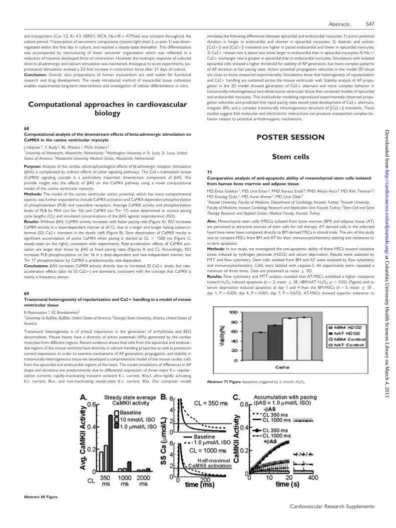
Purpose: Analysis of the cardiac electrophysiological effects of b-adrenergic receptor stimulation(bAS) is complicated by indirect effects of other signaling pathways. The Ca2+/calmodulin kinase(CaMKII) signaling cascade is a particularly important downstream component of bAS. Weprovide insight into the effects of bAS on the CaMKII pathway using a novel computationalmodel of the canine ventricular myocyte.Methods: The model of the canine ventricular action potential, which has many compartmentalaspects, was further expanded to include CaMKII activation and CaMKII-dependent phosphorylationof phospholamban (PLB) and ryanodine receptors. Average CaMKII activity and phosphorylationlevels of PLB by PKA (on Ser 16) and CaMKII (on Thr 17) were determined at various pacingcycle lengths (CL) and simulated concentrations of the bAS agonist isoproterenol (ISO).Results: Without bAS, CaMKII activity increases with faster pacing rate (Figure A). ISO increasesCaMKII activity in a dose-dependent manner at all CL due to a larger and longer lasting subsarco-lemmal (SS) Ca2+ transient in the dyadic cleft (Figure B). Slow deactivation of CaMKII results insignificant accumulation of active CaMKII when pacing is started at CL , 1500 ms (Figure C;steady-state on the right), consistent with experiments. Rate-acceleration effects of CaMKII acti-vation are larger than those by bAS at fixed pacing rates (Figures A and C). Accordingly, ISOincreases PLB phosphorylation on Ser 16 in a dose-dependent and rate-independent manner, butThr 17 phosphorylation by CaMKII is predominantly rate-dependent.Conclusions: bAS increases CaMKII activity directly due to increased SS Ca2+ levels, but rate-acceleration effects (also via SS Ca2+) are dominant, consistent with the concept that CaMKII ismainly a frequency sensor.
69Transmural heterogeneity of repolarization and Ca21 handling in a model of mouseventricular tissue
R. Rasmusson 1; VE. Bondarenko 2
1University at Buffalo, Buffalo, United States of America; 2Georgia State University, Atlanta, United States ofAmerica
Transmural heterogeneity is of critical importance in the generation of arrhythmias and EKGabnormalities. Mouse hearts have a diversity of action potentials (APs) generated by the cardiacmyocytes from different regions. Recent evidence shows that cells from the epicardial and endocar-dial regions of the mouse ventricle have diversity in calcium handling properties as well as potassiumcurrent expression. In order to examine mechanisms of AP generation, propagation, and stability intransmurally heterogeneous tissue we developed a comprehensive model of the mouse cardiac cellsfrom the epicardial and endocardial regions of the heart. The model simulations of differences in APshape and durations are predominantly due to differential expression of three major K+ repolar-ization currents: rapidly-inactivating transient outward K+ current, IKto,f, ultra-rapidly activatingK+ current, IKur, and non-inactivating steady-state K+ current, IKss. Our computer model
simulates the following differences between epicardial and endocardial myocytes: 1) action potentialduration is longer in endocardial and shorter in epicardial myocytes; 2) diastolic and systolic[Ca2+]i and [Ca2+]i transients are higher in paced endocardial and lower in epicardial myocytes;3) Ca2+ release rate is about two times larger in endocardial than in epicardial myocytes; 4) Na+/Ca2+ exchanger rate is greater in epicardial than in endocardial myocytes. Simulations with isolatedepicardial cells showed a higher threshold for stability of AP generation, but more complex patternsof AP duration at fast pacing rates. Action potential propagation velocities in the model 2D tissueare close to those measured experimentally. Simulations show that heterogeneity of repolarizationand Ca2+ handling are sustained across the mouse ventricular wall. Stability analysis of AP propa-gation in the 2D model showed generation of Ca2+ alternans and more complex behavior intransmurally-inhomogeneous two-dimensional ventricular tissue that contained models of epicardialand endocardial myocytes. This multicellular modeling reproduced experimentally observed propa-gation velocities and predicted that rapid pacing rates would yield development of Ca2+ alternans,irregular APs, and a complex transmurally inhomogeneous structure of [Ca2+]i transients. Thesestudies suggest that molecular and electrotonic interactions can produce unexpected complex be-havior related to potential arrhythmogenic mechanisms.
POSTER SESSION
Stem cells
71Comparative analysis of anti-apoptotic ability of mesenchymal stem cells isolatedfrom human bone marrow and adipose tissue
MD. Ertas Gokhan 1; MD. Ural Ertan 2; PHD. Karaoz Erdal 3; PHD. Aksoy Ayca 3; MD. Kilic Teoman 2;MD. Kozdag Guliz 1; MD. Vural Ahmet 1; MD. Ural Dilek 1
1Kocaeli University, Faculty of Medicine, Department of Cardiology, Kocaeli, Turkey; 2Kocaeli University,Faculty of Medicine, Invasive Cardiology Research and Application Unit, Kocaeli, Turkey; 3Stem Cell and GeneTherapy Research and Applied Center, Medical Faculty, Kocaeli, Turkey
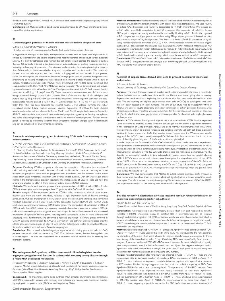
Aim: Mesenchymal stem cells (MSCs) isolated from bone marrow (BM) and adipose tissue (AT)are perceived as attractive sources of stem cells for cell therapy. AT derived cells in the infarctedheart have never been compared directly to BM derived MSCs in clinical trials. The aim of this studywas to compare MSCs from BM and AT for their immunocytochemistry staining and resistance toin-vitro apoptosis.Methods: In our study, we investigated the anti-apoptotic ability of these MSCs toward oxidativestress induced by hydrogen peroxide (H2O2) and serum deprivation. Results were assessed byMTT and flow cytometry. Stem cells isolated from BM and AT were analysed by flow cytometryand immunocytochemistry. Cells were labeled with caspase-3. All experiments were repeated aminimum of three times. Data are presented as mean + SD.Results: Flow cytometry and MTT analysis revealed that AT-MSCs exhibited a higher resistancetoward H2O2 induced apoptosis (n ¼ 3, mean + SE, hBM-hAT H2O2, p , 0.02) (Figure) and toserum deprivation induced apoptosis at day 1 and 4 than the BM-MSCs (n ¼ 3, mean + SE ,day 1, P ¼ 0.029, day 4, P ¼ 0.001, day 7, P ¼ 0.672). AT-MSCs showed superior tolerance to
Abstract 68 Figure
Abstract 71 Figure Apoptosis triggered by 2 mmol/L H2O2.
Abstracts S47
Cardiovascular Research Supplements
at Colum
bia University H
ealth Sciences Library on M
arch 4, 2013http://cardiovascres.oxfordjournals.org/
Dow
nloaded from
oxidative stress triggered by 2 mmol/L H2O2 and also have superior anti-apoptosis capacity towardserum-free culture.Conclusion: AT-MSCs could be a good source as an alternative to BM-MSCs and should be con-sidered for clinical applications.
72Cardiomyogenic potential of murine skeletal muscle-derived progenitor cells
C. Poulet 1; T. Christ 1; E. Wettwer 1; U. Ravens 1
1Dresden University of Technology, Medical Faculty Carl Gustav Carus, Dresden, Germany
In regenerative therapy of the heart, transplantation of stem cells to form new myocardium islimited by the inability of the cells to integrate into host myocardium and conduct cardiac electricalactivity. It is now hypothesized that refining the cell sorting could upgrade the results of such atherapy. Of particular interest is the description of subpopulations of skeletal muscle progenitorsfeaturing cardiomyogenic properties. Our aim was to characterize the electrophysiological proper-ties of these cells to determine whether they are compatible with cardiac function. We previouslyshowed that the cells express functional cardiac voltage-gated sodium channels. In the presentstudy, we investigated the presence of functional voltage-gated calcium channels. Progenitor cellsproliferating as floating myospheres were isolated from murine skeletal muscle. After 6 days ofculture, myospheres-derived cells (MDCs) were investigated with voltage-clamp technique andsemi-quantitative RT-PCR. With 10 mM Barium as charge carrier, MDCs demonstrated fast activat-ing inward currents with a threshold at -15 mV and peak activation at +5 mV. Peak current densityamounted to -18.2 + 1.2 pA/pF (n ¼ 30). These parameters are consistent with Ba2+ currentsbeing conducted through L-type Ca2+ channels. Block of the currents by 10 mM nifedipine andpotentiation by 10 mM Bay K 8644 confirmed this hypothesis. Interestingly the activation and inac-tivation rates (time to peak at +10 mV: 16.0 + 0.8 ms; tinact : 80.1 + 5.3 ms, n ¼ 30) were muchfaster than what has been described for skeletal muscle L-type calcium currents and ratherresembled cardiac L-type calcium currents kinetics. Expression of mRNA for both skeletalmuscle and cardiac calcium channel isoforms, Cav1.1 and Cav1.2 respectively, was confirmed byRT-PCR. We conclude that skeletal muscle-derived progenitors growing as floating myosphereshad some electrophysiological characteristics similar to those of cardiomyocytes. Further investi-gation is needed to determine whether these properties undergo changes upon differentiationand can be influenced by environmental conditions.
73A retinoic acid expression program in circulating CD34 cells from coronary arterydisease patients
CTM. Van Der Pouw Kraan 1; SH. Schirmer 2; JO. Fledderus 3; PD. Moerland 4; TA. Leyen 1; JJ. Piek 5;N. Van Royen 5; AJG. Horrevoets 1
1VU University Medical Center, Institute for Cardiovascular Research (ICaRVU), Amsterdam, Netherlands;2Universitatsklinikum des Saarlandes, Homburg, Germany; 3Academic Medical Center, University ofAmsterdam, Department of Medical Biochemistry, Amsterdam, Netherlands; 4Academic Medical Center,Department of Clinical Epidemiology Biostatistics & Bioinformatics, Amsterdam, Netherlands; 5AcademicMedical Center, Department of Cardiology at the University of Amsterdam, Amsterdam, Netherlands
Purpose: Circulating CD34+ progenitor cells have the potential to differentiate into a variety ofcells, including endothelial cells. Because of their multipotential character, autologous bonemarrow-, or peripheral blood derived progenitor cells have been used for ischemic cardiac tissuerepair after acute myocardial infarction with overall limited success. Our aim was to gain moreinsight in the transcriptional program regulating the multipotency of CD34+ cells and how thisprogram is affected in CD34+ from coronary artery disease (CAD) patients.Methods: We performed a whole genome transcriptome analysis of CD34+ cells, CD4+ T cells,CD14+ monocytes, and macrophages from 12 patients with CAD and 11 matched controls.Results: Analysis of the expression profile of CD34+ cells, compared to that of other mono-nuclear cells from the same individuals, revealed a high expression level of histone-encodinggenes, and KRAB box transcription factors, known to be involved in gene silencing. This correlatedwith high expression levels in CD34+ cells for the progenitor markers HOXA5 and HOXA9, whichare known to control expression of KRAB factor genes. The comparison of expression profiles ofCD34+ cells from CAD patients and controls revealed a less naıve phenotype in patients’ CD34+cells, with increased expression of genes from the Mitogen Activated Kinase network and a loweredexpression of a panel of histone genes, reaching levels comparable to that in more differentiatedcirculating cells. Furthermore, we observed a reduced expression of several genes involved inCXCR4-signalling and migration to CXCL12. Correlation- and pathway analysis indicated that thealtered gene expression profile of CD34+ cells in CAD patients was related to activation/differen-tiation by a retinoic acid-induced differentiation program.Conclusion: The reduced adhesive/migratory capacity of circulating precursor cells in CADpatients may restrict their recruitment by CXCL12 to ischemic tissues, thereby limiting their rolein tissue repair. The data are supportive for a causative role of retinoic acid in reducing themigratory capacity.
74The endogenous NO synthase inhibitor asymmetric dimethylarginine impairsangiogenic progenitor cell function in patients with coronary artery disease througha microRNA dependent mechanism
F. Fleissner 1; V. Jazbutyte 1; J. Fiedler 1; P. Galuppo 2; M. Mayr 3; G. Ertl 2; J. Bauersachs 2; T. Thum 1
1Hannover Medical School, Department of Molecular and Translational Therapeutic Strategies, Hannover,Germany; 2Julius-Maximilians University, Wurzburg, Germany; 3King’s College London, CardiovascularDivision, London, United Kingdom
Background: The endogenous nitric oxide synthase (NO) inhibitor asymmetric dimethylarginine(ADMA) is increased in patients with coronary artery disease and may regulate function of circulat-ing angiogenic progenitor cells (APC) by small regulatory RNAs.
Methods and Results: By using microarray-analyses we established microRNA expression profilesof human APC and showed major similarities with that of mature endothelial cells. We used ADMAto induce APC dysfunction and found 16 deregulated (p , 0.05) microRNAs. We focused onmiR-21 which was 3-fold upregulated by ADMA treatment. Overexpression of miR-21 in humanAPC impaired migratory capacity which could be rescued by blocking miR-21. To identify regulatedmiR-21 targets we employed proteome analysis using 2D-gel electrophoresis followed by massspectrometric analysis of regulated proteins. We found transfection of miR-21 precursors to signifi-cantly repress superoxide dismutase 2 (SOD2) in APC which increased intracellular reactive oxygenspecies (ROS) concentration and impaired NO bioavailability. ADMA-mediated impairment of NObioavailability in APC and migration defects could be rescued by miR-21 blockade. Importantly, APCfrom patients with coronary artery disease and high ADMA plasma levels displayed.4-fold elevatedmiR-21 levels and impaired migratory capacity, which could be normalized by miR-21 antagonism.Conclusions: We identified a novel miR-21-dependent mechanism of ADMA-mediated APC dys-function. MiR-21 antagonism therefore emerges as an interesting approach to improve dysfunctionalAPC in patients with coronary artery disease.
75Potential of adipose tissue-derived stem cells to prevent post-infarct ventriculararrhythmias
S. Protze ; A. Bussek ; U. RavensDresden University of Technology, Medical Faculty Carl Gustav Carus, Dresden, Germany
Purpose: The most frequent cause of sudden death after myocardial infarction is ventriculartachyarrhythmia due to conduction block within the fibrous scar tissue, giving rise to reentry.Regenerative medicine aims at replacing the scar tissue with functional tissue derived from stemcells. We are working on adipose tissue-derived stem cells (ADSCs) as autologous stem cellsthat are easily accessible in large numbers. The aim of our study was to investigated whetherADSCs are able to couple electrically and allow conduction of action potentials between areasof viable myocardium. Therefore we analysed ADSCs for the expression and function of connexin43 (Cx43), which is the major gap junction protein responsible for the electrical coupling betweencardiomyocytes.Results: ADSCs isolated from gonadal adipose tissue of six-month-old C57BL/6 mice expressedCx43 as shown by antibody staining. Western blot analysis did not detect a significant differencein the expression of Cx43 between ADSCs and bone marrow mesenchymal stem cells, whichwere previously shown to express functional gap junction channels, yet both cell types expressedsignificantly lower amounts of Cx43 than cardiac tissue. Furthermore the Western blots resultssuggested that ADSCs have correctly arranged Cx43 channels in their membrane due to the pres-ence of the fully phosphorylated p2 isoform in addition to p0 and p1.In order to prove the functionality of the Cx43 channels in vitro, conduction block experimentswere performed. For this Purpose neonatal mouse cardiomyocytes (nCMs) were cultured on multi-electrode arrays to form a synchronously beating monolayer. Propagation of electrical activity wasinterrupted by scratching a 400-200 mm-wide channel into the monolayer perpendicular to thedirection of conduction resulting in two independently beating areas. Into this scratch channel1x10^5 ADSCs were seeded and cultures were investigated for resynchronisation of the nCMswithin 24-72 h. Four out of six experiments resulted in resynchronisation of the nCM fields viaADSCs (66%, n ¼ 6). The conduction velocity in ADSCs was 3.0 + 1.3 mm/ms (n ¼ 4) comparedto 40 + 11 mm/ms (n ¼ 6) in nCMs. No resynchronisation was observed when the gap was filledwith cardiac fibroblasts (n ¼ 2).Conclusion: We have demonstrated that ADSCs do in fact express functional Cx43 channels intheir membranes that allow them to conduct electrical signals albeit at a slower speed than cardi-omyocytes. Therefore we are currently investigating if lentiviral-overexpression of Cx43 in ADSCscan improve conduction to the velocity seen in neonatal cardiomyocytes.
76Toll-like receptor 4 inactivation alleviates impaired vascular reendothelialisation byimproving endothelial progenitor cell adhesion
FYL. Li 1; RLC. Hoo 1; KSL. Lam 1; A. Xu 1
1Queen Mary Hospital, Department of Medicine, Hong Kong, Hong Kong SAR, People’s Republic of China
Introduction: Atherosclerosis is an inflammatory disease which is in part mediated by Toll-likereceptor 4 (TLR4). Endothelial injury, an initiating step in atherosclerosis, can be repairedthrough endothelial progenitor cell (EPC) activation, which has been shown to be diminished inpatients with diabetes and/or vascular diseases. Vascular inflammation appears to impair the capacityof EPC in mediating the reendothelialisation process. It remains to be determined whether TLR4 isinvolved in this impairment.Method: ApoE-deficient (ApoE2/2/TLR4+/+) mice and ApoE2/2 mice lacking functional TLR4(ApoE2/2/TLR42/2) were used in this study. Wire injury was introduced to the right commoncarotid artery of the mice which were allowed to recover. Vascular repair was assessed by Evansblue staining of the injured arteries after 3 days. Circulating EPCs were quantified by flow cytometryanalyses. Bone marrow-derived EPCs (BM-EPCs) were i) assessed for reendothelialisation capacityafter transplantation in vivo; ii) adhesion function in vitro and iii) reactive oxygen species production.ApoE2/2 mice were treated with N-acetyl CoA (NAC) for 21 days prior to vascular injury andassessment for reendothelialisation and circulating EPC number.Results: Reendothelialisation after wire injury was impaired in ApoE2/2/TLR4+/+ mice and wasconcomitant with an increased number of circulating EPCs. Inactivation of TLR4 in ApoE2/2/TLR42/2 mice conferred an improved reendothelialisation, together with a paradoxical decreasein EPC number. Further findings suggested that the repair and adhesion capacity of EPCs fromApoE2/2/TLR4+/+ mice was down-regulated. Transplantation of BM-EPC isolated fromApoE2/2/TLR42/2 mice improved vascular repair, compared to cells from ApoE2/2/TLR4+/+ mice. Adhesion was diminished in BM-EPCs isolated from ApoE2/2/TLR4+/+ mice,but was augmented in BM-EPCs from ApoE2/2/TLR42/2 mice. Oxidative stress was increasedin BM-EPCs isolated from ApoE2/2/TLR4+/+ mice, compared to those from ApoE2/2/TLR42/2 mice, suggesting a possible mechanism for EPC dysfunction. Antioxidant treatment of
S48 Abstracts
Cardiovascular Research Supplements
at Colum
bia University H
ealth Sciences Library on M
arch 4, 2013http://cardiovascres.oxfordjournals.org/
Dow
nloaded from
ApoE2/2 mice using NAC led to an improved vascular repair and lowered circulating EPCs,mimicking the effect of TLR4 inactivation.Conclusion: Impaired reendothelialisation in ApoE2/2 mice is attributable to a decreased abilityof EPC function, which can be improved by TLR4 inactivation. Thus modulation of TLR4 activitymay represent an effective strategy for improving EPC function and hence protection againstatherosclerosis.
77Lysophosphatidic acid receptors LPA1 and LPA3 induce neointima formation byrecruitment of smooth muscle progenitor cells
P.Subramanian 1; E.Karshovska 2; R.Megens 3; S.Akhtar 1; K.Heyll 1; Y. Jansen 1; C.Weber 1; A.Schober1
1Institute of Molecular Cardiovascular Research, University Hospital Aachen, RWTH, Aachen, Germany;2Ludwig-Maximilians University, Campus Innenstadt, Department of Cardiology, Munich, Germany;3Interdisciplinary Center for Clinical Research BIOMAT within the Faculty of Medicine, RWTH Aachen,Aachen, Germany
Objective: To evaluate the role of lysophosphatidic acid (LPA) receptors LPA1 and LPA3 in initi-ating CXCL12-dependent vascular repair by smooth muscle progenitor cells (SPCs).Methods: Intraluminal incubation of the carotid artery with LPA20:4 (40 mM, 30 min) was per-formed in C57BL/6 mice and neointimal area determined after 28 days. Some mice wereco-incubated with the LPA1/3 receptor antagonist Ki16425, systemically treated with the CXCR4antagonist POL5551, or perivascularly with LPA receptor-specific siRNA dissolved in Pluronicgel. Multi-photon microscopy was applied to study mice after transplantation with SM22-LacZbone marrow cells. Peripheral Sca-1+/Lin- SPC were studied after 1 day by FACS. ApoE2/2mice on western-type diet were treated with Ki16425 (ip, 5mg/kg/d, 3 weeks; n ¼ 5-6) aftercarotid wire injury. Neointimal area and cellular plaque composition including Sca-1+SMCs werestudied after 28 days. LPA receptor expression was examined by qRT-PCR.Results: CXCL12 protein in the vessel wall was increased by LPA20:4 from day 1 on. LPA20:4induced neointima formation by mobilization and recruitment of bone marrow-derived SPCs.Local inhibition of LPA1 or LPA3 by Ki16425 or siRNA revealed that both receptors independentlymediate the SPC-dependent neointima formation by LPA20:4. Systemic blockade of CXCR4 com-pletely blocked LPA-induced neointima formation and SPC mobilization. Whereas mRNAexpression of LPA1 was diminished, LPA3 was increased after vascular injury. Wire injury-inducedneointima formation was reduced by 70% through Ki16425. This was due to a decreased neointimalSMC, especially Sca-1+SMC, and macrophage content.Conclusions: LPA1 and LPA3 promote neointima formation through activation of CXCL12/CXCR4-mediated mobilization and recruitment of SPCs.
78Erythropoietin receptor expression and its role in resident stem cells of the adultmouse heart
MP. Zafeiriou 1; C. Noack 1; A. Renger 1; R. Dietz 2; L. Zelarayan 1; M. Bergmann 3
1Max Delbruck Center for Molecular Medicine, Berlin, Germany; 2Charite - Campus Virchow-Klinikum,Department of Cardiology, Berlin, Germany; 3Asklepios Clinic St. Georg, Hamburg, Germany
Several evidences support that a regeneration process contributes to cardiac replenish at baselineand after injury as well as the existence of cardiac stem cells that amplify and commit to the myocytelineage in response to increased workload. However, the fundamental mechanisms of cardiac regen-eration remain incompletely defined. We previously found that b-catenin depletion attenuates postinfarct left ventricule (LV) remodeling via increased differentiation of alphaMHCpos/GATA4pos/Sca-1pos/TropTneg subpopulation of resident cardiac progenitor cells. Next, we aimed tofurther dissect the identity and the signaling pathways driving proliferation and differentiation ofendogenous adult cardiac progenitor cells in cardiac remodeling, which may help to elucidate thepotentiality of cardiac progenitor cells for therapeutic intervention.In this study we observed that the above-mentioned identified progenitor cells expressed earlycardiac markers as Nkx2.5, eHand and TBX5 via immunofluorescence and real time PCR analysis.Interestingly, this set of activated transcription factors is characteristic for cells deriving from theembryonic first heart field (FHF) that gives rise to most of the mature left ventricle cardiomyocytes.Moreover, a sub-population of these progenitor cells showed expression of the Erythropoietinreceptor (EpoR). Epo has been shown to exert a therapeutic action in myocardial ischemia/ reper-fusion and congestive heart failure. EpoR was shown until now to be expressed in mesenchymal andendothelial progenitors cells but its expression and its role in cardiac committed progenitors was sofar not examined. To address this issue we established a co-culture system using adult mouse fibro-blasts as a feeder layer. Employing immunofluorescence and real time PCR analysis we found thatEpoR is expressed in cardiac committed progenitor cells expressing earlier cardiac specific tran-scription factors (Tbx5 and eHand) that differentiated to TroponinT and alphaMHC expressingcells. These cells proliferated and differentiated in culture. We observed expression of EpoR todecrease upon differentiation in vitro similarly to the loss of EpoR expression from neonatal toadult mouse heart.We report here a new role of Epo-EpoR signaling where apart from angiogenesis it promotes thedifferentiation of endogenous cardiac precursor cells to cardiomyocytes and we therefore believeEpo to be a potential candidate for regenerative therapy of patients affected by cardiac stress.
79Colony-forming unit-Hill cells express monocytic rather than endothelial markers
I. Meln 1; AB. Malashicheva 1; SV. Anisimov 1; NI. Kalinina 2; VY. Sysoeva 2; AY. Zaritskey 1
1V.A. Almazov Federal Center of Heart, Blood and Endocrinology, Saint Petersburg, Russian Federation;2State University, Moscow, Russian Federation
Endothelialcells (ECs) play an important role in many physiological functions. Disturbanceof endo-thelial function is a key early event in the development of manyvascular and cardiovascular diseases.
Overthe past years, neovascularization is thought to be tightly linked toendothelial progenitor cells(EPCs). EPCs represent a minor subpopulation ofperipheral blood mononuclear cells (PBMCs).Potential link between EPCsphenotype and severity of cardiovascular disease became a subjectof intensivestudies. Several Methods are available for in vitro cell culturing of EPCs. In an originalmethod, adult PBMCs are plated over fibronectin-coated dishes. After a 48 h pre-platingstep deplet-ing the sample of adherent macrophages and mature ECs, thenon-adherent cells are removed andre-plated on fresh fibronectin-coated dishes. Individual colonies emergein 3.5-5 days, comprised ofround cells located centrally with spindle-shapedcells sprouting at the periphery. Colonies of thistype are now widely referredto as colony-forming unit-ECs (CFU-ECs), also as colony-formingunit-Hill(CFU-Hill) cells or early outgrowth CE-EPCs. In the present study, we have analyzedtheantigene profile of CFU-Hill cells using immunofluorescence staining. Humanumbilical vein endo-thelial cells (HUVEC) and human fibroblast cell line servedas controls. Unexpectedly, we did notobserve positive signal for an assay of endothelialmarkers (PECAM-1 (CD31), VE-cadherin,VEGFR-2 (KDR, CD309), Von Willebrandfactor) and CD90 (marker specific to fibroblast cells).In contrast, CFU-Hillcells demonstrated strong positivity for CD68, indicating these cells mustbelongto a monocyte/macrophage lineage. Our results stress an importance of developingreliableand specific protocols for purifying EPCs from peripheral blood andexpanding those cells invitro. This issue is especially important fordeveloping cell and tissue therapy approaches.
80Differentiation and enrichment of pacemaker cardiomyocytes from testis-derivedadult pluripotent stem cells
A. Barbuti 1; A. Scavone 1; N. Mazzocchi 1; A. Crespi 1; D. Capilupo 1; D. Difrancesco 1
1University of Milano, Milano, Italy
Several studies have recently shown that multi/pluripotent stem cells can be derived in vitro frommouse and human spermatogonial stem cells (SSCs). We have recently isolated Pluripotent AdultSpermatogonial-derived Stem Cells (PA-SSCs) from mouse testes and verified that they possessseveral features typical of embryonic stem cells (ESCs).Purpose: Our aim is to differentiate PA-SSCs into sinoatrial-like cardiomyocytes and to optimizethe yield of cardiomyocytes by non-genetic selection of cardiac precursors.Methods and Results: We applied to PA-SSCs the same protocol used to induce ESCs to differ-entiate into cardiomyocytes with typical features of pacemaker cells which consist in the formationof embryoid bodies (EBs). We verified by immunofluorescence and RT-PCR experiments theexpression of cardiac proteins (sarcomeric a-actinin, myosin, connexin 43); also, patch clamp analy-sis and confocal microscopy showed the presence of spontaneously beating cells expressing thepacemaker If current (and specifically the HCN1 and HCN4 isoforms), thus confirming a sinoatrial-like phenotype. Preliminary data indicate that cardiac progenitors can be isolated, from both ESCsand PA-SSCs, based on the membrane expression of the protein CD166 at a specific stage (day 8)of differentiation. CD166+ cells were sorted by FACS and allowed to re-aggregate in EB-like struc-tures. After a few days in culture these cells started to beat spontaneously; immunofluorescenceanalysis indicated that about 70% of the CD166+ cells were a-actinin positive, and almost 40%of these cells expressed the pacemaker channel HCN4.Conclusions: Our data show that PA-SSCs, like ESCs, can be differentiated into sinoatrial-like car-diomyocytes and that the yield of cardiomyocytes can be increased by selecting CD166+ cardiacprecursor at a specific stage of differentiation. These findings, based on the properties and avail-ability of human spermatogonial-derived pluripotent cells, open new opportunities in stem cell-based therapies for cardiac interventions.
81Delivery of myogenic differentiated adult human mesenchymal stem cells tocryoinjured myocardium
L. Qian 1; W. Shim 1; Y. Gu 1; S. Mohammed 1; P. Wong 1
1National Heart Centre Singapore (NHCS), Singapore, Singapore
Introduction: We have previously reported improved cardiac functions in stem cell therapy andenhanced cardiac gene expressions on differentiated adult human mesenchymal stem cells culturedwith collagen V. This study investigates the delivery Methods and feasibility of cellular injection and acardiac patch in an infarcted rat’s model.Materials and methods: Myocardial infarction in Wistar rats was induced by cryoinjury using a3mm metal rod cooled by liquid nitrogen. Intracardiac cell transplantation of myogenic differentiatedadult human mesenchymal stem cells was then delivered by injection with a mix of collagen I and V orby a commercial collagen patch to the damaged area. Hemodynamic measurements were obtained forbaseline and 6 weeks post treatment using echocardiography while pressure volume catheterizationwas performed at end point. Hearts were explanted for histological and molecular studies.Results: Echocardiography results showed that systolic myocardial velocities of the posteroinferiorwalls of left ventricle in the cell injection group did the best out of the other groups (0.03% +0.08%) when compared to baseline results. From PV-loops data, cell injection group as well asthe cell patch group showed improved global LVEF when compared to serum free medium injectiongroup. LV dilatation was prevented by inhibiting further expansion of end-diastolic volume andreducing end-diastolic pressure as compared to the control groups.Conclusion: Transplanting stem cells into the damaged myocardium appears to prevent furtherdeterioration of the systolic function. Using differentiated cells with collagen patch improvedtissue compliance and diastolic function, preventing negative LV remodelling.
83A functional cross-talk of KLF15 to the Wnt signalling pathway and its role in adultcardiac precursor cell regulation
C. Noack 1; A. Renger 1; MP. Zafiriou 1; R. Dietz 1; HJ. Schaeffer 2; MW. Bergmann 3; LC. Zelarayan 1
1Charite - Campus Berlin Buch/Experimental & Clinical Research Center, Department of Cardiology, Berlin,Germany; 2MPI of Biochemistry, Munich, Germany; 3Asklepios Clinic St. Georg, Hamburg, Germany
Cardiac endogenous regeneration has recently been identified to play a fundamental role in tissuehomeostasis at baseline and following injury. The elucidation of signaling pathways governing cardiac
Abstracts S49
Cardiovascular Research Supplements
at Colum
bia University H
ealth Sciences Library on M
arch 4, 2013http://cardiovascres.oxfordjournals.org/
Dow
nloaded from
progenitor cells (CPCs) may lead to novel targets for heart failure therapy. We previously foundheart specific downregulation of b-catenin to be required for adaptive hypertrophy preservingleft ventricular function and to enhance endogenous CPC differentiation post-infarct. Thus, weaimed to identify a potential cardiac specific repressor of b-catenin and to dissect the signaling path-ways driving differentiation of endogenous CPCs in cardiac remodeling.We identified the transcriptional regulator KLF15 to interact and co-localize with b-catenin andNLK, a known inhibitorof Wnt/b-catenin canonical signaling. Employing different cell lines andprimary neonatal cardiomyocytes we found KLF15 to repress b-catenin-LEF/TCF dependentgene transcription in a dose-dependent manner (P , 0.001). Therefor KLF15 requires anN-terminal domain including amino acids 45 to 152 and the C-terminus containing the NLS. Analysisof adult Klf15 knockout (ko) mice revealed a significant decline in cardiac function by aging at base-line. Subjecting Klf15 ko and respective controls (wt) to experimental induced cardiac stress,echocardiography showed earlier left ventricle dilation in ko when compared to wt mice indicatinga faster cardiac deterioration, which is in line with previous observations showing that Klf15 ko miceare extremely sensitive to cardiac stress. Flow cytometry analysis of the non-cardiomyocyte cellfraction revealed a significant decrease of a cardiac committed stem cell population, marked bySca1pos/aMHCpos and Tbx5pos/cTntneg cells, in ko mice (P , 0.01), evidencing a reducedcardiac regeneration in mice lacking KLF15. Mechanistically, upregulation of the b-catenin target-gene TCF4 at mRNA (P , 0.01) and protein level was found in Klf15 ko mice. Moreover, down-regulation of NLK (P , 0.05) was found upon KLF15 deletion as well as upon cardiac specificb-catenin stabilization in vivo, indicating that KLF15 is an upstream modulator of b-catenin.These data showed that KLF15 regulates canonical (via b-catenin) and non-canonical (via NLK) Wntsignaling and plays an essential role in controlling adult CPC biology in the adult heart. This work isan important step forward to elucidate new signaling molecules that are emerging as possible targetsto therapeutically attenuate cardiac remodeling.
Atrial fibrillation
84NCX current is increased in human chronic atrial fibrillation: a possible explanationfor contractile dysfunction?
PP. Kovacs 1; J. Simon 2; T. Christ 2; E. Wettwer 2; A. Varro 1; U. Ravens 2
1University of Szeged, Faculty of Medicine, Department of Pharmacology & Pharmacotherapy, Szeged,Hungary; 2Dresden University of Technology, Department of Pharmacology and Toxicology, Dresden,Germany
Purpose: Chronic atrial fibrillation (AF) is characterised by contractile dysfunction, that can beexplained by reduction in transsarcolemmal Ca2+ influx via ICa,L. Furthermore increased Ca2+efflux could aggravate contractile dysfunction in AF. Since Ca2+ efflux via Na/Ca exchanger(NCX) is the major mechanism to extrude Ca2+ that has entered the cell, we have testedwhether NCX current is increased in AF. Relevance of NCX for excitation-contraction couplingwas examined by employing the newly developed NCX inhibitor SEA-0400 in intact atrial trabecu-lae in SR and AF.Methods: Atrial myocytes were isolated from patients with sinus rhythm (SR) and AF. Membranecurrents were measured from a constant holding potential of 210 mV in a bath solution free ofNa+ and Ca2+ with a pipette solution containing high Na+ and low Ca2+. NCX current wasmeasured as outward current evoked by elevating extracellular Ca2+ concentration [Ca2+]e to1 mM. Isolated trabeculae were used to measure action potentials and force of contraction (Fc)in normal Tyrode’s solution. All experiments were performed at 378C.Results: Outward NCX currents in response to increase in [Ca2+]e were significantly larger in AF8,96 + 0,37 (31/13) compared to SR 6,39 + 0,26 pA/pF (80/23, p , 0.001). Pretreatment withSEA-0400 completely blocked activation of outward currents in SR and AF. Sensitivity toSEA-0400 was similar in AF (-logEC50�0.2 mM). Trabecular action potentials were not alteredwith the highest SEA concentration studied (10 mM). Furthermore, SEA-0400 had no effect onFc generation. The positive inotrop effect of b-adrenoceptor stimulation with noradrenaline (10mM) was not modified by SEA-0400. Block of transient potassium outward currents with 4-aminopyridine was previously shown to enhance Fc by indirectly increasing ICa,L at more positiveplateau voltages. SEA-0400 also did not influence this positive inotropic intervention.Conclusions: NCX current is larger in human AF than in SR. Block of NCX with SEA-0400 did notaffect basal or inotropic interventions, thereby challenging the concept that NCX regulates intra-cellular Ca2+ in a manner relevant for force under steady-sate conditions. Further work isneeded to investigate whether the relevance of human atrial NCX is relevant for electrical stabilityin EC-coupling related phenomena.
85Changes in connexin43 distribution pattern are associated with postoperative newonset atrial fibrillation
P. Athias 1; JE. Wolf 2; O. Bouchot 3; D. Vandroux 4; A. Mathe 2; A. De Carvalho 2; G. Laurent 2
1Institute of CardioVascular Research (IRCV) - Hospital of Bocage, Dijon, France; 2Cardiology Unit, UniversityHospital Center, Dijon, France; 3Cardiovascular Surgery Unit, Dijon, France; 4NVH Medicinal, Dijon, France
Atrial fibrillation (AF) is a common complication after cardiac surgery, but the proarrhythmic sub-strates underlying the development of postoperative AF remain unclear. This study investigated thehypothesis that alteration of atrial connexin43 (Cx43) distribution may be a determinant of a pre-disposition to postoperative new onset AF. This study included 79 patients undergoing "on pump"cardiac surgery (coronary artery bypass grafts, valve replacements or both). Right atrial appendage(RAA) tissue samples were taken at the aortic clamping (baseline, BSL) and just before the clampwas removed (post ischaemic time, PIT). Cardiac rhythm was monitored continuously for 48hours, and then daily12-lead ECGs and additional ECGs were performed for symptoms. In a sub-group of 25 age-matched patients with normal left atrial size and no AF history (AF+ group,n ¼ 11 and AF- group, n ¼ 14) immuno-histological and Western blotting analyses were performedby two experienced observers blinded to the groups. Cx43 distribution was assessed using a four
level ranking scale ("side to side" vs "end to end" calculated ratio: score 1 ¼ 0-24%, score 2 ¼25-49%, score 3 ¼ 50-74%, score 4 ¼ 75-100%). New onset AF occurred in 35.5% of the patients.Older age (.65 years) was the only independent predictive factor for AF (OR ¼ 2.97, p , 0.01). Inthe AF+ group, 8/11 patients had a Cx43 distribution pattern scored 1 at BSL, and changed to 3 or4 at PIT, corresponding to a lateralization phenomenon. This phenomenon was not associated witha longer cross clamp time or a valve replacement. Three patients who had a score of 3-4 at BSLdeveloped AF after surgery. None of the AF- patients had a score of 3 or 4 at BSL or after PIT.The amount of atrial fibrosis at BSL (collagen I and III) as well as the phosphorylation status ofCx43 at BSL and at PIT were not correlated with AF incidence. Patients presenting postoperativeAF had a higher TNFa plasma level than those who did not (AF+ group: 8.6 +/22.9 pg/ml vs FA-group: 6.2 +/22.7 pg/ml, p ¼ 0.03). During "on pump" cardiac surgery, a lateral Cx43 distributionpattern in RAA at BSL or a lateralization phenomenon during the ischemic period may be associatedwith post operative new onset AF. Inflammation may be involved in these changes which could alterconduction anisotropy in the atria which in turn may become proarrhythmic.
86The primary bile-salt glycocholate induces arrhythmias in human atrial myocardium
PP. Rainer 1; MS. Huber 1; F. Edelmann 2; T. Stojakovic 3; A. Trantina-Yates 4; M. Trauner 5; BM. Pieske 1;D. Von Lewinski 1
1Medical University of Graz, Department of Cardiology, Graz, Austria; 2University of Gottingen, Departmentof Cardiology and Pneumonology, Gottingen, Germany; 3Medical University of Graz, Department ofLaboratory Medicine, Graz, Austria; 4Medical University of Graz, Department of Cardiac Surgery, Graz,Austria; 5Medical University of Graz, Department of Gastroenterology and Hepatology, Graz, Austria
Background: Clinical evidence indicates that high bile-acid levels may induce arrhythmias.Recently, in vitro studies on rat cardiomyocytes confirmed this arrhythmogenic effect. The conse-quences of elevated bile-acid concentrations on human cardiac tissue, however, remain unknown.Methods: Analysis of serum bile-acid levels in 138 patients and correlation with age, sex andcomorbidities.Human atrial endocardial trabeculae (12 patients); modified Tyrode’s solution; 37C8, pH 7.4; 2.5mMCa++; electrical stimulation with 1 and 0.5Hz.Dose-response curve for the predominant human primary bile-acid (glycin-conjugated cholic-acid,GCA). Analysis of the appearance of arrhythmic extra contractions (AEC).Results: Patients with atrial fibrillation have higher levels of serum bile-acids (6.2 + 1.36 vs. 4.2 +2.39 mmol/l, p , 0.05), which do not correlate with sex, comorbidities and medication.No arrhythmias were observed at concentrations ≤30 mM at 1Hz stimulation. At 100 mM 1/12(8.3%), at 300 mM 4/12 (33.3%), at 1mM 7/12 patients (58.3%) developed AECs (EC50 262.6mM, nH 2.0). At 0.5Hz stimulation AECs did frequently occur at lower concentrations. At 30mM in 6/9 (66.7%), at 100 mM in 7/9 (77.8%) at 300 mM in 8/9 patients (88.9%) (EC50 24.0 mM,nH 5.6). Arrhythmias were fully reversible by switching to bile-acid free perfusate.Conclusions: Atrial fibrillation is associated with an elevation of serum bile-acids irrespective ofage, medication and comorbidities. The mean bile-acid concentration in patients with atrial fibrilla-tion is still within the physiologic range.Human atrial myocardium develops arrhythmias upon glycocholic-acid administration. Thesearrhythmias are dose-dependent, reversible and occur at pathophysiologic relevant concentrations.
87Primary cell culture model for structural changes caused by stretch, occurringbefore onset of atrial fibrillation
AR. De Jong 1; AH. Maass 1; SU. Oberdorf-Maass 1; IC. Van Gelder 1
1University Medical Center, Groningen, Netherlands
Purpose: Atrial fibrillation (AF) occurs often in the presence of a morphological substrate, forexample caused by hypertension or heart failure. Already before the first episode of AF structuralremodeling occurs, which deteriorates once AF develops. Stretch is thought to play an importantrole in the remodeling process. Our aim is to develop a cell culture model mimicking early atrialremodeling in pressure overload of the atria, representing the atrial situation after start of hyperten-sion or heart failure.Methods: Neonatal rat atrial cardiomyocytes (NRAM) were cultured and set under cyclical stretchon elastic membranes. Cells were analyzed after 3-24hr of stretch for dedifferentiation via investi-gating mRNA expression of alpha and beta-myosin heavy chain and skeletal alpha-actin, and break-down of contractile elements using western blotting. Expression of stress specific markers, such asANP, BNP and growth differentiation factor-15 (GDF15) were determined after stretching as wellas after stimulation with the alpha-adrenergic agonist phenyleprine (PE).Results: Stretching of atrial cardiomyocytes for 3-24hrs with 3Hz and 10% elongation or 1Hz and15% elongation did not change troponin T protein expression. These stress regimens in NRAMshow no evidence for myolysis. Dedifferentiation was suggested, shown by 1.5-fold increasedmRNA expression of skeletal alpha-actin and 1.8-fold increased beta/alpha-myosin heavy chainratio after stretching with 1Hz and 15% elongation for 6hr and 24hr, respectively. ANP mRNAlevels were unchanged in NRAM after stretching at all timepoints. ANP protein levels were
Abstract 86 Figure AECs upon 300 mM GCA (*) @ 1 Hz
S50 Abstracts
Cardiovascular Research Supplements
at Colum
bia University H
ealth Sciences Library on M
arch 4, 2013http://cardiovascres.oxfordjournals.org/
Dow
nloaded from
decreased and secreted ANP levels were increased after stretching with 1Hz and 15% elongation.ANP mRNA levels were also unchanged after 24hr PE stimulation. In contrast, hormonal stimulationin ventricular cardiomyocytes increased ANP mRNA expression 4.9-fold. BNP mRNA levels inNRAM were 2-fold increased after stretch for 3hr with 1Hz and 15% elongation and 3-fold afterstretch for 3hr with 3Hz and 10% elongation. GDF15 mRNA levels were 1.4-fold increased after24hr of stretch. Hormonal stimulation of NRAM increased BNP mRNA expression 1.7-fold,however GDF15 expression was unchanged.Conclusions: Stretch of neonatal rat atrial cardiomyocytes results in cellular changes which mayresemble those seen before and in the early course of AF such as dedifferentiation and changesin expression of natriuretic peptides. In addition, stimulation with PE of atrial and ventricular cardi-omyocytes evokes different responses in terms of the stress marker ANP.
88Left ventricular posterior wall thickness is an independent risk factor for paroxysmalatrial fibrillation: a retrospective paired case-control study
Y. Lin 1; J. Li 1; F. Wang 1; YM. He 1; X. Li 1; HF. Xu 1; XJ. Yang 1
1Department of Cardiology,the First Affiliated Hospital of Soochow University, Suzhou, People’s, ChinaRepublic of
Background: Atrial fibrillation is the most common clinically significantly cardiac arrhythmia, butits risk factors remain unknown. We have hypothesized that left ventricular posterior wall thicknessis an independent risk factor for paroxysmal atrial fibrillation.Methods: A series of 166 consecutive patients, hospitalized in the Department of Cardiology for anepisode of paroxysmal atrial fibrillation from Jan. 1st, 2006 to Dec. 31st, 2008, were included in thisstudy. A group of 166 healthy check-up people strictly matched for age and sex were used as con-trols in the same period. Univariable analysis and multivariable conditional Logistic stepwiseregression analysis were conducted. Receiver operating characteristic (ROC) curve analysis wasperformed on those significant indices obtained from the multivariable Logistic regression analysis.Results: The multivariable stepwise analysis identified left ventricular posterior wall thickness (P ¼0.0024, OR ¼ 1.348, 95% confidence interval [CI] 1.111 to 1.635), left atrial diameter (P , 0.0001,OR ¼ 1.130, 95%[CI] 1.072 to 1.191),tricuspid insufficiency (P ¼ 0.0018, OR ¼ 2.876, 95%[CI]1.483 to 5.576) and residence (P ¼ 0.0014, OR ¼ 0.437, 95% [CI] 0.263 to 0.725) as independentpredictors for paroxysmal atrial fibrillation. ROC curve analysis demonstrated the area under thecurve (AUC) ¼ 0.644, 95%CI 0.548 to 0.655, P ¼ 0.0001, and the cut-off value 9mm in the leftventricular posterior wall thickness, AUC ¼ 0.743, 95%CI 0.692 to 0.789,P ¼ 0.0001 and thecut-off value 38mm in the left atrial diameter, AUC ¼ 0.643, 95%CI 0.589 to 0.695,P ¼ 0.0001and the cut-off value .0 in the tricuspid insufficiency, and AUC ¼ 0.602, 95%CI 0.590 to0.695,P ¼ 0.0001 in the residence.Conclusions: In this strictly age; sex-matched, population-based sample, left ventricular posteriorwall thickness, left atrial diameter,tricuspid insufficiency and residence, were predictive risks for par-oxysmal atrial fibrillation. This study offers novel information therapeutically beyond that providedby traditional clinical atrial fibrillation risk factors.
89Cellular mechanisms of contractile dysfunction in atrial fibrillation
R. Coppini 1; C. Ferrantini 2; C. Ferrara 2; A. Rossi 3; A. Mugelli 1; C. Poggesi 2; E. Cerbai 1
1Department of Pharmacology, Florence, Italy; 2Department of Physiology, Florence, Italy; 3Careggi UniversityHospital, Florence, Italy
Atrial Fibrillation (AF) is associated with contractile impairment. Down regulation of L-Type Ca2+current, by reducing Ca2+entry during Action potential, plays a major role in determining contrac-tile dysfunction. However other, potentially revertible, EC-Coupling changes could be involved inhuman AF.We dissected atrial trabeculae from right atrial appendages of AF patients undergoing cardiacsurgery and used them for force recordings. Cells isolated from the same samples were used forCa2+current, Ca2+transients and reticular Ca2+content (caffeine) measurements. Samplesfrom sinus rhythm (SR) patients were used as controls.Despite a 75%reduced basal force, positive inotropic responses to isoproterenol, endothelin-1,stimulation pauses and high external[Ca2+] were preserved in AF.Basal ICa and Ca2+-transientswere significantly reduced (30%of SR) but transients showed a greater increase upon inotropicstimuli, reducing the difference with SR. No difference was found in time-course of mechanicaland Ca2+transients’ restitution and in the rate of spontaneous mechanical oscillations, suggestingno major impairment of Ryanodine Receptor function.The finding that sarcoplasmic reticulum Ca2+ content wasn’t reduced in AF compared to SR madeus suggest that CAF-related Ca2+release impairment could be due to a change from synchronousEC Coupling to propagated Ca2+-induced Ca2+-release(CICR), where a fast subsarcolemmalCa2+rise is followed by Ca2+diffusion-mediated signal propagation toward the core.Supporting this idea, in AF:.1)transients showed a fast monophasic rise(subsarcolemmal Ca2+-release only) but they regaineda biphasic, dome-shaped aspect(peripheral rise followed by inward Ca2+-wave spread) upon ino-tropic stimulations.2)fraction of Ca2+recirculating to the sarcoplasmic reticulum decreased, suggesting an increasedNCX vs. SERCA role, consistent with a non-propagated peripheral Ca2+rise.3)in myocytes and trabeculae, Ca2+transients amplitude and twitch force transitorily increasedafter abrupt reduction of intracellular Ca2+ buffering obtained either with SERCA blocker CPAor with mitochondrial Ca2+-uniporter blocker Cu-360, improving cytosolic Ca2+diffusion.4)atrial myocytes’ capacitance/volume ratio was reduced, suggesting a diminished T-tubule density.In AF, reduction of T-tubules density and/or increased cytosolic Ca2+buffering (e.g.increased myo-filament Ca2+sensitivity,mitochondrial swelling)could be crucial for transition to propagated-CICRand Ca2+-wave spread impairment. Ca2+trigger enhancement or Ca2+diffusion improvementcould promote deeper myofibrils recruitment thus increasing force.
90Impaired nitric oxide regulation of L-type calcium current in patients with atrialfibrillation
N. Rozmaritsa ; N. Voigt ; T. Christ ; E. Wettwer ; D. Dobrev ; U. RavensDresden University of Technology, Department of Pharmacology and Toxicology, Dresden, Germany
Purpose: Oxidative stress and increased free nitric oxide (NO) levels are implicated in atrial fibril-lation (AF) pathophysiology. NO-mediated S-nitrosylation is suggested to contribute to theAF-related reduction of L-type calcium current (ICa,L), thereby shortening effective refractoryperiod and promoting reentry. However, little is known about the direct effects of NO on atrialICa,L of AF patients.Methods: ICa,L was measured with whole-cell voltage-clamp technique in right atrial myocytesfrom 11 sinus rhythm (SR) and 5 chronic AF patients. The NO donor S--nitroso-N-acetylpenicillamine (SNAP, 100 mM) was applied to increase intracellular NO levels.Results: In cAF basal current was confirmed to be smaller compared to SR (3.44 + 0.67 pA/pF,n ¼ 13/5 vs. 6.94 + 0.77 pA/pF, n ¼ 23/11 [myocytes/patients]; P , 0.05). Unexpectedly, appli-cation of SNAP (100 mM) increased basal ICa,L in SR to 10.97 + 1.44 pA/pF (n ¼ 23/11; P ,
0.0001), but had no effect in AF. Inhibition of PKA by Rp-8-Br-cAMP (1 mM) abolished the effectof SNAP on basal ICa,L (5.92 + 1.33 pA/pF before vs. 6.05 + 1.45 pA/pF after SNAP, n ¼ 5/4),suggesting PKA involvement in NO-mediated increase of ICa,L, that may be absent in cAF.b-adrenoreceptor stimulation with isoprenaline (Iso, 1 mM) was associated with a smaller ICa,Lincrease in cAF compared to SR (Iso-sensitive increase: 8.44 + 1.24 pA/pF, n ¼ 13/5 vs. 16.20+ 1.65 pA/pF, n ¼ 23/11; P , 0.05). SNAP had no effect on Iso-stimulated ICa,L in both groups.Conclusions: The NO donor SNAP enhances human atrial ICa,L by activation of PKA. Although itis not known by which mechanism SNAP fails to increase ICa,L in cAF, elevated phosphatase activityin AF could contribute. The lack of SNAP effect on decreased ICa,L of AF patients suggests that theblunted NO-mediated ICa,L increase may be an additional mechanism of the profibrillatoryreduction of ICa,L.
91Openings of pannexin channels underlie single large-conductance currents in atrialmyocytes and elicit action potentials
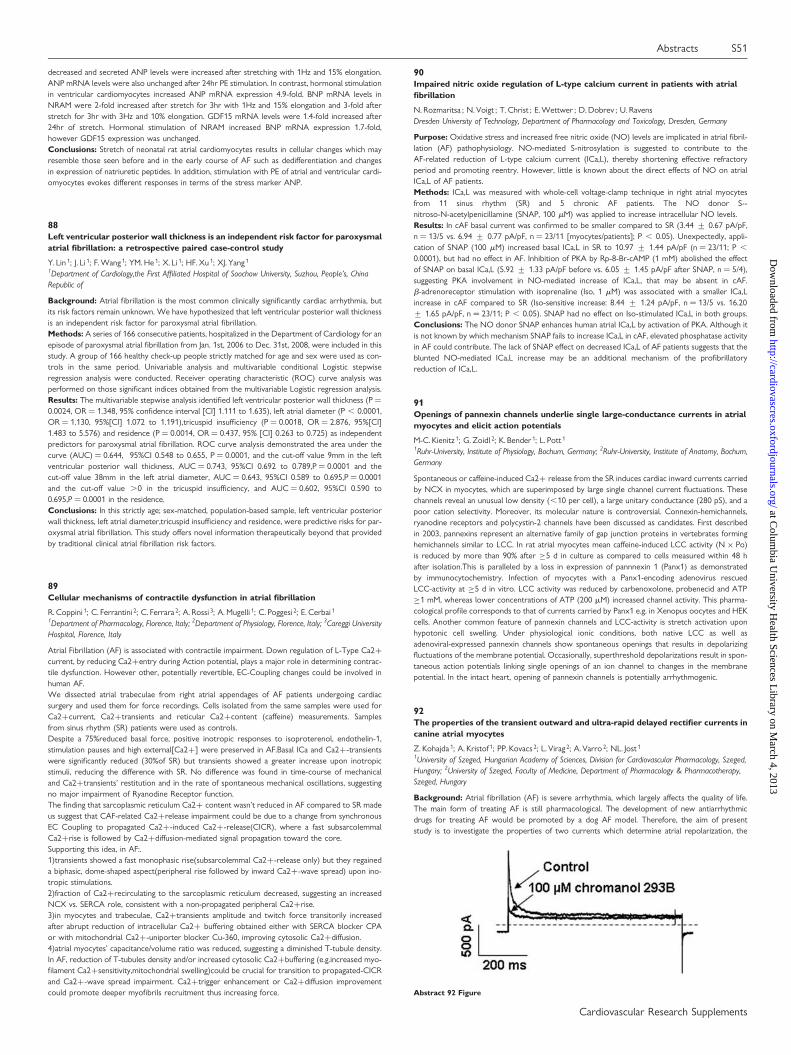
M-C. Kienitz 1; G. Zoidl 2; K. Bender 1; L. Pott 1
1Ruhr-University, Institute of Physiology, Bochum, Germany; 2Ruhr-University, Institute of Anatomy, Bochum,Germany
Spontaneous or caffeine-induced Ca2+ release from the SR induces cardiac inward currents carriedby NCX in myocytes, which are superimposed by large single channel current fluctuations. Thesechannels reveal an unusual low density (,10 per cell), a large unitary conductance (280 pS), and apoor cation selectivity. Moreover, its molecular nature is controversial. Connexin-hemichannels,ryanodine receptors and polycystin-2 channels have been discussed as candidates. First describedin 2003, pannexins represent an alternative family of gap junction proteins in vertebrates forminghemichannels similar to LCC. In rat atrial myocytes mean caffeine-induced LCC activity (N × Po)is reduced by more than 90% after ≥5 d in culture as compared to cells measured within 48 hafter isolation.This is paralleled by a loss in expression of pannnexin 1 (Panx1) as demonstratedby immunocytochemistry. Infection of myocytes with a Panx1-encoding adenovirus rescuedLCC-activity at ≥5 d in vitro. LCC activity was reduced by carbenoxolone, probenecid and ATP≥1 mM, whereas lower concentrations of ATP (200 mM) increased channel activity. This pharma-cological profile corresponds to that of currents carried by Panx1 e.g. in Xenopus oocytes and HEKcells. Another common feature of pannexin channels and LCC-activity is stretch activation uponhypotonic cell swelling. Under physiological ionic conditions, both native LCC as well asadenoviral-expressed pannexin channels show spontaneous openings that results in depolarizingfluctuations of the membrane potential. Occasionally, superthreshold depolarizations result in spon-taneous action potentials linking single openings of an ion channel to changes in the membranepotential. In the intact heart, opening of pannexin channels is potentially arrhythmogenic.
92The properties of the transient outward and ultra-rapid delayed rectifier currents incanine atrial myocytes
Z. Kohajda 1; A. Kristof 1; PP. Kovacs 2; L. Virag 2; A. Varro 2; NL. Jost 1
1University of Szeged, Hungarian Academy of Sciences, Division for Cardiovascular Pharmacology, Szeged,Hungary; 2University of Szeged, Faculty of Medicine, Department of Pharmacology & Pharmacotherapy,Szeged, Hungary
Background: Atrial fibrillation (AF) is severe arrhythmia, which largely affects the quality of life.The main form of treating AF is still pharmacological. The development of new antiarrhythmicdrugs for treating AF would be promoted by a dog AF model. Therefore, the aim of presentstudy is to investigate the properties of two currents which determine atrial repolarization, the
Abstract 92 Figure
Abstracts S51
Cardiovascular Research Supplements
at Colum
bia University H
ealth Sciences Library on M
arch 4, 2013http://cardiovascres.oxfordjournals.org/
Dow
nloaded from
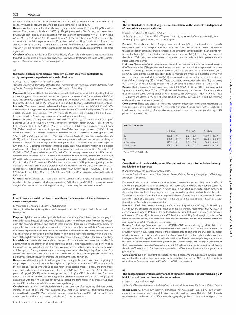
transient outward (Ito) and ultra-rapid delayed rectifier (IKur) potassium current in isolated atrialcanine myocytes by applying the whole cell patch clamp technique at 378C.Results: We have identified in all atrial canine myocytes a chromanol 293B (c ¼ 100 mM) sensitivecurrent. The current amplitude was 16730 + 390 pA (measured at 50 mV) and the current inac-tivation was best fitted by two exponentials with the following components: t1 ¼ 61 + 27 ms andA1 ¼ 474 + 97 pA ; t2 ¼ 27 + 15 ms and A2 ¼ 655 + 253 pA. Chromanol 293B accelerated theinactivation in the following manner: t1 ¼ 27 + 3 ms and A1 ¼ 113 + 31 pA; t2 ¼ 1,8 + 0,1 msand A2 ¼ 166 + 5 pA (Fig. 1). The IKur current was identified by 100 mM aminopyridine (4-AP).100 mM 4-AP did not significantly change either the peak or the steady state current in dog atrialmyocytes.Conclusion: We concluded that IKur plays a less significant role in the canine atrial repolarisationthan that was reported in human atrial myocytes. However, understanding the cause for these inter-species differences requires further investigations.
93Increased diastolic sarcoplasmic reticulum calcium leak may contribute toarrhythmogenesis in patients with atrial fibrillation
N. Voigt 1; AW. Trafford 2; U. Ravens 1; D. Dobrev 1
1Dresden University of Technology, Department of Pharmacology and Toxicology, Dresden, Germany; 2Unitof Cardiac Physiology, University of Manchester, Manchester, United Kingdom
Purpose: Chronic atrial fibrillation (cAF) is associated with impaired atrial Ca2+ signalling. Indirectevidence suggests that increased diastolic sarcoplasmic reticulum (SR) Ca2+ release (SR-Ca2+leak) may promote triggered activity, contributing to AF maintenance. The aim of this study wasto quantify SR-Ca2+ leak in cAF patients and to elucidate its poorly understood molecular basis.Methods: Membrane currents (whole-cell voltage-clamp technique) and [Ca2+]i (Fluo-3 AM)were measured in right atrial myocytes from 8 sinus rhythm (CTL) and 8 cAF patients. To quantifydiastolic SR-Ca2+ leak, tetracaine (10 mM) was applied to quiescent myocytes in Na+ and Ca2+free bath solution. Protein expression was assessed by immunoblotting.Results: Diastolic [Ca2+]i was similar in cAF and CTL (359.2 + 57.2 nM, n ¼ 8/5 [myocytes/patients] vs. 281.2 + 41.5 nM, n ¼ 9/5). Tetracaine unmasked higher SR-Ca2+ leak in cAF vs.CTL patients (90.8 + 12.7 nM vs. 59.7 + 7.3 nM; p , 0.05). This is unlikely to result fromSR Ca2+ overload, because integrating Na+-Ca2+ exchange current (INCX) duringcaffeine-induced Ca2+ release revealed comparable SR Ca2+ contents in both groups (cAF:1.12 + 0.21 pC/pF vs. CTL: 1.21 + 0.12 pC/pF). Protein levels of ryanodine receptor channels(RyR2) and their important regulator proteins calsequestrin, triadin and junctin were unchanged.However, phosphorylation of RyR2 at Ser2809 (PKA- and CaMKII-site) was �3 times higher incAF than in CTL patients, suggesting enhanced steady-state RyR2 phosphorylation as a potentialmechanism of enhanced SR-Ca2+ leak. Expression and autophosphorylation (activation) ofCaMKII at Thr287 were enhanced by 63% and 40%, respectively, whereas catalytic PKA subunitexpression was unaltered in cAF. To test whether increased CaMKII activity contributes to increasedSR-Ca2+ leak, we repeated the tetracaine protocol in the presence of the selective CaMKII blockerKN-93 (1 mM). KN-93 decreased SR-Ca2+ leak to levels seen in CTL patients, suggesting that theincrease of SR-Ca2+ leak in cAF is caused by CaMKII. In addition we found that the same SR Ca2+release due to caffeine application produced a larger INCX current (slope INCX/[Ca2+]i: 1.31 +0.13 A/F/mM, n ¼ 13/8 vs. 0.85 + 0.13 A/F/mM, n ¼ 12/8; p , 0.05), suggesting enhanced functionalNCX in cAF.Conclusions: The increased SR-Ca2+ leak due to CaMKII-mediated RyR2 hyperphosphorylationtogether with the generation of a larger depolarising INCX for a given SR Ca2+ release may causedelayed after depolarisations and triggered activity, contributing the maintenance of AF.
94Use of pro-brain atrial natriuretic peptide as the biomarker of tissue damage incardiac arrhythmias
B. Prnjavorac 1; E. Mujaric 2; J. Jukic 1; K. Abduzaimovic 1
1General Hospital Tesanj, Tesanj, Bosnia and Herzegovina; 2Cantonal Hospital, Zenica, Bosnia andHerzegovina
Purpose: High frequency cardiac dysrhythmias have very a strong effect of coronary blood supply formyocardial tissue. Because of shortening of diastole, there is no sufficient blood flow for the myocar-dium, that anaerobe glycolysis take place. In these situations there are no enaugh energy supply formyocardial function, so strenght of contraction of the heart muscle is not sufficient. Some streetchof complet myocardial walls take occur, nevertheless if distension of the heart muscle occur ornot. The stretch of miocardium provkes liberation of the atrial natriuretic peptide. What is the influ-ence of the high frequency dysrhythmias to the liberation of these peptides, is the aim of the stady.Materials and methods: We analyzed changes of concentration of N-terminal pro-BNP inplasma, which is the precursor of atrial natriuretic peptide. The measurement was performed atthe admittance in Hospital and one day after. We analyzed the patients with tachycardial paroxys-mal dysrhytmias. For any case we noted how many time passed after beginning of paroxysm. Cal-culation was performed using Spearman rank correlation test. At al,l we analyzed 43 patients withparoxysmal supraventricular tachycardia and paroxysmal atrial fibrillation.Results: We divided the patients in three groups, according to the time elapsed since beginning ofthe paroxysm to the admittance to the hospital. In all patients heart rate was 150/min or more. Inthe first group, elapsed time was up to one hour, in the second group 1-8 hour, and third groupmore than eight hour. The mean level of the pro-BNP were 146 pg/ml (SD 68) in the firstgroup, 374 pg/ml (SD 97) in the second group, and 449 pg/ml (SD 114) in the third. Spearmanrank correlation test showed statistical significant difference between second and third group, incomparison with the first one, but not between second and third group. In all three group levelof pro-BNP one day after admittance decrease significantly.Conclusion: In any case, with elapsed time more than one hour after beginning of the paroxysm,increase of level of pro-BNP was measured. Prolongation of paroxysmal tachycardia showedincreased level of pro-BNP. We concluded that measurement of the pro-BNP could be use for esti-mation how harmful are paroxysmal dysrhythmias for the myocardium.
95The antifibrillatory effects of vagus nerve stimulation on the ventricle is independentof muscarinic receptor activation
K. Brack 1; VH. Patel 2; JH. Coote 3; GA. Ng 1
1University of Leicester, Leicester, United Kingdom; 2University of Warwick, Coventry, United Kingdom;3University of Birmingham, Birmingham, United Kingdom
Purpose: Classically the effect of vagus nerve stimulation (VS) is considered to be entirelymediated via muscarinic receptor activation. We have previously shown that direct VS reducesthe slope of action potential duration restitution and simultaneously protects the heart against ven-tricular fibrillation (VF), effects that are mediated via nitric oxide (NO). In this study we measuredthe effects of VS during muscarinic receptor blockade in the isolated rabbit heart preparation withintact autonomic nerves.Methods: Monophasic Action Potential was recorded from the left ventricular surface and duration(MAPD) measured at 90% repolarisation. Standard restitution was studied with single ventricular extra-stimuli (S2) following a 20-beat drive train (300 ms), down to the effective refractory period (ERP).S2-MAPD were plotted against preceding diastolic intervals and fitted to exponential curves withmaximum Slope measured. VF threshold (VFT) was determined as the minimum current required toinduce VF with rapid pacing (30 × 30 ms). These parameters were studied at baseline (BL) and duringVS (7V, 10Hz), before and during perfusion with 0.1 mM atropine. Data are mean + SEM (n¼ 9).Results: During control, VS decreased heart rate (HR) (147.1 + 6.3 to 90.8 + 7.2 bpm) whilstsignificantly increasing both ERP and VFT (Table) and decreasing the maximum Slope of the elec-trical restitution curve. During perfusion with atropine, the bradycardia (143.5 + 9.8 vs. 142.2+ 9.3 bpm) and effects of VS on ERP were blocked whilst the increase in VFT and the flatteningof the restitution curve were preserved.Conclusions: These data suggest a muscarinic receptor independent mechanism underlying thevagal protection of the heart against VF. The context of these findings needs further explorationin relation to the possibility of alternative neurotransmitters and a tentative parallel vagus-NOpathway in the ventricle.
Abstract 95 Table
ERP (ms) VFT (mA) RT Slope
BL 130.0 + 7.8 2.2 + 0.3 1.679 + 0.267VS 147.8 + 6.7*** 5.3 + 0.5*** 0.868 + 0.154***BL&Atropine 125.6 + 3.1 2.0 + 0.5 1.708 + 0.181VS&Atropine 132.8 + 4.4 4.7 + 0.7*** 0.885 + 0.193***
Table: ***P , 0.001 vs BL
96Contribution of the slow delayed rectifier potassium current to the beta-adrenergicmodulation of heart rate
R. Wilders 1; ACG. Van Ginneken 1; AO. Verkerk 1
1Academic Medical Center, Heart Failure Research Center, Dept. of Anatomy, Embryology and Physiology,Amsterdam, Netherlands
Purpose: Under control conditions, the slow delayed rectifier K+ current (IKs) has little effect, ifany, on the pacemaker activity of sinoatrial (SA) node cells. However, this outward current isenhanced by b-adrenergic stimulation, in which case it may affect pacing rate, either through itsshortening effect on the action potential or through its inhibiting effect on diastolic depolarization.To assess the role of IKs in the b-adrenergic modulation of heart rate, we experimentally deter-mined the effect of b-adrenergic stimulation on IKs and used the thus obtained data in computersimulations of SA nodal pacemaker activity.Methods: HEK-293 cells were transiently transfected with 1 mg wild-type KCNQ1 cDNA and 1 mgKCNE1 cDNA, encoding the a and b subunits of the IKs channel, respectively. KCNQ1/KCNE1currents were studied at 378C using the perforated patch-clamp technique in absence and presenceof forskolin (10 mmol/L) to increase the cAMP level, thus mimicking b-adrenergic stimulation. SAnodal pacemaker activity was simulated using the mathematical model of a primary rabbit SAnode pacemaker cell by Kurata and coworkers.Results: Forskolin significantly increased the KCNQ1/KCNE1 current density by ≈25%, shifted thesteady-state activation curve to more negative membrane potentials by ≈15 mV, and increased theactivation rate by ≈50%. Incorporation of these experimental findings into the SA nodal cell modelresulted in a 6-ms decrease in cycle length, the shortening effect on action potential duration dom-inating over the inhibiting effect on diastolic depolarization. The decrease in cycle length is similar tothe 10-ms decrease observed upon incorporation of a +8-mV change in the voltage dependence ofthe hyperpolarization-activated ‘pacemaker current’ (If), reflecting our earlier experimental data onthe effect of forskolin on HCN4 current expressed in undifferentiated human cardiac myocyte pro-genitor cells.Conclusions: IKs is an important contributor to the b-adrenergic modulation of heart rate. Thismay explain the impaired heart rate response to exercise observed in LQT1 and LQT5 patientswith a loss-of-function mutation in KCNQ1 or KCNE1, respectively.
97The postganglionic antifibrillatory effect of vagal stimulation is preserved during VIPinhibition and does not involve the endothelium
K. Brack 1; JH. Coote 2; GA. Ng 1
1University of Leicester, Leicester, United Kingdom; 2University of Birmingham, Birmingham, United Kingdom
Background: We have shown that vagal stimulation (VS) releases nitric oxide (NO) in the ventri-cle which modulates action potential duration restitution and ventricular fibrillation (VF). There isno information on the source of NO or modulating signaling pathways. Here we investigated if the
S52 Abstracts
Cardiovascular Research Supplements
at Colum
bia University H
ealth Sciences Library on M
arch 4, 2013http://cardiovascres.oxfordjournals.org/
Dow
nloaded from

antifibrillatory effect of VS- released NO is dependent on activation of vasoactive intestinal peptide(VIP), involves post-ganglionic nerve fibers or the endothelium as an origin of NO.Methods: The isolated heart with intact autonomic nerves of adult male New Zealand rabbits (2.7+ 0.1Kg) was used with VS at 7-10Hz, 7-10V. Ventricular effective refractory period (ERP) wasmeasured with the single extrastimulus method following a 20-beat drive train whilst ventricularfibrillation threshold (VFT) was determined as the minimum current required to induce sustainedVF with rapid pacing. Parametes were studied at baseline (BL) and during VS, before and withVIP inhibitor 20nM VIP(6-28), 0.5mM Hexamethonium and after a bolus injection with 0.2%Triton X (0.1ml). Data are mean + SEM, n ¼ 3, statistical analysis using 2-factor ANOVA, P ,
0.05 was considered significant.Results: At BL, VS significantly increased ERP and VFT. During perfusion with VIP(6-28) andTriton-X, the effects of VS on ERP and VFT were maintained but inhibited during hexamethoniumperfusion.Conclusions: These data suggest that the electrophysiological cardiac effects of VS occur via post-ganglionic efferent neurons via a VIP and endothelium independent mechanism and not from ante-grade stimulation of afferent neurons. The context of these findings needs further exploration inrelation to the possibility of alternative neurotransmitters and a direct vagus-NO pathway.
Abstract 97 Table
ERP (ms) VFT (mA)
BL 126.7 + 14.8 2.5 + 1.5VS 136.9 + 7.1 * 5.5 + 2.5 *BL VIP(6-28) 125.0 + 7.6 3.8 + 1.1VS VIP(6-28) 136.7 + 4.4 * 8.2 + 2.5 *BL Hexamethonium 125.0 + 7.6 2.8 + 1.4VS Hexamethonium 126.7 + 14.5 NS 2.7 + 1.2 NSBL Triton X 123.3 + 6.7 2.7 + 0.9VS Triton X 146.7 + 6.0 * 4.2 + 1.4 *
Table. *,0.05, NS P.0.05, vs BL
Hypertension
98NADPH oxidase polymorphisms, aortic and brachial blood pressure levels: up, closeand personal?
P. Xaplanteris 1; C.Vlachopoulos 1; K. Baou 1; C.Vassiliadou 1; I. Dima 1; N. Ioakeimidis 1; C. Stefanadis 1
1Hippokration Hospital, University of Athens, 1st Department of Cardiology, Athens, Greece
Purpose: The nicotinamide adenine dinucleotide phosphate (NADPH) oxidase produces superox-ide, thus regulating redox state in the vessel wall. Three single nucleotide polymorphisms (-930A/G,A640G, C242T) of the p22phox subunit have been associated with hypertension; however, theirrole on peripheral and central pressures in normotensive individuals has not been addressed.Methods: 210 healthy, normotensive individuals were studied. Genotypes for the -930A/G, A640Gand C242T polymorphisms were determined by polymerase chain reaction. Peripheral pressureswere measured by mercury sphygmomanometer and aortic pressures by a validated device employ-ing applanation tonometry.Results: Both peripheral and central pressures differed across -930A/G genotypes. G allele carriersdemonstrated higher levels of peripheral systolic blood pressure (PSBP) (AA: 113 + 12, GG/AG:119 + 12 mmHg; p , 0.01) and diastolic blood pressure (PDBP) (AA: 70 + 9, GG/AG:73+ 10mmHg; p , 0.05). Regarding central pressures, AA homozygotes had lower central systolicblood pressure (CSBP) (AA: 103 + 12, GG/AG: 108 + 12 mmHg;p , 0.01) and diastolic bloodpressure (CDBP) (AA: 71 + 9, GG/AG: 74 + 10 mmHg;p , 0.05). In multiple linear regressionanalysis, presence of the G allele (AG or GG) independently predicted CSBP. Blood pressure levelsdid not differ across A640G and C242T genotypes.Conclusion: The -930A/G polymorphism of p22phox is a determinant of peripheral and centralpressures in normotensive individuals.The G allele is associated with higher blood pressure in thebrachial artery,as well as in the aorta. These findings further elucidate the role of this polymorphismin the regulation of blood pressure. In contrast, the A640G and C242T SNPs do not influence per-ipheral and central pressures in normotensives.
99Hypertensive REN2 rats have mild anemia and severe bone marrow dysfunction,which cannot be corrected by erythropoietin administration
WT. Ruifrok 1; C. Qian 1; HHW. Sillje 1; H. Van Goor 1; DJ. Van Veldhuisen 1; WH. Van Gilst 1;RA. De Boer 1
1University Medical Center, Groningen, Netherlands
Background: Heart failure (HF) is associated with a high prevalence of anemia, however the precisecause of this anemia is unknown. Possibly, the observed depressed function of bone marrow stemcells (BMSC) in HF plays a role. Erythropoietin (EPO) has been shown to exert cardioprotectiveeffects and is a known mitogenic factor for BMSC. We evaluated if EPO administration couldprevent the occurrence of anemia in experimental HF by improving bone marrow function.Methods: We employed male homozygous hypertensive REN2 rats that overexpress the mouserenin gene, causing severe left ventricular (LV) hypertrophy. 6 week old rats were randomizedto EPO (40 mg/kg) every 3 weeks (n ¼ 13) or placebo (n ¼ 13) for 6 weeks. Sprague Dawley(SD) rats were used as controls (n ¼ 8). An echocardiogram was made and blood was sampledat baseline, 3 and 6 weeks. After 6 weeks, at sacrifice, heart weight (HW) and hemodynamic par-ameters were measured (blood pressure, heart rate and intracardiac pressures). C-kit positive cellswere isolated from bone marrow by MACs column and cultured in 35 mm culture plate with semi-solid medium for 10 days. Burst forming units of the erythroid lineage (BFU-E) were quantified.Results: REN2 rats have severe hypertrophy and dilated hearts (assessed by echo). EPO did notdecrease HW nor improved hemodynamic parameters. Hemoglobin (Hb) levels increased in theSD EPO-treated group (after 6 weeks: 16.9 + 1.3 vs. 20.5 + 0.6 g/dl, P , 0.01), however inthe (mildly anemic) REN2 rats EPO treatment did not increase Hb (after 6 weeks: 14.7 + 0.6vs. 15.3 + 0.8 g/dl). This was paralleled by a severe decrease in number of BFU-E in the bonemarrow of REN2 rats compared to SD (50 + 6.2 vs. 6.4 + 1.7, P , 0.01). EPO administrationsignificantly improved the number of BFU-E in both SD (50 + 6.2 vs. 64.5 + 6.8, P , 0.05)and REN2 (6.4 + 1.7 vs. 16 + 4.3, P , 0.01) groups, but in the REN2-EPO group, it could notreverse the number to control level.Conclusions: In hypertensive REN2 rats with progressive cardiac dilatation there is mild anemiaand profound dysfunction of the erythroid lineage of the bone marrow. Response to EPO isblunted in REN2 rats and cannot rescue anemia. These results are in line with the known tendencytowards anemia in patients with HF.
100Single nucleotide polymorphisms within the connexin40 (GJA5 gene) promoterdirectly affects its activity
K. Schmidt 1; FJ. Kaiser 2; J. Erdmann 3; C. De Wit 1
1Universitat zu Lubeck, Institut fur Physiologie, Lubeck, Germany; 2Universitat zu Lubeck, Institut furHumangenetik, Lubeck, Germany; 3Universitat zu Lubeck, Medizinische Klinik II, Lubeck, Germany
Purpose: Connexins (Cx) are transmembrane proteins which provide a direct communicationpathway between adjacent cells by forming gap junction channels. This protein family comprises21 members in humans of which four are expressed in the cardiovascular system. Connexin40(Cx40) is encoded by GJA5 and is expressed in the vascular endothelium, in cardiomyocytes andin renin-producing cells in the kidney. The functional importance of Cx40 is highlighted by arterialhypertension, cardiac hypertrophy, high plasma renin activity, and arteriolar dysfunction in micedeficient for Cx40. Recently, a single nucleotide polymorphism (SNP) within the intron of GJA5was associated in a genome-wide study with left ventricular hypertrophy (LVH). Since LVH canresult from long-lasting hypertension and Cx40-deficient mice are remarkably hypertensive wesearched for functional polymorphisms in GJA5 in humans which may explain this geneticassociation.Methods: DNA from probands with or without a thickened left ventricle and the homozygous SNPin the intron of GJA5 was isolated. The coding and promoter region of this gene was amplified andsequenced. In addition, the promoter regions were inserted into the pGL4.10 promoterless fireflyluciferase-plasmid and the promoter activity was quantified in a luciferase assay in HeLa-cells aftertheir transfection.Results: GJA5 consists of two exons, however, only the second exon encodes for the functionalCx40 protein. Sequencing of this coding region (1077 bp) in 150 probands did not reveal any basepair change. Additional sequencing of the 695 bp upstream region, which consists of the5′-untranslated region as well as exon 1 and contains the promoter of GJA5, identified twoSNPs in complete linkage disequilibrium (LD) in 13 of 26 studied probands with LVH. In contrast,these SNPs were found with an allele frequency of 4% in a control population. Moreover, oneproband in this small study group carried four different base pair changes in the GJA5 promoterregion. Promoter activity as revealed in HeLa cells was unaffected by the two coupled SNPs, butthe four other identified SNPs decreased its activity by 40%.Conclusions: Although we did not identify nucleotide exchanges in the coding exon of GJA5 inselected probands, sequencing analyses of the promoter region exposed four SNPs which led tochanges in the luciferase activity. Further studies are required to verify cell-specific effects whichmay translate into functional consequences for affected humans.
101Disturbances in coronary microcirculation in hypertension
O. Barnett 1; Y. Kyyak 1
1Lviv National Medical University, Lviv, Ukraine
Introduction: Altered coronary microcirculation is an important consequences of hypertension.Ultrastructural changes involved in microvascular remodeling remain unclear.The purpose of the present study was to clarify the changes in coronary microcirculation induced byhypertension.Subject and Methods: myocardial express-necropsies from 18 patients (10 males, 8 females, agerange 45-79) who suffered from HT , and died predominantly from Acute Myocardial Infarction(AMI) complicated by Heart Failure Killip class III-IV. Transthorax necropsy of the heart wasAbstract 98 Figure Blood pressures across genotypes.
Abstracts S53
Cardiovascular Research Supplements
at Colum
bia University H
ealth Sciences Library on M
arch 4, 2013http://cardiovascres.oxfordjournals.org/
Dow
nloaded from
performed immediately (15 min. average) after patient’s death in clinic. Control group consisted of 5persons who died of extra cardiac causes in the same age. Intact and near infarction zones of the leftventricle were examined.Results: Coronary microvascular dysfunction caused by hypertension (HT) is severe enough tocause myocardial ischemia. HT leads to altered microcirculation which cause hypoperfusion andinterstitial fibrosis. In rturn interstitial fibrosis led to decreased density of vessels in the coronarymicrovasculature. As regards to endothelial cell (EC), HT led to their hypertrophy , proliferationand migration resulting in considerable decrease in microvessel inner diameter and loss of the capil-lary reserve. Blood stasis and cell migration into interstitium is present. The most important finding,regarding EC remodelling, is their hibernation following cellular hypertrophy. With the developmentof progressive ischemia, glycogen particles disappear from EC cytoplasm and secondary cell necrosismay develop. Impaired myocardial perfusion contributed to tissue ischemia, cardiomyocytedegeneration, stunning and hibernation and predisposed Heart Failure. Precipitating traditional cor-onary risk factors predispose patients to more severe microvascular remodeling and ischemia.These findings indicate that microcirculatory disturbances develop in parallel to HT persistence ,but more severe dysfunction is present in patients with hypertensive heart disease and the devel-opment of AMI.Conclusions: Hypertension causes EC hypertrophy, their structural and functional heterogeneity,preceding their hibernation and apoptosis. Therapeutic target in hypertensive patients should rep-resent microvascular protective agents.
102Arterial stiffness is associated with cardiac damage severity in essential hypertensionpatients
F. Cesana 1; L. Boffi 1; T. Mauri 2; M. Alloni 1; M. Betelli 1; S. Nava 1; C. Giannattasio 1; G. Mancia 1
1University of Milan-Bicocca, Department Clinical Medicine & Prevention, Milan, Italy; 2University ofMilan-Bicocca, Department of Experimental Medicine (DIMS), Milan, Italy
Purpose: Essential hypertension (EH) is often associated with cardiac damage and arterial stiffness,which are well established independent risk factors for cardiovascular disease. Aim of this study wasto describe whether there is an association between anatomical and functional signs of cardiac dys-function and the degree of arterial stiffness in a large EH patients population.Methods: We performed standard trans-thoracic echocardiography to measure anatomical (leftventricular mass indexed by body surface area [LVMI] and relative wall thickness [RWT]) and func-tional (ejection fraction [EF], diastolic function [E/A] and deceleration time [dec T]) cardiac par-ameters on 827 treated EH patients. On the same day, we measured blood pressure (BP) andcarotid-femoral pulse wave velocity (PWV) using the foot to foot velocity method, to estimatearterial stiffness. Data were analyzed by linear regressions or ANOVA and post-hoc Bonferronitest, as appropriate.Results: Patients were 53 + 14 years old and 50% were male. Systolic BP and diastolic BP were142.3 + 18.6/86.7 + 10.6 mmHg. LVMI was 111.36 + 32.7 g/m2, RWT 0.41 + 0.07, EF 63 + 4%, E/A 1.05 + 0.33, dec T was 211 + 47 mSec, while PWV was 10.7 m/Sec. PWV was significantlycorrelated with LVMI (r ¼ 0.214, p , 0.001), E/A (r ¼ 20.25, p , 0.001) and dec T (r ¼ 0.146, p, 0.001). Geometry of left ventricle, as defined by LVMI and RWT (ESC guidelines), was normal in336 (43%) patients, while in 163 (21%) we found concentric remodelling, concentric hypertrophy in173 (22%) and eccentric hypertrophy in 109 (14%) patients. PWV was significantly differentbetween the 4 subgroups (p ¼ 0.001), with concentric and eccentric hypertrophy patients havingsignificantly higher PWV values than normal patients (p ≤ 0.001 for both comparisons).Conclusions: Arterial stiffness may be associated with presence and severity of cardiac damage inEH patients. This finding may help unveil important interactions between arterial stiffness and targetend-organ dysfunction development during EH.
103Protective effects of L-carnitine and mildronate in salt-induced hypertension
R. Vilskersts 1; J. Kuka 1; B. Svalbe 1; E. Liepinsh 1; M. Dambrova 1
1Latvian Institute of Organic Synthesis, Riga, Latvia
Hypertension is a frequent cause of development of cardiovascular diseases and increased mortality.The aim of this study was to investigate the effects of the administration of L-carnitine or mildronate,an inhibitor of L-carnitine biosynthesis, or their combination on the development of hypertension-related complications in Dahl salt-sensitive (DS) rats fed with a high salt diet.At the age of 7 weeks male DS rats were divided into five groups. Control group animals (n ¼ 10)continued to consume a diet with normal salt content. Animals from other groups were switched toa high salt (8% NaCl) diet and treated for 8 weeks with vehicle (water; n ¼ 10), L-carnitine (100 mg/kg; n ¼ 10), mildronate (100 mg/kg; n ¼ 10) or a combination of L-carnitine and mildronate at thedoses above (n ¼ 10).Treatment with the combination significantly improved the survival rate compared to the vehiclereceiving group. Administration of mildronate and the combination for 8 weeks significantlydecreased resting heart rate by 12% and 10%, respectively; while none of the tested compoundsor their combination influenced high salt intake-induced hypertension or development of the leftventricular hypertrophy. In addition, hypertensive DS rats manifested impaired endothelium-dependent relaxation that was improved by the administration of the combination.In conclusion, our results show that treatment with a combination of L-carnitine and mildronateattenuates the development of the hypertension-related complications in a DS rat model.
104Endothelial hyaluronan synthase 2 and hyaluronan are increased by anatheroprotective flow profile
A.Zakrzewicz 1; J.Maroski 1; B.Vorderwuelbecke 1; K. Fiedorowicz 1; L.Da Silva-Azevedo 1; AR.Pries 1
1Charite - Campus Benjamin Franklin, Berlin, Germany
Objective: The endothelial surface layer (ESL) of which hyaluronan (HA) is a constitutive com-ponent is significantly reduced at atheroprone regions with oscillating low flow profiles without
any reason why this aegis against atherosclerosis is so greatly reduced. Therefore, this study wasdone to reveal if hyaluronan synthase 2 (HAS2) and subsequently HA in endothelial cells can beexperimentally changed by different flow profiles.Methods and Results: An atheroprotective flow profile, produced in a cone-and-plate system,increased HAS2-mRNA (as determined by real-time RT-PCR) in human umbilical vein endothelialcells fourfold while constant laminar flow or an atheroprone flow profile did not induce any differencestowards static control cultures. Likewise, HA (determined by ELISA) was increased by the atheropro-tective flow profile both in cell culture supernatants and on the surface of endothelial cells.Conclusions: These experiments provide a link between the production of the ESL by endothelialcells and different flow conditions.
105Entropy of skin microcirculation flow in subjects with familial predisposition ornewly diagnosed hypertension
B. Gryglewska 1; M. Necki 1; M. Zelawski 1; T. Grodzicki 1
1Jagiellonian University, Cracow, Poland
Purpose: To assess entropy of skin microcirculation flow in subjects with familial predisposition ornewly diagnosed hypertension.Methods: A 3-minutes rest flow (RF), minimal flow (BZ) during 3-minutes ischemia and 8-minutesheat flow (HF) were recorded (laser Doppler flowmetry) in patients with untreated hypertension[HT], and in normotensives with no [NT(2)] or with familial predisposition to hypertension[NT(+)]. Average 1-minutes surface areas under the curve of flow records and box dimensions(D) as a measure of entropy were calculated.Results: We studied 70 persons (36,1 + 10,3y, 39 men). HT (n¼ 31) had significantly higher valuesof both clinic BP (152,6 + 8,3/95,1 + 6,9 vs 122,1 + 11,5/80,1 + 6,3 vs 121,3 + 8,6/75,4 + 6,8mmHg) and 24-h ambulatory BP (132,6 + 10,2/83,2 + 8,3 vs 118,2 + 9,0/72,9 + 6,0 vs 113,8 +6,9//71,1 + 4,7 mmHg), body mass index, glucose, triglycerides and insulin than NT(2), (n ¼ 17) andNT(+), (n¼ 22) groups. Mean values of flows and surface area of RF, BZ, HF, and D RF and D HFwere comparable in studied groups, but D BZ differed (1,13 + 0,05 vs 1,15 + 0,05 vs 1,11 + 0,05;p ¼ 0,04). Family history of hypertension, insulin level and variability of 24-h diastolic BP were signifi-cant predictors of BZ entropy lower values in the multiple regression model.Conclusions: Subjects with familial predisposition to hypertension reveal altered homeodynamicsof microvascular flow.
Endothelial function
106Peroxisome Proliferator-Activated Receptor-gamma inhibits angiogenesis bysuppressing CREB-mediated cyclooxygenase-2 expression in the humanendothelium
E. Scoditti 1; M. Massaro 1; MA. Carluccio 1; A. Distante 1; C. Storelli 2; R. De Caterina 3
1Institute of Clinical Physiology-CNR, Lecce, Italy; 2University of Salento, Department of Biological andEnvironmental Science and Technology (DiSTeBA), Lecce, Italy; 3“G. d’Annunzio” University and Center ofExcellence on Aging, Chieti, Italy
Purpose: Neoangiogenesis contributes to diabetic vasculopathy and intraplaque hemorrhage inatherosclerosis. The activation of Peroxisome Proliferator-Activated Receptor(PPAR)g is knownto inhibit angiogenesis. We therefore aimed at examining the effects of PPARg agonists on thepro-angiogenic enzyme cyclooxygenase(COX)-2 in human umbilical vein endothelial cells chal-lenged with vascular endothelial growth factor (VEGF) and phorbol 12-myristate 13-acetate (PMA).Methods and Results: A 24 h exposure of HUVEC to the PPARgamma agonists rosiglitazone(RSG) and GW1929 significantly attenuated VEGF- and PMA-stimulated COX-2 activity (by 30%,immunoassay for 6-keto-PGF1a), as well as protein (by 40%, Western analysis) and mRNAexpression (by 40%, Northern analysis). This effect was abolished by the PPARg antagonists bisphe-nol A diglycidyl ether and GW9662 as well as well as by PPARg small-interfering RNAs (siRNAs).COX-2 promoter activity experiments revealed that the induction of COX-2 promoter was signifi-cantly inhibited by RSG through an interference with the cAMP response element (CRE) site.COX-2 downregulation after siRNA knockdown of the transcription factor CRE binding protein(CREB) confirmed the role of CREB in mediating COX-2 transcription. Correspondingly, PPARgagonists also attenuated CREB activation. Since Protein Kinase(PK)C is involved in VEGF-inducedCOX-2 expression and CREB activation, we also investigated which isoforms of PKC were affectedby RSG. While the inhibition of both conventional PKCa and b suppressed VEGF- andPMA-stimulated CREB activation and COX-2 expression, RGS only reduced VEGF- andPMA-stimulated PKCa membrane translocation.Conclusions: The anti-angiogenic effect of PPARg agonists is due, at least in part, to their inter-ference with the PKCa-mediated activation of CREB and the related expression of COX-2.PKCa may therefore be a novel therapeutic target for antidiabetic drugs in atherosclerosis.
107Effects of NO on vascular bradykinin type-2 receptor expression
O.Kocgirli 1; S.Valcaccia 1; VT.Dao 1; T.Suvorava 1; S.Kumpf 1; M.Floeren 1; M.Oppermann 1; G.Kojda 1
1Institute of Pharmacology, dusseldorf, Germany
Purpose: The activation of vascular bradykinin type-2 receptor (BKR-2) produces vasodilatoryeffects mediated by prostacyclin and NO. In eNOS knockout mice, coronary flow responses to bra-dykinin are largly increased. These data suggest that NO might act as a negative feed-back regulatoron vascular BKR-2 expression and/or activity.Methods: We used a human head and neck squamous carcinoma cell line expressing BKR-2(UDSCC-2) and another one without BKR-2 expression (UDSCC-3) which were established inour labaratory to confirm the detection of BKR-2. For in vivo experiments we used eNOS++mice which has more than 2 times higher endothelial NOS, eNOSn (transnegative litter mates)for negative control, eNOS knockout and C57Bl/6 mice. For in vitro experiments we used
S54 Abstracts
Cardiovascular Research Supplements
at Colum
bia University H
ealth Sciences Library on M
arch 4, 2013http://cardiovascres.oxfordjournals.org/
Dow
nloaded from
primary cultured porcine aortic endothelial cells and porcine aortic smooth muscle cells which wereincubated with 10 mM S-nitroso-N-acetylpenicillamine (SNAP) (NO-Donor) or Ca-Ionophortogether with NG-nitro-L-Arginine-Methyl-Ester (L-NAME) (NOS inhibitor). C57Bl/6 mice werefed with NO-donor pentaerythrityl-tetranitrate (PETN, 0mg, 6mg or 60mg kg/KG/d).Results: The expression of BKR-2 in eNOS++ compared to eNOSn mice lung (124.6 + 28.4%)and heart (114.5 + 18.1%) have not shown any significant difference. The knockout mice haveshown an increase of BKR-2 expression (lung 130.7 + 6.5%, p , 0.005, heart 172 + 26%, p ,
0.05). The C57Bl/6 mice which were treated with PETN and the cells which were incubatedwith SNAP or L-NAME showed no significant difference in BKR-2 expession.Conclusion: These data suggest that an increase of BKR-2 expression in knockout eNOS micecould cause the largely increase of coronary flow mediated by bradykinin. NO does not seem tohave any effect on vascular BKR-2 expression, in vitro and in vivo.
109Impairment of both nitric oxide and endothelium-derived hyperpolarizing factor instreptozotocin-induced diabetic small mesenteric artery
CH. Leo 1; J. Ziogas 2; JL. Favaloro 1; OL. Woodman 1
1RMIT University, Bundoora, Australia; 2The University of Melbourne, Melbourne, Australia
Nitric oxide (NO) and endothelium-derived hyperpolarizing factor (EDHF) contribute toendothelium-dependent relaxation in small vessels such as mesenteric artery. Diabetes is knownto impair endothelium-dependent relaxation, however, it is not established whether it affectseither or both NO and EDHF. The aim of this study is to evaluate the mechanism of endothelium-dependent relaxation of mesenteric arteries from type 1 diabetic rats. Wire myography wasemployed to examine ACh-induced relaxation of mesenteric arteries. Superoxide levels, measuredby L-012 and lucigenin-enhanced chemiluminescence, were significantly increased in diabetic mesen-teric arteries compared to normal arteries. Diabetes significantly reduced the sensitivity to ACh(pEC50, diabetic, 6.77 + 0.14 vs normal, 7.74 + 0.09, n ¼ 10-11, p , 0.0001) in mesentericarteries. When the contribution of NO to relaxation was abolished by L-NNA (100 mM) andODQ (10 mM), the sensitivity to ACh was significantly decreased in the diabetic arteries comparedto normal arteries (pEC50 diabetic, 6.68 + 0.12 vs pEC50 normal, 7.08 + 0.11, n ¼ 11-12, p ,
0.05), indicating an impaired contribution of EDHF. Conversely, when the contribution of EDHF wasinhibited with the combination of TRAM-34 (1 mM), apamin (1 mM) and iberiotoxin (100 nM), themaximum relaxation (Rmax) to ACh was significantly decreased in diabetic arteries (Rmax diabetic17 + 5 vs Rmax normal, 81 + 5, n ¼ 8-9, p , 0.0001), suggesting that the contribution of NOwas also impaired by diabetes. Western blot analysis also demonstrated eNOS uncoupling indiabetic mesenteric arteries. Taken together, this study demonstrated that endothelium-dependentrelaxation was impaired at 10 weeks of diabetes due to a reduced contribution of both NOand EDHF.
111Cyclic strain downregulates angiopoietin-2 expression in human endothelial cells
W. Goettsch 1; A. Marton 1; C. Goettsch 2; H. Morawietz 1
1Dresden University of Technology, Medical Clinic III, Dpt of Vascular Endothelium & Microcirculation,Dresden, Germany; 2Dresden University of Technology, Medical Clinic III, Dpt of Endocrin., Diabetes & Met.Bone Disease, Dresden, Germany
Purpose: Endothelial cells in vivo are constantly exposed to mechanical stimuli like shear stress andcyclic strain. A critical regulator of vessel maturation and endothelial cell quiescence isAngiopoietin-2 (Ang-2). In previous studies, we could show a VEGF-dependent induction ofAng-2 in low flow areas, and a FOXO1-dependent downregulation of Ang-2 in high flowareas. In contrast, the impact of low and high cyclic strain on the endothelial expression ofAng-2 and the role of nitric oxide (NO) and reactive oxygen species in this context are not wellunderstood.Methods: Primary cultures of human umbilical vein endothelial cells (HUVEC) were exposed to2%, 5%, 8%, 12%, 16% and 18% cyclic strain (1 Hz) for up to 24 h using the FX-4000 Flexcellsystem. In these cells, we analyzed the effect of cyclic strain on Ang-2 expression by RT-PCRand Western blot. NO formation was measured by Griess reaction.Results: Application of physiological levels of cyclic strain for 24 h resulted in downregulation ofAng-2 expression (8% strain: 39.1 + 7.4%, 12% strain: 29 + 3.2%, n ¼ 6 each, *P , 0.05 vs.control). In contrast, Ang-2 was not regulated by pathophysiolocial levels of cyclic strain (16%and 18%). Ang-2 was downregulated by 12% cyclic strain after 3 h (61.3%) and 7 h (34.8%) reachingits maximum after 24 h. Furthermore, long-term application of 12% cyclic strain increased endo-thelial NO formation (con: 0.31 + 0.02 mM NO; 12%: 1.88 + 0.23 mM NO, n ¼ 6, *P , 0.05)and eNOS expression (296 + 64%, n ¼ 6, *P , 0.05 vs. control). In additional studies, wefound a reduced formation of reactive oxygen species after application of 12% cyclic strain, butnot in response to higher levels of cyclic strain. We further tested the impact of reactive oxygenspecies on Ang-2 expression. Application of hydrogen peroxide (100 mM) for 24 h inducedAng-2 in human endothelial cells.Conclusions: Our data support a redox-sensitive reduction of the angiogenic potential of humanendothelial cells in response to chronic cyclic strain. This might involve a mechanosensitive balancebetween NO and reactive oxygen species, thus contributing to a vasoprotective potential of phys-iological cyclic strain.
112Increased intima media thickness in rheumatic mitral stenosis: correlation with highsensitivity C-reactive protein levels
ENAS. Khalifa 1; Z. Ashour 2
1Dar Al Fouad Hospital, cairo, Egypt; 2Cairo University, Faculty of Medicine, Cairo, Egypt
Background: The endothelium lining of the cardiovascular system plays an important regulatoryrole in vascular physiology and pathology. In similar fashion, the surfaces of valve leaflets are
presumed to be generally protected and regulated by the endothelium. Minor increases of C-reactive protein (CRP) levels have been reported in chronic rheumatic valve diseases as well asin vascular inflammatory processes. It is not clear whether the endocardial involvement in rheumaticfever is accompanied by affection of the vascular endothelium or not.Objectives: In this study we planed to determine carotid intima media thickness (cIMT) in patientswith rheumatic mitral stenosis (MS) and to correlate this with of high sensitivity (hs)-CRP as amarker of ingoing inflammation.Methods: A total of 80 patients with an established diagnosis of rheumatic mitral stenosis (meanage 34 + 8.1 years), and 36 age and sex matched healthy individuals (mean age 32 + 7.1years)were included in this study. All patients underwent transthoracic and transesophageal echocardio-graphy for accurate assessment of left atrial dimension (LAD) , mitral valve area (MVA), mean dias-tolic transmitral pressure gradient (MPG), and mitral valve scoring. According to mitral valve score,patients were divided into group I (valve score more than 8/16), group II (valve score less than 8/16)and group III (the control group). The common carotid arteries were examined using B-mode ultra-sonography and from which cIMT was measured. Ultrasensitive immunoassay was used to measureserum levels of hs-CRP.Results: The age did not differ significantly among the three groups (p . 0.05). No significantdifference between group I&II as regard MVA, MVG and LAD (P . 0.05) whereas these variableswere significantly higher in group I&II when compared to controls (P , 0.0001). hs-CRP and cIMTwere significantly higher in group I compared to group II and controls (5.9 + 0.3 mg/L vs 3.4 + 0.5mg/L vs 2.2 + 0.08 mg/L, & 0.71 + 0.02 mm vs 0.62 + 0.08 mm vs 0.51 + 0.04 mm respectively,p , 0.001 for all). Strong positive correlations were identified between hs-CRP and cIMT(r ¼ 0.86in group I, r ¼ 0.67 in group II, p , 0.001) and also between hs-CRP and LAD(r ¼ 0.77 in group I,r ¼ 0.71 in group II, p , 0.001).Conclusion: In rheumatic MS, increased cIMT was associated with more evident inflammatoryprocess with high hs-CRP levels. These results suggest that in rheumatic MS valvular damage isaccompanied by systemic vascular endothelial damage.
113Effect of physical activity on the expression of vasoprotective proteins in venoustissue: Role of vascular catalase
VT. Dao 1; M. Floeren 1; S. Kumpf 1; T. Suvorava 1; G. Kojda 1
1Heinrich-Heine University of Dusseldorf, Institute of Pharmacology and Clinical Pharmacology, Dusseldorf,Germany
Purpose: Many observations in animals and humans provide evidence for profound changes ofarterial gene expression and protein activity induced by exercise. In striking contrast little isknown about its effect in venous vessels and the pulmonary circulation. We sought to investigateto what extent the effect of physical forces differ in pulmonary tissues such as the vein (venacava) compared to the aorta and the heart by measuring the expression of endothelial nitricoxide synthase (eNOS) and extracellular superoxide dismutase (ecSOD) after physical activity.Methods: 2 months old male C57Bl/6 mice were randomized into three groups and singularized ina 350cm2 small cage for 6 weeks. Mice were trained for four weeks using two different exerciseprotocols, forced exercise training (5 days a week for 30min at 0.25m/s) and voluntary running.Results: Both training protocols significantly increased relative heart weight vs. body weight andresulted in upregulation of aortic and myocardial eNOS (P , 0.05, n ¼ 6) in exercised mice com-pared to sedentary controls. In striking contrast, eNOS expression in the lung (P . 0.05, n ¼ 6)and in the vein (P . 0.05, n ¼ 6) remained unchanged vs. sedentary controls. Furthermore bothtraining protocols had no effect on eNOS mRNA expression level in lung tissue normalized toHPRT (Hypoxanthin-Phosphoribosyl-Transferase 1) as compared to non exercised mice (P .
0.05; n ¼ 6). Likewise, exercise significantly upregulated ecSOD in the aorta and in the heart(P , 0.05, n ¼ 6) but not in lung (P . 0.05, n ¼ 6) and vein tissue (P . 0.05, n ¼ 6). Further-more, we observed a comparable increase of eNOS-Ser1177 phosphorylation in all tissuessuggesting that exercise increased shear stress in both arterial and venous tissue. To evaluatethe underlying mechanism we investigated the catalase expression in vein compared to theaorta at rest and after exercise training. The expression of catalase in vein at rest was 5 foldhigher than in the aorta (P , 0.01, n ¼ 6). After exercise catalase expression increased to1042 + 250 (P , 0.05, n ¼ 6) in the vein compared to the aorta. Preliminary data showedthat 6 weeks of treatment with the catalase inhibitor aminotriazol (10mg/ml) elicited an exerciseinduced increase of eNOS and ecSOD expression in lung tissue (130.2 + 14.35, n ¼ 4; 132.9 +22.16, n ¼ 4; respectively).Conclusion: Our data suggest an important role of vein catalase expression and thus hydrogenperoxide levels for exercise induced changes of the expression of vasoprotective proteins. Thus,the development and progression of impaired pulmonary circulation, e.g. following chronicsmoking, is unlikely prevented by exercise.
114Resveratrol decreases asymmetrical dimethylarginine levels in human endothelialcells via SIRT1 signaling
V. Rupprecht 1; F. Scalera 1; J. Martens-Lobenhoffer 1; SM. Bode-Boeger 1
1Otto-von-Guericke University of Magdeburg, Institute of Clinical Pharmacology, Magdeburg, Germany
Purpose: Asymmetric dimethylarginine (ADMA), an endogenous inhibitor of nitric oxide (NO)synthase, is metabolized by dimethylarginine dimethylaminohydrolase (DDAH) 1 and 2. Recently,it has been shown that DDAH 1 is upregulated by inhibiting signaling of sterol regulatoryelement binding protein (SREBP) 1c, while inhibition of SREBP2 signaling leads to downregulatedDDAH 1. SREBPs are transcription factors that regulate cholesterol and lipid synthesis. Becauseresveratrol (trans-3,5,4′-trihydroxystilbene) inhibits SREBP 1c expression via activating NAD+-dependent class III histone deacetylase sirtuin1 (SIRT1) signaling, we tested the hypothesis thatresveratrol decreases ADMA via SIRT1-SREBP signaling pathway.Methods: Human endothelial cells (EC) from the third passage were incubated in presence ofdifferent concentrations of resveratrol (0.1, 1, 10, 25, 50, and 100 mmol/L) for 24 h. The level ofADMA, nitrite and nitrate, and 8-iso-prostaglandin F2a (8-iso-PGF2a), as a marker for oxidative
Abstracts S55
Cardiovascular Research Supplements
at Colum
bia University H
ealth Sciences Library on M
arch 4, 2013http://cardiovascres.oxfordjournals.org/
Dow
nloaded from
stress in conditioned media, the activity and protein expression of DDAH 1 and DDAH 2, andprotein expression of SREBP 1c , SREBP 2, and SIRT1, in endothelial cells were determined.Results: When EC were exposed to low concentration of resveratrol, ADMA concentrationdecreased significantly versus corresponding control. This effect was associated with a significantincrease in activity and protein expression of DDAH 2, whereas the protein expression ofDDAH 1 was significantly downregulated. Furthermore, an increase in NO synthesis, a decreasein 8-iso-PGF2a formation and in protein expression of SREBP 1c and SREBP2, and enhancedprotein expression of SIRT1 were observed. Surprisingly, inhibition of SREBP 1c signaling downre-gulated DDAH 1 expression, whereas inhibiting signaling of SREBP 2 did not affect the expression ofDDAH 1. Moreover, resveratrol decreased SREBP 1c and SREBP 2 expression independently ofSIRT1 signaling. In contrast, suppression of SIRT1 signaling prevented the effect of resveratrol onDDAH-ADMA-NO axis.Conclusions: We have demonstrated that resveratol by activating SIRT1 signaling modulates thecatabolism and release of ADMA. We therefore propose that independently of SREBP signalingresveratrol translational and posttranslational upregulates DDAH 2, causing ADMA to diminish suf-ficient to increase NO synthesis.
115Possible mechanisms underlying abacavir-related cardiovascular complications
WS. Li 1; YW. Kwan 2; GPH. Leung 1
1The University of Hong Kong, Hong Kong, People’s Republic of China; 2The Chinese University of HongKong, Hong Kong, Hong Kong SAR, People’s Republic of China
Abacavir is a nucleoside reverse transcriptase inhibitor commonly used for the treatment of HIVinfection. However, several clinical studies indicated that patients receiving abacavir treatmentwere connected to an increase in cardiovascular risks. Since the underlying mechanism is stillunclear, we would like to find out the reason of abacavir-induced cardiovascular risks. Theeffects of abacavir on vascular functions and platelet activities were observed.Sprague-Dawley rats (330-350 g) were fed with two doses of abacavir (16 mg/kg/day and 160 mg/kg/day) intragastrically for 28 days. Isometric tensions of basilar arteries were measured afterwards.Messenger RNA levels of eNOS, COX-2, MCP-1, I-CAM and V-CAM in aortae, mesenteric arteriesand pulmonary arteries were measured by RT-PCR. For the determination of platelet activation,plasma level of CD40L, a platelet-derived factor, was measured by ELLSA kit. Moreover, theeffect of abacavir on viability of human brain microvascular endothelial cells (HBMEC) was testedby MTT assay.Results from myograph studies showed that, after the treatment of abacavir, although thedose-response curve for acetylcholine-induced relaxation is slightly right-shifted, the maximumrelaxation was not changed. Besides, there was no significant change in the mRNA levels ofCOX-2, MCP-1, I-CAM and V-CAM in rat aortae, mesenteric arteries and pulmonary arteriesafter the abacavir treatment. However, the mRNA level of eNOS was decreased in pulmonaryarteries in abacavir-treated group. Also, a higher plasma level of CD40L was detected in theabacavir-treated group. The results of MTT assay showed that abacavir inhibited the growth ofHBMEC.Abacavir did not affect the endothelium-dependent relaxation in basilar arteries nor change themRNA level of eNOS in aortae and mesenteric arteries. However, mRNA level of eNOS wasdecreased in pulmonary arteries. It shows that the effect of abacavir on the vascular tone maybe localized to certain vascular beds. The inhibition of endothelial cell growth suggested that aba-cavir may be toxic to endothelial cells. Abacavir may also increase the chance of developing throm-bosis through the upregulation of the platelet activity. These findings may give hint to the possiblemechanisms involved in the increase in cardiovascular risks by abacavir.
116MiR-492 acts as an antiangiogenic microRNA in endothelial cells
F. Patella 1; A. Mercatanti 2; L. Pitto 2; G. Rainaldi 3
1Scuola Superiore Sant’Anna, Pisa, Italy; 2Laboratory of Gene and Molecular Therapy, Institute of ClinicalPhysiology, CNR, Pisa, Italy; 3Istituto Toscano Tumori, Florence, Italy
It is already known that endothelial cells growing in high glucose have a reduced proliferation rateand a pronounced increase in apoptosis compared with normal glucose. Moreover, an effect ofhigh glucose on migration and angiogenesis has been also reported. The idea is that the numerouscellular responses induced by high glucose are mediated by microRNAs, a class of endogenous22-25nt single-stranded RNA molecules that regulate gene expression post-transcriptionally.Purpose: The aim of this work is to investigate whether the glucose stress affects the expressionprofile of miRNAs and to gain information on the role of miRNAs in high glucose signalling network.Methods: We determined the miRNA expression profile of HUVEC after 3 days growth inmedium containing 5 mM (G-5) or 30 mM (G-30) glucose. We validated the microarray resultsby qRT PCR and used transfection of mature miRNA to study its effects on HUVEC angiogenicpotential (tube formation, cell proliferation, cell migration). Western blot and luciferase reporterassay were used to detect GATA2 and to validate miR-492/3′UTR-GATA2 interaction,respectively.Results: MiRNA microarray analysis revealed that 13 miRNAs were over-expressed and 1 miRNAwas under-expressed in G-30 cells. The most upregulated microRNA was miR-492. The transfec-tion of this miRNA in exponentially growing HUVEC reduced tube number, cell proliferation andmigration similarly to high glucose. Moreover, we found that eNOS transcriptional activity was pro-foundly decreased. Looking at the molecular targets of miR-492, we noticed that the 3′UTR ofeNOS contains a binding site for miR-492. We also focused on genes which control eNOS at tran-scriptional level. We found that GATA2 is a transcriptional activator of eNOS and contains abinding site for miR-492. We verified that endogenous expression of GATA2 protein wasreduced after miR-492 transfection and proved that miR-492 binds 3′UTR-GATA2 by means ofa gene reporter assay.Conclusion: This work demonstrates that miR-492 acts as an antiangiogenic microRNA and thatits overexpression accounts for the impaired angiogenic properties of HUVEC grown in high
glucose. The probable mechanism seems to be due to dysregulation of the expression axismiR-492/GATA2/eNOS.
117A human fused protein IO-3 for stimulation of endothelial cells.
I. Tsimafeyeu 1; Y. Tishova 2; N. Wynn 3; S. Kalinchenko 2
1I.M. Sechenov Moscow Medical Academy, Moscow, Russian Federation; 2Peoples Friendship University ofRussia (RPFU), Moscow, Russian Federation; 3University of Toronto, Toronto, Canada
Previous studies have shown that several growth factors induce therapeutic angiogenesis. Fibroblastgrowth factor receptor 1 (FGFR1), a tyrosine kinase receptor, has been shown to be a potentialtherapeutic target for treatment of angiogenesis-associated diseases. We describe here a fusionhuman protein IO-3 containing part of basic fibroblast growth factor (bFGF), and Ig-like domainof FGFR1. This fusion protein, produced by a CHO expression system, stimulated growth ofHuman Umbilical Vein Endothelial Cells (HUVEC) in vitro; about 4-fold greater increase in cellnumber after 14 days in comparison with control (bFGF, 10 nmol/l). Also, IO-3 protein stimulatedphosphorylation of FGFR1, activation of MAP kinase and Akt downstream signaling pathways inFGFR1 expressing endothelial (HUVEC). A fusion protein significantly increased the number ofendothelial cells in a concentration-dependent manner (from 1 nmol/l to 50 nmol/l). Number ofendothelial cells was significantly higher (P ¼ 0.0001) after 7 days incubation with our FGFR1 neu-tralizing monoclonal antibody and IO-3 protein than incubation with bFGF and antibody.These results indicate that fusion human protein IO-3 is suitable for in vitro culture of endothelialcells and that this agent can be used in future studies.
118Pharmacological characterization of mechanisms of arterial hypotension in K-Ras4Aknockout mice
MM. Clemente Lorenzo 1; MT. Grande 2; F. Barriocanal 2; MA. Aparicio 1; A. Martin 1; JM. Hernandez 1;JM. Lopez Novoa 2; C. Martin Luengo 1
1University Hospital of Salamanca, Salamanca, Spain; 2Physiology and Pharmacology Deparment,Salamanca University, Salamanca, Spain
Ras GTPases superfamily (H-Ras, N-Ras, K-Ras4A and K-Ras4B) regulate an astonishing diversity ofcellular functions and current research relate them with cardiovascular diseases, such as cardiachypertrophy, heart failure and endothelial disfunction. Recently, we generated K-Ras4A knockoutmice and found that they had no major phenotype except for hypotension and tachycardia. Theunderlying mechanisms of arterial hypotension were assessed by echocardiography, radiotelemetryand direct hemodynamic cannulation method for monitoring blood pressure and isolated perfusedhindlimb model for vascular resistance evaluation in peripheral arteries in K-Ras4A2/2 and wild-type mice K-Ras4A+/+. Echocardiographic imaging showed no differences in cardiac chambers orsystolic function variables. Both kind of animals had similar responses to administration of angioten-sin II, losartan, sodium nitroprusside, L-NAME and GMPc infused via jugular catheterization but thesubstancial hypotensive effect induced by acetylcholine in K-Ras4A2/2 was markedly reduced inK-Ras4A+/+. Similar responses after intraperitoneal administration of these vasoactive substanceswere observed by radiotelemetry method. However, when drugs were administered in isolatedhindlimbs, significative differences were found not only after acetylcholine administration, butalso after sodium nitroprusside, L-NAME and GMPc with a mayor effect in K-Ras4A2/2.Urinary and plasma concentrations of nitrites, a nitric oxide metabolite, were higher inK-Ras4A2/2. Likewise, levels of endothelial nitric oxide synthase (eNOS) expression werehigher in K-Ras4A2/2 than in K-Ras4A+/+ mice in both kidneys and lungs. These data suggesta relationship between increased endothelium-dependent vasodilation and arterial hypotension inK-Ras4A knockout mice.
119Ischemic versus non-ischemic dilated cardiomyopathy: biochemical andhemodynamic predictors of poor outcomes
AK. Kurlianskaya 1; TL. Denisevich 1
1Research & Practical Centre "Cardiology", Minsk, Belarus
Aim: Contractility of myocardium and endothelial function damage in different ways in ischemic ornon-ischemic dilated cardiomyopathy and therefore hemodynamics, coronary flow response,plasma endothelial markers would be change in dissimilar ways during and after dobutaminestress-echocardiography (DSE) in these three groups of patients.Methods: DSE with evaluation of two-dimensional standard wall motion images, myocardial per-formance index (MPI), the estimation of TDI date of the lateral mitral annulus - relation Ea/Aa,Em/Ea and circulating marker of endothelial function (level of endothelin-1(E-1), activity of von Will-ebrand factor (vWF)), IL1,6 were performed on advance heart failure (HF) (III-IV functional class)patients (pts) with ischemic cardiomyopathy (IC) (35), dilated cardiomyopathy (DC) (27), alcoholiccardiomyopathy (AC) (15). It’s been collected at baseline, intermediate, peak and rest stages of DSE.Results: At baseline all of pts have heightened level of E-1: DC pts have the highest (4,66 + 0,34fmol/ml), AC pts the lowest (1,34 + 0,14 fmol/ml), IC pts average - (1,87 + 0,42 fmol/ml) (p ,
0,05). E-1 changed in different ways in these groups at peak and rest stages of DSE. vWF: DC ptshave the highest (205,4 + 33%), IC pts the lowest (165,22 + 21%), AC pts (181,7 + 24,5%). Ejec-tion fraction grew up during DSE in IC pts only. At baseline all of pts have heightened MPI: DC ptshave the highest (1,53 + 0,21), IC pts the lowest (1,04 + 0,19), AC pts (1,32 + 0,12) (p , 0,05)and MPI changed in different ways in these groups at peak and rest stages of DSE. Em/Ea grew upduring DSE in IC (from 7,27 + 0,11 to 16,7 + 0,11) (p , 0,01) and AC pts during DSE, but did notsignificant change in DC and AC pts.Conclusion: Combined the estimation of TDI date and endothelial dysfunction assessment duringDSE is feasible, noninvasive method and can become a useful surrogate, differentiating markers indiagnosis of HF patients with ischemic or non-ischemic dilated cardiomyopathy.
S56 Abstracts
Cardiovascular Research Supplements
at Colum
bia University H
ealth Sciences Library on M
arch 4, 2013http://cardiovascres.oxfordjournals.org/
Dow
nloaded from
1207 day treatment with DiOHF prevents endothelial dysfunction in mesenteric arteriesfrom streptozotocin-induced diabetic rats
CH. Leo 1; J. Ziogas 2; JL. Favaloro 1; OL. Woodman 1
1RMIT University, Bundoora, Australia; 2The University of Melbourne, Melbourne, Australia
3′ , 4′-dihydroxyflavonol (DiOHF) prevents diabetes-induced endothelial dysfunction in aorta,however, it is unclear whether it is vasoprotective in the microvasculature. The aim of this studywas to evaluate the effect of treating diabetic rats with DiOHF (7 days, 1mg/kg per day, s.c) onmesenteric artery function. Superoxide levels, determined by lucigenin-enhanced chemilumines-cence, were significantly increased in diabetic mesenteric arteries but treatment with DiOHFreversed that effect. ACh-induced relaxation of mesenteric arteries was assessed using wire myo-graphy. Diabetes significantly reduced the sensitivity to ACh and treatment with DiOHF preventedendothelial dysfunction (pEC50 diabetic, 6.86 + 0.12 vs diabetic+DiOHF, 7.49 + 0.13, n ¼ 11, p, 0.05) in mesenteric arteries. When the contribution of nitric oxide (NO) to relaxation was elimi-nated by L-NNA (100 mM) and ODQ (10 mM), the sensitivity to ACh was significantly decreased inthe diabetic arteries (pEC50 diabetic, 6.63 + 0.15 vs normal, 7.14 + 0.12, n ¼ 12, p , 0.05), andtreatment with DiOHF had no effect (pEC50 diabetic, 6.85 + 0.12). Thus, DiOHF did not affect thecontribution of endothelium-derived hyperpolarizing factor (EDHF) to relaxation. When the con-tribution of EDHF was inhibited with the potassium channel blockers, TRAM-34 (1 mM), apamin(1 mM) and iberiotoxin (100 nM), the maximum relaxation (Rmax) to ACh was significantlydecreased in diabetic arteries (Rmax diabetic 31 + 9 vs normal, 68 + 10, n ¼ 8-9, p , 0.05),suggesting that diabetes impaired NO activity but DiOHF treatment prevented that effect (Rmaxdiabetic+DiOHF 69 + 6, n ¼ 11). Treatment with DiOHF in normal rats had no effect on super-oxide levels or endothelial function. The antioxidant DiOHF prevents endothelial dysfunction bypreserving NO activity.
Therapeutic angiogenesis
121Growth factor induced proliferation, migration and sprouting ability of humanendothelial cells depends on A kinase anchoring protein (AKAP) 12
N. Barth 1; AE. Loot 1; I. Fleming 1
1J.W. Goethe-University, Institute for Vascular Signalling, Frankfurt/M., Germany
The ubiquitous A-kinase anchoring protein (AKAP) 12 is a scaffold for various protein kinases, phos-phatases, cyclins and other signalling molecules and interacts with the cytoskeleton. Expression ofAKAP12 in astrocytes and cancer cells has been described to negatively regulate angiogenesis byreducing VEGF release and controlling cell cycle progression. However, the role of AKAP12 inthe endothelium, a primary target for VEGF, is unknown. We therefore studied the role ofAKAP12 in the angiogenic response of endothelial cells to VEGF.In primary human umbilical vein endothelial cells, VEGF (30 ng/mL) transiently induced AKAP12mRNA expression, reaching a maximum after 6 hours of stimulation, and returning to baselineafter 24 hours. In cells transfected with a control siRNA, 72 hours stimulation with VEGFinduced significant proliferation, increasing total cell numbers approximately 2-fold compared tonon-stimulated cells (P , 0.05). In cells treated with siRNA against AKAP12, no significantVEGF-induced proliferation was observed. In a scratch wound assay, VEGF improved the numberof cells invading the denuded area and this migration was impaired in cells treated with AKAP12siRNA. Furthermore AKAP12 siRNA severely impeded (50%) the sprouting of endothelial cellsin a modified spheroid assay.Taken together, our data indicate that, endothelial AKAP12 expression is essential forVEGF-induced angiogenesis in human endothelial cells. These observations are possibly related toa role for AKAP12 in regulating cell-cycle progression and cytoskeletal changes that has beendescribed in other cell types.
122YKL-40 and inflammatory genes expression in chronic ischemic myocardium
Y. Wang 1; A. Gabrielsen 2; R. Ripa 3; E. Jorgensen 4; J. Kastrup 4
1Holbaek University Hospital, Department of Medicine, Holbaek, Denmark; 2Karolinska University Hospital,Stockholm, Sweden; 3Frederiksberg Hospital, Copenhagen, Denmark; 4Rigshospitalet, Copenhagen UniversityHospital, Copenhagen, Denmark
Purpose: YKL-40 is a pro-inflammatory protein highly expressed in atherosclerotic plaques andYKL-40 levels are elevated in patients with chronic coronary artery disease (CAD). The studyaim was to assess YKL-40 and inflammatory gene expression in myocardium in patients with CAD.Methods: 15 patients with CAD referred for CABG were included. Biopsies were taken from myo-cardium subtended by stenotic or occluded arteries and from areas with no apparent disease, I) atbaseline before initiation of cardioplegia; II) after termination of CPB grafting; III) 30 minutes afterblood reperfusion.Results: The YKL-40 expression was similar in chronic ischemic myocardium and in normally per-fused myocardium at baseline and during acute ischemia and reperfusion. Overexpression of IL-6,IL-8 and MCP-1 were found in normally perfused myocardium during reperfusion. YKL-40 andIL-6 was significantly associated (r ¼ 0.81, P ¼ 0.05) in chronic ischemic myocardium but not in nor-mally perfused myocardium (r ¼ 20.34, P ¼ 0.54) at baseline.Conclusions: The increase YKL-40 level in patient with chronic CAD seemed not to be induced byan activation of the genes in the cardiomyocytes. YKL-40 is correlated with the inflammatory cyto-kines, which might serve as a useful molecular marker for the predicting myocardial ischemia, remo-delling and prognosis of patients with CAD.
123Tissue factor regulates angiogenesis via activation of the ETS of transcription factorfamily
G. Arderiu 1; E. Pena 1; L. Badimon 1
1Hospital de la Santa Creu i Sant Pau, Cardiovascular Research Center, CSIC-ICCC, Barcelona, Spain
Purpose : Elevated levels of tissue factor expression have been closely linked to angiogenesis, butthe signaling events involved in such regulation remain to be determined. This study investigated thesignalling mechanisms involved in TF-induced angiogenesis and its potential triggering of neovesselformation and stabilization.Methods: To show the role of TF in neovessels stabilization, we used microvascular endothelialcells (HMEC-1) transfected with siRNA against TF. A nucleofactor device was used for TF silencing.cDNA was used to evaluated 84 key genes involved in angiogenesis. PCR and WB were use forvalidation of candidate genes.Results: Inhibition of TF expression was accompanied by an inhibition of capillary-like branches inHMEC-1 cells cultures on 3DBM. Using a gene array analysis, TF silencing was found to reduceexpression of several angiogenic genes related to the ETS transcription factor family (erytroblastosisvirus E26 oncogene homologs). TF silencing induced a significant reduction in Ets1 mRNA andprotein expression as well as Ets1 DNA binding. Inhibition of TF further regulated some Ets1target molecules including VE-cadherin, a key regulator of endothelial intercellular junctions. TFsilencing significantly down-regulates VE-cadherin mRNA and protein expression.Conclusions: Based on these findings, we suggest that TF has a key role in coordinating the for-mation of neovessels. Moreover, a new pathway regulating angiogenesis via TF through ETS and theadhesion molecule VE-cadherin has been identified.This work has been possible thanks to PNS-MICINN SAF2006-10091, CIBERobn CB06/03.
124Increased gene expression of flow-sensitive transcription factor Kruppel-like factor 2and angiopoetin-2 in the duodenum of patients with liver cirrhosis
K. Kobus 1; J. Czyszek 1; A. Kozlowska-Wiechowska 2; P. Milkiewicz 2; M. Milkiewicz 1
1Pomeranian Medical University, Medical Biology Laboratory, Szczecin, Poland; 2Pomeranian MedicalUniversity, Liver Unit,, Szczecin, Poland
Purpose: Portal hypertension (PH) and hyperdynamic circulation are serious complications of livercirrosis. PH is responsible for the formation of the collateral vessels in the form of oesophageal andgastric varices, which may cause life-threatening bleeds. Exact mechanisms involved in the develop-ment of these verices in humans remain to be elucidated. This refers, in particular, to processes thattrigger the synchronized regulation of specific flow-mediated transcriptional signaling pathways thatlead to PH. The ability to inhibit pathological angiogenesis could be of importance in developingtherapies for conditions associated with portal hypertension.The aim of this project was to study the gene expression of transcription factor Kruppel-like factor2 (KLF2) that is selectively induced in endothelial cells exposed to a biomechanical stimulus andpro-angiogenic factors such as angiopoetin-2 (Ang-2), vascular endothelial growth factor (VEGF)and its receptor VEGFR2 in duodenal tissue from patients with portal hypertension.Methods: Eighteen consecutive patients with liver cirrhosis (9 males, 9 females, mean age 54,4 +2.9) were enrolled. The diagnosis of cirrhosis was confirmed with liver biopsy and/or typical clinical,laboratory and imaging features. Duodenal biopsies were obtained during a routine upper gastro-intestinal (GI) endoscopy. Ten patients (5 males, 5 females, mean age 58,6 + 4.7), without cirrhosis,who underwent upper GI endoscopy for other reasons, served as controls. Amongst patients withcirrhosis 10 received treatment with beta-blockers. The study was accepted by local ethics commit-tee and the patients signed appropriate consent forms. Total RNA was isolated and reverse-transcribed into complementary DNA. Real-time quantitative PCR assays for KLF2, Ang-2, VEGF,VEGFR2 and control GAPDH transcripts were carried out using gene-specific FAM-labeled probes.Results: Endoscopic examinations confirmed presence of esophageal varices in 16 patients withcirrhosis. Enhanced expression of KLF2 mRNA was observed (2.3-fold increase vs. controls, p ,
0.05), that was accompanied by upregulation of Ang-2 mRNA (5-fold increase vs. controls, p ,
0.05). Treatments with beta-blockers had no significant effects on the transcript levels of bothexamined genes. No changes in VEGF and VEGFR2 mRNA levels were found in patients as com-pared to controls.Conclusions: This study identified KLF2 and Ang-2 as new factors in the signaling pathways reg-ulating the formation of an extensive network of portosystemic collateral vessels induced by theobstruction of portal blood flow in liver cirrhiosis.
125High glucose promotes angiogenesis by induction of COX-2 and MMP in humanendothelial cells via hyperosmolarity changes involving aquaporin-1 and Na/Hexchanger-1
R. Madonna 1; E. Montebello 1; YJ. Geng 2; R. De Caterina 1
1G. D’Annunzio University, Chieti, Italy; 2Texas Heart Institute, Houston, United States of America
Objective: Cycloxygenase-2 (COX-2) is induced by proinflammatory stimuli and plays an essentialrole in angiogenesis and instabilization of atherosclerotic plaques, where it co-localizes with matrixmetalloproteinase (MMPs). We previously demonstrated the contribution of hyperosmolarityaccompanying diabetic hyperglycemia in proinflammatory endothelial effects (VCAM-1 andICAM-1 expression). Here we examined the role of hyperosmolarity in COX-2 and MMP inductionin human macrovascular endothelial cells and its possible relation with neoangiogenesis and plaquedestabilization in diabetes.Methods: Human aortic endothelial cells (HAEC) were incubated – in short-term (1 day) or long-term (3 days) - with 5.5 mmol/L glucose (normoglycemia, basal), high glucose (25 and 45 mmol/L,HG), or a hyperosmolar control (mannitol 25 and 45 mmol/L, HM), in the presence or absence ofthe COX-2 inhibitor NS-398 or selective inhibitors of aquaporin-1 or the sodium/hydrogenexchanger-1 (NHE-1).
Abstracts S57
Cardiovascular Research Supplements
at Colum
bia University H
ealth Sciences Library on M
arch 4, 2013http://cardiovascres.oxfordjournals.org/
Dow
nloaded from
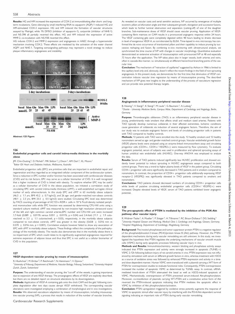
Results: HG and HM increased the expression of COX-2 at immunoblotting after short- and long-term incubations. Gene silencing by small interfering RNA to aquaporin (AQP)-1 reduced HG andHM-stimulated COX-2 expression. HG and HM induced the formation of vascular structuresassayed by Matrigel, while 1% DMSO (inhibitor of aquaporin-1), cariporide (inhibitor of NHE-1)and NS-398 all partially reverted this effect. HG and HM induced the expression of activeMMP-2, co-incubation with NS-938 reverted this effect.HG induces COX-2 and MMP-2 expression and angiogenesis in HAEC through a hyperosmolarmechanism involving COX.2. These effects are mediated by the activation of the water channelAQP1 and NHE-1. Targeting osmosignaling pathways may represent a novel strategy to reduceplaque inflammatory angiogenesis and instability.
126Endothelial progenitor cells and carotid intima-media thickness in the morbidlyobese
JPF. Chin-Dusting 1; D. Michell 1; MR. Skilton 1; J. Dixon 1; AM. Dart 1; XL. Moore 1
1Baker IDI Heart and Diabetes Institute, Melbourne, Australia
Endothelial progenitor cells (EPC) are primitive cells that are important in endothelial repair andregeneration and thus regarded as an integrated cellular component of the cardiovascular system.Since a reduction in EPC number and/or function has been associated with cardiovascular diseases(CVD) and its risk factors, EPC may serve as a cellular biomarker of CVD. It is well recognizedthat a higher incidence of CVD is linked with obesity. To explore whether EPC may be usefulas a cellular biomarker of CVD in the obese population, we initiated a correlation study ofcirculating EPC with carotid intima-media thickness (cIMT), a well-established surrogate clinicalmarker of early atherosclerosis. In this study EPC and cIMT in 63 morbidly obese subjects(46.3 + 1.1 yrs, BMI 45.2 + 0.7 kg/m2), and 26 age- and gender-matched non-obese controls(48.1 + 2.3 yrs, BMI 25.5 + 0.5 kg/m2) were studied. Circulating EPC level was determinedby FACS counting of percentage of AC133+/KDR+ cells in 10^6 Ficoll-density isolated periph-eral mononuclear cells while EPC function was assessed by determining CFU-Hill units using astandard culture method. cIMT was measured by non-invasive high resolution ultrasound scansof the left and right common carotid arteries.EPC number and CFU-Hill unit were increased1.7-fold (0.089 + 0.011% versus 0.051 + 0.011%, p ¼ 0.04) and 3.4-fold (11.1 + 1.9 colo-nies/well vs 3.3 + 1.1 colonies/well, p ¼ 0.03), respectively, in the morbidly obese subjectscompared to non-obese controls. cIMT was greater in the obesity (0.658 + 0.016 mm vs0.570 + 0.016 mm, p ¼ 0.001). Correlation analysis however failed to show any association ofEPC with cIMT in morbidly obese subjects. These findings reflect the complexity of the pathophy-siology of the morbidly obesity. The results also demonstrate that in the morbidly obese there isno impairment of EPC which could relate to its significantly augmented angiogenesis required forextreme expansion of adipose tissue and thus that EPC is not useful as a cellular biomarker ofCVD in this population.
127VEGF-dependent vascular pruning by means of intussusception
R. Hlushchuk 1; M. Ehrbar 2; P. Reichmuth 1; N. Heinimann 1; V. Djonov 1
1University of Fribourg, Department of Medicine, Vascular Biology, Fribourg, Switzerland; 2University HospitalZurich, Zurich, Switzerland
Purpose: The understanding of vascular pruning, the "cut-off" of the vessels, is gaining importancedue to expansion of anti-VEGF therapy. The proangiogenic effects of VEGF are explicitly described,but there are no detailed report on structural alterations by its downregulation.Methods: Application of VEGF-A containing gels onto the chick CAM and the gels’ following com-plete degradation after two days causes abrupt VEGF withdrawal. The corresponding vascularalterations were investigated employing a combination of morphological and in vivo investigations.Results: We observed vasculature adaptation by means of intussusception, including intussuscep-tive vascular pruning (IVP), a process that results in reduction of the number of vascular branches.
As revealed on vascular casts and serial semithin sections, IVP occurred by emergence of multipleeccentric pillars at bifurcation angle and their subsequent growth, elongation and successive fusions,which leads to further luminal obstruction and separation (cutting-off) of one of the daughterbranches. Sub-maintenance doses of VEGF should cause vascular pruning. Application of VEGF-containing fibrin matrices on CAM results in a pronounced angiogenic response within 24 hours.The VEGF containing gels were completely digested within 48 hours leading to drastic decreasein level of exogenous VEGF-A, as corroborated by ELISA. Time-lapsed in-vivo microscopy has con-firmed the de novo occurrence of transluminal pillars and their capability to induce pruning by suc-cessive reshaping and fusion. By combining in-vivo monitoring with ultrastructural analysis, wesynchronized the time course of IVP with changes in vascular morphology. Quantitative evaluationdemonstrated an extensive activation of intussusception with pronounced IVP at 48 and especially72 hours after the application. The IVP takes place also in larger vessels, both arteries and veins,often in cascade-like manner, i.e. simultaneously at different hierarchical branching points of the vas-cular tree.Conclusion: The mechanism of "retraction of capillaries" suggested by Ashton in 1966 is limited tothe capillary level only and, obviously, doesn’t reflect the recent findings in the field of non-sproutingangiogenesis. In this present study, we demonstrate for the first time that diminution of VEGF con-centration induces vascular tree regression by means of intussusceptive pruning. The describedmechanism of IVP gives new insights to the understanding of the process of vasculature regressionand can provide new potential therapy targets.
128Angiogenesis in inflammatory peripheral vascular disease
B. Hewing 1; V. Stangl 1; K. Stangl 1; M. Laule 1; G. Baumann 1; A. Ludwig 1
1Charite - University Medicine Berlin, Campus Mitte, Department of Cardiology and Angiology, Berlin,Germany
Purpose: Thromboangiitis obliterans (TAO) is an inflammatory peripheral vascular disease inyoung, predominantly male smokers that affects small and medium sized arteries. Patients withTAO typically develop numerous collaterals in their affected extremities. Ischemic conditionscause generation of collaterals via induction of several angiogenic factors. Therefore, the aim ofour study was to evaluate angiogenic factors and levels of circulating progenitor cells in patientswith TAO compared to healthy controls.Methods: 12 patients with TAO were enrolled into the study. 12 healthy smokers and 12 healthynonsmokers served as age- and gender-matched control groups. Vascular endothelial growth factor(VEGF) plasma levels were analyzed using an enzyme-linked immunosorbent assay and circulatingprogenitor cells (CD34+, CD34+/ VEGFR2+) were measured by flow cytometry. To evaluateangiogenic potential, serum of subjects was used in proliferation and spheroid sprouting assays ofhuman umbilical vein endothelial cells (HUVEC). One-way ANOVA was employed to analyzethe data.Results: Serum of TAO patients induced significantly less HUVEC proliferation and showed evi-dently lower potential to induce sprouting in HUVEC angiogenesis assays compared to bothcontrol groups. There was a trend to higher plasma levels of VEGF in the patient group. CirculatingCD34+ progenitor cells were significantly decreased in TAO patients and in smokers compared tononsmokers. In contrast, the proportion of CD34+ progenitor cells additionally expressing VEGFreceptor-2 (VEGFR2) was significantly elevated in TAO patients compared to smokers andnonsmokers.Conclusions: TAO patients showed decreased numbers of circulating CD34+ progenitor cellswhile levels of putative circulating endothelial progenitor cells (CD34+/ VEGFR2+) wereincreased. Despite elevated levels of VEGF, serum of TAO patients exhibited lower angiogenicpotential.
129The pro-apoptotic effect of PTEN is mediated by the inhibition of the PI3K-Aktpathway after vascular injury.
R. Widmer-Teske 1; A. Mueller 1; P. Stieger 1; H. Tillmanns 1; RC. Braun-Dullaeus 2; DG. Sedding 1
1University Hospital Giessen and Marburg, Medical Clinic I, Cardiology and Angiology, Giessen, Germany;2Hospital Magdeburg, Department of Cardiology, Magdeburg, Germany
Background: The inositol phosphatase and tumor suppressor protein PTEN is a negative regulatorof the phosphatidylinositol-3 kinase (PI3-K)/protein kinase B (Akt) pathway. However, the PTEN-dependent mechanisms during early vascular remodeling are still unknown. In this study, we inves-tigated the hypothesis that PTEN regulates the underlying mechanisms of vascular smooth musclecells (VSMC) during early apoptotic processes following vascular injury in vivo.Methods and Results: Immunohistochemistry, western blotting and phosphatase activity assaysindicated that PTEN expression and activity were strongly increased in apoptotic (TUNEL+)VSMC 2-12h following balloon-injury of rat carotid arteries in vivo. PTEN expression was not influ-enced by stimulation with serum or different growth factors in vitro, whereas treatment with H2O2as a source of oxidative stress was followed by enhanced PTEN expression and activity in a time-and dose-dependent manner. Human VSMC were transduced with a plasmid carrying a WT form ofPTEN for overexpression studies to evaluate functional roles. Consequently, PTEN overexpressionincreased the number of apoptotic VSMC as determined by TUNEL assay. In contrast, siRNA-mediated knock-down of PTEN attenuated the basal as well as H2O2-induced apoptosis ofVSMC. Mechanistically, overexpression of PTEN prevented serum-induced Akt-phosphorylation.Moreover, co-transfection of plasmids carrying WT-PTEN and a constitutive active form of Aktreversed the PTEN-induced apoptosis indicating that PTEN mediates the apoptotic effect inVSMC by inhibition of Akt phosphorylation/activation.Conclusion: PTEN upregulation triggered by oxidative stress potently augments the response ofVSMC to apoptotic stimuli after vascular injury by interfering with the PI3-K/Akt-dependent survivalsignaling indicating an important role of PTEN during early vascular remodeling.
Abstract 125 Figure
S58 Abstracts
Cardiovascular Research Supplements
at Colum
bia University H
ealth Sciences Library on M
arch 4, 2013http://cardiovascres.oxfordjournals.org/
Dow
nloaded from
130Fluid shear stress-responsive microRNAs are involved in arteriogenesis
K.Troidl1; L.Eller 1; I.Benli 2; H.Apfelbeck 2; W.Schierling3; C.Troidl 4; W.Schaper1; T.Schmitz-Rixen2
1Max Planck Institute for Heart and Lung Research, Bad Nauheim, Germany; 2Division of Vascular andEndovascular Surgery, Johan-Wolfgang-Goethe, Frankfurt, Germany; 3University Hospital Regensburg,Regensburg, Germany; 4Franz-Groedel Institute of the Kerckhoff Clinic, Bad Nauheim, Germany
Objective: MicroRNAs (miRNA) are implicated in post-transcriptional regulation of geneexpression. Arteriogenesis – the growth of pre-existing collateral arterioles to functional arteries– is triggered by increased fluid shear stress (FSS). The growth process is associated with a pheno-typic switch of vascular smooth muscle cells (VSMC) and a re-expression of fetal or cardiac genes.We investigated the involvement of miRNAs in arteriogenesis in a rat model of chronically elevatedFSS in collateral arteries.Methods: 6 sprague dawley rats were subjected to femoral artery ligature (FAL). A side-to-sideanastomosis distal to the ligature was created between the femoral artery and the accompanyingvein, which leads to chronically elevated FSS inside the collaterals. Following dissection of collateraltissue 7d after surgery, miRNA was isolated and an expression profile was generated by microarrayanalysis. Differential expression was confirmed by qRT-PCR. Cellular localization of selectedmiRNAs was assessed by in situ hybridisation.Results: Surgical FSS-stimulation provoked an extreme arteriogenic response and the growingcollateral arteries showed increased levels of 29 miRNAs when compared to sham operatedcontrols. A subset of 6 miRNAs turned out to be significantly up-regulated (p , 0.05) asdetermined by qRT-PCR. miR-143, miR-195, and miR-24 were localized in the media of growingcollateral arteries.Conclusions: These data indicate that miRNAs are involved in arteriogenesis. miR-143 belongs toa cluster which is implicated in the phenotypic switch of VSMCs. miR-195 and mir-24 are alsoup-regulated in human heart failure and hypertrophic mouse heart. A potential functional associ-ation of the identified miRNAs with arteriogenesis has to be confirmed.
131Therapeutic neovascularization via Thymosin beta4 overexpression requires AKTactivation and capillary sprouting in the calf muscles: evidence for backwardsignaling
R. Hinkel 1; T. Trenkwalder 1; A. Pfosser 1; F. Globisch 1; G. Stachel 1; C. Lebherz 1; I. Bock-Marquette 2;C. Kupatt 1
1Ludwig-Maximilians University, Medical Clinic and Policlinic I, Clinic Grosshadern, Munich, Germany;2University of Texas Southwestern Medical Center, Dallas, United States of America
Thymosin beta4, an endogenously occurring peptide of 5 kDa, is cardioprotective after ischemia andreperfusion in a Protein Kinase B (AKT) dependent manner. Furthermore it is essential for coronaryvessel development and provides angiogenesis during wound healing of the adult organism. Here wetested if long-term overexpression of thymosin beta4 with an adeno-associated virus is beneficial fortherapeutic neovascularisation in a chronic in vivo model of hindlimb ischemia.Methods: In rabbits (n ¼ 5 per group), femoral artery excision was performed at d0 and 5 ×10E12 AAV2/9 expressing Tb4 or Lac-Z (control group) were applied through intramuscular injec-tion. Tb4 was either injected over the whole limb or only into the calf muscles. In addition a thirdgroup received total limb Tb4 transfection and 1 × 10E13 AAV 2/9 AKT-DN into the lower limbonly (i.m.). At day 7 and day 35 angiography was performed for collateral quantification (% of day 7)and frame count score (% of day 7) and fluorescent microspheres were applied to determine theregional blood flow. At day 35 tissue was harvested for determination of capillary density via immu-nostaining for PECAM-1 (capillaries/muscle fiber ¼ c/mf).Results: rAAV-Tb4 significantly increasd capillary density in the ischemic limb (1.6 + 0.2 c/mf)compared to the control group (0.96 + 0.1c/mf). Quantification of collateral growth (175 +15% vs. 95 + 6% in controls) and microspheres (157 + 10 vs. 110 + 6 in controls) yieldedsimilar results after Tb4 transfection. However co-transfection of AKT-DN blunted the effect (c/mf 1.3 + 0.2, collateral growth 126 + 11%; microspheres 107 + 3). Moreover, calf muscle trans-duction of Tb4 sufficed to induce collateral growth (176.84 + 4,1%) and gain of perfusion (micro-spheres: 160 + 12.1) to the same extent as whole limb Tb4 transduction.Conclusion: Thymosin b4 long-term overexpression exerts therapeutic neovascularisation inchronic ischemia. The down-regulation of activated AKT using a dominant negative mutant in thelower limb only suppresses capillary formation and subsequent collateral growth. In contrast, stimu-lation of microcirculatory growth using Tb4 in calf muscles only sufficed to promote arteriogenesisin the upper leg. These results suggest a backward signalling from microcirculation to conductancevessel formation.
Smooth muscle
132Inhibition of two-pore domain potassium channels TASK-1 by endothelin-1 is Rho-kinase dependent with Ser393 as the predominant phosphorylation site
C.Seyler 1; E.Duthil-Straub 1; E.Zitron 2; EP. Scholz 2; D.Thomas 2; J.Gierten 1; CA.Karle 2; RHA.Fink 1
1The Medical Faculty of Heidelberg, Heidelberg, Germany; 2University Hospital of Heidelberg, Heidelberg,Germany
Purpose: TASK-1 channels contribute substantially to the resting membrane potential in humanpulmonary artery smooth muscle cells (PASMCs). Endothelin-1 (ET-1) is a potent vasoconstrictorand mitogen for smooth muscle. ET-1 binds to two types of receptors, ETA and ETB. Both recep-tors are found in smooth muscle cells. ETA- receptors predominate in the large human pulmonaryarteries while ETB- receptors prevail in airway smooth muscle, alveolar wall tissue and capillaries.Tang et al. 2009 already showed that TASK-1 channels are inhibited by ET-1 in hPASMCs. However,they only investigated the ETA pathway. Furthermore, the exact underlying molecular mechanismremained unclear.
Methods: Cloned human endothelin A and B receptors and human TASK-1 channels wereco-expressed in Xenopus oocytes and pharmacological experiments were performed using thedouble electrode voltage clamp technique.Results: Application of ET-1 (10 nmol/l) to Xenopus oocytes expressing human TASK-1 channelscaused a strong inhibition of currents if the channels were co-expressed with endothelin-receptors. Using ETA receptors, current amplitudes were reduced to 25.9 + 6.0 %.Dose-response relations were analyzed and yielded an IC50 of 0.08 nmol/l. After co-expressionof ETB receptors with TASK-1 channels, exposure to ET-1 (10 nmol/l) lead to a reduction ofcurrents to 40.2 + 8.0 %. The dose-response curve yielded an IC50 of 0.23 nmol/l. A screeningwith the use of small molecule protein kinase inhibitors indicated that TASK-1 is regulated viaRho-kinase dependent signalling by both receptors. Hence, we generated mutant channelslacking Rho-kinase phosphorylation sites Ser-336 and Ser-393. With ETA receptors, the effectof ET-1 (10 nmol/l) was completely abolished in mutant TASK1-S393A channels resulting in acurrent increase to 104.2 + 6.6 %. The effect was also attenuated in TASK1-S336A channelswith a current decrease to 78.9 + 5.7 %. With ETB receptors, mutant TASK1-S393A channelsshowed a current increase to 114.7 + 7.7 % after ET-1 application, whereas TASK1-S336Achannels did not show any desensitization to ET-1, resulting in a current decrease of60.5 + 8.1 %.Conclusions: This study shows that ET-1 binding to either ETA or ETB receptors causes an inhi-bition of human TASK-1 channels via a Rho-kinase dependent pathway. Rho-kinase phosphorylationsite Ser-393 is essential for this regulation, whereas the functional significance of Ser-336 differsbetween ETA- and ETB-dependent signalling. The Rho-kinase pathway may provide novel thera-peutic targets downstream of the ETA and ETB receptors.
133Angiotensin II promotes migration of arterial smooth muscle cells exposed toaggregated LDL by a UPA dependent mechanism
T. Padro 1; R. Lugano 1; M. Garcia-Arguinzonis 1; L. Badimon 1
1Hospital de la Santa Creu i Sant Pau, Cardiovascular Research Center, CSIC-ICCC, Barcelona, Spain
Purpose: Urokinase-type plasminogen activator (UPA) is increased in vascular smooth muscle cell(VSMC)-rich areas of early atherosclerotic lesions and in vascular remodelling. Low density lipopro-teins (LDL) and angiotensin II (AngII) are directly associated with the development of atherosclero-tic lesions. Recent evidences suggest that AngII enhances atherosclerosis in hyperlipemic animals. Itis our hypothesis that this effect might be dependent on the UPA/UPAR system. Thus, we aimed toinvestigate the effects of AngII in UPA expression and migration in VSMC exposed to pathologicalconcentrations of atherogenic matrix-bound aggregated LDL (agLDL).Methods: Human VSMC were treated with/without 1 mM AngII in the presence/absence of 100mg/mL agLDL (24h). Cell migration was studied using an in vitro model of wound repair. UPAmRNAs levels were analyzed by RTqPCR. Cell UPA localization was assessed by immunostaining.UPA expression was knocked down by small interfering RNAs.Results: AngII increased migration kinetics of VSMC in the presence of agLDL (2-fold increase, p ,
0.05), whereas AngII did not show any significant effect on control VSMC migration (absence ofagLDL). AngII treatment resulted in a 3-fold higher expression of UPA mRNA (p , 0.05) in cellsexposed to agLDL. In contrast, PAI-1mRNA levels (UPA inhibitor) only increased by 1.4-foldunder these conditions. AngII markedly enhanced UPA detection at the cell membrane ofagLDL-treated cells, as assessed by WB and immunocytochemistry. UPA signals were mainlylocated at the front and rear edges of the migrating cells. AngII effects on VSMC exposed toagLDL were abrogated by endogenous UPA blockade with a polyclonal antibody (see table).Similar results were observed by silencing the UPA gene. Furthermore, the addition of 2 mg/mLexogenous UPA enhanced by more than 95% (p , 0.05) the migration kinetics of VSMCexposed to agLDL.Conclusions: Ang II-triggered cell migration in VSMC exposed to pathologic concentrations ofagLDL is dependent on UPA. Our results suggest a new mechanism by which AngII may contributeto progression of atherosclerotic lesions.This work was performed thanks to FIS PI07-1070, PN MICINN SAF2006-10091, CIBERobn CB06/03.
Abstract 133 Table Wound repair at 8 hours after injury
Control agLDL agLDL+AngII agLDL+AngII + anti-UPA ANOVA Test
83 + 4% 34 + 4% 79 + 6% 35 + 7% p , 0.05
134The dinucleotide uridine adenosine tetraphosphate (Up4A) enhances in vitromineralization via transformation of VSMCs to osteoblast-like cells
M. Schuchardt 1; J. Pruefer 1; M. Toelle 1; N. Pruefer 1; V. Jankowski 1; J. Jankowski 1; W. Zidek 1;M. Van Der Giet 1
1Charite - Campus Benjamin Franklin, Berlin, Germany
Purpose: Vascular calcification is a major complication in advanced chronic kidney disease (CKD)and dialysis patients. Vascular calcification has long been considered to be a passive process;however recent evidence indicates actively inducing changes in vascular smooth muscle cell behav-ior toward an osteoblast-like phenotype, whereas the clear pathomechanism is not clear. The aim ofthis study was to explore the influence of Up4A, which accumulates in serum from dialysis patients,on the induction of in vitro calcification in smooth muscle cells.Methods: In vitro calcification in rat vascular smooth muscle cells (VSMC) were induced with cal-cification medium (DMEM supplemented with 15% FCS, 10 mM sodium pyruvat, 50 mg/ml VitaminC, and 10 mM b-glycero phosphate) in the presence and absence of Up4A. Calcium deposition was
Abstracts S59
Cardiovascular Research Supplements
at Colum
bia University H
ealth Sciences Library on M
arch 4, 2013http://cardiovascres.oxfordjournals.org/
Dow
nloaded from
monitored by Alizarin staining and quantified by O-cresolphthalein complexone method. Alkalinephosphatase (ALP) activity was measured using a commercial available assay kit. a-actin wasstained immunofluorescently with an FITC-conjugated anti-a-actin-smooth-muscle antibody.Cbfa1 mRNA expression was measured via real-time PCR.Results: Up4A [10 mmol/l] led to a significantly increased calcium deposition monitored by Alizarinstaining and quantified by O-cresolphthalein complexone method (control: 7.7 + 1.2 mg/dl; calci-fication medium: 32.1 + 6.2 mg/dl vs. calcification medium containing Up4A: 63.6 + 13.2 mg/dl; p, 0.05, n ¼ 11). The specific marker for smooth muscle cells, a-actin, decreases during calcificationand is significantly down-regulated in the Up4A stimulated cells compared with calcificationmedium, indicating the phenotype switch of the cells. Furthermore, ALP activity increases time-dependently in cells stimulated with calcification medium and is significantly higher in the presenceof Up4A (after 14 days [IU/g] – control: 3.3 + 0.7; calcification medium: 10.8 + 1.2 vs. calcificationmedium containing Up4A: 19.5 + 2.8; p , 0.05; n ¼ 11). Up4A is able to significantly increase corebinding factor a-1 (Cbfa1) mRNA expression, a highly regulatory transcription factor for osteogen-esis, in a dose-dependent manner.Conclusions: Our results could show that Up4A might be a potent inductor of arterial calcificationby transformation of smooth muscle cells in osteoblast-like cells via induction of the osteogenenicmarkers. Therefore, Up4A, which is up-regulated in dialysis patients, could be having potential impli-cation in arteriosclerosis under dialysis condition.
135Tissue factor signaling in migrating human vascular smooth muscle cells
E. Pena ; G. Arderiu ; L. BadimonHospital de la Santa Creu i Sant Pau, Cardiovascular Research Center, CSIC-ICCC, Barcelona, Spain
Purpose: Tissue factor (TF) is the most relevant physiological trigger of blood coagulation andthrombosis in response to vascular injury. Phosphorylation of the cytoplasmic tail of TF byprotein kinase C and interaction with actin-binding protein 280 has been described. The aim ofthis study has been to elucidate by which mechanism TF induces HVSMC migration.Methods: Primary cultures of HVSMC were obtained from coronary arteries of explanted hearts(Transplant Unit). A nucleofector device was used for TF silencing (siRNA). cDNA was used toevaluated 84 key genes of 18 different signal transduction pathways. Validation by PCR and WBwere performed.Results: The most affected network by inhibition of TF expression was the Hedgehog pathwaywith down regulation of BMP2, BMP4, EN1, FOXA2 and PTCH1. Transcription factor EGR1 alsowas down regulated by inhibition of TF. EGR1 regulates heparanase (HPSE) gene transcription intumor cells and we tested whether HPSE expression gene was modified in TF siRNA tranfectedHVSMC. Indeed the HPSE gene that encodes heparanase, an enzyme that targets both the cellsurface and the extracellular matrix to degrade polymeric heparan sulphate molecules intoshorter chain length oligosaccharides, was significantly down-regulated by TF silencing in HVSMC.Conclusions: TF has a significant role in cell migration through numerous pathways alternative tothe classic ones. sRNA-TF in HVSMCs modifies a wide spectrum of transduction pathways, whichplay a crucial role in many intracellular events. Most genes identified were not previously recognizedas TF-regulated, and they encode proteins involved in cell-cycle control and proteins involved inmatrix remodeling as HPSE that by degrading extracellular matrix, facilitates HVSMC migration.This work has been possible thanks to PNS-MICINN SAF2006-10091, CIBERobn CB06/03
136Impact of window L-type calcium influx on activity of calcium channel blockers inaortic smooth muscle cells of C57Bl6 mice
P. Fransen 1; CE. Van Hove 1; C. Michiels 1; J. Van Langen 1; H. Bult 1
1University of Antwerp, Antwerp, Belgium
Purpose: L-type calcium channel (CC) blockers (CCBs) reduce blood pressure more effectively inhypertensive than in normotensive subjects and are more effective in vascular smooth muscle(VSM) than in cardiac muscle. Several attempts have been made to explain these unique features.The depolarization of the resting potential by hypertension and the depolarized resting potentialof VSM in comparison with the heart not only favour the inactivated state of the CC, but alsoincrease window L-type calcium influx.Methods: The present study investigated whether the CC agonist BAY K8644 and whether nife-dipine, verapamil or diltiazem, representatives of different CCB classes, alone or in combination,modulated window calcium influx (Fura-2 method) and isometric contraction during depolarisationof C57Bl6 mouse aorta segments with 50 mM K+.Results: High K+-induced contractions were biphasic. The fast force component coincided withcalcium influx via a population of CC that exhibited fast activation and then inactivation. Theslow, sustained force component was attributed to voltage-dependent, but time-independentcalcium influx displaying incomplete inactivation (window) and was significantly more sensitive toCCBs than the fast component. Stimulation of CC calcium influx with 30 nM BAY K8644 decreased,whereas conditions that favoured window calcium influx increased the efficacy of CCBs to inhibitisometric contractions.Conclusion: Results suggest that inhibition of window L-type calcium influx may play a dominantrole in the anti-hypertensive effects of CCBs.
137Pulmonary vascular cell dysfunction in chronic thromboembolic pulmonaryhypertension: a role of c-reactive protein?
R. Quarck 1; M. Wynants 1; E. Alfaro-Moreno 2; M. Rosario Sepulveda 3; F. Wuytack 3;D. Van Raemdonck 4; B. Meyns 5; M. Delcroix 1
1Katholieke Universiteit Leuven, Respiratory Disease Division, LEUVEN, Belgium; 2Instituto Nacional deCancerologia, Basic Research, Mexico City, Mexico; 3Katholieke Universiteit Leuven, Molecular Cell BiologyDepartment, LEUVEN, Belgium; 4Universitaire Ziekenhuizen Leuven, Thoracic Surgery Unit, LEUVEN,Belgium; 5Katholieke Universiteit Leuven, Cardiac Surgery Unit, LEUVEN, Belgium
Purpose: Chronic thromboembolic pulmonary hypertension (CTEPH) is associated with a proxi-mal pulmonary vascular obstruction whose origin is not elucidated. Our hypothesis is that this vas-cular obstruction is not only a consequence of dysregulated thrombosis and/or thrombolysis, butthat uncontrolled inflammation may also be involved in the pathogenesis of CTEPH. We have pre-viously found that circulating C-reactive protein (CRP), an inflammatory biomarker, levels wereelevated in CTEPH patients compared to healthy subjects. The current objective is to investigatethe potential involvement of CRP in promoting pulmonary vascular dysfunction observed in largepulmonary arteries of CTEPH patients.Methods: Proximal pulmonary arteries have been obtained from CTEPH patients, from patientswith other causes of pulmonary hypertension (PH) and from lung donors. Primary cultures of prox-imal pulmonary arterial endothelial (PAEC) and smooth muscle cells (PASMC) have been estab-lished. mRNA and protein expression of CRP and its receptor, CD32, has been measured inproximal pulmonary vascular tissue and primary cultures of PAEC and PASMC. The effect ofCRP on PAEC and SMC mitogenic activity was measured. CRP-induced adhesion capacity ofPAEC to a human monocytic cell line, U937 cells has been evaluated. CRP-induced ET-1 secretionby PAEC has been studied by ELISA. Localization and tissue distribution of CRP and CD32 has beenperformed on pulmonary vascular tissue, PAEC and PASMC by immunofluorescence.Results: An upregulation of CRP and CD32 was observed in proximal pulmonary arteries fromCTEPH patients. Increasing concentrations of CRP significantly increased CTEPH-SMC mitogenicactivity leveling off at 140% (p ¼ 0.001), whereas it had no effect on PH- and normal PASMC.CRP had no effect on PAEC mitogenic activity. CRP increased the adhesion of U937 cells toCTEPH-PAEC by 37% (p ¼ 0.0002), by inducing the expression of the adhesion molecule Inter-Cellular Adhesion Molecule 1 (ICAM-1) at their surface. Similarly, CRP tended to increase thesecretion of ET-1 by CTEPH-PAEC by 129% (p ¼ 0.08), whereas it did not affect ET-1 secretionby PH-PAEC or normal PAEC. FBS-stimulated CTEPH-SMC displayed a late upregulation of CRPmRNA whereas PH-PASMC and PH-PASMC did not.Conclusions: We evidenced a CRP-driven dysfunction of pulmonary vascular cells of CTEPHpatients, suggesting a potential role for CRP in the pathophysiology of CTEPH. Our results alsosuggest that CRP may contribute to vascular remodeling, directly via PASMC and indirectly viaPAEC.
138Expression and function of the tissue renin-angiotensin system in human vascularsmooth muscle cells in culture
F. Christofi 1; S. Wijetunge 1; PS. Sever 1; AD. Hughes 1
1Imperial College London, National Heart & Lung Institute, London, United Kingdom
Purpose: The renin-angiotensin system (RAS) plays an integral role in cardiovascular homeostasisby influencing vascular tone, fluid and electrolyte balance. Abnormalities of RAS have been impli-cated in the development of hypertension, diabetes and cardiovascular disease. It has been pro-posed that RAS exists locally in tissue including the blood vessels, the heart and brain. Weexamined the expression of components of the RAS in human vascular smooth muscle cells inculture and investigated their role in VSMC proliferation.Methods: Human saphenous vein cells (hSVSMC), human coronary artery cells (hCAC) andimmortalised human VSMC derived from saphenous vein cells (hVTSM1) were grown in SmoothMuscle Growth Medium. Expression of components of the RAS was examined by conventionalPCR methodology. RNA was isolated from the cells and reverse transcribed to complimentaryDNA (cDNA). Standard PCR methodology was carried out on the cDNA using primer setsdesigned for the genes: renin, angiotensinogen (AGT), angiotensin converting enzyme-1 (ACE-1),angiotensin converting enzyme-2 (ACE-2), angiotensin II type 1 and type 2 receptors (AT1R &AT2R), the receptor for angiotensin [1-7] (MAS-1), and the renin receptors, mannose-6-phosphate(M6P-R) and ATP6AP2. The PCR products were run on agarose gels and visualised by ethidiumbromide staining and UV light exposure.Protein expression of the RAS components was measured by SDS-PAGE and Western blottingusing antibodies directed against renin, and its receptors M6P-R and ATP6AP2.VSMC proliferation was studied using the Quick Cell Proliferation Assay Kit, based on cleavage oftetrazolium salt WST-1 to formazan by mitochondrial dehydrogenase activity. Cells were treatedwith renin and viable cells quantified by measuring formazan by spectrophotometery.Results: hSVSMC, hVTSM1 and hCAC expressed genes for (pro)renin, ATP6AP2, M6P-R, AGT,ACE-1, and AT1R (n ¼ 3). Expression of AT2R, ACE-2 and MAS-1 genes was not detected inthese cells. Protein expression of renin, M6P-R and ATP6AP2 were detected in both hVTSM1and hSVSMC (n ¼ 4).Treatment of saphenous vein cells with renin (10nM) for 24hrs increased cell proliferation by 23 +4% (n ¼ 4, p , 0.03).Conclusions: Multiple components of RAS are expressed in vascular smooth muscle cells at RNAand protein level. Local RAS may play a role in VSMC proliferation and this may be of functionalimportance in vascular remodelling and in the atherosclerotic process.
139Activation of neutral sphingomyelinase by endothelin-1 is implicated in vascularinflammation of intact small arteries
J. Ohanian 1; SP. Forman 1; V. Ohanian 1
1University of Manchester, Manchester, United Kingdom
Purpose: Endothelin-1 (ET-1) is a potent vasoconstrictor with multiple effects on the cardiovascu-lar system which range from pro-inflammatory actions to induction of endothelial dysfunction andremodelling of vascular tissue. Sphingolipids are implicated in cardiovascular homeostasis includingvascular inflammation. Sphingomyelinases (Smase) hydrolyse sphingomyelin to produce the bio-active sphingolipid ceramide and its metabolite sphingosine-1-phosphate both of which are impli-cated in the vascular inflammatory actions of cytokines. We studied the effect of ET-1 onSmases in intact rat mesenteric small arteries.Methods: Smase activity was measured by the production of ceramide from sphingomyelin. Vas-cular cell adhesion molecule-1 (VCAM-1) expression was determined by western blotting.
S60 Abstracts
Cardiovascular Research Supplements
at Colum
bia University H
ealth Sciences Library on M
arch 4, 2013http://cardiovascres.oxfordjournals.org/
Dow
nloaded from
Results: We identified neutral (NSmase) and acid (ASmase) activity and showed that ET-1 stimu-lated only the former. NSmase activity was localised predominantly with caveolae/lipid raft fractionswithin the membrane. The time course of NSmase activation was similar to the hydrolysis of sphin-gomyelin and accumulation of ceramide induced by ET-1. ET-1 activation of NSmase was dependenton p38MAPK activation and inhibited by the NSmase inhibitor GW4869. Prolonged (16 hour) incu-bation with ET-1 induced VCAM-1 expression, a marker of inflammation. This effect was inhibitedby GW4869.Conclusion: These data show that pro-inflammatory effects of ET-1 in vascular tissue are throughthe NSmase signalling pathway.
140Src family tyrosine kinases mediate hypotonicity- induced activation of L-typecalcium channels in vascular smooth muscle cells
S. Wijetunge 1; AD. Hughes 1
1Imperial College London, National Heart & Lung Institute, London, United Kingdom
Purpose: A hyposmotic stimulus causes phosphorylation of src family tyrosine kinases (SFK) andinduces arterial vasoconstriction in vitro by activation of L-type calcium channels. We investigatedwhether a hyposmotic stimulus increases L-type Ca2+channel currents in voltage clamped singlearterial smooth muscle cells and whether SFK mediate this effect.Methods: Vascular smooth muscle cells (VSMC) were isolated enzymatically from rat tail artery.Calcium channel currents (IBa) were measured by conventional whole cell voltage clamp techniquesusing Ba2+ as a charge carrier. Hyposmolarity was induced by removal of mannitol from the cellbathing solution. Role of SFK was investigated by using structurally different selective inhibitorsof SFK, SU6656 and PP1.Results: A reduction in extracellular osmolarity from 300mOsm/L to 275mOsm/L increased IBa by38 + 8 % (n ¼ 3, p , 0.05), and a further reduction in osmolarity to 250mOsm/L caused a further51 + 12% (n ¼ 8, p ¼ 0.005) increase in IBa. The increase in IBa induced by a reduction in osmo-larity to 250mOsm/L was prevented by pre-incubation with SU6656 (10 mM) and PP1 (1 mM) (n ¼3 & 5 respectively).Conclusion: A reduction in osmolarity results in increased L-type Ca2+ channel currents and thiseffect is mediated by SFK. This mechanism may be important in linking biomechanical stimuli tovasoconstriction in the vasculature including vasoconstriction induced by increased transmuralpressure and stretch.
141The role of adducin in reorganisation of the actin cytoskeleton in vascular smoothmuscle
C. Gibbons 1; J. Ohanian 1; V. Ohanian 1
1University of Manchester, Manchester, United Kingdom
Reorganisation of the vascular smooth muscle (VSM) actin cytoskeleton plays a fundamental rolein contraction. VSM contains a substantial pool of unpolymerised (G) actin which exists in con-stant flux with filamentous (F) actin creating a dynamic cytoskeleton which can adapt rapidly toenvironmental changes. The importance of the G/F actin ratio in contraction has been shownin a number of studies. The cytoskeletal protein adducin acts as an actin binding and cappingprotein, binding to the fast growing ends of actin filaments and preventing loss or gain of actinsubunits, thereby stabilising filament turnover. Adducin also bundles actin filaments at highconcentrations.We have investigated the role of adducin in actin cytoskeleton reorganisation in rat mesentericsmall arteries (RMSA). We have shown that a-adducin is present in smooth muscle cells, both inculture and in RMSA sections. In non stimulated arteries approximately 50% of a-adducin is associatedwith the cytoskeleton. After stimulation for 1 minute with noradrenaline (NA) adducin becomesphosphorylated on a site specific for protein kinase C and translocates from the cytoskeleton tothe cytosol. We have also shown that there is little change in the G/F actin ratio in RMSA aftertreatment with NA for 1min, however after 5min NA stimulation there is an increase in theproportion of F actin. Therefore the phosphorylation and subsequent dissociation of adducin fromthe actin cytoskeleton may be important to allow elongation of actin filaments in vascular smoothmuscle.
142Selective role of SREBP isoforms in the regulation of vascular LRP-1 by aggregatedLDL
P. Costales 1; R. Aledo 1; S. Vernia 2; A. Das 3; V. Shah 3; M. Casado 2; L. Badimon 1; V. Llorente-Cortes 1
1Cardiovascular Research Center, CSIC-ICCC, Barcelona, Spain; 2Institute of Biomedicine of Valencia (CSIC),Valencia, Spain; 3Mayo Clinic, Rochester, United States of America
Aims: We aimed to investigate the role of SREBP-1a and SREBP-1c in the modulation of LRP1 tran-scription in human vascular smooth muscle cells (VSMC).Methods & Results: Specific loss of SREBP-1 or SREBP-2 significantly increased LRP1 proteinexpression by 2.71 and 2.26-fold, respectively. Overexpression of an active form of SREBP-1a orSREBP-2, but not of SREBP-1c, decreased LRP1 expression. Gel mobility shift and ChIP assaysdemonstrated that SREBP-1 and SREBP-2 were able to bind to three putative SRE sequences;SRE-A (-1042 to -1028), SRE-B (-115 to -101) and the previously described SRE-C sequence(+226 to +234). ChIP assays performed in agLDL-exposed cells demonstrated that agLDL (100mg/mL, 24 hours) significantly and specifically decreased SREBP-2 enrichment of the LRP1 promo-ter. Luciferase assays demonstrated that agLDL increased the transcriptional activity of A/B or A/Cdouble mutants but failed to increase that of the double B/C mutant. Our results show that bothSREBP-1a and SREBP-2 negatively modulated LRP1 transcription. Furthermore, agLDL exerted anupregulatory effect on LRP1 expression by physical displacement of SREBP-2 from the LRP1 pro-moter. Two SRE-like sequences control the response of LRP1 to agLDL.Conclusions: These results underscore a SREBP-2 targeted mechanism to prevent the LRP1 over-expression induced by hypercholesterolemia in the vascular wall.
143Significance of window L-type calcium influx for contraction of C57Bl6 mouse aorticsegments
P. Fransen ; CE. Van Hove ; J. Van Langen ; H. BultUniversity of Antwerp, Antwerp, Belgium
Purpose: Recently, differential expression of spliced isoforms of the voltage-gated calcium channel(VGCC) gene Cav1.2 was demonstrated along the vascular tree. These isoforms display cell-specificelectrophysiological properties with respect to the window VGCC calcium influx and sensitivity toL-type calcium channel blockers. We investigated whether the window VGCC calcium influx plays aphysiological role for blood vessel constriction.Methods: Calcium mobilisation (Fura-2 assay) and isometric contraction were studied in aortic seg-ments of C57Bl6 mice. The membrane potential was clamped at fixed potentials by increasingexternal K+ concentration.Results: Above 20 mM, K+ induced biphasic contractions. The fast component coincided withcalcium influx via a VGCC population that exhibited fast activation and then inactivation. Theslow force component was temporally related to a slow voltage-dependent, but time-independentcalcium influx. The slow contraction was not due to calcium induced calcium release, as it was notaffected by dantrolene, ryanodine or depletion of sarcoplasmic reticulum calcium stores with cyclo-piazonic acid. By modulating external calcium levels before and after depolarisation with extracellu-lar K+, we could attribute the sustained contraction to a population of VGCCs displayingincomplete inactivation (window). At normal extracellular K+ concentration, this window was oper-ative, allowing a continuous VGCC-mediated calcium influx leading to basal tone. Basal influx andforce were both suppressed upon hyperpolarization by adding the ATP-dependent K+ channelagonist levcromakalim.Conclusion: The window L-type calcium influx has a profound physiological impact on basaltension and isometric contraction of C57Bl6 mouse aortic smooth muscle cells.
144The miR-143/145 cluster: keyplayer in vascular proliferative diseases
W. Bielenberg 1; JM. Daniel 1; HH. Tillmanns 1; DG. Sedding 1
1University Hospital Giessen and Marburg, Medical Clinic I, Cardiology and Angiology, Giessen, Germany
Background: MicroRNAs (miRNA) are crucial regulators for a multiplicity of cellular events. Theirexpression during vascular proliferative diseases still remains elusive and may offer an interestingapproach for the prevention or treatment in these items.Methods/Results: Using microarray based expression analysis we screened for regulated miRNAsduring atherosclerosis and restenosis. For this, aortic arches of ApoE/LDLr2/2 mice wereextracted after 2 weeks (control), 6 and 12 months, miRNA was isolated and applied to microar-rays. Approximately half of all miRNAs emerged to be regulated.Restenosis was induced in C57BL6/N by dilation of the femoral artery and miRNA was isolated 10and 21 days after injury. Strikingly, the majority of all miRNAs was regulated.Noticeably downregulated miRNAs in both diseases were miR-143 and miR-145.An expression analysis in different cell types of the vascular wall assigned the expression of thiscluster to vascular smooth muscle cells (VSMCs).To analyze what stimuli might evoke the observed downregulation in vivo, proliferative (10% FCS)and inflammatory (INF-g, TNF-a, IL-1b, each 20 ng) stimuli were applied to VSMCs in vitro and theexpression of miR-143 and miR-145 was determined by real-time PCR. For both miRNAs, thereoccured a significantly reduced expression following mitogenic as well as inflammatory stimulation.To further assess the functional role of the miR-143/145 cluster, VSMCs were transfected with theprecursor forms of miR-143 and -145, and a combination of both, resulting in a significantly pre-vented proliferation and cell count as assessed by BrdU incorporation and Wst-1 conversion.However, no effect on apoptosis and migration was detectable.A PCR Array, comprising genes involved in the human cell cycle machinery, revealed a profoundantiproliferative expression pattern following increased concentrations of miR-143/145. Thisstrongly suggests a decisive role of the cluster for the regulation of genes upstream of theseevents and so potentially with great impact for proliferative diseases. In an in vivo setting thishypothesis was confirmed. The transfection of injured murine femoral arteries with precursorsof miR-143 and miR-145 significantly prevented neointima formation, highlighting the impact ofthis miRNA-cluster for vascular proliferative diseases.Conclusion: We conclude that the miR-143/145 cluster is a pivotal modulator of vascular eventsand therefore inhibits extensive implications for the therapy of vascular proliferative diseases.
145Factor Seven Activating Protease (FSAP) counteracts vascular stenosis progressionthrough cleavage of Platelet Derived Growth Factor (PDGF)-BB.
J-M. Daniel 1; K. Hersemeyer 2; T. Schmidt-Woell 2; DN. Kaetzel 3; H. Tillmans 1; DG. Sedding 1;SM. Kanse 2
1Justus Liebig University Giessen, Med. Clinic I, Cardiology, Giessen, Germany; 2Institute for Biochemistry,Justus Liebig University, Giessen, Germany; 3University of Kentucky, Kentucky, United States of America
Purpose: The Marburg I (MI) single nucleotide polymorphism (SNP) (G534E) in the factor sevenactivating protease (FSAP) gene is associated with reduced enzymatic activity and represents a riskfactor for carotid stenosis in humans. Since FSAP inhibits PDGF-BB mediated vascular smoothmuscle cell (VSMC) proliferation, we investigated the effects of FSAP in a wire-induced injurymodel of neointima (NI) formation.Methods: Dilatation of the femoral artery was followed by exogenous application of wild-type(WT)-FSAP, MI-FSAP or control. FSAP2/2 mice were also dilated, and neointimal size, PCNAstaining and PDGF-BB expression levels were determined at 2 weeks after injury. The effects ofWT- and MI-FSAP on PDGF-BB activity and VSMC proliferation were analyzed in vitro as wasthe influence of mutating PDGF-BB at its cleavage site (Arg160-Lys161-Lys162) (RKK to EEE).Results: Application of exogenous WT-FSAP but not MI-FSAP significantly reduced NI formation andinhibited VSMC proliferation within the vessel wall (n¼ 6 mice per group, P , 0.05). In contrast, the
Abstracts S61
Cardiovascular Research Supplements
at Colum
bia University H
ealth Sciences Library on M
arch 4, 2013http://cardiovascres.oxfordjournals.org/
Dow
nloaded from
neointimal size in FSAP2/2 mice was enhanced (n ¼ 6, P , 0.05) and higher expression levelsof PDGF-BB were detected by immunostaining. Furthermore, we show by Western blottinganalysis that dilated arteries of FSAP2/2 mice exhibited higher ratios of PDGF-BB/ tubulinexpression (n ¼ 6, P , 0.05). WT-FSAP cleaved PDGF-BB and reduced proliferation of VSMCin vitro, whereas MI-FSAP showed substantially decreased activity. Moreover, the RKK to EEEmutant of PDGF-BB was not cleaved by FSAP and its activity towards VSMC proliferationremained unchanged.Conclusions: By demonstrating that WT-FSAP but not MI-FSAP inhibits VSMC proliferation andNI formation via inactivation of PDGF-BB at its cleavage site, we identify FSAP as an endogenousregulator of PDGF-BB activity and thus a key player in vascular remodeling in vivo.
Calcium handling and excitation contractionhandling
146Phosphorylation of ryanodine receptors plays important role in rat heart functionduring maturation
E. Tuncay 1; HB. Kandilci 1; EN. Zeydanli 1; NN. Sozmen 1; D. Akman 1; S. Yildirim 1; B. Turan 1
1Ankara University, Faculty of Medicine, Ankara, Turkey
It is known that not only incidence but also prevalence of certain diseases vary with age orsexes, or both and recent studies focused to clarify the underlying mechanisms of these impor-tant events. We aimed to determine whether functional alterations occur in the heart duringmaturation and if it proves to case to elicit the underlying molecular steps involved in thisphenomenon. We used 3 and 6 month old male Wistar rats and compared their functional par-ameters as followed. First, we evaluated and compared mechanical function of left ventricle andintracellular action potential of left ventricle papillary muscle. We also measured field-stimulatedCa2+ transients and L-type Ca2+ channel currents (ICaL) in isolated ventricular cardiomyo-cytes from 3 or 6 month old rats. The critical Ca2+ handling proteins, phosphorylationstatus of ryanodine receptors (RyR2) and Ca2+/calmodulin-dependent kinase II (CaMKII),protein kinase A (PKA) were also studied. We further compared the protein levels of sarcoplas-mic reticulum Ca2+ pump (SERCA), FKBP 12.6 in cardiac tissues of 3 and 6 month old rats.Oxidation levels of protein sulfhydryl (SH) groups in isolated cardiomyocytes were also deter-mined. In mature hearts, we observed a depression in the cardiac contractile function (LVDP) aswell as b-adrenergic receptor (bAR) mediated contractile responses despite the unchangedLVEDP parameter. Moreover, action potential was significantly prolonged in isolated maturepapillary muscles. Maturation also leads to decreased Ca2+- transient, caffeine response, andICaL which are addressing altered Ca2+ homeostasis. Therefore, we have investigated therole of Ca2+ handling proteins which might be responsible from the declined functionalresponse during maturation. Maturation was accompanied by significant decrease in the b-ARdensity, protein levels of RyR2 and FKBP12.6 but CaMKII, PKA and SERCA protein levelswere unchanged. However, phosphorylation level of RyR2, CaMKII and PKA were increasedby maturation. Such observations may lead to hyperphosphorylated unstable RyR2 functionand altered Ca2+ homeostasis. Supporting this, in 6 month old rats, both total and free SHlevels were significantly less than 3 month old myocytes. Taken together, our data demonstratethat altered activity and expression levels of Ca2+ handling proteins of cytoplasm and intra-cellular Ca2+ stores accompanied by increased oxidant stress might affect cardiac functionduring maturation.(Supported by TUBITAK-SBAG-107S304)
147Dynamic regulation of IK1 by increased [Ca21]i enhances repolarization reserve indog myocardium
N. Nagy 1; K. Acsai 2; A. Farkas 3; JGY. Papp 2; A. Varro 1; A. Toth 1
1University of Szeged, Faculty of Medicine, Department of Pharmacology & Pharmacotherapy, Szeged,Hungary; 2University of Szeged, Hungarian Academy of Sciences, Division for Cardiovascular Pharmacology,Szeged, Hungary; 3University of Szeged, Faculty of Medicine, 2nd Dept of Internal Medicine & CardiologyCenter, Szeged, Hungary
Background: The "cardiac repolarization reserve" is established by interactions of several potass-ium channels and plays a crucial role in ensuring safe repolarization kinetics in the heart. In the lit-erature there are number of assumptions, but not much evidence, that certain potassium currents(IKr, IKs, IK1) may be modulated by intracellular calcium level ([Ca2+]i). IK1 is considered a keycomponent of the repolarization reserve. Therefore, the aim of this study – carried out in dog ven-tricular myocardium – was to elucidate the [Ca2+]i dependence of the IK1 and to estimate thepossible consequences on the cardiac repolarization reserve.Methods and key results: The APD lengthening effect of selective IK1 inhibition by 10 mM BaCl2was investigated using the conventional microelectrode technique. When [Ca2+]o was increasedto 4 mM, the effect of BaCl was almost doubled. In contrast, in the presence of 4 mM [Ca2+]o theAPD lengthening effect of complete IKr blockade by 300 nM dofetilide was substantially reduced.Separate voltage clamp experiments revealed that when the [Ca2+]i was increased by a prepulse,the magnitude of IK1 was also significantly increased as compared to low [Ca2+]i condition. In con-trast, statically increased [Ca2+]i failed to influence the IK1.Conclusions and implications: These results indicate that an increase in [Ca2+]i may inducedynamic augmentation of IK1. IK1 elevation may occur during the systolic period of the cardiaccycle, while the resting membrane potential is not affected. While an increase in [Ca2+]i is animportant factor in physiological adaptation, it also can induce life threatening arrhythmias underpathological conditions. These results suggest that the modulation of IK1 by increased [Ca2+]imay play an endogenous antiarrhythmic role via enhancing repolarization reserve.
149A hydrophobic region of the ryr2 pore is critical for its function, as evidenced byshaker peptides
C. Viero 1; S. Mason 1; A. Williams 1
1Wales Heart Research Institute, Cardiff, United Kingdom
Catecholaminergic polymorphic ventricular tachycardias (CPVT) are inherited arrhythmias affectingpredominantly the cardiac ryanodine receptor type 2 (calcium release channel of the sarcoplasmicreticulum or RyR2). Recently, CPVT mutations were identified within the pore of the channel, in thetransmembrane domain 10, in a region especially rich in hydrophobic residues (I4848V, F4851C andI4867M). These findings suggest that the stability of hydrophobic areas in the pore plays an impor-tant role in the function of RyR2. To test this hypothesis, we employed short peptides of 20 aminoacids with the particularity of having a distinct very hydrophobic N-terminal amino acid sequence.They derived from the NH2-terminus of Shaker B (ShB) K+ channels. Homologues of the wild-typeShB peptide (ShBP) were all able to block the single channel activity of isolated and purified mouseRyR2 channels incorporated in planar lipid bilayers in a concentration and voltage-dependentmanner, in 210 mM KCl. Residue substitutions led to the design of peptides with a lower degreeof hydrophobicity (LHBP) and peptides with a higher degree of hydrophobicity (MHBP), whilethe net charge was preserved. Noteworthy is that LHBP block was less efficient than the wild-typepeptide block and was characterised by an increase of the rate of dissociation (253.52 + 24.49 s-1for LHBP and 115.30 + 7.90 s-1 for ShBP, 3 to 14 channels, P , 0.001, at a holding potential of+50 mV). On the contrary, MHBP displayed a decrease of the rate of dissociation (82.14 + 5.48s-1 for MHBP and 115.30 + 7.90 s-1 for ShBP, 3 to 14 channels, P ¼ 0.01, at a holding potential of+50 mV). All peptides were acting on the cytosolic face of the channel, and their blocking effectswere reversed by increasing the salt concentration on the luminal side. These results indicate thathydrophobic interactions are important for the block of RyR2 by ShBP analogues and that theseinteractions take place within the pore of the channel. Hence, the exploration of hydrophobicpockets in the RyR2 pore might lead to a better understanding of its structural and ion translocationproperties, which could help the design of specifically targeted drugs.
150Familial dilated cardiomyopathy is caused by uncoupling myofilamentCa21-sensitivity from troponin phosphorylation by PKA
S. Marston 1; D. Stuckey 2; E. Dyer 1; W. Song 1
1Imperial College London, London, United Kingdom; 2University of Oxford, Laboratory of Physiology, Oxford,United Kingdom
In vitro studies using recombinant thin filament proteins with DCM-causing mutations consistentlyshow a reduced Ca2+-sensitivity and sometimes a reduced crossbridge turnover rate. In this studywe have investigated the E361G mutation in cardiac actin expressed at 50% of actin in transgenicmouse heart and a heart biopsy from a patient with the cTnC G159D mutation. Thin filamentswere reconstituted with human actin, tropomyosin and troponin which contain native post-translational modifications including phosphorylation. Movement over a bed of myosin wasmeasured by quantitative in vitro motility assay. Actin E361G mutant thin filaments were not signifi-cantly different from wild-type actin whilst the TnC G159D thin filaments showed an increasedCa2+-sensitivity of motility (ratio EC50 mutant/donor or NTG ¼ 0.55 + 0.13 for TnC G159Dand 0.96 + 0.1 for actin E361G). We hypothesised that the different results obtained with recom-binant or native thin filaments was related to the phosphorylation levels of troponin I. When wecompared thin filaments containing natively phosphorylated and dephosphorylated DCM mutantactin or TnC we found in both cases that Ca2+-sensitivity was independent of the level of troponinphosphorylation (ratio EC50 native/dephosphorylated ¼ 1.18 + 0.09 for cTnC G159D and 1.0 +0.1 for actin E361G). In contrast, donor heart or NTG mouse heart dephosphorylation caused anincrease in Ca2+-sensitivity (2.5 + 0.2-fold and 3.0 + 0.3 -fold respectively).We propose that the uncoupling of Ca2+-sensitivity from TnI phosphorylation, rather than changesin the absolute Ca2+-sensitivity, causes the DCM phenotype. This would be expected to blunt theresponse to adrenergic stimulation and is consistent with our observation that E361G mice at restexhibit no DCM phenotype. To test the hypothesis we stimulated anaesthetised mice with dobu-tamine (i.p. injection of 1.5ug/g). Measurement of heart contractility by MRI and echocardiography5 min after injection showed a significantly smaller increase in heart rate (102 + 35 bpm vs 169 +58 bpm in NTG) and ejection fraction in the actin E361G transgenic mice (7.2 + 6.2% vs 15.7 +6.6% increase in NTG). Thus the ACTC E361G DCM-causing mutation blunts the heart’s responseto adrenergic stimulation.
151Rapid changes in unitary Ca21 current in the presence of reduced Ca21 channelopen probability modulates calcium induced calcium release
M. El Kadri 1; G. Hart 1; M. Hussain 2
1University of Liverpool, Liverpool, United Kingdom; 2University of Bradford, Bradford, United Kingdom
Purpose: Calcium induced calcium release (CICR) is the main mechanism responsible for Ca2+release from the sarcoplasmic reticulum (SR). In general, the amount of Ca2+ released is pro-portional to the amplitude of the Ca2+ current passing through L-type Ca2+ channels (ICa).We studied whether this remains the case when unitary Ca2+ current amplitude is rapidlyaltered while Ca2+ channel open probability is low.Methods: Rat ventricular myocytes (n ¼ 6-8) were loaded with Fura-2 AM and voltage-clampedusing the perforated patch clamp technique. ICa and [Ca2+]i transients were recorded using 10mV depolarisations. SR Ca2+ load was standardised by applying 5 conditioning pulses prior toeach test pulse. A piezoelectric solution changer was used to rapidly switch the solution bathingthe myocytes. External solutions containing 0.5 mM nifedipine (calcium channel blocker) witheither 0.2, 1.0, or 5.0 mM [Ca2+]o, were applied for 2.5 S immediately after the last conditioningpulse.Results: Compared to control (1.0 mM [Ca2+]o), the rapid application of nifedipine and 0.2 mM[Ca2+]o solution was effective in reducing both ICa (20 + 3 %, 32 + 2 % and 29 + 3 %) and
S62 Abstracts
Cardiovascular Research Supplements
at Colum
bia University H
ealth Sciences Library on M
arch 4, 2013http://cardiovascres.oxfordjournals.org/
Dow
nloaded from
[Ca2+]i transient amplitude (57 + 11 %, 37 + % and 34 + 10 %) at -20, 0 and +30 mV, respect-ively (P , 0.001). However, when nifedipine and 1.0 mM [Ca2+]o were applied, a significantreduction in ICa amplitude was seen at all potentials (62 + 6 %, 69 + 4 % and 53 + 5 %, P ,
0.001) without a significant decrease in [Ca2+]i transient amplitude (97 + 4 %, 89 + 5 % and67 + 4 %) at-20, 0 and +30 mV, respectively. On the other hand, high [Ca2+]o appears to coun-teract the effects of nifedipine on ICa only at positive potentials (122 + 18 % at +30 mV) but notnegative potentials (88 + 7 %, 73 + 11 % at -20 and 0 mV). A similar effect was noted on [Ca2+]itransients (114 + 5 %, 94 + 3 % and 165 + 30 % at -20, 0 and +30 mV, respectively).Conclusion: Changes in [Ca2+]o concentration can modify the effects of nifedipine on CICR,possibly by affecting the number of Ca2+ ions available to trigger Ca2+ release.
152Effect of domain peptides of the cardiac ryanodine receptor on RyR2 activity and onthe stability of bilayer lipid membranes
A. Faltinova 1; J. Gaburjakova 1; L. Urbanikova 2; M. Hajduk 2; B. Tomaskova 3; M. Antalik 3;A. Zahradnikova 1
1Slovak Academy of Sciences, Institute of Molecular Physiology and Genetics, Bratislava, Slovak Republic;2Slovak Academy of Sciences, Institute of Molecular Biology, Bratislava, Slovak Republic; 3Slovak Academy ofSciences, Institute of Experimental Physics, Kosice, Slovak Republic
Purpose: Contraction of cardiac myocytes during the systole is induced by a transient elevation of cyto-solic Ca2+ as a result of Ca2+ release from the intracellular stores through the Ca2+ release channel -ryanodine receptor (RyR2). RyR2 contains one N-terminal, one central and two C-terminal domainswhere mutations related to the cardiac arrhythmia, CPVT, tend to be clustered. It is assumed that inter-action between the N-terminal and the central domain plays a role in forming the “domain switch” thatregulates the stability of the resting (closed) state of the RyR2. The aim of our study was to test whetherthe regions of the RyR2 affected by mutations participate in the “domain switch”, since in that case pep-tides with an identical sequence should suppress the stability of the closed conformation.Methods: We constructed two peptides, DPcpvtN2 and DPcpvtC, corresponding to the N-terminal and central part of the RyR2, respectively, with the highest occurrence of CPVT mutations.We examined their effect on the resting activity of the RyR2 at 90 nM cytosolic Ca2+. Both pep-tides were applied from the cytosolic side of the artificial bilayer lipid membrane (BLM) containing areconstituted rat RyR2 channel.Result: We found that DPcpvtC (20 – 30 mM) moderately increased the RyR2 open probability.While DPcpvtC had no effect on BLM stability, DPcpvtN2 (0.5 – 2.0 mM) perforated the BLMwithin minutes of its application, regardless of the presence of the RyR2. Therefore it was not poss-ible to collect sufficient amount of data to analyze its effect on RyR2 open probability. Secondarystructure analysis using bioinformatics and CD-spectroscopy suggested a high incidence of a helix(45–76 %) as well as ascending hydrophobicity gradient in the DPcpvtN2, and a lower incidence ofa helix and the presence of b strand segments in the DPcpvtC. Mapping of DPcpvtN2 on theknown tertiary structure of the IP3R ligand-binding domain, homological with the distal part ofthe N-terminal domain, confirmed the predominantly helical structure of this peptide.Conclusions: The secondary structure of DPcpvtN2 is similar to that of some pore-forming pep-tides, which might explain the observed effect of DPcpvtN2 on the stability of BLM. Our resultsindicate that peptides derived from protein domains may have unexpected biophysical effects unre-lated to their physiological function.
153Impact of phosphorylation of FHC-inducing cTnI-R145G on cardiac myocytecontraction
P. Steinwascher 1; K. Jaquet 1; A. Muegge 2
1Ruhr-University Bochum, St. Josef Hospital, Research Laboratory Molecular Cardiology, Bochum, Germany;2BG University Hospital Bergmannsheil, Medical Clinic II, Bochum, Germany
More than 500 mutations in at least 12 different genes are linked to Familial Hypertrophic Cardiomyo-pathy (FHC). Some of these mutations are located in the gene encoding cardiac troponin I (cTnI), theinhibitory subunit of the heterotrimeric Tn complex being composed of cTnI, cTnT and cTnC. Tn, acomponent of the thin filament, regulates muscle contraction through reversible binding of Ca2+ tocTnC and phosphorylation of Ser/Thr residues within cTnI and cTnT. Fine tuning of contraction ismainly due to b-adrenergic pathways. Upon b-adrenergic stimulation Ser residues 22 and 23 withinthe heart specific N-terminal extension of cTnI are phosphorylated by protein kinase A (PKA). It is scar-cely known, how mutations linked to FHC affect the consequences of b-adrenergic stimulation.Measuring the Ca-dependence of the actomyosin-S1 ATPase activity revealed that phosphorylationeffects were impaired, suggesting an impaired transduction of the phosphorylation signal. Adult ratcardiac myocytes expressing cTnI-R145G especially show suppressed relaxation rate or relaxationand contraction rates dependent on b1- or b2-adrenergic stimulation, respectively.To investigate the effects of cTnI-R145G phosphorylation by PKA, adenoviruses for expression ofpseudo-dephosphorylated (S22,23A) or -bisphosphorylated (S22,23D) cTnI-R145G were created.Using the edge detection method contractile properties of isolated ventricular adult rat cardiacmyocytes expressing these forms of cTnI were measured.At a stimulation frequency of 1 Hz no effect on sarcomere shortening was observed due to pseudo-phosphorylated cTnI-R145G, only at a stimulation frequency of 2 Hz there seems to be a significantenhancement of 50-100 % in sarcomere shortening. Addition of isoproterenol (ISO) to cTnI-R145Gexpressing cells enhanced sarcomere shortening by up to 200 %. These results indicate that PKAdependent phosphorylations of other proteins than cTnI-R145G have a larger effect on sarcomereshortening than cTnI phosphorylation. Similar results were obtained regarding the rate of relength-ening. The shortening rates of cells expressing pseudo-phosphorylated cTnI-R145G were nearlycomparable to those obtained with cTnI-R145G expressing cells stimulated with ISO. These findingsmight indeed indicate an impaired signal transduction, however further experiments using pseudo-phosphorylated wildtype cTnI as well as contraction measurements using ISO are needed. Further-more cTnI-S22,23A-R145G does not seem to be a suitable model for non-phosphorylatable cTnIsince increased kinetic properties were observed.
154Mechanical consequences of T-tubules disruption in cardiac muscle.
C. Ferrantini 1; R. Coppini 1; GL. Wang 2; ML. Zhang 2; E. Cerbai 1; C. Tesi 1; C. Poggesi 1; H. Ter Keurs 2
1University of Florence, Interuniversity Center of Molecular Medicine and Applied Biophysics (CIMMBA),Florence, Italy; 2University of Calgary, Calgary, Canada
Action potential driven Ca2+ currents via the transverse tubular membrane synchronously triggerCa2+release from the SR close to the myofibril rapidly activating contraction. Loss of T-tubules hasbeen reported in disease including heart failure, but the effect of detubulation on muscle mechanicshas never been investigated.We dissected thin (50-200 mm) right ventricular and left atrial trabeculae from adult rats and mice.Force and sarcomere length were recorded. To achieve acute detubulation, we exposed trabeculaeto formamide 1.5M for 20 minutes. Return to normo-osmotic solution increases cell-volume sud-denly and disconnects T-tubules from the sarcolemma as was directly verified using di-8-aneppsstaining and confocal microscopy.In 8 rat ventricular trabeculae, detubulation prolonged the twitch, i.e. time to peak force (by 31 +9%) and time to 50% relaxation (by 46 + 14%) at 1Hz. Detubulation reduced the inotropic effectof increased stimulus-rate (by 29 + 7%). Maximal twitch force after post-rest potentiation wasunchanged. Detubulation increased the fraction of Ca2+ recirculating to the SR (by 17 + 5%)measured by the decay of potentiation, suggesting an increased SERCA vs. NCX activity. Similarresults were obtained in 7 mice ventricular trabeculae. None of these effects was seen informamide-treated atrial trabeculae, which constitutively lack T-tubules in rodents.T-tubular disruption from the membrane implies loss of 80%L-Type ICa and 60% INCX. Mathemat-ical modeling shows that in myocytes with EC-Coupling via T tubules and SR alone the loss of theaforementioned currents is not sufficient to explain the differences between control and detubu-lated trabeculae. Such differences can be predicted assuming that EC-coupling is maintained by afast Ca2+ rise near the sarcolemma but now followed by, Ca2+-diffusion mediated, propagatedCa2+ induced SR-Ca2+ release toward the core. Enhanced Ca2+wave spread and recruitmentof all myofibril layers can contribute to maintain maximal contractile force in the absence ofT-tubules.
155Left atrial cellular hypertrophy accompanying a left ventricular infarction isaccompanied by altered intracellular Ca21 handling in the presence of adrenergicstimulation
S. Kettlewell 1; GL. Smith 1; AJ. Workman 2
1University of Glasgow, Faculty of Biomedical and Life Sciences, Glasgow, United Kingdom; 2University ofGlasgow, BHF Glasgow Cardiovascular Research Centre, Glasgow, United Kingdom
Purpose: Heart failure, commonly occurring as a result of myocardial infarction (MI), causes struc-tural and functional remodeling of the atria, and increased adrenergic activity, which may contributeto a predisposition to atrial fibrillation. The aim was to investigate whether chronic MI in rabbitsremodels atrial intracellular Ca2+-handling characteristics, in the absence and presence ofb-adrenergic stimulation.Methods: The left descending coronary artery was ligated via thoracotomy in adult male NewZealand White rabbits (�2.5 kg). Eight weeks later, single myocytes were isolated from the leftatrium (LA) by enzymatic dissociation, and superfused at 36–378C with a Krebs–Henseleit solution(1.8 mM Ca2+). Steady state intracellular Ca2+ concentration ([Ca2+]i; measured using Fura-2)and L-type Ca2+ currents (ICa,L) were simultaneously recorded, using the whole cell voltage clamptechnique.Results: Left atrial myocytes from the MI group (151 cells, 8 hearts) were wider (17.7 + 0.4 mmvs. 14.9 + 0.5 mm) and longer (134 + 3 mm vs. 111 + 3 mm) than those from sham operatedcontrols (n ¼ 70 cells, 4 hearts; P , 0.001). Cell capacitance was greater in the MI group (128+ 6 pF; n ¼ 46 cells, 13 hearts) compared to controls (89 + 5 pF; n ¼ 44 cells, 9 hearts; P ,
0.001). ICa,L density, and peak systolic and diastolic [Ca2+]i, were not significantly differentbetween the two experimental groups. On addition of the b-adrenergic agonist isoproterenol at1 mM (ISO), ICa,L density was increased from 10.9 + 0.9 pA/pF (n ¼ 21 cells, 9 hearts) to 29.1+ 3.6 pA/pF (12 cells, 6 hearts, P , 0.001). This was accompanied by an increase in peak systolic[Ca2+]i, from 427 + 39 nM (n ¼ 21 cells, 9 hearts) to 1146 + 154 nM (n ¼ 12 cells, 6 hearts; P ,
0.001). However, in the presence of ISO, ICa,L density was smaller in the MI group (18.1 + 1.4 pA/pF; 20 cells, 10 hearts) compared to the sham group (29.1 + 3.6 pA/pF, n ¼ 12 cells, 6 hearts; P ,
0.01). Similar changes were observed in the ICa,L integral. Although a smaller ICa,L density after ISOwas observed in the MI than in the control group, the peak systolic [Ca2+]i was not significantlydifferent between the groups.Conclusions: Despite significant cellular hypertrophy in atrial myocytes from the LA of rabbitspost MI, the ICa,L and Ca2+ transient characteristics were unchanged in the absence ofb-adrenergic stimulation. However, the smaller ICa,L observed in the MI group in the presenceof ISO suggests that defects in atrial excitation-contraction coupling as a result of the remodelingassociated with the MI are revealed only in the presence of b-adrenergic stimulation.
156Crosstalk between reduced NCX activity and Ca channel inactivation throughchanges in local Ca in ventricular myocytes of the heterozygous NCX knockoutmice.
K. Acsai 1; I. Lenaerts 1; P. Holemans 1; S. Sokolow 2; S. Schurmans 2; A. Herchuelz 2; KR. Sipido 1;G. Antoons 1
1Catholic University of Leuven, Department of Cardiovascular Diseases, Leuven, Belgium; 2Free University ofBrussels, Lab of Pharmacodynamics, Brussels, Belgium
Introduction: The Na/Ca exchanger (NCX) maintains the Ca balance of the cell by removing Cathat has entered through Ca currents (ICaL) during a cardiac cycle. In the diseased heart, increasedNCX activity can produce arrhythmogenic currents. Partial inhibition of NCX may have anti-arrhythmic potential, but not without functional consequences on Ca handling. Here we used
Abstracts S63
Cardiovascular Research Supplements
at Colum
bia University H
ealth Sciences Library on M
arch 4, 2013http://cardiovascres.oxfordjournals.org/
Dow
nloaded from
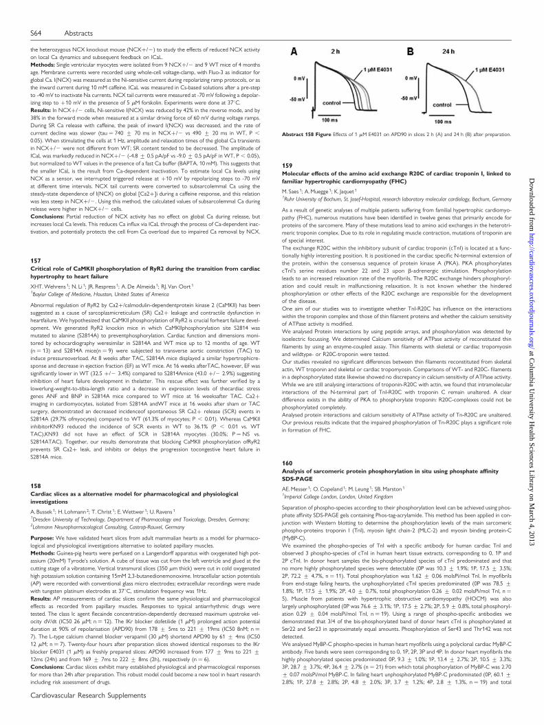
the heterozygous NCX knockout mouse (NCX+/2) to study the effects of reduced NCX activityon local Ca dynamics and subsequent feedback on ICaL.Methods: Single ventricular myocytes were isolated from 9 NCX+/2 and 9 WT mice of 4 monthsage. Membrane currents were recorded using whole-cell voltage-clamp, with Fluo-3 as indicator forglobal Ca. I(NCX) was measured as the Ni-sensitive current during repolarizing ramp protocols, or asthe inward current during 10 mM caffeine. ICaL was measured in Cs-based solutions after a pre-stepto -40 mV to inactivate Na currents. NCX tail currents were measured at -70 mV following a depolar-izing step to +10 mV in the presence of 5 mM forskolin. Experiments were done at 378C.Results: In NCX+/2 cells, Ni-sensitive I(NCX) was reduced by 42% in the reverse mode, and by38% in the forward mode when measured at a similar driving force of 60 mV during voltage ramps.During SR Ca release with caffeine, the peak of inward I(NCX) was decreased, and the rate ofcurrent decline was slower (tau ¼ 740 + 70 ms in NCX+/2 vs 490 + 20 ms in WT, P ,
0.05). When stimulating the cells at 1 Hz, amplitude and relaxation times of the global Ca transientsin NCX+/2 were not different from WT; SR content tended to be decreased. The amplitude ofICaL was markedly reduced in NCX+/2 (-4.8 + 0.5 pA/pF vs -9.0 + 0.5 pA/pF in WT, P , 0.05),but normalized to WT values in the presence of a fast Ca buffer (BAPTA, 10 mM). This suggests thatthe smaller ICaL is the result from Ca-dependent inactivation. To estimate local Ca levels usingNCX as a sensor, we interrupted triggered release at +10 mV by repolarizing steps to -70 mVat different time intervals. NCX tail currents were converted to subsarcolemmal Ca using thesteady-state dependence of I(NCX) on global [Ca2+]i during a caffeine response, and this relationwas less steep in NCX+/2. Using this method, the calculated values of subsarcolemmal Ca duringrelease were higher in NCX+/2 cells.Conclusions: Partial reduction of NCX activity has no effect on global Ca during release, butincreases local Ca levels. This reduces Ca influx via ICaL through the process of Ca-dependent inac-tivation, and potentially protects the cell from Ca overload due to impaired Ca removal by NCX.
157Critical role of CaMKII phosphorylation of RyR2 during the transition from cardiachypertrophy to heart failure
XHT. Wehrens 1; N. Li 1; JR. Respress 1; A. De Almeida 1; RJ. Van Oort 1
1Baylor College of Medicine, Houston, United States of America
Abnormal regulation of RyR2 by Ca2+/calmodulin-dependentprotein kinase 2 (CaMKII) has beensuggested as a cause of sarcoplasmicreticulum (SR) Ca2+ leakage and contractile dysfunction inheartfailure. We hypothesized that CaMKII phosphorylation of RyR2 is crucial forheart failure devel-opment. We generated RyR2 knockin mice in which CaMKIIphosphorylation site S2814 wasmutated to alanine (S2814A) to preventphosphorylation. Cardiac function and dimensions moni-tored by echocardiography weresimilar in S2814A and WT mice up to 12 months of age. WT(n ¼ 13) and S2814A mice(n ¼ 9) were subjected to transverse aortic constriction (TAC) toinduce pressureoverload. At 8 weeks after TAC, S2814A mice displayed a similar hypertrophicre-sponse and decrease in ejection fraction (EF) as WT mice. At 16 weeks afterTAC, however, EF wassignificantly lower in WT (32.5 +/2 3.4%) compared to S2814Amice (43.0 +/2 2.9%) suggestinginhibition of heart failure development in thelatter. This rescue effect was further verified by alowerlung-weight-to-tibia-length ratio and a decrease in expression levels of thecardiac stressgenes ANF and BNP in S2814A mice compared to WT mice at 16 weeksafter TAC. Ca2+imaging in cardiomyocytes, isolated from S2814A andWT mice at 16 weeks after sham or TACsurgery, demonstrated an decreased incidenceof spontaneous SR Ca2+ release (SCR) events inS2814A (29.7% ofmyocytes) compared to WT (61.3% of myocytes; P , 0.01). Whereas CaMKIIinhibitorKN93 reduced the incidence of SCR events in WT to 36.1% (P , 0.01 vs. WTTAC),KN93 did not have an effect of SCR in S2814A myocytes (30.0%; P ¼ NS vs.S2814ATAC). Together, our results demonstrate that blocking CaMKII phosphorylation ofRyR2prevents SR Ca2+ leak, and inhibits or delays the progression tocongestive heart failure inS2814A mice.
158Cardiac slices as a alternative model for pharmacological and physiologicalinvestigations
A. Bussek 1; H. Lohmann 2; T. Christ 1; E. Wettwer 1; U. Ravens 1
1Dresden University of Technology, Department of Pharmacology and Toxicology, Dresden, Germany;2Lohmann Neuropharmacological Consulting, Castrop-Rauxel, Germany
Purpose: We have validated heart slices from adult mammalian hearts as a model for pharmaco-logical and physiological investigations alternative to isolated papillary muscles.Methods: Guinea-pig hearts were perfused on a Langendorff apparatus with oxygenated high pot-assium (20mM) Tyrode’s solution. A cube of tissue was cut from the left ventricle and glued at thecutting stage of a vibratome. Vertical transmural slices (350 mm thick) were cut in cold oxygenatedhigh potassium solution containing 15mM 2,3-butanedionemonoxime. Intracellular action potentials(AP) were recorded with conventional glass micro electrodes; extracellular recordings were madewith tungsten platinum electrodes at 378C, stimulation frequency was 1Hz.Results: AP measurements of cardiac slices confirm the same physiological and pharmacologicaleffects as recorded from papillary muscles. Responses to typical antiarrhythmic drugs weretested. The class Ic agent flecainde concentration-dependently decreased maximum upstroke vel-ocity dV/dt (IC50 26 mM; n ¼ 12). The IKr blocker dofetilide (1 mM) prolonged action potentialduration at 90% of repolarisation (APD90) from 178 + 5ms to 221 + 19ms (IC50 8nM; n ¼7). The L-type calcium channel blocker verapamil (30 mM) shortend APD90 by 61 + 4ms (IC5012 mM; n ¼ 7). Twenty-four hours after preparation slices showed identical responses to the IKrblocker E4031 (1 mM) as freshly prepared slices: APD90 increased from 177 + 9ms to 221 +12ms (24h) and from 169 + 7ms to 222 + 8ms (2h), respectively (n ¼ 6).Conclusions: Cardiac slices exhibit many established physiological and pharmacological responsesfor more than 24h after preparation. This robust model could become a new tool in heart researchincluding risk assessment of drugs.
159Molecular effects of the amino acid exchange R20C of cardiac troponin I, linked tofamiliar hypertrophic cardiomyopathy (FHC)
M. Saes 1; A. Muegge 1; K. Jaquet 1
1Ruhr University of Bochum, St. Josef-Hospital, research laboratory molecular cardiology, Bochum, Germany
As a result of genetic analyses of multiple patients suffering from familial hypertrophic cardiomyo-pathy (FHC), numerous mutations have been identified in twelve genes that primarily encode forproteins of the sarcomere. Many of these mutations lead to amino acid exchanges in the heterotri-meric troponin complex. Due to its role in regulating muscle contraction, mutations of troponin areof special interest.The exchange R20C within the inhibitory subunit of cardiac troponin (cTnI) is located at a func-tionally highly interesting position. It is positioned in the cardiac specific N-terminal extension ofthe protein, within the consensus sequence of protein kinase A (PKA). PKA phosphorylatescTnI’s serine residues number 22 and 23 upon b-adrenergic stimulation. Phosphorylationleads to an increased relaxation rate of the myofibrils. The R20C exchange hinders phosphoryl-ation and could result in malfunctioning relaxation. It is not known whether the hinderedphosphorylation or other effects of the R20C exchange are responsible for the developmentof the disease.One aim of our studies was to investigate whether TnI-R20C has influence on the interactionswithin the troponin complex and those of thin filament proteins and whether the calcium sensitivityof ATPase activity is modified.We analysed Protein interactions by using peptide arrays, and phosphorylation was detected byisoelectric focussing. We determined Calcium sensitivity of ATPase activity of reconstituted thinfilaments by using an enzyme-coupled assay. Thin filaments with skeletal or cardiac tropomyosinand wildtype- or R20C-troponin were tested.Our studies revealed no significant differences between thin filaments reconstituted from skeletalactin, WT troponin and skeletal or cardiac tropomyosin. Comparisons of WT- and R20C- filamentsin a dephosphorylated state likewise showed no discrepancy in calcium sensitivity of ATPase activity.While we are still analysing interactions of troponin-R20C with actin, we found that intramolecularinteractions of the N-terminal part of TnI-R20C with troponin C remain unaltered. A cleardifference exists in the ability of PKA to phosphorylate troponin: R20C-complexes could not bephosphorylated completely.Analysed protein interactions and calcium sensitivity of ATPase activity of Tn-R20C are unaltered.Our previous results indicate that the impaired phosphorylation of Tn-R20C plays a significant rolein formation of FHC.
160Analysis of sarcomeric protein phosphorylation in situ using phosphate affinitySDS-PAGE
AE. Messer 1; O. Copeland 1; M. Leung 1; SB. Marston 1
1Imperial College London, London, United Kingdom
Separation of phospho-species according to their phosphorylation level can be achieved using phos-phate affinity SDS-PAGE gels containing Phos-tag-acrylamide. This method has been applied in con-junction with Western blotting to determine the phosphorylation levels of the main sarcomericphospho-proteins troponin I (TnI), myosin light chain-2 (MLC-2) and myosin binding protein-C(MyBP-C).We examined the phospho-species of TnI with a specific antibody for human cardiac TnI andobserved 3 phospho-species of cTnI in human heart tissue extracts, corresponding to 0, 1P and2P cTnI. In donor heart samples the bis-phosphorylated species of cTnI predominated and thatno more highly phosphorylated species were detectable (0P was 10.3 + 1.9%; 1P, 17.5 + 3.5%;2P, 72.2 + 4.7%, n ¼ 11). Total phosphorylation was 1.62 + 0.06 molsPi/mol TnI. In myofibrilsfrom end-stage failing hearts, the unphosphorylated cTnI species predominated (0P was 78.5 +1.8%; 1P, 17.5 + 1.9%; 2P, 4.0 + 0.7%, total phosphorylation 0.26 + 0.02 molsPi/mol TnI, n ¼5). Muscle from patients with hypertrophic obstructive cardiomyopathy (HOCM) was alsolargely unphosphorylated (0P was 76.6 + 3.1%; 1P, 17.5 + 2.7%; 2P, 5.9 + 0.8%, total phosphoryl-ation 0.29 + 0.04 molsPi/mol TnI, n ¼ 19). Using a range of phospho-specific antibodies wedemonstrated that 3/4 of the bis-phosphorylated band of donor heart cTnI is phosphorylated atSer22 and Ser23 in approximately equal amounts. Phosphorylation of Ser43 and Thr142 was notdetected.We analysed MyBP-C phospho-species in human heart myofibrils using a polyclonal cardiac MyBP-Cantibody. Five bands were seen corresponding to 0, 1P, 2P, 3P and 4P. In donor heart myofibrils thehighly phosphorylated species predominated: 0P, 9.3 + 1.0%; 1P, 13.4 + 2.7%; 2P, 10.5 + 3.3%;3P, 28.7 + 3.7%; 4P, 36.4 + 2.7% (n ¼ 21) from which total phosphorylation of MyBP-C was 2.70+ 0.07 molsPi/mol MyBP-C. In failing heart unphosphorylated MyBP-C predominated (0P, 60.1 +2.8%; 1P, 27.8 + 2.8%; 2P, 4.8 + 2.0%; 3P, 3.7 + 1.2%; 4P, 2.8 + 1.3%, n ¼ 19) and total
Abstract 158 Figure Effects of 1 mM E4031 on APD90 in slices 2 h (A) and 24 h (B) after preparation.
S64 Abstracts
Cardiovascular Research Supplements
at Colum
bia University H
ealth Sciences Library on M
arch 4, 2013http://cardiovascres.oxfordjournals.org/
Dow
nloaded from
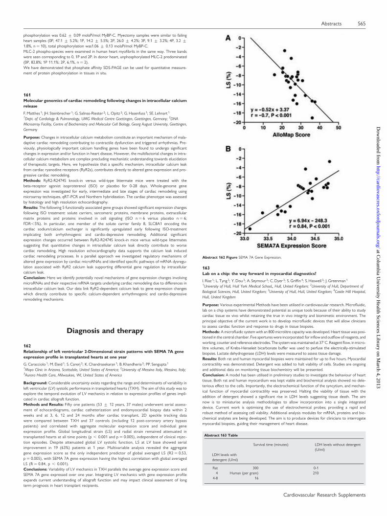
phosphorylation was 0.62 + 0.09 molsPi/mol MyBP-C. Myectomy samples were similar to failingheart samples (0P, 47.1 + 5.2%; 1P, 14.2 + 5.5%; 2P, 26.0 + 4.2%; 3P, 9.1 + 3.2%; 4P, 3.2 +1.8%, n ¼ 10), total phosphorylation was1.06 + 0.13 molsPi/mol MyBP-C.MLC-2 phospho-species were examined in human heart myofibrils in the same way. Three bandswere seen corresponding to 0, 1P and 2P. In donor heart, unphosphorylated MLC-2 predominated(0P, 82.8%; 1P 11.1%; 2P, 6.1%, n ¼ 2).We have demonstrated that phosphate affinity SDS-PAGE can be used for quantitative measure-ment of protein phosphorylation in tissues in situ.
161Molecular genomics of cardiac remodeling following changes in intracellular calciumrelease
F. Matthes 1; JH. Steinbrecher 1; G. Salinas-Riester 2; L. Opitz 2; G. Hasenfuss 1; SE. Lehnart 1
1Dept. of Cardiology & Pulmonology, UMG Medical Centre Goettingen, Goettingen, Germany; 2DNAMicroarray Facility, Centre of Biochemistry and Molecular Cell Biology, Georg August University, Goettingen,Germany
Purpose: Changes in intracellular calcium metabolism constitute an important mechanism of mala-daptive cardiac remodeling contributing to contractile dysfunction and triggered arrhythmias. Pre-viously, physiologically important calcium handling genes have been found to undergo significantchanges in expression and/or function in heart disease. However, the multifactorial changes in intra-cellular calcium metabolism are complex precluding mechanistic understanding towards elucidationof therapeutic targets. Here, we hypothesize that a specific mechanism, intracellular calcium leakfrom cardiac ryanodine receptors (RyR2s), contributes directly to altered gene expression and pro-gressive cardiac remodeling.Methods: RyR2-R2474S knock-in versus wild-type littermate mice were treated with thebeta-receptor agonist isoproterenol (ISO) or placebo for 0-28 days. Whole-genome geneexpression was investigated for early, intermediate and late stages of cardiac remodeling usingmicroarray techniques, qRT-PCR and Northern hybridization. The cardiac phenotype was assessedby histology and high resolution echocardiography.Results: The following 5 functionally associated gene groups showed significant expression changesfollowing ISO treatment: solute carriers, sarcomeric proteins, membrane proteins, extracellularmatrix proteins and proteins involved in cell signaling (ISO n ¼ 6 versus placebo n ¼ 6;FDR,5%). In particular, one member of the solute carrier family 8, SLC8A1 encoding thecardiac sodium/calcium exchanger is significantly upregulated early following ISO-treatmentimplicating both arrhythmogenic and cardio-depressive remodeling. Additional significantexpression changes occurred between RyR2-R2474S knock-in mice versus wild-type littermatessuggesting that quantitative changes in intracellular calcium leak directly contribute to worsecardiac remodeling. High resolution echocardiography data supports the calcium leak inducedcardiac remodeling processes. In a parallel approach we investigated regulatory mechanisms ofaltered gene expression by cardiac microRNAs and identified specific pathways of mRNA dysregu-lation associated with RyR2 calcium leak supporting differential gene regulation by intracellularcalcium leak.Conclusion: Here we identify potentially novel mechanisms of gene expression changes involvingmicroRNAs and their respective mRNA targets underlying cardiac remodeling due to differences inintracellular calcium leak. Our data link RyR2-dependent calcium leak to gene expression changeswhich directly contribute to specific calcium-dependent arrhythmogenic and cardio-depressiveremodeling mechanisms.
Diagnosis and therapy
162Relationship of left ventricular 2-Dimensional strain patterns with SEMA 7A geneexpression profile in transplanted hearts at one year
G. Caracciolo 1; M. Eleid 1; S. Carerj 2; K. Chandrasekaran 1; B. Khandheria 3; PP. Sengupta 1
1Mayo Clinic in Arizona, Scottsdale, United States of America; 2University of Messina Italy, Messina, Italy;3Aurora Health Care, Milwaukee, WI, United States of America
Background: Considerable uncertainty exists regarding the range and determinants of variability inleft ventricular (LV) systolic performance in transplanted hearts (TXH). The aim of this study was toexplore the temporal evolution of LV mechanics in relation to expression profiles of genes impli-cated in cardiac allograft function.Methods and Results: Fifty one patients (53 + 12 years, 37 males) underwent serial assess-ment of echocardiograms, cardiac catheterization and endomyocardial biopsy data within 2weeks and at 3, 6, 12 and 24 months after cardiac transplant. 2D speckle tracking datawere compared between TXH and 37 controls (including 12 post-coronary artery bypasspatients) and correlated with aggregate molecular expression score and individual geneexpression profile. Global longitudinal strain (LS) and radial strain remained attenuated intransplanted hearts at all time points (p , 0.001 and p ¼ 0.005), independent of clinical rejec-tion episodes. Despite attenuated global LV systolic function, LS at LV base showed serialimprovement in 19 (63%) patients at 1 year. Multivariable analysis revealed the aggregategene expression score as the only independent predictor of global averaged LS (R2 ¼ 0.53,p ¼ 0.005), with SEMA 7A gene expression having the highest correlation with global averagedLS (R ¼ 0.84, p , 0.001).Conclusions: Variability of LV mechanics in TXH parallels the average gene expression score andSEMA 7A gene expressed over one year. Integrating LV mechanics with gene expression profileexpands current understanding of allograft function and may impact clinical assessment of longterm prognosis in heart transplant recipients.
163Lab on a chip: the way forward in myocardial diagnostics?
I. Riaz 1; L. Tyng 2; Y. Dou 3; A. Seymour 2; C. Dyer 3; S. Griffin 4; S. Haswell 3; J. Greenman 1
1University of Hull, Hull York Medical School,, Hull, United Kingdom; 2University of Hull, Department ofBiological Sciences, Hull, United Kingdom; 3University of Hull, Hull, United Kingdom; 4Castle Hill Hospital,Hull, United Kingdom
Purpose: Various experimental Methods have been utilised in cardiovascular research. Microfluidic,lab on a chip systems have demonstrated potential as unique tools because of their ability to studycardiac tissue ex vivo whilst retaining the true in vivo integrity and biomimetic environment. Theprincipal objective of the current work is to develop microfluidic devices that will allow cliniciansto assess cardiac function and response to drugs in tissue biopsies.Methods: A microfluidic system with an 800 microlitre capacity was developed. Heart tissue was posi-tioned in the central chamber. Five apertures were incorporated: for inflow and outflow of reagents, andworking, counter and reference electrodes. The system was maintained at 378C. Reagent flow, in micro-litre volumes, of Krebs-Henseleit bicarbonate buffer was used to perfuse the electrically-stimulatedbiopsies. Lactate dehydrogenase (LDH) levels were measured to assess tissue damage.Results: Both rat and human myocardial biopsies were maintained for up to five hours. Myocardialcontractility was demonstrated. Detergent was added to halt viability of cells. Studies are ongoingand additional data on monitoring tissue biochemistry will be presented.Conclusion: A model has been utilised in preliminary studies to investigate the behaviour of hearttissue. Both rat and human myocardium was kept viable and biochemical analysis showed no dele-terious effect to the cells. Importantly, the electrochemical function of the syncytium, and mechan-ical function of myocardial contractility was preserved. Halting the viability of tissue with theaddition of detergent showed a significant rise in LDH levels suggesting tissue death. The aimnow is to miniaturise analysis methodologies to allow incorporation into a single integrateddevice. Current work is optimising the use of electrochemical probes; providing a rapid androbust method of assessing cell viability. Additional analysis modules for mRNA, proteins and bio-chemical analytes are being developed. The aim is to produce devices for clinicians to interrogatemyocardial biopsies, guiding their management of heart disease.
Abstract 163 Table
Survival time (minutes) LDH levels without detergent(U/ml)
LDH levels withdetergent (U/ml)
Rat 300 0-14 Human (per gram) 210
4-8 16
Abstract 162 Figure SEMA 7A Gene Expression.
Abstracts S65
Cardiovascular Research Supplements
at Colum
bia University H
ealth Sciences Library on M
arch 4, 2013http://cardiovascres.oxfordjournals.org/
Dow
nloaded from
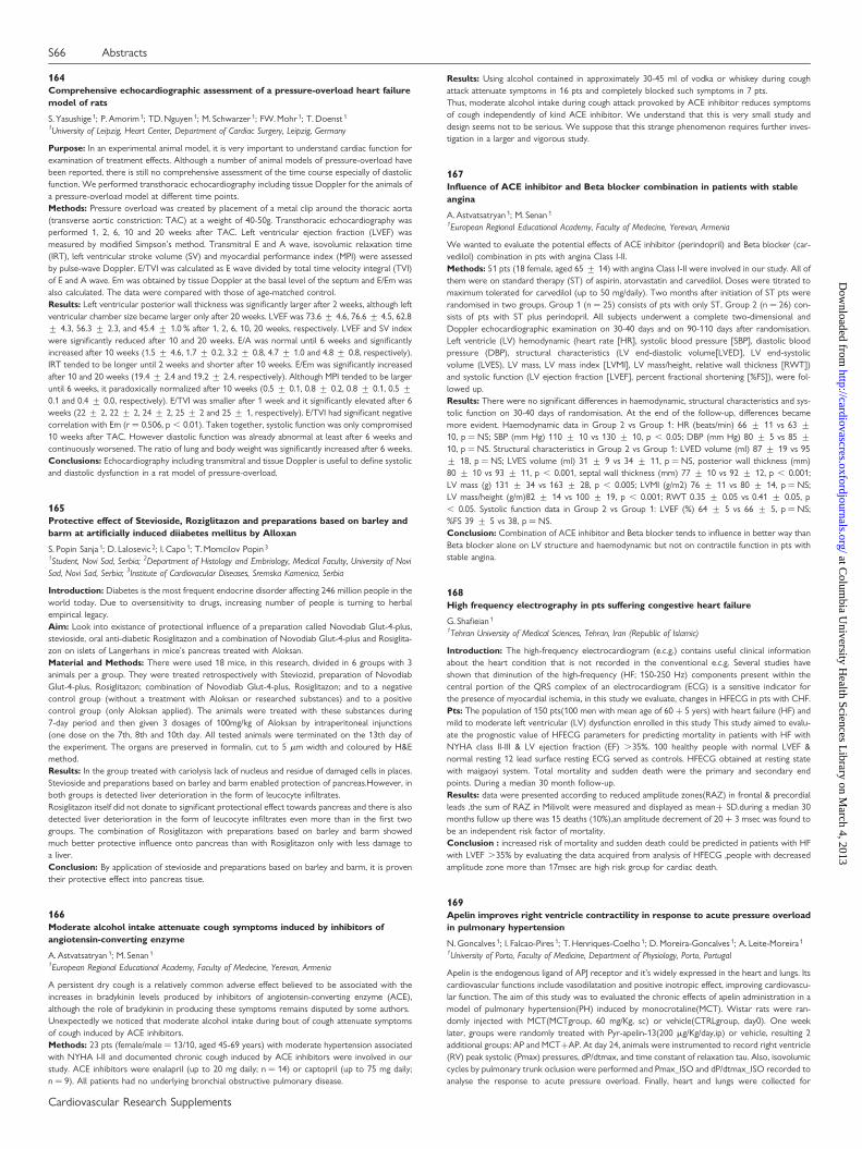
164Comprehensive echocardiographic assessment of a pressure-overload heart failuremodel of rats
S. Yasushige 1; P. Amorim 1; TD. Nguyen 1; M. Schwarzer 1; FW. Mohr 1; T. Doenst 1
1University of Leipzig, Heart Center, Department of Cardiac Surgery, Leipzig, Germany
Purpose: In an experimental animal model, it is very important to understand cardiac function forexamination of treatment effects. Although a number of animal models of pressure-overload havebeen reported, there is still no comprehensive assessment of the time course especially of diastolicfunction. We performed transthoracic echocardiography including tissue Doppler for the animals ofa pressure-overload model at different time points.Methods: Pressure overload was created by placement of a metal clip around the thoracic aorta(transverse aortic constriction: TAC) at a weight of 40-50g. Transthoracic echocardiography wasperformed 1, 2, 6, 10 and 20 weeks after TAC. Left ventricular ejection fraction (LVEF) wasmeasured by modified Simpson’s method. Transmitral E and A wave, isovolumic relaxation time(IRT), left ventricular stroke volume (SV) and myocardial performance index (MPI) were assessedby pulse-wave Doppler. E/TVI was calculated as E wave divided by total time velocity integral (TVI)of E and A wave. Em was obtained by tissue Doppler at the basal level of the septum and E/Em wasalso calculated. The data were compared with those of age-matched control.Results: Left ventricular posterior wall thickness was significantly larger after 2 weeks, although leftventricular chamber size became larger only after 20 weeks. LVEF was 73.6 + 4.6, 76.6 + 4.5, 62.8+ 4.3, 56.3 + 2.3, and 45.4 + 1.0 % after 1, 2, 6, 10, 20 weeks, respectively. LVEF and SV indexwere significantly reduced after 10 and 20 weeks. E/A was normal until 6 weeks and significantlyincreased after 10 weeks (1.5 + 4.6, 1.7 + 0.2, 3.2 + 0.8, 4.7 + 1.0 and 4.8 + 0.8, respectively).IRT tended to be longer until 2 weeks and shorter after 10 weeks. E/Em was significantly increasedafter 10 and 20 weeks (19.4 + 2.4 and 19.2 + 2.4, respectively). Although MPI tended to be largeruntil 6 weeks, it paradoxically normalized after 10 weeks (0.5 + 0.1, 0.8 + 0.2, 0.8 + 0.1, 0.5 +0.1 and 0.4 + 0.0, respectively). E/TVI was smaller after 1 week and it significantly elevated after 6weeks (22 + 2, 22 + 2, 24 + 2, 25 + 2 and 25 + 1, respectively). E/TVI had significant negativecorrelation with Em (r ¼ 0.506, p , 0.01). Taken together, systolic function was only compromised10 weeks after TAC. However diastolic function was already abnormal at least after 6 weeks andcontinuously worsened. The ratio of lung and body weight was significantly increased after 6 weeks.Conclusions: Echocardiography including transmitral and tissue Doppler is useful to define systolicand diastolic dysfunction in a rat model of pressure-overload.
165Protective effect of Stevioside, Roziglitazon and preparations based on barley andbarm at artificially induced diiabetes mellitus by Alloxan
S. Popin Sanja 1; D. Lalosevic 2; I. Capo 1; T. Momcilov Popin 3
1Student, Novi Sad, Serbia; 2Department of Histology and Embriology, Medical Faculty, University of NoviSad, Novi Sad, Serbia; 3Institute of Cardiovacular Diseases, Sremska Kamenica, Serbia
Introduction: Diabetes is the most frequent endocrine disorder affecting 246 million people in theworld today. Due to oversensitivity to drugs, increasing number of people is turning to herbalempirical legacy.Aim: Look into existance of protectional influence of a preparation called Novodiab Glut-4-plus,stevioside, oral anti-diabetic Rosiglitazon and a combination of Novodiab Glut-4-plus and Rosiglita-zon on islets of Langerhans in mice’s pancreas treated with Aloksan.Material and Methods: There were used 18 mice, in this research, divided in 6 groups with 3animals per a group. They were treated retrospectively with Steviozid, preparation of NovodiabGlut-4-plus, Rosiglitazon; combination of Novodiab Glut-4-plus, Rosiglitazon; and to a negativecontrol group (without a treatment with Aloksan or researched substances) and to a positivecontrol group (only Aloksan applied). The animals were treated with these substances during7-day period and then given 3 dosages of 100mg/kg of Aloksan by intraperitoneal injunctions(one dose on the 7th, 8th and 10th day. All tested animals were terminated on the 13th day ofthe experiment. The organs are preserved in formalin, cut to 5 mm width and coloured by H&Emethod.Results: In the group treated with cariolysis lack of nucleus and residue of damaged cells in places.Stevioside and preparations based on barley and barm enabled protection of pancreas.However, inboth groups is detected liver deterioration in the form of leucocyte infiltrates.Rosiglitazon itself did not donate to significant protectional effect towards pancreas and there is alsodetected liver deterioration in the form of leucocyte infiltrates even more than in the first twogroups. The combination of Rosiglitazon with preparations based on barley and barm showedmuch better protective influence onto pancreas than with Rosiglitazon only with less damage toa liver.Conclusion: By application of stevioside and preparations based on barley and barm, it is proventheir protective effect into pancreas tisue.
166Moderate alcohol intake attenuate cough symptoms induced by inhibitors ofangiotensin-converting enzyme
A. Astvatsatryan 1; M. Senan 1
1European Regional Educational Academy, Faculty of Medecine, Yerevan, Armenia
A persistent dry cough is a relatively common adverse effect believed to be associated with theincreases in bradykinin levels produced by inhibitors of angiotensin-converting enzyme (ACE),although the role of bradykinin in producing these symptoms remains disputed by some authors.Unexpectedly we noticed that moderate alcohol intake during bout of cough attenuate symptomsof cough induced by ACE inhibitors.Methods: 23 pts (female/male ¼ 13/10, aged 45-69 years) with moderate hypertension associatedwith NYHA I-II and documented chronic cough induced by ACE inhibitors were involved in ourstudy. ACE inhibitors were enalapril (up to 20 mg daily; n ¼ 14) or captopril (up to 75 mg daily;n ¼ 9). All patients had no underlying bronchial obstructive pulmonary disease.
Results: Using alcohol contained in approximately 30-45 ml of vodka or whiskey during coughattack attenuate symptoms in 16 pts and completely blocked such symptoms in 7 pts.Thus, moderate alcohol intake during cough attack provoked by ACE inhibitor reduces symptomsof cough independently of kind ACE inhibitor. We understand that this is very small study anddesign seems not to be serious. We suppose that this strange phenomenon requires further inves-tigation in a larger and vigorous study.
167Influence of ACE inhibitor and Beta blocker combination in patients with stableangina
A. Astvatsatryan 1; M. Senan 1
1European Regional Educational Academy, Faculty of Medecine, Yerevan, Armenia
We wanted to evaluate the potential effects of ACE inhibitor (perindopril) and Beta blocker (car-vedilol) combination in pts with angina Class I-II.Methods: 51 pts (18 female, aged 65 + 14) with angina Class I-II were involved in our study. All ofthem were on standard therapy (ST) of aspirin, atorvastatin and carvedilol. Doses were titrated tomaximum tolerated for carvedilol (up to 50 mg/daily). Two months after initiation of ST pts wererandomised in two groups. Group 1 (n ¼ 25) consists of pts with only ST, Group 2 (n ¼ 26) con-sists of pts with ST plus perindopril. All subjects underwent a complete two-dimensional andDoppler echocardiographic examination on 30-40 days and on 90-110 days after randomisation.Left ventricle (LV) hemodynamic (heart rate [HR], systolic blood pressure [SBP], diastolic bloodpressure (DBP), structural characteristics (LV end-diastolic volume[LVED], LV end-systolicvolume (LVES), LV mass, LV mass index [LVMI], LV mass/height, relative wall thickness [RWT])and systolic function (LV ejection fraction [LVEF], percent fractional shortening [%FS]), were fol-lowed up.Results: There were no significant differences in haemodynamic, structural characteristics and sys-tolic function on 30-40 days of randomisation. At the end of the follow-up, differences becamemore evident. Haemodynamic data in Group 2 vs Group 1: HR (beats/min) 66 + 11 vs 63 +10, p ¼ NS; SBP (mm Hg) 110 + 10 vs 130 + 10, p , 0.05; DBP (mm Hg) 80 + 5 vs 85 +10, p ¼ NS. Structural characteristics in Group 2 vs Group 1: LVED volume (ml) 87 + 19 vs 95+ 18, p ¼ NS; LVES volume (ml) 31 + 9 vs 34 + 11, p ¼ NS, posterior wall thickness (mm)80 + 10 vs 93 + 11, p , 0.001, septal wall thickness (mm) 77 + 10 vs 92 + 12, p , 0.001;LV mass (g) 131 + 34 vs 163 + 28, p , 0.005; LVMI (g/m2) 76 + 11 vs 80 + 14, p ¼ NS;LV mass/height (g/m)82 + 14 vs 100 + 19, p , 0.001; RWT 0.35 + 0.05 vs 0.41 + 0.05, p, 0.05. Systolic function data in Group 2 vs Group 1: LVEF (%) 64 + 5 vs 66 + 5, p ¼ NS;%FS 39 + 5 vs 38, p ¼ NS.Conclusion: Combination of ACE inhibitor and Beta blocker tends to influence in better way thanBeta blocker alone on LV structure and haemodynamic but not on contractile function in pts withstable angina.
168High frequency electrography in pts suffering congestive heart failure
G. Shafieian 1
1Tehran University of Medical Sciences, Tehran, Iran (Republic of Islamic)
Introduction: The high-frequency electrocardiogram (e.c.g.) contains useful clinical informationabout the heart condition that is not recorded in the conventional e.c.g. Several studies haveshown that diminution of the high-frequency (HF; 150-250 Hz) components present within thecentral portion of the QRS complex of an electrocardiogram (ECG) is a sensitive indicator forthe presence of myocardial ischemia, in this study we evaluate, changes in HFECG in pts with CHF.Pts: The population of 150 pts(100 men with mean age of 60 + 5 yers) with heart failure (HF) andmild to moderate left ventricular (LV) dysfunction enrolled in this study This study aimed to evalu-ate the prognostic value of HFECG parameters for predicting mortality in patients with HF withNYHA class II-III & LV ejection fraction (EF) .35%. 100 healthy people with normal LVEF &normal resting 12 lead surface resting ECG served as controls. HFECG obtained at resting statewith maigaoyi system. Total mortality and sudden death were the primary and secondary endpoints. During a median 30 month follow-up.Results: data were presented according to reduced amplitude zones(RAZ) in frontal & precordialleads ,the sum of RAZ in Milivolt were measured and displayed as mean+ SD.during a median 30months fullow up there was 15 deaths (10%),an amplitude decrement of 20 + 3 msec was found tobe an independent risk factor of mortality.Conclusion : increased risk of mortality and sudden death could be predicted in patients with HFwith LVEF .35% by evaluating the data acquired from analysis of HFECG ,people with decreasedamplitude zone more than 17msec are high risk group for cardiac death.
169Apelin improves right ventricle contractility in response to acute pressure overloadin pulmonary hypertension
N. Goncalves 1; I. Falcao-Pires 1; T. Henriques-Coelho 1; D. Moreira-Goncalves 1; A. Leite-Moreira 1
1University of Porto, Faculty of Medicine, Department of Physiology, Porto, Portugal
Apelin is the endogenous ligand of APJ receptor and it’s widely expressed in the heart and lungs. Itscardiovascular functions include vasodilatation and positive inotropic effect, improving cardiovascu-lar function. The aim of this study was to evaluated the chronic effects of apelin administration in amodel of pulmonary hypertension(PH) induced by monocrotaline(MCT). Wistar rats were ran-domly injected with MCT(MCTgroup, 60 mg/Kg, sc) or vehicle(CTRLgroup, day0). One weeklater, groups were randomly treated with Pyr-apelin-13(200 mg/Kg/day,ip) or vehicle, resulting 2additional groups: AP and MCT+AP. At day 24, animals were instrumented to record right ventricle(RV) peak systolic (Pmax) pressures, dP/dtmax, and time constant of relaxation tau. Also, isovolumiccycles by pulmonary trunk oclusion were performed and Pmax_ISO and dP/dtmax_ISO recorded toanalyse the response to acute pressure overload. Finally, heart and lungs were collected for
S66 Abstracts
Cardiovascular Research Supplements
at Colum
bia University H
ealth Sciences Library on M
arch 4, 2013http://cardiovascres.oxfordjournals.org/
Dow
nloaded from
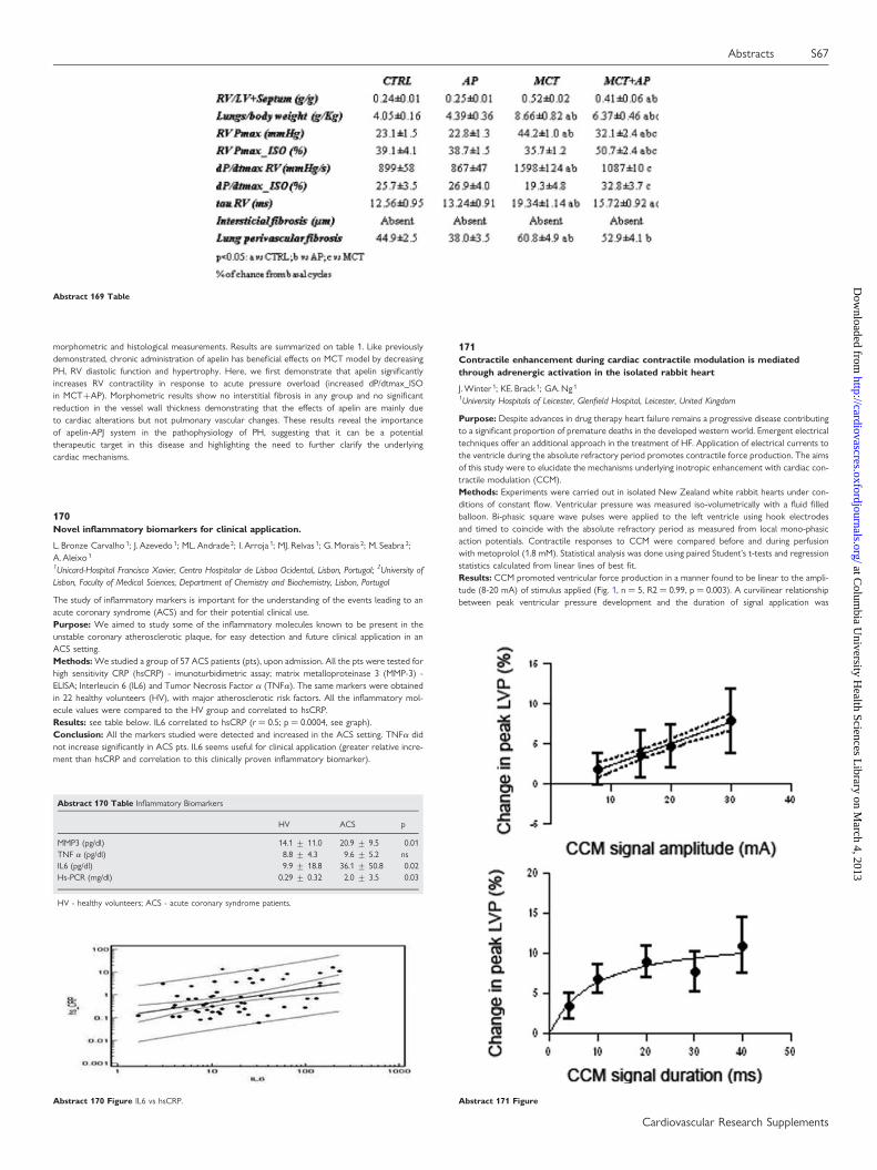
morphometric and histological measurements. Results are summarized on table 1. Like previouslydemonstrated, chronic administration of apelin has beneficial effects on MCT model by decreasingPH, RV diastolic function and hypertrophy. Here, we first demonstrate that apelin significantlyincreases RV contractility in response to acute pressure overload (increased dP/dtmax_ISOin MCT+AP). Morphometric results show no interstitial fibrosis in any group and no significantreduction in the vessel wall thickness demonstrating that the effects of apelin are mainly dueto cardiac alterations but not pulmonary vascular changes. These results reveal the importanceof apelin-APJ system in the pathophysiology of PH, suggesting that it can be a potentialtherapeutic target in this disease and highlighting the need to further clarify the underlyingcardiac mechanisms.
170Novel inflammatory biomarkers for clinical application.
L. Bronze Carvalho 1; J. Azevedo 1; ML. Andrade 2; I. Arroja 1; MJ. Relvas 1; G. Morais 2; M. Seabra 2;A. Aleixo 1
1Unicard-Hospital Francisco Xavier, Centro Hospitalar de Lisboa Ocidental, Lisbon, Portugal; 2University ofLisbon, Faculty of Medical Sciences, Department of Chemistry and Biochemistry, Lisbon, Portugal
The study of inflammatory markers is important for the understanding of the events leading to anacute coronary syndrome (ACS) and for their potential clinical use.Purpose: We aimed to study some of the inflammatory molecules known to be present in theunstable coronary atherosclerotic plaque, for easy detection and future clinical application in anACS setting.Methods: We studied a group of 57 ACS patients (pts), upon admission. All the pts were tested forhigh sensitivity CRP (hsCRP) - imunoturbidimetric assay; matrix metalloproteinase 3 (MMP-3) -ELISA; Interleucin 6 (IL6) and Tumor Necrosis Factor a (TNFa). The same markers were obtainedin 22 healthy volunteers (HV), with major atherosclerotic risk factors. All the inflammatory mol-ecule values were compared to the HV group and correlated to hsCRP.Results: see table below. IL6 correlated to hsCRP (r ¼ 0.5; p ¼ 0.0004, see graph).Conclusion: All the markers studied were detected and increased in the ACS setting. TNFa didnot increase significantly in ACS pts. IL6 seems useful for clinical application (greater relative incre-ment than hsCRP and correlation to this clinically proven inflammatory biomarker).
Abstract 170 Table Inflammatory Biomarkers
HV ACS p
MMP3 (pg/dl) 14.1 + 11.0 20.9 + 9.5 0.01TNF a (pg/dl) 8.8 + 4.3 9.6 + 5.2 nsIL6 (pg/dl) 9.9 + 18.8 36.1 + 50.8 0.02Hs-PCR (mg/dl) 0.29 + 0.32 2.0 + 3.5 0.03
HV - healthy volunteers; ACS - acute coronary syndrome patients.
171Contractile enhancement during cardiac contractile modulation is mediatedthrough adrenergic activation in the isolated rabbit heart
J. Winter 1; KE. Brack 1; GA. Ng 1
1University Hospitals of Leicester, Glenfield Hospital, Leicester, United Kingdom
Purpose: Despite advances in drug therapy heart failure remains a progressive disease contributingto a significant proportion of premature deaths in the developed western world. Emergent electricaltechniques offer an additional approach in the treatment of HF. Application of electrical currents tothe ventricle during the absolute refractory period promotes contractile force production. The aimsof this study were to elucidate the mechanisms underlying inotropic enhancement with cardiac con-tractile modulation (CCM).Methods: Experiments were carried out in isolated New Zealand white rabbit hearts under con-ditions of constant flow. Ventricular pressure was measured iso-volumetrically with a fluid filledballoon. Bi-phasic square wave pulses were applied to the left ventricle using hook electrodesand timed to coincide with the absolute refractory period as measured from local mono-phasicaction potentials. Contractile responses to CCM were compared before and during perfusionwith metoprolol (1.8 mM). Statistical analysis was done using paired Student’s t-tests and regressionstatistics calculated from linear lines of best fit.Results: CCM promoted ventricular force production in a manner found to be linear to the ampli-tude (8-20 mA) of stimulus applied (Fig. 1, n ¼ 5, R2 ¼ 0.99, p ¼ 0.003). A curvilinear relationshipbetween peak ventricular pressure development and the duration of signal application was
Abstract 169 Table
Abstract 170 Figure IL6 vs hsCRP. Abstract 171 Figure
Abstracts S67
Cardiovascular Research Supplements
at Colum
bia University H
ealth Sciences Library on M
arch 4, 2013http://cardiovascres.oxfordjournals.org/
Dow
nloaded from
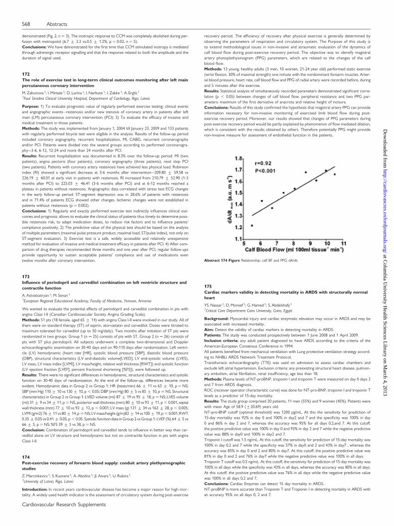
demonstrated (Fig. 2, n ¼ 3). The inotropic response to CCM was completely abolished during per-fusion with metropolol (6.7 + 2.2 vs.0.5 + 1.2%, p ¼ 0.02, n ¼ 5).Conclusions: We have demonstrated for the first time that CCM stimulated inotropy is mediatedthrough adrenergic receptor signalling and that the response related to both the amplitude and theduration of signal used.
172The role of exercise test in long-term clinical outcomes monitoring after left mainpercutaneous coronary intervention
M. Zabunova 1; I. Mintale 1; D. Lurina 1; I. Narbute 1; I. Zakke 1; A. Erglis 1
1Paul Stradins Clinical University Hospital, Department of Cardiology, Riga, Latvia
Purpose: 1) To evaluate prognostic value of regularly performed exercise testing: clinical eventsand angiographic events–restenosis and/or new stenosis of coronary artery in patients after leftmain (LM) percutaneous coronary intervention (PCI); 2) To evaluate the efficacy of invasive andmedical treatment in those patients.Methods: The study was implemented from January 1, 2004 till January 23, 2009 and 133 patientswith regularly performed bicycle test were eligible in the analysis. Results of the follow-up periodincluded coronary angiography, recurrent hospitalization, MI, CABG, recurrent coronarographyand/or PCI. Patients were divided into the several groups according to performed coronarogra-phy–3-6, 6-12, 12-24 and more than 24 months after PCI.Results: Recurrent hospitalization was documented in 8.3% over the follow-up period: MI (twopatients), angina pectoris (four patients), coronary angiography (three patients), next step PCI(two patients). Patients with coronary artery restenosis have achieved less physical load. Robinsonindex (RI) showed a significant decrease at 3-6 months after intervention–209.80 + 59.58 vs226.19 + 60.01 at early visit in patients with restenosis. RI increased from 210.79 + 52.90 (1-3months after PCI) to 225.03 + 46.41 (3-6 months after PCI) and at 6-12 months reached aplateau in patients without restenosis. Angiographic data correlated with stress test ECG changesin the early follow-up period: ST-segment depression was in 28.6% of patients with restenosisand in 71.4% of patients ECG showed other changes. Ischemic changes were not established inpatients without restenosis (p ¼ 0.002).Conclusions: 1) Regularly and exactly performed exercise test indirectly influences clinical out-comes and prognosis: allows to evaluate the clinical status of patients thus timely to determine poss-ible restenosis risk, to adapt medication doses, to reduce risk factors and to influence patients’compliance positively. 2) The predictive value of the physical test should be based on the analysisof multiple parameters (maximal pulse pressure product, maximal load, ST/pulse index), not only onST-segment evaluation. 3) Exercise test is a safe, widely accessible and relatively unexpensivemethod for evaluation of invasive and medical treatment efficacy in patients after PCI. 4) After com-parison of drug therapies recommended three months and one year after PCI, regular follow-upsprovide opportunity to sustain acceptable patients’ compliance and use of medications eventwelve months after coronary intervention.
173Influence of perindopril and carvedilol combination on left ventricle structure andcontractile function
A. Astvatsatryan 1; M. Senan 1
1European Regional Educational Academy, Faculty of Medecine, Yerevan, Armenia
We wanted to evaluate the potential effects of perindopril and carvedilol combination in pts withangina Class I-II (Canadian Cardiovascular Society Angina Grading Scale).Methods: 51 pts (18 female, aged 65 + 14) with angina Class I-II were involved in our study. All ofthem were on standard therapy (ST) of aspirin, atorvastatin and carvedilol. Doses were titrated tomaximum tolerated for carvedilol (up to 50 mg/daily). Two months after initiation of ST pts wererandomized in two groups. Group 1 (n ¼ 25) consists of pts with ST, Group 2 (n ¼ 26) consists ofpts with ST plus perindopril. All subjects underwent a complete two-dimensional and Dopplerechocardiographic examination on 30-40 days and on 90-110 days after randomization. Left ventri-cle (LV) hemodynamic (heart rate [HR], systolic blood pressure [SBP], diastolic blood pressure(DBP), structural characteristics (LV end-diastolic volume[LVED], LV end-systolic volume (LVES),LV mass, LV mass index [LVMI], LV mass/height, relative wall thickness [RWT]) and systolic function(LV ejection fraction [LVEF], percent fractional shortening [%FS]), were followed up.Results: There were no significant differences in hemodynamic, structural characteristics and systolicfunction on 30-40 days of randomization. At the end of the follow-up, differences became moreevident. Hemodynamic data in Group 2 vs Group 1: HR (beats/min) 66 + 11 vs 63 + 10, p ¼ NS;SBP (mm Hg) 110 + 10 vs 130 + 10, p , 0.05; DBP (mm Hg) 80 + 5 vs 85 + 10, p ¼ NS. Structuralcharacteristics in Group 2 vs Group 1: LVED volume (ml) 87 + 19 vs 95 + 18, p ¼ NS; LVES volume(ml) 31 + 9 vs 34 + 11, p ¼ NS, posterior wall thickness (mm) 80 + 10 vs 93 + 11, p , 0.001, septalwall thickness (mm) 77 + 10 vs 92 + 12, p , 0.001; LV mass (g) 131 + 34 vs 163 + 28, p , 0.005;LVMI (g/m2) 76 + 11 vs 80 + 14, p ¼ NS; LV mass/height (g/m)82 + 14 vs 100 + 19, p , 0.001; RWT0.35 + 0.05 vs 0.41 + 0.05, p , 0.05. Systolic function data in Group 2 vs Group 1: LVEF (%) 64 + 5 vs66 + 5, p ¼ NS; %FS 39 + 5 vs 38, p ¼ NS.Conclusion: Combination of perindopril and carvedilol tends to influence in better way than car-vedilol alone on LV structure and hemodynamic but not on contractile function in pts with anginaClass I-II.
174Post-exercise recovery of forearm blood supply: conduit artery plethysmographicstudies
Z. Marcinkevics 1; S. Kusnere 1; A. Abolins 1; JI. Aivars 1; U. Rubins 1
1University of Latvia, Riga, Latvia
Introduction: In recent years cardiovascular disease has become a major reason for high mor-tality. A widely used health indicator is the assessment of circulatory system during post-exercise
recovery period. The efficiency of recovery after physical exercise is generally determined byobserving the parameters of respiration and circulatory system. The Purpose of this study isto extend methodological issues in non-invasive and atraumatic evaluation of the dynamics ofcalf blood flow during post-exercise recovery period. The objective was to identify magistralartery photoplethysmogram (PPG) parameters, which are related to the changes of the calfblood flow.Methods: 13 young, healthy adults (3 men, 10 women, 21-24 year old) performed static exercise(wrist flexion, 30% of maximal strength) one minute with the nondominant forearm muscles. Arter-ial blood pressure, heart rate, calf blood flow and PPG of radial artery were recorded before, duringand 5 minutes after the exercise.Results: Statistical analysis of simultaneously recorded parameters demonstrated significant corre-lation (p , 0.05) between changes of calf blood flow, peripheral resistance and two PPG par-ameters: maximum of the first derivative of anacrota and relative height of incisura.Conclusions: Results of this study confirmed the hypothesis that magistral artery PPG can provideinformation necessary for non-invasive monitoring of exercised limb blood flow during post-exercise recovery period. Moreover, our results showed that changes of PPG parameters duringpost-exercise recovery period would be partly explained by phenomenon of flow mediated dilation,which is consistent with the results obtained by others. Therefore potentially PPG might providenon-invasive measure for assessment of endothelial function in the patients.
175Cardiac markers validity in detecting mortality in ARDS with structurally normalheart
YS. Nassar 1; D. Monsef 1; G. Hamed 1; S. Abdelshafy 1
1Critical Care Department Cairo University, Cairo, Egypt
Background: Myocardial injury and cardiac enzymatic elevation may occur in ARDS and may beassociated with increased mortality.Aim: Detect the validity of cardiac markers in detecting mortality in ARDS.Patients: The study was conducted prospectively between 1 June 2008 and 1 April 2009.Inclusion criteria: any adult patient diagnosed to have ARDS according to the criteria of theAmerican-European Consensus Conference in 1994.All patients benefited from mechanical ventilation with Lung protective ventilation strategy accord-ing to NHBLI ARDS Network Treatment Protocol.Transthoracic echocardiography (TTE) was used on admission to assess cardiac chambers andexclude left atrial hypertension. Exclusion criteria: any preexisting structural heart disease, pulmon-ary embolism, atrial fibrillation, renal insufficiency, age less than 18.Methods: Plasma levels of NT-proBNP, troponin I and troponin T were measured on day 0 days 2and 7 from ARDS diagnosis.ROC (receiver operator characteristic curve) was done for NT-pro-BNP, troponin I and troponin Tlevels as a predictor of 15-day mortality.Results: The study group comprised 20 patients, 11 men (55%) and 9 women (45%). Patients werewith mean Age of 58.9 (+20.69) years old.NT-pro-BNP cutoff (optimal threshold) was 1200 pg/mL. At this the sensitivity for prediction of15-day mortality was 92% in day 0 and 100% in day2 and 7 and the specificity was 100% in day0 and 86% in day 2 and 7, whereas the accuracy was 95% for all days 0,2,and 7. At this cutoff,the positive predictive value was 100% in day 0 and 93% in day 2 and 7 while the negative predictivevalue was 88% in day0 and 100% in day2 and 7.Troponin I cutoff was 1.5 ng/mL. At this cutoff, the sensitivity for prediction of 15-day mortality was100% in day 0,2 and 7 while the specificity was 57% in day0 and 2 and 43% in day7 , whereas theaccuracy was 85% in day 0 and 2 and 80% in day7. At this cutoff, the positive predictive value was81% in day 0 and 2 and 76% in day7 while the negative predictive value was 100% in all days.Troponin T cutoff was 0.5 ng/mL. At this cutoff, the sensitivity for prediction of 15-day mortality was100% in all days while the specificity was 43% in all days, whereas the accuracy was 80% in all days.At this cutoff, the positive predictive value was 76% in all days while the negative predictive valuewas 100% in all days 0,2 and 7.Conclusions: Cardiac Enzymes can detect 15 day mortality in ARDS.NT-proBNP is more accurate than Troponin T and Troponin I in detecting mortality in ARDS withan accuracy 95% on all days 0, 2 and 7.
Abstract 174 Figure Relationship: calf BF and PPG dA/dt.
S68 Abstracts
Cardiovascular Research Supplements
at Colum
bia University H
ealth Sciences Library on M
arch 4, 2013http://cardiovascres.oxfordjournals.org/
Dow
nloaded from
Inflammation and atherosclerosis
176Echo-guided, quantitative approach for establishment of infective endocarditis inmice model
LW. Chen 1; YH. Wu 2; JH. Wang 3; CF. Cheng 4
1Division of Cardiology, Department of Internal Medicine, National Yang-Ming University Hospital, Yilan,Taiwan; 2Graduate Institute of Clinical Medicine, Tzu Chi University, Hualien, Taiwan; 3Division of Cardiology,Department of Internal Medicine, Buddhist Tzu Chi General Hospital, Hualien, Taiwan; 4Department ofResearch, Buddhist Tzu Chi General Hospital, Hualien, Taiwan
Purpose: Infective endocarditis involves the endocardium and valvular structure which remainsclinical challenge with high mortality. An animal model with reproducible consistency and stabilityis of great help in evaluating newer therapies. Mice model using intra-cardiac polyurethane tube toprovoke valvular damage has been developed, but the degree of valvular injury was variable as noaccurate in vivo evaluation was available. The objective of this study was to modify currentmethod of experimental infective endocarditis in mice, using an echo-guided approach tocontrol the extent of valvular damage, with subsequently different amount of systemic bacterialloading.Methods: 7 weeks old, female ICR mice weighing 30 to 35 grams underwent general anesthesiawith proper tissue dissection, then the submandible to suprasternal region was exposed andaortic valve damage was induced by inserting a 32-gauge polyurethane tube from left carotidartery to the aorto-ventricular junction. Trans-thoracic echocardiography was applied to ensurethe tube has been placed in the accurate position and that injury of aortic valve was similar. Thetube was secured in place by sutures at proximal and distal to the insertion sites, with incisionwound closed and the mice recovered. Twenty-four hours later, mouse-virulent, methicillin-susceptible S. aureus suspension was injected from lateral tail vein with 0.1ml of 6 log10, 7log10, and 8 log10, respectively. The mice were left untreated for forty-eight hours, then euthanizedand the hearts were harvested, transversely sectioned and stained with hematoxylin and eosin. Micewith same amount of systemic bacterial loading but without the tube implant procedure were usedas control group.Results: Five catheterized mice were used for each group, and one non-catheterized was taken ascontrol. The hearts of all mice were harvested and sections were examined. Under microscopeinspection, those with induced aortic valve injury developed endocardial inflammation change,while non-catheterized mice had no signs of endocarditis. Furthermore, using 8 log10 methicillin-susceptible S. aureus strain provoked the most significant pathological result without mortalityduring incubation period.Conclusions: With echocardiographic guidance, induced endocarditis can be controlled withinsimilar severity. Systemic bacterial loading by 8 log10 methicillin-susceptible S. aureus strain canthen result in significant pathology with vegetation formed after forty-eight hours incubation.Our method provides a stable, consistent model that can be used as a platform for further studyof endocarditis.
177Vasoprotective role of nicotinamide N-methyltransferase (NNMT) in the liver
M. Sternak 1; TI. Khomich 2; A. Jakubowski 1; M. Szafarz 3; W. Szczepanski 4; L. Mateuszuk 1;J. Szymura-Oleksiak 3; S. Chlopicki 1
1Jagiellonian University Medical College, Department of Experimental Pharmacology, Krakow, Poland;2Instytute of Pharmacology and Biochemistry, NAS of Bielarus, Grodno, Belarus; 3Jagiellonian UniversityMedical College, Department of Pharmacokinetics and Physical Pharmacy,, Krakow, Poland; 4JagiellonianUniversity Medical College, Chair of Pathomorphology, Krakow, Poland
It was recently suggested that 1-methylnicotinamide (MNA) displays anti-thrombotic and anti-inflammatory action dependent on COX2/ PGI2 pathway and reverse endothelial dysfunction in dia-betes. Accordingly, endogenous nicotinamide N-methyltrasferase (NNMT) expressed mainly in theliver, converting nicotinamide (NA) to 1-methylnicotinamide (MNA) may limit propagation ofinflammatory cardiovascular diseases by its vasoprotective action. It is not known however,whether NNMT-pathway is involved in liver inflammatory response that is considered to contributeto cardiovascular diseases.The aim of this study was to analyze a possible involvement of NNMT-derived MNA in the mousemodel of hepatitis.Activity of NNMT in the liver and concentration of endogenous MNA and its metabolites (Met-2PY,Met-4PY) were analyzed in T-cell dependent liver inflammatory response induced by ConA inBALB/c mice. Liver injury was quantified by measuring plasma enzyme activities of alanine amino-transaminase (ALT) and aspartate aminotransferase (AST), by measuring of inflammatory cytokines(INFgamma, TNFa, IL-6) and assessed histologically.ConA-induced cytokine release (INFgamma, TNFa, IL-6) as early as 2 hours after ConA,followed by ALT elevation and liver injury assessed histologically (inflammatory cells infiltratesand disseminated hepatocellular necrosis). Liver NNMT activity was significantly elevated(ca. three-folds) 8, 16 and 24 h after ConA administration and this response was associatedwith the transient elevation of the plasma concentration of MNA and its metabolites(Met-2PY, Met-4PY). On the other hand, exogenous MNA (100 mg/kg) blunted ConA-inducedrise in ALT and this effect was reversed in the presence of IP receptor antagonist, RO 3244794.In conclusion, these findings demonstrated that T-cell dependent inflammatory response involving(INFgamma, TNFa, IL-6) is associated with activation of NNMT in the liver and increased MNAconcentration that may limit propagation of liver inflammatory response by a PGI2-dependentmechanism. Interestingly, NNMT activity was also elevated along the progression of atherosclerosisand MNA displayed anti-atherosclerotic effect, suggesting that NNMT-MNA pathways may provideanti-inflammatory mechanisms in cardiovascular diseases.
178Increased number of pro-inflammatory double negative T lymphocytes in youngadult survivors of acute lymphoblastic leukemia 5 years after chemotherapy.
J. Sulicka 1; M. Strach 2; I. Kierzkowska 2; A. Surdacki 3; T. Mikolajczyk 4; W. Balwierz 5; TJ. Guzik 4;T. Grodzicki 2
1University Hospital, Department of Internal Medicine and Geriatrics, Krakow, Poland; 2JagiellonianUniversity Medical College, Department of Internal Medicine and Gerontology, Krakow, Poland; 3JagiellonianUniversity Medical College, 2nd Department of Cardiology, Krakow, Poland; 4Jagiellonian University,Department of Internal and Agricultural Medcine, Krakow, Poland; 5Children University Hospital,Department of Oncology and Hematology, Krakow, Poland
Objective: Adaptive immunity which involves lymphocyte T subsets plays an important role in vas-cular inflammation and subsequent atheromathosis. Chemotherapy is related to increased cardio-vascular morbidity and mortality in cancer survivors, but little is known about the underlyingmechanisms. The aim of the study was to see if changes in T lymphocyte subsets are present inyoung adult patients longterm after chemotherapy due to acute lymphoblastic leukemia (ALL).Methods: We present preliminary results of 11 young survivors of ALL in whom chemotherapywas completed at least 5 years ago and 12 healthy controls. Medical history, physical examination,fasting glucose, cholesterol, fibrinogen, hsCRP were measured. Lymphocyte subsets within the CD4and CD8 were identified using flow cytometry, subsets of naive (CD45RA+ CCR7+), centralmemory (CD45RA-CCR7+) and effector (CD45RO+CD45RA-CCR7+) lymphocytes were quan-tified within CD4 and CD8 lymphocyte subsets.Results: The mean age of ALL survivors (7 female) was 21 + 2,91 years. Risk factor profiles werecomparable between ALL and control subjects. 3 subjects from ALL group presented 1 or more CVrisk factors such as smoking (5 in CTR), 2 positive family history of CVD (1 in CTR), 2 hypercho-lesterolemia (3 in CTR), and 1 masked hypertension (0 in CTR). ALL patients showed more naiveand less effector T cells within CD8, while there was a doubling in CD3+CD4-CD8- cells.Conclusions: Increased cardiovascular risk in young adult survivors of ALL evaluated many yearsafter chemotherapy may be associated with immune dysregulation, particularly with increaseddouble negative T cells (CD3+CD4-CD8-), rather than with the presence of typical effector T cells.
Abstract 178 Table
post ALL (mean + SD) control (mean + SD)
SBP/DBP mmHg 113,2 + 8,7/74,5 + 6,1 113,6 + 15,3/77,7 + 7,2BMI kg/m2 22,1 + 2 23,2 + 2,2TCHOL mmol/l 4,5 + 0,8 5,2 + 0,6glucose mmol/l 4,7 + 0,4 4,9 + 0,5hsCRP mg/l 0,9 + 1,4 0,6 + 1,4fibrynogen g/l 3 + 0,6 2,8 + 0,7CD8+effector % 57 + 5 67 + 6 p , 0.05CD3+CD4-CD8- % 13,3 + 2,8 6,6 + 1,3 p , 0.05
179Gender differences of methylglyoxal and arterial stiffness in the hypertensivepatients
V. Dmitriev 1; E. Oschepkova 1; O. Polovitkina 1; V. Titov 1; A. Rogoza 1
1Russian Cardiology Research and Production Center, Moscow, Russian Federation
Objective: The aim of our study was to investigate association of methylglyoxal (MG) withbrachial-ankle Pulsative Wave Velocity (baPWV) and Ancle-brachial index (ABI) in the patientswith Essential Hypertension (EH).Design and method: 56 EH pts (41M, 15 F), grade 1 or 2, av. age 42,6 + 1,9 years; without anty-hypertensive therapy for 2 weeks before study. Patients with acute inflammatory diseases, notearlier than 2 months were included in study. MG level was defined by liquid chromatography(normal level is 12,1-29,8 nmol/ml). High-sensitivity C-reactive protein (CRP) was defined by amethod of turbidymetry. BaPWV and ABI were measured by VaSera VS-1000, Fukuda Denshi.24 hour blood pressure monitoring (24-h BPM) was carried out by Meditech ABPM-04(Hungary). The statistical analysis was carried out by nonparametric Methods of Spearman withSTATISTICA 6. The data is presented as M + m.Results: In the whole group EH patients 24-h SBP was 137,4 + 1,7 mm Hg, DBP 81,5 + 1,3 mm Hg,.MG levels were 36,0 + 3,5 nmol/ml. Increase of MG level (. 29,8 nml/ml) was marked in the 33pts (59%). There were found tendentious association MG and baPWV (r ¼ 0,25; p ¼ 0,07) in thewhole group of hypertensive pts. There were no differences in BaPWV between the hypertensivemen and women (13,52 + 0,31 and 14,54 + 0,60 m/sec, n/s, respectively). Significant associationbetween MG and baPWV (r ¼ 0,31; p , 0,05) and ABI (r ¼ 0,41; p , 0,05) was found in subgrouphypertensive men. Moreover, we found association between MG and CRP (r ¼ 0,52; p , 0,01) inthe subgroup of hypertensive men, but not in the women’s one.Conclusion: MG may cause hypertensive and atherosclerotic vascular lesions in the middle age EHmen unlike women of the same age.
180Preventing chronic rejection post transplantion: the effects of statins andimmunosuppressive drugs on IL1beta and IL1 receptor antagonist release from thehuman myelo-monocytic cell line U937
R. Shakur 1; S. Metcalfe 1; JA. Bradley 1
1John Radcliffe Hospital, Nuffield Department of Medicine, Oxford, United Kingdom
Purpose: We investigated the effect of statins on the bio-activity of the pro-inflammatory cytokineInterleukin 1beta (IL-1b), referring to the level of interleukin 1 receptor antagonist (IL1-Ra)/IL-1b,
Abstracts S69
Cardiovascular Research Supplements
at Colum
bia University H
ealth Sciences Library on M
arch 4, 2013http://cardiovascres.oxfordjournals.org/
Dow
nloaded from
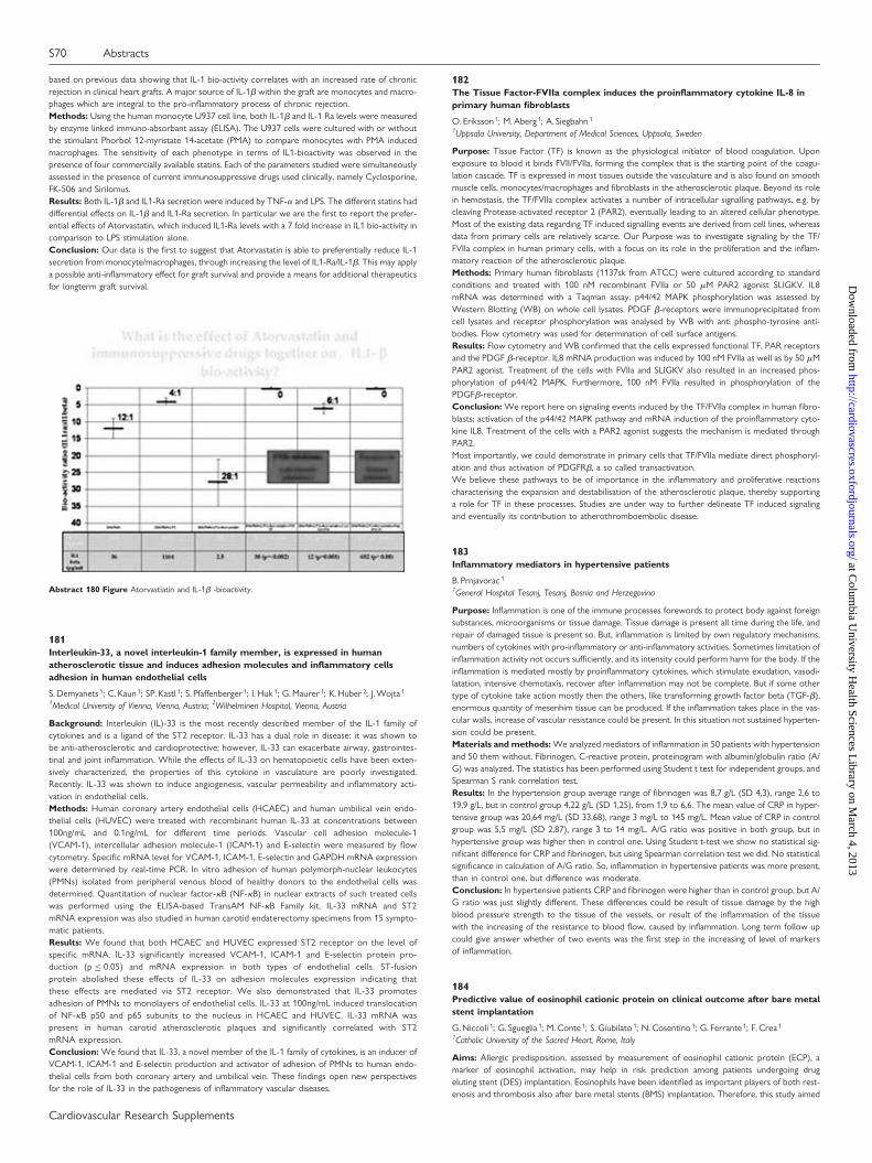
based on previous data showing that IL-1 bio-activity correlates with an increased rate of chronicrejection in clinical heart grafts. A major source of IL-1b within the graft are monocytes and macro-phages which are integral to the pro-inflammatory process of chronic rejection.Methods: Using the human monocyte U937 cell line, both IL-1b and IL-1 Ra levels were measuredby enzyme linked immuno-absorbant assay (ELISA). The U937 cells were cultured with or withoutthe stimulant Phorbol 12-myristate 14-acetate (PMA) to compare monocytes with PMA inducedmacrophages. The sensitivity of each phenotype in terms of IL1-bioactivity was observed in thepresence of four commercially available statins. Each of the parameters studied were simultaneouslyassessed in the presence of current immunosuppressive drugs used clinically, namely Cyclosporine,FK-506 and Sirilomus.Results: Both IL-1b and IL1-Ra secretion were induced by TNF-a and LPS. The different statins haddifferential effects on IL-1b and IL1-Ra secretion. In particular we are the first to report the prefer-ential effects of Atorvastatin, which induced IL1-Ra levels with a 7 fold increase in IL1 bio-activity incomparison to LPS stimulation alone.Conclusion: Our data is the first to suggest that Atorvastatin is able to preferentially reduce IL-1secretion from monocyte/macrophages, through increasing the level of IL1-Ra/IL-1b. This may applya possible anti-inflammatory effect for graft survival and provide a means for additional therapeuticsfor longterm graft survival.
181Interleukin-33, a novel interleukin-1 family member, is expressed in humanatherosclerotic tissue and induces adhesion molecules and inflammatory cellsadhesion in human endothelial cells
S. Demyanets 1; C. Kaun 1; SP. Kastl 1; S. Pfaffenberger 1; I. Huk 1; G. Maurer 1; K. Huber 2; J. Wojta 1
1Medical University of Vienna, Vienna, Austria; 2Wilhelminen Hospital, Vienna, Austria
Background: Interleukin (IL)-33 is the most recently described member of the IL-1 family ofcytokines and is a ligand of the ST2 receptor. IL-33 has a dual role in disease: it was shown tobe anti-atherosclerotic and cardioprotective; however, IL-33 can exacerbate airway, gastrointes-tinal and joint inflammation. While the effects of IL-33 on hematopoietic cells have been exten-sively characterized, the properties of this cytokine in vasculature are poorly investigated.Recently, IL-33 was shown to induce angiogenesis, vascular permeability and inflammatory acti-vation in endothelial cells.Methods: Human coronary artery endothelial cells (HCAEC) and human umbilical vein endo-thelial cells (HUVEC) were treated with recombinant human IL-33 at concentrations between100ng/mL and 0.1ng/mL for different time periods. Vascular cell adhesion molecule-1(VCAM-1), intercellular adhesion molecule-1 (ICAM-1) and E-selectin were measured by flowcytometry. Specific mRNA level for VCAM-1, ICAM-1, E-selectin and GAPDH mRNA expressionwere determined by real-time PCR. In vitro adhesion of human polymorph-nuclear leukocytes(PMNs) isolated from peripheral venous blood of healthy donors to the endothelial cells wasdetermined. Quantitation of nuclear factor-kB (NF-kB) in nuclear extracts of such treated cellswas performed using the ELISA-based TransAM NF-kB Family kit. IL-33 mRNA and ST2mRNA expression was also studied in human carotid endaterectomy specimens from 15 sympto-matic patients.Results: We found that both HCAEC and HUVEC expressed ST2 receptor on the level ofspecific mRNA. IL-33 significantly increased VCAM-1, ICAM-1 and E-selectin protein pro-duction (p ≤ 0.05) and mRNA expression in both types of endothelial cells. ST-fusionprotein abolished these effects of IL-33 on adhesion molecules expression indicating thatthese effects are mediated via ST2 receptor. We also demonstrated that IL-33 promotesadhesion of PMNs to monolayers of endothelial cells. IL-33 at 100ng/mL induced translocationof NF-kB p50 and p65 subunits to the nucleus in HCAEC and HUVEC. IL-33 mRNA waspresent in human carotid atherosclerotic plaques and significantly correlated with ST2mRNA expression.Conclusion: We found that IL-33, a novel member of the IL-1 family of cytokines, is an inducer ofVCAM-1, ICAM-1 and E-selectin production and activator of adhesion of PMNs to human endo-thelial cells from both coronary artery and umbilical vein. These findings open new perspectivesfor the role of IL-33 in the pathogenesis of inflammatory vascular diseases.
182The Tissue Factor-FVIIa complex induces the proinflammatory cytokine IL-8 inprimary human fibroblasts
O. Eriksson 1; M. Aberg 1; A. Siegbahn 1
1Uppsala University, Department of Medical Sciences, Uppsala, Sweden
Purpose: Tissue Factor (TF) is known as the physiological initiator of blood coagulation. Uponexposure to blood it binds FVII/FVIIa, forming the complex that is the starting point of the coagu-lation cascade. TF is expressed in most tissues outside the vasculature and is also found on smoothmuscle cells, monocytes/macrophages and fibroblasts in the atherosclerotic plaque. Beyond its rolein hemostasis, the TF/FVIIa complex activates a number of intracellular signalling pathways, e.g. bycleaving Protease-activated receptor 2 (PAR2), eventually leading to an altered cellular phenotype.Most of the existing data regarding TF induced signalling events are derived from cell lines, whereasdata from primary cells are relatively scarce. Our Purpose was to investigate signaling by the TF/FVIIa complex in human primary cells, with a focus on its role in the proliferation and the inflam-matory reaction of the atherosclerotic plaque.Methods: Primary human fibroblasts (1137sk from ATCC) were cultured according to standardconditions and treated with 100 nM recombinant FVIIa or 50 mM PAR2 agonist SLIGKV. IL8mRNA was determined with a Taqman assay. p44/42 MAPK phosphorylation was assessed byWestern Blotting (WB) on whole cell lysates. PDGF b-receptors were immunoprecipitated fromcell lysates and receptor phosphorylation was analysed by WB with anti phospho-tyrosine anti-bodies. Flow cytometry was used for determination of cell surface antigens.Results: Flow cytometry and WB confirmed that the cells expressed functional TF, PAR receptorsand the PDGF b-receptor. IL8 mRNA production was induced by 100 nM FVIIa as well as by 50 mMPAR2 agonist. Treatment of the cells with FVIIa and SLIGKV also resulted in an increased phos-phorylation of p44/42 MAPK. Furthermore, 100 nM FVIIa resulted in phosphorylation of thePDGFb-receptor.Conclusion: We report here on signaling events induced by the TF/FVIIa complex in human fibro-blasts; activation of the p44/42 MAPK pathway and mRNA induction of the proinflammatory cyto-kine IL8. Treatment of the cells with a PAR2 agonist suggests the mechanism is mediated throughPAR2.Most importantly, we could demonstrate in primary cells that TF/FVIIa mediate direct phosphoryl-ation and thus activation of PDGFRb, a so called transactivation.We believe these pathways to be of importance in the inflammatory and proliferative reactionscharacterising the expansion and destabilisation of the atherosclerotic plaque, thereby supportinga role for TF in these processes. Studies are under way to further delineate TF induced signalingand eventually its contribution to atherothromboembolic disease.
183Inflammatory mediators in hypertensive patients
B. Prnjavorac 1
1General Hospital Tesanj, Tesanj, Bosnia and Herzegovina
Purpose: Inflammation is one of the immune processes forewords to protect body against foreignsubstances, microorganisms or tissue damage. Tissue damage is present all time during the life, andrepair of damaged tissue is present so. But, inflammation is limited by own regulatory mechanisms,numbers of cytokines with pro-inflammatory or anti-inflammatory activities. Sometimes limitation ofinflammation activity not occurs sufficiently, and its intensity could perform harm for the body. If theinflammation is mediated mostly by proinflammatory cytokines, which stimulate exudation, vasodi-latation, intensive chemotaxis, recover after inflammation may not be complete. But if some othertype of cytokine take action mostly then the others, like transforming growth factor beta (TGF-b),enormous quantity of mesenhim tissue can be produced. If the inflammation takes place in the vas-cular walls, increase of vascular resistance could be present. In this situation not sustained hyperten-sion could be present.Materials and methods: We analyzed mediators of inflammation in 50 patients with hypertensionand 50 them without. Fibrinogen, C-reactive protein, proteinogram with albumin/globulin ratio (A/G) was analyzed. The statistics has been performed using Student t test for independent groups, andSpearman S rank correlation test.Results: In the hypertension group average range of fibrinogen was 8,7 g/L (SD 4,3), range 2,6 to19,9 g/L, but in control group 4,22 g/L (SD 1,25), from 1,9 to 6,6. The mean value of CRP in hyper-tensive group was 20,64 mg/L (SD 33,68), range 3 mg/L to 145 mg/L. Mean value of CRP in controlgroup was 5,5 mg/L (SD 2,87), range 3 to 14 mg/L. A/G ratio was positive in both group, but inhypertensive group was higher then in control one. Using Student t-test we show no statistical sig-nificant difference for CRP and fibrinogen, but using Spearman correlation test we did. No statisticalsignificance in calculation of A/G ratio. So, inflammation in hypertensive patients was more present,than in control one, but difference was moderate.Conclusion: In hypertensive patients CRP and fibrinogen were higher than in control group, but A/G ratio was just slightly different. These differences could be result of tissue damage by the highblood pressure strength to the tissue of the vessels, or result of the inflammation of the tissuewith the increasing of the resistance to blood flow, caused by inflammation. Long term follow upcould give answer whether of two events was the first step in the increasing of level of markersof inflammation.
184Predictive value of eosinophil cationic protein on clinical outcome after bare metalstent implantation
G. Niccoli 1; G. Sgueglia 1; M. Conte 1; S. Giubilato 1; N. Cosentino 1; G. Ferrante 1; F. Crea 1
1Catholic University of the Sacred Heart, Rome, Italy
Aims: Allergic predisposition, assessed by measurement of eosinophil cationic protein (ECP), amarker of eosinophil activation, may help in risk prediction among patients undergoing drugeluting stent (DES) implantation. Eosinophils have been identified as important players of both rest-enosis and thrombosis also after bare metal stents (BMS) implantation. Therefore, this study aimed
Abstract 180 Figure Atorvastiatin and IL-1b -bioactivity.
S70 Abstracts
Cardiovascular Research Supplements
at Colum
bia University H
ealth Sciences Library on M
arch 4, 2013http://cardiovascres.oxfordjournals.org/
Dow
nloaded from
at asssessing the association between baseline ECP levels, and recurrence of clinical events in a con-secutive serie of patients who underwent BMS implantation.Methods and Results: One hundred and ten patients (69 + 11 years, 88 men) undergoingimplantation of BMS for the treatment of stable chronic angina or an acute coronary syndromewere enrolled. Prior to percutaneous coronary intervention, serum levels of ECP were measuredby enzyme-linked immunosorbent assay. A clinical follow-up was performed 24 months after dis-charge in all patients. Major adverse cardiac events (MACEs), defined as cardiac death, recurrentmyocardial infarction, or clinically driven target lesion revascularization, were the endpoint of thestudy. Eighteen (16.4%) patients had MACEs and showed higher serum levels of ECP comparedwith those without MACEs [20.1 (9.8–47.3) vs. 9.5 (5.0–27.2) g/L, p ¼ 0.02). At Cox regressionanalysis, ECP level.11 g/L was and stenosis length.18 mm were the only significant predictorsof MACEs (hazard ratio 3.5, 95% confidence interval 1.1– 10.4, p ¼ 0.032 and hazard ratio 2.8,95% confidence interval 1.1-7.0 p ¼ 0.03, respectively).Conclusion: This study demonstrates that ECP, a sensitive marker of eosinophil activation, isassociated with MACEs after BMS implantation. As a similar finding was previously observed forDES, thus allergic mediated inflammation could explain some adverse reactions occurring after cor-onary artery stenting.
185Uric acid in urine and subclinical inflammation in the hypertensive patients
V. Dmitriev 1; E. Oschepkova 1; O. Polovitkina 1; V. Titov 1
1Russian Cardiology Research and Production Center, Moscow, Russian Federation
Objective: some authors hypothesised that cytokines may increase the uric acid production andthrough the enhancement of xanthine oxidase activity the reactive oxygen species mediated celldamage and the promotion of apoptosis in hypertensive animals. The aim of our study was to inves-tigate association uric acid in urine (UAU) and subclinical inflammation markers in the hypertensivepatients.Design and method: 58 EH pts (46 M, 12 F) grade 1 or 2, av. age 44,4 + 1,7 years; without anty-hypertensive therapy for 2 weeks before study. Patients with acute inflammatory diseases, notearlier than 2 months were included in study. Serum uric acid (SUA) and UAU (normal level is1,48-4,43 mmol/day) was defined by a UF method on the uricase selective analyzer. High-sensitivityC-reactive protein (CRP) and Uric albumin excretion (UAE) was defined by a turbidimetry method.24 hour blood pressure monitoring (24-h BPM) was carried out by Meditech ABPM-04 (Hungary).The statistical analysis was carried out by nonparametric Methods of Spearman and Mann-Whitneywith STATISTICA 6. The data is presented as M + m.Results: According UAU the all EH pts was divided into the two groups. 24-h SBP level were con-siderably higher in the increase UAU group (n ¼ 21) compared with normal UAU group (n ¼ 37),(143,5 + 2,9 and 136,1 + 1,9 mm Hg, p , 0,05, respectively), 24-h DBP levels was not different(84,1 + 2,2 and 81,1 + 1,5 mm Hg, n/s,respectively). The average levels of CRP, SUA and UAUwere significant higher in the increase UAU pts (n ¼ 21) compared to normal UAU pts (n ¼ 37):5,7 + 1,9 and 2,7 + 0,4 mg/L, p , 0,05; 397 + 21 and 300 + 12 mmol/L, p , 0,001; 26,6 + 8,1and 15,9 + 3,1 mg/L, p ¼ 0,005, respectively. Also, in the increase UAU group was found significantassociation between CRP and 24-h SBP (r ¼ 0,67; p ¼ 0,001). The same association was notdemonstrated in the normal UAU group.Conclusion: Determination of the increased levels UAU might be the early renal damage markerin the hypertensive patients.
186Correlation between biochemical and echografic markers of inflammation inpatients with cardiometabolic syndrome
D. Ilisei 1; M. Leon 1; F. Mitu 1
1Grigore T. Popa University of Medicine and Pharmacy, Iasi, Romania
Purpose: Adipose tissue is an endocrine organ secreting several adipokines which regulate a widespectrum of metabolic and immune processes. Obesity is associated with development of adiposetissue inflammation. Besides specific action curative factors used in obesity, metabolic syndrome anddiabetes mellitus also produce anti-inflammatory effect and thus diminish risk factors of cardiovas-cular diseases, rate of their development, and alleviate manifestations of atherosclerosis.Methods: We used the measurement of carotid intima-media thickness (IMT) as a marker of earlyatherosclerosis and established the values of this biochemical parameters: total cholesterol, LDLcholesterol, triglycerides, hs-C-reactive protein(CRP) and fibrinogen.Results: IMT has been shown to correlate significantly with the presence of coronary arterydisease (CAD) and to predict fatal and not fatal cerebro- and cardio-vascular events. It has beenestablished positive correlation of the degree of stenosis and intima-media thickness of carotidarteries with the biochemical parameters studied. Negative correlation was stated with respectto HDL cholesterol and protein C. Additionally, C-reactive protein levels rise with increasinglevels of visceral adipose tissue.Conclusion: The strong relationship between hs-CRP and IMT may potentially account for thecomplex role of hs-CRP and IMT in the pathogenesis of cardiovascular events.
187The role of EGFR transactivation in relation to signals generated by conditionedmedium from activated CD1d-restricted human NKT cells in human vascular cells
E. Kyriakakis 1; M. Philippova 1; M. Cavallari 1; V. Bochkov 2; B. Biedermann 1; G. De Libero 1; P. Erne 3;TJ. Resink 1
1University Hospital Basel, Basel, Switzerland; 2Medical University of Vienna, Vienna, Austria; 3CantonalHospital Lucerne, Lucerne, Switzerland
Introduction: Inflammation and intraplaque angiogenesis play an important role in atherosclerosis.We hypothesize that activation of immune cells by lipid antigens may constitute a proangiogenic
mechanism in atherosclerosis. Our goal is to identify CD1d-restricted NKT activation as a relevantangiogenic regulator and to elucidate the angiogenic signals generated.Methods: Different APC‘s which expressed either neglible levels of CD1d (Molt4) or higher levels(C1R and HeLa) were preincubated in the presence or absence of a glycosphingolipid,alpha-galactosylceramide (aGalCer), before incubation with Cd1d-restricted human NKT cell clones.Conditioned media (CM) supernatants were collected 2 days later and activation of NKT clonesinresponse to the antigen presentation was confirmed by measuring IL-4 and INF-g. We measuredangiogenic rates in vitro, migration, proliferation and tried to look at signalling pathways involved inthe activation.Results: We observed that CD1d-NKT activation from APC‘s overexpressing CD1d , significantlyaltered the morphology of the endothelial cells and increased sprouting, as compared to non-stimulated APC‘sand APC‘s expressing low levels of CD1d. RT-PCR analysis of genes expressedafter incubation of HMEC-1 cells with CM showed that IL-8 and Cox-2 are significantly upregulated.Motility was also induced in an IL-8 and EGFR dependent manner, but cell growth was not affectedat early time points. Several proteins involved in cell migration and cell survival were highly upre-gulated or activated in the presence of CM from activated iNKT cells, but matrix metalloproteinasesremained unaffected.Conclusions: We demonstrated that activation ofCD1d-restricted human NKT cells could inducemigration and angiogenesis in vitro through the transactivation of EGFR. Attenuation of IL8 andEGFR signals by use of blocking antidodies could reverse these effect. Identification ofCD1d-restricted NKT activation as a relevant angiogenic regulator will enable development ofnovel immune-cell targeted therapies.
188The use of the theory of biological function and in the investigations pathogenesis ofdiseases widely spread in a population
V. Titov 1
1Russian Cardiology Research & Production Center, Moscow, Russian Federation
Impairments of biological functions that superimpose specific individual functions of morphologicallyseparated systems lay the pathogenetic basis of numerous diseases. These functions are: "purity" ofthe intercellular medium (endoecology), exotrophy, homeostasis, locomotion, stress, adaptationand compensation. We believe that impaired endoecology and trophic function are the commonlink in the pathogeneses of arterial hypertension, atherosclerosis, diabetes, obesity and metabolicsyndrome. The impairments involve three biological reactions: inflammation, filtration via the sorp-tion filter of the nephron, and activated passive transcytosis by endothelial cells (pinocytosis) due toa hydraulic pressure increase in a local intravascular pool of the intercellular medium. Removal ofendogenous small-molecular-mass initiators of inflammation occurs by filtration through the biologi-cal sorption filter and macromolecules are removed by "scavenger" resident macrophages by pha-gocytosis in the loci of collection and utilization of biological "waste", i.e., by inflammatory response.When physiological capacities of these reactions and functions of peripheral peristaltic pumps(muscle type arteriole) are exhausted, passive steps of the reactions are activated by arterialblood pressure elevation. This elevation acts in vivo acts as a physical regulatory factor of homeo-stasis and endoecology. Arterial blood pressure rise, a simultaneous increase in blood content ofC-reactive protein and heightened urinary excretion of albumin reflect the accumulation ofsmall- and large-molecular weight biological "waste" in the intercellular medium. Removal of this"waste" is activated by elevation of hydraulic pressure in a local intravascular pool by moreintense functioning of the heart which acts as the central pump. Biological reactions of hydraulicpressure elevation, higher excretion of “waste” with urine, and inflammatory response are involvedin realization of all biological functions, thus forming a universal pathogenesis of the majority ofwidely spread diseases.
189Atorvastatin directly improves vascular redox state by affecting eNOS coupling andNADPH-oxidase activity in human vein grafts ex-vivo
C. Bakogiannis 1; C. Antoniades 2; D. Tousoulis 1; M. Demosthenous 1; C. Psarros 1; N. Sfyras 1;KM. Channon 2; C. Stefanadis 1
11st Cardiology Department, Athens University Medical School., Athens, Greece; 2Department ofcardiovascular Medicine, University of Oxford., Oxford, United Kingdom
Purpose: Statins generally improve survival of patients after coronary bypass surgery (CABG), buttheir effect on vascular wall is unclear. We examined the direct impact of atorvastatin on vascularredox of saphenous vein grafts (SVG) used for CABG, and explored the underlying mechanisms ofits vascular effects.Methods: SVGs from ten patients undergoing CABG were exposed to atorvastatin 0, 5 and 10mmol/L for 6 hours in an ex vivo model system. Vascular O2- was measured by lucigenin chemilu-minescence +/2 endothelial nitric oxide synthase (eNOS) inhibitor LNAME (to estimate eNOScoupling) or NADPH (to estimate NADPH-oxidase activity). The direct O2- scavenging effect ofatorvastatin was tested in a cell free system of xanthine/xanthine oxidase (X/XO), by usingvitamin C as positive control.Results: Atorvastatin induced a rapid reduction of vascular O2- (Fig. A) and reduced theLNAME-inhibitable O2- generation (Fig. B) in SV grafts ex vivo. Moreover, atorvastatin alsoreduced NADPH-oxidase activity in these vessels (Fig C). Importantly, atorvastatin had directscavenging effects of on O2- produced by X/XO system (Fig. D).Conclusions: These novel findings suggest that atorvastatin exerts a rapid, direct effect on vas-cular redox state in human SV grafts used for CABG, independently of LDL-mediated mechan-ism. Moreover, the direct effects of atorvastatin on SV grafts are mediated through theimprovement of eNOS coupling and the reduction of NADPH oxidase activity in the vascularwall.
Abstracts S71
Cardiovascular Research Supplements
at Colum
bia University H
ealth Sciences Library on M
arch 4, 2013http://cardiovascres.oxfordjournals.org/
Dow
nloaded from
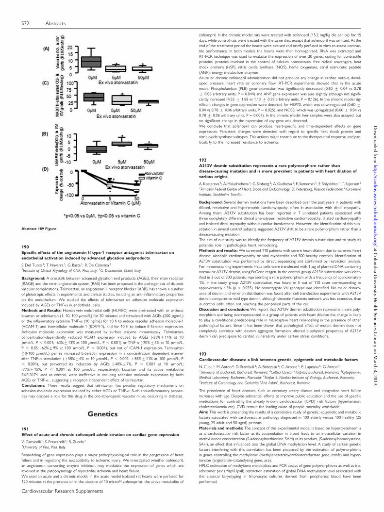
190Specific effects of the angiotensin II type-1 receptor antagonist telmisartan onendothelial activation induced by advanced glycation endproducts
S. Del Turco 1; T. Navarra 1; G. Basta 1; R. De Caterina 2
1Institute of Clinical Physiology of CNR, Pisa, Italy; 2G. D’annunzio, Chieti, Italy
Background: A crosstalk between advanced glycation end products (AGEs), their main receptor(RAGE) and the renin-angiotensin system (RAS) has been proposed in the pathogenesis of diabeticvascular complications. Telmisartan, an angiotensin II receptor blocker (ARB), has shown a numberof pleiotropic effects in experimental and clinical studies, including an anti-inflammatory propertieson the endothelium. We studied the effects of telmisartan on adhesion molecule expressioninduced by AGEs or TNF-a in endothelial cells.Methods and Results: Human vein endothelial cells (HUVEC) were pretreated with or withoutlosartan or telmisartan (1, 10, 100 mmol/L) for 30 minutes and stimulated with AGEs (200 mg/mL)or the inflammatory cytokine TNF-a (10 ng/mL) for 18 h to induce vascular adhesion molecule-1(VCAM-1) and intercellular molecule-1 (ICAM-1), and for 10 h to induce E-Selectin expression.Adhesion molecule expression was measured by surface enzyme immunoassay. Telmisartan,concentration-dependently reduced VCAM expression induced by AGEs (-33%+11% at 10mmol/L, P , 0.001; -63%+13% at 100 mmol/L, P , 0.001) or TNF-a (-20%+5% at 10 mmol/L,P , 0.05; -62%+9% at 100 mmol/L, P , 0.001), but not of ICAM-1 expression. Telmisartan(10-100 mmol/L) per se increased E-Selectin expression in a concentration dependent mannerafter TNF-a stimulation (+58%+6% at 10 mmol/L, P , 0.001; +88%+11% at 100 mmol/L, P, 0.001), but prevented its induction by AGEs (-40%+7%, P , 0.001 at 10 mmol/L;-77%+15%, P , 0.001 at 100 mmol/L, respectively). Losartan and its active metaboliteEXP-3174 used as control, were ineffective in reducing adhesion molecule expression by bothAGEs or TNF-a , suggesting a receptor-independent effect of telmisartan.Conclusions: These results suggest that telmisartan has peculiar regulatory mechanisms onadhesion molecule expression induced by either AGEs or TNF-a. Such anti-inflammatory proper-ties may disclose a role for this drug in the pro-atherogenic vascular milieu occurring in diabetes.
Genetics
191Effect of acute and chronic zofenopril administration on cardiac gene expression
V. Carnicelli 1; S. Frascarelli 1; R. Zucchi 1
1University of Pisa, Pisa, Italy
Remodeling of gene expression plays a major pathophysiological role in the progression of heartfailure and in regulating the susceptibility to ischemic injury. We investigated whether zofenopril,an angiotensin converting enzyme inhibitor, may modulate the expression of genes which areinvolved in the patophysiology of myocardial ischemia and heart failure.We used an acute and a chronic model. In the acute model isolated rat hearts were perfused for120 minutes in the presence or in the absence of 10 microM zofenoprilat, the active metabolite of
zofenopril. In the chronic model rats were treated with zofenopril (15.2 mg/Kg die per os) for 15days, while control rats were treated with the same diet, except that zofenopril was omitted. At theend of the treatment period the hearts were excised and briefly perfused in vitro to assess contrac-tile performance. In both models the hearts were then homogenized, RNA was extracted andRT-PCR technique was used to evaluate the expression of over 20 genes, coding for contractileproteins, proteins involved in the control of calcium homeostasis, free radical scavengers, heatshock proteins (HSP), nitric oxide synthase (NOS), heme oxygenase, atrial natriuretic peptide(ANP), energy metabolism enzymes.Acute or chronic zofenopril administration did not produce any change in cardiac output, devel-oped pressure, heart rate or coronary flow. RT-PCR experiments showed that in the acutemodel Phospholamban (PLB) gene expression was significantly decreased (0.60 + 0.04 vs 0.78+ 0.06 arbitrary units, P ¼ 0.044) and ANP gene expression was also slighltly although not signifi-cantly increased (4.55 + 1.88 vs 1.13 + 0.29 arbitrary units, P ¼ 0,126). In the chronic model sig-nificant changes in gene expression were detected for HSP70, which was downregulated (0.60 +0.04 vs 0.78 + 0.06 arbitrary units, P ¼ 0.025), and NOS3, which was upregulated (0.60 + 0.04 vs0.78 + 0.06 arbitrary units, P ¼ 0.007). In the chronic model liver samples were also assayed, butno significant change in the expression of any gene was detected.We conclude that zofenopril can produce heart-specific and time-dependent effects on geneexpression. Persistent changes were detected with regard to specific heat shock protein andnitric oxide synthase subtypes. This actions might contribute to the therapeutical response, and par-ticularly to the increased resistance to ischemia.
192A213V desmin substitution represents a rare polymorphism rather thandisease-causing mutation and is more prevalent in patients with heart dilation ofvarious origins.
A. Kostareva 1; A. Malashicheva 1; G. Sjoberg 2; A. Gudkova 1; E. Semernin 1; E. Shlyakhto 1; T. Sejersen 2
1Almazov Federal Centre of Heart, Blood and Endocrinology, St. Petersburg, Russian Federation; 2KarolinskaInstitute, Stockholm, Sweden
Background: Several desmin mutations have been described over the past years in patients withdilated, restrictive and hypertrophic cardiomyopathy, often in association with distal myopathy.Among them, A213V substitution has been reported in 7 unrelated patients associated withthree completely different clinical phenotypes: restrictive cardiomyopathy, dilated cardiomyopathyand isolated distal myopathy without cardiac involvement. However, the identification of this sub-stitution in several control subjects suggested A213V shift to be a rare polymorphism rather than adisease-causing mutation.The aim of our study was to identify the frequency of A213V desmin substitution and to study itspotential role in pathological heart remodelling.Methods and results: We screened 110 patients with severe heart dilation due to ischemic heartdisease, alcoholic cardiomyopathy or viral myocarditis and 300 healthy controls. Identification ofA213V substitution was performed by direct sequencing and confirmed by restriction analysis.For immunostaining experiments HeLa cells were transfected with 1 mg of plasmid DNA containingnormal or A213V desmin, using FuGene reagen. In the control group A213V substitution was ident-ified in 3 out of 300 patients, representing a rare polymorphism with a frequency of approximately1%. In the study group A213V substitution was found in 5 out of 110 cases corresponding toapproximately 4.5% (p , 0.035). No homozygote Val genotype was identified. No major disturb-ance of desmin and vimentin distribution was found after cell transfection experiments with A213Vdesmin compares to wild type desmin, although vimentin filaments network was less extensive, thanin control cells, often not reaching the peripheral parts of the cell.Discussion and conclusion: We report that A213V desmin substitution represents a rare poly-morphism and being overrepresented in a group of patients with heart dilation the change is likelyto play a conditional predisposing role in maladaptive heart remodelling in the presence of otherpathological factors. Since it has been shown that pathological effect of mutant desmin does notcompletely correlate with desmin aggregate formation, altered biophysical properties of A213Vdesmin can predispose to cardiac vulnerability under certain stress conditions.
193Cardiovascular diseases: a link between genetic, epigenetic and metabolic factors
N. Cucu 1; M. Anton 2; D. Stambuli 3; A. Botezatu 4; C. Arsene 1; E. Lupeanu 5; G. Anton 4
1University of Bucharest, Bucharest, Romania; 2Coltea Clinical Hospital, Bucharest, Romania; 3CytogenomicMedical Laboratory, Bucharest, Romania; 4Stefan S. Nicolau Institute of Virology, Bucharest, Romania;5Institute of Gerontology and Geriatrics "Ana Aslan", Bucharest, Romania
The prevalence of heart diseases, such as coronary artery disease and congestive heart failureincreases with age. Despite substantial efforts to improve public education and the use of specificmedications for controlling the already known cardiovascular (CVD) risk factors (hypertension,cholesterolaemia etc), CVD remain the leading cause of people mortality worldwide.Aim: This work is presenting the results of a correlative study of genetic, epigenetic and metabolicfactors associated with cardiovascular pathology diagnosed in 100 elderly versus 100 healthy (25young, 25 adult and 50 aged) persons.Materials and methods: The concept of this experimental model is based on hypercysteinaemiaas a cardiovascular risk factor as its accumulation in blood leads to an intracellular variation inmethyl donor concentration (S-adenosylmethionine, SAM) vs its product, (S-adenosylhomocysteine,SAH), an effect that influenced also the global DNA methylation level. A study of certain geneticfactors interfering with this correlation has been proposed by the estimation of polymorphismsin genes controlling the methylome (methylenetetrahydrofolatereductase gene, mthfr) and hyper-tension (angiotensin-catabolysing gene, ace).HPLC estimation of methylome metabolites and PCR assays of gene polymorphisms as well as iso-schisomer pair (MspI/HpaII) restriction estimation of global DNA methylation level associated withthe classical karyotyping in limphocyte cultures derived from periphereal blood have beenperformed.
Abstract 189 Figure
S72 Abstracts
Cardiovascular Research Supplements
at Colum
bia University H
ealth Sciences Library on M
arch 4, 2013http://cardiovascres.oxfordjournals.org/
Dow
nloaded from
Results: The results showed interesting correlations between the modification of normal SAM toSAH ratio towards the increased level of SAH and decreased level of SAM, a pronounced hypo-methylation of global DNA. These metabolites levels and epigenetic factors were correlated withgenetic aspects in terms of an altered karyotype, as well as in high frequency of polymorphism inmthfr (25% homozygotes, 8% heterozygotes for C677-WT, 25% homozygotes and 10% heterozy-gotes for 1298C-WT) and ace (ID-40%, DD-50%, II-18%) genes.In conclusion, we demonstrated that an altered metabolites level, as high SAH concentration con-comitant with a lowered SAM concentration, determined a global DNA hypomethylation. This, inturn may be considered a cause of alteration in pericentromeric heterochromatin of chromosomesthat explains the genomic instability in terms of aneuploidies and frequent premature chromatideseparation phenomena. These genetic and epigenetic factors are well correlated with a high fre-quency of abnormal allelic expression of the mthfr and ace gene due to their SNPs.
194Secretoneurin gene therapy improves nerve function in diabetic neuropathy
AGE.Beer 1; MT.Theurl 1; WS.Schgoer 1; KA.Albrecht 1; JRP.Patsch 1; EH.Huber 1; PS.Schratzberger 1;RK. Kirchmair 1
1Innsbruck Medical University, Department of Internal Medicine I, Innsbruck, Austria
1. Purpose: Pathogenesis of diabetic neuropathy remains enigmatic and metabolic changes due tohyperglycemia as well as impact of diabetes on vasa nervorum might lead to this disease.2. Methods: As an animal model for diabetic neuropathy db/db mice were used. Heterozygotemice served as control.Gene therapy was accomplished with intra-muscular injection of plasmid DNA expressing theangiogenic factor Secretoneurin (SN). Sciatic nerve motor and sensory nerve conduction velocity(MCV and SCV) and tail flick temperature was measured. Vascularity of sciatic nerves was assessedby in situ fluorescent staining using BS-1 lectin. In vitro effects of SN on Schwann cells weredetermined.3. Results: Heterozygote mice showed normal values for MCV and SCV that stayed unchanged forthe observational period of 9 weeks (MCV: baseline 52.8 + 1.7 m/s, 9 wk 52.5 + 0.9 m/s; SCV:baseline 58.7 + 2.1 m/s, 9 wk 59.8 + 0.8 m/s; n ¼ 10). Homozygous mice showed severelyreduced nerve conduction velocities. Mice received 100 ug of SN or control plasmid. Aftercontrol plasmid injection nerve conduction velocities did not change significantly (MCV: baseline32.5 + 0.9 m/s, 9 wk 31.5 + 0.9 m/s; SCV: baseline 36.7 + 1.3 m/s, 9 wk 33.1 + 1.4 m/s;n ¼ 24). After SN gene therapy, MCV and SCV increased significantly (MCV: baseline 31.9 +0.7 m/s, 9 wk 46.7 + 1.3 m/s; SCV: baseline 36.5 + 1.2 m/s, 9 wk 49.3 + 1.3 m/s; n ¼ 26) andyielded a significant better outcome of SN treated mice (P , 0.01 for MCV and SCV SN vs.Ctr.). Tail-flick temperature was: heterozygote mice: baseline 44.3 + 0.258C; 4 wks 44.4 +0.28C, n ¼ 5; ctr treated mice: baseline 45.9 + 0.128C, 4 wks 45.8 + 0.148C; SN treated mice:baseline 45.8 + 0.188C, 4 wks 44.9 + 0.128C; P , 0.05 SN vs. Ctr. after 4 wks; n ¼ 12 eachgroup. Improved density of vasa nervorum in SN treated mice was observed. Schwann cellswere stained by DAPI for evaluation of cell proliferation. SN treatment increased cell number (rela-tive cell number vs. Ctr: SN 100 ng/ml 2.7 + 0.15; n ¼ 4; P , 0.05 SN vs. Ctr; n ¼ 4). For induction ofapoptosis cells were treated by H2O2 and stained by DAPI. SN significantly decreased apoptosis (%apoptotic cells: Ctr 34.4 + 2.7%, SN 100 ng/ml 18.5 + 2.0%; n ¼ 4; P , 0.05 SN vs. Ctr; n ¼ 4).4. Conclusions: The present observations indicate that SN exerts beneficial effects on nerve func-tion and vascularity in diabetic neuropathy in vivo and additionally on Schwann cells in vitro.
195Ribozymes as specific therapeutic agents against smooth muscle cell migration andproliferation: a new perspective in gene therapy.
C. Lande 1; A. Cecchettini 2; L. Tedeschi 1; MG. Trivella 1; L. Citti 1
1Institute of Clinical Physiology of CNR, Pisa, Italy; 2University, Pisa, Italy
Purpose: Cell migration and proliferation are still an open issues concerning atherogenesis andrestenosis. The exploitation of ribozyme for the inactivation of genes involved in the phenotypechanges responsible for the pathological state, could be a promising and innovative technique.The Purpose of this study was to set up a method for the delivery and the validation of a ribozymein Vascular Smooth Muscle Cells (VSM cells).Methods: A minimal ribozyme was tailored on PDGFR-b mRNA target sequence by an "in silico"approach and synthesized with an oligonucleotide synthesizer apparatus. The delivery was accom-plished with a new polyplexed vehicle. The complex vehicle-ribozyme was administered to porcineVSM cells in culture in the presence of serum. Cells were stimulated by PDGF-BB after 24 hours andtheir migration activity monitored according to an "in vitro wound model".Results: A differential migratory activity was observed: cells stimulated with PDGF-BB, invadedextensively the scratched surface, whereas cells pre-treated with ribozyme (before growth factorstimulation) displayed a considerably reduction of this repair activity. A computational measureof repair demonstrated that the ribozyme reduced the cell migration of about 90%.Conclusions: These results are very encouraging since the activity of ribozyme was demonstrated in abiological model. Moreover, the vehicle preserved the gene-drug from nucleases and it was highly effec-tive also in the presence of serum, condition that mimic the effective environment of vascular tissue. Suchpilot experiments open actual perspectives for "in vivo" local gene knock-down treatments suggestingtherapeutic strategies based on multiple ribozymes targeting different crucial pathological genes.
196Construction of recombinant adeno-associated virus serotype 9 with ribozyme genetargeting NF kappaB and its suppression of NF kappaB activity in HeLa cells
BD. Chen 1; YT. Ma 1; YINING. Yang 1; XIANG. Ma 1; FEN. Liu 1
1Cardiovascular Institute of 1st affiliated hospital in Xinjiang Medical University, Urumuqi, China, People’sRepublic of
Objective: To construct the recombinant adeno-associated virus serotype 9 containing ribozymegene(R65) targeting nuclear factor-kappa B(NF-kB), and investigate the inhibitory effect ofrAAV9-eGFP-R65 on activation of NF-kB as well as on protein expression of NF-kB P65 in HeLa cells.Methods: The synthesized ribozyme gene targeting NF-kB was inserted into the plasmidpFB-CMV-eGFP with definite direction, and packaged into the recombinant adeno-associatedvirus serotype 9 by three plasmids co-transfection, then recombinant adeno-associated virus waspurified by cesium choride density centrifugation. The purity of recombinant virusrAAV9-eGFP-R65 was observed and verified by transmission electron microscopy andSDS-PAGE and viral tite was checked by GFP. Finally, HeLa cells were infected by the recombinantadeno-associated virus , the activation of NF-kB and protein expression of P65 were analyzed byelectrophoretic mobility shift assay[] and Western blot.Results: The high expression of green fluorescence protein expression in HEK293 and HeLa celllines were found under fluorescent microscope. Electron microscopy and SDS-PAGE test indicatedthat recombinant adeno-associated virus rAAV9-eGFP-R65 was successfully constructed, and thetiter of the virus reached 4.63 × 10¹² vg/ml. Western blot and EMSA showed that the proteinexpression of P65 and the activation of NF-kB in HeLa cells were markedly inhibited after transfec-tion with rAAV9-eGFP-R65.Conclusion: Activation of NF-kB and expression of NF-kB p65 protein in HeLa cells are effectivelyinhibited by recombinant adeno-associated virus rAAV9-eGFP-R65, which lays the foundation forfurther researches into therapy nuclear factor-kappa B related ischemic diseases.
197Association of eNOS genetic polymorphism with coronary artery disease in theIranian patients
M.Hasanzad 1; L.Rejali 1; M.Fathi 1; A.Minassian 1; R.Mohammad Hassani 1; A.Najafi 1; MR.Sarzaeem 2;SH. Sezavar 3
1Islamic Azad University, Tehran Medical Branch, Tehran, Iran (Republic of Islamic); 2Tehran University ofMedical Sciences, Shariati Hospital, Tehran, Iran (Republic of Islamic); 3Iran University of Medical Sciences,Tehran, Iran (Republic of Islamic)
Coronary artery disease (CAD) is a leading cause of mortality and morbidity in Iranian population.But little is known about CAD and its relation with genetic factors in the Iranian population. Thisstudy was designed for two Purposes ; collecting the CAD samples for future genetic studies andalso investigation about the association between CAD and Glu298Asp polymorphism of the endo-thelial nitric oxide synthase (eNOS) gene by RFLP method. Previous eNOS Glu298Asp polymorph-ism studies suggested its important role in cardiovascular disorders and specially its association withCAD. We determined the prevalence of eNOS Glu298Asp polymorphism in healthy volunteersfrom Iraninan population and also in the patients suffering from CAD. A number of 90 patientsand 100 controls were collected during a period of six month. Distribution of genotypes GGwas around 70% for control groups but the G allele was not the susceptible allele for CAD subjectsin this study. The patients group showed an increase in the frequency of T allele compared to con-trols, there was significant association between the TT genotype and CAD. The present study whichis under progress was the first in the Iranian population. Further evaluation with larger samples maybe helpful for assessing the risk, determining the management strategy and Iran targeting preventiveintervention for coronary artery disease.
198Differential gene expression in thrombus-derived leukocytes of patients with acutecoronary syndrome
A. Akhmedov 1; R. Klingenberg 1; K. Yonekawa 1; C. Lohmann 1; S. Gay 1; W. Maier 1; M. Neithard 1;TF. Luescher 1
1University Hospital Zurich, Zurich, Switzerland
Aim : Previous work from our group has identified monocytes/macrophages as the predominantinflammatory cell type in coronary thrombi. This study aimed to provide enhanced insight intothe pathophysiology of coronary thrombus formation by analysis of the activation pattern ofleukocytes.Methods and Results : Transcriptional profiling (Affymetrix Gene Array)was performed compar-ing coronary thrombi from individual ACS patients (n ¼ 4) and their respective peripheral bloodmononuclear cells (PBML). Among 10337 differentially expressed genes, 378 fulfilled the selectioncriteria of a . ¼ 2-fold upregulation and a p value of , ¼ 0.01. Thrombi were distinguished byupregulation of transcripts indicative of alternatively activated (M2) macrophages (CXCL2,CXCL3, CCL18, MSR1, MRC1, SPHK1, TGFBI, FN1,) along with markers of macrophage foamcells (OLR1, APOC1, FABP4, FABP5) and activated dendritic cells (GPNMB, SERPINE1, PLAUR,CXCL16). Distinct signs of adaptive immunity were detected (CD83, CD86, BCAP29, MR1) withactivation of TNFR superfamily signal transduction (PVRL2, CD40, NFKBIE). Thrombi showed astrong upregulation of transcripts involved in extracellular matrix turnover and fibrotic/calcifictissue repair (MMP12, CTGF, MMP19, CTSL1, MMP2, TIMP1, TGFBI, TGIF1, POSTN, NRP1,NRP2, SPP1, BMP2K, SDC2, DSE). Furthermore, markers of hypoxia and neoangiogenesis weremarkedly upregulated (NR4A3, EPAS1, P4HA1, CTSLL3, FLT1, VEGFA, ACVRL1). In addition, tran-scripts involved in coagulation/platelet activation were enhanced (S100A10, SERPINB8, THBD,PLAU, PLAUR).Conclusions : Our data suggest a pivotal role for alternatively activated macrophages (M2) in cor-onary thrombi with known functions in resolution of inflammation via phagocytic clearance andtrophic factor synthesis along with activated dendritic cells promoting adaptive immunity.Abstract 195 Figure
Abstracts S73
Cardiovascular Research Supplements
at Colum
bia University H
ealth Sciences Library on M
arch 4, 2013http://cardiovascres.oxfordjournals.org/
Dow
nloaded from
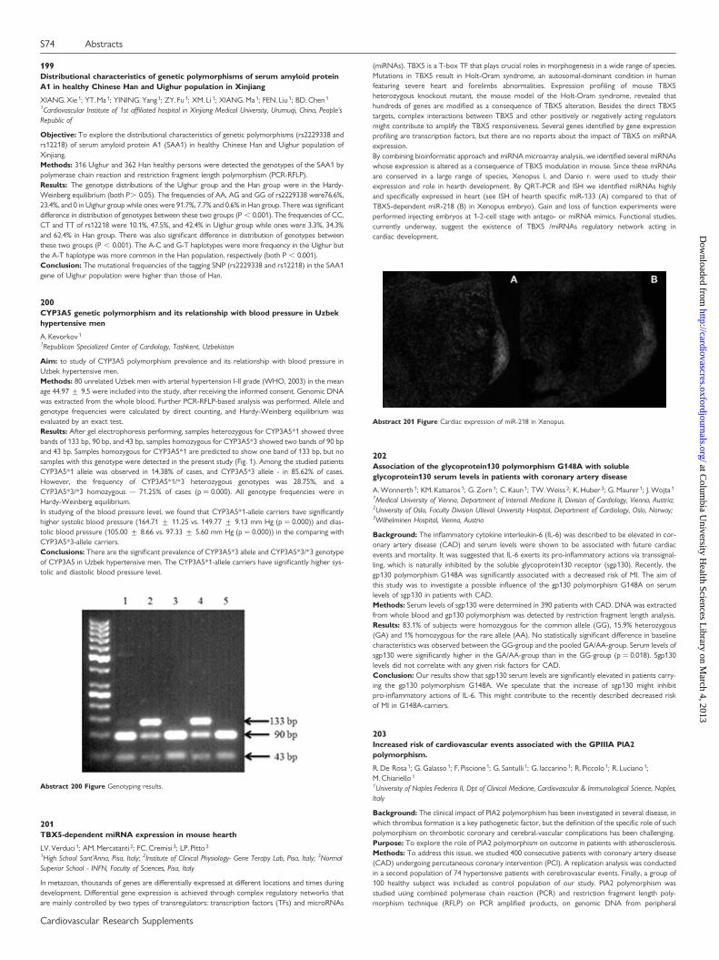
199Distributional characteristics of genetic polymorphisms of serum amyloid proteinA1 in healthy Chinese Han and Uighur population in Xinjiang
XIANG. Xie 1; YT. Ma 1; YINING. Yang 1; ZY. Fu 1; XM. Li 1; XIANG. Ma 1; FEN. Liu 1; BD. Chen 1
1Cardiovascular Institute of 1st affiliated hospital in Xinjiang Medical University, Urumuqi, China, People’sRepublic of
Objective: To explore the distributional characteristics of genetic polymorphisms (rs2229338 andrs12218) of serum amyloid protein A1 (SAA1) in healthy Chinese Han and Uighur population ofXinjiang.Methods: 316 Uighur and 362 Han healthy persons were detected the genotypes of the SAA1 bypolymerase chain reaction and restriction fragment length polymorphism (PCR-RFLP).Results: The genotype distributions of the Uighur group and the Han group were in the Hardy-Weinberg equilibrium (both P. 0.05). The frequencies of AA, AG and GG of rs2229338 were76.6%,23.4%, and 0 in Uighur group while ones were 91.7%, 7.7% and 0.6% in Han group. There was significantdifference in distribution of genotypes between these two groups (P , 0.001). The frequencies of CC,CT and TT of rs12218 were 10.1%, 47.5%, and 42.4% in Uighur group while ones were 3.3%, 34.3%and 62.4% in Han group. There was also significant difference in distribution of genotypes betweenthese two groups (P , 0.001). The A-C and G-T haplotypes were more frequency in the Uighur butthe A-T haplotype was more common in the Han population, respectively (both P , 0.001).Conclusion: The mutational frequencies of the tagging SNP (rs2229338 and rs12218) in the SAA1gene of Uighur population were higher than those of Han.
200CYP3A5 genetic polymorphism and its relationship with blood pressure in Uzbekhypertensive men
A. Kevorkov 1
1Republican Specialized Center of Cardiology, Tashkent, Uzbekistan
Aim: to study of CYP3A5 polymorphism prevalence and its relationship with blood pressure inUzbek hypertensive men.Methods: 80 unrelated Uzbek men with arterial hypertension I-II grade (WHO, 2003) in the meanage 44.97 + 9.5 were included into the study, after receiving the informed consent. Genomic DNAwas extracted from the whole blood. Further PCR-RFLP-based analysis was performed. Allele andgenotype frequencies were calculated by direct counting, and Hardy-Weinberg equilibrium wasevaluated by an exact test.Results: After gel electrophoresis performing, samples heterozygous for CYP3A5*1 showed threebands of 133 bp, 90 bp, and 43 bp, samples homozygous for CYP3A5*3 showed two bands of 90 bpand 43 bp. Samples homozygous for CYP3A5*1 are predicted to show one band of 133 bp, but nosamples with this genotype were detected in the present study (Fig. 1). Among the studied patientsCYP3A5*1 allele was observed in 14.38% of cases, and CYP3A5*3 allele - in 85.62% of cases.However, the frequency of CYP3A5*1/*3 heterozygous genotypes was 28.75%, and aCYP3A5*3/*3 homozygous – 71.25% of cases (p ¼ 0.000). All genotype frequencies were inHardy-Weinberg equilibrium.In studying of the blood pressure level, we found that CYP3A5*1-allele carriers have significantlyhigher systolic blood pressure (164.71 + 11.25 vs. 149.77 + 9.13 mm Hg (p ¼ 0.000)) and dias-tolic blood pressure (105.00 + 8.66 vs. 97.33 + 5.60 mm Hg (p ¼ 0.000)) in the comparing withCYP3A5*3-allele carriers.Conclusions: There are the significant prevalence of CYP3A5*3 allele and CYP3A5*3/*3 genotypeof CYP3A5 in Uzbek hypertensive men. The CYP3A5*1-allele carriers have significantly higher sys-tolic and diastolic blood pressure level.
201TBX5-dependent miRNA expression in mouse hearth
LV. Verduci 1; AM. Mercatanti 2; FC. Cremisi 3; LP. Pitto 2
1High School Sant’Anna, Pisa, Italy; 2Institute of Clinical Physiology- Gene Terapy Lab, Pisa, Italy; 3NormalSuperior School - INFN, Faculty of Sciences, Pisa, Italy
In metazoan, thousands of genes are differentially expressed at different locations and times duringdevelopment. Differential gene expression is achieved through complex regulatory networks thatare mainly controlled by two types of transregulators: transcription factors (TFs) and microRNAs
(miRNAs). TBX5 is a T-box TF that plays crucial roles in morphogenesis in a wide range of species.Mutations in TBX5 result in Holt-Oram syndrome, an autosomal-dominant condition in humanfeaturing severe heart and forelimbs abnormalities. Expression profiling of mouse TBX5heterozygous knockout mutant, the mouse model of the Holt-Oram syndrome, revealed thathundreds of genes are modified as a consequence of TBX5 alteration. Besides the direct TBX5targets, complex interactions between TBX5 and other positively or negatively acting regulatorsmight contribute to amplify the TBX5 responsiveness. Several genes identified by gene expressionprofiling are transcription factors, but there are no reports about the impact of TBX5 on miRNAexpression.By combining bioinformatic approach and miRNA microarray analysis, we identified several miRNAswhose expression is altered as a consequence of TBX5 modulation in mouse. Since these miRNAsare conserved in a large range of species, Xenopus l. and Danio r. were used to study theirexpression and role in hearth development. By QRT-PCR and ISH we identified miRNAs highlyand specifically expressed in heart (see ISH of hearth specific miR-133 (A) compared to that ofTBX5-dependent miR-218 (B) in Xenopus embryo). Gain and loss of function experiments wereperformed injecting embryos at 1-2-cell stage with antago- or miRNA mimics. Functional studies,currently underway, suggest the existence of TBX5 /miRNAs regulatory network acting incardiac development.
202Association of the glycoprotein130 polymorphism G148A with solubleglycoprotein130 serum levels in patients with coronary artery disease
A. Wonnerth 1; KM. Katsaros 1; G. Zorn 1; C. Kaun 1; TW. Weiss 2; K. Huber 3; G. Maurer 1; J. Wojta 1
1Medical University of Vienna, Department of Internal Medicine II, Division of Cardiology, Vienna, Austria;2University of Oslo, Faculty Division Ulleval University Hospital, Department of Cardiology, Oslo, Norway;3Wilhelminen Hospital, Vienna, Austria
Background: The inflammatory cytokine interleukin-6 (IL-6) was described to be elevated in cor-onary artery disease (CAD) and serum levels were shown to be associated with future cardiacevents and mortality. It was suggested that IL-6 exerts its pro-inflammatory actions via transsignal-ling, which is naturally inhibited by the soluble glycoprotein130 receptor (sgp130). Recently, thegp130 polymorphism G148A was significantly associated with a decreased risk of MI. The aim ofthis study was to investigate a possible influence of the gp130 polymorphism G148A on serumlevels of sgp130 in patients with CAD.Methods: Serum levels of sgp130 were determined in 390 patients with CAD. DNA was extractedfrom whole blood and gp130 polymorphism was detected by restriction fragment length analysis.Results: 83.1% of subjects were homozygous for the common allele (GG), 15.9% heterozygous(GA) and 1% homozygous for the rare allele (AA). No statistically significant difference in baselinecharacteristics was observed between the GG-group and the pooled GA/AA-group. Serum levels ofsgp130 were significantly higher in the GA/AA-group than in the GG-group (p ¼ 0.018). Sgp130levels did not correlate with any given risk factors for CAD.Conclusion: Our results show that sgp130 serum levels are significantly elevated in patients carry-ing the gp130 polymorphism G148A. We speculate that the increase of sgp130 might inhibitpro-inflammatory actions of IL-6. This might contribute to the recently described decreased riskof MI in G148A-carriers.
203Increased risk of cardiovascular events associated with the GPIIIA PlA2polymorphism.
R. De Rosa 1; G. Galasso 1; F. Piscione 1; G. Santulli 1; G. Iaccarino 1; R. Piccolo 1; R. Luciano 1;M. Chiariello 1
1University of Naples Federico II, Dpt of Clinical Medicine, Cardiovascular & Immunological Science, Naples,Italy
Background: The clinical impact of PlA2 polymorphism has been investigated in several disease, inwhich thrombus formation is a key pathogenetic factor, but the definition of the specific role of suchpolymorphism on thrombotic coronary and cerebral-vascular complications has been challenging.Purpose: To explore the role of PlA2 polymorphism on outcome in patients with atherosclerosis.Methods: To address this issue, we studied 400 consecutive patients with coronary artery disease(CAD) undergoing percutaneous coronary intervention (PCI). A replication analysis was conductedin a second population of 74 hypertensive patients with cerebrovascular events. Finally, a group of100 healthy subject was included as control population of our study. PlA2 polymorphism wasstudied using combined polymerase chain reaction (PCR) and restriction fragment length poly-morphism technique (RFLP) on PCR amplified products, on genomic DNA from peripheral
Abstract 200 Figure Genotyping results.
Abstract 201 Figure Cardiac expression of miR-218 in Xenopus.
S74 Abstracts
Cardiovascular Research Supplements
at Colum
bia University H
ealth Sciences Library on M
arch 4, 2013http://cardiovascres.oxfordjournals.org/
Dow
nloaded from
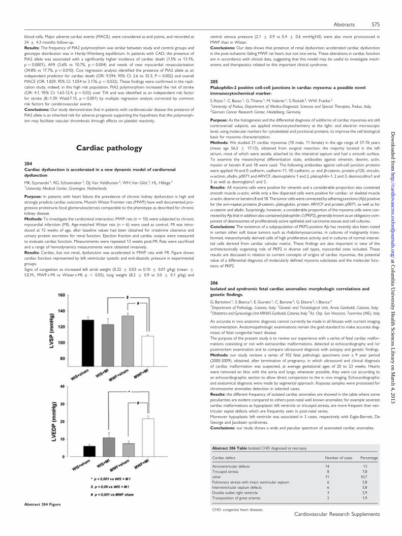
blood cells. Major adverse cardiac events (MACE), were considered as end points, and recorded at24 + 4.3 months follow-up.Results: The frequency of PlA2 polymorphism was similar between study and control groups andgenotype distribution was in Hardy-Weinberg equilibrium. In patients with CAD, the presence ofPlA2 allele was associated with a significantly higher incidence of cardiac death (1.5% vs 13.1%,p ¼ 0.0001), AMI (2.6% vs 10.7%, p ¼ 0.004) and needs of new myocardial revascularization(34.8% vs 17.7%, p ¼ 0.010). Cox regression analysis identified the presence of PlA2 allele as anindependent predictor for cardiac death (OR: 9.594, 95% CI: 2.6 to 35.3, P ¼ 0.002) and overallMACE (OR: 1.829, 95% CI: 1.054 to 3.176, p ¼ 0.032). These findings were confirmed in the repli-cation study, indeed, in this high risk population, PlA2 polymorphism increased the risk of stroke(OR: 4.1, 95% CI: 1.63-12.4, p ¼ 0.02) over TIA and was identified as an independent risk factorfor stroke (B:-1.39; Wald:7.15, p ¼ 0.001) by multiple regression analysis corrected by commonrisk factors for cerebrovascular events.Conclusions: Our study demonstrates that in patients with cardiovascular disease the presence ofPlA2 allele is an inherited risk for adverse prognosis supporting the hypothesis that this polymorph-ism may facilitate vascular thrombosis through effects on platelet reactivity.
Cardiac pathology
204Cardiac dysfunction is accelerated in a new dynamic model of cardiorenaldysfunction
MK. Szymanski 1; RG. Schoemaker 1; DJ. Van Veldhuisen 1; WH. Van Gilst 1; HL. Hillege 1
1University Medical Center, Groningen, Netherlands
Purpose: In patients with heart failure the prevalence of chronic kidney dysfunction is high andstrongly predicts cardiac outcome. Munich Wistar Fromter rats (MWF) have well documented pro-gressive proteinuria focal glomerulosclerosis comparable to the phenotype as described for chronickidney disease.Methods: To investigate the cardiorenal interaction, MWF rats (n ¼ 10) were subjected to chronicmyocardial infarction (MI). Age-matched Wistar rats (n ¼ 6) were used as control. MI was intro-duced at 12 weeks of age, after baseline values had been obtained for creatinine clearance andurinary protein excretion for renal function. Ejection fraction and cardiac output were measuredto evaluate cardiac function. Measurements were repeated 12 weeks post-MI. Rats were sacrificedand a range of hemodynamics measurements were obtained invasively.Results: Cardiac, but not renal, dysfunction was accelerated in MWF rats with MI. Figure showscardiac function represented by left ventricular systolic and end-diastolic pressure in experimentalgroups.Signs of congestion as increased left atrial weight (0.32 + 0.03 vs 0.10 + 0.01 g/kg) (mean +S.E.M., MWF+MI vs Wistar+MI; p , 0.05), lung weight (8.2 + 0.9 vs 3.0 + 0.1 g/kg) and
central venous pressure (2.1 + 0.9 vs 0.4 + 0.6 mmHg;NS) were also more pronounced inMWF than in Wistar.Conclusions: Our data shows that presence of renal dysfunction accelerated cardiac dysfunctionin the post-ischaemic failing MWF rat heart, but not vice-versa. These alterations in cardiac functionare in accordance with clinical data, suggesting that this model may be useful to investigate mech-anisms and therapeutics related to this important clinical syndrome.
205Plakophilin-2 positive cell-cell junctions in cardiac myxoma: a possible novelimmunocytochemical marker.
S. Rizzo 1; C. Basso 1; G. Thiene 1; M. Valente 1; S. Rickelt 2; WW. Franke 2
1University of Padua, Department of Medico-Diagnostic Sciences and Special Therapies, Padua, Italy;2German Cancer Research Center, Heidelberg, Germany
Purpose: As the histogenesis and the differential diagnosis of subforms of cardiac myxomas are stillcontroversial subjects, we applied immunocytochemistry at the light- and electron microscopiclevel, using molecular markers for cytoskeletal and junctional proteins, to improve the cell biologicalbasis for myxoma characterization.Methods: We studied 21 cardiac myxomas (10 male, 11 female) in the age range of 37-76 years(mean age 56,5 + 17,13), obtained from surgical resection, the majority located in the leftatrium, most of which were sessile, attached to the interatrial septum and had a smooth surface.To examine the mesenchymal differentiation state, antibodies against vimentin, desmin, actin,myosin or keratin 8 and 18 were used. The following antibodies against cell-cell junction proteinswere applied: N-and E-cadherin, cadherin-11, VE-cadherin, a- and b-catenin, protein p120, vinculin,a-actinin, afadin, p0071 and ARVCF, desmoplakins 1 and 2, plakophilin-1, 2 and 3; desmocollins1 and3 as well as desmogleins1 and 2.Results: All myxoma cells were positive for vimentin and a considerable proportion also containedsmooth muscle a-actin, while only a few dispersed cells were positive for cardiac- or skeletal musclea-actin, desminor keratins 8 and 18. The tumor cellswere connected byadhering junctions (AJs) positivefor the arm-repeat proteins b-catenin, plakoglobin, protein ARVCF and protein p0071, as well as fora-catenin and afadin. Surprisingly, however, a considerable proportion of the myxoma cells were con-nected by AJs that in addition also contained plakophilin-2 (PKP2), generally known as an obligatory com-ponent of desmosomes of proliferatively active epithelial and carcinoma tissues and cell cultures.Conclusions: The existence of a subpopulation of PKP2-positive AJs has recently also been notedin certain other soft tissue tumors such as rhabdomyosarcomas, in cultures of malignantly trans-formed, mesenchymally derived cells of high proliferative activity and in cultures of normal intersti-tial cells derived from cardiac valvular matrix. These findings are also important in view of thearchitectonically organizing role of PKP2 in diverse cell types, myocardial ones included. Theseresults are discussed in relation to current concepts of origins of cardiac myxomas, the potentialvalue of a differential diagnosis of molecularly defined myxoma subclasses and the molecular func-tions of PKP2.
206Isolated and syndromic fetal cardiac anomalies: morphologic correlations andgenetic findings.
G. Bartoloni 1; S. Bianca 2; E. Giurato 1; C. Barone 2; G. Ettore 3; I. Bianca 4
1Department of Pathology, Catania, Italy; 2Genetic and Teratological Unit, Arnas Garibaldi, Catania, Italy;3Obstetrics and Gynecology Unit ARNAS Garibaldi, Catania, Italy; 4Az. Osp. San Vincenzo, Taormina (ME), Italy
An accurate in vivo anatomic diagnosis cannot currently be made in all fetuses with current imaginginstrumentation. Anatomopathologic examinations remain the gold standard to make accurate diag-noses of fetal congenital heart disease.The purpose of the present study is to review our experience with a series of fetal cardiac malfor-mations coexisting or not with extracardiac malformations, detected at echocardiography and /orpostmortem examination and to compare ultrasound diagnosis with autopsy and genetic findings.Methods: our study reviews a series of 102 fetal pathologic specimens over a 9 year period(2000-2009), obtained, after termination of pregnancy, in which ultrasound and clinical diagnosisof cardiac malformation was suspected, at average gestational ages of 20 to 23 weeks. Heartswere removed en bloc with the aorta and lungs; whenever possible, they were cut according toan echocardiographic section to allow direct comparison to the in vivo imaging. Echocardiographicand anatomical diagnosis were made by segmental approach. Iliopsoas samples were processed forchromosome anomalies detection in selected cases.Results: the different frequency of isolated cardiac anomalies are showed in the table where somepeculiarities are evident compared to others post-natal well known anomalies; for example severestcardiac malformations as hypoplastic left ventricle or tricuspid atresia, are more frequent than ven-tricular septal defects which are frequently seen in post-natal series.Moreover hypoplastic left ventricle was associated in 3 cases, respectively with Eagle-Barrett, DeGeorge and Jacobsen syndromes.Conclusions: our study shows a wide and peculiar spectrum of associated cardiac anomalies.
Abstract 206 Table Isolated CHD diagnosed at necropsy
Cardiac defect Number of cases Percentage
Atrioventricular defects 14 13Tricuspid atresia 8 7,8other 11 10,7Pulmonary atresia with intact ventricular septum 6 5,8Interventricular septum defects 6 5,8Double outlet right ventricle 3 2,9Transposition of great arteries 2 1,9
CHD: congenital heart diseases.Abstract 204 Figure
Abstracts S75
Cardiovascular Research Supplements
at Colum
bia University H
ealth Sciences Library on M
arch 4, 2013http://cardiovascres.oxfordjournals.org/
Dow
nloaded from

207High prevalence of pharmacologically active autoantibodies in heart failure, withdual action on serotoninergic 5-HT4 and muscarinic M2 receptors
P. Eftekhari 1; G. Wallukat 1; A. Bekel 1; F. Heinrich 1; M. Fu 1; M. Briedert 1; JP. Briand 1; JC. Roegel 1
1Pharmacology, Strsbourg, France
Purpose: In adult in congestive heart failure the expression of 5-HT4 receptor is increased 4 folds.Therefore we looked for the presence of anti-5HT4 receptor autoantibodies (AAb) in sera ofpatients with heart failure.Methods: Sera from 333 heart failure patients and 150 controls were screened against secondextra cellular loop (SEC) of 5-HT4, b1, and 2 adrenoceptors, AT1 and M2 muscarinic receptor,using indirect and inhibition ELISA. The ability of AAbs to bind native corresponding receptorswas explored in fluocytometry. AAbs’ pharmacological activity was explored using chronothropyon neanatla rat cardiomyocytes and HL-1 cells. Five different truncated peptides from SEC of5-HT4 were used for eptiope mapping.Results: We could confirme specific immunoreactivity only for 5-HT4 and M2.We found 21 and19% of sera positive for 5-HT4 and M2 receptors respectively with 90% of exact identity for bothreceptors. We found the same result in control population. Peptides derived from SEL of bothreceptors cross-inhibit sera interactions with 5-HT4 and M2 receptors. They recognised all oneand same epitope. IgG fractions patients and controls had negative chronotropic effect. The speci-ficity of the AAbs for 5-HT4 and M2 receptors were verified pharmacologically with ML10375 andatropine specific 5-HT4 antagonist and M2 agonist. Clinical studies revealed that the entire positivecontrol individuals suffered from hypertension, so was the case of heart failure patients positive forthe two receptors. There is a high prevalence of pharmacological active cross- reacting AAbs againstSEC of 5-HT4 and M2 receptors in hypertensinve heart failure patients. These AAbs may play animportant role in on set and pathophysiology of heart heart failure as they are found in hypertesiovepatients without yet clinical manifestation of heart disease.
208TGF-beta1 dipendent acquisition of specific adherens junctions in mitral valveprolapse
S. Rizzo 1; K. Pilichou 1; C. Basso 1; G. Thiene 1
1University of Padua, Department of Medico-Diagnostic Sciences and Special Therapies, Padua, Italy
Purpose: Mitral valve prolapse (MVP) is the leading cause of mitral incompetence in WesternCountries. The pathogenesis of the disease is still unknown, although the dysregulation of TGF-bpathway seems to play a role in familial MVP. TGF-b may induce the expression of stress fibers(a-smooth muscle actin, SMA) in fibroblasts and the acquisition of a myofibroblastic phenotypeas well as a switch of the cadherin pattern at junctional level. There are no data on molecular com-position of cell-cell junctions in valvular interstitial cells (VICs) from MVP. Aim of this study was toevaluate whether TGF-b pathway is activated in isolated MVP and to study the effect of this acti-vation on differentiation of VICs in myofibroblasts and on the molecular pattern of their junctions.Methods: Floppy mitral valves were obtained at surgery from patients (12 cases, 10 M and 2 F,mean age 55.5 + 12.7 years) who underwent valve repair. Age and sex-matched controlsamples were obtained from Homograft Tissue Bank (5 cases, mean age 49 + 9 years). Valve mor-phology was assessed on routinely stained histology sections. Antibodies recognizing active form ofSmad2 (p-Smad2) in VICs were used. The following antibodies against junctional proteins wereapplied: N-cadherin, cadherin-11, VE-cadherin, a- and b-catenin, plakoglobin, protein p120. Ki67was used to assess the proliferation.Results: MVP leaflets exhibited alterations in architecture with fourfold increase in thickness (2,5+ 0,8 vs 0,6 + 0,3 mm, p , 0.0001) and increased cell density (110,9 + 64,6 vs 51,8 + 27,5 cellsper high power field, p ¼ 0.04) compared to the normal valves. A higher density of nuclear stainingfor p-Smad2 (38% vs 12%, p , 0.05) was also observed, indicating an increased activation ofTGF-beta pathway.The VICs in myxomatous leaflets showed a myofibroblast differentiation (expression of a-SMA) andAJs containing high levels of cadherin-11, either exclusively or in colocalization with N-cadherin,unlike VICs from controls that were a-SMA negative and exhibited predominant expression of N-cadherin at AJs level.Conclusions: Our data support the hypothesis that an increased activation of the TGF-b pathwaymay contribute to the pathogenesis of sporadic MVP, either with promotion of myofibroblast differ-entiation of VICs and acquisition of a specific cadherin pattern in AJs, that confer higher resistenceof cell-cell contacts in a stress environment. In its turn, myofibroblast contraction may activatelatent TGF-beta from extracellular matrix.Targeting of myofibroblast AJs by specific anti-cadherin peptides may be used to prevent or retardmitral myxoid degeneration.
209Comparison of myocardial structural and molecular changes in rat models of type 1and type 2 diabetes mellitus
S. Korkmaz ; T. Radovits ; S. Pali ; K. Hirschberg ; S. Zoellner ; S. Loganathan ; M. Karck ; G. SzaboUniversity of Heidelberg, Department of Cardiac Surgery, Heidelberg, Germany
Cardiovascular complications are one of the main causes of mortality in diabetes mellitus (DM).There is obvious evidence demonstrating that DM leads to structural and functional changes atthe level of the myocardium. In this study, we investigated and compared myocardial structuraland molecular changes in rat models of type-1 and type-2 DM.Type-1 DM was induced in Sprague-Dawley rats by single injection of streptozotocin (60mg/kg, i.p.).Control rats receive only vehicle. Experiments were performed 8 wk after the confirmation of dia-betes. For type-2 DM, Zucker-diabetic fatty (ZDF) and ZDF-lean rats were used and experimentswere performed at the age of 30–32 wk. Heart samples were stained with hematoxylin-eosin andimmunohistochemical analysis was performed for nitrotyrosine. TUNEL (terminal deoxynucleotidyltransferase dUTP nick end labeling) was used for detection of DNA-strand breaks. Different geneexpression analysis was performed by quantitative real-time polymerase chain reaction.
Histopathological examination showed the characteristics of diabetic cardiomyopathy. DNA-damage in the myocardium of diabetic group was more higher and the nitro-oxidative stress wassignificantly more pronounced in type-1 compared to type-2 DM. Relative mRNA-expression foralpha-myosin heavy chain (MHC) was decreased whereas beta-MHC and endothelin-1 mRNA-levels were significantly increased in both diabetic compared to corresponding control-groups.Transforming growth factor (TGF)-beta1 mRNA-level was significantly up-regulated only intype-2, whereas c-fos and c-jun only in type-1 DM. Moreover, endothelial NOS, collagen I and IIIwere significantly down-regulated only in type-1 DM. These alterations were significantly more pro-nounced in type-1 DM.Our results show that rat model of type-1 DM is associated with significantly higher structural andmolecular changes compared with the type-2 DM.
210Incomplete Kawasaki disease as cause of unexpected death in 2 infants:immunophenotype characterization of coronary artery aneurysms
G. Bartoloni 1; A. Pucci 2; J. Pantaleo 2; S. Martino 3
1Department of Pathology, Catania, Italy; 2Molecular, Ultrastructural and Surgical Pathology Department,University Hospital, Pisa, Pisa, Italy; 3Cardiology, Immunology and Infectious Disease; Regina MargheritaHospital, Torino, Turin, Italy
Introduction: incomplete or atypical Kawasaki Disease (KD) is more common in children youngerthan one year. In these patients, the diagnosis can be difficult because KD may mimic common child-hood illnesses, but the rate of coronary artery aneurysms is much higher if not treated and conse-quences of missed diagnoses might be serious morbidity or even death.Purpose: aim of this study is the evaluation of the immunohistochemical profile observed in post-mortem coronary lesions of two infants deceased for KD.Methods: we describe two patients (a 3-month-old female infant and a 6-month-old male child)with fatal incomplete KD; in the first case a 2D echocardiography showed giant aneurisms inboth main coronary arteries; in the second one, diagnosis was done only after post-mortem exam-ination. In both cases a complete autopsy was performed and pathologic specimens were processedfor routine histology, histochemical and immunohistochemical stains. The following specific antiserawere tested: anti-endothelial antigen CD31, anti-TNF-alpha, anti-Tissue Factor (TF),anti-Thrombopoietin receptor (r-TPO), anti-HLA-DR (Major Histocompatibility Complex, ClassII antigen) and anti-monocyte/macrophage antigen CD68.Results: histopathology showed similar coronary artery lesions in both cases, mainly consisting ofaneurysms with diffuse macrophage infiltration and prominent neo-angiogenesis in the arterial wall.Coronary artery aneurysms, and particularly giant aneurysms showed strong immunoreactivity forTNF-alpha, TF and r-TPO, mainly localized in endothelial, smooth muscle cells and macrophages. TFexpression was also detected in thrombus. TNF-alfa and HLA-DR expression appeared particularlyhigh in the areas with dense macrophage infiltration. MAcrophage infiltration and HLA-DR immu-noreactivity were also shown in the area of the atrioventricular node.Conclusions : our findings suggest a pathogenetic association of macrophages infiltration, immuno-logical activation (i.e., HLA-DR positivity), tumor necrosis factor and procoagulant factors with cor-onary artery aneurysms and thrombosis in incomplete KD.
211Time course and distribution of TUNEL nuclear positivity of cardiomyocytes in achronic rat model of coronary occlusion with and without reperfusion
G. Pelosi 1; M. Matteucci 2; C. Kusmic 1; N. Vesentini 1; F. Piccolomini 2; F. Viglione 1; MG. Trivella 1;A. L’abbate 2
1Institute of Clinical Physiology of CNR, Pisa, Italy; 2High School Sant’Anna, Pisa, Italy
Occlusion of a coronary artery either permanent or followed by reperfusion is known to cause irre-versible myocellular death mainly by "oncotic" modality. DNA fragmentation by in situ terminaldeoxynucleotidyl transferase (TdT)-mediated dUTP nick end-labeling (TUNEL) has been frequentlydetected at light microscopy (LM) in oncotic cells and believed to be an index of a caspase-mediated"apoptotic" type of cellular death, although typical apoptotic ultrastructural features have neverbeen observed. The contribution of this DNA fragmentation- associated oncotic myocellulardeath to overall irreversible myocardial necrosis after ischemia and ischemia reperfusion is stillquestionable and its time course unknown.Purpose: To assess the time course and distribution of TUNEL nuclear positivity of cardiomyo-cytes within the infarcted and ischemic-reperfused myocardium of rat heart over a 3 day time span.Methods: Male rats underwent permanent (I, n ¼ 12) or transient (I-R, 30min occlusion followedby reperfusion, n ¼ 12) ligation of the left anterior descending coronary artery and hearts explantedat 1h (groups 1h I, 1h I-R), 1 day (groups 1d I, 1d I-R) and 3 days (groups 3d I and 3d I-R).Sham-operated animals served as controls (C, n ¼ 6). Histology (H&E and Masson trichromicstain) and TUNEL assay were performed on 10 consecutive serial slices at two levels distal tothe ligature for LM morphometric assessment of necrosis and nuclear TUNEL analysis.Results: In groups 1h I and 1h I-R neither cellular necrosis or TUNEL positivity could be detectedboth in risk and control areas as well as in C hearts. In group 1d I contraction bands in the peripheryand coagulation necrotic myocells in the central part of infarction showed 30 + 5% and 45 + 3%of TUNEL positive nuclei respectively. In group 1d I-R 50 + 5% of contraction band necrotic cellshad TUNEL positive nuclei. In groups 3d I and 3d I-R most of the still visible nuclei of necrotic myo-cytes were positive (94 + 5 and 95 + 4% respectively). TUNEL positivity was never observed inmyocytes without LM evidence of cellular necrosis.Conclusion: The present findings demonstrate that DNA fragmentation: a) invariably coexists withmyocellular necrosis prior to complete nuclear demise over a 3 day time span, b) it has a compar-able incidence in I and I-R groups, c) it persists at 3 days with an even higher incidence. The possi-bility that TUNEL positivity could identify a specific caspase-dependent subpopulation of irreversiblydamaged cardiomyocytes within the context of ischemic and ischemic-reperfusion necrosis issuggested.
S76 Abstracts
Cardiovascular Research Supplements
at Colum
bia University H
ealth Sciences Library on M
arch 4, 2013http://cardiovascres.oxfordjournals.org/
Dow
nloaded from
212Receptor system of sensory innervation in the rat heart and its regulation inlong-term diabetes
J. Slavikova 1; M. Chottova Dvorakova 1; W. Kummer 2
1Charles University Prague, Faculty of Medicine in Pilsen, Department of Physiology, Pilsen, Czech Republic;2Institute for Anatomy and Cell Biology, Justus-Liebig-University, Giessen, Germany
Substance P (SP) is co-stored with calcitonin gene-related peptide (CGRP) in a special class ofnociceptive neurons that have both afferent and efferent functions. SP/CGRP-containing sensorynerve fibers within the heart derive from cell bodies located in the dorsal root and sensory vagal(jugular-nodose) ganglia. Upon stimulation both SP and CGRP are released and modulate heartrate, force of contraction, and coronary vascular resistance. Their actions are mediated throughspecific G protein-coupled receptors, NK1-NK3 and calcitonin receptor-like receptor (CRLR),respectively. The recently discovered peptides adrenomedullin (ADM) and intermedin (IMD)with significant vasodilator and cardiac protective actions are members of the CGRP peptide super-family and also signal through CRLR. Ligand affinity of CRLR is determined by receptor activity mod-ifying proteins (RAMP1-3). CGRP binds to the complex formed by CRLR/RAMP1, whereas CRLR/RAMP2 and CRLR/RAMP3 serve as receptors for ADM. Here, we investigated these signallingsystems in the control rat heart and compared them to those in diabetic rats.Tissue distribution of CGRP and NK1 receptor in the control rat hearts has been studied by meansof immunofluorescence. The separated rat heart compartments from animals 26 weeks after admin-istration of streptozotocin (STZ; 65 mg/kg i.v) and in the age-matched controls (n ¼ 6 per group)were analyzed by real-time RT-PCR. Relative expression of ADM, IMD, CGRP, CRLR, RAMP1-3,and NK1 receptor mRNA was expressed as a ratio of target gene CT value to CT value of house-keeping gene – beta-actin. The results were considered significantly different when p , 0.05.Strong CGRP-immunoreactivity (IR) was found in varicosities of nerve fibres in close associationwith small and large neuronal cell bodies of intrinsic ganglia of the cardiac plexus and around cor-onary vessels. Also, NK1 receptor-IR was identified on the surface of smooth muscle cells of thecoronary vessels. Neither distribution nor quantity of the immunofluorescence was visiblychanged in the course of diabetes. The results of RT-PCR indicate that relative expressions ofNK1 receptor, ADM, IMD, CGRP, and CRLR were not significantly altered 26 weeks after inductionof diabetes with the exception of RAMP3 which was upregulated 2fold in the right atrium (p ,
0,009) and 4fold in the left ventricle (,0,026).In summary, the shifts observed in long-term diabetes may favour a trend of a pronounced ADMsignalling.Granted by MSM 0021620819 and DFG, 436 TSE 113/51/0-1.
213Elevated g protein-coupled receptor kinases 2 (GRK2) levels in lymphocytesassociate with worse cardiac function in acute ST segment elevation myocardialinfarction (STEMI)
A. Campanile 1; L. Spinelli 1; G. Santulli 1; M. Ciccarelli 1; S. De Gennaro 1; E. Assante Di Panzillo 1;B. Trimarco 1; G. Iaccarino 1
1University of Naples Federico II, Dpt of Clinical Medicine, Cardiovascular & Immunological Science, Naples,Italy
We have shown that heart and lymphocyte GRK2 levels increase in Chronic Heart Failure (CHF)and correlate with a severe cardiac function representing a possible prognostic marker in this con-dition. To analyze GRK2 in acute myocardial dysfunction due to ST segment elevation (STE) myo-cardial infarction (MI) we enrolled 32 patients with acute STE-MI (,24hr) admitted to intensivecare unit. Patients were re-assessed with a second examination after 2 years from beginningevent. Lymphocytes were extracted from 20ml blood sample by means of phicoll purification atadmission and after 24 and 48h. GRK2 expression was assessed by western blotting and correctedby actin expression (Corrected Densitometry Units, CDU) and lymphocyte levels for each patient.MI was confirmed by increased serum CK and assessed by cardiac ultrasound using the Wall MotionScore Index (WMSI). Ventricular Filling was evaluated with E/A ratio, deceleration time (DT) andTissue Doppler Imaging of mitral annulus (E/E′). Ventricular remodelling was evaluated by longtime gain of end-systolic volume (D%-ESV/BSA). Patients with STE-MI presented higher GRK2levels when compared with patients affected by unstable angina (0.75 + 0.16 vs 0.27 + 0.07, p, 0.05; ANOVA). STE-MI patients were grouped based on GRK2 median value on the first dayof analysis: (Hi-GRK2:.0.47; Lo-GRK2:,0.47). Patients with Lo-GRK2 had higher stroke volumevs Hi-GRK2 (43.30 + 2.87 vs 36.53 + 1.25, p , 0.05; ANOVA). Moreover, Hi-GRK2 groupshowed a lower DT value when compared with Lo-GRK2 (143.5 + 10.4 vs 183 + 15.1, p ,
0.05; ANOVA) and GRK2 levels were in correlation with DT and E/E′ ratio (p , 0.05, Pearson)and in linear regression with these two parameters (r2 ¼ 0.1503 and r2 ¼ 1302, respectively, p, 0.05). After 2 years, Hi-GRK2 group still presented worse cardiac function with a lower SVthan Lo-GRK2 (45.54 + 2.27 vs 53.84 + 2.46, p , 0.05; ANOVA). Moreover, admissionGRK2 levels were in correlation (p , 0.05, Pearson) and in linear regression with D%-ESV/BSA(r2 ¼ 0.25, p , 0.05). Admission variables associated with D%-ESV/BSA were: SF, WMSI, SV,AMI index, E/E′ ratio, CPK peak and GRK2 levels. In a multiple regression analysis the best predictivemodel for D%-ESV/BSA was constituted by CPK peak (b ¼ 0.341), AMI index (b ¼ 0.429) andadmission GRK2 levels (b ¼ 0.378). In conclusion GRK2 enhances during MI and it is associatedwith a worse cardiac function. This study suggests that lymphocyte GRK2 levels predict ventricularremodeling after myocardial infarction and poses the ground for investigating the prognostic role ofGRK2 in acute coronary syndromes.
214The role of p53, bax, bcl2, and 8-OHdG in human acute myocardial infarction
R. Akbarzadeh Najar 1; SMH. Ghaderian 1; AS. Tabatabaei Panah 2; H. Vakili 3; A. Rezaei Farimani 4;G. Rezaie 4; A. Beigi Harchegani 4
1Department of Medical Genetics, Shahid Beheshti University of Medical Sciences and Health Services,Tehran, Iran (Islamic Republic of); 2Department of Biology,Basic Sciences Faculty,East Tehran Branch(Ghiamdasht)-Islamic Azad University, Tehran, Iran (Islamic Republic of); 3Department of Cardiology, Shahid
Modarress Hospital, Shahid Beheshti University of Medical Sciences, Tehran, Iran (Islamic Republic of);4Department of Clinical Biochemistry, Shahid Beheshti University of Medical Sciences, Tehran, Iran (IslamicRepublic of)
Purpose: Apoptosis is implicated in unfavorable remodeling of the left ventricle during acute myo-cardial infarction (AMI). DNA damage and p53 play an important role in regulating apoptosis.Expression patterns of apoptotic regulating genes such as p53, bax, and bcl-2 highlighted the neces-sity of inhibition of ventricle remodeling and subsequent injuries. In present study, serum levels ofp53 and 8-hydroxy-2-deoxyguanosine (8-OHdG) as well as p53, bax, and bcl-2 expression afteronset of AMI in Iranian patients were examined.Methods: Serum levels of p53 and 8-OHdG were measured by enzyme-linked immunosorbentassay (ELISA). Presence of p53 protein and mRNA expression of p53, bax, and bcl-2 were analyzedby western blotting and real time reverse transcriptase-polymerase chain reaction (RT-PCR)Methods respectively.Results: Circulating levels of p53 were increased in the patients comparing with the controls (4.45+ 1.71 vs. 0.62 + 0.16 respectively). The plasma levels of 8-OHdG were higher in the patientsthan in the control subjects (1.39 + 1.16 vs. 0.25 + 0.15 respectively). Comparison of thepatient and control subjects demonstrated that in patient subjects, p53 and bax mRNA expressionwere 4.5-fold and 3-fold higher than control samples respectively. In contrast, bcl-2 expression ofthe patients decreased to about 0.75-fold comparing with controls. Molecular weight of p53 proteinwas detected at 53 kDa.Conclusions: p53 and 8-OHdG might be used as markers of apoptosis and DNA damage respect-ively following AMI. Our results revealed that apoptosis occurs in concert with up-regulation of p53and bax versus down-regulation of bcl-2 which may suggests a therapeutic approach in recovery AMI.
215Diabetes mellitus significantly impacts left ventricular myocardial structure andfunction in aortic stenosis before valve replacement
I. Falcao-Pires 1; N. Hamdani 2; C. Gavina 3; J. Van Der Velden 2; HWM. Niessen 2; GJ. Stienen 2;AF. Leite-Moreira 1; WJ. Paulus 2
1University of Porto, Faculty of Medicine, Porto, Portugal; 2VU University Medical Center, Amsterdam,Netherlands; 3Sao Joao Hospital, Porto, Portugal
Purpose: Diabetes mellitus (DM) is an independent risk factor for progression of aortic valve ste-nosis (AS) and significantly impacts longterm outcome after valve replacement. High incidence ofresidual heart failure may account for this prognosis. We aimed to assess the impact of DM on dias-tolic (dys)function of AS patients.Methods: Patients with severe isolated AS (n ¼ 46) and AS plus type-II diabetes patients(AS-DM+, n ¼ 16) with preserved left ventricular (LV) ejection fraction and no clinical or angio-graphic signs of coronary artery disease were studied. Doppler echocardiographic data was usedto compare in vivo LV function. Biopsies were used to assess fibrosis, cardiomyocyte hypertrophy(MyD), advanced glycation endproducts (AGEs) and phosphorylation of myofilamentary proteins.Cardiomyocytes were also isolated and permeabilized to measure active force (Factive), restingforce (Fpassive) and calcium sensitivity (pCa50).Results: In isolated AS, LV deceleration time and end-diastolic pressure were augmented and thelatter significantly correlated with increased fibrosis (r ¼ 0.40, p ¼ 0.04) and MyD (r ¼ 0.60, p ,
0.001). In AS-DM+ patients, diastolic dysfunction was exacerbated in comparison with isolatedAS, as fibrosis, cardiomyocytes hypertrophy and AGEs were further increased and Fpassive signifi-cantly rose. Furthermore, AS-DM+ patients presented with a higher PKA-induced drop of pCa50,which was correlated with higher levels of PKA-induced phosphorylation of Troponin I.Conclusions: We characterized the diastolic disturbances associated with AS chronic pressureoverload alone and in the presence of diabetes. DM exacerbates the existing diastolic dysfunctionof AS patients through extracellular matrix alterations (fibrosis and AGEs), Fpassive raise and PKA-mediated hyperphosphorylation status of troponin I. This study highlights the need for earlier thera-peutic interventions in order to prevent the faster progression of diastolic dysfunction in diabetic ASpatients.
216Effects of diabetes mellitus, pressure overload and their association on myocardialstructure and function
N. Goncalves 1; I. Falcao-Pires 1; C. Moura 1; I. Lamego 1; C. Eloy 2; HWM. Niessen 3; JC. Areias 1;A. Leite-Moreira 1
1University of Porto, Faculty of Medicine, Department of Physiology, Porto, Portugal; 2University of Porto,Faculty of Medicine, Porto, Portugal; 3VU University Medical Center, Amsterdam, Netherlands
Background: Structural and functional changes involved in cardiac injury induced by diabetes mel-litus, pressure overload or both conditions were evaluated.Methods: Pressure-overload was established by supra-renal aortic banding in rats. Six-weeks later,diabetes was induced by streptozotocin (65mg/kg, ip), resulting in 4 groups: SHAM, banded (BA),diabetic (DM) and diabetic-banded (DM-BA). On the 12th week, left ventricular (LV) structureand function were evaluated. LV function was assessed in vivo with pressure-volume cathetersand in vitro by papillary muscles’ performance at baseline and in response to isoprenaline (ISO,10-8-10-5M). Finally, left and RV were weight and collected for morphometric and histologicalmeasurements.Results: Compared to SHAM, we observed a significant increase of type-B natriuretic peptide(BA ¼ 370 + 110%; DM-BA ¼ 580 + 210%), LV mass (BA ¼ 36.8 + 3.6%; DM-BA ¼ 32.1 +3.1%), cardiomyocytes’ diameter (BA ¼ 19.5 + 2.3%; DM ¼ 14.3 + 1.9%; DM-BA ¼ 11.4 +2.0%), fibrosis (BA ¼ 85 + 14%; DM ¼ 145 + 28%; DM-BA ¼ 155 + 14%), AGEs deposition(DM ¼ 141 + 29%; DM-BA ¼ 166 + 46%), contraction (tAT: DM ¼ 13.7 + 2.4%; DM-BA ¼26.3 + 7.1%); a delayed relaxation (tHR: DM ¼ 13.8 + 2.6%; DM-BA ¼ 25.5 + 9.2%) and adecrease of collagen type-I/Type-III ratio (DM ¼ 266.1 + 4.6%; DM-BA ¼ 251.9 + 5.5). InSHAM animals, ISO (10-5M) increased 86.5 + 26.2% active tension, 105.3 + 20.2% dT/dtmaxand 166.8 + 29.9% dT/dtmin. Similar effects were observed in BA and DM animals, while in
Abstracts S77
Cardiovascular Research Supplements
at Colum
bia University H
ealth Sciences Library on M
arch 4, 2013http://cardiovascres.oxfordjournals.org/
Dow
nloaded from
DM-BA these inotropic and lusitropic responses were blunted. Moreover, at a similar resting musclelength, ISO decreased passive tension by 12 + 3% in SHAM and 11 + 3% in BA, indicating anincrease in myocardial distensibility, an effect that was absent in both diabetic groups.Conclusion: Longstanding pressure-overload increased LV mass, while diabetes promoted AGEs andcollagen deposition, which might explain the abolition of ISO-induced increased myocardial distensibil-ity. Association of pressure-overload and diabetes completely blunted the inotropic and lusitropicresponses to ISO, with no additional structural damages than in pressure-overload or diabetes alone.
217Increased atrial expression of connective tissue growth factor predictspost-cardiosurgery atrial fibrillation
T. Bonda 1; M. Dziemidowicz 1; T. Hirnle 2; I. Dmitruk 2; K. Kaminski 3; WJ. Musial 3; MM. Winnicka 1
1Medical University of Bialystok, Department of General and Experimental Pathology, Bialystok, Poland;2Medical University of Bialystok, Department of Cardiosurgery, Bialystok, Poland; 3Medical University ofBialystok, Department of Cardiology, Bialystok, Poland
Structural remodeling of the atria plays an important role in pathogenesis of atrial fibrillation. Fibro-sis is a constant finding in chronically fibrillating atria and forms basis for morphological substrate ofthe arrhythmia. Connective tissue growth factor (CTGF) may be involved modulating the fibrosis aswell as other aspects of atrial remodeling. The aim of the present work was to estimate theexpression of CTGF in human atria in relation to presence of atrial fibrillation.Methods: Right atrial appendages were collected from 76 patients undergoing cardiac surgery.According to history and postoperative follow-up patients were assigned to following groups:chronic AF group (CAF, n ¼ 19), paroxysmal AF group (PAF, n ¼ 8), postoperative AF group(PoAF, n ¼ 13) and control group that was in sinus rhytm (SR, n ¼ 36). Protein expression wasmeasured in tissue homogenates using western blot method.Results: The level of CTGF was significantly higher in patients with postoperative AF in relation toany other group (vs SR p ¼ 0.012; vs CAF p ¼ 0.02; vs PAF p ¼ 0.042). There were no differencesin CTGF expression between SR, CAF and PAF groups. Like in no other group, only in PoAFpatients the level of CTGF significantly correlated positively with the left atrium size (p ¼ 0.004)and negatively with the ejection fraction of the left ventricle (p ¼ 0.02).Conclusion: CTGF may be involved in initiation of atrial fibrillation, but it does not seem to play animportant role in chronic form of this arrhythmia.
Growth factors and cytokines incardiovascular disease
218Mutant mice lacking BAMBI (BMP and Activin Membrane Bound Inhibitor) exhibitlusitropic dysfunction involving altered beta 1 adrenergic signalling
AV. Villar 1; D. Merino 1; M. Ares 2; F. Pilar 3; E. Valdizan 3; MA. Hurle 1; JF. Nistal 2
1University of Cantabria, Santander, Spain; 2University Hospital Marques de Valdecilla, Santander, Spain;3Institute of Biomedicine and Biotechnology of Cantabria (IBBTEC), Santander, Spain
Purpose: It is known that transforming growth factor-b (TGF-b) overexpression promotes altera-tions in b-adrenergic receptor (b-AR) signalling and, as a result, the myocardial responses elicited bycatecholamines are affected. BAMBI (BMP and Activin Membrane Bound Inhibitor) is a membranepseudoreceptor that regulates negatively the cardiomyocyte response to TGF-b. The lack of BAMBIin mutant mice results in increased levels of TGF-b signalling activity. In this study, we assessedwhether the absence of BAMBI would have effects on LV physiology and b-AR signalling.Methods: The study was carried-out in BAMBI-KO and wild type (WT) mice. High fidelity LVpressures were recorded at baseline and during IV perfusion of dobutamine (30, 80 and 200 ng/g/min) with 1.4 Fr microtransducer-tipped catheters (Millar) and appropriate hard- and software(Power Lab and Lab Chart, ADInstruments). Myocardial gene (q-PCR) and protein (WesternBlot) expressions of b1-AR, GRK2, and b-arrestin were determined. b-AR coupling to Gas pro-teins (GTPgS binding) and cAMP production were assessed in LV myocardial membranes.Results: KO mice exhibited an abnormal behaviour of LV relaxation as assessed by the indices ofheart rate-relaxation and contraction-relaxation coupling (heart rate-tau and dP/dt max-tau) duringincreasing dobutamine dosage (p , 0.05 for the slopes of both regression lines vs WT). Theresponse of myocardial membranes from KO mice to b-AR stimulation was significantly depressed,as reflected GTPgammaS specific binding to GalphaS proteins (KO: 104.5 + 3 % vs WT: 126.0 + 5%; p , 0.01) and cAMP production (KO: 118.3 + 2 % vs WT: 145.9 + 5; p , 0.01). Lower myo-cardial gene expression levels of b1-AR (WT: 0.31 + 0.08 vs KO: 0.18 + 0.08, p , 0.05), GRK2(WT: 2.14 + 0.55 vs KO: 1.31 + 0.70; p , 0.05) and b-arrestin (WT: 0.43 + 0.05 vs KO: 0.36 +0.11; p , 0.05) were observed in KO mice. These differences were confirmed at the protein level.Gene expression of b1-adrenergic receptor correlated directly with BAMBI’s (r ¼ 0.81; p , 0.05)and inversely with TGF-b’s (r ¼ 20.7; p , 0.01).Conclusions: Our results indicate that the absence of BAMBI induces alterations in left ventricularrelaxation, via malfunction of the adrenergic pathway. We suggest that a normal TGF-b signalling activityis necessary for a physiologic cardiac b-adrenergic transmission. The absence of BAMBI reveals aninverse relationship between TGF-b signalling activity and b-adrenergic function that could be involvedin the adrenergic dysfunction of myocardial pathologies with increased TGF-b activity.
219Increased uridine adenosine tetraphosphate concentrations cause increasedproliferation rates of IMT and correlate with blood pressure
V. Vera 1; M. Toelle 1; M. Van Der Giet 1; W. Zidek 1; J. Jankowski 1
1Charite - Campus Benjamin Franklin, Berlin, Germany
Background: Uridine adenosine tetraphosphate (Up4A) was been recently characterized as apotent vasoconstrictor 1. Up4A occurs in plasma from healthy subjects at concentrations suffi-cient
to cause strong vasoconstrictive effects. In this study, Up4A concentrations in plasma from juvenilehypertensives and normotensives were determined.Methods: Up4A was purified to homogeneity by preparative reverse phase high performanceliquid-chromatography (HPLC), affinity HPLC and analytic reverse phase HPLC from depro-teinizedplasma of juvenile hypertensives and normotensives.Results and Discussion: Mean total plasma Up4A concentration was significantly increased injuvenile hypertensives compared to juvenile normotensives (33.0 +/2 25.4 vs. 3.7 +/2 0.9nmol/L; mean +/2 SEM, n ¼ 40 and 38; respectively, p , 0.005). Accordingly, Up4A showed asignificant association with juvenile hypertension (OR for ln(Up4A): 1.82; 95% CI 1.12, 2.95).Plasma Up4A concentrations correlated with left ventricular mass (Kendall-t correlation coefficient0.220, n ¼ 40; p , 0.05) and intima media wall thickness (Kendall-t correlation coefficient 0.296,n ¼ 40; p , 0.05) in the hypertensives. Since the increased intima media thickness may berelated to proliferative effects of Up4A, we studied the effects of Up4A on human vascularsmooth muscle cell (VSMC) proliferation. The maximum proliferative effect of Up4A was 83.7+/2 18.0% above control (p , 0.01).Conclusion: Circulating levels of Up4A are strongly associated with juvenile hypertension. Theendothelium-derived vasoconstrictor Up4A may contribute to the early development of primaryhypertension and is moreover an important risk factor of juvenile hypertension.
220Prognostic value of early interleukin-10 elevation in patients with acute coronarysyndrome and left bundle branch block
A. Astvatsatryan 1; M. Senan 1
1European Regional Educational Academy, Faculty of Medecine, Yerevan, Armenia
Aim: We analysed the prognostic value of early elevation of interleukin-10 (IL-10) in patientsadmitted to the CCU with acute coronary syndrome (ACS) and left bundle branch block(LBBB).Methods: 84 consecutive patients (51 male and 33 female, aged 51.9 + 11.1) without history ofdiabetes mellitus, inflammatory or neoplastic disease were involved in our study. All patientswere undergone by standard examination and IL-10 on admission. IL-10 was measured by com-mercially available ELISA test. Patients were divided in two groups: G1 (n ¼ 40) consisted ofpatients with early IL-10 (. 0.38 pg/ml), G2 (n ¼ 44) consisted of patients without IL-10elevation. The endpoints were cardiac mortality and non-fatal acute myocardial infarction(AMI) at 12 months.Results: In G1 tertile distribution showed that elevation of level IL-10 was related to cardiac mor-tality (10.2%, 7.8% and 31.0%, p , 0.03) and it was an independent predictor of cardiac mortality(HR ¼ 2.3, 95%, CI 1.3-3.8; p , 0.005), in G2 level of IL-10 was independent predictor of non-fatalAMI (HR ¼ 1.3, 95%, CI 1.1- 2.2; p ¼ 0,01).Conclusion: Early elevation of level IL-10 is independent predictor of cardiac mortality at 12month in patients with ACS and LBBB. In the absence of AMI IL-10 elevation on admission predictsnon-fatal AMI.
221Does remote ischaemia modulate cytokines and growth factors? A clinical study inpatients undergoing coronary artery bypass graft surgery
P. Karuppasamy 1; S. Chaubey 1; T. Dew 1; R. Sherwood 1; J. Desai 1; L. John 1; M. Marber 2; G. Kunst 1
1King’s College Hospital, London, United Kingdom; 2St Thomas’ Hospital, London, United Kingdom
Purpose: Remote ischaemic preconditioning is a strategy of protecting the heart againstischaemia-reperfusion injury. Little is known about its underlying mechanism of translating limbischaemia into transferable signals to the heart. We investigated whether remote ischaemia per-formed as left arm ischaemia in patients undergoing coronary artery bypass graft (CABG)surgery modulates cytokines and growth factors.Methods: After ethical approval, 54 adult patients undergoing CABG surgery were prospectivelyrandomized into a single blind study. 27 patients received three five-minute cycles of left arm ischae-mia after anaesthetic induction and before the start of surgery, whereas 27 patients had a bloodpressure cuff identically placed but without inflation. Within one minute of each cuff deflationand at 24 hrs and 48 hrs after surgery, and at identical time points in the control group, bloodsamples were taken from the superior vena cava. We analyzed a broad panel of cytokines andgrowth factors including IGF-1, Myostatin, VEGF, TNFa, IL-1A, IL-1B, IL-2, IL-4, IL-6, IL-8, IL-10.In addition, the marker for myocardial ischaemia TnI was measured. Multilevel models werefitted to the data for statistical analysis.Results: Despite significant changes over time in cytokine markers, there was no difference in con-centrations between the remote ischaemia and the control groups. Interestingly, cardiac TnI alsochanged significantly postoperatively, but there was no difference between groups.Conclusions: Our results demonstrate that the assayed specific cytokines and growth factorsshow indiscriminable profiles in patients under anaesthesia with and without remote ischaemia;they are thus unlikely to act as humoral factors released during remote ischaemia. There is noadditional benefit on cardiac TnI as a marker of cardiac ischaemic damage from remote ischaemiaover anaesthesia alone.
222A novel CaMKII/ERK interaction in the heart sustains cardiac hypertrophy inspontaneously hypertensive rats
E. Cipolletta 1; G. Santulli 1; A. Attanasio 1; C. Del Giudice 1; P. Campiglia 2; M. Illario 3; G. Iaccarino 1
1University of Naples Federico II, Dpt of Clinical Medicine, Cardiovascular & Immunological Science, Naples,Italy; 2University of Salerno, Salerno, Italy; 3University of Naples Federico II, Dpt of Patholgy and Cellular andMolecular biology, Naples, Italy
Cardiac hypertrophy is an adaptive response to sustain cardiac output in response to cardiovascularconditions. The molecular mechanism underlying it involves, among others, abnormal intracellular
S78 Abstracts
Cardiovascular Research Supplements
at Colum
bia University H
ealth Sciences Library on M
arch 4, 2013http://cardiovascres.oxfordjournals.org/
Dow
nloaded from
Ca++ dynamics. Impaired Ca++/Calmodulin protein kinase II (CaMKII) activation mediatesCa++ signaling in the hypertrophic transduction pathways. Nevertheless, the mechanism bywhich CaMKII integrates with other pathways to develop cardiac hypertrophy is incompletelyunderstood. We previously showed that phenylephrine (PE) induces cell proliferation throughCaMKII and ERK complex formation, which promotes phosphorylation and nuclear localizationof both proteins. We hypothesize that CaMKII through its association with ERK, promotesnuclear localization in hypertrophic cardiomyocytes. In H9C2 cardiomyoblasts, PE induces bothERK and CaMKII activation, which can be reduced by the non selective CaMKs inhibitor KN93,by the selective CaMKII inhibitor AntCaNtide, and by the MEK/ERK inhibitor UO126. Coimmuno-precipitating studies demonstrated that PE induces CaMKII/ERK interaction, which was preventedby pretreatment with KN93, ANtCaNTide or UO126. Next, we tracked the intracellular localiz-ation of ERK and CaMKII; PE induces a time-dependent accumulation of both ERK and CaMKIIinto the nucleus. AnTCaNtide and UO126 pretreatment preventes both ERK and CaMKIInuclear localization, supporting the hypothesis that ERK activation is required for CaMKII nuclearlocalization. In order to determine the role of CaMKII in vivo cardiac hypertrophy, SpontaneouslyHypertensive Rats (SHR) were subjected to intracardiac injection of AnTCaNtide (ANTS, 50 mg/kg,n ¼ 8). Sham groups of SHR (SS, n ¼ 8) and normotensive rats (WKY, n ¼ 7) were subjected tointracardiac injection of saline. After 3 from the treatment, we performed an ultrasound (VeVo-Visualsonic) cardiac evaluation and an invasive (Millar) arterial blood pressure (BP) measurementin the rats. Mean arterial pressure and body weight were similar between SS and ANTS, but weobserved in ANTS a significant reduction of both cardiac size (ANTS vs SS: Heart weight/bodyratio 3.961 + 0.02586 vs 4.177 + 0.02583, p , 0.05) and thickness of interventricular septum(ANTS vs SS: 1.821 + 0.01063 vs 1.918 + 0.01797, p , 0.05). In ANTS hearts, by westernblot, we observed a significant reduction of CaMKII and ERK phosphorylation levels. In conclusion,a crosstalk between CaMKII and ERK sustain the activation of both kinases both in vitro and in vivo.This CaMKII-ERK interaction offers a novel therapeutic approach to limit pathological cardiachypertrophy.
223Osteopontin plasma level is reflected severity of both symptomatic heart failure andcardiac remodelling
A. Berezin 1; EYU. Koretskaya 1
1State Medical University, Zaporozhye, Ukraine
Aim: To investigate interrelations between plasma level of osteopontin with both neurohumoral/ proinflammatory activation and severity of cardiac remodeling in patients with mild-to-severeof HF.Methods: Peripheral blood was collected from 64 patients with mild-to-severe HF due toischemic heart disease and 30 healthy control subjects. Left ventricular ejection fraction (LVEF)was calculated using a modified Simpson’s rule. The 64 patients were classified into 3 classesaccording to the New York Heart Association (NYHA) functional classification. Osteopontin,tumor necrosis factor-alpha and NT-pro-brain natriuretic peptide in plasma were examined byELISA.Results: Plasma levels of osteopontin, tumor necrosis factor-alpha and NT-pro-brain natriureticpeptide were higher in patients with HF than in controls (p ¼ 0.012; p , 0.05 and p , 0.001).Furthermore, both the plasma osteopontin levels and NT-pro-brain natriuretic peptide increasedin proportion to the severity of the NYHA functional class. Tumor necrosis factor-alpha in plasmais correlated well with NYHA functional class in patients with moderate-to-severe HF only(r ¼ 20.380, p , 0.01). It this patients population tumor necrosis factor-alpha level was corre-lated with body mass index (r ¼ 20.328, p ¼ 0.014) and LVEF (r ¼ 20.388, p ¼ 0.0036). Theosteopontin plasma level was significantly correlated with LVEF (r ¼ 20.406, p ¼ 0.0022),mitral regurgitation (r ¼ 0.40, p , 0.05), area of both left ventricle and left atrial (r ¼ 0.402, p, 0.01 and r ¼ 0.406, p , 0.001), log plasma NT-pro-brain natriuretic peptide level (r ¼ 0.45,p ¼ 0.002), and tumor necrosis factor-alpha level (r ¼ 0.301, p ¼ 0.012). However, after adjust-ment to age, sex, body mass index and treatment regimes osteopontin plasma level had been pre-served significant interrelationships with both severity of symptomatic HF and cardiacremodelling.In conclusions, osteopontin plasma level reflects the severity of symptomatic HF and correlates wellwith both LVEF and NT-pro-brain natriuretic peptide concentration. Osteopontin could be con-sidered as target in the assessment of severity both HF and cardiac remodelling.
224Examination of the mechanisms underlying changes in monocyte chemoattractantprotein (MCP1) levels in endothelial cells exposed to cigarette smoke extracts.
EL. Bishop 1; IM. Fearon 1
1British American Tobacco, Southampton, United Kingdom
Objective: To investigate the effects of cigarette smoke extracts on endothelial cell expression ofthe monocyte chemoattractant protein, MCP1, an important protein in atherosclerotic lesionformation.Methods: Human umbilical vein endothelial cells (HUVECs) were exposed to concentrations ofbetween 6 mg/ml and 96 mg/ml of cigarette smoke total particulate matter (TPM) for 24 hours.In experiments performed with the antioxidants n-acetylcysteine (NAC; 5 mM) or ascorbic acid(200 mM), the antioxidants were added to HUVECs 5 hours prior to, and also during, TPMexposure. mRNA levels were examined by PCR microarray. Media MCP-1 protein levels wereexamined by electrochemiluminescence detection and localisation of this protein was studied byimmunocytochemistry. Responses to TPM were examined in HUVECs obtained from three differ-ent donors and in triplicate in each donor.Results: In HUVECs treated with a non-cytotoxic concentration of TPM (88 mg/ml) we observed adecrease in MCP1 mRNA levels, which was consistently seen in all three donors examined. Simi-larly, in MSD studies the amount of secreted MCP1 protein found in culture media was reducedin response to TPM treatment in a concentration dependent manner. In contrast to the decreasein MCP1 protein secreted into the culture media, when MCP1 protein levels were examined by
immunocytochemistry we observed an increase in expression at the endothelial cell surface inresponse to TPM. The decrease in MCP1 protein levels, assessed by MSD analysis, was abrogatedin cells pre-treated with either NAC or ascorbic acid.Conclusions: The reduction in MCP1 mRNA levels and secreted protein, along with the observedincrease in immunocytochemical staining, suggests a complex response to TPM. This potentiallyinvolves enhanced retention of the MCP1 protein at the cell surface in response to exposure toTPM. Our antioxidant data further suggest a role for oxidative stress in mediating the MCP1protein response to TPM.
225TGFbeta receptor activation enhances cardiac apoptosis via SMAD activation andconcomitant NO release
J. Heger 1; B. Warga 1; Y. Abdallah 1; B. Meyering 1; KD. Schlueter 1; HM. Piper 2; G. Euler 1
1Justus-Liebig University, Giessen, Germany; 2Heinrich-Heine University, Dusseldorf, Germany
TGFb expression is induced in myocardium during transition from compensated hypertrophy toheart failure. In cardiomyocytes, stimulation with TGFb results in restricted cardiac function andenhanced apoptosis, two fundamental processes in the development of heart failure. BesidesTGFb, nitric oxide (NO) induces apoptosis and influences cardiac function. Since there arereports that TGFb can enhance NO-release, we wanted to know whether NO is causally involvedin TGFb induced apoptosis.In isolated ventricular cardiomyocytes incubation with TGFb1 increased NO-release up to 148.2+ 25.5 % (n ¼ 7; p , 0.05). This NO release was inhibited by the NO-synthase (NOS) inhibitorETU. Real-time RT-PCR experiments revealed that RNA expression of NOS isoforms was notincreased under TGFb; instead, phosphorylation of endothelial NOS was enhanced. Beyondthat, apoptosis induction under TGFb was blocked upon NOS inhibition, but not with inducibleNOS (1400W) or neuronal NOS (TFA) specific inhibitor. Furthermore, TGFb induced apoptosisand NO formation depends on TGFb receptor activation, because ALK5 receptor blockerSB431543 significantly decreased apoptosis and NO release. Effects of TGFb on apoptosis aresequential mediated by soluble guanylate cyclase (sGC) and protein kinase G since inhibition ofsGC by ODQ and PKG by KT5823 abolished these effects. Classical mediators of TGFb signallingare SMADs. To investigate, whether SMAD transcription factors are downstream of NO/sGC/PKG pathway, we analyzed SMAD activation. Remarkably, SMAD2 phosphorylation could notbe blocked with NOS inhibitor (ETU) nor could SMAD binding activity be reduced by sGCblocker. However, SMAD decoys and SMAD4 antisense oligonucleotides inhibited TGFbinduced apoptosis.Conclusion: In cardiomyocytes TGFb induced apoptosis is mediated via TGFb receptor activationthat concomitantly activates SMAD transcription factors and eNOS/NO/sGC/PGK pathway. Thisdiscloses a new perspective of cardiac NO-release and NO as a possible contributor to heartfailure progression under TGFb.
226Raised plasma concentrations of insulin-like growth factor-1 and erythropoietin inpatients with ischemic heart disease and metabolic syndrome
A. Lavorgna 1; S. Cecchetti 1; T. Rio 1; G. Coluzzi 1; C. Carrozza 2; E. Conti 3; F. Crea 1; F. Andreotti 1
1Catholic University of the Sacred Heart, Department of Cardiovascular Medicine, Rome, Italy; 2CatholicUniversity of the Sacred Heart, Institute of Clinical and Chemical Biochemistry, Rome, Italy; 32nd Faculty LaSapienza, Insitute of Cardiology, Rome, Italy
Purpose: Experimental and clinical studies indicate insulin-sensitising, vasodilator, antiplatelet, anti-apoptotic, anti-inflammatory and constitutive nitric oxide (NO) enhancing effects of insulin-likegrowth factor-1 (IGF-1) and erythropoietin (EPO). Besides promoting erythropoiesis, EPO protectsagainst myocardial ischemia/reperfusion injury, favours progenitor cell mobilization and angiogen-esis. The metabolic syndrome (MS) is considered an insulin-resistant state. Our aim was tomeasure circulating levels of IGF-1 and EPO in patients with ischemic heart disease (IHD) withand without MS.Methods: Ninety-two consecutive patients (acute myocardial infarction in 29, unstable angina in 33,stable angina in 30) were grouped according to presence or absence of MS, using NCEP-ATP III andrecent IDF/AHA/NHLBI criteria. Plasma EPO, free IGF-1, and observed (O) over predicted (P) EPOratio (based on haematocrit and inversely related to EPO sensitivity) were measured 4 + 2 daysafter admission.Results: Patients with or without MS did not differ significantly in age, gender distribution, left ven-tricular ejection fraction, coronary disease, clinical diagnosis, blood haemoglobin and renal function(all p . 0.2). Free IGF-1, EPO and O/P ratio were significantly higher in patients with MS comparedto those without, both by NCEP (0.82 + 0.61 vs 0.47 + 0.53 ng/ml, p ¼ 0.001; 7.7 + 3.6 vs 5.1 +2.1 mIU/ml, p ¼ 0.033; and 0.76 + 0.20 vs 0.58 + 0.22, p ¼ 0.025, respectively) and IDF/AHA/NHLBI criteria (0.91 + 0.53 vs 0.42 + 0.31 ng/ml, p ¼ 0.001; 7.9 + 3.1 vs 4.7 + 1.9 mIU/ml,p ¼ 0.02; and 0.82 + 0.25 vs 0.52 + 0.18, p ¼ 0.02, respectively). IGF-1 levels increased withthe number of MS components present (p ¼ 0.02, r ¼ 0.27), particularly when the more recentIDF/AHA/NHLBI definition were applied (p ¼ 0.002, r ¼ 0.35). Patients with ,4 criteria hadlower IGF-1 levels compared with those with 5 criteria, both by NCEP (0.74 + 0.47 ng/ml vs1.39 + 0.88 ng/ml, p ¼ 0.04) and IDF/AHA/NHLBI criteria (0.77 + 0.44 vs 1.34 + 0.63 ng/ml,p ¼ 0.02). Waist circumference was the only MS component that correlated positively withIGF-1, EPO and O/P ratio (p ¼ 0.001, r ¼ 0.37; p ¼ 0.027, r ¼ 0.40; and p ¼ 0.002, r ¼ 0.55respectively).Conclusions: IHD patients with MS, compared to those without MS, exhibit increased plasma freeIGF-1 and EPO concentrations, and EPO-resistance expressed as increased O/P ratio. Thesechanges may represent compensatory or adaptive metabolic processes within the context of mul-tiple resistance to these hormones. In virtue of the cardiovasculoprotective effects of IGF-1 andEPO, resistance to their effects might play a crucial part in linking the MS with adverse outcomes.
Abstracts S79
Cardiovascular Research Supplements
at Colum
bia University H
ealth Sciences Library on M
arch 4, 2013http://cardiovascres.oxfordjournals.org/
Dow
nloaded from
227Relationship of heart rate variability to transforming growth factor-beta1 forpatients with arterial hypertension and diabetes mellitus
A. Berezin 1; A. Glavatskiy 1
1State Medical University, Zaporozhye, Ukraine
Background: Autonomic nervous dysfunction is considered as valid marker of high cardiovascularrisk. However, the interrelation between biological markers, such as TGF beta1, and severity ofautonomic nervous dysfunction has not been still defined.The aim: To investigate of the relationship of indexes of heart rate variability (HRV) to the serumconcentration of transforming growth factor-beta1 (TGF-beta1) for patients with arterial hyperten-sion associated with 2nd type diabetes mellitus (DM) with documented left ventricular hypertrophy(LVH).Methods: We examined 52 patients with arterial hypertension associated with DM. All patientswere carried out of 24-hours ambulatory monitoring of ECG with assessment of time and frequentdomains of HRV for 24 hours by the instrumentality of standards of European Society of Cardiologyand North American Society for Pacing and Electrophysiology (1996). The serum concentration ofTGF-beta1 was determined from each subject with the use of solid-phase TGF-betab1–specificsandwich ELISA. The LVH was assessed by the instrumentality of standard cardiac sonographicmethod. Blood sampling, 24-hours ambulatory monitoring of ECG and Echo were performed atthe baseline after writing informed consent.Results: We determined that LVH diagnosed from patients in 88.5% cases. For patients with LVHthe serum concentration of TGF-beta1 had significant negative relationship to SD (r ¼ 20.54),SDNN (r ¼ 20.55), SDANN (r ¼ 20.49), RMSSD (r ¼ 20.43), pNN50% (r ¼ 20.40), LF(r ¼ 20.51), TP (r ¼ 20.53) (p , 0.05 for all cases). About patients without LVH (11.5% cases)the serum concentration of TGF-beta1 had no significant negative relationship to RMSSD(r ¼ 20.41) and pNN50% (r ¼ 20.39) (p . 0.05 for both cases).Conclusion: For patients of arterial hypertension associated with DM against a background LVHincrease of the serum concentration of TGF-beta1 is accompanied significant decrease of row ofindexes of HRV characterizing both sympathetic and parasympathetic autonomous nervoussystems.
Atherosclerosis and lipids
228The relationship between coronary calcification and the metabolic markers ofosteopontin, fetuin-A, and visfatin
O. Uz 1; E. Kardesoglu 1; O. Yiginer 1; S. Bas 2; OM. Ipcioglu 1; N. Ozmen 1; M. Aparci 1; BY. Cingozbay 1
1GATA Haydarpasa Training Hospital, Istanbul, Turkey; 2Gaziosmanpasa Private Hospital, Istanbul, Turkey
Objectives: We investigated whether coronary calcification detected by multislice computed tom-ography (MSCT) was correlated with plasma osteopontin, serum fetuin-A, and visfatin levels.Methods: The study included 64 consecutive patients (51 males, 13 females; mean age 49.5 + 10.9years; range 33 to 78 years) who underwent MSCT for suspected coronary artery disease. Coron-ary artery calcification (CAC) scores of the patients were calculated using the Agatston scoringmethod. Plasma osteopontin, serum fetuin-A, and visfatin levels were measured from fastingblood samples and correlations were sought with calcium scores.Results: Coronary calcification was detected in 32 patients (50%). The mean CAC score was 146.5+ 333.7 Agatston units (AU), indicating an intermediate risk for coronary artery disease. In 10patients (15.6%), the CAC score exceeded 400 AU. The mean fetuin-A, visfatin, and osteopontinlevels were 25.6 + 6.4 ng/ml, 19.7 + 47.2 ng/ml, and 20.4 + 16.1 ng/ml, respectively. Serum vis-fatin (r ¼ 0.15, p ¼ 0.37) and fetuin-A (r ¼ 0.17, p ¼ 0.22) were not correlated with the CACscore, whereas plasma osteopontin level showed a moderate correlation with the CAC score(r ¼ 0.35; p ¼ 0.008). In ROC analysis, the area under the curve for identification of CAC wasgreatest for osteopontin (0.741; p ¼ 0.004), followed by fetuin-A (0.574; p ¼ 0.31), and visfatin(0.580; p ¼ 0.27). The cut-off value was 18.45 ng/ml for osteopontin, with a sensitivity of 72%and specificity of 73%.Conclusion: Our results suggest that there might be an association between CAC and plasmaosteopontin levels. Research should continue to find out a metabolic parameter that will stronglyindicate coronary calcification.
229Plasma aldosterone predicts mortality in patients with coronary artery disease
F. Ivanes 1; MAK. Hillaert 2; S. Susen 1; F. Mouquet 1; PA. Doevendans 2; B. Jude 1; G. Montalescot 3;E. Van Belle 1
1Hospital Regional University of Lille, Lille, France; 2University Medical Center Utrecht, Utrecht, Netherlands;3AP-HP - Hospital Pitie-Salpetriere, Paris, France
Purpose: Recent studies have demonstrated that aldosterone levels in patients with heart failure oracute MI can predict long-term mortality. The predictive value of aldosterone in patients with cor-onary artery disease (CAD) outside these settings is unknown. This study evaluated the relationshipbetween the plasma level of aldosterone and the risk of death in CAD patients with a preserved leftventricle (LV) function and no acute MI.Methods and Results: In 807 consecutive patients referred for elective coronary angioplasty,baseline aldosterone (median ¼ 25pg/mL) was measured, as well as BNP (median ¼ 35pg/mL),hsCRP (median ¼ 4.17mg/L) and LVEF (mean ¼ 58%). During a median follow-up of 14.9months, the primary endpoint of cardiovascular (CV) death occurred in 41 and the secondary end-point of total mortality in 52 patients. Plasma aldosterone was related to BMI, hypertension andNYHA functional class and inversely related to age, creatinine clearance and use of beta-blockers.Univariate analysis showed that aldosterone had a positive relationship with CV mortality (p ¼0.0007) and total mortality (p ¼ 0.005). In multivariable Cox models, 5 variables were significantlyassociated with CV mortality: aldosterone (p ¼ 0.0009), BNP (p ¼ 0.0009), diabetes (p ¼ 0.02),
recent (2-7 days) acute coronary syndrome (ACS) (p ¼ 0.03), and LVEF (p ¼ 0.04). Similarly, 5 vari-ables were significantly associated with total mortality: aldosterone (p ¼ 0.001), BNP (p ¼ 0.009),age (p ¼ 0.01), hsCRP (p ¼ 0.03), and gender (p ¼ 0.03).Conclusion: Our results demonstrate that, in patients with CAD without heart failure or acute MI,aldosterone is a strong and independent predictor of total and cardiovascular mortality.
Abstract 229 Table Hazard ratio’s of CV mortality
Independent variable Hazard ratio 95% CI p-value
Aldosterone (1 Log increase) 4.22 1.81-9.83 0.0009BNP (1 Log increase) 4.99 1.93-12.95 0.0009Diabetes mellitus 2.40 1.11-5.20 0.02Recent (2-7 days) ACS 2.93 1.09-7.91 0.03LVEF (10% decrease) 1.30 1.01-1.68 0.04
Other data in the model are age, gender, hypertension, smoking, BMI, family history of CAD, NYHA,severity of CAD, LVEF, total cholesterol, creatinine clearance, hsCRP and treatment with beta-blockers,statins and ACE inhibitors.
230Markers of atherosclerosis as prognostic factors of cardiovascular disease in patientwith cardiometabolic syndrome
M. Leon 1; D. Ilisei 1; F. Mitu 1
1Grigore T. Popa University of Medicine and Pharmacy, Iasi, Romania
Purpose: Heart disease is the primary cause of death worldwide. The cardio-ankle vascular index(CAVI) has been proposed as a new noninvasive marker of arterial stiffness independent of bloodpressure.Methods: Carotid intima-media thickness (CIMT), as measured by B-mode ultrasound, is a surro-gate marker for atherosclerosis and can be used to detect an accelerated disease process and sub-clinical disease. We investigated the association of the CAVI with coronary atherosclerosis at a totalof 150 subjects: CAVI, CIMT measurement and echocardiography were performed.Results: The CAVI was significantly correlated with age, systolic blood pressure and diastolic bloodpressure. Multivariate analysis revealed an independent correlation between the parameters and allparameters were independently correlated with each other in subjects,70 years. Cardio-ankle vas-cular index was positively correlated with visceral adipose tissue area and negatively correlated withweight. We also found the positive association of the number of metabolic syndrome componentswith CAVI in both sexes. CAVI values were significantly higher in metabolic syndrome than in non-metabolic syndrome patients, whereas there was no significant difference in body mass index, totalcholesterol, and low-density lipoprotein-cholesterol. The individual components of the metabolicsyndrome, except for a low HDL-cholesterol level, were associated with increased CAVI. Hyperch-olesterolemia was also associated with increase in CAVI. A low HDL-cholesterol level was associ-ated with an increased CAVI.Conclusion: Presence of significant correlation between carotid intima-media thickness andcardio-ankle vascular index in patients with cardiometabolic syndrome suggests that this correlationis stronger with increasing extent of atherosclerosis.
231Immunolocalisation of glycophorin A in cardiac allograft vasculopathy: implicationfor plaque progression
C.Castellani 1; A.Angelini 1; O.De Boer 2; C.Van Der Loos 2; G.Gerosa 3; G.Thiene 1; A.Van Der Wal2
1University of Padua, Department of Medico-Diagnostic Sciences and Special Therapies, Padua, Italy;2Academic Medical Center, Amsterdam, Netherlands; 3University of Padua, Department of Cardiac Thoracicand Vascular Sciences, Padua, Italy
The progression of atherosclerosis is a complex phenomenon. Recent studies have demonstratedthat intraplaque hemorrhages may cause plaque instability but are also involved in the growth ofplaque. Aim of our study was to investigate whether and to what extent such hemorrhages alsoplay a role in the progression of transplant arteriosclerotic lesions (allograft vasculopathy).Methods: a total of 70 coronary plaques were obtained from 12 patients who had been trans-planted because of chronic ischemic heart disease (6 pts) and dilated cardiomyopathy (6 pts) anddied because allograft vasculopathy. Of each patient we had both the native heart and heartafter transplantation at time of death. Of each plaque immunohistochemistry for CD31, von Will-ebrand factor (vWF) for endothelial cells, CD45 for leukocytic infiltrations, CD68 for macrophagesand glycophorin A (GFA, reactive with component of erythrocyte membranes) was applied toidentify inflammation, plaque vessels and plaque hemorrhages. A semi quantitative analysis wasperformed.Results: In our series the incidence of GFA positive hemorrhages was greater in plaques in trans-plant hearts with positivity in 20 of 37 lesions (54%) positive compared to native atheroscleroticplaques with positivity in 4 out of 23 positive lesions (8.6%). GFA staining was localized at the sub-endotelial level in most of the cases. Similar to native plaques, post transplants plaques showedareas with neovascularization and with diffuse perivascular staining for von Willebrand factor sug-gestive of leakage. Inflammatory cells were present in the most of the plaques after transplantation(89 percent) and in some of the atherosclerotic plaque of native hearts. In post heart transplantplaques no difference was identified between superficial or deep inflammation (27/37 versus 25/37 lesions respectively).Conclusions: Our results suggest for the first time that intraplaque hemorrhage may stimulate theprogression and remodeling of plaque in the setting of graft vasculopathy
S80 Abstracts
Cardiovascular Research Supplements
at Colum
bia University H
ealth Sciences Library on M
arch 4, 2013http://cardiovascres.oxfordjournals.org/
Dow
nloaded from
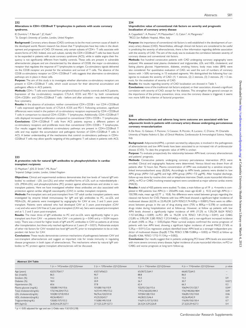
232Alterations in CD41CD28null T lymphocytes in patients with acute coronarysyndrome.
IE. Dumitriu 1; P. Baruah 1; JC. Kaski 1
1St. George’s University of London, London, United Kingdom
Background: Coronary artery disease (CAD) continues to be the most common cause of death inthe developed world. Recent research has shown that T lymphocytes have key roles in the devel-opment and progression of CAD. Of interest, only certain subsets of CD4+ T cells associate withsevere forms of CAD. Indeed, one such subset, namely the CD4+CD28null T cells has been foundto be expanded in patients with acute coronary syndrome (ACS), while in stable angina their fre-quency is not significantly different from healthy controls. These cells are present in vulnerableatherosclerotic plaques and are characterized by the absence of CD28, the major co-stimulatoryreceptor that regulates the response of T lymphocytes to antigen. Co-stimulatory signals deliveredvia the CD28 receptor are pivotal for the activation and survival of T cells. The absence of theCD28 co-stimulatory receptor on CD4+CD28null T cells suggests that alternative co-stimulatorypathways are in place in these cells.Purpose: The aim of this study is to investigate whether alternative co-stimulatory receptors arepresent in CD4+CD28null T cells, which could account for their abnormal accumulation andpathogenic effects in ACS patients.Methods: CD4+ T cells were isolated from peripheral blood of healthy controls and ACS patients.Expression of the co-stimulatory receptors CTLA-4, ICOS and PD-1 by both conventionalCD4+CD28+ and CD4+CD28null T cells - before and after activation - was determined usingflow cytometry.Results: In the absence of activation, neither conventional CD4+CD28+ nor CD4+CD28nullT cells expressed significant levels of CTLA-4, ICOS and PD-1. Following activation, significantdifferences were noted in the pattern of co-stimulatory receptors expressed by CD4+CD28nullT cells in comparison to classical CD4+CD28+ T lymphocytes. Additionally, CD4+CD28null Tcells displayed increased proliferation compared to conventional CD4+CD28+ T lymphocytes.Conclusions: CD4+CD28null T cells from ACS patients express a different pattern ofco-stimulatory receptors and proliferate more than conventional CD4+CD28+ T lymphocytes.Our results suggest that additional co-stimulatory pathways are present in CD4+CD28null Tcells and may explain the accumulation and pathogenic function of CD4+CD28null T cells inACS. A better understanding of the mechanisms that control co-stimulatory pathways in CD4+CD28null T cells may allow specific targeting of this pathogenic T cell subset in patients with ACS.
233A protective role for natural IgM antibodies to phosphorylcholine in cardiactransplant recipients
O. Maytham 1; JDS. D Smith 1; ML. Rose 1
1Imperial College London, London, United Kingdom
Objectives: Clinical and experimental evidence demonstrates that low levels of ‘natural IgM anti-bodies’ to oxidised - LDL (ox-LDL) and other modified forms of LDL such as malondialdehyde -LDL (MDA-LDL) and phosphorylcholine (PC) protect against atherosclerosis and stroke in non-transplant patients. Here we have investigated whether these antibodies are also associated withprotection against cardiac allograft vasculopathy (CAV) in cardiac transplant recipients.Methods: Pre-transplant sera and post-transplant from 137 adult cardiac transplant patients wereinvestigated by enzyme linked immunoassay for IgM and IgG antibodies to PC, ox-LDL andMDA-LDL. All patients were investigated by angiography for CAV at one, 3 and 5 years post-transplant. Patients were selected who had developed CAV at 3 years post-transplant (CAV+ve) and who were CAV free at 5 year post-transplant (CAV-ve). Sera were assayed pre-transplantand at 1, 2 and 5 years post-transplant.Results: The mean titres of IgM antibodies to PC and ox-LDL were significantly higher in pre-transplant sera from CAV –ve patients than CAV +ve patients (p ¼ 0.045 and p ¼ 0.034 respect-ively). There was a trend for higher levels of IgM anti-PC antibodies in CAV-ve patients at all timesafter transplantation, but these only reached significance at 2 years (P ¼ 0.021). Multivariate analysisof other risk factors for CAV revealed low level IgM anti-PC prior to transplantation to be an inde-pendent risk factor for CAV.Conclusions: These results demonstrate common mechanisms of pathogenesis between CAV andnon-transplant atherosclerosis and suggest an important role for innate immunity in regulatingdisease progression in both types of atherosclerosis. The mechanisms where by natural IgM anti-bodies to PC protect against transplant atherosclerosis will be discussed.
234Unpredictive value of conventional risk factors on severity and prognosticlocalization of coronary artery disease
A. Cappelletti 1; A. Pessina 1; M. Mazzavillani 1; G. Calori 1; A. Margonato 1
1IRCCS San Raffaele Hospital, Milan, Italy
Purpose: the importance of conventional risk factors is well established in the development of cor-onary artery disease (CAD). Nevertheless, although clinical risk factors are considered to be usefulin predicting the severity of atherosclerosis, there is few information regarding definite associationwith the severity of CAD. The aim of the study was to evaluate the correlation between severity ofcoronary disease and conventional risk factors.Methods: five hundred consecutive patients with CAD undergoing coronary angiography wereanalysed. We assessed total plasma cholesterol and triglycerides, LDL and HDL cholesterol, andfasting plasma glucose. Hypertension, diabetes, smoking history, body mass index (BMI) werealso considered. For quantitative evaluation of CAD, we used the sum of numbers of coronarylesions with .50% narrowing, in 13 evaluated segments. We distinguished the following four cat-egories to evaluate the severity of CAD: (1) 1 stenosis, (2) 2 stenosis, (3) 3 stenosis, (4) .3 ste-nosis, for the evaluation of severity of CAD.Results: the results regarding severity of CAD localization are showed in Table 1.Conclusions: none of the traditional risk factors analyzed, or their association, showed a significantcorrelation with severity of CAD, except for the diabetes. This strengthen the general concept onthe importance of the primary prevention, since, once the coronary disease is triggered, it seemsnon more fulfill the criterion of factorial proportion.
235Severe atherosclerosis and adverse long term outcome are associated with lowadiponectin levels in patients with coronary artery disease undergoing percutaneouscoronary revascularization
R. De Rosa ; G. Galasso ; F. Piscione ; S. Cassese ; R. Piccolo ; R. Luciano ; C. D’anna ; M. ChiarielloUniversity of Naples Federico II, Dpt. of Clinical Medicine, Cardiovascular & Immunological Science, Naples,Italy
Background: Adiponectin(APN), a protein secreted by adipocytes, is involved in the pathogenesisof atherosclerosis and low APN levels have been associated to an increased risk of cardiovasculardisease (CVD). To date the prognostic value of APN is unclear.Purpose: To evaluate prospectively the relationship between APN level, coronary atherosclerosisand patients’ prognosis.Methods: Consecutive patients undergoing coronary percutaneous intervention (PCI) wereenrolled. Clinical and angiographic features were determined. Venous blood was drawn from allpatients after 12 hours fast. Plasma concentration of APN was evaluated by a sensitive enzyme-linked immunoadsorbent assay (ELISA). Accordingly to APN levels, patients were divided in: lowAPN group (APN,5.0 mg/Ml) and high APN group (APN.7.0 mg/Ml). After hospital discharge,follow-up was done by routine clinic visit or telephone interview. Death, acute myocardial infarction(AMI), re-PCI or CABG involving treated segment were considered as major adverse cardiac events(MACE).Results: A total of 420 patients were studied. To date, a main follow up of 19 + 4 months is com-pleted in 400 patients: low APN (n ¼ 250,69% male, mean age 62.60 + 10.3) and high APN (n ¼150,78% male, mean age 62.77 + 10.8). No difference were noted between groups regarding theclassical risk factors for CVD. Patients with low APN level showed a significant higher incidence ofmultivessel disease (62.5% vs 23,4%,OR 2,674 95%CI:1.74-4,09,p ¼ 0.0001).There were no differ-ences between groups in the use of drug eluting stent (75% vs 80%,p ¼ 0.378) or cardioactivedrugs intake during hospitalization and at follow-up. However, at follow up patients with lowAPN levels showed a significantly higher incidence of AMI (11.2% vs 1.3%,OR 8,624 95%CI1.157-64.286,p ¼ 0.009) re-PCI (8% vs 1%,OR 6.16 95%CI 1.81-31.4,p ¼ 0.041) and CABG(12,8% vs 3.9%,OR 3.285 95%CI 1.213-9,460,p ¼ 0.035), and a non-significant increased incidenceof death (4.8% vs 3%,p . 0.05).Kaplan Meier survival analysis confirmed the worse prognosis ofpatients with low APN level, showing a significant higher incidence of overall MACE (17.6% vs5.2%,p ¼ 0.011).Cox regression analysis identified lower APN level as a stronger independent pre-dictor of multivessel disease (Exp(B) 7.756, 95%CI 3.788-15.884,p ¼ 0.002) or MACE at follow up(Exp(B) 4.366, 95%CI 1.712-11.134,p ¼ 0.002).Conclusions: Our results suggest that in patients undergoing PCI lower APN levels are associatedwith more severe coronary artery disease, higher incidence of acute myocardial infarction, re-PCI orCABG and worse prognosis at long term follow-up.
Abstract 234 Table
1 (n ¼ 141)median (Q1;Q3)mean 2 (n ¼ 129)median (Q1;Q3)mean 3 (n ¼ 116)median (Q1;Q3)mean 4 (n ¼ 114)median (Q1;Q3)mean p value
Age (years) 62(55;70)61.7 63(57;69)62.5 65(58;72.5)64.1 66(60;72)64.5 2
Sex (male%) 86.5 90.7 88.8 86.0 2
Smokers (%) 14.2 22.5 15.5 13.2 0.9Diabetes (%) 18.4 20.9 26.7 30.7 0.02*†Hypertension (%) 60.6 57.8 62.4 66.3 0.2Plasma glucose (mg/dL) 100(88;114)110.6 101(88;116)110.9 102(92;126)115.6 106(94;131)120.7 0.04*Total cholesterol(mg/dL) 172(146;204)175.5 167(141;199)172,2 179.5(160;205)184.3 176(148;206)1804 0.09LDL cholesterol(mg/dL) 103(80;127.8)105.0 96(76.6;120)101.8 107(84;128)110.0 106(85;135)110.8 0.06HDL cholesterol(mg/dL) 40(36;48)43.1 41(35;50)43.7 44(38;53.5)46.4 42(36;49)42.9 0.9Triglycerides(mg/dL) 120(82;157)132.2 113(88;149)133.8 116(91.5;157.5)130.5 116(90;150)130.4 0.7BMI (kg/m2) 26.8(24.6;29.3)27.1 27.1(25.2;29.1)27.3 26.5(24.3;29.2)26.8 27.2(25;29.4)27.3 0.4
* p , 0.05 adjusted for age and sex † Odds ratio 1.5(1.03-2.18)
Abstracts S81
Cardiovascular Research Supplements
at Colum
bia University H
ealth Sciences Library on M
arch 4, 2013http://cardiovascres.oxfordjournals.org/
Dow
nloaded from
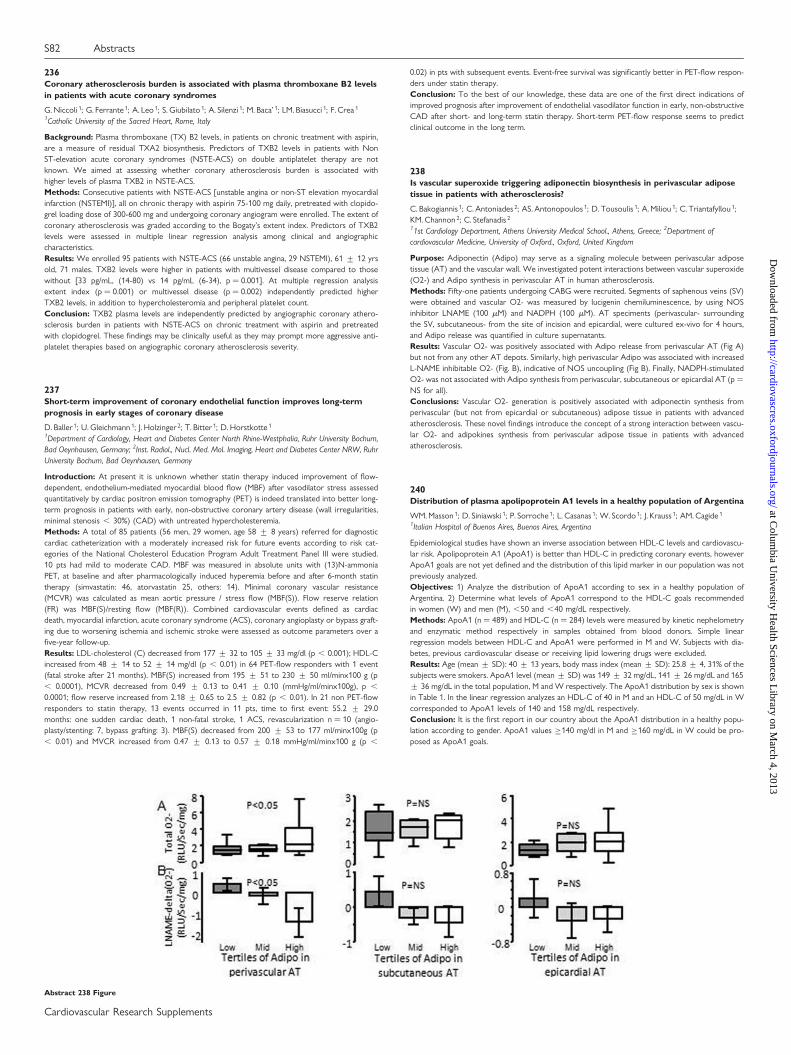
236Coronary atherosclerosis burden is associated with plasma thromboxane B2 levelsin patients with acute coronary syndromes
G. Niccoli 1; G. Ferrante 1; A. Leo 1; S. Giubilato 1; A. Silenzi 1; M. Baca’ 1; LM. Biasucci 1; F. Crea 1
1Catholic University of the Sacred Heart, Rome, Italy
Background: Plasma thromboxane (TX) B2 levels, in patients on chronic treatment with aspirin,are a measure of residual TXA2 biosynthesis. Predictors of TXB2 levels in patients with NonST-elevation acute coronary syndromes (NSTE-ACS) on double antiplatelet therapy are notknown. We aimed at assessing whether coronary atherosclerosis burden is associated withhigher levels of plasma TXB2 in NSTE-ACS.Methods: Consecutive patients with NSTE-ACS [unstable angina or non-ST elevation myocardialinfarction (NSTEMI)], all on chronic therapy with aspirin 75-100 mg daily, pretreated with clopido-grel loading dose of 300-600 mg and undergoing coronary angiogram were enrolled. The extent ofcoronary atherosclerosis was graded according to the Bogaty’s extent index. Predictors of TXB2levels were assessed in multiple linear regression analysis among clinical and angiographiccharacteristics.Results: We enrolled 95 patients with NSTE-ACS (66 unstable angina, 29 NSTEMI), 61 + 12 yrsold, 71 males. TXB2 levels were higher in patients with multivessel disease compared to thosewithout [33 pg/mL, (14-80) vs 14 pg/mL (6-34), p ¼ 0.001]. At multiple regression analysisextent index (p ¼ 0.001) or multivessel disease (p ¼ 0.002) independently predicted higherTXB2 levels, in addition to hypercholesteromia and peripheral platelet count.Conclusion: TXB2 plasma levels are independently predicted by angiographic coronary athero-sclerosis burden in patients with NSTE-ACS on chronic treatment with aspirin and pretreatedwith clopidogrel. These findings may be clinically useful as they may prompt more aggressive anti-platelet therapies based on angiographic coronary atherosclerosis severity.
237Short-term improvement of coronary endothelial function improves long-termprognosis in early stages of coronary disease
D. Baller 1; U. Gleichmann 1; J. Holzinger 2; T. Bitter 1; D. Horstkotte 1
1Department of Cardiology, Heart and Diabetes Center North Rhine-Westphalia, Ruhr University Bochum,Bad Oeynhausen, Germany; 2Inst. Radiol., Nucl. Med. Mol. Imaging, Heart and Diabetes Center NRW, RuhrUniversity Bochum, Bad Oeynhausen, Germany
Introduction: At present it is unknown whether statin therapy induced improvement of flow-dependent, endothelium-mediated myocardial blood flow (MBF) after vasodilator stress assessedquantitatively by cardiac positron emission tomography (PET) is indeed translated into better long-term prognosis in patients with early, non-obstructive coronary artery disease (wall irregularities,minimal stenosis , 30%) (CAD) with untreated hypercholesteremia.Methods: A total of 85 patients (56 men, 29 women, age 58 + 8 years) referred for diagnosticcardiac catheterization with a moderately increased risk for future events according to risk cat-egories of the National Cholesterol Education Program Adult Treatment Panel III were studied.10 pts had mild to moderate CAD. MBF was measured in absolute units with (13)N-ammoniaPET, at baseline and after pharmacologically induced hyperemia before and after 6-month statintherapy (simvastatin: 46, atorvastatin 25, others: 14). Minimal coronary vascular resistance(MCVR) was calculated as mean aortic pressure / stress flow (MBF(S)). Flow reserve relation(FR) was MBF(S)/resting flow (MBF(R)). Combined cardiovascular events defined as cardiacdeath, myocardial infarction, acute coronary syndrome (ACS), coronary angioplasty or bypass graft-ing due to worsening ischemia and ischemic stroke were assessed as outcome parameters over afive-year follow-up.Results: LDL-cholesterol (C) decreased from 177 + 32 to 105 + 33 mg/dl (p , 0.001); HDL-Cincreased from 48 + 14 to 52 + 14 mg/dl (p , 0.01) in 64 PET-flow responders with 1 event(fatal stroke after 21 months). MBF(S) increased from 195 + 51 to 230 + 50 ml/minx100 g (p, 0.0001), MCVR decreased from 0.49 + 0.13 to 0.41 + 0.10 (mmHg/ml/minx100g), p ,
0.0001; flow reserve increased from 2.18 + 0.65 to 2.5 + 0.82 (p , 0.01). In 21 non PET-flowresponders to statin therapy, 13 events occurred in 11 pts, time to first event: 55.2 + 29.0months: one sudden cardiac death, 1 non-fatal stroke, 1 ACS, revascularization n ¼ 10 (angio-plasty/stenting: 7, bypass grafting: 3). MBF(S) decreased from 200 + 53 to 177 ml/minx100g (p, 0.01) and MVCR increased from 0.47 + 0.13 to 0.57 + 0.18 mmHg/ml/minx100 g (p ,
0.02) in pts with subsequent events. Event-free survival was significantly better in PET-flow respon-ders under statin therapy.Conclusion: To the best of our knowledge, these data are one of the first direct indications ofimproved prognosis after improvement of endothelial vasodilator function in early, non-obstructiveCAD after short- and long-term statin therapy. Short-term PET-flow response seems to predictclinical outcome in the long term.
238Is vascular superoxide triggering adiponectin biosynthesis in perivascular adiposetissue in patients with atherosclerosis?
C. Bakogiannis 1; C. Antoniades 2; AS. Antonopoulos 1; D. Tousoulis 1; A. Miliou 1; C. Triantafyllou 1;KM. Channon 2; C. Stefanadis 2
11st Cardiology Department, Athens University Medical School., Athens, Greece; 2Department ofcardiovascular Medicine, University of Oxford., Oxford, United Kingdom
Purpose: Adiponectin (Adipo) may serve as a signaling molecule between perivascular adiposetissue (AT) and the vascular wall. We investigated potent interactions between vascular superoxide(O2-) and Adipo synthesis in perivascular AT in human atherosclerosis.Methods: Fifty-one patients undergoing CABG were recruited. Segments of saphenous veins (SV)were obtained and vascular O2- was measured by lucigenin chemiluminescence, by using NOSinhibitor LNAME (100 mM) and NADPH (100 mM). AT speciments (perivascular- surroundingthe SV, subcutaneous- from the site of incision and epicardial, were cultured ex-vivo for 4 hours,and Adipo release was quantified in culture supernatants.Results: Vascular O2- was positively associated with Adipo release from perivascular AT (Fig A)but not from any other AT depots. Similarly, high perivascular Adipo was associated with increasedL-NAME inhibitable O2- (Fig. B), indicative of NOS uncoupling (Fig B). Finally, NADPH-stimulatedO2- was not associated with Adipo synthesis from perivascular, subcutaneous or epicardial AT (p ¼NS for all).Conclusions: Vascular O2- generation is positively associated with adiponectin synthesis fromperivascular (but not from epicardial or subcutaneous) adipose tissue in patients with advancedatherosclerosis. These novel findings introduce the concept of a strong interaction between vascu-lar O2- and adipokines synthesis from perivascular adipose tissue in patients with advancedatherosclerosis.
240Distribution of plasma apolipoprotein A1 levels in a healthy population of Argentina
WM. Masson 1; D. Siniawski 1; P. Sorroche 1; L. Casanas 1; W. Scordo 1; J. Krauss 1; AM. Cagide 1
1Italian Hospital of Buenos Aires, Buenos Aires, Argentina
Epidemiological studies have shown an inverse association between HDL-C levels and cardiovascu-lar risk. Apolipoprotein A1 (ApoA1) is better than HDL-C in predicting coronary events, howeverApoA1 goals are not yet defined and the distribution of this lipid marker in our population was notpreviously analyzed.Objectives: 1) Analyze the distribution of ApoA1 according to sex in a healthy population ofArgentina. 2) Determine what levels of ApoA1 correspond to the HDL-C goals recommendedin women (W) and men (M), ,50 and ,40 mg/dL respectively.Methods: ApoA1 (n ¼ 489) and HDL-C (n ¼ 284) levels were measured by kinetic nephelometryand enzymatic method respectively in samples obtained from blood donors. Simple linearregression models between HDL-C and ApoA1 were performed in M and W. Subjects with dia-betes, previous cardiovascular disease or receiving lipid lowering drugs were excluded.Results: Age (mean + SD): 40 + 13 years, body mass index (mean + SD): 25.8 + 4, 31% of thesubjects were smokers. ApoA1 level (mean + SD) was 149 + 32 mg/dL, 141 + 26 mg/dL and 165+ 36 mg/dL in the total population, M and W respectively. The ApoA1 distribution by sex is shownin Table 1. In the linear regression analyzes an HDL-C of 40 in M and an HDL-C of 50 mg/dL in Wcorresponded to ApoA1 levels of 140 and 158 mg/dL respectively.Conclusion: It is the first report in our country about the ApoA1 distribution in a healthy popu-lation according to gender. ApoA1 values ≥140 mg/dl in M and ≥160 mg/dL in W could be pro-posed as ApoA1 goals.
Abstract 238 Figure
S82 Abstracts
Cardiovascular Research Supplements
at Colum
bia University H
ealth Sciences Library on M
arch 4, 2013http://cardiovascres.oxfordjournals.org/
Dow
nloaded from
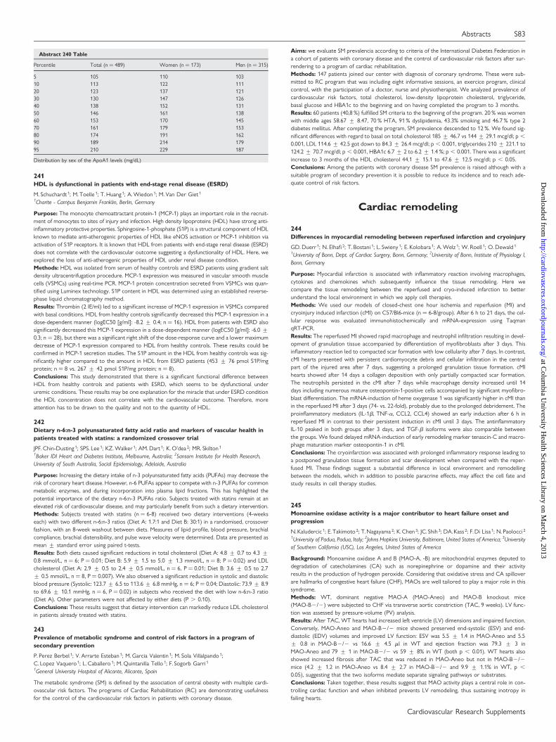
241HDL is dysfunctional in patients with end-stage renal disease (ESRD)
M. Schuchardt 1; M. Toelle 1; T. Huang 1; A. Wiedon 1; M. Van Der Giet 1
1Charite - Campus Benjamin Franklin, Berlin, Germany
Purpose: The monocyte chemoattractant protein-1 (MCP-1) plays an important role in the recruit-ment of monocytes to sites of injury and infection. High density lipoproteins (HDL) have strong anti-inflammatory protective properties. Sphingosine-1-phosphate (S1P) is a structural component of HDLknown to mediate anti-atherogenic properties of HDL like eNOS activation or MCP-1 inhibition viaactivation of S1P receptors. It is known that HDL from patients with end-stage renal disease (ESRD)does not correlate with the cardiovascular outcome suggesting a dysfunctionality of HDL. Here, weexplored the loss of anti-atherogenic properties of HDL under renal disease condition.Methods: HDL was isolated from serum of healthy controls and ESRD patients using gradient saltdensity ultracentrifugation procedure. MCP-1 expression was measured in vascular smooth musclecells (VSMCs) using real-time PCR. MCP-1 protein concentration secreted from VSMCs was quan-tified using Luminex technology. S1P content in HDL was determined using an established reverse-phase liquid chromatography method.Results: Thrombin (2 IE/ml) led to a significant increase of MCP-1 expression in VSMCs comparedwith basal conditions. HDL from healthy controls significantly decreased this MCP-1 expression in adose-dependent manner (logEC50 [g/ml]: -8.2 + 0.4; n ¼ 16). HDL from patients with ESRD alsosignificantly decreased this MCP-1 expression in a dose-dependent manner (logEC50 [g/ml]: -6.0 +0.3; n ¼ 28), but there was a significant right shift of the dose-response curve and a lower maximumdecrease of MCP-1 expression compared to HDL from healthy controls. These results could beconfirmed in MCP-1 secretion studies. The S1P amount in the HDL from healthy controls was sig-nificantly higher compared to the amount in HDL from ESRD patients (453 + 76 pmol S1P/mgprotein; n ¼ 8 vs. 267 + 42 pmol S1P/mg protein; n ¼ 8).Conclusions: This study demonstrated that there is a significant functional difference betweenHDL from healthy controls and patients with ESRD, which seems to be dysfunctional underuremic conditions. These results may be one explanation for the miracle that under ESRD conditionthe HDL concentration does not correlate with the cardiovascular outcome. Therefore, moreattention has to be drawn to the quality and not to the quantity of HDL.
242Dietary n-6:n-3 polyunsaturated fatty acid ratio and markers of vascular health inpatients treated with statins: a randomized crossover trial
JPF. Chin-Dusting 1; SPS. Lee 1; KZ. Walker 1; AM. Dart 1; K. O’dea 2; MR. Skilton 1
1Baker IDI Heart and Diabetes Institute, Melbourne, Australia; 2Sansom Institute for Health Research,University of South Australia, Social Epidemiology, Adelaide, Australia
Purpose: Increasing the dietary intake of n-3 polyunsaturated fatty acids (PUFAs) may decrease therisk of coronary heart disease. However, n-6 PUFAs appear to compete with n-3 PUFAs for commonmetabolic enzymes, and during incorporation into plasma lipid fractions. This has highlighted thepotential importance of the dietary n-6:n-3 PUFAs ratio. Subjects treated with statins remain at anelevated risk of cardiovascular disease, and may particularly benefit from such a dietary intervention.Methods: Subjects treated with statins (n ¼ 6-8) received two dietary interventions (4-weekseach) with two different n-6:n-3 ratios (Diet A: 1.7:1 and Diet B: 30:1) in a randomised, crossoverfashion, with an 8-week washout between diets. Measures of lipid profile, blood pressure, brachialcompliance, brachial distensibility, and pulse wave velocity were determined. Data are presented asmean + standard error using paired t-tests.Results: Both diets caused significant reductions in total cholesterol (Diet A: 4.8 + 0.7 to 4.3 +0.8 mmol/L, n ¼ 6; P ¼ 0.01; Diet B: 5.9 + 1.5 to 5.0 + 1.3 mmol/L, n ¼ 8; P ¼ 0.02) and LDLcholesterol (Diet A: 2.9 + 0.5 to 2.4 + 0.5 mmol/L, n ¼ 6, P ¼ 0.01; Diet B: 3.6 + 0.5 to 2.7+ 0.5 mmol/L, n ¼ 8, P ¼ 0.007). We also observed a significant reduction in systolic and diastolicblood pressure (Systolic: 123.7 + 6.5 to 113.6 + 6.8 mmHg, n ¼ 6; P ¼ 0.04; Diastolic: 73.9 + 8.9to 69.6 + 10.1 mmHg, n ¼ 6, P ¼ 0.02) in subjects who received the diet with low n-6:n-3 ratio(Diet A). Other parameters were not affected by either diets (P . 0.10).Conclusions: These results suggest that dietary intervention can markedly reduce LDL cholesterolin patients already treated with statins.
243Prevalence of metabolic syndrome and control of risk factors in a program ofsecondary prevention
P. Perez Berbel 1; V. Arrarte Esteban 1; M. Garcia Valentin 1; M. Sola Villalpando 1;C. Lopez Vaquero 1; L. Caballero 1; M. Quintanilla Tello 1; F. Sogorb Garri 1
1General University Hospital of Alicante, Alicante, Spain
The metabolic syndrome (SM) is defined by the association of central obesity with multiple cardi-ovascular risk factors. The programs of Cardiac Rehabilitation (RC) are demonstrating usefulnessfor the control of the cardiovascular risk factors in patients with coronary disease.
Aims: we evaluate SM prevalencia according to criteria of the International Diabetes Federation ina cohort of patients with coronary disease and the control of cardiovascular risk factors after sur-rendering to a program of cardiac rehabilitation.Methods: 147 patients joined our center with diagnosis of coronary syndrome. These were sub-mitted to RC program that was including eight informative sessions, an exercice program, clinicalcontrol, with the participation of a doctor, nurse and physiotherapist. We analyzed prevalence ofcardiovascular risk factors, total cholesterol, low-density lipoprotein cholesterol, triglyceride,basal glucose and HBA1c to the beginning and on having completed the program to 3 months.Results: 60 patients (40,8 %) fulfilled SM criteria to the beginning of the program. 20 % was womenwith middle ages 58.67 + 8.47, 70 % HTA, 91 % dyslipidemia, 43.3% smoking and 46.7 % type 2diabetes mellitus. After completing the program, SM prevalence descended to 12 %. We found sig-nificant differences with regard to basal on total cholesterol 185 + 46.7 vs 144 + 29.1 mcg/dl; p ,
0.001, LDL 114.6 + 42.5 got down to 84.3 + 26.4 mcg/dl; p , 0.001, triglycerides 210 + 221.1 to124.2 + 70.7 mcg/dl; p , 0.001, HBA1c 6.7 + 2 to 6.2 + 1.4 %; p , 0.001. There was a significantincrease to 3 months of the HDL cholesterol 44.1 + 15.1 to 47.6 + 12.5 mcg/dl; p , 0.05.Conclusions: Among the patients with coronary disease SM prevalence is raised although with asuitable program of secondary prevention it is possible to reduce its incidence and to reach ade-quate control of risk factors.
Cardiac remodeling
244Differences in myocardial remodeling between reperfused infarction and cryoinjury
GD. Duerr 1; N. Elhafi 2; T. Bostani 1; L. Swieny 1; E. Kolobara 1; A. Welz 1; W. Roell 1; O. Dewald 1
1University of Bonn, Dept. of Cardiac Surgery, Bonn, Germany; 2University of Bonn, Institute of Physiology I,Bonn, Germany
Purpose: Myocardial infarction is associated with inflammatory reaction involving macrophages,cytokines and chemokines which subsequently influence the tissue remodeling. Here wecompare the tissue remodeling between the reperfused and cryo-induced infarction to betterunderstand the local environment in which we apply cell therapies.Methods: We used our models of closed-chest one hour ischemia and reperfusion (MI) andcryoinjury induced infarction (cMI) on C57/Bl6-mice (n ¼ 6-8/group). After 6 h to 21 days, the cel-lular response was evaluated immunohistochemically and mRNA-expression using TaqmanqRT-PCR.Results: The reperfused MI showed rapid macrophage and neutrophil infiltration resulting in devel-opment of granulation tissue accompanied by differentiation of myofibroblasts after 3 days. Thisinflammatory reaction led to compacted scar formation with low cellularity after 7 days. In contrast,cMI hearts presented with persistent cardiomyocyte debris and cellular infiltration in the centralpart of the injured area after 7 days, suggesting a prolonged granulation tissue formation. cMIhearts showed after 14 days a collagen deposition with only partially compacted scar formation.The neutrophils persisted in the cMI after 7 days while macrophage density increased until 14days including numerous mature osteopontin-1-positive cells accompanied by significant myofibro-blast differentiation. The mRNA-induction of heme oxygenase 1 was significantly higher in cMI thanin the reperfused MI after 3 days (74- vs. 22-fold), probably due to the prolonged debridement. Theproinflammatory mediators (IL-1b, TNF-a, CCL2, CCL4) showed an early induction after 6 h inreperfused MI in contrast to their persistent induction in cMI until 3 days. The antinflammatoryIL-10 peaked in both groups after 3 days, and TGF-b isoforms were also comparable betweenthe groups. We found delayed mRNA-induction of early remodeling marker tenascin-C and macro-phage maturation marker osteopontin-1 in cMI.Conclusions: The cryoinfarction was associated with prolonged inflammatory response leading toa postponed granulation tissue formation and scar development when compared with the reper-fused MI. These findings suggest a substantial difference in local environment and remodellingbetween the models, which in addition to possible paracrine effects, may affect the cell fate andstudy results in cell therapy studies.
245Monoamine oxidase activity is a major contributor to heart failure onset andprogression
N.Kaludercic 1; E.Takimoto 2; T.Nagayama 2; K. Chen 3; JC. Shih 3; DA.Kass 2; F.Di Lisa 1; N.Paolocci 2
1University of Padua, Padua, Italy; 2Johns Hopkins University, Baltimore, United States of America; 3Universityof Southern California (USC), Los Angeles, United States of America
Background: Monoamine oxidase A and B (MAO-A, -B) are mitochondrial enzymes deputed todegradation of catecholamines (CA) such as norepinephrine or dopamine and their activityresults in the production of hydrogen peroxide. Considering that oxidative stress and CA spilloverare hallmarks of congestive heart failure (CHF), MAOs are well tailored to play a major role in thissyndrome.Methods: WT, dominant negative MAO-A (MAO-Aneo) and MAO-B knockout mice(MAO-B2/2) were subjected to CHF via transverse aortic constriction (TAC, 9 weeks). LV func-tion was assessed by pressure-volume (PV) analysis.Results: After TAC, WT hearts had increased left ventricle (LV) dimensions and impaired function.Conversely, MAO-Aneo and MAO-B2/2 mice showed preserved end-systolic (ESV) and end-diastolic (EDV) volumes and improved LV function: ESV was 5.5 + 1.4 in MAO-Aneo and 5.5+ 0.8 in MAO-B2/2 vs 16.6 + 4.5 ml in WT and ejection fraction was 79.3 + 3 inMAO-Aneo and 79 + 1 in MAO-B2/2 vs 59 + 8% in WT (both p , 0.01). WT hearts alsoshowed increased fibrosis after TAC that was reduced in MAO-Aneo but not in MAO-B2/2mice (4.2 + 1.2 in MAO-Aneo vs 8.4 + 2.7 in MAO-B2/2 and 9.9 + 1.1% in WT, p ,
0.05), suggesting that the two isoforms mediate separate signaling pathways or substrates.Conclusions: Taken together, these results suggest that MAO activity plays a central role in con-trolling cardiac function and when inhibited prevents LV remodeling, thus sustaining inotropy infailing hearts.
Abstract 240 Table
Percentile Total (n ¼ 489) Women (n ¼ 173) Men (n ¼ 315)
5 105 110 10310 113 122 11120 123 137 12130 130 147 12640 138 152 13150 146 161 13860 153 170 14570 161 179 15380 174 191 16290 189 214 17995 210 229 187
Distribution by sex of the ApoA1 levels (mg/dL)
Abstracts S83
Cardiovascular Research Supplements
at Colum
bia University H
ealth Sciences Library on M
arch 4, 2013http://cardiovascres.oxfordjournals.org/
Dow
nloaded from
246Decreased sensitivity to beta-adrenergic stimulation participates in reducedsusceptibility to VT in stress conditions in TAC mice with cardiac FKBP12.6overexpression
L. Vinet 1; M. Pezet 1; F. Briec 2; M. Previlon 1; P. Rouet-Benzineb 1; A. Hivonnait 2; F. Charpentier 2;JJ. Mercadier 1
1Inserm U698 - Hospital Bichat-Claude Bernard, Paris, France; 2Inserm UMR 915, Institut of the Thorax,CNRS ERL3147, University of Nantes, Nantes, France
Through its involvement in the RyR2 macromolecular complex, FKBP12.6 (calstabin 2) has beensuggested to have anti-arrhythmic properties. It was shown before that FKBP12.6 overexpressionin the mouse heart prevents from ventricular tachycardia (VT) induced by burst pacing (BP) instress conditions. We examined whether such a protection also exists in hypertrophied ventriclesand if so, whether an alteration in cardiac sensitivity to beta-adrenergic stimulation could be involved.Double transgenic mice (DT) with cardiac-specific FKBP12.6 overexpression were obtained by cross-ing tetO-FKBP12.6 mice with the alpha-MHCtTA transactivator mouse strain (tTA). Wild-type andtTA mice were used as controls (Ctr). Constriction of the thoracic aorta (TAC) and sham-operation(Sham) were performed in 5-week-old male mice. Mice were studied at 1 and 2 months (MO) aftersurgery. In Ctr, LV peak systolic pressure almost doubled at 1 and 2 MO post-TAC (P , 0.01 vsSham). LVEDP increased at 1 MO (P , 0.01 vs Sham) with no further increase at 2 MO and tau,the time constant of LV relaxation, increased at 1 MO and further increased at 2 MO (P , 0.01).LVW/BW increased by 86% at 1 MO and by 108% at 2 MO. Similar alterations were seen in DTmice. At 1 MO, whereas 3 ng/g/min dobutamine (Dob) infusion was associated with a 15% increasein heart rate (HR) in Sham-Ctr (p , 0.01), 30 ng/g/min Dob were necessary to increase HR inSham-DT, a feature also observed in TAC-Ctr and TAC-DT. At 2 MO, the positive chronotropiceffect of Dob was totally blunted in TAC-Ctr and TAC-DT. At 1 and 2 MO, a 20% increase in dP/dtmax/P (p , 0.01) in response to 30 ng/g/min Dob was seen in Sham-Ctr whereas the responsewas blunted in TAC-Ctr and in the 2 DT groups. Programmed electrical stimulation and BP were per-formed at 2 MO. At baseline, ventricular effective refractory period (VERP) was similar in Sham-Ctrand Sham-DT. Iso injection (0.2 mg/kg ip) shortened VERP in the 2 groups (p , 0.05). VERP wasshorter in TAC-DT than in TAC-Ctr (p , 0.05). Iso injection markedly shortened VERP inTAC-Ctr (p , 0.01), an effect that was blunted in TAC-DT. Nevertheless, VERP was shorter inTAC-DT than in TAC-Ctr (p , 0.05). BP induced VT in 1 out of 19 (5%) TAC-Ctr and 1 out of11 (9%) TAC-DT mice at baseline (NS). After Iso injection, BP induced VT in 8 out of 16TAC-Ctr (47%) but in none of the 7 TAC-DT (P¼ 0.052). Taken together, our results indicatethat FKBP12.6 overexpression reduces susceptibility of the hypertrophied heart to VT in stress con-ditions at least in part due to shortened VERP and reduced sensitivity to beta-adrenergic stimulation.
247The cardiac hypertrophic phenotype of BAMBI (BMP and Activin Membrane BoundInhibitor) defective mice associates increased myocardial expression of micro-RNAmiR-21
AV. Villar 1; M. Cobo 2; M. Llano 2; C. Montalvo 1; V. Exposito 2; JF. Nistal 2; MA. Hurle 1
1University of Cantabria, Santander, Spain; 2University Hospital Marques de Valdecilla, Santander, Spain
Purpose: Micro-RNAs are endogenous non-coding small RNAs that act as posttranscriptionalrepressors of gene function and whose role in cardiac pathologies, including hypertrophy, has beenrecently recognized. TGF-b is a potent hypertrophic cytokine whose signalling efficacy is negativelymodulated by the transmembrane pseudoreceptor BAMBI. Here, we assessed the relationshipbetween miR-21 and the LV hypertrophy caused by TGF-b overactivity in mutant mice lacking BAMBI.Methods: Experiments were performed in BAMBI-KO mice and in cultured HeLa cells. Echocardio-graphic measurements were made with a Vevo-770 equipment (VisualSonics). BAMBI was silenced inHeLa cells by transfection with small interference-RNA (siRNA). MicroRNA and total RNA wereextracted. Gene and protein expressions were determined by qPCR and Western Blot, respectively.Results: BAMBI-KO mice displayed an echocardiographic phenotype characterized by: a) IncreasedLV mass (KO: 95.0 + 5.7 mg vs WT: 70.8 + 0.04 mg; p , 0.01); and b) Eccentric hypertrophy,with increased PWT (BAMBI-KO: 0.79 + 4.9 mm vs WT 0.68 + 0.04 mm; p , 0.05) and LVEDD(BAMBI-KO: 4.02 + 0.01 mm vs WT: 3.76 + 0.08 mm; p , 0.05), without differences in relativePWT. Biochemical analysis evidenced the augmented TGF-b signalling in KO mice, reflected by theincreased nuclear presence of SMAD4. Cardiac hypertrophy coursed with elevated myocardialexpression levels of the TGF-b target gene, alpha-myosin heavy chain (alpha-MHC). In LVsamples from BAMBI-KO mice, miR-21 was overexpressed (KO: 8.8 + 1.3 vs WT: 3.9 + 0.6; p, 0.05). Other hypertrophy-related miRNAs, such as miR-92, miR-100, miR133a or miR133b,were not altered. In cultured HeLa cells, TGF-b induced the expression of miR-21 (control: 0.0+ 0.0; TGF-b: 514.5 + 117.6), and the effect was significantly potentiated after BAMBI siRNAtransfection (TGF-b + BAMBI-siRNA: 763.1 + 82.7; p , 0.05).Conclusions: The absence of BAMBI in KO mice leads to increased myocardial TGF-b signalling,that associates LV eccentric hypertrophy and up-regulation of miR-21 expression. Accordingly, incultured HeLa cells, RNA interference-mediated downregulation of BAMBI significantly potentiatedTGF-b-induced miR-21 expression. Our data further support the role of miR-21 as a critical regu-lator of myocardial hypertrophy. Our results offer new insights on a TGF-b-directed miR-21 regu-latory circuit, and disclose its relevance for hypertrophy progression in cardiovascular diseases thatassociate increased levels of TGF-b.
248Erythropoietin receptor deficient mice have impaired cardiac adaptation duringvoluntary exercise
WT. Ruifrok 1; LMG. Meems 1; BD. Westenbrink 1; AH. Maass 1; HHW. Sillje 1; DJ. Van Veldhuisen 1;WH. Van Gilst 1; RA. De Boer 1
1University Medical Center, Groningen, Netherlands
Background: We have demonstrated that erythropoietin (EPO) via its receptor (EPOR) exertscardioprotective effects in pathophysiological hypertrophy (e.g. myocardial infarction and heart
failure). It is unknown however if the EPO-EPOR signalling pathway exerts similar results in phys-iological hypertrophy.Methods: We employed transgenic-rescued EPOR-null mutant mice (EPOR2/2rescued, KO)that express EPOR exclusively in the hematopoietic cells. C57Bl/6 mice were used as controls(WT). Both groups had unlimited access to a running wheel for 28 days (EX, N ¼ 50). Furthermore,sedentary mice of both groups were studied (SED, N ¼ 42). We measured exercise performance:average distance, average daily speed and average running time. Cardiac function was assessed byechocardiography. At sacrifice heart weights and hemodynamic parameters were measured. Weevaluated markers of lipid oxidation (CPT-1b, MCIP-1 and MCAD).Results: WT-EX mice ran 8.0 + 2.1 km/day, whereas KO-EX mice ran 5.5 + 2.4 km/day (P ,
0.01). This was mainly due to a lesser average speed (WT-EX 1.6 + 0.3 km/hrs vs. KO-EX 1.3+ 0.3 km/hrs, P , 0.01). In the WT-EX group, heart weight increased (heart weight-to-bodyweight ratio: 7.3 + 1.1 vs. WT-SED 5.8 + 0.6 mg/g, P , 0.001). In the KO-EX mice runningdid not increase heart weight. Stroke volume assessed with echocardiography was increased inWT-EX mice (74 + 12 vs. WT-SED 62 + 13 ml, P , 0.05), whereas exercise in the KO-EXmice did not increase stroke volume. Indices of contractility and relaxation were increased afterexercise in both the controls (WT-EX dPdtmax: 10611 + 1369 vs. WT-SED 8969 + 973mmHg/s, P , 0.01; WT-EX dPdtmin: -10057 + 867 vs. WT-SED -8318 + 1604 mmHg/s, P ,
0.05), as well in the KO mice (KO-EX dPdtmax: 11081 + 1491 vs. KO-SED 8901 + 1432mmHg/s, P , 0.05; KO-EX dPdtmin: -11122 + 1656 vs. KO-SED -8278 + 1092 mmHg/s, P ,
0.01). MCIP-1 was 1.4 fold increased (P , 0.05) in the WT-ex mice, but not in the KO-ex mice;CPT-1b and MCAD were unchanged.Conclusion: The absence of EPO-EPOR signalling leads to a lesser exercise capacity upon volun-tary wheel running. We suggest that EPO-EPOR signalling is important for physiological adaptationduring exercise.
249Variable ultrastructural remodeling and cellular functional changes in myocardiumremote to the MI
L. Biesmans 1; V. Bito 1; RB. Driessen 1; P. Holemans 1; I. Lenaerts 1; C. Huysmans 1; KR. Sipido 1
1Catholic University of Leuven, Leuven, Belgium
Background: While intermittent ischemia is an important stimulus for remodeling in the area adja-cent to a myocardial infarction (MI), stretch will be more important in the area at a distance(Remote) and dependent on the hemodynamic impact of the MI. We investigated remodelingand changes in Ca2+ handling in the remote area of pigs with different extent of MI.Methods: A copper-coated metal stent inducing intima proliferation was implanted in the LAD ofpigs resulting in.90% stenosis. We studied a subgroup of pigs with variable extent of MI (6 to 13 %of LV mass). Matched sham-operated animals (Sham, 5 pigs) served as controls. Ultrastructuralremodeling was quantified with histology and electron microscopy. Six weeks after stent implan-tation myocytes were enzymatically isolated from the remote wall. Cells were studied at 378Cduring field stimulation and whole-cell voltage clamp; K5-Fluo-3 was used as Ca2+ indicator.Results: The remote area had a variable degree of ultrastructural remodeling. Based on semi-quantitative evaluation of glycogen deposition in the perinuclear area and between myofilaments,and the degree of myolysis, pigs were rated as moderate (RemoteMOD: % remodeling. average+stdev of sham group, 3 pigs) or severe (RemoteSEV: % remodeling. average+3*stdev of shamgroup, 4 pigs). Myocytes were hypertrophied to the same extent in both groups. Functional remo-deling was also present in both groups. Cell shortening was significantly slower compared to Sham.[Ca2+]i transient amplitude and sarcoplasmic reticulum Ca2+ content remained comparable toSham, while L-type Ca2+ current was reduced, indicating an increased excitation-contraction coup-ling gain. Spontaneous spark frequency and duration were significantly increased compared to Sham,suggesting an increased diastolic Ca2+ leak. In addition, myocytes isolated from the RemoteSEVgroup also showed a significant increase in the amplitude of fractional shortening (4.6 + 0.2%vs. 3.7 + 0.2% in RemoteMOD and 3.8 + 0.2% in Sham, P , 0.05) and a shorter action potentialduration (at 50% of repolarization: 335 + 26ms vs. 407 + 36ms in RemoteMOD; 303 + 40ms inSham, P , 0.05), indicating that electrical remodeling was only present in RemoteSEV.Conclusion: The degree of cellular remodeling in the remote area is variable for both ultrastruc-tural and functional parameters. This suggests the existence of complex signaling cascades for remo-deling relating to local stimuli.
250Thyroid hormone receptor alpha1: a novel molecular switch to the progression toheart failure after acute myocardial infarction?
I. Mourouzis 1; C. Pantos 1; G. Galanopoulos 1; M. Gavra 1; P. Perimenis 1; D. Spanou 1; DV. Cokkinos 2
1University of Athens, Athens, Greece; 2Onassis Cardiac Surgery Center, 1st Department of Cardiology,Athens, Greece
Thyroid hormone receptor a1 (TRa1) is shown to act as a molecular switch between physiologicaland pathological growth due to its dual mode of action depending on the thyroid hormone avail-ability (unliganded vs liganded state). Thus, the present study investigated potential role of TRa1in the pathophysiology of postischaemic cardiac remodeling. Acute myocardial infarction inducedin rats by coronary artery ligation (AMI) resulted in animals with (AMI-HF) and without heartfailure (AMI-NHF) after 34 weeks. SHAM operated animals served as controls (SHAM). InAMI-HF, nuclear TRa1 expression in both border and remote myocardium was found to be mark-edly decreased (2.0 fold and 1.5 fold) compared either to SHAM or to AMI-NHF, P , 0.05. Thisresponse resulted in tissue hypothyroidism with suppression of triiodothyronine (T3) regulatedmyosin isoform expression and cardiac dysfunction. Furthermore, phospho-Akt and phospho-ERKlevels were significantly decreased in AMI-HF as compared to AMI-NHF and may account for altera-tions in nuclear TRa1 expression. In fact, in a neonatal cardiomyocytes model of phenylephrine (PE)induced hypertrophy, inactivation of Akt/mTOR and /or ERK signaling was shown to result inmarked suppression of the amount of TRa1 in nucleus. In conclusion, nuclear TRa1 expressionappears to be suppressed in the failing post-infarcted heart resulting in tissue hypothyroidism
S84 Abstracts
Cardiovascular Research Supplements
at Colum
bia University H
ealth Sciences Library on M
arch 4, 2013http://cardiovascres.oxfordjournals.org/
Dow
nloaded from
which may trigger cardiac atrophy and suppression of T3 regulated contractile protein expression.Akt/mTOR and/ or ERK signaling pathway may be involved in this response.
251Circulating matrix metalloproteinase-3 plasma level as a marker of early cardiacremodelling after myocardial infarction
A. Berezin 1; T. Panasenko 1
1State Medical University, Zaporozhye, Ukraine
Background: It has been reported that the rate of both production of metalloproteinases andaccumulation of extracellular matrix are the important markers of cardiac remodelling, associatedwith survival of patients after Q-wave myocardial infarction independently of age, gender, signs andsymptoms of chronic heart failure.The aim of our study was to investigate the interrelation between the level of circulating matrixmetalloproteinase-3 (MMP-3) and sings of cardiac remodeling in patients after Q-wave myocardialinfarction in acute and early post myocardial infarction period.Methods: 67 patients (51 men, mean age 62.4 + 3.11 years) with Q-wave myocardial infarctionwere included. All patients gave the written informed consent on participating in research. Trans-thorasic echocardiography and Doppler were performed in order to assess ejection fraction (EF),end-systolic volume (ESV) and end-diastolic volume (EDV) of left ventricle, isovolumic relaxationtime (IVRT). Doppler index, values of myocardial stress, indexes of sphericity local contractilityof left ventricle. Investigation of the level of MMP-3 by enzyme linked immunosorbent assaywere performed. All measurings were done on day 3 and was repeated after 12 weeks of treatment.Results: It was shown a significant correlation between the level of MMP-3 and EF (r ¼ 0.52; P ¼0.012), index of local contractility (r ¼ 0.50; P , 0.02), Doppler index (r ¼ 0.44; P , 0.05), EDV(r ¼ 0.46; P , 0.001) and indexes of sphericity of left ventricle (r ¼ 20.52; P , 0.05) accordingly.In 12 weeks after Q-wave myocardial infarction in patients with EF 41-45% the level of MMP-3 wassignificantly lower than in patients with EF ≤40%. Only for the last group of patients it was set thatthere was a significant correlation between the level of MMP-3 and EDV (r ¼ 0.54; P , 0.01),indexes of sphericity of left ventricle (r ¼ 20.52; P , 0.05) and mitral regurgitation velocity(r ¼ 0.48; P , 0.02) respectively.Conclusion: Monitoring of the concentration of MMP-3 in plasma can be used for authenticationof patients with the high risk of phenotypic and prognostic unfavorable forms of cardiac remodelingon the early stages of this process.
252YB-1 inhibits apoptosis and hypertrophy in ventricular cardiomyocytes of rat
G. Euler 1; S. Partsch 1; C. Harjung 1; J. Heger 1
1Justus-Liebig University Giessen, Institute of Physiological, Giessen, Germany
Cardiac hypertrophy and apoptosis contribute to the development of heart failure. The transcrip-tion factors AP-1 and SMAD have been identified as mediators of at least one of these processesand both factors can be inhibited by the transcription factor YB-1. Thus, YB-1 may be an inhibitor ofcardiac hypertrophy and apoptosis. To test this hypothesis, a YB-1-adenovirus was constructed andinfluence of YB-1 overexpression on apoptosis or hypertrophy was analysed in isolated cardiomyo-cytes of adult rat.Cardiomyocytes were infected with 1000 MOI of AdYB-1. This increased YB1-RNA expression to205 + 23% within 12 h and to 2506 + 633 % within 24 h (n ¼ 6, p , 0.05). YB-1 proteinexpression increased significantly to 150 + 13 % after 12 h and to 1816 + 417 % after 36 h(n ¼ 6, p , 0.05). Induction of apoptosis by TGFb1 (1 ng/ml) to 13.7 + 1.6 % apoptotic cellswas abolished in YB-1 overexpressing cardiomyocytes (6.6 + 1.1 % apoptotic cells, n ¼ 7, p ,
0.05). In addition, hypertrophic growth response in cardiomyocytes due to a-adrenergic stimulationby phenylephrine (10 mM) (139 + 47 % rate of proteinsynthesis, n ¼ 8) was abrogated in YB-1overexpressing cells (87 + 35 %, n ¼ 8, p , 0.05). An influence on phosphorylation or expressionof SMADs by YB-1 overexpression was not observed, since the phosphorylation of SMAD2/3 underTGFb1 stimulation was still present in AdYB-1 transfected cells.In conclusion, the transcription factor YB-1 efficiently inhibits apoptosis and hypertrophy in cardi-omyocytes. YB-1 may, therefore, be a promising candidate for heart failure therapies, although itsintracellular targets still have to be defined.
Mechanisms of cardiac protectionin ischaemia
253Two faces of nitric oxide: behind cardioprotective effects of erythropoietin
A. Bogdanova 1; D. Mihov 1; P. Mocharla 2; S. Yakushev 1; J. Vogel 1; M. Gassmann 1; R. Tavakoli 3
1Vetsuisse Faculty at the University of Zurich, Zurich, Switzerland; 2University of Zurich-Irchel, Department ofAnatomy and Physiology, Cardiovascular Research, Zurich, Switzerland; 3Department of Thoracic andCardiovascular Surgery, Canton Hospital Lucerne, Luzern, Switzerland
Purpose: The study was performed to characterize the targets of hrEpo in the heart and cardio-protective potential of the cytokine in treatment of cold global ischemia-reperfusion injury.Methods: In vivo heterotopic rat heart transplantation model and ex vivo isolated blood-perfusedrat heart model of cold global IR, isolated cardiomyocytes and HAECs have been used in the study.We have monitored localization of Epo in the heart. Site-specific activation status of PI3K/PKB andeNOS was assessed and plasma nitrite levels used as an indicator of NO production and tyrosinenitration. Tissue GSH:GSSG levels served as a redox indicator. Efficiency of capillary perfusion wasassessed in cryosections using Evans Blue, tissue ion content measured by flame photometry andthe Na,K ATPase activity as ouabain-sensitive 86Rb influx.Results: Intravenous administration of hrEpo to the recipient animals 20 minutes prior to therestoration of blood flow in the transplanted heart reduced ischemia-reperfusion injury
(plasma cTnT levels). Immediate interaction of hrEpo with coronary endothelium, but not withcardiomyocytes, resulted in acute transient activation of PI3 and Akt kinases and stimulation ofNO production by eNOS. Transient increase in NO levels facilitated rapid recovery fromischemia-induced myocardial oedema and Na+ accumulation. This was achieved by restorationof efficient capillary perfusion in post-ischemic heart and NO-induced activation of theNa,K-ATPase function in cardiomyocytes. The protective effects of hrEpo were abolished bythe inhibition of NO production by L-NAME or L-arginine depletion. Acute reperfusion-inducedoxidative stress in the graft could also be reduced by NO. The hrEpo-induced activation of eNOSwas transient due to the rapid internalization and degradation of the cytokine-receptor complexin the endothelial cells. Delayed activation of NOS in control non-treated hearts was observedduring inflammatory burst six hours after the transplantation. Uncontrolled generation reactivenitrogen species during inflammation followed by GSH oxidation and extensive nitration of tyro-sine residues was suppressed by hrEpo.Conclusions: Acute transient activation of NO production in the Epo-treated coronary endo-thelium improved microcirculation in the post-ischemic heart thus rapidly restoring ion andwater balance in the myocardium. Temporal special effect of hrEpo on eNOS reduced acute oxi-dative stress at the onset of blood perfusion and prevented delayed long-term activation ofNOSes during inflammation thereby protecting the myocardium from the second wave ofoxidation.
254Heptanol triggers cardio protection via mitochondrial potassium channels and notby gap junction uncoupling
D. Johansen 1; E. Sanden 2; C. Xi 1; R. Sundset 2; K. Ytrehus 1
1University of Tromso, Faculty of Medicine, Department of Medical Biology, Tromso, Norway; 2University ofTromso, Faculty of Medicine, Department of Clinical Medicine, Tromso, Norway
The aim of this study was to investigate mechanisms behind heptanol induced infarct size reductionand in particular if protection by pre-treatment is triggered by gap junction uncoupling or by mito-chondrial mechanisms.Results: In Langendorff perfused rat hearts, heptanol 2mM was given before regional ischemia. Thisresulted in reduced infarct size compared to controls (29.7 + 3.4% vs.12.6 + 2.1% P, 0.001).Mitochondrial potassium channel blockers (5HD blocking mitoKATP and PAX blocking mitoKCa)abolished cardio protective effect of heptanol (5HD 36.7 + 2.9% and PAX 40.2 + 2.8%). Measure-ments of oxygen consumption in isolated mitochondria (oxygraph) showed that heptanol signifi-cantly reduced respiratory control ratio in both subsarcolemmal (SSM) and interfibrillarmitochondria (IFM) in a dose dependent manner (0.5-5.0 mM). The P/O-ratio was also significantreduced. Laser scanning confocal microscopy of TMRM loaded isolated adult rat cardiomyocytesusing line-scan mode showed that heptanol significantly increased time to mitochondrialPTP-opening. Western blot analysis showed that pre-treatment with heptanol significantly increasedphosphorylation of AKT and GSK-3b, and the difference in phosphorylation was maintained at theend of the ischemic period.In conclusion, the results show that pre-treatment with heptanol protects the heart against ische-mia. This protection is most likely mediated via mitochondrial mechanisms and not via gap junctionuncoupling.
255Phosphorylation of survival kinases is not a guarantee for protection ofischemia-reperfusion injury in the isolated, perfused mouse heart
M. Bliksoen 1; A. Rutkovskiy 1; LH. Mariero 1; IJ. Vaage 1; KO. Stenslokken 2
1Oslo University Hospital, Ulleval, Oslo, Norway; 2Institute of Molecular Biosciences, University of Oslo, Oslo,Norway
Background: Isolated, perfused hearts are major tools in cardiovascular research. Phosphoryl-ation of survival protein kinases, including AKT, ERK, P38 and JNK has been suggested to beimportant for cardioprotection. We recently found that the perfusion procedure per se phos-phorylated these kinases. The phosphorylation was further increased by the presence of an intra-ventricular balloon.Hypotheses: 1: The MAPKinases are phosphorylated in a time depended manner during perfusion. 2:The presence of an intraventricular balloon will reduce infarct size compared to hearts perfusedwithout the balloon. 3: The inadvertent phosphorylation of ERK, P38 and JNK caused by the per-fusion procedure per se may interfere with studies of protective pathways.Methods and results: Series 1: Langendorff-perfused mouse hearts with an intraventricularballoon were sampled after 5, 10, 20, 40 and 60 minutes (n ¼ 8). Western blots showed a rapidincrease in phosphorylation of ERK, P38 (, 5 minutes, P , 0,05) and JNK (, 10 minutes, P ¼0,05), and a gradual reduction of AKT phosphorylation (60 minutes, P ¼ 0,05).Series 2: Isolated, perfused mouse hearts underwent 20 minutes of stabilization, 35 minutes of ische-mia and 120 minutes of reperfusion. In group 2.1. (n ¼ 8) the balloon was left in place, while theballoon was removed after ischemia in group 2.2. (n ¼ 8). Infarct size, evaluated by staining withtriphenyl tetrazolium chloride, was larger in the group with the intraventricular balloon. (group2.1., 50 + 7 vs. 32 + 4% in group 2.2., mean + SD, p ¼ 0.039).Series 3: An intraventricular balloon was present only during stabilization (group 3.1., n ¼ 10), whileno balloon was inserted in group 3.2 (n ¼ 10). After 35 minutes of ischemia and 120 minutes ofreperfusion, the infarct size was larger in group 3.1. (54 + 11 vs. 31 + 9 % in group 3.2., p ,
0.001). In both series 2 and 3, coronary flow increased with the intraventricular balloon present(p , 0.05 in both groups).Conclusion: perfusion of isolated mouse hearts induces a rapid phosphorylation of ERK, P38, andJNK. The presence of an intraventricular balloon either during reperfusion or during stabilizationincreases infarct size, despite massive increase of survival kinase phosphorylation. The present find-ings question the potential protective role of some of the survival kinases and may even suggest thattheir phosphorylation prior to ischemia may be detrimental.
Abstracts S85
Cardiovascular Research Supplements
at Colum
bia University H
ealth Sciences Library on M
arch 4, 2013http://cardiovascres.oxfordjournals.org/
Dow
nloaded from
256Metabolic and antioxidant effects of dinitrosyl iron complex with glutathione inischaemic rat heart
O. Pisarenko 1; V. Shulzhenko 1; I. Studneva 1; L. Serebryakova 1; O. Tskitishvili 1; Y. Pelogeykina 1;A. Timoshin 1; A. Vanin 2
1Russian Cardiology Research-and-Production Complex, Moscow, Russian Federation; 2N.N. SemenovInstitute of Chemical Physics, Moscow, Russian Federation
The aim of this study was to assess influence of synthesized dinitrosyl iron complex with reducedglutathione (DNIC-GS) on myocardial energy metabolism and formation of reactive oxygen species(ROS) following ischaemia and reperfusion of rat heart. Isolated working rat hearts were subjectedto 30-min global ischaemia and 30-min reperfusion. 70 nM DNIC-GS was infused into the heart for5 min before ischaemia. An acute myocardial infarction was modeled in narcotized rats by 40-minLAD occlusion and 60-min reperfusion. DNIC-GS was injected intravenously (3.1 mmol/kg bodywt.) before LAD occlusion. Metabolites and enzyme activities were assessed by enzymaticMethods. DNIC-GS contents in blood and myocardial tissue were determined using EPR signalanalysis. Cardiac microdialysis and the spin-trapping with 5,5-dimethyl-1-pyrroline-N-oxide(DMPO) were used to monitor hydroxyl radical formation. DNIC-GS infusion before ischaemia sig-nificantly improved recovery of coronary flow, the LVDP-HR product, minute and stroke volume ofisolated rat heart during reperfusion. This effect was combined with 1.4-fold increase in myocardialATP, reduction of ADP and AMP levels and augmented perservation of the total adenine nucleotidepool. Preischemic DNIC-GS infusion reduced lactate and pyruvate contents in reperfused hearts tothe initial values. Additionally DNIC-GS-treated group exhibited significantly less lactate dehydro-genase (LDH) leakage into myocardial effluent on early reperfusion and nearly complete preser-vation of myocardial total creatine at the end of reperfusion. Intravenous bolus injection ofDNIC-GS before LAD occlusion increased protein-bound DNIC concentration in the wholeblood and initiated transfer of Fe+(NO+)2 groups into myocardial tissue leading to a rise ofDNIC content in area at risk (AR) and nonischaemic area (NA) of the heart. PreischemicDNIC-GS administration significantly attenuated DMPO-OH adduct concentration in AR duringreperfusion but did not affect DMPO-OH formation in NA. Reduction of ROS generation wasaccompanied by limitation of infarct size and decrease in LDH and creatine kinase-MB fractionactivities in blood plasma at the end of reperfusion. Metabolism of AR was ameliorated afterDNIC-GS injection: the energy charge potential and myocardial ATP/ADP, PCr/Cr ratios were sig-nificantly higher while tissue lactate/pyruvate ratio was lower at the end of reperfusion than incontrol. Therefore, cardioprotective mechanisms of DNIC-GS action are tightly related to preser-vation of aerobic metabolism and antioxidant capability to provide a better membrane integrity inpostischaemic cardiomyocytes.
257Anti-ischemic activity of anthocyanins in the isolated rat heart isbilitranslocase-mediated
L. Ziberna 1; M. Lunder 1; G. Drevensek 1; S. Passamonti 2
1University of Ljubljana, Faculty of Medicine, Institute of Pharmacology, Ljubljana, Slovenia; 2University ofTrieste, Department of Life Sciences, Trieste, Italy
Purpose: Bilitranslocase is a membrane protein responsible for the ATP-independent transport offlavonoids into vascular endothelial cells, operating as a uniporter. The aim of the study was toexamine the role of bilitranslocase-mediated endothelial transport in the protective activity ofanthocyanins in the ischemia-reperfusion injury in the isolated rat heart.Methods: As a representative of anthocyanins we used cyanidin 3-glucoside (C3G) and bilberryanthocyanins. Experiments were carried out on the isolated rat hearts from Wistar rats of bothsexes according to the Langendorff method. Post-ischemic myocardial injuries during reperfusionwere determined by changes in coronary flow rate, lactate dehydrogenase (LDH) release rate,electrocardiogram analysis, and duration of arrhythmias. To study the bilitranslocase activity, iso-lated rat hearts were immediately after the surgical preparation retrogradely perfused with themono-specific polyclonal bilitranslocase antibodies solution. In addition, we performed immunohis-tochemistry of rat myocardium and coronary arteries to confirm the presence of the biltiranslocasein the heart.Results: In the control groups, both C3G (1 mmol/L) and bilberry anthocyanins (1 mmol/Lexpressed as C3G equivalents) increased coronary flow, decreased LDH release rate, and shor-tened the duration of arrhythmias compared to the non-treated groups. Inhibition of bilitranslocaseactivity by antibodies reversed the protective activity of anthocyanins.Conclusions: Our results confirm the presence of bilitranslocase in the heart and show its impor-tant role as flavonoid plasma membrane transporter in anti-ischemic activity.
258Myofibrillar protein carbonylation and loss of troponin-T and desmin integrity:parallel investigations in ischemic human and rat hearts.
L. Gorza 1; B. Ravara 1; C. Scapin 1; M. Vitadello 2; FM. Zigrino 3; G. Gerosa 3; JK. Gwathmey 4;F. Del Monte 5
1University of Padua, Department of Biomedical Sciences, Padua, Italy; 2CNR, Institute of Neuroscience,University of Padua, Padua, Italy; 3University of Padua, Department of Cardiac Thoracic and VascularSciences, Padua, Italy; 4Boston University Medical Center, Cambridge, United States of America; 5Beth IsraelDeaconess Medical Center, Boston, United States of America
Purpose: In addition to be released from irreversibly injured cardiomyocytes, troponin T and I canundergo proteolysis and cross-linking, which have been claimed to occur also in viable ischemic car-diomyocytes, accounting for their depressed contractility. Another mechanism leading to contractilederangement is represented by oxidation of myofibrillar proteins. To determine whether these twoevents occur simultaneoulsy, we investigated the degree and the distribution of myofibrillar proteinabnormalities in ischemic and remote regions of human and experimental rat hearts.Methods and Results: Samples of infarcted, ischemic and/or remote human myocardium wereobtained from 16 patients affected with ischemic cardiomyopathy, including 6 acute MI, and from
6 unused donors and analyzed using anti-troponin and -desmin antibodies, coupled with cell-deathand carbonylation markers, by Western blotting and immunohistochemistry. Western blottinganalysis of infarcted regions showed the presence of degradation and cross-linking of troponins Tand I and desmin. The use in immunohistochemistry of antibodies recognizing only modified tropo-nin T (BN-59) or desmin confirmed the loss of integrity of these proteins in irreversibly damagedcardiomyocytes and revealed it in several, apparently viable, cardiomyocytes of the acutely infarctedregions of three patients, whereas both remote and peri-infarctual regions lacked any reactivity.Such a regional distribution was not observed when cardiomyocyte oxidation was investigated. Car-bonylation of myofibrillar proteins increased significantly in both the infarcted and remote regionsof the ischemic hearts, compared to those of unused donors’ hearts (P , 0.04). In order to assesswhether loss of troponin T integrity occurred in still viable ischemic cardiomyocytes, regional heartischemia was performed for 30min in 30 rats, preinjected with pimonidazole to identify the ischemicregion, and was relieved or not by either 30min-, 60min-, or 24h-reperfusion. After a 30min-reperfusion, BN-59 antibody labelled a number of pimonidazole-positive cardiomyocytes, whichcorresponded to infarcted cardiomyocytes after 60min/24h-reperfusion, whereas viable non-ischemic and ischemic cardiomyocytes appeared unlabelled. In contrast, carbonylation immunostain-ing increased in both infarcted and remote regions.Conclusions: Loss of troponin T and desmin integrity occurs in ischemic cardiomyocytes justbefore irreversible damage. Conversely, carbonylation of myofibrillar proteins affects both ischemicand remote regions, possibly leading to post-ischemic contractile derangement.
259A sustained raise in mTOR/Akt/P70S6k is associated to the surge of TGFbeta inreperfused myocardium
G. Vilahur 1; O. Juan-Babot 1; B. Onate 1; L. Casani 1; L. Badimon 1
1Hospital de la Santa Creu i Sant Pau, Cardiovascular Research Center, CSIC-ICCC, Barcelona, Spain
Purpose: The extent of cardiac remodelling is recognized to be a major determinant of survivalafter myocardial infarction (MI). However, the precise mechanisms driving cardiac remodellingremain unknown. We sought to evaluate, in a pig model of AMI, the effect of 90 minutes of ischemiaand reperfusion on myocardial changes up to 6 days post-MI.Methods: Pigs (N ¼ 36) were subjected to AMI by closed-chest 1.5h mid-LAD balloon occlusionand then reperfused (R). Animal hearts were analyzed 2.5 h, 1 day, 3 days, and 6 days afterwards.LVEF was similarly worsen in all animals after 1.5h occlusion (≈30% reduction; p , 0.05 vs. basal).At sacrifice, tissue from ischemic myocardium (IM) was obtained for molecular analysis of: 1)protein synthesis-related markers [Akt/mTOR/P70S6K axis]; and 2) fibrosis (TGF-b1, collagen1-A1,collagen1-A3, MMP-2-9). Histopathological assessment of collagen and fibroblast content, andzymographic analysis of metalloproteinase activities were performed.Results: The Akt/mTOR and its downstream target P70s6K are activated upon R and remain elev-ated up to 6D-R (P , 0.05). TGFb-1 mRNA peaks at ischemia, remains elevated up to 1D-R andthereafter returns to basal values. TGF-b1 signalling induces a gradual rise in collagen-Type I and IIImRNA up to day 6 post-R (p , 0.001). Total collagen fibrils and fibroblasts content shows a time-dependent increase from 3D-R onwards (p , 0.005). Finally, MMP-2 activity increases by about25% during ischemia (P ¼ 0.058), progressively rises to 40% upon 2.5H-R and then remains highup to 3D post-R (P , 0.05) whereas no changes are detected in MMP-9 activity.Conclusion: In unprotected reperfusion in a human-like model (hearts of similar size, structure andfunction) the TGF-b1 signalling pathway triggering fibrosis and the Akt/mTOR/P70S6k axis inducingthe cellular machinery for protein translation are temporarily induced in the left ventricle. Thesemolecular changes translate into tissue fibrotic scar formation as early as 6 days post reperfusion.To understand the time-dependent cardiac alterations upon MI is essential to better define andtimely apply novel cardioprotective and/or regenerative strategies that may help to regress orhalt the early cardiac remodelling process.This work has been possible thanks to PNS-MICINN SAF2006-10091, MICINN-RYC.
260Myocardial ischemic postconditioning: importance of the overshoot ofphosphocreatine in early reperfusion period
S.Lemoine 1; G.Calmettes 2; B. Jaspard-Vinassa 1; C.Duplaa 1; T.Couffinhal 1; P.Diolez 2; P.Dos Santos 1
1INSERM U.828, Pessac, France; 2UMR 5536-CNRS, Bordeaux, France
Purpose: Timely reperfusion is the only way to stop the progressing irreversible damage duringmyocardial ischemia. However, reperfusion is a double-edged sword and can itself induce severeand also irreversible damage to the myocardium: the reperfusion injury. Ischemic preconditioninglimits the damage induced by subsequent ischemia/reperfusion. However, preconditioning is oflittle practical use as the onset of an infarction is usually unpredictable. Experimental and clinicalevidences indicate that modulations of reperfusion or postconditioning (PostC) during the onsetof reperfusion reduce cardiac ischemia-reperfusion injury. Mitochondrial permeability transitionpore (mPTP) inhibition plays a relevant role in PostC. However, the dynamics of energetic metab-olism in response to PostC has never been investigated. Here, we examined the kinetics of altera-tions in high energy phosphates (ATP, Phosphocreatine, DGp), using 31P-NMR in isolated mousehearts submitted either to ischemic PostC (IPostC) or administration of cyclosporin A (CsA), aninhibitor of mPTP.Methods-Results: The hearts isolated from C57BL/6J mice were subjected to no flow ischemia for40 min followed by 2 hours of reperfusion (Control, n ¼ 8). IPostC was performed by 10 cycles of5s reperfusion/5s ischemia at the onset of reperfusion (n ¼ 8), CsA 0.2 mM was administered duringthe first 10 min of reperfusion (n ¼ 8).IPostC and CsA significantly enhanced postischemic recovery of phosphocreatine levels (PCr) ascompared to Control, and IPostC was associated with an overshoot of PCr (at 20 min reperfusion,PCr: 147 + 13% of baseline in IPostC, 98 + 20% in CsA vs. 59 + 14% in Control, P , 0.05). Atthe end of reperfusion, PCr: 107 + 28% of baseline in IPostC and 84 + 37% in CsA, vs. in Control52 + 8%, P , 0.05). The pHi, Pi, DGp and ATP levels were not statistically different between the 3groups. At the end of reperfusion, IPostC and CsA significantly reduced myocardial infarct size (27+ 3% and 37 + 4% of area vs. 56 + 3% in Control, P , 0.05), and induced a significantly better
S86 Abstracts
Cardiovascular Research Supplements
at Colum
bia University H
ealth Sciences Library on M
arch 4, 2013http://cardiovascres.oxfordjournals.org/
Dow
nloaded from

recovery of contractile function (57 + 8% in IPostC, 41 + 12% in CsA vs. in Control 18 + 5% ofinitial rate pressure product, P , 0.05).Conclusions: Our results show, for the first time, that IPostC and CsA induced-cardioprotectionare linked to an overshoot of PCr in early reperfusion. Nevertheless, cardioprotective effectinduced by inhibition of mPTP opening was less important than IPostC effect, suggesting that sup-plementary signalling pathway would be recruited in response to IPostC.
261Mitochondrial localization unveils a novel role for GRK2 in the regulation ofoxidative metabolism
A. Fusco 1; G. Santulli 1; E. Cipolletta 1; D. Sorriento 1; P. Cervero 1; B. Trimarco 1; A. Feliciello 2;G. Iaccarino 1
1University of Naples Federico II, Dpt of Clinical Medicine, Cardiovascular & Immunological Science, Naples,Italy; 2University of Naples Federico II, Dpt of Patholgy and Cellular and Molecular biology, Naples, Italy
Recent studies demonstrate the role of GRK2 in the control of insulin signaling and cellular metab-olism. Here, we investigate the role of this kinase in cellular energy production.In HEK-293 cells, GRK2 targets within mitochondrial compartments and the RH domain within theamino-terminus of GRK2 is required and sufficient for mitochondria localization. Within the mito-chondria, GRK2 binds and phosphorylates some proteins that are still to be identified. In HEK-293s,stable overexpression of GRK2, increases the number of mitochondria, which are giant, with regularcrest organization, suggestive of increased biogenesis. Accordingly, two mitochondrial genes,NADHd and cytochrome B, are increased in copy numbers and expression after GRK2 overexpres-sion. This effect is lost when a catalytically inactive mutant is overexpressed in HEK-293. The reci-procal evidence was collected in mouse aorta cells lacking the GRK2 gene. In this setup, we found areduction in the number of copies and the level of expression of the NADHd and cytocrome B ascompared to control cells. Since increased mitochondrial DNA content often associates with elev-ated oxidative respiratory chain activity, we assessed total and mitochondrial ATP, and found thatGRK2 overexpression increases while inactive GRK2 decreases total and mitochondrial ATP levels.Acute hypoxia promotes a transient increase of total and mitochondria-associated GRK2 levels, thesuccessive re-oxygenation for 15 minutes and 1 hour restores the levels of the kinase. Also, ATPdecreases during hypoxia and slowly returns to basal levels after oxygen replenishment. The over-expression of GRK2 attenuates the loss of ATP production during hypoxia and after oxygen restor-ation, compared to controls. Finally, in mice, GRK2 gene deletion in the skeletal muscle causes asignificant reduction of ATP levels under basal conditions and attenuates the recovery of ATPlevels following ischemia/reperfusion.Our data show an unexpected role of GRK2 in the mitochondria, supporting a positive regulation ofthe bioenergetic pathway.
262Activation of soluable guanylate cyclase by cinaciguat reduces ischemia/reperfusioninjury after heart transplantation
S. Loganathan 1; E. Barnucz 1; S. Korkmaz 1; K. Hirschberg 1; M. Karck 1; G. Szabo 1
1University Hospital of Heidelberg, Heidelberg, Germany
Ischemia/reperfusion injury is one of the main limiting factors of cardiac surgery. We previouslyshowed that upregulation of the cGMP-signaling has protective effects on cardiac tissue. In thepresent study, we investigated the effects of cinaciguat, an activator of the soluble guanylatecyclase (sGC), on ischemia/reperfusion injury in a rat model of heterotopic heart transplantation.In our study young Lewis rats were used for allogenic intraabdominal heart transplantation. Threeexperimental groups were assigned: A cinaciguat preconditioning group, an ischemic control and anonischemic control. The treatment group received 10mg/d/kg body weight cinaciguat orally for 4days prior explantation. Heterotopic heart transplantation was performed by anastomosing aortaand pulmonary artery of the donor heart with the abdominal aorta or the caval vein of the recipient,respectively. Nonischemic transplantation was achieved by a slight modification of the model of het-erotopic heart transplantation using cuff technique. The ischemic time period in the treatmentgroup and the ischemic control group was standardized to 1h. The reperfusion time was 1h inall groups. Left ventricular pressure (LVP), its first derivative (dP/dt) and end-diastolic pressure(LVEDP) were evaluated. ATP-content of the myocardial tissue was measured and the energycharge potential, as marker for the energy state of the myocardial tissue, was calculated.Hemodynamic parameters were significantly increased in the cinaciguat group (Pmax: 82 + 4 vs.123 + 13* vs. 127 + 13* mmHg; dP/dtmax: 1740 + 116 vs. 3350 + 444* vs. 4397 + 602*mmHg/s; ischemic control vs. cinaciguat vs. nonischemic control; *p , 0.05 vs. ischemic control).Furthermore we recorded increased ATP-levels and an improved energy charge potential (ATP:1.86 + 0.41 vs. 6.56 + 0.84* vs. 6.58 + 1.12* mmol/g; energy charge potential: 0.49 + 0.04vs. 0.76 + 0.08* vs. 0.69 + 0.07*; ischemic control vs. cinaciguat vs. nonischemic control; *p ,
0.05 vs. ischemic control).Our current results support the concept that the NO-cGMP-PKG-pathway plays an important rolein ischemia/reperfusion injury. We conclude that upregulating this pathway using cinaciguat has apreconditioning-like effect and can effectively reduce ischemia/reperfusion injury.
263Myocardial ischemic tolerance and expression/distribution of protein kinase C invarious forms of chronic hypoxia
K. Kozichova 1; M. Hlavackova 1; J. Neckar 2; F. Kolar 2; O. Novakova 1; F. Novak 1
1Charles University Prague, Faculty of Science, Prague, Czech Republic; 2Academy of Sciences of the CzechRepublic, Institute of Physiology, Prague, Czech Republic
The adaptation to chronic hypoxia (CH) confers long-lasting cardiac protection against acute ische-mia/reperfusion injury, and protein kinase C (PKC) appears to be involved in its protective mech-anism. Studies dealing with the mechanism of cardioprotection induced by CH providecontroversial results, which may be caused by using different CH models. The aim of our study
was to compare the expression and distribution of PKCd, PKCe and their phosphorylated formsin the left ventricular myocardium of rats exposed to various forms of CH. Adult male Wistarrats were either kept under normoxic conditions or exposed to intermittent hypobaric hypoxia(IHH); (8 hours/day, 5 weeks, 7000 m), continuous normobaric hypoxia (CNH); (24 hours/day, 3weeks, 10.5 % O2) and intermittent normobaric hypoxia (INH); (23 hours/day, 3 weeks, 10.5 %O2). The susceptibility of the hearts to myocardial infarction was evaluated in rats subjected to20-min coronary artery occlusion and 3-h reperfusion (tetrazolium staining). The expression ofPKC isoforms d, e and their phosphorylated forms was determined by Western blotting. The adap-tation to IHH decreased the infarct size from 60.6 + 2.2% of the area at risk in normoxic controlsto 48.0 + 2.2%. IHH increased the expression of PKCd in homogenate, cytosolic and particularfractions (to 160 %, 193 % and 126 % of normoxic controls, respectively) and decreased theexpression of PKCe in homogenate and particular fraction (to 58 % and 60 % of normoxic controls,respectively) without affecting the level of phosphorylated PKCe (Ser 729). CNH decreased theinfarct size to 41.5 + 3.0%, but did not influence the relative amount of PKCd, PKCe and phos-phorylated PKCe . Surprisingly, INH did not affect the infarct size and the expression of the totaland phosphorylated PKCe , but led to the translocation of PKCd from the membranes to the cyto-solic fraction. Our study suggests that degree of protection and involvement of PKC isoforms inprotective signaling pathway strongly depends on the models of CH and adaptation protocol.
264Heme oxygenase over-expression reduces tissue damage and improvesmicrovascular function in the rat model of chronic myocardial infarction
C. Kusmic 1; M. Matteucci 2; G. Pelosi 1; N. Vesentini 1; C. Barsanti 2; MG. Trivella 1; NG. Abraham 3;A. L’abbate 2
1Institute of Clinical Physiology of CNR, Pisa, Italy; 2High School Sant’Anna, Pisa, Italy; 3Department ofPhysiology and Pharmacology, Toledo, Ohio,, United States of America
Occlusion of a coronary artery causes metabolic derangement and immediate loss of contractilefunction in downstream ischaemic myocardium as well as irreversible myocardial damage withtissue necrosis followed by scar formation. Both the infarcted segment and the residual viable myo-cardium undergo profound remodeling leading to progressive loss in pump efficiency up to heartfailure. Although size of area at risk and occlusion duration influence cell death type and distribution,other factors as microvascular vasoconstriction, oxidative stress and inflammation affect both infarctsize and remodeling via apoptosis and hypertrophy. Heme-oxygenases are a complex system mod-ulating vascular tone, oxidative stress and cellular apoptosis. The two isoforms HO-1 and HO-2break down heme, thereby generating carbon monoxide (a vasodilator gas with anti-inflammatoryproperties), bilirubin (antioxidant) and iron. HO-2 is constitutive, while HO-1 is induced by freeheme, as well as by oxidative stress.Purpose: To assess the effect of HO-1 over-expression during acute event, on ischemic damage,ventricular remodeling and microvascular reactivity in a rat model of chronic infarct.Methods: Male rats underwent permanent ligation of the descendant anterior coronary artery(LAD). In one group of animals HO-1 expression was pharmacologically induced by cobalt proto-porphyrin (CoPP) administration 10 min following LAD ligation. Thereafter, CoPP was administeredi.p. once a week over four weeks. In parallel, a second group was injected with saline (placebo). Theeffect of CoPP treatment was evaluated in comparison with placebo at one month from surgery onthe basis of in vivo ventricular function (2D-echography), plasma levels of natriuretic peptide B(BNP), amount of fibrosis as assayed by morphometric analysis and response of coronary resistanceto changes of perfusion pressure in isolated heart experiments.Results: In a rat model of myocardial infarct HO-1 over-expression in the early phase of ischemiaand during the following weeks, as compared to placebo, significantly: a) preserved ejection fraction(75% + 2 vs 52% + 4); b) reduced BNP levels (50 + 23 vs 92 + 17 pg/ml) and infarct size (23%+ 1 vs 36% + 1 of left ventricle); c) prevented coronary microvascular constriction in response tolowering perfusion pressure (paradoxical vasoconstriction).Conclusion: The current findings support the concept that HO-1 over-expression might be auseful strategy for myocardial protection against ischemic damage and progression toward heartfailure.
265Cardioprotection at reperfusion by magnesium orotate in Langendorff perfused rathearts and isolated mitochondria
D. Muntean 1; S. Mirica 1; O. Duicu 1; A. Raducan 1; M. Hancu 1; O. Fira-Mladinescu 1; V. Ordodi 1
1University of Medicine and Pharmacy, Timisoara, Romania
Orotic acid, and its salt, magnesium orotate, have been previously shown to markedly improve theenergy status and heart function in several pathological settings associated with ischaemia/reperfu-sion injury. This study was purported to characterize the effects of magnesium orotate (Mg-orotate)administrated at reperfusion on contractile function in Langendorff perfused rat hearts and oxidativephosphorylation in rat heart mitochondria. To this aim isolated rat hearts were subjected to 30 minof global ischemia and 60 min of reperfusion in the presence or absence of 1 mM Mg-orotate givenfor the whole duration of the postischaemic reperfusion. Recovery of post-ischaemic ventricularfunction was assessed at 30 min of reperfusion by the left ventricular developed pressure(LVDP) and the maximal and minimal first derivatives of left ventricular pressure and (dLVP/dtmax and dLVP/dtmin) as indices of contractility and relaxation, respectively. All contractile par-ameters were expressed as percentage of their pre-ischaemic values. In an additional set of exper-iments, mitochondria were isolated at 30 min of reperfusion from both treated and non-treatedhearts and oxygen consumption was measured at 378C by polarographic oxymetry in the presenceof NAD and FAD-linked substrates, respectively. Basal (state 2) and ADP-stimulated (state 3) res-piratory rates were recorded and expressed as nanoatoms oxygen/min/mg mitochondrial protein.Respiratory control index (RCI) was calculated as the ratio between state 3 and 2 respiratory rates.At 30 min of reperfusion, Mg-orotate induced a substantial recovery of LVDP (62 + 3% in treatedvs. 45 + 4% in non-treated animals, p , 0.01) and a similar significant improvement of dLVP/dtmaxvalues. With respect to mitochondrial function, no differences were found in respiratory ratesbetween the two groups (13% increase in state 2 and 21% increase in state 3, respectively, p ¼
Abstracts S87
Cardiovascular Research Supplements
at Colum
bia University H
ealth Sciences Library on M
arch 4, 2013http://cardiovascres.oxfordjournals.org/
Dow
nloaded from
NS) with complex I-dependent substrates (glutamate + malate). In contrast, in the presence ofcomplex II-dependent substrates (succinate + amytal to inhibit CI) respiratory rates increased sig-nificantly by 75% in state 2 and by 38% in state 3 (p , 0.01) and RCI decreased by 21% (p , 0.05)in treated vs. non-treated animals. In isolated rat hearts, Mg-orotate given during the postischemicreperfusion was able to elicit substrate-dependent partial mitochondrial uncoupling, an effect thatmay contribute to its cardioprotective effect against reperfusion injury.Research supported by grant 42-122/2008 and the contract 8478/2009.
266Loss of mkk7 influences cardiac ischemia/reperfusion injury
JGJ. Voelkl 1; B. Haubner 1; G. Neely 2; C. Moriell 1; S. Seidl 1; O. Pachinger 1; J. Penninger 2; B. Metzler 1
1Innsbruck Medical University, Department of Internal Medicine III, Cardiology, Innsbruck, Austria; 2IMBA,Vienna, Austria
Background: The highly conserved mitogen-activated protein kinases have proven to be of greatimportance regarding myocardial development, hypertrophy, and survival. Mitogen-activatedprotein kinase kinase 7 (Mkk7) – JNK pathways demonstrated dichotomous properties during myo-cardial survival and remodeling. The in vivo role of Mkk7, using a muscle-specific knock-out strategy,in the setting of cardiac ischemia and reperfusion remained unclear.
Methods: We therefore subjected muscle-specific Mkk7 knock-out (KO) mice compared toMKK7 wild-type (WT) rodents to experimental myocardial ischemia and reperfusion. Cardiovas-cular magnetic resonance (CMR), echocardiography and histological Methods were used tocharacterize the transgenic phenotype. Western Blot analysis was performed to determinedifferent activation of signalling pathways between KO and WT mice at varying reperfusiontimes.Results: The extent of ischemia/reperfusion injury was significantly reduced in Mkk7 KO com-pared to WT mice. Western Blot analysis revealed modified activation of multiple stress signallingpathways after different reperfusion times. Following 30 minutes of ischemia and 3 hours ofreperfusion, Mkk7 mutagenic rodents presented significantly reduced levels of troponin T(WT: 2,97 + 0,39 vs. KO: 1,78 + 0,26 ng/ml; p , 0,05; n ¼ 13 per group) followed bysmaller areas of infarction after 1 week (WT: 3,58 + 0,50 vs. KO: 1,77 + 0,30mm2; p ,
0,05; n ¼ 14 per group; sum of 3 sections per heart). Concordantly, functional analysis after 1week of reperfusion showed a greater reduction of contractility in Mkk7 WT mice comparedto the transgenic strain.Conclusion: Our data provide the first in vivo knock-out evidence for the critical role of MKK7during myocardial ischemia/reperfusion injury. Various signalling pathways are impacted by loss ofMkk7 after cardiac ischemia/reperfusion. Mkk7 deficient mice show a significantly better outcomeconcerning troponin T elevation, infarction area, and loss of contractility at all stages of reperfusion.
S88 Abstracts
Cardiovascular Research Supplements
at Colum
bia University H
ealth Sciences Library on M
arch 4, 2013http://cardiovascres.oxfordjournals.org/
Dow
nloaded from


