Embed Size (px)
Citation preview

Organelle and translocatable forms of glyoxysomal malatedehydrogenase
The effect of the N-terminal presequence
Bryan Cox1, Ma May Chit2, Todd Weaver3, Christine Gietl4, Jaclyn Bailey5, Ellis Bell6
and Leonard Banaszak1
1 Department of Biochemistry, Molecular Biology and Biophysics, University of Minnesota, MN, USA
2 Western University of Health Sciences, Pomona, CA, USA
3 Department of Chemistry, University of Wisconsin La Crosse, WI, USA
4 Institute of Botany, Technical University of Munich, Germany
5 Gustavus Adolphus College, St Peter, MN, USA
6 Department of Chemistry, University of Richmond, VA, USA
Keywords
glyoxysome organelle-precursor; malate
dehydrogenase; protein translocation; X-ray
diffraction
Correspondence
L. Banaszak, 6–155 Jackson Hall University
of Minnesota 321 Church St. S.E.
Minneapolis, MN 55455, USA
Fax: +1 612 6245121
Tel: +1 612 6266597
E-mail: [email protected]
(Received 24 June 2004, revised 2
November 2004, accepted 10 November
2004)
doi:10.1111/j.1742-4658.2004.04475.x
Many organelle enzymes coded for by nuclear genes have N-terminal
sequences, which directs them into the organelle (precursor) and are
removed upon import (mature). The experiments described below charac-
terize the differences between the precursor and mature forms of water-
melon glyoxysomal malate dehydrogenase. Using recombinant protein
methods, the precursor (p-gMDH) and mature (gMDH) forms were puri-
fied to homogeneity using Ni2+–NTA affinity chromatography. Gel filtra-
tion and dynamic light scattering have shown both gMDH and p-gMDH
to be dimers in solution with p-gMDH having a correspondingly higher
molecular weight. p-gMDH also exhibited a smaller translational diffusion
coefficient (Dt) at temperatures between 4 and 32 �C resulting from the
extra amino acids on the N-terminal. Differential scanning calorimetry des-
cribed marked differences in the unfolding properties of the two proteins
with p-gMDH showing additional temperature dependent transitions. In
addition, some differences were found in the steady state kinetic constants
and the pH dependence of the Km for oxaloacetate. Both the organelle-
precursor and the mature form of this glyoxysomal enzyme were crystal-
lized under identical conditions. The crystal structure of p-gMDH, the first
structure of a cleavable and translocatable protein, was solved to a resolu-
tion of 2.55 A. GMDH is the first glyoxysomal MDH structure and was
solved to a resolution of 2.50 A. A comparison of the two structures shows
that there are few visible tertiary or quaternary structural differences
between corresponding elements of p-gMDH, gMDH and other MDHs.
Maps from both the mature and translocatable proteins lack significant
electron density prior to G44. While no portion of the translocation
sequences from either monomer in the biological dimer was visible, all of
the other solution properties indicated measurable effects of the additional
residues at the N-terminal.
Abbreviations
Dt, translational diffusion coefficient; DSC, differential scanning calorimetry; DLS, dynamic light scattering; ER, endoplasmic reticulum;
gMDH, MDH from watermelon glyoxysomes; MDH, malate dehydrogenase; p-gMDH, precursor of watermelon glyoxysomal MDH; PTS1 or
PTS2, peroxisomal targeting signal 1 or 2; Rh, equivalent radius (sphere) of hydration.
FEBS Journal 272 (2005) 643–654 ª 2005 FEBS 643

Organelles are subcellular particles present in all
eukaryotic cells, and are typically bounded by one or
more protein-containing membranes. In eukaryotic
cells, examples may include: nuclei, mitochondria, per-
oxisomes, chloroplasts and the endoplasmic reticulum
(ER) system. With some exceptions, the internalized
enzymes and membrane proteins are coded for by nuc-
lear genes and translocated into the organelle from the
cytosol [1]. The ER system is relatively unique as
translocation appears to most often occur simulta-
neously with translation. This means that folding
occurs in the lumen or membranes of the ER system.
Aside from the ER system, other organelles present
in the cell rely on some partially defined recognition
and translocation processes. The recognition element
directing proteins to the appropriate organelle includes
a variety of amino acid sequences, which may be
located at the N-terminus, the C-terminus, or inter-
nally. Frequently, N-terminal fragments are removed
during translocation and consequently the primary
structure found in the organelle is different from the
precursor form. A number of organelle systems fit this
category, and there are translocation sequences on
nuclear-coded mitochondrial, glyoxysomal and chloro-
plast proteins [2].
The assortment of proteins and metabolic enzymes
present within these various types of microbodies char-
acterizes their cellular functions. For example, in
plants, the distinction is made between glyoxysomes,
which are involved in mobilization of stored fat and
leaf-type peroxisomes that are involved in photo-
respiration. The presence and amounts of organelle
enzymes for different metabolic pathways may vary
depending on species, growth conditions and upon the
age of the cells [3].
Because there is overlap in catalytic requirements,
there is also overlap in the class of enzymes present
within different organelles. For example, malate
dehydrogenases (MDHs) are found in mitochondria,
glyoxysomes and chloroplasts, with each organelle hav-
ing a different nuclear gene. It is presently unclear how
much of the recognition and translocation processes
are common for different organelle-types.
The study described below focuses on a malate
dehydrogenase found within glyoxysomes but MDHs
are needed wherever the citric acid and glyoxylate
cycles are operating. Two peroxisomal targeting signals
(PTS) are known to contribute to the import of matrix
proteins into peroxisomes and glyoxysomes. PTS1 is a
noncleavable, C-terminal tripeptide, generally having
the amino acid sequence -S-K-L, while PTS2s are at the
N-terminus and is generally removed upon import [4].
Like other extended N-terminal sequences, p-gMDH is
cleaved upon import into peroxisomes and glyoxysomes
in higher eukaryotes such as mammals and plants but
not in lower eukaryotes such as yeast [5].
There are but a few crystal structures of translocata-
ble proteins. Yeast thiolase, the last enzyme in the per-
oxisomal b-oxidation pathway contains a noncleavable
PTS2 sequence on its N-terminus [6]. The crystal struc-
ture has been reported, but no visible electron density
was found for the first 27 amino acids [6]. A crystallo-
graphic structure of aldolase from Trypanosoma glyco-
somes, a type of peroxisome, which contain the
glycolytic enzymes, has also been obtained. Like yeast
thiolase, the aldolase has a noncleavable peroxisomal
targeting sequence [7]. Unlike yeast thiolase in the
crystal structure of this tetrameric aldolase, the target-
ing segment forms an important part of a subunit
interface.
In order to compare the properties of translocatable
proteins and their processed forms, we have undertaken
a study of the MDHs. As noted above, there exist
unique isoforms of MDH for glyoxysomes, mitochon-
dria and chloroplasts, and in many species there is also
a cytosolic form [8]. Examples of the N-terminal prese-
quences of organelle isoforms of MDH showing mainly
their translocation segments are shown in Fig. 1. The
alignment in Fig. 1 is based on the consensus sequence
of the NAD+ binding domains: –VLGAAGGIGQP–.
This omnipresent amino acid sequence is a reliable
common feature identifying the beginning of the
coenzyme-binding domain. In general, the processed
MDH subunits are very similar in terms of the start site
of the N-termini. The precursor form, p-gMDH, has a
37-residue signal motif at the N-terminus and after
Fig. 1. N-Terminal amino acid sequence of MDHs. The amino acid sequences at the N-termini of the MDHs from different organelles, and
for a cytosolic and prokaryote enzyme are aligned using the NAD+ binding consensus sequence -G-A-A-G-G-I-G-. The amino acids shown in
italic ⁄ bold mark the position of the proteolytic cleavage site that commonly occurs upon organelle uptake. Glyox, enzyme derived from gly-
oxysomes; mito, MDHs found within mitochondria; chlor, an example of an MDH derived from chloroplasts; cyto, a form of cytosolic MDH.
For the sake of brevity, 25 amino acids are missing from the N-terminal of the chloroplast enzyme derived from sorghum [26].
Translocatable malate dehydrogenase B. Cox et al.
644 FEBS Journal 272 (2005) 643–654 ª 2005 FEBS

import, the proteolytically processed or organelle
gMDH consists of 319 amino acids [9].
In the known structures of MDH, the N-termini of
the mature form of the protein encompass a b-strand,followed by a turn and the beginning of an a-helix with
this segment nestling close to the bound coenzyme in
the holo-form. As it has no extra amino acids on the
b-strand that starts the consensus structure (Fig. 1,
Escherichia coli), the prokaryotic form of MDH may be
regarded as the minimal fold required for the catalytic
function. Note there is also a cytosolic form of MDH
listed in Fig. 1. Like the prokaryotic form, it has essen-
tially no N-terminal extension. The study described
below evaluates the impact of the additional amino
acids. We report the X-ray structure of the precursor
and mature forms of the novel glyoxysomal MDH as
well as the steady state parameters, the quaternary
structure and the thermal stability of the two forms.
Results
The experiments described here focus on establishing
the similarities and dissimilarities between gMDH and
the organelle translocatable form, p-gMDH. Signifi-
cant differences were found in the overall stability and
the unfolding mechanism. On the other hand the cata-
lytic parameters differed only slightly and no major
conformational differences were observed in the X-ray
crystal structures.
Purification and characterization of p-gMDH
and gMDH
Approximately 2 mg of either gMDH or p-gMDH
were obtainable from 1 g of E. coli cells (JM105) con-
taining the appropriate plasmid. Three independent
experiments were used to characterize the translocata-
ble form of the protein. The results are summarized in
Fig. 2. In the early stages of this study, many prepara-
tions showed evidence of proteolytic modification. A
typical example is shown in the insert of Fig. 2. In this
preparation represented by lanes 5–8, the presence of a
proteolytic contaminant is visible especially in the lanes
with higher concentrations of protein.
The MALDI-TOF mass spectrum also shown in
Fig. 2 was obtained from a preparation that was free of
any proteolytic cleavage product. A single species is pre-
sent at an m ⁄ z ratio of 38 615 (MWcalculated 38 517).
N-Terminal sequencing of the protein prior to crystal-
lization and after diffraction data collection, also veri-
fied the presence of the presequence.
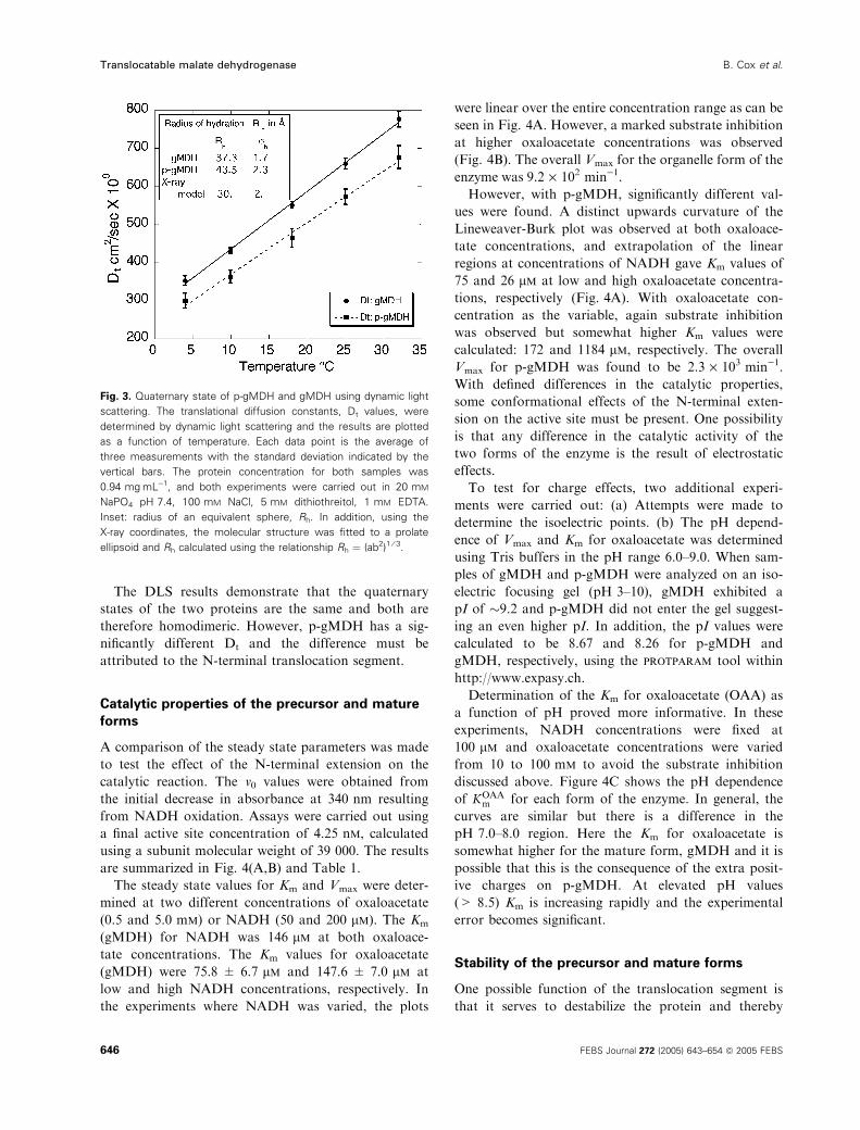
To compare the quaternary structure of the two
forms of the enzyme, dynamic light scattering was used
with the results shown in Fig. 3. Both the mature and
translocatable forms of the protein were linear as
expected in the 4–32 �C temperature range and the
translational diffusion constants are similar enough to
indicate the equivalent quaternary structure. As expec-
ted, p-gMDH has a somewhat smaller Dt than the
mature enzyme, and this can only result from the pres-
ence of the translocation sequence. The Dt values may
also be used to calculate the equivalent spherical
radius of hydration (Rh), and the results are summar-
ized in the insert of Fig. 3. The Rh calculations suggest
an approximate 15% increase in size for the translocat-
able form, p-gMDH. To make more direct comparisons
with the X-ray results, the Rh from the dynamic light
scattering (DLS) studies were also compared to esti-
mates obtained from the X-ray model and the results
shown in the insert of Fig. 3.
Fig. 2. Characterization of recombinant p-gMDH and gMDH. Three
methods were used to demonstrate the presence of the N-terminal
translocation segment in preparations of the recombinant p-gMDH.
In this composite, data is shown for samples free of proteolytic chan-
ges and in the inset an example of an SDS ⁄ PAGE experiment on a
purified sample that was not useful since some proteolysis had
occurred during the purification. The graph represents a MALDI-TOF
mass spectrum of purified p-gMDH free of any proteolytic modifica-
tion. Theoretical mass of p-gMDH is 38 517 Da, gMDH is 34 686 Da
and a frequently observed proteolytically modified form is 34 839 Da.
The inset contains an SDS ⁄ PAGE stained with Coomassie Brilliant
Blue with a sample showing some proteolytic modification. The eight
lanes from left to right contained (1) 0.3 mgÆmL)1 gMDH, (2)
0.15 mgÆmL)1 gMDH, (3) 0.075 mgÆmL)1 gMDH, (4) molecular mass
markers, (5) 0.025 mgÆmL)1 p-gMDH, (6) 0.05 mgÆmL)1 p-gMDH, (7)
0.1 mgÆmL)1 p-gMDH, (8) 0.2 mgÆmL)1 p-gMDH. Different amounts
of protein were applied to visualize any trace contamination. In lanes
5–8, a second band is visible indicating some proteolytic cleavage.
The upper left corner shows the results of an N-terminal sequencing
experiment, used in the early stages before mass spectroscopy
became readily available. The results of the experiment indicate a
preparation uncontaminated by proteolysis.
B. Cox et al. Translocatable malate dehydrogenase
FEBS Journal 272 (2005) 643–654 ª 2005 FEBS 645
The DLS results demonstrate that the quaternary
states of the two proteins are the same and both are
therefore homodimeric. However, p-gMDH has a sig-
nificantly different Dt and the difference must be
attributed to the N-terminal translocation segment.
Catalytic properties of the precursor and mature
forms
A comparison of the steady state parameters was made
to test the effect of the N-terminal extension on the
catalytic reaction. The v0 values were obtained from
the initial decrease in absorbance at 340 nm resulting
from NADH oxidation. Assays were carried out using
a final active site concentration of 4.25 nm, calculated
using a subunit molecular weight of 39 000. The results
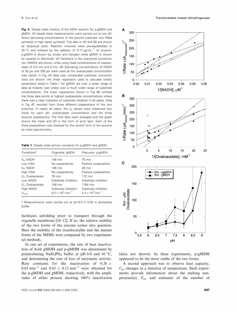
are summarized in Fig. 4(A,B) and Table 1.
The steady state values for Km and Vmax were deter-
mined at two different concentrations of oxaloacetate
(0.5 and 5.0 mm) or NADH (50 and 200 lm). The Km
(gMDH) for NADH was 146 lm at both oxaloace-
tate concentrations. The Km values for oxaloacetate
(gMDH) were 75.8 ± 6.7 lm and 147.6 ± 7.0 lm at
low and high NADH concentrations, respectively. In
the experiments where NADH was varied, the plots
were linear over the entire concentration range as can be
seen in Fig. 4A. However, a marked substrate inhibition
at higher oxaloacetate concentrations was observed
(Fig. 4B). The overall Vmax for the organelle form of the
enzyme was 9.2 · 102 min)1.
However, with p-gMDH, significantly different val-
ues were found. A distinct upwards curvature of the
Lineweaver-Burk plot was observed at both oxaloace-
tate concentrations, and extrapolation of the linear
regions at concentrations of NADH gave Km values of
75 and 26 lm at low and high oxaloacetate concentra-
tions, respectively (Fig. 4A). With oxaloacetate con-
centration as the variable, again substrate inhibition
was observed but somewhat higher Km values were
calculated: 172 and 1184 lm, respectively. The overall
Vmax for p-gMDH was found to be 2.3 · 103 min)1.
With defined differences in the catalytic properties,
some conformational effects of the N-terminal exten-
sion on the active site must be present. One possibility
is that any difference in the catalytic activity of the
two forms of the enzyme is the result of electrostatic
effects.
To test for charge effects, two additional experi-
ments were carried out: (a) Attempts were made to
determine the isoelectric points. (b) The pH depend-
ence of Vmax and Km for oxaloacetate was determined
using Tris buffers in the pH range 6.0–9.0. When sam-
ples of gMDH and p-gMDH were analyzed on an iso-
electric focusing gel (pH 3–10), gMDH exhibited a
pI of �9.2 and p-gMDH did not enter the gel suggest-
ing an even higher pI. In addition, the pI values were
calculated to be 8.67 and 8.26 for p-gMDH and
gMDH, respectively, using the protparam tool within
http://www.expasy.ch.
Determination of the Km for oxaloacetate (OAA) as
a function of pH proved more informative. In these
experiments, NADH concentrations were fixed at
100 lm and oxaloacetate concentrations were varied
from 10 to 100 mm to avoid the substrate inhibition
discussed above. Figure 4C shows the pH dependence
of KOAAm for each form of the enzyme. In general, the
curves are similar but there is a difference in the
pH 7.0–8.0 region. Here the Km for oxaloacetate is
somewhat higher for the mature form, gMDH and it is
possible that this is the consequence of the extra posit-
ive charges on p-gMDH. At elevated pH values
(> 8.5) Km is increasing rapidly and the experimental
error becomes significant.
Stability of the precursor and mature forms
One possible function of the translocation segment is
that it serves to destabilize the protein and thereby
Fig. 3. Quaternary state of p-gMDH and gMDH using dynamic light
scattering. The translational diffusion constants, Dt values, were
determined by dynamic light scattering and the results are plotted
as a function of temperature. Each data point is the average of
three measurements with the standard deviation indicated by the
vertical bars. The protein concentration for both samples was
0.94 mgÆmL)1, and both experiments were carried out in 20 mM
NaPO4 pH 7.4, 100 mM NaCl, 5 mM dithiothreitol, 1 mM EDTA.
Inset: radius of an equivalent sphere, Rh. In addition, using the
X-ray coordinates, the molecular structure was fitted to a prolate
ellipsoid and Rh calculated using the relationship Rh ¼ (ab2)1 ⁄ 3.
Translocatable malate dehydrogenase B. Cox et al.
646 FEBS Journal 272 (2005) 643–654 ª 2005 FEBS
facilitates unfolding prior to transport through the
organelle membrane [10–12]. If so, the relative stability
of the two forms of the enzyme comes into question.
Here the stability of the translocatable and the mature
forms of the MDHs were compared by two experimen-
tal methods.
In one set of experiments, the rate of heat inactiva-
tion of both gMDH and p-gMDH was determined by
preincubating NaH2PO4 buffer at pH 8.0 and 45 �C,and determining the rate of loss of enzymatic activity.
Rate constants for the inactivation of 0.20 ±
0.03 min)1 and 0.63 ± 0.12 min)1 were obtained for
the p-gMDH and gMDH, respectively, with the ampli-
tudes of either process showing 100% inactivation
(data not shown). In these experiments, p-gMDH
appeared to be the more stable of the two forms.
A second approach was to observe heat capacity,
Cp, changes as a function of temperature. Such experi-
ments provide information about the melting tem-
perature(s), Tm, and estimates of the number of
Fig. 4. Steady state kinetics of the MDH reaction for p-gMDH and
gMDH. All steady state measurements were carried out at two dif-
ferent saturating concentrations of the second substrate: low (filled
symbols) or high (open symbols). The data in (A) and (B) are shown
as reciprocal plots. Reaction mixtures were pre-equilibrated to
25 �C and initiated by the addition of 0.17 lgÆmL)1 of enzyme.
p-gMDH is shown by circles and triangles while gMDH is shown
by squares or diamonds. (A) Variations in the coenzyme concentra-
tion (NADH) are shown while using fixed concentrations of oxaloac-
etate of 0.5 mM and 5 mM. (B) Saturating concentrations of NADH
of 50 lM and 200 lM were used as the oxaloacetate concentration
was varied. In Fig. 4A data over comparable substrate concentra-
tions are shown: the linear regression used to calculate kinetic
parameters listed in Table 1 for gMDH are over a wider range of
data as linearity was visible over a much wider range of substrate
concentrations. The linear regressions shown in Fig. 4B omitted
the three data points at highest oxaloacetate concentrations where
there was a clear indication of substrate inhibition in all cases. Data
in Fig. 4C resulted from three different preparations of the two
enzymes. In nearly all cases, the v0 values were measured four
times for each pH, oxaloacetate concentration and the three
enzyme preparations. The final data were averaged and the graph
shows the mean and SD in the form of error bars. Each of the
three preparations was checked for the correct form of the enzyme
by mass spectrometry.
Table 1. Steady state activity constants for p-gMDH and gMDH.
Conditionsa Organelle: gMDH Precursor: p-gMDH
Km NADH 146 mM 75 mM
Low OAA No cooperativity Positive cooperativity
Km NADH 146 mM 26 mM
High OAA No cooperativity Positive cooperativity
Km Oxaloacetate 76 mM 172 mM
Low NADH Substrate inhibition Substrate inhibition
Km Oxaloacetate 148 mM 1184 mM
High NADH Substrate inhibition Substrate inhibition
Vmax 9.2 · 102 min)1 2.3 · 103 min)1
a Measurements were carried out at pH 8.0 in 0.02 M phosphate
buffer.
B. Cox et al. Translocatable malate dehydrogenase
FEBS Journal 272 (2005) 643–654 ª 2005 FEBS 647
transition(s) during the unfolding reaction [13]. If dif-
ferent transitions were apparent in the comparable
unfolding processes for gMDH and p-gMDH, the
results would suggest some unique structural compo-
nents such as additional domains, dissimilar states of
quaternary structure and ⁄or different enthalpic proper-
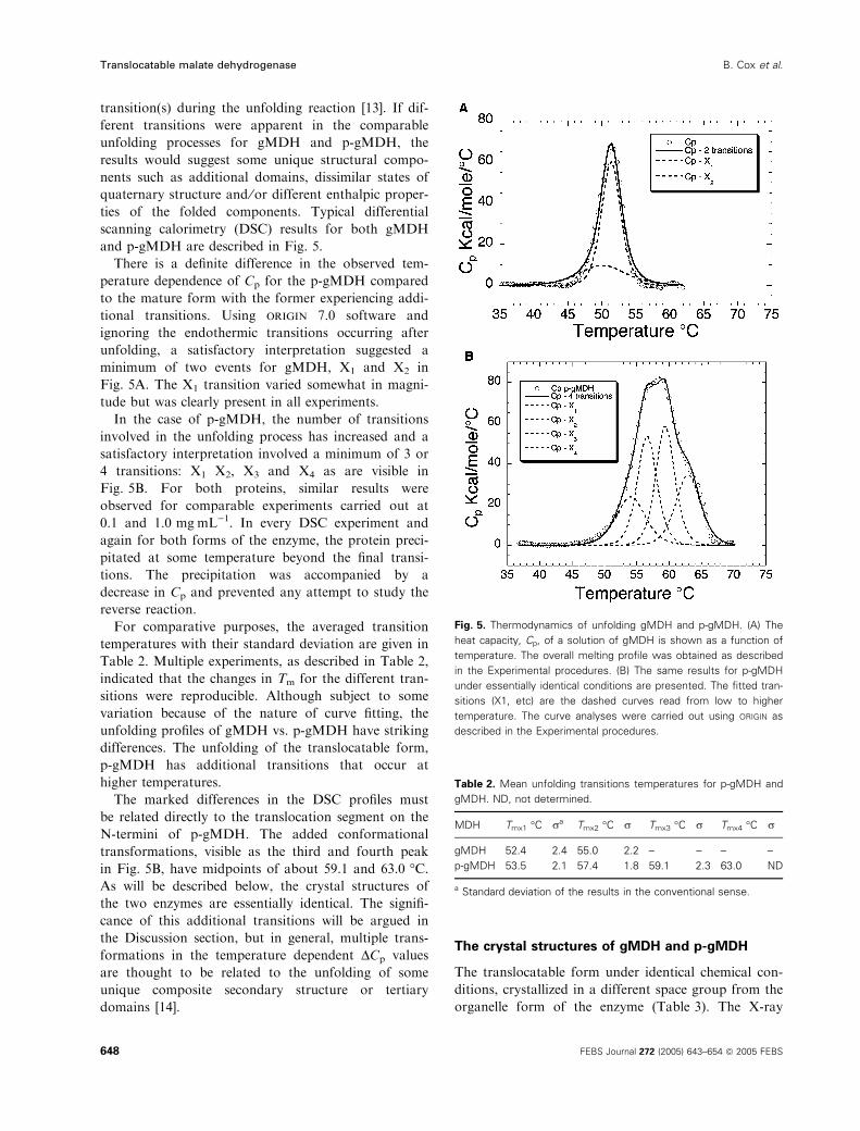
ties of the folded components. Typical differential
scanning calorimetry (DSC) results for both gMDH
and p-gMDH are described in Fig. 5.
There is a definite difference in the observed tem-
perature dependence of Cp for the p-gMDH compared
to the mature form with the former experiencing addi-
tional transitions. Using origin 7.0 software and
ignoring the endothermic transitions occurring after
unfolding, a satisfactory interpretation suggested a
minimum of two events for gMDH, X1 and X2 in
Fig. 5A. The X1 transition varied somewhat in magni-
tude but was clearly present in all experiments.
In the case of p-gMDH, the number of transitions
involved in the unfolding process has increased and a
satisfactory interpretation involved a minimum of 3 or
4 transitions: X1 X2, X3 and X4 as are visible in
Fig. 5B. For both proteins, similar results were
observed for comparable experiments carried out at
0.1 and 1.0 mgÆmL)1. In every DSC experiment and
again for both forms of the enzyme, the protein preci-
pitated at some temperature beyond the final transi-
tions. The precipitation was accompanied by a
decrease in Cp and prevented any attempt to study the
reverse reaction.
For comparative purposes, the averaged transition
temperatures with their standard deviation are given in
Table 2. Multiple experiments, as described in Table 2,
indicated that the changes in Tm for the different tran-
sitions were reproducible. Although subject to some
variation because of the nature of curve fitting, the
unfolding profiles of gMDH vs. p-gMDH have striking
differences. The unfolding of the translocatable form,
p-gMDH has additional transitions that occur at
higher temperatures.
The marked differences in the DSC profiles must
be related directly to the translocation segment on the
N-termini of p-gMDH. The added conformational
transformations, visible as the third and fourth peak
in Fig. 5B, have midpoints of about 59.1 and 63.0 �C.As will be described below, the crystal structures of
the two enzymes are essentially identical. The signifi-
cance of this additional transitions will be argued in
the Discussion section, but in general, multiple trans-
formations in the temperature dependent DCp values
are thought to be related to the unfolding of some
unique composite secondary structure or tertiary
domains [14].
The crystal structures of gMDH and p-gMDH
The translocatable form under identical chemical con-
ditions, crystallized in a different space group from the
organelle form of the enzyme (Table 3). The X-ray
Fig. 5. Thermodynamics of unfolding gMDH and p-gMDH. (A) The
heat capacity, Cp, of a solution of gMDH is shown as a function of
temperature. The overall melting profile was obtained as described
in the Experimental procedures. (B) The same results for p-gMDH
under essentially identical conditions are presented. The fitted tran-
sitions (X1, etc) are the dashed curves read from low to higher
temperature. The curve analyses were carried out using ORIGIN as
described in the Experimental procedures.
Table 2. Mean unfolding transitions temperatures for p-gMDH and
gMDH. ND, not determined.
MDH Tmx1 �C ra Tmx2 �C r Tmx3 �C r Tmx4 �C r
gMDH 52.4 2.4 55.0 2.2 – – – –
p-gMDH 53.5 2.1 57.4 1.8 59.1 2.3 63.0 ND
a Standard deviation of the results in the conventional sense.
Translocatable malate dehydrogenase B. Cox et al.
648 FEBS Journal 272 (2005) 643–654 ª 2005 FEBS
studies are summarized as follows. p-gMDH crystals
diffracted to 2.55 A and have a Matthews’ coefficient
(Vm) of 3.3 A3ÆDa)1, with 1 dimer in the crystallo-
graphic asymmetric unit. The structure of p-gMDH
was solved by molecular replacement using the poly-
alanine equivalent of the dimer coordinates from por-
cine heart mitochondrial MDH (1mld, PDB) [15].
GMDH crystals diffracted to a resolution of 2.50 A
under cryogenic conditions, and gave a Vm of 2.45
A3ÆDa)1 with four dimers in the asymmetric unit. The
molecular packing in the crystal lattice produces a
pseudo space group, C2221. Many hours were spent
trying to process the raw data using the centered or-
thorhombic system but the refinement and predictions
were always short of satisfactory, and the X-ray struc-
ture was solved in the monoclinic space group using
the coordinates of p-gMDH and the method of
molecular replacement. The refinement results for both
crystal structures and their PDB accession codes are
given in Table 4.
Discussion of the quaternary and tertiary structure
of the gMDHs seems unnecessary in lieu of the many
crystal structures in the PDB. Instead using the avail-
able coordinates for the enzymes shown in Fig. 1 and
the method of least squares, studies to compare the
main chain conformations were carried out. The main
chain conformation of gMDH is shown by the red
tubular representation in Fig. 6. In both forms of the
enzyme, electron density was first visible beginning at
G44 (precursor numbering scheme), seven amino acids
into the N-termini of the mature form. At least in
these two crystal structures, the dynamically disordered
translocation segment begins at an amino acid present
in the N-terminus of the cleaved enzyme.
The different crystal packing between gMDH and
p-gMDH under identical chemical conditions corro-
borates but does not prove the presences of the prese-
quence in the one lattice. In the crystal structure of
p-gMDH, the first visible N-terminal residue points
directly into a solvent channel of the lattice suggesting
that there is adequate space for the substantial but dis-
ordered translocation segment.
Because of the large asymmetric unit for gMDH
(four molecules, eight subunits) coupled with the two
subunits determined for p-gMDH, the coordinates of a
single subunit of this glyoxysomal enzyme are tenfold
redundant. The reliability of the coordinates and the
site of any significant differences could be estimated by
cross-comparisons (45 in total). To do this, crystal
coordinates of each independent subunit were overlaid
pairwise by the method of least squares and the result-
ing root mean square deviations (A) compared. The
outcome is summarized in Fig. S1 (supplementary
data). Overall, the coordinates for the two forms of
the protein are in close agreement. Among the 10 cop-
ies of the gMDH monomer in the crystal structures,
the root mean square deviation between Ca atoms is
between 0.2 and 0.5 A, near to the anticipated experi-
mental error. However there are two regions that
appear to contain conformational variability.
The first region is around residues 123–132, the
so-called active site loop. The electron density in this
region was generally low in both of the crystal struc-
tures and most of the subunits suggesting that this
loop is not always in the same conformation. This
disordered segment has the amino acid sequence:
-P-R-K-P-G-M-T-R-D-D- (123–132), and overall these
residues have the highest B-factors of the entire struc-
ture. R124 near the tip of a loop is believed to be
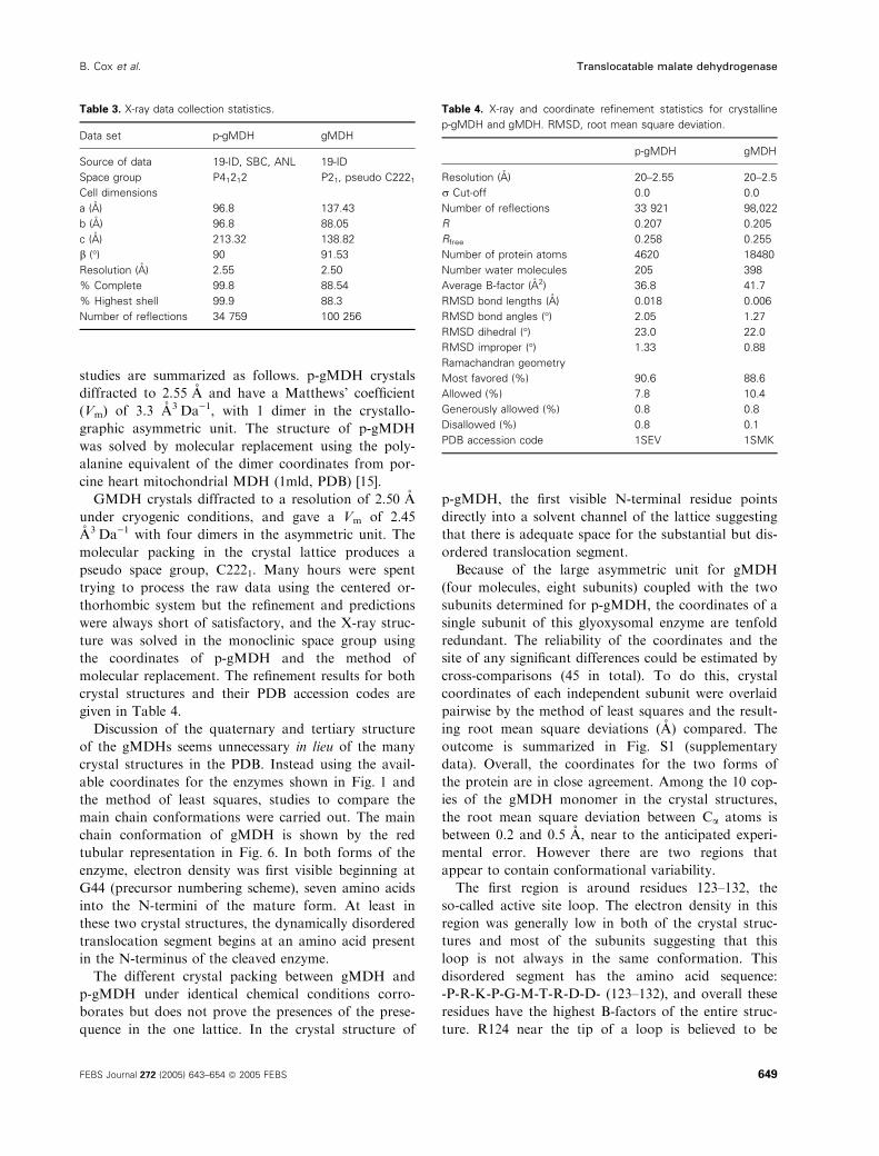
Table 3. X-ray data collection statistics.
Data set p-gMDH gMDH
Source of data 19-ID, SBC, ANL 19-ID
Space group P41212 P21, pseudo C2221
Cell dimensions
a (A) 96.8 137.43
b (A) 96.8 88.05
c (A) 213.32 138.82
b (�) 90 91.53
Resolution (A) 2.55 2.50
% Complete 99.8 88.54
% Highest shell 99.9 88.3
Number of reflections 34 759 100 256
Table 4. X-ray and coordinate refinement statistics for crystalline
p-gMDH and gMDH. RMSD, root mean square deviation.
p-gMDH gMDH
Resolution (A) 20–2.55 20–2.5
r Cut-off 0.0 0.0
Number of reflections 33 921 98,022
R 0.207 0.205
Rfree 0.258 0.255
Number of protein atoms 4620 18480
Number water molecules 205 398
Average B-factor (A2) 36.8 41.7
RMSD bond lengths (A) 0.018 0.006
RMSD bond angles (�) 2.05 1.27
RMSD dihedral (�) 23.0 22.0
RMSD improper (�) 1.33 0.88
Ramachandran geometry
Most favored (%) 90.6 88.6
Allowed (%) 7.8 10.4
Generously allowed (%) 0.8 0.8
Disallowed (%) 0.8 0.1
PDB accession code 1SEV 1SMK
B. Cox et al. Translocatable malate dehydrogenase
FEBS Journal 272 (2005) 643–654 ª 2005 FEBS 649
involved in interaction with the dicarboxylic acid sub-
strates during the formation of catalytic intermediates.
The binding of substrate is believed to draw the loop
further into the active site and the conformational
change may be necessary for catalysis [16].
The second conformational variation occurs in the
polypeptide chains at residues 256–268. In the dimeric
MDH family, the interface is formed by the packing of
three a-helices from each subunit. In the amino acid
sequence, the last of the three helices begins at residue
268. To examine the dimer interface for each of the
two enzymes, the X-ray structures were used to esti-
mate the change in the solvent accessible surface area
as a result of dimer formation. For gMDH the loss
was 3180 ± 63 A2. For p-gMDH crystallographic
coordinates indicated a loss of 3160 ± 41 A2, and
using this criterion, the two interfaces are identical.
Discussion
Recognition and translocation of nuclear-encoded
organelle proteins appear to have many aspects in
common even though the current dogma explaining
these processes differs for organelle classes [17].
Common elements include a variety of protein factors,
both cytosolic and membranous, that appear to be spe-
cific for a given organelle type. For the organelles
mentioned in the Introduction and proteins destined
for translocation, segments of extra amino acids
mostly on the N-termini are required for the import
process. Questions arise as to whether or not there are
discernible differences in the properties of these pro-
teins with and without these so-called translocation
segments.
In the experiments described above, one enzyme was
compared in both its translocatable and organelle
form. In the crystallographic portion of the experi-
ments, two conformational variations were identified
between the two forms, but no electron density was
visible for the translocation segment. Lack of electron
density in a protein crystal structure is explainable by
dynamic disorder; that is, the structural element is in
multiply interchangeable conformations in the crystal
lattice.
The comparison of the physical and catalytic proper-
ties of the two nearly identical enzymes has none-
theless produced some surprising differences. For
example, one possible consequence of the presence of
the translocation segment would be to alter the quater-
nary structure. GMDH and most but not all MDHs
are dimers. The DLS results as described above indica-
ted that p-gMDH is homodimeric like the organelle
form. However, the precursor had a significantly smal-
ler Dt, and using the assumption of a spherical
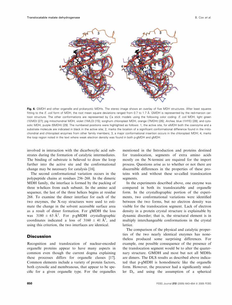
Fig. 6. GMDH and other organelle and prokaryotic MDHs. The stereo image shows an overlay of five MDH structures. After least squares
fitting to the E. coli form of MDH, the root mean square deviations ranged from 0.7 to 1.7 A. GMDH is represented by the red-maroon car-
toon structure. The other conformations are represented by Ca stick models using the following color coding: E. coli MDH, light green
(1EMD) [27]; pig mitochondrial MDH, violet (1MLD) [15]; sorghum chloroplast MDH, orange (7MDH) [26]; Archea, blue (1HYE) [28]; and cyto-
solic MDH, purple (5MDH) [29]. The numbered positions were highlighted as follows: 1, the active site, for eMDH both the coenzyme and a
substrate molecule are indicated in black in the active site; 2, marks the location of a significant conformational difference found in the mito-
chondrial and chloroplast enzymes from other family members; 3, a major conformational insertion occurs in the chloroplast MDH; 4, marks
the loop region noted in the text where weak electron density was found in both p-gMDH and gMDH.
Translocatable malate dehydrogenase B. Cox et al.
650 FEBS Journal 272 (2005) 643–654 ª 2005 FEBS
molecule in analyzing translational diffusion, it is
approximately 15% larger. Although the crystallo-
graphic studies show that the dimers are not spherical,
the additional N-terminal segment has measurable
effects on the overall hydrodynamic properties of the
enzyme.
Enzyme catalysis is generally a function of a finely
tuned atomic array at the active site and the transloca-
tion segment appears to have no dramatic effect on this
constellation. However, comparison of the steady state
properties demonstrated that the active sites are nearly
the same but small differences are identifiable. At high
concentrations of substrates, the turnover number for
p-gMDH was improved by nearly a factor of two over
the organelle form. How could this happen? In most of
the NAD+-dependent dehydrogenase reactions, the
catalytic step of hydride transfer, or a linked conforma-
tional change, is probably rate limiting. During sub-
strate inhibition, there must be either formation of
abortive complexes or substrate hindrance of normally
rate limiting conformational changes.
One explanation for changes in catalytic parameters
is the fact that the precursor with the translocation
segment adds seven extra positive charges (if two histi-
dines are included), but only three acidic residues.
Depending on the pK values of the histidines, the net
charge change ranges from +2 to +4. Vmax for both
forms of the enzyme showed similar pH dependence
with a broad optimum between pH 7.5 and 8.0. In this
relatively narrow pH region, the Km for oxaloacetate is
smaller for the precursor form of the enzyme. Assu-
ming the same mechanism occurs in both forms of the
enzyme, it is possible that the additional basic residues
present in the translocation segment are increasing the
affinity for the anionic substrate, oxaloacetate.
The results of superimposition of the available crys-
tallographic coordinates of MDHs presented in Fig. 6
above was carried out to analyze both conformation
and properties that might distinguish organelle
enzymes from other forms. In terms of molecular
structure, only two sites on a MDH monomer were
visibly different. At one site, the mitochondrial and
chloroplast enzymes have a small insertion labeled as
‘2’ in Fig. 6. The chloroplast MDH also contains a
large insertion at ‘3’ that, in fact, is adjacent to the
N-terminus. However overall, the conformations inclu-
ding the prokaryotic forms of MDH are very similar.
The conclusion is that if translocation operates
with folded proteins [17,18], the presence of the
translocation segment may be related solely to the
recognition process.
The removal of an N-terminal segment from many
of the nuclear-coded proteins upon reaching the matrix
of an organelle is another interesting step of the trans-
location phenomena. Because it is proteolytically
cleaved during movement into the organelle, a region
of the translocation segment must be accessible to the
cellular protease whether or not the protein is already
folded. The crystallographic results on p-gMDH and
gMDH indicate that conformational variation near
the N-terminal appears to begin several residues into
the chain of the mature protein. This would make the
upstream cleavage site more available to a protease.
The proposed mitochondrial import mechanism is
purported to involve threading the random coil form
through a preformed pore [1]. On the other hand,
recent evidence seems to show that glyoxysomal ⁄per-oxisomal import occurs with fully folded proteins [18].
The studies described above cannot attest to either
proposal. However, the gMDH vs. p-gMDH compar-
ison has shown that unfolding of the precursor form
is more energy requiring than the unfolding of the
organelle form of the protein lacking the translocation
segment.
To summarize, the results described above illustrate
methods for preparing quantities of translocatable
protein by recombinant methods. For glyoxysomal
MDH, the precursor form folds normally but the
N-terminal segment is extremely sensitive to proteo-
lysis. Hydrodynamic and enzymatic characterization
of p-gMDH compared to gMDH showed that the
two proteins are similar but not identical. The most
surprising result stems from the DSC studies. The
results of the heat capacity changes as a function of
temperature point to additional structure in p-gMDH.
Preliminary results (not shown) suggest that NADP-
dependent isocitrate dehydrogenase belonging to yeast
mitochondria produce similar DSC results when com-
pared to the identical enzyme without the N-terminal
translocation segment. If these observations can be
demonstrated in other organelle enzymes, unfolding
prior to translocation does not appear to be a viable
hypothesis.
Experimental procedures
Reagents
The plasmid pQE 60 containing the cDNA sequence of
either p-gMDH or gMDH and a His6 tag at the C-terminus
was generated as previously described [19]. Ni2+–nitrilotri-
acetate (Ni2+–NTA) resin was purchased from Qiagen
(Valencia, CA, USA). Pefabloc was purchased from Roche
Biochemicals (Indianapolis, IN, USA). All other reagents
were obtained from Sigma Chemical Co. (St. Louis, MO,
USA).
B. Cox et al. Translocatable malate dehydrogenase
FEBS Journal 272 (2005) 643–654 ª 2005 FEBS 651

Purification of p-gMDH and gMDH
P-gMDH and gMDH were expressed in the E. coli strain
JM105 from a derivative of pQE 60 that carries either
p-gMDH or gMDH cDNA with an inserted codon for gly-
cine (GGA) between the start codon and the second codon
of both cDNAs [19]. Cells were grown at 37 �C in LB med-
ium in six 1 L flasks supplemented with ampicillin
(100 lgÆmL)1). When the cells reached D600 � 0.6 the tem-
perature was lowered to 32 �C and grown for 5 h after
induction with 1.0 mm isopropyl thio-b-d-galactoside. Cellswere harvested by centrifugation at 4000 g for 30 min and
stored at )80 �C.To isolate the MDHs, cell pastes were thawed and re-sus-
pended to 0.5 gÆmL)1 in Buffer A (50 mm Tris pH 7.2,
300 mm NaCl). All steps were conducted at 4 �C unless
otherwise noted. A cocktail of protease inhibitors was
added to Buffer A to a final concentration of 4 mm
Pefabloc, 100 mgÆmL)1 phenylmethylsulfonyl fluoride,
1.0 mgÆmL)1 pepstatin, 2.0 mm leupeptin, 1.0 mm benzami-
dine, and 1 mL of Sigma protease inhibitor cocktail P8849
per 20 g cell paste. The suspension was subjected to five,
30 s cycles of sonication in a dry ice ⁄ isopropanol bath fol-
lowed by the addition of 0.35% polyethylenimine to preci-
pitate the DNA. Cell debris and DNA were pelleted at
17 500 g for 15 min.
The supernatant was mixed for 5 min with Ni2+–NTA
resin (1 mL resin per 5 g cells) equilibrated with the Buf-
fer A. The column was washed three times with three col-
umn volumes of Buffer B (50 mm Tris pH 7.2, 300 mm
NaCl, 60 mm imidazole). p-gMDH or gMDH was eluted
with two column volumes of Buffer C (50 mm Tris
pH 7.2, 300 mm NaCl, 500 mm imidazole). Protease inhib-
itors were not included beyond the cell lyses step. Both
proteins were then dialyzed against 4 L of buffer contain-
ing 50 mm NaH2PO4 pH 7.4, 300 mm NaCl, then dialyzed
into 50 mm NaH2PO4 pH 7.4, 300 mm NaCl, 1 mm
dithiothreitol, and 1 mm EDTA, using four, one liter buf-
fer changes. One way of insuring success in the purifica-
tion of p-gMDH was to complete the entire procedure,
cells to pure protein, in about 1 h.
Steady state measurements
The catalytic activities of p-gMDH and gMDH were deter-
mined by following the disappearance of NADH by moni-
toring the absorbance at 340 nm in a reaction solution
containing 50 mm phosphate at pH 8.0, 100 lm NADH
and 1 mm oxaloacetate. To obtain the steady state parame-
ters as a function of pH, 50 mm Tris HCl buffers were
used. Reactions were initiated by the addition of 10–60 ng
of protein to 3 mL of assay solution. All measurements
were minimally carried out in triplicate and values of Km
and Vmax are averages of three to five independent determi-
nations of v0.
Characterization of p-gMDH and gMDH
The purity of the protein was verified by SDS ⁄PAGE with
Coomassie brilliant blue staining. The concentrations of
purified p-gMDH and gMDH were determined spectro-
photometrically using an e280nm ¼ 0.475 mLÆmg)1Æcm)1.
The translational diffusion coefficient (Dt) was determined
for both p-gMDH and gMDH at different temperatures by
dynamic light scattering at a concentration of 0.94 mgÆmL)1
using a Protein Solutions DynaPro Micro Sampler. All pro-
tein samples used for light scattering experiments were pre-
filtered with 0.02 lm Whatman Anotop Plus filter discs.
Verification of the presequence
Because of the sensitivity of p-gMDH to proteases, the puri-
fied fractions were always examined either by N-terminal
sequencing analysis (University of Minnesota Micro-Chem-
ical Facility) or mass spectrometry (Mass Spectrometry
Consortium for the Life Sciences, University of Minnesota)
and SDS ⁄PAGE. Protein samples were sequenced from
solutions containing 50 mm NaH2PO4 pH 7.4, and 300 mm
NaCl. In one experiment, upon completion of X-ray data
collection, a p-gMDH crystal was re-dissolved into 50 mm
NaH2PO4 pH 7.4, 300 mm NaCl and the N-terminal
sequence determined (data not shown).
The instrument used for the collection of MALDI-TOF
data was a Bruker Biflex III, equipped with a N2 laser
(337 nm, 3 nanosecond pulse length) and a microchannel
plate (MCP) detector. The data was collected in the linear
mode, positive polarity, with an accelerating potential of
19 kV. Calibration was performed using bovine serum albu-
min (MH+ ⁄Z ¼ 66431, MH2+ ⁄Z ¼ 33216).
Thermal transitions
Heat capacity changes as a function of temperature were
obtained with a VP-DSC differential scanning calorimeter
(MicroCal, Northampton, MA, USA) in 50 mm NaH2PO4
pH 7.4 and 100 mm NaCl, with a heating rate of 2 �CÆmin)1.
The resulting curve was analyzed with the origin 7.0 (http://
www.micocal.com) software using the minimal number of
transitions to fit the observations. For both proteins, some
precipitate was present at the completion of the experiment.
Crystallization and X-ray data collection
Crystallization was carried out by first dialyzing the puri-
fied enzyme against a buffer containing 10 mm NaH2PO4
pH 7.4, 100 mm NaCl, 5 mm dithiothreitol, 1 mm EDTA.
The dialyzed protein was concentrated to 10 mgÆmL)1 using
an Amicon concentrator (PM10, 43 mm). Crystals of
p-gMDH and gMDH were grown at 20 �C by hanging
drop – vapor diffusion from mother liquor containing
Translocatable malate dehydrogenase B. Cox et al.
652 FEBS Journal 272 (2005) 643–654 ª 2005 FEBS
100 mm sodium citrate pH 4.5, 1 mm dithiothreitol, and
PEG 8000. Needle-shaped crystals of both p-gMDH and
gMDH appeared in a few days in the range of 6.5–11.0%
and 9.0–13.5% (w ⁄ v) of PEG 8000, respectively.
Crystals of p-gMDH and gMDH were cryo-protected by
adding a 50% glycerol solution to the crystallization drop
resulting in a final concentration of 33% (v ⁄ v) glycerol.
Data sets for p-gMDH and gMDH were collected at the
SBC Beamline 19-ID (ANL-APS) with the 3 · 3 CCD area
detector set at 2h ¼ 0�. Data were indexed and integrated
with denzo and scaled with scalepack [20].
Phases were found for p-gMDH by molecular replace-
ment using the dimeric structure of porcine heart mitoch-
ondrial malate dehydrogenase substituted with poly-alanine
as a search model using x-plor [21]. One dimer was present
in the asymmetric unit. The phases of gMDH were also
found by molecular replacement using the partially refined
model of p-gMDH and the program EPMR [22]. Four
dimers were present in the asymmetric unit of a P21 unit
cell. During the refinement, the space group was found to
be pseudo C-centered. A further explanation of the space
group ambiguity is given in the Results section.
Both X-ray structures were refined using cns v.0.3 [23].
The coordinates were refined by rigid body, then simulated
annealing, positional refinement, and B-factor refinement.
Five percent of the data were omitted from refinement and
used for Rfree calculations [24]. Non-crystallographic sym-
metry was used to restrain the four dimers of gMDH dur-
ing the early rounds of positional and B-factor refinement,
and then released during subsequent macro-cycles. The pro-
gram o was used for model rebuilding after each round of
crystallographic refinement [25].
Acknowledgements
The authors recognize the Mass Spectrometry Consor-
tium for the Life Sciences at the University of
Minnesota and various supporting agencies, including
the National Science Foundation for Major Research
Instrumentation grant 9871237, used to purchase the
instruments described in this study. We thank the
Microchemical Facility (http://mcf.ahc.umn.edu) at
the University of Minnesota for help in characterizing
the proteins. We remain continually grateful to Ed Hoe-
ffner for the maintenance of hardware and software
associated with the X-ray diffraction, DSC and DLS
instrumentation. The SBC station at the Argonne
National Laboratory was used for collection of some X-
ray data. We are also grateful to the Minnesota
Supercomputer Institute for continuing use of their
facilities necessary for X-ray crystallography especially
the use of the Biomedical Computing Science Laborat-
ory and software maintenance by Dr Patton Fast. This
work was partially supported by a grant from the
National Institutes of Health (GM 13925). Bryan Cox is
grateful for support from the NIH Biophysical Training
Grant (GM08277) and initially by a research grant from
the NSF.
References
1 Schatz G (1998) The doors to organelles. Nature 395,
439–440.
2 Steer C & Hanover J (1991) Intracellular Trafficking
of Proteins. Cambridge University Press, Cambridge,
UK.
3 Veenhuis M, Kiel JAKW & Van Der Klei IJ (2003) Per-
oxisome assembly in yeast. Microsc Res Technique 61,
139–150.
4 Faber KN, Haima P, Gietl C, Harder W, Geert AB &
Veenhuis M (1994) The methylotrophic yeast Hansenula
polymorpha contains an inducible import pathway for
peroxisomal matrix proteins with an N-terminal target-
ing signal (PTS2 proteins), Proc Natl Acad Sci USA 91,
12985–12989.
5 Fransen M, Terlecky SR & Subramani S (1998) Identifi-
cation of a human Pts1 receptor docking protein
directly required for peroxisomal protein import. Proc
Natl Acad Sci USA 95, 8087–8092.
6 Mathieu M, Zeelen J, Pauptit RA, Erdmann R,
Kunau WH & Wierenga RK (1994) The 2.8 A crystal
structure of peroxisomal 3-ketoacyl-CoA thiolase of
Saccharomyces cerevisiae: a five-layered ABABA struc-
ture constructed from two core domains of identical
topology. Structure 2, 797–808.
7 Chudzik DM, Michels PA, de Walque S & Hol WG
(2000) Structures of type 2 peroxisomal targeting signal
in two trypanosomatid aldolases. J Mol Biol 300,
697–707.
8 Gietl C (1992) Malate dehydrogenase isoenzymes: cellu-
lar locations and role in the flow of metabolites between
the cytoplasm and cell organelles. Biochim Biophys Acta
1100, 217–234.
9 Gietl C (1990) Glyoxysomal malate dehydrogenase from
watermelon is synthesized with an amino-terminal pep-
tide. Proc Natl Acad Sci USA 87, 5773–5777.
10 Huang S, Ratliff K & Matouschek A (2002) Protein
unfolding by the mitochondrial membrane potential.
Nat Struct Biol 9, 301–307.
11 Huang S, Ratiff KS, Schwartz M, Spenner J &
Matouschek A (1999) Mitochondria unfold precursor
proteins by unraveling them from their N-termini. Nat
Struct Biol 6, 1132–1138.
12 Pfanner N, Rassow J, Guiard B, Sollner T, Hartl F &
Neupert W (1990) Energy requirements for unfolding
and membrane translocation of precursor proteins dur-
ing import into mitochondria. J Biol Chem 265, 16324–
16329.
B. Cox et al. Translocatable malate dehydrogenase
FEBS Journal 272 (2005) 643–654 ª 2005 FEBS 653
13 Makhatadze G & Privalov P (1995) Energetics of pro-
tein structure. Adv Protein Chem 47, 307–425.
14 Privalov P (1989) Thermodynamic problems of pro-
tein structure. Ann Rev Biophys Biophys Chem 18,
47–69.
15 Gleason WB, Fu ZJ, Birktoft J & Banaszak L (1994)
Refined crystal structure of mitochondrial malate dehy-
drogenase from porcine heart and the consensus struc-
ture for dicarboxylic acid oxidoreductases. Biochemistry
33, 2078–2088.
16 Bell J, Yennawar H, Wright SK, Thompson J, Viola R
& Banaszak L (2001) Structural analyses of a malate
dehydrogenase with a variable active site. J Biol Chem
276, 31156–31162.
17 Smith MD & Schnell DJ (2001) Peroxisomal protein
import: the paradigm shifts. Cell 105, 293–296.
18 Dammai V & Subramani S (2001) The human peroxiso-
mal targeting signal receptor, Pex5p, is translocated into
the peroxisomal matrix and recycled to the cytosol. Cell
105, 187–196.
19 Gietl C, Seidel C & Svendsen I (1996) Plant glyoxyso-
mal but not mitochondrial malate dehydrogenase can
fold without chaperone assistance. Biochim Biophys Acta
1274, 48–58.
20 Otwinowski Z & Minor W (1997) Processing of X-ray
data collected in oscillation mode. Methods Enzymol
276, 307–326.
21 Brunger AT, Adams PD, Clore GM, Delano WL,
Gros P, Grossekunstleve RW, Jiang JS, Kuszewski J,
Nilges M, Pannu NS, Read RJ, Rice LM, Simonson T
& Warren GL (1998) Crystallography and NMR system
– a new software suite for macromolecular structure
determination. Acta Crystallogr Sect D Biol Crystallogr
54, 905–921.
22 Kissinger CR (2001) Molecular replacement by evolu-
tionary search. Acta Crystallogr Sect D Biol Crystallogr
57, 1474–1479.
23 Brunger AT, Adams PD & Rice LM (1998) Recent
developments in the efficient crystallographic refinement
of macromolecular structures. Curr Opin Struct Biol 8,
606–611.
24 Kleywegt GJ & Brunger AT (1996) Checking your ima-
gination: applications of the free R value. Structure 4,
897–904.
25 Jones T, Zou J, Cowan S & Kjeldgaard M (1991)
Improved methods for building protein models in
electron density maps and the location of errors in these
models. Acta Crystallogr Sect A Found Crystallogr A47,
110–119.
26 Johansson K, Ramaswamy S, Saarinen M, Lemaire-
Chamley M, Issakidis-Bourguet E, Miginiac-Maslow M
& Eklund H (1999) Structural basis for light activation
of a chloroplast enzyme: the structure of sorghum
NADP-malate dehydrogenase in its oxidized form.
Biochemistry 38, 4319–4326.
27 Hall MD & Banaszak LJ (1993) Crystal structure of a
ternary complex of Escherichia coli malate dehydrogen-
ase and NAD at 1.9 A resolution. J Mol Biol 232,
213–222.
28 Panisko E & Mcalister-Henn L (2001) Subunit interac-
tions of yeast NAD+-specific isocitrate dehydrogenase.
J Biol Chem 276, 1204–1210.
29 Chapman ADM, Cortes A, Dafforn TR, Clarke AR &
Brady RL (1999) Structural basis of substrate specificity
in malate dehydrogenases: crystal structure of a ternary
complex of porcine cytoplasmic malate dehydrogenase,
alpha-ketomalonate and tetrahydonad. J Mol Biol 285,
703–712.
Supplementary Material
The following material is available from http://www.
blackwellpublishing.com/products/journals/suppmat/EJB/
EJB4475/EJB4475sm.htm
Fig. S1. Pairwise comparison of the X-ray crystal
structures of gMDH and p-gMDH. The X-ray studies
described in the accompanying text resulted in eight
sets of atomic coordinates for the gMDH subunit.
After superimposition by the method of least squares
over the entire monomer, pairwise comparisons of the
coordinates were calculated at each Ca position. These
were averaged and the standard deviation calculated.
The monomers of p-gMDH were also superimposed
and a pairwise comparison was done. The results are
shown in the plot. The solid line is r, the standard
deviation, at each Ca position and the mean RMSD
positional difference is represented by the dotted line.
To avoid confusion, the baseline of the p-gMDH data
was raised from 0 to 2.
Translocatable malate dehydrogenase B. Cox et al.
654 FEBS Journal 272 (2005) 643–654 ª 2005 FEBS


