Embed Size (px)
Citation preview
Approach to a child with hematemesis or melena
Avijeet k. Mishra
1st year Resident
Guide – Dr Surya Bahadur Thapa
DOCH
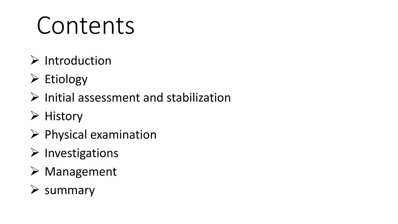
Contents Introduction
Etiology
Initial assessment and stabilization
History
Physical examination
Investigations
Management
summary
Case
A 32 months old male presented to us with a history of 2 days of abdominal pain , 3 episodes of black colored stool and 1 episode of fresh blood mixed with feces small in quantity without any similar past history. He had uneventful neonatal period and no history of rashes or bleeding from other sites.

Introduction
Upper gastrointestinal bleeding-
Bleeding from a site proximal to the ligament of Treitz
Hematemesis is the cardinal sign
Some may present with melena
Lower gastrointestinal bleed-
Bleeding from site distal to the ligament of Treitz
Hematochezia is the usual presentation

Contd.. Hematemesis-
Vomiting of blood which may be red or coffee grounds
Melena-
Passage of black tarry stools
Action of digestive enzymes and bacteria change the color of stool to
black tarry and foul smelling
Hematochezia –
Passage of fresh blood per anus, usually in or with stool

Etiology
Neonate Swallowed maternal blood
- During delivery
- From mother’s nipple
Coagulopathy
- Hemorrhagic disease of the newborn
- Septicemia, DIC
- Hemophilia
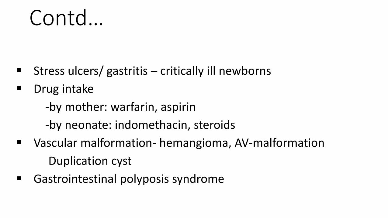
Contd…
Stress ulcers/ gastritis – critically ill newborns
Drug intake
-by mother: warfarin, aspirin
-by neonate: indomethacin, steroids
Vascular malformation- hemangioma, AV-malformation
Duplication cyst
Gastrointestinal polyposis syndrome
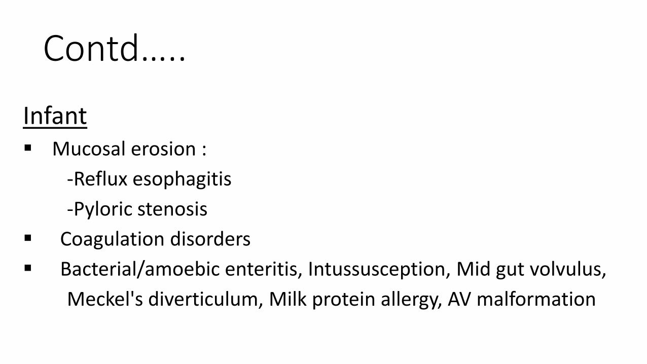
Contd…..
Infant Mucosal erosion :
-Reflux esophagitis
-Pyloric stenosis
Coagulation disorders
Bacterial/amoebic enteritis, Intussusception, Mid gut volvulus,
Meckel's diverticulum, Milk protein allergy, AV malformation
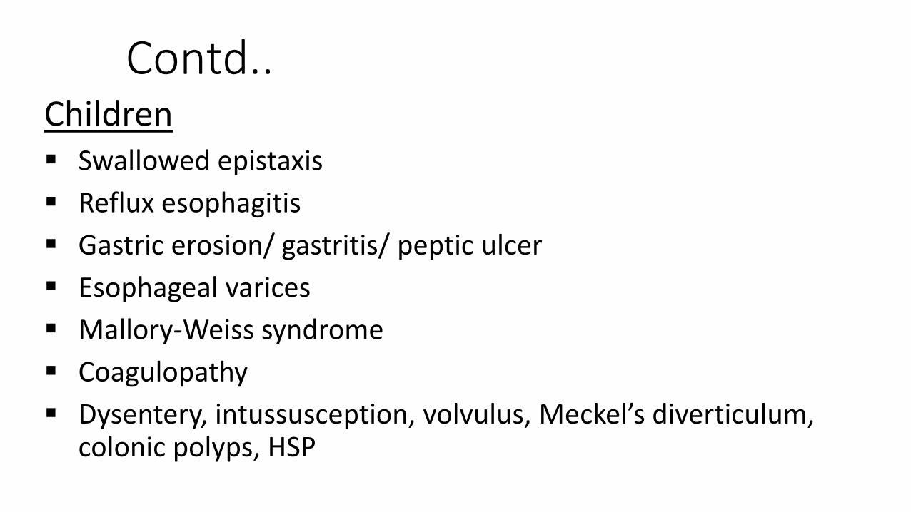
Contd..Children Swallowed epistaxis
Reflux esophagitis
Gastric erosion/ gastritis/ peptic ulcer
Esophageal varices
Mallory-Weiss syndrome
Coagulopathy
Dysentery, intussusception, volvulus, Meckel’s diverticulum, colonic polyps, HSP
Contd..
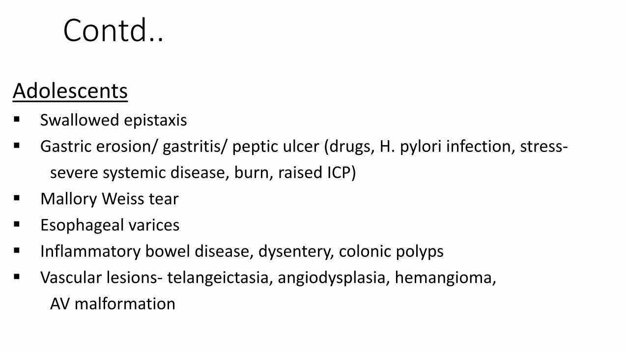
Adolescents Swallowed epistaxis
Gastric erosion/ gastritis/ peptic ulcer (drugs, H. pylori infection, stress-
severe systemic disease, burn, raised ICP)
Mallory Weiss tear
Esophageal varices
Inflammatory bowel disease, dysentery, colonic polyps
Vascular lesions- telangeictasia, angiodysplasia, hemangioma,
AV malformation
Contd….
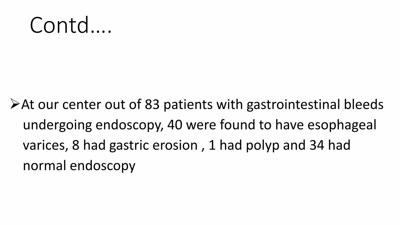
At our center out of 83 patients with gastrointestinal bleeds
undergoing endoscopy, 40 were found to have esophageal
varices, 8 had gastric erosion , 1 had polyp and 34 had
normal endoscopy
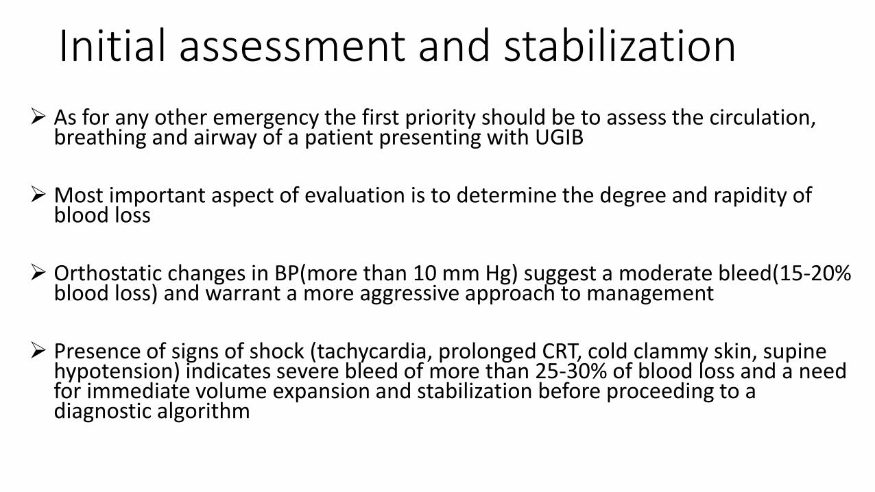
Initial assessment and stabilization
As for any other emergency the first priority should be to assess the circulation, breathing and airway of a patient presenting with UGIB
Most important aspect of evaluation is to determine the degree and rapidity of blood loss
Orthostatic changes in BP(more than 10 mm Hg) suggest a moderate bleed(15-20% blood loss) and warrant a more aggressive approach to management
Presence of signs of shock (tachycardia, prolonged CRT, cold clammy skin, supine hypotension) indicates severe bleed of more than 25-30% of blood loss and a need for immediate volume expansion and stabilization before proceeding to a diagnostic algorithm

contd..
1. Whether actual blood loss or ingested substances
• Hematemesis
food coloring
red candy
colored gelatin
beets
tomato skin
rifampin
phenytoin
• Melena
bismuth
iron preparations
licorice
spinach
grapes
blueberries
charcoal
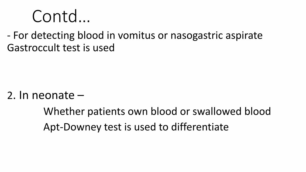
Contd…- For detecting blood in vomitus or nasogastric aspirate Gastroccult test is used
2. In neonate –Whether patients own blood or swallowed blood
Apt-Downey test is used to differentiate
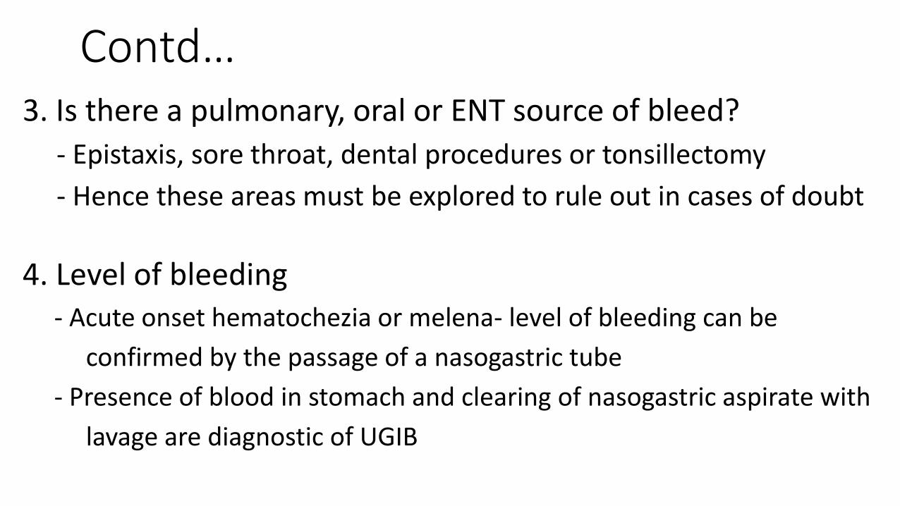
Contd…3. Is there a pulmonary, oral or ENT source of bleed?
- Epistaxis, sore throat, dental procedures or tonsillectomy
- Hence these areas must be explored to rule out in cases of doubt
4. Level of bleeding- Acute onset hematochezia or melena- level of bleeding can be
confirmed by the passage of a nasogastric tube
- Presence of blood in stomach and clearing of nasogastric aspirate with
lavage are diagnostic of UGIB
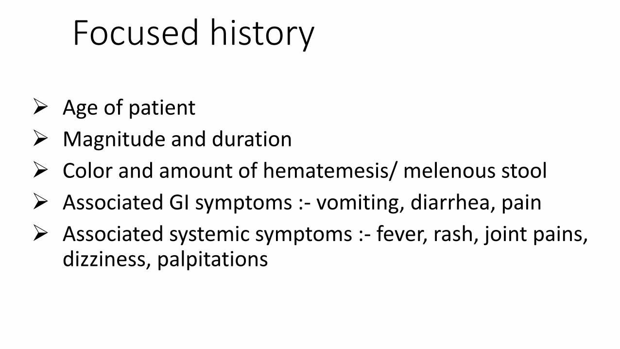
Focused history
Age of patient
Magnitude and duration
Color and amount of hematemesis/ melenous stool
Associated GI symptoms :- vomiting, diarrhea, pain
Associated systemic symptoms :- fever, rash, joint pains, dizziness, palpitations
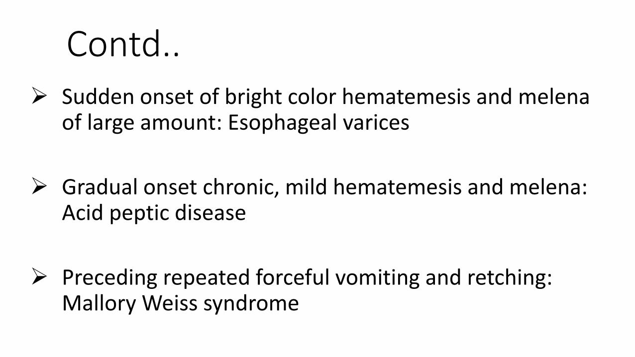
Contd.. Sudden onset of bright color hematemesis and melena
of large amount: Esophageal varices
Gradual onset chronic, mild hematemesis and melena: Acid peptic disease
Preceding repeated forceful vomiting and retching: Mallory Weiss syndrome

Contd..
Acid regurgitation, nausea, vomiting, water brash, retrosternal pain: Reflux esophagitis
Anorexia, nausea, vomiting and epigastric pain with relation to food: Peptic ulcer
Bloody diarrhea, vomiting, abdominal pain, fever: Dysentery
History of easy bruising or bleeding: coagulation, platelet dysfunction or thrombocytopenia

Contd.. History of drug intake: NSAIDS, corticosteroids, Mucosal irritants, iron
preparation: Gastritis.
Poisoning : Paracetamol, iron.
Risk factors for portal HTN: umbilical sepsis / catheterization, jaundice, liver disease- Esophageal varices
H/o chronic cough, recurrent lung infections: Cystic fibrosis, Bronchiectasis.
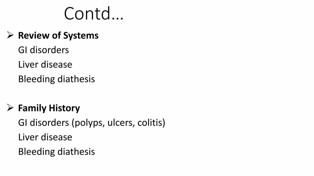
Contd… Review of Systems
GI disorders
Liver disease
Bleeding diathesis
Family History
GI disorders (polyps, ulcers, colitis)
Liver disease
Bleeding diathesis
Physical examination
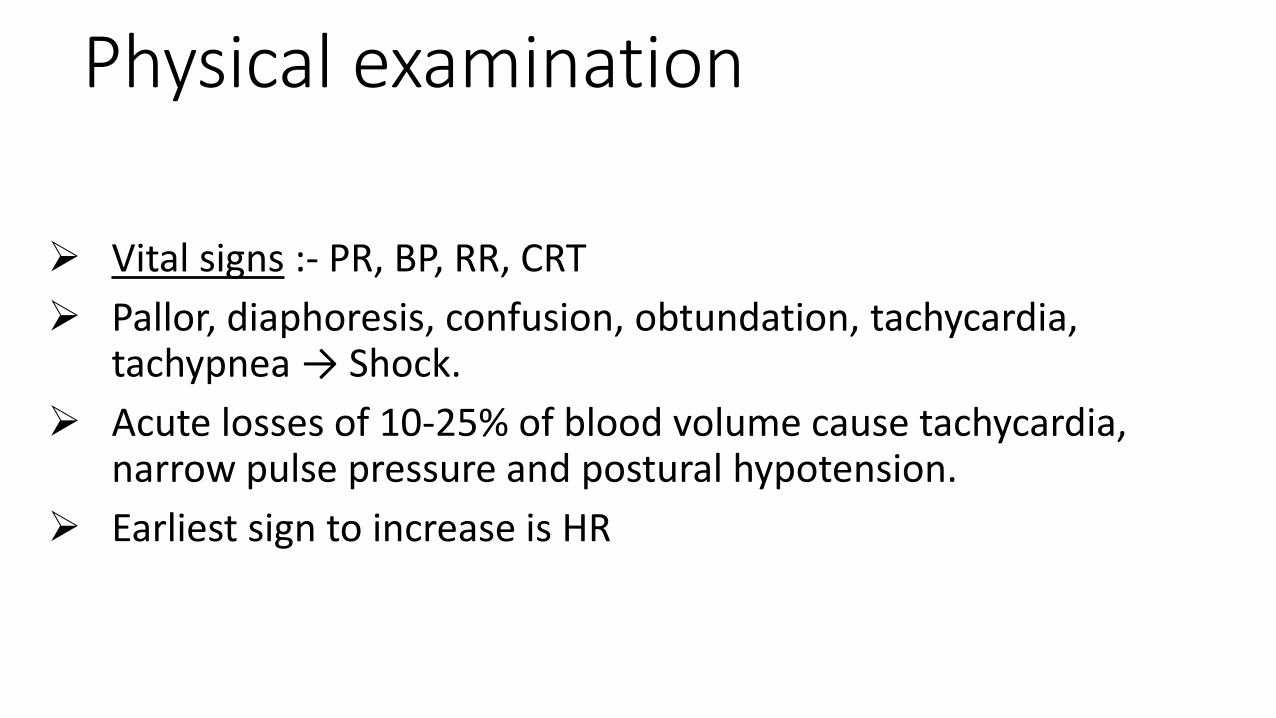
Vital signs :- PR, BP, RR, CRT
Pallor, diaphoresis, confusion, obtundation, tachycardia, tachypnea → Shock.
Acute losses of 10-25% of blood volume cause tachycardia, narrow pulse pressure and postural hypotension.
Earliest sign to increase is HR

Contd..
Pallor- Increased paleness will point towards ongoing blood loss
Icterus- chronic liver disease
Skin- petechiae, Purpura, ecchymoses, vascular malformations, stigmata for chronic liver disease like spider angioma, palmar erythema
Examination of nose, oral cavity and throat
Contd…..
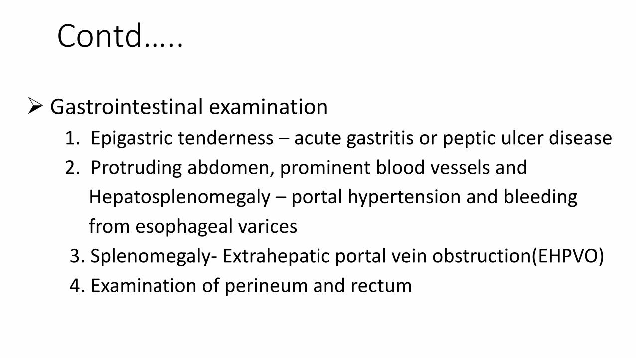
Gastrointestinal examination
1. Epigastric tenderness – acute gastritis or peptic ulcer disease
2. Protruding abdomen, prominent blood vessels and
Hepatosplenomegaly – portal hypertension and bleeding
from esophageal varices
3. Splenomegaly- Extrahepatic portal vein obstruction(EHPVO)
4. Examination of perineum and rectum

Investigations In an emergency setting only a few tests are essential in the beginning to
evaluate UGIB
CBC
PT/INR
APTT
LFT
Blood grouping and Cross matching
Further investigations
1. abdominal USG- EHPVO, portal hypertension due to liver disease, large vessel anomalies, splenic artery aneurysm

Contd..2. Endoscopy-
- UGI endoscopy is the gold standard for diagnosis and treatment of UGIB
- Procedure of choice for all patients with UGIB.
- In the skilled hands diagnosis of etiology in 85-90% of cases
- Contraindicated in in hemodynamically unstable patients
3. CT angiography- Vascular malformations beyond the duodenum , in areas not
accessed by routine UGI endoscopy
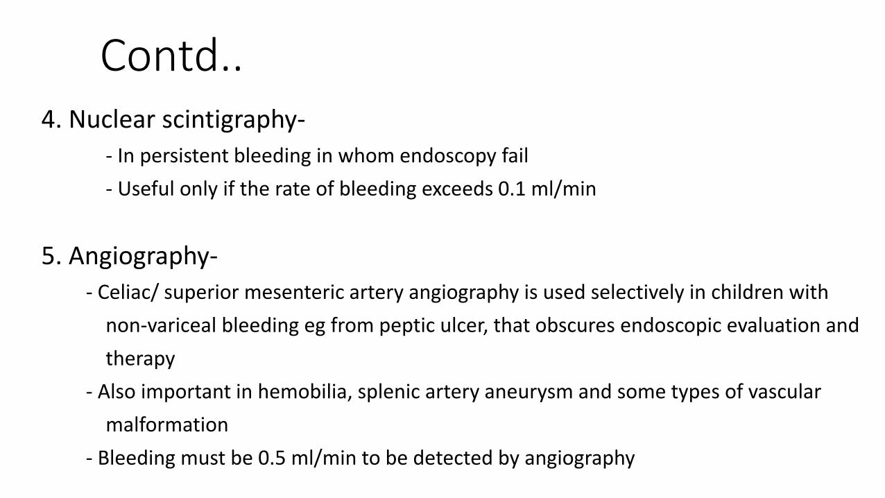
Contd..4. Nuclear scintigraphy-
- In persistent bleeding in whom endoscopy fail
- Useful only if the rate of bleeding exceeds 0.1 ml/min
5. Angiography-
- Celiac/ superior mesenteric artery angiography is used selectively in children with
non-variceal bleeding eg from peptic ulcer, that obscures endoscopic evaluation and
therapy
- Also important in hemobilia, splenic artery aneurysm and some types of vascular
malformation
- Bleeding must be 0.5 ml/min to be detected by angiography

Management The initial steps in the management of severe UGIB include assessment,
resuscitation, re- evaluation, identification of the cause and source of bleeding and commencing appropriate treatment
Resuscitation and stabilization
1. Circulation-
large bore venous access to restore blood volume
crystalloids initially
blood transfusion
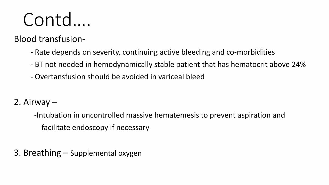
Contd….Blood transfusion-
- Rate depends on severity, continuing active bleeding and co-morbidities
- BT not needed in hemodynamically stable patient that has hematocrit above 24%
- Overtansfusion should be avoided in variceal bleed
2. Airway –
-Intubation in uncontrolled massive hematemesis to prevent aspiration and
facilitate endoscopy if necessary
3. Breathing – Supplemental oxygen

Contd..Reassessment and monitoring-
-Vitals should be monitor every 10- 15 minutes till stabilized
-Then hourly for 24 hours after bleeding stops
Nasogastric aspiration-Aspiration and saline lavage indicated in all patients with UGIB to confirm
- Presence of intragastric blood
- Rate of gross bleeding
- Check for ongoing or recurrent bleeding
- Clear gastric field for endoscopic visualization
- Prevent aspiration
- Prevent hepatic encephalopathy in patients of cirrhosis
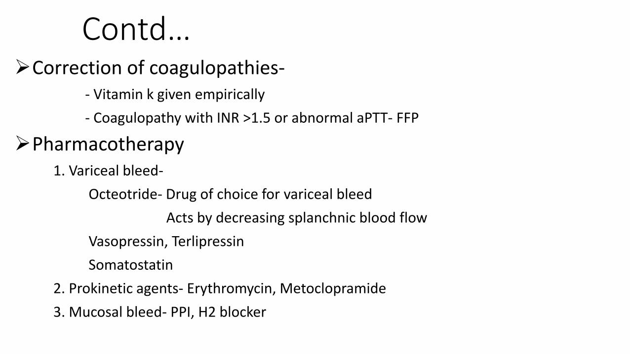
Contd…Correction of coagulopathies-
- Vitamin k given empirically
- Coagulopathy with INR >1.5 or abnormal aPTT- FFP
Pharmacotherapy1. Variceal bleed-
Octeotride- Drug of choice for variceal bleed
Acts by decreasing splanchnic blood flow
Vasopressin, Terlipressin
Somatostatin
2. Prokinetic agents- Erythromycin, Metoclopramide
3. Mucosal bleed- PPI, H2 blocker

Contd…. Endoscopic techniques
1. Variceal bleed-
Endoscopic sclerotherapy is the mainstay of treatment in this group
Endoscopic variceal ligation
2. Nonvariceal bleed
Injection adrenaline and saline
Endoclip devices
Balloon tamponade
Sengstaken-blakemore tube
Used in whom bleeding continues despite pharmacotherapy and endoscopic
methods

Case
Our case presented to us in the ER. At presentation he was an average build child with pallor and vitals of T-98*F, PR- 170, BP-90/60, RR- 36. He was pale and anicteric. On per abdomen he had splenomegaly of about 3 cm. Rest of the exam was normal.
Lab investigations-
Hb- 5.7, TLC – 13,000, platelets 17,600
LFT-N , RFT- N
PT/INR- N, aPTT- N
Contd…
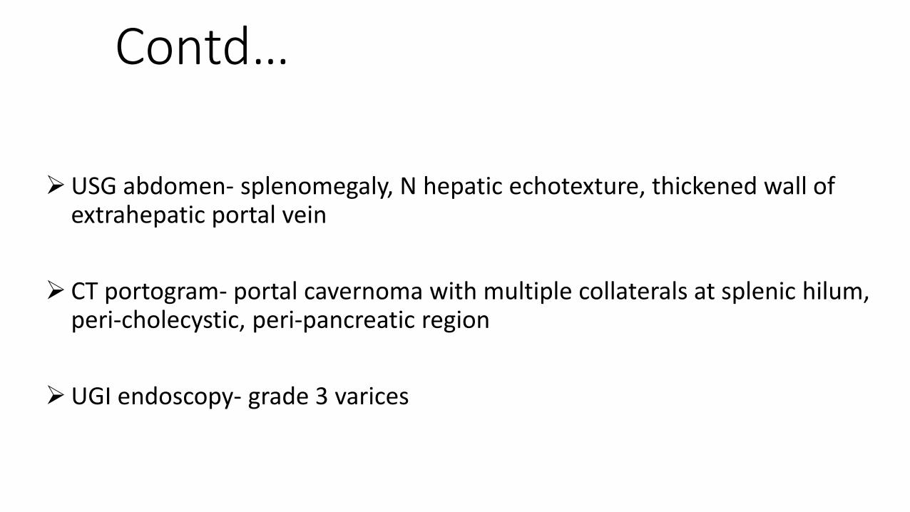
USG abdomen- splenomegaly, N hepatic echotexture, thickened wall of extrahepatic portal vein
CT portogram- portal cavernoma with multiple collaterals at splenic hilum, peri-cholecystic, peri-pancreatic region
UGI endoscopy- grade 3 varices

Summary
UGI bleeding is a potentially life threatening emergency requiring an appropriate diagnostic and therapeutic approach
Therefore primary focus in a child with UGI bleed is resuscitation and stabilization followed by a diagnostic evaluation
In infants and toddlers mucosal erosion is the most common cause while in older children variceal bleeding due to EHPVO is most common
UGI endoscopy is the most accurate and useful diagnostic tool to evaluate UGI bleed
Treatment depends on the cause
Reference
Indian journal of pediatrics
Pediatric in review
Nelson textbook of pediatrics
www.Wikipedia.com