Embed Size (px)
Citation preview
of March 28, 2022.This information is current as
MHC Class IIIgG Constant Domains for Presentation by Processing of an Antigenic Sequence from
and Inger SandlieBogenFrigstad, Gøril Berntzen, Terje E. Michaelsen, Bjarne
Morten Flobakk, Ingunn B. Rasmussen, Elin Lunde, Terje
http://www.jimmunol.org/content/181/10/7062doi: 10.4049/jimmunol.181.10.7062
2008; 181:7062-7072; ;J Immunol
Referenceshttp://www.jimmunol.org/content/181/10/7062.full#ref-list-1
, 25 of which you can access for free at: cites 61 articlesThis article
average*
4 weeks from acceptance to publicationFast Publication! •
Every submission reviewed by practicing scientistsNo Triage! •
from submission to initial decisionRapid Reviews! 30 days* •
Submit online. ?The JIWhy
Subscriptionhttp://jimmunol.org/subscription
is online at: The Journal of ImmunologyInformation about subscribing to
Permissionshttp://www.aai.org/About/Publications/JI/copyright.htmlSubmit copyright permission requests at:
Email Alertshttp://jimmunol.org/alertsReceive free email-alerts when new articles cite this article. Sign up at:
Print ISSN: 0022-1767 Online ISSN: 1550-6606. Immunologists All rights reserved.Copyright © 2008 by The American Association of1451 Rockville Pike, Suite 650, Rockville, MD 20852The American Association of Immunologists, Inc.,
is published twice each month byThe Journal of Immunology
by guest on March 28, 2022
http://ww
w.jim
munol.org/
Dow
nloaded from
by guest on March 28, 2022
http://ww
w.jim
munol.org/
Dow
nloaded from
Processing of an Antigenic Sequence from IgG ConstantDomains for Presentation by MHC Class II1
Morten Flobakk,2*‡ Ingunn B. Rasmussen,*‡ Elin Lunde,*‡ Terje Frigstad,†‡ Gøril Berntzen,*‡
Terje E. Michaelsen,§ Bjarne Bogen,†‡ and Inger Sandlie3*‡
Targeting of T cell epitopes to APC enhances T cell responses. We used an APC-specific Ab (anti-IgD) and substituted either of18 loops connecting � strands in human IgG constant H (CH) domains with a characterized T cell peptide epitope. All Ab-epitopefusion molecules were secreted from producing cells except IgG-loop 2(BC)CH1, and comparing levels, a hierarchy appeared withfusions involving CH2>CH1>CH3. Within each domain, fusion at loop 6(FG) showed best secretion, while low secretion correlatedwith the substitution of native loops that contain conserved amino acids buried within the folded molecule. Comparing theAPC-specific rAb molecules for their ability to induce T cell activation in vitro, the six mutants with epitope in CH2 were the mosteffective, with loop 4CH2 ranking on top. The CH1 mutants were more resistant to processing, and the loop 6CH1 mutant onlyinduced detectable activation. The efficiency of the CH3 mutants varied, with loop 6CH3 being the least effective and equal to loop6 CH1. Considering both rAb secretion level and T cell activation efficiency, a total of eight loops may carry T cell epitopes to APCfor processing and presentation to T cells, namely, all in CH2 in addition to loop 6 in CH1 and CH3. Comparing loop 4CH2 withloop 6CH1 mutants after injection of Ab in BALB/c mice, the former was by far the most efficient and induced specific T cellactivation at concentrations at least 100-fold lower than loop 6CH1. The Journal of Immunology, 2008, 181: 7062–7072.
I ncreased presentation of antigenic peptide in complex withMHC molecules enhances T cell responses, and Ab- medi-ated targeting of peptide or whole Ag to APC is a way to
increase the number of peptide-MHC complexes on the surface ofAPC (1). This is important since major efforts are concentrated onthe development of safe vaccines that generate strong, specific Tcell responses. Abs with T cell epitopes within their V region CDRloops have been described previously (2). Although such rAbs mayenter APC by way of Fc�Rs, rAbs with peptides or whole Agadded C-terminally to Fab (3) or to Ab (4, 5) may target a preferredand defined APC surface molecule by way of V region specificity.We have introduced peptide epitopes into loops connecting �strands in constant heavy (CH)4 domains, and denoted the epitope-loaded rAb “Troybodies.” The loop-grafting experiments have in-volved the amino acid sequence 91–101 of the MOPC315 plas-macytoma �2 L chain (91–101 �2315), which represents a minimalstimulating T cell epitope (6) presented on I-Ed MHC class II forCD4� T cells (7). In initial experiments, this epitope was ex-changed with or inserted into either of three loops (BC, DE, or FG)in CH1 of human (h) IgG3 (8) and murine (m) IgG2b (9). For
simplicity, these loops are hereby denoted loop 2CH1, 4CH1, and6CH1, respectively (see Fig. 1A). The loop 2CH1 hIgG3 rAb mu-tant was retained, while all other single loop substitution mutantswere secreted. We also showed that loop 6CH1 hIgG3 could besubstituted with model epitopes that show great variation in aminoacid sequence, length, and secondary structure, namely, aa 323–339 from OVA, aa 110–120 from hemagglutinin, and aa 46–61from hen egg lysozyme (10). Initially, the epitope was grafted intohapten-specific rAbs (8) and, subsequently, the rAbs wereequipped with V genes encoding APC specificity. Following invitro targeting, the epitopes were excised from the rAbs, loaded onMHC class II molecules, and presented to specific T cells. SuchTroybody targeting to murine IgDa resulted in a 103-fold improve-ment in presentation efficiency compared with rAbs with irrelevantspecificity. Importantly, T cell activation was improved up to 105-fold compared with that achieved using synthetic peptide or wholeprotein (10, 11). The same results were obtained in experimentswith (91–101 �2315) in loop 6CH1 where the target was MHC classII (I-E) (12). Furthermore, a 102-fold enhanced presentation wasseen in vivo in mice (12).
Comparing the in vitro presentation efficiency of APC loadedwith mouse and human rAbs with 91–101 �2315epitopes grafted invarious loops in CH1 (8, 9), differences were found whichprompted us to initiate a comprehensive analysis that involvedgrafting in all loops in all three CH domains. Each domain has sixloops connecting � strands, offering 18 possibilities for loop re-placement. To identify the loops that are best suited for epitopeinsertion, we exchanged every loop in the three hIgG3 CH domainswith the amino acid sequence 89–105 from �2315 (89–105) �2315
and show here that a total of 17 such fusion molecules were se-creted from transiently transfected 293E cells, although in differentamounts. Levels of secretion are compared with variability as wellas hydropathicity and solvent accessibility score for each aminoacid within each domain.
For each domain, the loop 6 mutant was secreted at the highestlevel. Furthermore, the CH1 and CH2 mutants (except loop 2CH1)
*Department of Molecular Biosciences, †Institute of Immunology, and ‡Centre forImmune Regulation, University of Oslo, Oslo, Norway; and §Department of Vac-cines, National Institute of Public Health, Oslo, Norway
Received for publication July 11, 2007. Accepted for publication September 6, 2008.
The costs of publication of this article were defrayed in part by the payment of pagecharges. This article must therefore be hereby marked advertisement in accordancewith 18 U.S.C. Section 1734 solely to indicate this fact.1 This work was funded by the Norwegian Cancer Society (to M.F. and I.B.R.) andthe Research Council of Norway (to E.L., T.F., and G.B.).2 Current address: BioInn, 2317 Hamar, Norway.3 Address correspondence and reprint requests to Dr. Inger Sandlie, Department ofMolecular Biosciences, University of Oslo, PO Box 1041 Blindern, NO-0316 Oslo,Norway. E-mail address: [email protected] Abbreviations used in this paper: CH, constant region of Ig H chain; RT, roomtemperature; AEP, asparaginyl endopeptidase; wt, wild type.
Copyright © 2008 by The American Association of Immunologists, Inc. 0022-1767/08/$2.00
The Journal of Immunology
www.jimmunol.org
by guest on March 28, 2022
http://ww
w.jim
munol.org/
Dow
nloaded from
were secreted better than the CH3 domain mutants, of which theloop 6 mutant only was secreted well. In general, low secretioncorrelated with the removal of conserved amino acids that wereburied either within a domain core or between two interactingdomains.
All secreted rAbs were tested for the ability to stimulate CD4�
T cells in vitro in T cell activation and growth inhibition assays.Importantly, T cell epitope loop grafting on all three CH domainswas compatible with efficient stimulation of T cells. Eight mutantswere both secreted in good amounts and found to induce T cellactivation, namely, loop 6CH1, all CH2 mutants and loop 6CH3.The difference in induction potential was large, as the best activa-tor (loop 4CH2) induced T cell activation at a concentration thatwas 102-fold lower than the weakest (loop 6CH1). These two Abswere selected for further studies in vivo and both were injectedinto the tail vein of normal mice at various concentrations. Fol-lowing in vivo targeting, isolated spleen cells as APC stimulatedspecific T cells in vitro and again the loop 4CH2 mutant was atleast 102-fold more efficient than the loop 6CH1 mutant.
Materials and MethodsMice, cell lines, and Abs
BALB/c mice were bred by Taconic Farms. The study was approved by theNational Committee for Animal Experiments (Oslo, Norway). 293E (CRL-10852) cells were obtained from American Type Culture Collection. The Tcell clone 7A10B2 recognizes aa 91–101 of �2315 in complex with I-Ed (7).Spleen cells from BALB/c mice were used as APC, as were A20 B lym-phoma cells transfected with a 2,4,6-trinitrophenyl-specific IgD BCR(A20�; gift from Dr. N. Hozumi, Department of Medical Genetics, Uni-versity of Toronto, Toronto, Ontario, Canada) (13).
The 4B2A1 and 7A10B2 T cell clones recognize the same �2315 I-Ed
complex (7). The T cell hybridoma BW4B2A1 was produced by fusinglymph node cells from 4B2A1 TCR-transgenic SCID mice (14) with theTCR-negative T cell hybridoma line BW51.47 �/� (15) using polyethyleneglycol under standard conditions. Hypoxanthine/aminopterin/thymidine-resistant clones were selected for surface expression of the 4B2A1 TCR.Functionality was confirmed by coculturing hybridoma cells with irradi-ated BALB/c splenocytes (20 Gy) and synthetic �2315 peptide (89–107),with subsequent detection of IL-2 in supernatant by sandwich ELISA (datanot shown).
All cells were cultured in DMEM (BioWhittaker) or RPMI 1640 sup-plemented with 10% heat-inactivated FCS (PAA), 2 mM L-glutamine(DMEM only), 25 �g/ml streptomycin, and 25 U/ml penicillin (both fromBioWhittaker) under standard conditions. Abs for IL-2 detection were ratanti-mouse IL-2 (clone JES6-1A12) and biotin rat anti-mouse IL-2 (cloneJES6-5H4), both from BD Pharmingen. Abs for IFN-� detection were ratanti-mouse IFN-� (AN18 (16)) and biotin rat anti-mouse IFN-� (XMG1.2-bio (17)). mAb HP-6050 is specific for hIgG3 hinge (Sigma-Aldrich). Allother Abs used were produced by us using standard procedures. The mAbK13 is specific for the human � L chain (18). The mAbs 132c8 and HP-6050 (19) were compared for binding to all four hIgG isotypes side-by-sidein ELISA and found to bind the hIgG3 hinge in a similar fashion. Affinity-purified sheep polyclonal Abs, s303 (20) and s12 (21), are specific for hIgGFab and hIgG3 hinge, respectively.
Structure analysis of hIgG3 H chain constant domains
Secondary structure analysis (� strand or loop) was conducted using dataand evaluation programs given in the PDB database (http://www.rcsb.org/pdb/) (22) and as described previously (9). Briefly, crystal structures of1FC1 (hIgG1), 1IGY (mIgG1), and 1IGT (mIgG2a) were studied regardingboth loop length and limits as determined by three-dimensional visualiza-tion and the program Structure Explorer (http://www.pdb.org). Loops werenumbered 1–6 in each domain starting from the N-terminal end of thepolypeptide chain. Variability of each amino acid position in Ig H chainswas analyzed using the sequence analysis program provided by S. M. J.Searle (The Sanger Institute, Cambridge, U.K.) as described previously (9).
The relative total side chain accessibility of each amino acid in the CH1,CH2, and CH3 domains was investigated using Naccess (http://wolf.bms.umist.ac.uk/naccess/) and PDB ID: 1HZH (human IgG1) (23). Hydro-pathicity analysis of hIgG3 domains CH1, CH2, and CH3 was performedusing the program ProtScale (window size � 9; http://www.expasy.org/
cgi-bin/protscale.pl) (24) and the Kyte-Doolittle scale (25), as provided bythe Swiss Institute of Bioinformatics.
Production of mutant hIgG3: construction of loop exchangemutants
The CH chain gene encoding the G3m (b0) allotype (26) was a gift fromDr. M. P. LeFranc (International ImMunoGeneTics Information System,Montpellier, France) and cloned as a 2.8-kb fragment into HindIII-SphIsites in the polylinker of pUC19 (Sigma-Aldrich). The resulting plasmid,pUC19�3wt, was template in all mutagenesis reactions. All predicted loopsequences in C�3 CH1, CH2, and CH3, were exchanged with aa 89–105from �2315 (FAALWFRNHFVFGGGTK) in individual H chains. The cys-teine in position 90 of the endogenous sequence was exchanged with ala-nine. Mutagenesis was performed by QuikChange mutagenesis (Strat-agene). Table I shows loop sequences and Table II shows mutagenic primersequences. Colonies were screened for the presence of the mutation by thesimultaneous introduction of a silent DraIII restriction site and DraIII di-gestion of plasmid DNA. All 18 mutations were subsequently confirmed bysequencing by GATC. Sequenced fragments containing mutations weresubcloned into unmanipulated vectors as HindII-BglII, BglII-PmlI, PmlI-NsiI, or PmlI-SphI (Fig. 1B) to exclude possible amplification errors out-side the sequenced areas. Complete mutant H chain genes were assembledin pLNOH2IgD, which encode IgD-specific H chains, as described else-where (11, 27). The corresponding L chain gene, encoded on pLNOKIgD
(11, 27) and each of the 18 pLNOH2IgD variants were transiently cotrans-fected in 293E cells (28). Portions of supernatant were harvested and re-placed with fresh medium every 2–3 days for 14 days as described. Asubstitution of loop 4CH1 with 91–101 �2315 has been described elsewhere(8) and the complete H chain gene was assembled in pLNOH2IgD. Thecorresponding loop 6CH1 substitution in an IgD-specific H chain has beendescribed previously (11). Both H chain genes were transiently cotrans-fected with pLNOKIgD and supernatant harvested as described above. Thecomplete IgD-specific rAbs are denoted “loop 4 or loop 6CH1 (91–101),”respectively.
Sandwich ELISA for detection of hIgG3 variant concentrations
The amounts of IgD-specific rAb mutants secreted after each transfectionwere measured as follows: 96-well microtiter plates were coated with ahIgG3-specific Ab and incubated overnight at room temperature (RT).Then, samples of 100 �l of diluted supernatants were added to each welland detected with a second hIgG3-specific Ab. A hIgG3wt preparation wasdiluted in a 3-fold series and used as standard. Three different Ab combi-nations were used as coat and detecting agent: s303 (2 �g/ml) and 132c8-bio (1/6000), s12 (10 �g/ml) and 132c8-bio (1/6000), as well as K13 (2�g/ml) and s303-bio (1/6000), respectively. Detection was done with thesubstrate for alkaline phosphatase, p-nitrophenyl phosphate (Sigma-Al-drich) diluted in diethanolamine buffer to 1 mg/ml.
Table I. Loop positions in hIgG3
Domain Loopa EU No.
CH1 1 129–1382 150–1533 159–1664 175–1795 189–1966 205–208
CH2 1 244–2572 265–2733 280–2894 296–2995 308–3186 325–331
CH3 1 352–3612 371–3773 384–3904 394–4055 414–4226 429–436
a Loops are numbered 1–6 in each domain from the N- to the C-terminal, e.g.,such that the loop connecting � strand A and B is loop 1 (Fig 1A).
7063The Journal of Immunology
by guest on March 28, 2022
http://ww
w.jim
munol.org/
Dow
nloaded from
Isolation of mutant hIgG3
Proteins in supernatants were precipitated by 1:1 addition of portions of asaturated ammonium sulfate solution. Incubation at RT for 20 min wasfollowed by a 10-min centrifugation at 17,000 � g using a Sorvall centri-fuge (OneMed). Pellets were dissolved in dH2O and dialyzed three times toPBS/azide and two times to RPMI 1640 at 4°C before sterile filtration. Absused in in vivo targeting experiments were affinity purified from cell su-pernatant by use of protein G-conjugated Sepharose columns.
Western blotting
Western blots were performed using Criterion XT Precast Gels (Bio-Rad).Briefly, the Ab samples were preheated at 95°C for 3 min before they wereloaded onto the gel and separated at 140 V for 100 min. By Semi Drytransfer at 20 V for 30 min, proteins were transferred to an Immobilon-Ppolyvinylidene difluoride membrane (Sigma-Aldrich). Detection reagentswere biotinylated mouse anti-hIgG3 (HP-6050) (Sigma-Aldrich) and HRP-conjugated streptavidin (GE Healthcare). Detection was performed usingthe ECL Plus Western Blotting Detection Reagents kit (GE Healthcare) asdescribed by the manufacturer, and membranes were analyzed using Im-ageQuant TL version 2003.02 software (GE Healthcare). Individual Abconcentrations were normalized to a known concentration of purifiedhIgG3.
Binding to soluble Fc�RIIA or protein G
The extracellular domains of hFc�RIIA were cloned and expressed as sol-uble fusion to GST (Fc�RIIA-GST) as described elsewhere (29). ELISAplates were coated overnight at 4°C with mouse IgD at 4 �g/ml. Thefollowing day, the wells were blocked in 2% skim milk, washed three timesin PBS/0.05% Tween 20, and incubated for 1 h at RT with the mutant Absamples at 1 �g/ml. After washing, the wells were incubated for 1 h at RTwith protein G-conjugated to HRP (VWR) or Fc�RIIA-GST at 1 �g/mlfollowed by anti-GST conjugated to HRP (GE Healthcare). After threewashes, the plates were developed in ABTS (Sigma-Aldrich) in citratebuffer at pH 2.2. Absorbance at 405 nm was read after 15–60 min.
In vitro Ag presentation assays
Growth inhibition assay Growth inhibition assays were performed as de-scribed by Bogen et al. (7). All Ab mutants were diluted in 5-fold seriesstarting at 1 �g/ml and added as triplicates in flat-bottom 96-well microtiterplates. 7A10B2 T cells on day 10 after the last stimulation were irradiated(20 Gy) and 103 cells were added to each well along with 5000 A20� cellsin exponential growth expressing the IgDa allotype (13). Synthetic 89–107�2315 peptide was included as positive control. Supernatant from cells thathad had been treated with transfection reagent without DNA addition wasammonium sulfate precipitated and used as medium control. Negative con-trol was hIgG3wt with IgD specificity. After incubation for 24 h, the cul-tures were pulsed with 1 �Ci of [3H]dThd for 16–24 h, harvested ontofilters, and counted using a TopCount NXT scintillation counter (GMI).T cell proliferation assay Samples of 5 � 105 irradiated (20 Gy) BALB/cspleen APC were cultured with 2 � 104 7A10B2 T cells and variousamounts of rAb or synthetic 89–107 �2315 peptide in triplicates for 48 h.The cultures were then pulsed with 1 �Ci of [3H]dThd for 16–24 h, har-vested onto filters, and counted using the TopCount1 counter (GMI).
In vivo experiments
BALB/c mice were injected i.v. in the tail with titrated amounts of loop6CH1 and loop 4CH2 rAbs. Two mice received PBS only. Ninety minutesafter i.v. injections, the mice were killed by cervical dislocation and thespleens were removed. Irradiated (8 Gy) spleen cells (5 � 105/well) werecultured with responder T cells, namely, 7A10B2 or T cell hybridomasBW4B2A1 (both 2 � 104/well). An optimal concentration of the �2315
synthetic peptide (10 �g/ml) was added to the positive control. After 72 h,portions of 100 �l of supernatant were collected for cytokine measure-ments, and the cultures were pulsed for 24 h with 1 �Ci of [3H]dThd. Thecultures were harvested and incorporated [3H]dThd was measured usingthe TopCount1 counter. Detection of IFN-� and IL-2 in supernatants wasperformed in microtiter plates coated with AN18 or JES6-1A12 (both 2�g/ml in PBS), respectively. XMG1.2-bio (1 �g/ml in PBST) was used asdetection Ab for IFN-�, whereas JES6-5H4 (1 �g/ml in PBST) was used
Table II. Mutagenesis primersa
Name Sequence
1CH1(�) CCCATCGGTCTTCCCCCTGTTCGCAGCTCTATGGTTCAGAAACCACTTTGTGTTCGGTGGTGGAACCAAGACAGCGGCCCTGGGCTGC1CH1(�) GCAGCCCAGGGCCGCTGTCTTGGTTCCACCACCGAACACAAAGTGGTTTCTGAACCATAGAGCTGCGAACAGGGGGAAGACCGATGGG2CH1(�) GCTGCCTGGTCAAGGACTACTTCGCAGCTCTATGGTTCAGAAACCACTTTGTGTTCGGTGGTGGAACCAAGGTGACGGTGTCGTGGAACTCA2CH1(�) TGAGTTCCACGACACCGTCACCTTGGTTCCACCACCGAACACAAAGTGGTTTCTGAACCATAGAGCTGCGAAGTAGTCCTTGACCAGGCAGC3CH1(�) ACCGGTGACGGTGTCGTGGTTCGCAGCTCTATGGTTCAGAAACCACTTTGTGTTCGGTGGTGGAACCAAGGTGCACACCTTCCCGGCTG3CH1(�) CAGCCGGGAAGGTGTGCACCTTGGTTCCACCACCGAACACAAAGTGGTTTCTGAACCATAGAGCTGCGAACCACGACACCGTCACCGGT4CH1(�) ACACCTTCCCGGCTGTCCTATTCGCAGCTCTATGGTTCAGAAACCACTTTGTGTTCGGTGGTGGAACCAAGTACTCCCTCAGCAGCGTGGT4CH1(�) ACCACGCTGCTGAGGGAGTACTTGGTTCCACCACCGAACACAAAGTGGTTTCTGAACCATAGAGCTGCGAATAGGACAGCCGGGAAGGTGT5CH1(�) TCAGCAGCGTGGTGACCGTGTTCGCAGCTCTATGGTTCAGAAACCACTTTGTGTTCGGTGGTGGAACCAAGACCTACACCTGCAACGTGAATC5CH1(�) GATTCACGTTGCAGGTGTAGGTCTTGGTTCCACCACCGAACACAAAGTGGTTTCTGAACCATAGAGCTGCGAACACGGTCACCACGCTGCTGA6CH1(�) CTACACCTGCAACGTGAATCACTTCGCAGCTCTATGGTTCAGAAACCACTTTGTGTTCGGTGGTGGAACCAAGACCAAGGTGGACAAGAGAGTTG6CH1(�) CAACTCTCTTGTCCACCTTGGTCTTGGTTCCACCACCGAACACAAAGTGGTTTCTGAACCATAGAGCTGCGAAGTGATTCACGTTGCAGGTGTAG1CH2(�) GGACCGTCAGTCTTCCTCTTCTTCGCAGCTCTATGGTTCAGAAACCACTTTGTGTTCGGTGGTGGAACCAAGGAGGTCACGTGCGTGGTGGT1CH2(�) ACCACCACGCACGTGACCTCCTTGGTTCCACCACCGAACACAAAGTGGTTTCTGAACCATAGAGCTGCGAAGAAGAGGAAGACTGACGGTCC2CH2(�) GAGGTCACGTGCGTGGTGGTGTTCGCAGCTCTATGGTTCAGAAACCACTTTGTGTTCGGTGGTGGAACCAAGCAGTTCAAGTGGTACGTGGACG2CH2(�) CGTCCACGTACCACTTGAACTGCTTGGTTCCACCACCGAACACAAAGTGGTTTCTGAACCATAGAGCTGCGAACACCACCACGCACGTGACCTC3CH2(�) GGTCCAGTTCAAGTGGTACGTGTTCGCAGCTCTATGGTTCAGAAACCACTTTGTGTTCGGTGGTGGAACCAAGAAGCCGCGGGAGGAGCAGTAC3CH2(�) GTACTGCTCCTCCCGCGGCTTCTTGGTTCCACCACCGAACACAAAGTGGTTTCTGAACCATAGAGCTGCGAACACGTACCACTTGAACTGGACC4CH2(�) ACAAAGCCGCGGGAGGAGCAGTTCGCAGCTCTATGGTTCAGAAACCACTTTGTGTTCGGTGGTGGAACCAAGTTCCGTGTGGTCAGCGTCCTCA4CH2(�) TGAGGACGCTGACCACACGGAACTTGGTTCCACCACCGAACACAAAGTGGTTTCTGAACCATAGAGCTGCGAACTGCTCCTCCCGCGGCTTTGT5CH2(�) CGTGTGGTCAGCGTCCTCACCTTCGCAGCTCTATGGTTCAGAAACCACTTTGTGTTCGGTGGTGGAACCAAGTACAAGTGCAAGGTCTCCAACAAAG5CH2(�) CTTTGTTGGAGACCTTGCACTTGTACTTGGTTCCACCACCGAACACAAAGTGGTTTCTGAACCATAGAGCTGCGAAGGTGAGGACGCTGACCACACG6CH2(�) GGAGTACAAGTGCAAGGTCTCCTTCGCAGCTCTATGGTTCAGAAACCACTTTGTGTTCGGTGGTGGAACCAAGATCGAGAAAACCATCTCCAAAACC6CH2(�) GGTTTTGGAGATGGTTTTCTCGATCTTGGTTCCACCACCGAACACAAAGTGGTTTCTGAACCATAGAGCTGCGAAGGAGACCTTGCACTTGTACTCC1CH3(�) GAACCACAGGTGTACACCCTGTTCGCAGCTCTATGGTTCAGAAACCACTTTGTGTTCGGTGGTGGAACCAAGCAGGTCAGCCTGACCTGCCTG1CH3(�) CAGGCAGGTCAGGCTGACCTGCTTGGTTCCACCACCGAACACAAAGTGGTTTCTGAACCATAGAGCTGCGAACAGGGTGTACACCTGTGGTTC2CH3(�) CAGCCTGACCTGCCTGGTCAAATTCGCAGCTCTATGGTTCAGAAACCACTTTGTGTTCGGTGGTGGAACCAAGGCCGTGGAGTGGGAGAGCAGC2CH3(�) GCTGCTCTCCCACTCCACGGCCTTGGTTCCACCACCGAACACAAAGTGGTTTCTGAACCATAGAGCTGCGAATTTGACCAGGCAGGTCAGGCTG3CH3(�) CGCCGTGGAGTGGGAGAGCTTCGCAGCTCTATGGTTCAGAAACCACTTTGTGTTCGGTGGTGGAACCAAGTACAACACCACGCCTCCCATG3CH3(�) CATGGGAGGCGTGGTGTTGTACTTGGTTCCACCACCGAACACAAAGTGGTTTCTGAACCATAGAGCTGCGAAGCTCTCCCACTCCACGGCG4CH3(�) GCCGGAGAACAACTACAACACCTTCGCAGCTCTATGGTTCAGAAACCACTTTGTGTTCGGTGGTGGAACCAAGCTCTACAGCAAGCTCACCGTG4CH3(�) CACGGTGAGCTTGCTGTAGAGCTTGGTTCCACCACCGAACACAAAGTGGTTTCTGAACCATAGAGCTGCGAAGGTGTTGTAGTTGTTCTCCGGC5CH3(�) TACAGCAAGCTCACCGTGGACTTCGCAGCTCTATGGTTCAGAAACCACTTTGTGTTCGGTGGTGGAACCAAGTTCTCATGCTCCGTGATGCATGA5CH3(�) TCATGCATCACGGAGCATGAGAACTTGGTTCCACCACCGAACACAAAGTGGTTTCTGAACCATAGAGCTGCGAAGTCCACGGTGAGCTTGCTGTA6CH3(�) CATCTTCTCATGCTCCGTGATGTTCGCAGCTCTATGGTTCAGAAACCACTTTGTGTTCGGTGGTGGAACCAAGACGCAGAAGAGCCTCTCCCTG6CH3(�) CAGGGAGAGGCTCTTCTGCGTCTTGGTTCCACCACCGAACACAAAGTGGTTTCTGAACCATAGAGCTGCGAACATCACGGAGCATGAGAAGATG
a Letters in bold indicate nucleotidet encoding the (89–105)�2315 peptide while underlining indicates nucleotide substitutions for the introduction of a silent DraIII site. Allprimers were designed with the 33 nt encoding (89–105)�2315 in the center flanked by 18–21 nt complementary to the C�3wt sequence. Name according to loop of epitopegrafting and domain, e.g. 1CH1 � epitope in loop 1 of CH1. �/� indicates template or complementary direction, respectively.
7064 IgG CONSTANT DOMAIN LOOP SUBSTITUTION
by guest on March 28, 2022
http://ww
w.jim
munol.org/
Dow
nloaded from
for IL-2, followed by streptavidin-alkaline phosphatase (1/3000) and 1mg/ml p-nitrophenyl phosphate in diethanolamine buffer. Standard curveswere prepared from a 3-fold dilution starting at 35 ng/ml IFN-� supernatantor a 2-fold dilution starting at 2 ng/ml rIL-2 (BD Pharmingen).
Prediction of asparaginyl endopeptidase (AEP) cleavage sites
Prediction of AEP cleavage sites within every mutated hIgG3 H chain wasperformed with NetAEP (http://theory.bio.uu.nl/kesmir/AEP/) as providedby C. Kesmir at Utrecht University (Utrecht, The Netherlands). Distribu-tion of every asparagine residue, from those positively identified as AEPcleavage sites to totally overlooked residues, were positioned relative to theinserted epitope and within the original secondary structure.
Prediction of MHC II peptide presentation
The presence of I-Ed binding peptides in hIgG3 H chains was predictedusing the PredBALB/c server (30). Briefly, each mutant IgG3 H chain wasscanned for I-Ed binding nonameric peptides. Nonamers of lower scorethan aa 92–100 in the �2315 epitope (31) were excluded.
ResultsConstruction of mutant C�3 genes and secretion ofrecombinant Abs
Because Abs are stable molecules and may be given unique spec-ificities that allow targeting to APC, they are ideal vehicles fordelivery of amino acid sequences that contain T cell epitopes. Wetherefore wished to study how such a sequence of 17 aa that con-tains a model epitope could be introduced into a hIgG molecule. Itis essential that the fusion proteins are secreted from producingcells. We focused on loop sequences that link � strands in the CH
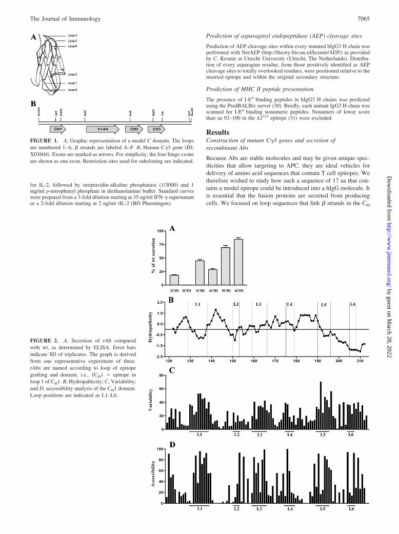
FIGURE 2. A, Secretion of rAb comparedwith wt, as determined by ELISA. Error barsindicate SD of triplicates. The graph is derivedfrom one representative experiment of three.rAbs are named according to loop of epitopegrafting and domain, i.e., 1CH1 � epitope inloop 1 of CH1. B, Hydropathicity; C, Variability;and D, accessibility analysis of the CH1 domain.Loop positions are indicated as L1–L6.
FIGURE 1. A, Graphic representation of a model C domain. The loopsare numbered 1–6, � strands are labeled A–F. B, Human C�3 gene (ID:X03604). Exons are marked as arrows. For simplicity, the four hinge exonsare shown as one exon. Restriction sites used for subcloning are indicated.
7065The Journal of Immunology
by guest on March 28, 2022
http://ww
w.jim
munol.org/
Dow
nloaded from
domains of hIgG3. The mutant molecules should necessarily passthe endoplasmic reticulum quality control and be secreted as com-plete H2 plus L2 Ig (32). We did an analysis of the domain archi-tecture and then took an empirical approach and exchanged everyloop in every CH domain with the model sequence and estimatedthe amounts of rAbs secreted (Figs. 2A, 3A, and 4A).
Since no crystal structure of a hIgG3 has been published, sec-ondary structure analysis of individual C region domains wasbased on knowledge of the structure of representative IgGs, asdescribed in Materials and Methods. Amino acids located in loopsconnecting � strands were determined and the results are presentedin Table I. Although loops 1, 4, and 6 in each domain connect �strands within the same sheet, loops 2, 3, and 5 connect � strandson two opposing sheets. One-half of the 18 loops had a proline ator close to the N- or C-terminal boundary and these were definedas part of the loop rather than the framework. Hydropathicity andsolvent exposure of each amino acids in all three domains wereanalyzed as described in Materials and Methods. The results aredescribed for hydropathicity (Figs. 2B, 3B, and 4B) and solventexposure (Figs. 2C, 3C, and 4C). We found that in CH1, four of sixloops are mostly hydrophilic while two are hydrophobic. All loops
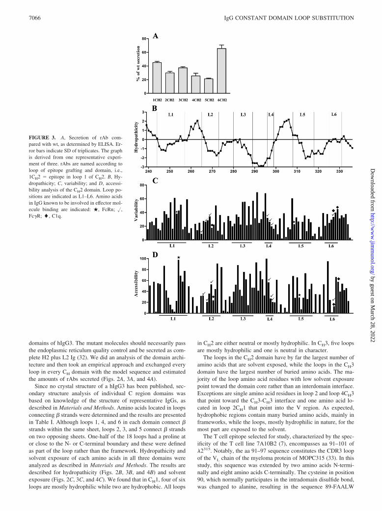
in CH2 are either neutral or mostly hydrophilic. In CH3, five loopsare mostly hydrophilic and one is neutral in character.
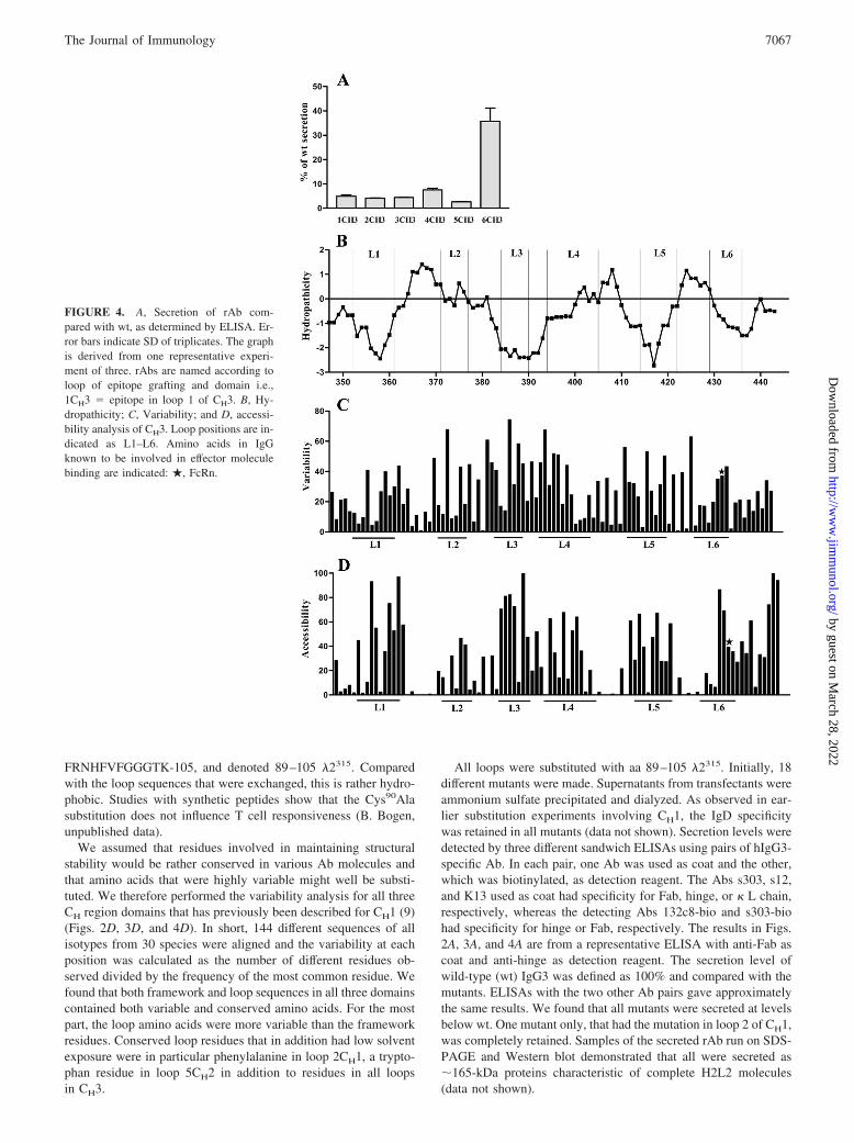
The loops in the CH2 domain have by far the largest number ofamino acids that are solvent exposed, while the loops in the CH3domain have the largest number of buried amino acids. The ma-jority of the loop amino acid residues with low solvent exposurepoint toward the domain core rather than an interdomain interface.Exceptions are single amino acid residues in loop 2 and loop 4CH3that point toward the CH3-CH3 interface and one amino acid lo-cated in loop 2CH1 that point into the V region. As expected,hydrophobic regions contain many buried amino acids, mainly inframeworks, while the loops, mostly hydrophilic in nature, for themost part are exposed to the solvent.
The T cell epitope selected for study, characterized by the spec-ificity of the T cell line 7A10B2 (7), encompasses aa 91–101 of�2315. Notably, the aa 91–97 sequence constitutes the CDR3 loopof the VL chain of the myeloma protein of MOPC315 (33). In thisstudy, this sequence was extended by two amino acids N-termi-nally and eight amino acids C-terminally. The cysteine in position90, which normally participates in the intradomain disulfide bond,was changed to alanine, resulting in the sequence 89-FAALW
FIGURE 3. A, Secretion of rAb com-pared with wt, as determined by ELISA. Er-ror bars indicate SD of triplicates. The graphis derived from one representative experi-ment of three. rAbs are named according toloop of epitope grafting and domain, i.e.,1CH2 � epitope in loop 1 of CH2. B, Hy-dropathicity; C, variability; and D, accessi-bility analysis of the CH2 domain. Loop po-sitions are indicated as L1–L6. Amino acidsin IgG known to be involved in effector mol-ecule binding are indicated: �, FcRn; �,Fc�R; �, C1q.
7066 IgG CONSTANT DOMAIN LOOP SUBSTITUTION
by guest on March 28, 2022
http://ww
w.jim
munol.org/
Dow
nloaded from
FRNHFVFGGGTK-105, and denoted 89–105 �2315. Comparedwith the loop sequences that were exchanged, this is rather hydro-phobic. Studies with synthetic peptides show that the Cys90Alasubstitution does not influence T cell responsiveness (B. Bogen,unpublished data).
We assumed that residues involved in maintaining structuralstability would be rather conserved in various Ab molecules andthat amino acids that were highly variable might well be substi-tuted. We therefore performed the variability analysis for all threeCH region domains that has previously been described for CH1 (9)(Figs. 2D, 3D, and 4D). In short, 144 different sequences of allisotypes from 30 species were aligned and the variability at eachposition was calculated as the number of different residues ob-served divided by the frequency of the most common residue. Wefound that both framework and loop sequences in all three domainscontained both variable and conserved amino acids. For the mostpart, the loop amino acids were more variable than the frameworkresidues. Conserved loop residues that in addition had low solventexposure were in particular phenylalanine in loop 2CH1, a trypto-phan residue in loop 5CH2 in addition to residues in all loopsin CH3.
All loops were substituted with aa 89–105 �2315. Initially, 18different mutants were made. Supernatants from transfectants wereammonium sulfate precipitated and dialyzed. As observed in ear-lier substitution experiments involving CH1, the IgD specificitywas retained in all mutants (data not shown). Secretion levels weredetected by three different sandwich ELISAs using pairs of hIgG3-specific Ab. In each pair, one Ab was used as coat and the other,which was biotinylated, as detection reagent. The Abs s303, s12,and K13 used as coat had specificity for Fab, hinge, or � L chain,respectively, whereas the detecting Abs 132c8-bio and s303-biohad specificity for hinge or Fab, respectively. The results in Figs.2A, 3A, and 4A are from a representative ELISA with anti-Fab ascoat and anti-hinge as detection reagent. The secretion level ofwild-type (wt) IgG3 was defined as 100% and compared with themutants. ELISAs with the two other Ab pairs gave approximatelythe same results. We found that all mutants were secreted at levelsbelow wt. One mutant only, that had the mutation in loop 2 of CH1,was completely retained. Samples of the secreted rAb run on SDS-PAGE and Western blot demonstrated that all were secreted as�165-kDa proteins characteristic of complete H2L2 molecules(data not shown).
FIGURE 4. A, Secretion of rAb com-pared with wt, as determined by ELISA. Er-ror bars indicate SD of triplicates. The graphis derived from one representative experi-ment of three. rAbs are named according toloop of epitope grafting and domain i.e.,1CH3 � epitope in loop 1 of CH3. B, Hy-dropathicity; C, Variability; and D, accessi-bility analysis of CH3. Loop positions are in-dicated as L1–L6. Amino acids in IgGknown to be involved in effector moleculebinding are indicated: �, FcRn.
7067The Journal of Immunology
by guest on March 28, 2022
http://ww
w.jim
munol.org/
Dow
nloaded from
Comparing the secretion levels of the mutants, those with 89–105 �2315 in loop 6CH1 and loop 6CH2 were secreted in relativelyhigh amounts. Excluding loop 2CH1, all mutants with substitutionsin CH1 were secreted rather well at levels between 20 and 80% ofwt. The CH2 mutants were all secreted at 20–60% of wt, whereasfor CH3, the loop 6 mutant only was secreted in high amounts. Theremaining CH3 rAbs were secreted at levels below 10% of wt. Allin all, 17 of 18 loop positions may be exchanged without completeretention and 12 without more than a 5-fold reduction in secretion.
Activation of specific T cells
It is crucial that the mutants are internalized by APC so as to enter theAg-processing pathway and that the specific epitopes are properlyexcised from the rAb carrier to bind MHC and transported as peptide-MHC complexes to the cell surface. In this study, the mutant hIgG3swere tested in two different in vitro T cell activation assays, namely,a growth inhibition assay and a T cell proliferation assay.
Growth inhibition assay
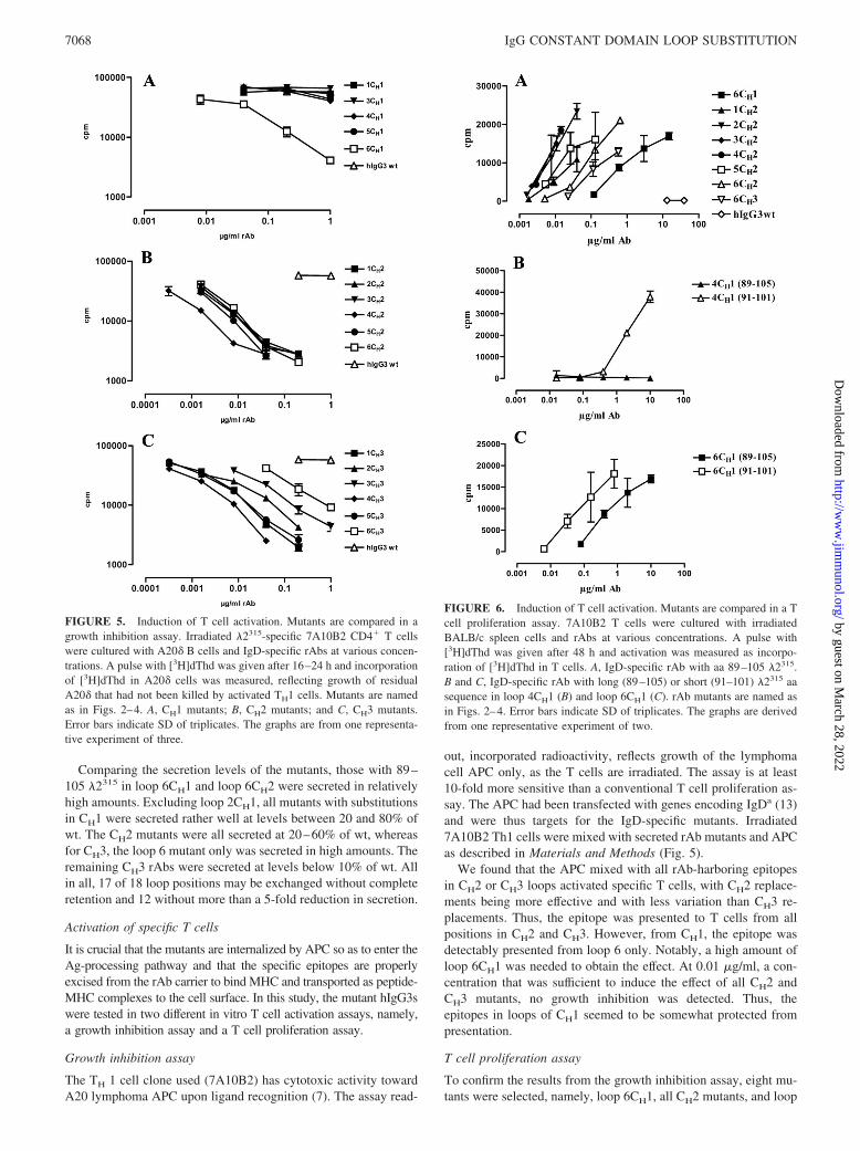
The TH 1 cell clone used (7A10B2) has cytotoxic activity towardA20 lymphoma APC upon ligand recognition (7). The assay read-
out, incorporated radioactivity, reflects growth of the lymphomacell APC only, as the T cells are irradiated. The assay is at least10-fold more sensitive than a conventional T cell proliferation as-say. The APC had been transfected with genes encoding IgDa (13)and were thus targets for the IgD-specific mutants. Irradiated7A10B2 Th1 cells were mixed with secreted rAb mutants and APCas described in Materials and Methods (Fig. 5).
We found that the APC mixed with all rAb-harboring epitopesin CH2 or CH3 loops activated specific T cells, with CH2 replace-ments being more effective and with less variation than CH3 re-placements. Thus, the epitope was presented to T cells from allpositions in CH2 and CH3. However, from CH1, the epitope wasdetectably presented from loop 6 only. Notably, a high amount ofloop 6CH1 was needed to obtain the effect. At 0.01 �g/ml, a con-centration that was sufficient to induce the effect of all CH2 andCH3 mutants, no growth inhibition was detected. Thus, theepitopes in loops of CH1 seemed to be somewhat protected frompresentation.
T cell proliferation assay
To confirm the results from the growth inhibition assay, eight mu-tants were selected, namely, loop 6CH1, all CH2 mutants, and loop
FIGURE 5. Induction of T cell activation. Mutants are compared in agrowth inhibition assay. Irradiated �2315-specific 7A10B2 CD4� T cellswere cultured with A20� B cells and IgD-specific rAbs at various concen-trations. A pulse with [3H]dThd was given after 16–24 h and incorporationof [3H]dThd in A20� cells was measured, reflecting growth of residualA20� that had not been killed by activated TH1 cells. Mutants are namedas in Figs. 2–4. A, CH1 mutants; B, CH2 mutants; and C, CH3 mutants.Error bars indicate SD of triplicates. The graphs are from one representa-tive experiment of three.
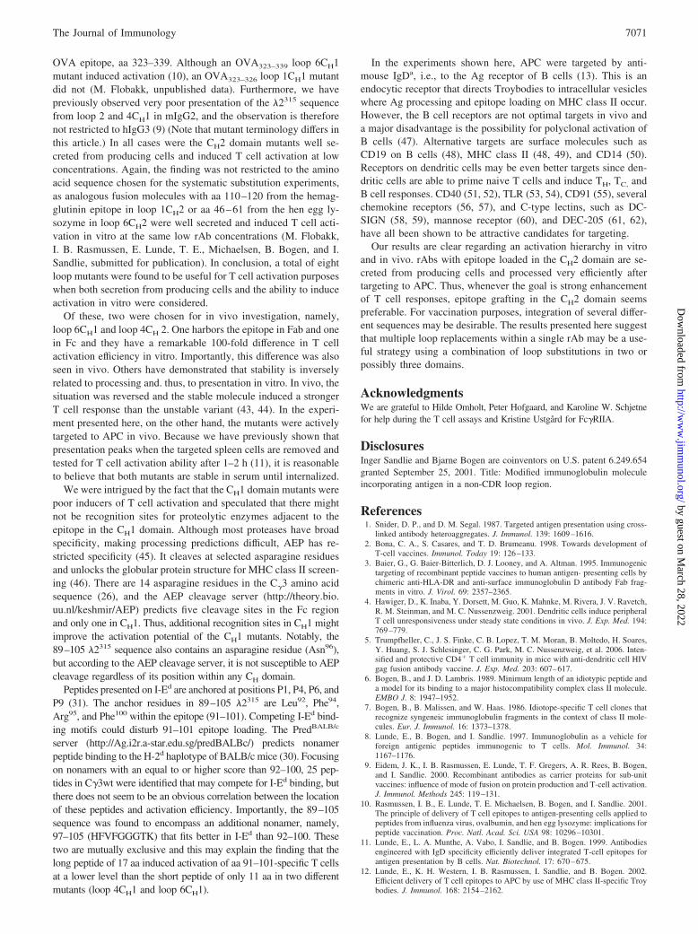
FIGURE 6. Induction of T cell activation. Mutants are compared in a Tcell proliferation assay. 7A10B2 T cells were cultured with irradiatedBALB/c spleen cells and rAbs at various concentrations. A pulse with[3H]dThd was given after 48 h and activation was measured as incorpo-ration of [3H]dThd in T cells. A, IgD-specific rAb with aa 89–105 �2315.B and C, IgD-specific rAb with long (89–105) or short (91–101) �2315 aasequence in loop 4CH1 (B) and loop 6CH1 (C). rAb mutants are named asin Figs. 2–4. Error bars indicate SD of triplicates. The graphs are derivedfrom one representative experiment of two.
7068 IgG CONSTANT DOMAIN LOOP SUBSTITUTION
by guest on March 28, 2022
http://ww
w.jim
munol.org/
Dow
nloaded from
6CH3, all of which were secreted at levels above 20% of wt. Thesewere further tested in a T cell proliferation assay. BALB/c spleencells as APC, T cells, and rAbs were combined. The APC wereirradiated and, thus, in this case, incorporation of radioactivity re-flects T cell proliferation upon Ag stimulation. All mutants testedinduced proliferation of the specific T cells (Fig. 6A). As in thegrowth inhibition assays, the mutants with epitopes in either ofloops 2, 3, or 4CH2 were the most efficient T cell activators, fol-lowed by those with epitope in loops 1, 5, or 6 CH2, which showedintermediate activation ability. Both loop 6CH1 and loop 6CH3mutants required �100� higher concentration than the best mu-tants to induce proper activation. A hierarchy appeared where loop2CH2 � 3CH2 � 4CH2 � 5CH2 � 1CH2 � 6CH2 � 6 CH3 �6CH1. The results correspond well to those obtained in the APCgrowth inhibition assay. A summary of results from the secretionand T cell activation assays are presented in Table III.
A loop 4CH1 mutant has previously proved to induce T cellactivation (8). This mutant was identical to the one tested here,except for the fact that a short sequence of 11 aa, namely, 91–101�2315, was introduced. To test whether the 89–105 sequence of 17aa was less efficient than 91–101, two mutants with the long andtwo mutants with the short sequence in loop 4 or 6 of CH1, re-spectively, were compared side-by-side in the T cell proliferationassay. In each case, the mutant with short sequence was indeedfound to be more efficient than the corresponding mutant with along sequence (Fig. 6, B and C).
T cell activation after in vivo targeting
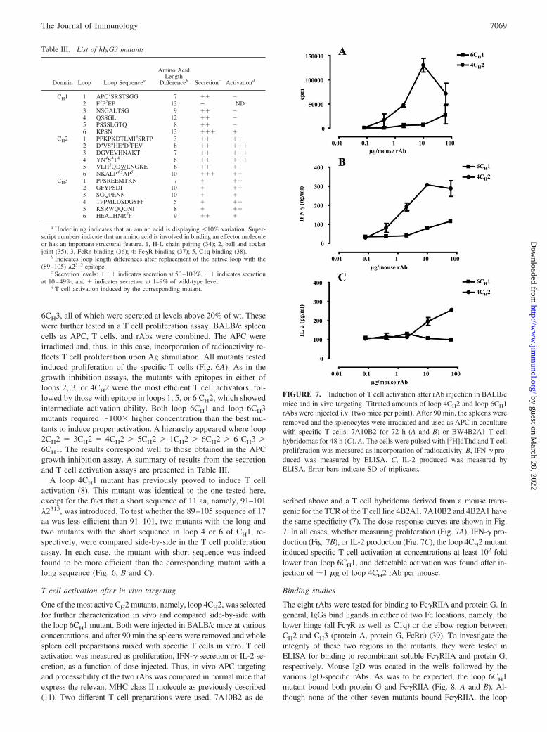
One of the most active CH2 mutants, namely, loop 4CH2, was selectedfor further characterization in vivo and compared side-by-side withthe loop 6CH1 mutant. Both were injected in BALB/c mice at variousconcentrations, and after 90 min the spleens were removed and wholespleen cell preparations mixed with specific T cells in vitro. T cellactivation was measured as proliferation, IFN-� secretion or IL-2 se-cretion, as a function of dose injected. Thus, in vivo APC targetingand processability of the two rAbs was compared in normal mice thatexpress the relevant MHC class II molecule as previously described(11). Two different T cell preparations were used, 7A10B2 as de-
scribed above and a T cell hybridoma derived from a mouse trans-genic for the TCR of the T cell line 4B2A1. 7A10B2 and 4B2A1 havethe same specificity (7). The dose-response curves are shown in Fig.7. In all cases, whether measuring proliferation (Fig. 7A), IFN-� pro-duction (Fig. 7B), or IL-2 production (Fig. 7C), the loop 4CH2 mutantinduced specific T cell activation at concentrations at least 102-foldlower than loop 6CH1, and detectable activation was found after in-jection of �1 �g of loop 4CH2 rAb per mouse.
Binding studies
The eight rAbs were tested for binding to Fc�RIIA and protein G. Ingeneral, IgGs bind ligands in either of two Fc locations, namely, thelower hinge (all Fc�R as well as C1q) or the elbow region betweenCH2 and CH3 (protein A, protein G, FcRn) (39). To investigate theintegrity of these two regions in the mutants, they were tested inELISA for binding to recombinant soluble Fc�RIIA and protein G,respectively. Mouse IgD was coated in the wells followed by thevarious IgD-specific rAbs. As was to be expected, the loop 6CH1mutant bound both protein G and Fc�RIIA (Fig. 8, A and B). Al-though none of the other seven mutants bound Fc�RIIA, the loop
FIGURE 7. Induction of T cell activation after rAb injection in BALB/cmice and in vivo targeting. Titrated amounts of loop 4CH2 and loop 6CH1rAbs were injected i.v. (two mice per point). After 90 min, the spleens wereremoved and the splenocytes were irradiated and used as APC in coculturewith specific T cells: 7A10B2 for 72 h (A and B) or BW4B2A1 T cellhybridomas for 48 h (C). A, The cells were pulsed with [3H]dThd and T cellproliferation was measured as incorporation of radioactivity. B, IFN-� pro-duced was measured by ELISA. C, IL-2 produced was measured byELISA. Error bars indicate SD of triplicates.
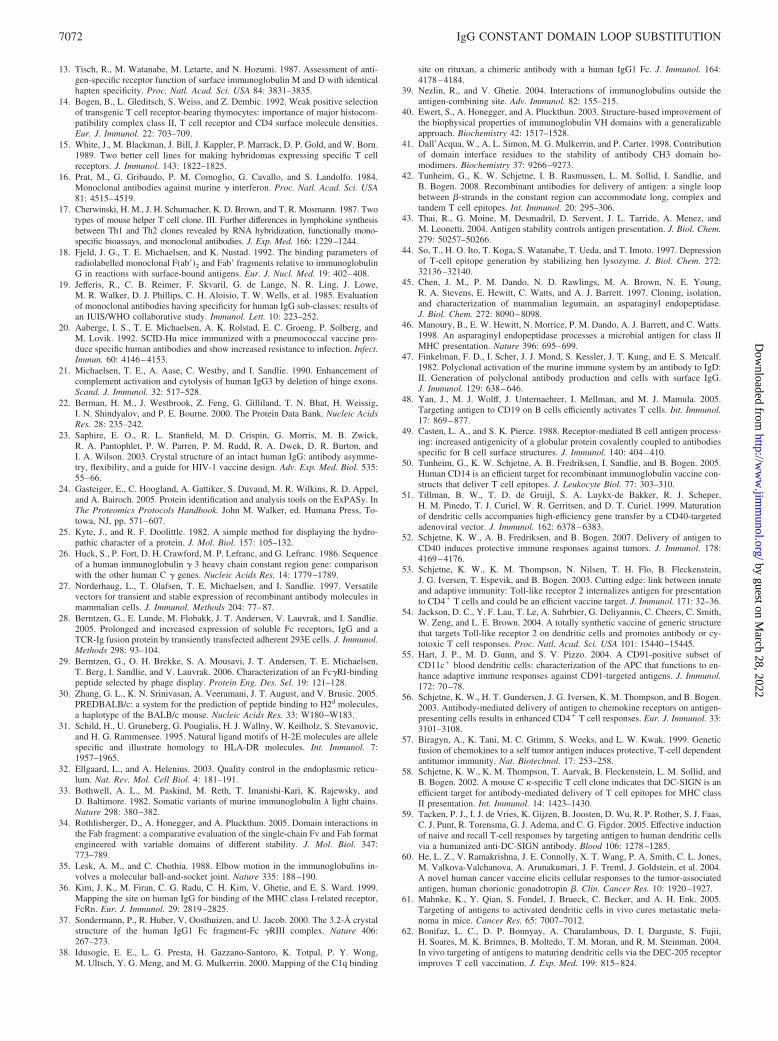
Table III. List of hIgG3 mutants
Domain Loop Loop Sequencea
Amino AcidLength
Differenceb Secretionc Activationd
CH1 1 APC1SRSTSGG 7 �� �2 F2P2EP 13 � ND3 NSGALTSG 9 �� �4 QSSGL 12 �� �5 PSSSLGTQ 8 �� �6 KPSN 13 ��� �
CH2 1 PPKPKDTLMI3SRTP 3 �� ��2 D4VS4HE4D5PEV 8 �� ���3 DGVEVHNAKT 7 �� ���4 YN4S4T4 8 �� ���5 VLH3QDWLNGKE 6 �� ��6 NKALP4,5AP5 10 ��� ��
CH3 1 PPSREEMTKN 7 � ��2 GFYPSDI 10 � ��3 SGQPENN 10 � �4 TPPMLDSDGSFF 5 � ��5 KSRWQQGNI 8 � ��6 HEALHNR3F 9 �� �
a Underlining indicates that an amino acid is displaying �10% variation. Super-script numbers indicate that an amino acid is involved in binding an effector moleculeor has an important structural feature. 1, H-L chain pairing (34); 2, ball and socketjoint (35); 3, FcRn binding (36); 4: Fc�R binding (37); 5, C1q binding (38).
b Indicates loop length differences after replacement of the native loop with the(89–105) �2315 epitope.
c Secretion levels: ��� indicates secretion at 50–100%, �� indicates secretionat 10–49%, and � indicates secretion at 1–9% of wild-type level.
d T cell activation induced by the corresponding mutant.
7069The Journal of Immunology
by guest on March 28, 2022
http://ww
w.jim
munol.org/
Dow
nloaded from
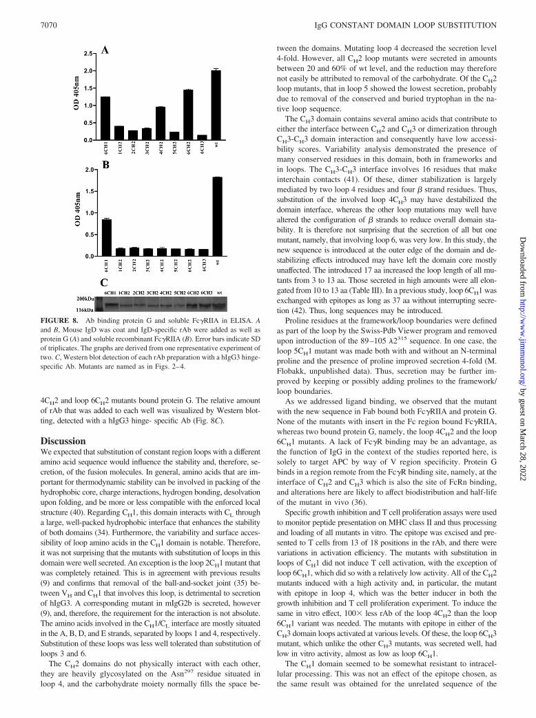
4CH2 and loop 6CH2 mutants bound protein G. The relative amountof rAb that was added to each well was visualized by Western blot-ting, detected with a hIgG3 hinge- specific Ab (Fig. 8C).
DiscussionWe expected that substitution of constant region loops with a differentamino acid sequence would influence the stability and, therefore, se-cretion, of the fusion molecules. In general, amino acids that are im-portant for thermodynamic stability can be involved in packing of thehydrophobic core, charge interactions, hydrogen bonding, desolvationupon folding, and be more or less compatible with the enforced localstructure (40). Regarding CH1, this domain interacts with CL througha large, well-packed hydrophobic interface that enhances the stabilityof both domains (34). Furthermore, the variability and surface acces-sibility of loop amino acids in the CH1 domain is notable. Therefore,it was not surprising that the mutants with substitution of loops in thisdomain were well secreted. An exception is the loop 2CH1 mutant thatwas completely retained. This is in agreement with previous results(9) and confirms that removal of the ball-and-socket joint (35) be-tween VH and CH1 that involves this loop, is detrimental to secretionof hIgG3. A corresponding mutant in mIgG2b is secreted, however(9), and, therefore, the requirement for the interaction is not absolute.The amino acids involved in the CH1/CL interface are mostly situatedin the A, B, D, and E strands, separated by loops 1 and 4, respectively.Substitution of these loops was less well tolerated than substitution ofloops 3 and 6.
The CH2 domains do not physically interact with each other,they are heavily glycosylated on the Asn297 residue situated inloop 4, and the carbohydrate moiety normally fills the space be-
tween the domains. Mutating loop 4 decreased the secretion level4-fold. However, all CH2 loop mutants were secreted in amountsbetween 20 and 60% of wt level, and the reduction may thereforenot easily be attributed to removal of the carbohydrate. Of the CH2loop mutants, that in loop 5 showed the lowest secretion, probablydue to removal of the conserved and buried tryptophan in the na-tive loop sequence.
The CH3 domain contains several amino acids that contribute toeither the interface between CH2 and CH3 or dimerization throughCH3-CH3 domain interaction and consequently have low accessi-bility scores. Variability analysis demonstrated the presence ofmany conserved residues in this domain, both in frameworks andin loops. The CH3-CH3 interface involves 16 residues that makeinterchain contacts (41). Of these, dimer stabilization is largelymediated by two loop 4 residues and four � strand residues. Thus,substitution of the involved loop 4CH3 may have destabilized thedomain interface, whereas the other loop mutations may well havealtered the configuration of � strands to reduce overall domain sta-bility. It is therefore not surprising that the secretion of all but onemutant, namely, that involving loop 6, was very low. In this study, thenew sequence is introduced at the outer edge of the domain and de-stabilizing effects introduced may have left the domain core mostlyunaffected. The introduced 17 aa increased the loop length of all mu-tants from 3 to 13 aa. Those secreted in high amounts were all elon-gated from 10 to 13 aa (Table III). In a previous study, loop 6CH1 wasexchanged with epitopes as long as 37 aa without interrupting secre-tion (42). Thus, long sequences may be introduced.
Proline residues at the framework/loop boundaries were definedas part of the loop by the Swiss-Pdb Viewer program and removedupon introduction of the 89–105 �2315 sequence. In one case, theloop 5CH1 mutant was made both with and without an N-terminalproline and the presence of proline improved secretion 4-fold (M.Flobakk, unpublished data). Thus, secretion may be further im-proved by keeping or possibly adding prolines to the framework/loop boundaries.
As we addressed ligand binding, we observed that the mutantwith the new sequence in Fab bound both Fc�RIIA and protein G.None of the mutants with insert in the Fc region bound Fc�RIIA,whereas two bound protein G, namely, the loop 4CH2 and the loop6CH1 mutants. A lack of Fc�R binding may be an advantage, asthe function of IgG in the context of the studies reported here, issolely to target APC by way of V region specificity. Protein Gbinds in a region remote from the Fc�R binding site, namely, at theinterface of CH2 and CH3 which is also the site of FcRn binding,and alterations here are likely to affect biodistribution and half-lifeof the mutant in vivo (36).
Specific growth inhibition and T cell proliferation assays were usedto monitor peptide presentation on MHC class II and thus processingand loading of all mutants in vitro. The epitope was excised and pre-sented to T cells from 13 of 18 positions in the rAb, and there werevariations in activation efficiency. The mutants with substitution inloops of CH1 did not induce T cell activation, with the exception ofloop 6CH1, which did so with a relatively low activity. All of the CH2mutants induced with a high activity and, in particular, the mutantwith epitope in loop 4, which was the better inducer in both thegrowth inhibition and T cell proliferation experiment. To induce thesame in vitro effect, 100� less rAb of the loop 4CH2 than the loop6CH1 variant was needed. The mutants with epitope in either of theCH3 domain loops activated at various levels. Of these, the loop 6CH3mutant, which unlike the other CH3 mutants, was secreted well, hadlow in vitro activity, almost as low as loop 6CH1.
The CH1 domain seemed to be somewhat resistant to intracel-lular processing. This was not an effect of the epitope chosen, asthe same result was obtained for the unrelated sequence of the
FIGURE 8. Ab binding protein G and soluble Fc�RIIA in ELISA. Aand B, Mouse IgD was coat and IgD-specific rAb were added as well asprotein G (A) and soluble recombinant Fc�RIIA (B). Error bars indicate SDof triplicates. The graphs are derived from one representative experiment oftwo. C, Western blot detection of each rAb preparation with a hIgG3 hinge-specific Ab. Mutants are named as in Figs. 2–4.
7070 IgG CONSTANT DOMAIN LOOP SUBSTITUTION
by guest on March 28, 2022
http://ww
w.jim
munol.org/
Dow
nloaded from
OVA epitope, aa 323–339. Although an OVA323–339 loop 6CH1mutant induced activation (10), an OVA323–326 loop 1CH1 mutantdid not (M. Flobakk, unpublished data). Furthermore, we havepreviously observed very poor presentation of the �2315 sequencefrom loop 2 and 4CH1 in mIgG2, and the observation is thereforenot restricted to hIgG3 (9) (Note that mutant terminology differs inthis article.) In all cases were the CH2 domain mutants well se-creted from producing cells and induced T cell activation at lowconcentrations. Again, the finding was not restricted to the aminoacid sequence chosen for the systematic substitution experiments,as analogous fusion molecules with aa 110–120 from the hemag-glutinin epitope in loop 1CH2 or aa 46–61 from the hen egg ly-sozyme in loop 6CH2 were well secreted and induced T cell acti-vation in vitro at the same low rAb concentrations (M. Flobakk,I. B. Rasmussen, E. Lunde, T. E., Michaelsen, B. Bogen, and I.Sandlie, submitted for publication). In conclusion, a total of eightloop mutants were found to be useful for T cell activation purposeswhen both secretion from producing cells and the ability to induceactivation in vitro were considered.
Of these, two were chosen for in vivo investigation, namely,loop 6CH1 and loop 4CH 2. One harbors the epitope in Fab and onein Fc and they have a remarkable 100-fold difference in T cellactivation efficiency in vitro. Importantly, this difference was alsoseen in vivo. Others have demonstrated that stability is inverselyrelated to processing and. thus, to presentation in vitro. In vivo, thesituation was reversed and the stable molecule induced a strongerT cell response than the unstable variant (43, 44). In the experi-ment presented here, on the other hand, the mutants were activelytargeted to APC in vivo. Because we have previously shown thatpresentation peaks when the targeted spleen cells are removed andtested for T cell activation ability after 1–2 h (11), it is reasonableto believe that both mutants are stable in serum until internalized.
We were intrigued by the fact that the CH1 domain mutants werepoor inducers of T cell activation and speculated that there mightnot be recognition sites for proteolytic enzymes adjacent to theepitope in the CH1 domain. Although most proteases have broadspecificity, making processing predictions difficult, AEP has re-stricted specificity (45). It cleaves at selected asparagine residuesand unlocks the globular protein structure for MHC class II screen-ing (46). There are 14 asparagine residues in the C�3 amino acidsequence (26), and the AEP cleavage server (http://theory.bio.uu.nl/keshmir/AEP) predicts five cleavage sites in the Fc regionand only one in CH1. Thus, additional recognition sites in CH1 mightimprove the activation potential of the CH1 mutants. Notably, the89–105 �2315 sequence also contains an asparagine residue (Asn96),but according to the AEP cleavage server, it is not susceptible to AEPcleavage regardless of its position within any CH domain.
Peptides presented on I-Ed are anchored at positions P1, P4, P6, andP9 (31). The anchor residues in 89–105 �2315 are Leu92, Phe94,Arg95, and Phe100 within the epitope (91–101). Competing I-Ed bind-ing motifs could disturb 91–101 epitope loading. The PredBALB/c
server (http://Ag.i2r.a-star.edu.sg/predBALBc/) predicts nonamerpeptide binding to the H-2d haplotype of BALB/c mice (30). Focusingon nonamers with an equal to or higher score than 92–100, 25 pep-tides in C�3wt were identified that may compete for I-Ed binding, butthere does not seem to be an obvious correlation between the locationof these peptides and activation efficiency. Importantly, the 89–105sequence was found to encompass an additional nonamer, namely,97–105 (HFVFGGGTK) that fits better in I-Ed than 92–100. Thesetwo are mutually exclusive and this may explain the finding that thelong peptide of 17 aa induced activation of aa 91–101-specific T cellsat a lower level than the short peptide of only 11 aa in two differentmutants (loop 4CH1 and loop 6CH1).
In the experiments shown here, APC were targeted by anti-mouse IgDa, i.e., to the Ag receptor of B cells (13). This is anendocytic receptor that directs Troybodies to intracellular vesicleswhere Ag processing and epitope loading on MHC class II occur.However, the B cell receptors are not optimal targets in vivo anda major disadvantage is the possibility for polyclonal activation ofB cells (47). Alternative targets are surface molecules such asCD19 on B cells (48), MHC class II (48, 49), and CD14 (50).Receptors on dendritic cells may be even better targets since den-dritic cells are able to prime naive T cells and induce TH, TC, andB cell responses. CD40 (51, 52), TLR (53, 54), CD91 (55), severalchemokine receptors (56, 57), and C-type lectins, such as DC-SIGN (58, 59), mannose receptor (60), and DEC-205 (61, 62),have all been shown to be attractive candidates for targeting.
Our results are clear regarding an activation hierarchy in vitroand in vivo. rAbs with epitope loaded in the CH2 domain are se-creted from producing cells and processed very efficiently aftertargeting to APC. Thus, whenever the goal is strong enhancementof T cell responses, epitope grafting in the CH2 domain seemspreferable. For vaccination purposes, integration of several differ-ent sequences may be desirable. The results presented here suggestthat multiple loop replacements within a single rAb may be a use-ful strategy using a combination of loop substitutions in two orpossibly three domains.
AcknowledgmentsWe are grateful to Hilde Omholt, Peter Hofgaard, and Karoline W. Schjetnefor help during the T cell assays and Kristine Ustgård for Fc�RIIA.
DisclosuresInger Sandlie and Bjarne Bogen are coinventors on U.S. patent 6.249.654granted September 25, 2001. Title: Modified immunoglobulin moleculeincorporating antigen in a non-CDR loop region.
References1. Snider, D. P., and D. M. Segal. 1987. Targeted antigen presentation using cross-
linked antibody heteroaggregates. J. Immunol. 139: 1609–1616.2. Bona, C. A., S. Casares, and T. D. Brumeanu. 1998. Towards development of
T-cell vaccines. Immunol. Today 19: 126–133.3. Baier, G., G. Baier-Bitterlich, D. J. Looney, and A. Altman. 1995. Immunogenic
targeting of recombinant peptide vaccines to human antigen- presenting cells bychimeric anti-HLA-DR and anti-surface immunoglobulin D antibody Fab frag-ments in vitro. J. Virol. 69: 2357–2365.
4. Hawiger, D., K. Inaba, Y. Dorsett, M. Guo, K. Mahnke, M. Rivera, J. V. Ravetch,R. M. Steinman, and M. C. Nussenzweig. 2001. Dendritic cells induce peripheralT cell unresponsiveness under steady state conditions in vivo. J. Exp. Med. 194:769–779.
5. Trumpfheller, C., J. S. Finke, C. B. Lopez, T. M. Moran, B. Moltedo, H. Soares,Y. Huang, S. J. Schlesinger, C. G. Park, M. C. Nussenzweig, et al. 2006. Inten-sified and protective CD4� T cell immunity in mice with anti-dendritic cell HIVgag fusion antibody vaccine. J. Exp. Med. 203: 607–617.
6. Bogen, B., and J. D. Lambris. 1989. Minimum length of an idiotypic peptide anda model for its binding to a major histocompatibility complex class II molecule.EMBO J. 8: 1947–1952.
7. Bogen, B., B. Malissen, and W. Haas. 1986. Idiotope-specific T cell clones thatrecognize syngeneic immunoglobulin fragments in the context of class II mole-cules. Eur. J. Immunol. 16: 1373–1378.
8. Lunde, E., B. Bogen, and I. Sandlie. 1997. Immunoglobulin as a vehicle forforeign antigenic peptides immunogenic to T cells. Mol. Immunol. 34:1167–1176.
9. Eidem, J. K., I. B. Rasmussen, E. Lunde, T. F. Gregers, A. R. Rees, B. Bogen,and I. Sandlie. 2000. Recombinant antibodies as carrier proteins for sub-unitvaccines: influence of mode of fusion on protein production and T-cell activation.J. Immunol. Methods 245: 119–131.
10. Rasmussen, I. B., E. Lunde, T. E. Michaelsen, B. Bogen, and I. Sandlie. 2001.The principle of delivery of T cell epitopes to antigen-presenting cells applied topeptides from influenza virus, ovalbumin, and hen egg lysozyme: implications forpeptide vaccination. Proc. Natl. Acad. Sci. USA 98: 10296–10301.
11. Lunde, E., L. A. Munthe, A. Vabo, I. Sandlie, and B. Bogen. 1999. Antibodiesengineered with IgD specificity efficiently deliver integrated T-cell epitopes forantigen presentation by B cells. Nat. Biotechnol. 17: 670–675.
12. Lunde, E., K. H. Western, I. B. Rasmussen, I. Sandlie, and B. Bogen. 2002.Efficient delivery of T cell epitopes to APC by use of MHC class II-specific Troybodies. J. Immunol. 168: 2154–2162.
7071The Journal of Immunology
by guest on March 28, 2022
http://ww
w.jim
munol.org/
Dow
nloaded from
13. Tisch, R., M. Watanabe, M. Letarte, and N. Hozumi. 1987. Assessment of anti-gen-specific receptor function of surface immunoglobulin M and D with identicalhapten specificity. Proc. Natl. Acad. Sci. USA 84: 3831–3835.
14. Bogen, B., L. Gleditsch, S. Weiss, and Z. Dembic. 1992. Weak positive selectionof transgenic T cell receptor-bearing thymocytes: importance of major histocom-patibility complex class II, T cell receptor and CD4 surface molecule densities.Eur. J. Immunol. 22: 703–709.
15. White, J., M. Blackman, J. Bill, J. Kappler, P. Marrack, D. P. Gold, and W. Born.1989. Two better cell lines for making hybridomas expressing specific T cellreceptors. J. Immunol. 143: 1822–1825.
16. Prat, M., G. Gribaudo, P. M. Comoglio, G. Cavallo, and S. Landolfo. 1984.Monoclonal antibodies against murine � interferon. Proc. Natl. Acad. Sci. USA81: 4515–4519.
17. Cherwinski, H. M., J. H. Schumacher, K. D. Brown, and T. R. Mosmann. 1987. Twotypes of mouse helper T cell clone. III. Further differences in lymphokine synthesisbetween Th1 and Th2 clones revealed by RNA hybridization, functionally mono-specific bioassays, and monoclonal antibodies. J. Exp. Med. 166: 1229–1244.
18. Fjeld, J. G., T. E. Michaelsen, and K. Nustad. 1992. The binding parameters ofradiolabelled monoclonal F(ab�)2 and Fab� fragments relative to immunoglobulinG in reactions with surface-bound antigens. Eur. J. Nucl. Med. 19: 402–408.
19. Jefferis, R., C. B. Reimer, F. Skvaril, G. de Lange, N. R. Ling, J. Lowe,M. R. Walker, D. J. Phillips, C. H. Aloisio, T. W. Wells, et al. 1985. Evaluationof monoclonal antibodies having specificity for human IgG sub-classes: results ofan IUIS/WHO collaborative study. Immunol. Lett. 10: 223–252.
20. Aaberge, I. S., T. E. Michaelsen, A. K. Rolstad, E. C. Groeng, P. Solberg, andM. Lovik. 1992. SCID-Hu mice immunized with a pneumococcal vaccine pro-duce specific human antibodies and show increased resistance to infection. Infect.Immun. 60: 4146–4153.
21. Michaelsen, T. E., A. Aase, C. Westby, and I. Sandlie. 1990. Enhancement ofcomplement activation and cytolysis of human IgG3 by deletion of hinge exons.Scand. J. Immunol. 32: 517–528.
22. Berman, H. M., J. Westbrook, Z. Feng, G. Gilliland, T. N. Bhat, H. Weissig,I. N. Shindyalov, and P. E. Bourne. 2000. The Protein Data Bank. Nucleic AcidsRes. 28: 235–242.
23. Saphire, E. O., R. L. Stanfield, M. D. Crispin, G. Morris, M. B. Zwick,R. A. Pantophlet, P. W. Parren, P. M. Rudd, R. A. Dwek, D. R. Burton, andI. A. Wilson. 2003. Crystal structure of an intact human IgG: antibody asymme-try, flexibility, and a guide for HIV-1 vaccine design. Adv. Exp. Med. Biol. 535:55–66.
24. Gasteiger, E., C. Hoogland, A. Gattiker, S. Duvaud, M. R. Wilkins, R. D. Appel,and A. Bairoch. 2005. Protein identification and analysis tools on the ExPASy. InThe Proteomics Protocols Handbook. John M. Walker, ed. Humana Press, To-towa, NJ, pp. 571–607.
25. Kyte, J., and R. F. Doolittle. 1982. A simple method for displaying the hydro-pathic character of a protein. J. Mol. Biol. 157: 105–132.
26. Huck, S., P. Fort, D. H. Crawford, M. P. Lefranc, and G. Lefranc. 1986. Sequenceof a human immunoglobulin � 3 heavy chain constant region gene: comparisonwith the other human C � genes. Nucleic Acids Res. 14: 1779–1789.
27. Norderhaug, L., T. Olafsen, T. E. Michaelsen, and I. Sandlie. 1997. Versatilevectors for transient and stable expression of recombinant antibody molecules inmammalian cells. J. Immunol. Methods 204: 77–87.
28. Berntzen, G., E. Lunde, M. Flobakk, J. T. Andersen, V. Lauvrak, and I. Sandlie.2005. Prolonged and increased expression of soluble Fc receptors, IgG and aTCR-Ig fusion protein by transiently transfected adherent 293E cells. J. Immunol.Methods 298: 93–104.
29. Berntzen, G., O. H. Brekke, S. A. Mousavi, J. T. Andersen, T. E. Michaelsen,T. Berg, I. Sandlie, and V. Lauvrak. 2006. Characterization of an Fc�RI-bindingpeptide selected by phage display. Protein Eng. Des. Sel. 19: 121–128.
30. Zhang, G. L., K. N. Srinivasan, A. Veeramani, J. T. August, and V. Brusic. 2005.PREDBALB/c: a system for the prediction of peptide binding to H2d molecules,a haplotype of the BALB/c mouse. Nucleic Acids Res. 33: W180–W183.
31. Schild, H., U. Gruneberg, G. Pougialis, H. J. Wallny, W. Keilholz, S. Stevanovic,and H. G. Rammensee. 1995. Natural ligand motifs of H-2E molecules are allelespecific and illustrate homology to HLA-DR molecules. Int. Immunol. 7:1957–1965.
32. Ellgaard, L., and A. Helenius. 2003. Quality control in the endoplasmic reticu-lum. Nat. Rev. Mol. Cell Biol. 4: 181–191.
33. Bothwell, A. L., M. Paskind, M. Reth, T. Imanishi-Kari, K. Rajewsky, andD. Baltimore. 1982. Somatic variants of murine immunoglobulin � light chains.Nature 298: 380–382.
34. Rothlisberger, D., A. Honegger, and A. Pluckthun. 2005. Domain interactions inthe Fab fragment: a comparative evaluation of the single-chain Fv and Fab formatengineered with variable domains of different stability. J. Mol. Biol. 347:773–789.
35. Lesk, A. M., and C. Chothia. 1988. Elbow motion in the immunoglobulins in-volves a molecular ball-and-socket joint. Nature 335: 188–190.
36. Kim, J. K., M. Firan, C. G. Radu, C. H. Kim, V. Ghetie, and E. S. Ward. 1999.Mapping the site on human IgG for binding of the MHC class I-related receptor,FcRn. Eur. J. Immunol. 29: 2819–2825.
37. Sondermann, P., R. Huber, V. Oosthuizen, and U. Jacob. 2000. The 3.2-Å crystalstructure of the human IgG1 Fc fragment-Fc �RIII complex. Nature 406:267–273.
38. Idusogie, E. E., L. G. Presta, H. Gazzano-Santoro, K. Totpal, P. Y. Wong,M. Ultsch, Y. G. Meng, and M. G. Mulkerrin. 2000. Mapping of the C1q binding
site on rituxan, a chimeric antibody with a human IgG1 Fc. J. Immunol. 164:4178–4184.
39. Nezlin, R., and V. Ghetie. 2004. Interactions of immunoglobulins outside theantigen-combining site. Adv. Immunol. 82: 155–215.
40. Ewert, S., A. Honegger, and A. Pluckthun. 2003. Structure-based improvement ofthe biophysical properties of immunoglobulin VH domains with a generalizableapproach. Biochemistry 42: 1517–1528.
41. Dall’Acqua, W., A. L. Simon, M. G. Mulkerrin, and P. Carter. 1998. Contributionof domain interface residues to the stability of antibody CH3 domain ho-modimers. Biochemistry 37: 9266–9273.
42. Tunheim, G., K. W. Schjetne, I. B. Rasmussen, L. M. Sollid, I. Sandlie, andB. Bogen. 2008. Recombinant antibodies for delivery of antigen: a single loopbetween �-strands in the constant region can accommodate long, complex andtandem T cell epitopes. Int. Immunol. 20: 295–306.
43. Thai, R., G. Moine, M. Desmadril, D. Servent, J. L. Tarride, A. Menez, andM. Leonetti. 2004. Antigen stability controls antigen presentation. J. Biol. Chem.279: 50257–50266.
44. So, T., H. O. Ito, T. Koga, S. Watanabe, T. Ueda, and T. Imoto. 1997. Depressionof T-cell epitope generation by stabilizing hen lysozyme. J. Biol. Chem. 272:32136–32140.
45. Chen, J. M., P. M. Dando, N. D. Rawlings, M. A. Brown, N. E. Young,R. A. Stevens, E. Hewitt, C. Watts, and A. J. Barrett. 1997. Cloning, isolation,and characterization of mammalian legumain, an asparaginyl endopeptidase.J. Biol. Chem. 272: 8090–8098.
46. Manoury, B., E. W. Hewitt, N. Morrice, P. M. Dando, A. J. Barrett, and C. Watts.1998. An asparaginyl endopeptidase processes a microbial antigen for class IIMHC presentation. Nature 396: 695–699.
47. Finkelman, F. D., I. Scher, J. J. Mond, S. Kessler, J. T. Kung, and E. S. Metcalf.1982. Polyclonal activation of the murine immune system by an antibody to IgD:II. Generation of polyclonal antibody production and cells with surface IgG.J. Immunol. 129: 638–646.
48. Yan, J., M. J. Wolff, J. Unternaehrer, I. Mellman, and M. J. Mamula. 2005.Targeting antigen to CD19 on B cells efficiently activates T cells. Int. Immunol.17: 869–877.
49. Casten, L. A., and S. K. Pierce. 1988. Receptor-mediated B cell antigen process-ing: increased antigenicity of a globular protein covalently coupled to antibodiesspecific for B cell surface structures. J. Immunol. 140: 404–410.
50. Tunheim, G., K. W. Schjetne, A. B. Fredriksen, I. Sandlie, and B. Bogen. 2005.Human CD14 is an efficient target for recombinant immunoglobulin vaccine con-structs that deliver T cell epitopes. J. Leukocyte Biol. 77: 303–310.
51. Tillman, B. W., T. D. de Gruijl, S. A. Luykx-de Bakker, R. J. Scheper,H. M. Pinedo, T. J. Curiel, W. R. Gerritsen, and D. T. Curiel. 1999. Maturationof dendritic cells accompanies high-efficiency gene transfer by a CD40-targetedadenoviral vector. J. Immunol. 162: 6378–6383.
52. Schjetne, K. W., A. B. Fredriksen, and B. Bogen. 2007. Delivery of antigen toCD40 induces protective immune responses against tumors. J. Immunol. 178:4169–4176.
53. Schjetne, K. W., K. M. Thompson, N. Nilsen, T. H. Flo, B. Fleckenstein,J. G. Iversen, T. Espevik, and B. Bogen. 2003. Cutting edge: link between innateand adaptive immunity: Toll-like receptor 2 internalizes antigen for presentationto CD4� T cells and could be an efficient vaccine target. J. Immunol. 171: 32–36.
54. Jackson, D. C., Y. F. Lau, T. Le, A. Suhrbier, G. Deliyannis, C. Cheers, C. Smith,W. Zeng, and L. E. Brown. 2004. A totally synthetic vaccine of generic structurethat targets Toll-like receptor 2 on dendritic cells and promotes antibody or cy-totoxic T cell responses. Proc. Natl. Acad. Sci. USA 101: 15440–15445.
55. Hart, J. P., M. D. Gunn, and S. V. Pizzo. 2004. A CD91-positive subset ofCD11c� blood dendritic cells: characterization of the APC that functions to en-hance adaptive immune responses against CD91-targeted antigens. J. Immunol.172: 70–78.
56. Schjetne, K. W., H. T. Gundersen, J. G. Iversen, K. M. Thompson, and B. Bogen.2003. Antibody-mediated delivery of antigen to chemokine receptors on antigen-presenting cells results in enhanced CD4� T cell responses. Eur. J. Immunol. 33:3101–3108.
57. Biragyn, A., K. Tani, M. C. Grimm, S. Weeks, and L. W. Kwak. 1999. Geneticfusion of chemokines to a self tumor antigen induces protective, T-cell dependentantitumor immunity. Nat. Biotechnol. 17: 253–258.
58. Schjetne, K. W., K. M. Thompson, T. Aarvak, B. Fleckenstein, L. M. Sollid, andB. Bogen. 2002. A mouse C �-specific T cell clone indicates that DC-SIGN is anefficient target for antibody-mediated delivery of T cell epitopes for MHC classII presentation. Int. Immunol. 14: 1423–1430.
59. Tacken, P. J., I. J. de Vries, K. Gijzen, B. Joosten, D. Wu, R. P. Rother, S. J. Faas,C. J. Punt, R. Torensma, G. J. Adema, and C. G. Figdor. 2005. Effective inductionof naive and recall T-cell responses by targeting antigen to human dendritic cellsvia a humanized anti-DC-SIGN antibody. Blood 106: 1278–1285.
60. He, L. Z., V. Ramakrishna, J. E. Connolly, X. T. Wang, P. A. Smith, C. L. Jones,M. Valkova-Valchanova, A. Arunakumari, J. F. Treml, J. Goldstein, et al. 2004.A novel human cancer vaccine elicits cellular responses to the tumor-associatedantigen, human chorionic gonadotropin �. Clin. Cancer Res. 10: 1920–1927.
61. Mahnke, K., Y. Qian, S. Fondel, J. Brueck, C. Becker, and A. H. Enk. 2005.Targeting of antigens to activated dendritic cells in vivo cures metastatic mela-noma in mice. Cancer Res. 65: 7007–7012.
62. Bonifaz, L. C., D. P. Bonnyay, A. Charalambous, D. I. Darguste, S. Fujii,H. Soares, M. K. Brimnes, B. Moltedo, T. M. Moran, and R. M. Steinman. 2004.In vivo targeting of antigens to maturing dendritic cells via the DEC-205 receptorimproves T cell vaccination. J. Exp. Med. 199: 815–824.
7072 IgG CONSTANT DOMAIN LOOP SUBSTITUTION
by guest on March 28, 2022
http://ww
w.jim
munol.org/
Dow
nloaded from