Embed Size (px)
Citation preview

Department of Otorhinolaryngology
University of Helsinki
Finland
PERIPHERAL FACIAL PALSY
Grading, Etiology, and Melkersson-Rosenthal Syndrome
Mervi Kanerva
Academic dissertation
To be publicly discussed,
with the permission of the Medical Faculty of the University of Helsinki,
in the Lecture Room of the Department of Otorhinolaryngology,
Haartmaninkatu 4E, Helsinki, on February 29th
, 2008, at 12 noon.
Helsinki 2008

Supervised by
Professor Anne Pitkäranta
Department of Otorhinolaryngology
University of Helsinki
Reviewed by
Professor Veijo Hukkanen
Department of Microbiology
University of Oulu
Docent Kalle Aitasalo
Department of Otorhinolaryngology-Head and Neck Surgery
University of Turku
Opponent
Professor Ilmari Pyykkö
University of Tampere
Medical School
ISBN 978-952-92-3376-2 (paperback)
ISBN 978-952-10-4541-7 (PDF)
Yliopistopaino
Helsinki 2008

3
CONTENTS
List of original publications……………….………..
Abstract……………………………………………....
Abbreviations……………………………..………....
Introduction…………………………………….........
Review of the literature……………………….……. Peripheral facial palsy………………………………….
Grading facial function…………………………………
Acute idiopathic peripheral facial palsy–Bell’s palsy….
Human herpesvirus-6 and -7…………………………...
Treatment of Bell’s palsy………………………………
Prognosis of Bell’s palsy……………………………….
Peripheral facial palsy in children……………………...
Melkersson-Rosenthal syndrome………………………
Aims of the study………………………………….....
Subjects and methods…………………………….…
Results…………………………...…………………...
Discussion……………………………………………. Grading…………………………………………………
Etiology………………………………………………...
Melkersson-Rosenthal syndrome………………………
Conclusions……………………………...…………...
Acknowledgments……..…………………………….
References………………………...………………….
Appendix…………………………………………......
Original publications
4
5
7
8
10 10
13
19
21
22
23
24
25
28
29
34
42 42
44
46
49
50
52
63

4
LIST OF ORIGINAL PUBLICATIONS
This study is based on the following original publications, which are referred to in the
text by their Roman numerals.
I Kanerva M, Poussa T, Pitkäranta A
Sunnybrook and House-Brackmann Facial Grading Systems: Intrarater repeatability
and interrater agreement
Otolaryngology-Head and Neck Surgery 135(6):865–71, 2006
II Kanerva M, Mannonen L, Piiparinen H, Peltomaa M, Vaheri A, Pitkäranta A
Search for Herpesviruses in cerebrospinal fluid of facial palsy patients by PCR
Acta Oto-Laryngologica 127(7):775–9, 2007
III Kanerva M, Jääskeläinen AJ, Suvela M, Piiparinen H, Vaheri A, Pitkäranta A
Human herpesvirus-6 and -7 DNA in cerebrospinal fluid of facial palsy patients
Acta Oto-Laryngologica, in press
IV Kanerva M, Moilanen K, Virolainen S, Vaheri A, Pitkäranta A
Melkersson-Rosenthal syndrome
Otolaryngology-Head and Neck Surgery, in press
The original articles have been reprinted with the kind permission of their copyright
holders.

5
ABSTRACT
The grading of facial appearance and function in peripheral facial palsy (FP) is
inconsistent and a subject of much dispute. We assessed two grading scales to determine
whether one might be superior for use in everyday clinical practice (I). Eight video-
recorded FP patients were graded in two sittings by 26 doctors. Repeatability and
agreement for the Sunnybrook facial grading system (SFGS) were measured by
intraclass correlation coefficient and coefficient of repeatability, and for the House-
Brackmann facial grading system (H-B FGS) by agreement percentage and kappa
coefficients.
Repeatability for SFGS proved to be from good to excellent and for H-B FGS from fair
to good depending on the statistical method used (I). Agreement between doctors for
SFGS was from moderate to excellent and for H-B FGS from poor to fair. Because
SFGS was at least as good in repeatability as H-B FGS and showed more reliable results
in agreement between doctors, we encourage the use of SFGS over H-B FGS.
The etiology of acute peripheral FP is unverified, as is the question of whether the
central nervous system is affected at some phase of the disease. Our objective was to
determine whether herpesviral DNA could be found in cerebrospinal fluid (CSF) of
peripheral FP patients (II, III). CSF samples from 33 peripheral FP patients (34
samples) and 36 controls were retrospectively examined for DNA of herpes simplex
virus-1 (HSV-1), varicella-zoster virus (VZV), and human herpesvirus-6 (HHV-6) by
polymerase chain reaction (PCR) (II) and for DNA of HSV-1 and -2, VZV, HHV-6A,
-6B, and -7, Epstein-Barr virus (EBV), and cytomegalovirus (CMV) by multiplex-PCR
and oligonucleotide microarray methods (III).
Three patients and five controls had HHV-6 or -7 DNA in CSF (II, III). No DNA of
HSV-1 or -2, VZV, EBV, or CMV was found. HHV-6 and-7 DNA was detectable in
about 10% of the CSF samples evaluated from immunocompetent adolescents and
adults without severe disease (III), an important finding that indicates caution when
interpreting CSF results. Detecting HHV-7 and dual HHV-6A and -6B DNA in CSF of
FP patients is intriguing, but these DNA findings in association with FP and the other
diseases that they accompanied require further exploration.
Melkersson-Rosenthal syndrome (MRS) is classically defined as a triad of recurrent
labial or oro-facial edema, recurrent peripheral FP, and plicated tongue. All three signs
are present in the minority of patients. Edema-dominated forms are more common in the
literature, while MRS with FP has received little attention. The etiology and true
incidence of MRS are unknown. We investigated characteristics of MRS with FP (IV)
and also compared MRS patients treated at the Departments of Otorhinolaryngology
and Dermatology. We hypothesized that in MRS FP patients edema would not be the
dominating feature, nor would progression with time occur, contrary to existing
knowledge. Patient charts at both departments were evaluated for MRS. Patients with
FP were mailed a questionnaire and clinically examined. When appropriate, a tissue
biopsy was taken to search for the nonnecrotizing granulomatous infiltrations typical of
MRS. Herpesviruses, among many other possibilities, have been suspected as etiologic

6
factors in MRS. We searched peripheral blood DNA for gene mutations leading to
UNC-93B protein deficiency, which would predispose to HSV-1 infections.
Thirty-five MRS patients were found, 20 with FP and 11 with the triad form of MRS.
At the Department of Otorhinolaryngology, every MRS patient had FP. Two had tissue
biopsies taken during an acute edema episode, with nonnecrotizing granulomatous
findings. Edema in most MRS FP patients did not dominate the clinical picture, and no
progression of the disease was observed, consistent with our hypotheses. Two triad
patients had recurring anterior uveitis. No UNC-93B1 gene mutations were found. At
the Department of Dermatology, two patients had triad MRS and 15 had
monosymptomatic granulomatous cheilitis with frequent or persistent edema and typical
MRS tissue histology. The clinical picture of MRS varied according to the department
where the patient was treated. More studies from otorhinolaryngology departments and
on patients with FP would clarify the actual incidence and clinical picture of the
syndrome.
FP is a phenomenon with many unconquered aspects (I, II, III, IV) that await future
explorations.

7
ABBREVIATIONS
AAO-HNS American Academy of Otolaryngology-Head and Neck Surgery
CG cheilitis granulomatosa
CI confidence interval
CMV cytomegalovirus
CNS central nervous system
CR coefficient of repeatability
CSF cerebrospinal fluid
EBV Epstein-Barr virus
ENoG electroneuronography
FP facial palsy
H-B FGS House-Brackmann facial grading system
HHV human herpesvirus
HSV herpes simplex virus
ICC intraclass correlation coefficient
LP lingua plicata
MRS Melkersson-Rosenthal syndrome
PCR polymerase chain reaction
SD standard deviation
SFGS Sunnybrook facial grading system
VP virus particle
VZV varicella-zoster virus
σe within-patient/doctor standard deviation

8
INTRODUCTION
Peripheral facial palsy (FP) is a common condition, in most cases without known cause.
Being clearly visible, the cosmetic drawback for the patient is obvious, as is the inability
to mimic normal communication, but the effects on vision, eating, and drinking are
easily overlooked. Even though most patients recover, outcome is not predictable at
palsy onset. Patients are typically under great psychological stress in addition to their
physical limitations. Not knowing the cause, no effective treatments exist to offset
sequelae or persistent palsy in the approximately 30% of patients who fail to recover
completely (Peitersen 2002). Grading facial function is necessary for determining and
reporting the spontaneous course of FP and especially the results of medical or surgical
treatments. However, FP studies are hindered by the lack of an objective, standardized
evaluation method. The subjective methods used vary and are prone to intra- and
interrater variation.
The etiology of acute idiopathic peripheral FP (Bell’s palsy) is still under debate
although herpesviruses, especially herpes simplex virus-1 (HSV-1) and varicella-zoster
virus (VZV), have gained support as etiologic factors (Furuta et al. 2000). HSV-1
reactivation has been reported to accompany Bell’s palsy, but causality is uncertain
(Murakami et al. 1996). VZV is known to cause Ramsay Hunt syndrome, a peripheral
FP with herpes vesicles most commonly in the ear or mouth. In Ramsay Hunt
syndrome, VZV is assumed to be able to spread widely in neural and mucocutaneous
tissue and in cerebrospinal fluid (CSF) (Murakami et al. 1998). VZV infection may
present without visible vesicles as zoster sine herpete, and has been suspected to be a
causative agent in Bell’s palsy in up to 30% of cases (Furuta et al. 2000). Human
herpesvirus (HHV)-6 and -7 infections occur commonly in early childhood and the
viruses persist latently after primary infection (Ward 2005). Both HHV-6 and -7 have
been detected in normal brain tissue at autopsy, indicating that they are able to invade
and persist asymptomatically in the central nervous system (CNS) and can be expected
to reactivate occasionally (Chan et al. 2000, Ward 2005). HHV-6 and -7 have seldom
been studied in association with FP, but we previously found HHV-6 DNA in the tear
fluid of Bell’s palsy patients more often than in controls and treated a toddler with FP
following exanthem subitum, a childhood rash caused by HHV-6 or -7 (Pitkäranta et al.
2000, Pitkäranta et al. 2004). CSF studies on FP patients are scarce and especially rare
for HHV-6 and -7.
Melkersson-Rosenthal syndrome (MRS) is another entity of peripheral FP of unknown
etiology. In triad form, it consists of recurrent peripheral FP, recurrent oro-facial
edemas, and plicated tongue (lingua plicata, LP). All symptoms are not needed for
diagnosis, and they most often occur on separate occasions (Hornstein 1997). As a rare
syndrome, studies on MRS are scarce, mainly concentrating on patients with edema
dominating the clinical picture, and studies from otorhinolaryngology departments and
on patients with FP are few. MRS is thought to be multifactorial in origin and based on
hereditary predisposition (Meisel-Stosiek et al. 1990). Many etiologic factors are
considered, including herpesviruses because of the resemblance to Bell’s palsy (Ziem et
al. 2000).

9
The objective of this study was to assess the utility of two subjective facial grading
systems, to evaluate the etiologic role of human herpesviruses in peripheral FP, and to
explore characteristics of MRS.

10
REVIEW OF THE LITERATURE
Peripheral facial palsy
The facial nerve, the seventh cranial nerve, has its nuclei in the pons of the brainstem
(Fig. 1) (May 2000). Nerve function disturbance at this level or distal to it, may lead to
ipsilateral peripheral FP (Fig. 2), which affects voluntary and involuntary movements of
all facial muscles. Muscles involved in raising and wrinkling the forehead and closing
the eyes are bilaterally innervated proximal to the facial motor nucleus in the pons, and
thus, function disturbances in cortical areas result in central FP, where the lower facial
muscles are affected, but the forehead and eyes are spared, unlike in peripheral FP (Fig.
2) (May 2000). The facial nerve also carries parasympathetic fibers to the salivary and
lacrimal glands, taste fibers to the anterior two-thirds of the tongue, other sensory fibers
to mucous membranes of the pharynx, nose, and palatine, and less well-defined sensory
fibers to the skin of the external auditory canal, pinna, and possibly the mastoid area
(May 2000, Eshraghi et al. 2002). Variable symptoms may accompany FP resulting
from dysfunctions of these nerve fibers.
1. Nucleus nervus facialis
2. Nucleus salivatorius superior
3. Nervus intermedius (fibers to
lacrimal, salivary, and mucous
glands)
4. Nucleus tractus solitarii
5. Nervus intermedius (taste fibers)
6. Porus acusticus internus
7. Foramen acusticus internus
8. Ganglion geniculi
9. Nn. petrosus major and minor
10. Second genu
11. Nervus stapedius
12. Chorda tympani
13. Foramen stylomastoideum
Figure 1. Course of the facial nerve from the brainstem through internal auditory and
fallopian canals (labyrinthine, tympanic, and mastoid segments) to the stylomastoid
foramen (modified from Fisch and Mattox 1988, May 2000).

11
Figure 2. A) Right-sided central facial palsy. B) Right-sided peripheral facial palsy. (Reprinted from Kanerva and Pitkäranta 2006 with permission of Duodecim Medical Journal.)
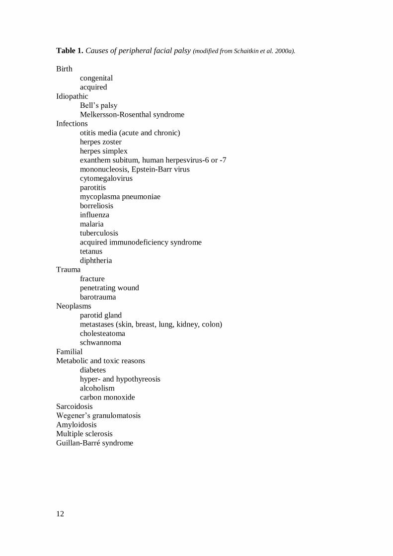
Peripheral FP is associated with many diseases and phenomena; some causes are listed
in Table 1.
The history of documented peripheral FP stems from the ancient times of Egyptians,
Greeks, Romans, Incas, and other native cultures with preserved art representing
deformed faces with peripheral FP (Peitersen 2002). In the medical literature, Stalpart
van der Wiel described in 1686 a woman with peripheral FP in the puerperium, 14 days
postpartum (van de Graaf and Nicolai 2005). In addition, an unpublished description of
acute peripheral FP without known cause by Douglas in 1704 and published
documentation of three patients by Friedreich in 1797 are the first references to Bell’s
palsy over 100 years and over 20 years before Bell’s studies (Peitersen 2002, van de
Graaf and Nicolai 2005). Bell demonstrated that the facial nerve controlled facial
motion and the trigeminal nerve facial sensibility (Bell 1821, 1829). The facial nerve
became known as “Bell’s nerve”. For a while, all cases of peripheral flaccid paralysis
were called “Bell’s palsy”, but later the term narrowed to only include idiopathic palsies
without known cause. Bell mentions temporary diseases of the facial nerve in his work
from 1829, but the concept of acute idiopathic peripheral paralysis was not contained in
these early descriptions (Bell 1821, 1829).
12
Table 1. Causes of peripheral facial palsy (modified from Schaitkin et al. 2000a).
Birth
congenital
acquired
Idiopathic
Bell’s palsy
Melkersson-Rosenthal syndrome
Infections
otitis media (acute and chronic)
herpes zoster
herpes simplex
exanthem subitum, human herpesvirus-6 or -7
mononucleosis, Epstein-Barr virus
cytomegalovirus
parotitis
mycoplasma pneumoniae
borreliosis
influenza
malaria
tuberculosis
acquired immunodeficiency syndrome
tetanus
diphtheria
Trauma
fracture
penetrating wound
barotrauma
Neoplasms
parotid gland
metastases (skin, breast, lung, kidney, colon)
cholesteatoma
schwannoma
Familial
Metabolic and toxic reasons
diabetes
hyper- and hypothyreosis
alcoholism
carbon monoxide
Sarcoidosis
Wegener’s granulomatosis
Amyloidosis
Multiple sclerosis
Guillan-Barré syndrome

13
Grading facial function
A reliable way of grading is needed to define the severity of facial dysfunction, to
follow the progression of FP, and to compare results of interventions. An internationally
accepted and implemented system has not yet been developed. To assess correctly
function and dysfunction of the facial nerve, the different aspects of its physiology need
to be considered. The facial nerve innervates 23 paired facial muscles and the
orbicularis oris, and the functional defect can vary in different parts of the face. In
addition, lacrimation, salivation, and taste may be affected to varying degrees. When
overall facial nerve function is assessed, an attempt to qualify and quantify these
different types of function should be made. After facial nerve injury, secondary defects
such as synkinesis, contracture, and hemifacial spasms may affect facial appearance and
function variably and need to be considered in the assessment.
House (1983) reviewed the existing facial grading systems and divided them into three
categories: gross, regional, and specific. The general scales are called gross because
they consider overall facial function, including degree of paralysis, and secondary
effects simultaneously. They are descriptive, meant to categorize patients in a simple
and practical way and not to give specific details about a patient’s facial function. In a
regional system, the assessor scores different areas of facial function independently.
Regional scales can be weighted or unweighted. In a weighted regional scale, certain
areas of the face are considered less important because they are less likely to have a
good return of function or are cosmetically or functionally less relevant (e.g. forehead)
(House 1983). Specific systems ask questions about specific areas of the face and
address the presence or absence of associated symptoms and signs (House 1983, Chee
and Nedzelski 2000).
The first facial grading system was introduced by Botman and Jongkees (1955). It was a
simple five-category scale to judge the degree of paralysis (0 = normal, IV = total
paralysis). Contractures were the only secondary defects mentioned in the grading in
total paralysis. House (1983) considered this inappropriate since, according to him, in
total paralysis secondary defects cannot develop. Peitersen (2002) modified the system
and used that scale in his studies (Table 2). After analyzing the pre-existing grading
systems, House (1983) introduced his gross scale system with six categories.
Brackmann and Barrs (1984) meanwhile published a measuring system for side
differences in facial movements. In 1985, the Facial Nerve Disorders Committee of the
American Academy of Otolaryngology-Head and Neck Surgery (AAO-HNS) adopted a
universal standard of grading facial function based on the works of House and
Brackmann (1985) (Table 3). The original House scale was modified and Brackmann’s
measuring scale was added to assist in placing patients in the proper group. Clinicians
were encouraged to convert their existing grading system to the House-Brackmann
facial grading system (H-B FGS) when reporting their results, and the use of the system
was required for articles in Otolaryngology-Head and Neck Surgery, the official journal
for the AAO-HNS.

14
Table 2. Peitersen grading system.
Grade Degree of palsy Description of palsy
0 None Normal function
I Slight Only visible when patient grimaces
II Moderate Visible with small facial movements
III Severe Function just visible
IV Complete No function
Table 3. House-Brackmann facial grading system.
Grade Description Characteristics
I Normal Normal facial function in all areas
II Mild
dysfunction
Gross: Slight weakness noticeable on close inspection; may
have very slight synkinesis
At rest: Normal symmetry and tone
Motion: Forehead: moderate to good function
Eye: complete closure with minimal effort
Mouth: slight asymmetry
III Moderate
dysfunction
Gross: Obvious but not disfiguring difference between two
sides; noticeable but not severe synkinesis,
contracture, and/or hemifacial spasm
At rest: Normal symmetry and tone
Motion: Forehead: slight to moderate movement
Eye: complete closure with effort
Mouth: slightly weak with maximum effort
IV Moderately
severe
dysfunction
Gross: Obvious weakness and/or disfiguring asymmetry
At rest: Normal symmetry and tone
Motion: Forehead: none
Eye: incomplete closure
Mouth: asymmetric with maximum effort
V Severe
dysfunction
Gross: Only barely perceptible motion
At rest: Asymmetry
Motion: Forehead: none
Eye: incomplete closure
Mouth: slight movement
VI Total
paralysis
No movement

15
Although widely used in the United States and Europe, H-B FGS failed to reach
worldwide acceptance. Originally created as a gross scale, it has been criticized as not
being sufficiently sensitive to document clinically significant changes (Murty et al.
1994, Ross et al. 1996, Rickenmann et al. 1997). It is also prone to interobserver
variation (Croxson et al. 1990, King et al. 1993, Murty et al. 1994, Ahrens et al. 1999,
Coulson et al. 2005), and assigning only one grade may be difficult because of the
different degrees of dysfunction in upper and lower parts of the face (Rickenmann et al.
1997, Scriba et al. 1999, Yen et al. 2003). The article by House and Brackmann (1985)
describing the grading system has become the most cited article in otolaryngology-head
and neck surgery literature (Wormald et al. 2007). Demands for validation, reliability,
and reproducibility assessments of the H-B FGS and its “golden standard” status have
been made (Browning 2007). In Japan, the Yanagihara grading system is generally used
(Satoh et al. 2000, Ikeda et al. 2003). It is an unweighted regional scale that assesses ten
areas of the face without taking secondary effects into account (Table 4). Tables have
been provided to convert Yanagihara scores to H-B FGS scores (Satoh et al. 2000).
Table 4. Yanagihara grading system.
Normal Partial palsy/weak No motion
1 At rest 4 2 0
2 Wrinkle forehead 4 2 0
3 Close eyes normally 4 2 0
4 Close eyes forcefully 4 2 0
5 Close eyes on the involved side only 4 2 0
6 Wrinkle nose 4 2 0
7 Blow out cheeks 4 2 0
8 Whistle 4 2 0
9 Grin 4 2 0
10 Depress lower lip 4 2 0
Recently, the Sunnybrook (Toronto) Facial Grading System (SFGS) (referred to also
simply as the Facial Grading System) (Ross et al. 1996, Ross and Nedzelski 1998, Hu et
al. 2001) has received good reviews (Ahrens et al. 1999, Kayhan et al. 2000, Schaitkin
and May 2000, Coulson et al. 2004, 2005) and is considered as a leading instrument in
clinical use (Rogers et al. 2007). It is a regional scale that measures also synkinesis
regionally (Table 5). The regional scores are weighted for the composite score. Berg et
al. (2004) considered the scale promising and suggested adding objective measurements
and secondary effects other than synkinesis to the scale to make it even better.

16
Table 5. Sunnybrook facial grading system (modified from Ross et al. 1996). Resting Symmetry Compared to normal side
Symmetry of Voluntary Movement Degree of muscle excursion compared to normal side
Synkinesis Rate the degree of involuntary muscle contraction associated with each expression
Eye (choose one only) normal 0 narrow 1 wide 1 eyelid surgery 1 Cheek (naso-labial fold) normal 0 absent 2 less pronounced 1 more pronounced 1 Mouth normal 0 corner drooped 1 corner pulled up/out 1 Total_____ Resting symmetry score: Total 5_____
Standard expressions Brow lift Gentle eye closure Open mouth smile Snarl
Lip pucker
Unable to Initiates Initiates Movement Movement initiate slight movement with almost complete movement movement mild excursion complete 1 2 3 4 5 1 2 3 4 5 1 2 3 4 5 1 2 3 4 5 1 2 3 4 5 Gross Severe Moderate Mild Normal asymmetry asymmetry asymmetry asymmetry symmetry Total_____ Voluntary movement score: Total 4_____
None* Mild† Moderate‡ Severe§ 0 1 2 3 0 1 2 3 0 1 2 3 0 1 2 3 0 1 2 3 Synkinesis score: Total_____
Voluntary movement score – (minus) Resting symmetry score – (minus) Synkinesis score = Composite Score_____
*no synkinesis or mass movement † slight synkinesis of one or more muscles ‡obvious synkinesis of one or more muscles §disfiguring synkinesis/gross mass movement of several muscles

17
Impairment and disability experienced by the patient may differ greatly from the
assessor’s grading (Bagger-Sjöbäck et al. 2005). Some investigators would include
subjective assessment in the composite grading of facial function (de Ru et al. 2006),
and others have created separate grading instruments for subjective dysfunction
measurement (VanSwearingen and Brach 1996, Kahn et al. 2001, Mehta et al. 2007).
All of the grading scales mentioned here and many others are subjective. One major
problem with grading systems is finding a balance between exact descriptions of
sequelae and minimizing the number of groups into which the patients are classified
(Peitersen 2002). The need for an objective, simple-to-use method to measure facial
dynamics is obvious, but it appears to be difficult to obtain. Significant differences exist
in facial expressions from one individual to another, in sides of the face, and between
genders and age groups (Giovanoli et al. 2003).
Burres and Fisch (1986) introduced an objective method to measure distances between
specific facial landmarks at rest and five standard expressions comparing the affected
side of the face to the normal side (Burres-Fisch Linear Measurement Index) by using
photographs and still video images. However, their method is time-consuming, difficult
to use, and seems to underestimate the degree of dysfunction in severe paralysis and to
overestimate it in mild paralysis (Croxson et al. 1990, Murty et al. 1994). Murty et al.
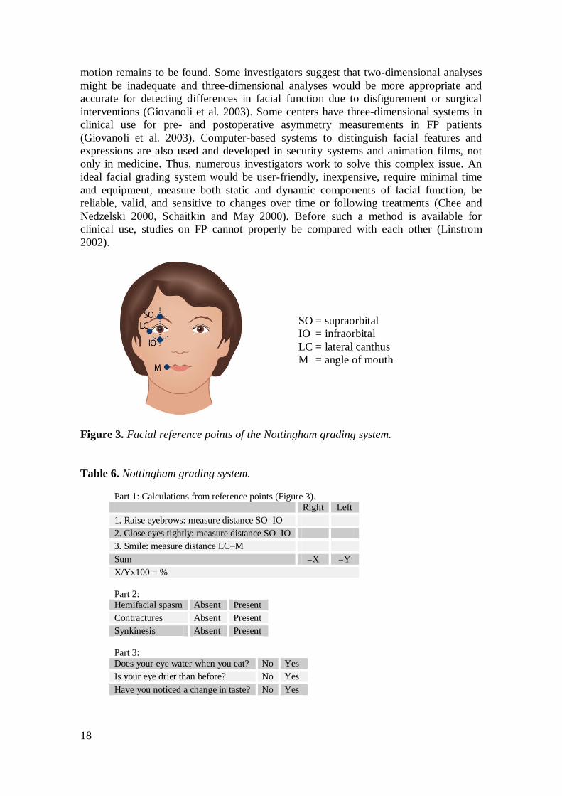
(1994) simplified the system to the Nottingham System (Fig. 3, Table 6) by preserving
objective measurements of three facial expressions (measuring the movements of four
points on the face and comparing the abnormal side with the normal side as a
percentage) and specifying whether secondary effects are present or absent and whether
the patient experiences crocodile tears, dry eyes, or taste disturbances. The associated
sequelae do not interfere with the measurement score and are not rated by severity. The
system does not take into account possible normal variance in facial expression between
the halves of the face (Chee and Nedzelski 2000) and is not applicable in bilateral FP
because the affected side is compared with the unaffected side (Kang et al. 2002). Some
investigators have found it promising, although more systemic evaluations are needed to
determine whether a widespread application is appropriate (Kang et al. 2002). Others
consider all linear measurements inadequate (Meier-Gallati et al. 1998).
Many computer-aided analysis systems have been created to measure dysfunction of
one part of the face (Tomat and Manktelow 2005), but to create a clinically usable and
affordable method that takes into account whole-face function and secondary defects is
challenging and still underway. Systems based on video-recording of facial expressions
and light reflection (Neely et al. 1992, Yuen et al. 1997, Meier-Gallati et al. 1998) have
been introduced, but because special equipment or techniques are required, these
systems have not been taken into wide use (Chee and Nedzelski 2000). Automated
facial image analysis (Cohn et al. 1999), originally created to detect, extract, and
recognize emotion and paralinguistic expressions, has been used in clinical studies to
distinguish subtle changes in facial movement after interventions (Rogers et al. 2007).
The system requires manual marking of 40 points (with the computer mouse to video
pictures), increasing the possibility for repeatability and agreement errors. Linstrom
(2002) used a commercially available video-computer interactive system, The Peak
Motus Motion Measurement System, to objectively measure the side-to-side
displacement (asymmetry) of selected marker sites on the face during eye closure and
smile, and concluded that the ideal objective system to both quantify and classify facial
18
motion remains to be found. Some investigators suggest that two-dimensional analyses
might be inadequate and three-dimensional analyses would be more appropriate and
accurate for detecting differences in facial function due to disfigurement or surgical
interventions (Giovanoli et al. 2003). Some centers have three-dimensional systems in
clinical use for pre- and postoperative asymmetry measurements in FP patients
(Giovanoli et al. 2003). Computer-based systems to distinguish facial features and
expressions are also used and developed in security systems and animation films, not
only in medicine. Thus, numerous investigators work to solve this complex issue. An
ideal facial grading system would be user-friendly, inexpensive, require minimal time
and equipment, measure both static and dynamic components of facial function, be
reliable, valid, and sensitive to changes over time or following treatments (Chee and
Nedzelski 2000, Schaitkin and May 2000). Before such a method is available for
clinical use, studies on FP cannot properly be compared with each other (Linstrom
2002).
SO = supraorbital
IO = infraorbital
LC = lateral canthus
M = angle of mouth
Figure 3. Facial reference points of the Nottingham grading system.
Table 6. Nottingham grading system.
Part 1: Calculations from reference points (Figure 3).
Right Left
1. Raise eyebrows: measure distance SO–IO
2. Close eyes tightly: measure distance SO–IO
3. Smile: measure distance LC–M
Sum =X =Y
X/Yx100 = %
Part 2:
Hemifacial spasm Absent Present
Contractures Absent Present
Synkinesis Absent Present
Part 3:
Does your eye water when you eat? No Yes
Is your eye drier than before? No Yes
Have you noticed a change in taste? No Yes

19
Acute idiopathic peripheral facial palsy–Bell’s palsy
In about 70% of adult peripheral FPs, the cause is unknown and the palsy is termed
idiopathic (Bell’s palsy) (Adour et al. 1978, Peitersen 2002). Bell’s palsy is acute onset,
usually reaching maximal intensity in a few days. The definition for the time limit
within which the palsy has to reach its maximum to be Bell’s palsy varies from one to
two weeks (Adour and Hetzler 1984) to up to three weeks (May and Hughes 1987).
Criteria for Bell’s palsy have been unilateral FP without any other cranial neuropathies
or neurologic deficits. The definition has been modified over the years with
accumulating knowledge of the disease. Accompanying symptoms may arise from
cranial nerves other than the facial nerve in acute peripheral FP without identifiable
cause (Adour 2002, Benatar and Edlow 2004). Some investigators (Peitersen 2002)
exclude patients with underlying conditions possibly predisposing to FP (e.g. diabetes
or pregnancy) from the Bell’s palsy diagnosis, but in FP studies the cause of action is
not uniform. Also spontaneous recovery to some degree within six months is considered
a feature of Bell’s palsy by some investigators (May and Hughes 1987). In this thesis,
Bell’s palsy is used as a synonym for acute idiopathic peripheral FP.
Incidence of Bell’s palsy is about 20–30/100 000/year worldwide (Hauser et al. 1971,
Adour et al. 1978, Katusic et al. 1986, Yanagihara 1988, Peitersen 2002, Rowlands et
al. 2002, Ljøstad et al. 2005). In most studies, no racial, gender, or seasonal
predisposition has been noted (Adour et al. 1978, Peitersen 2002, Rowlands et al. 2002).
In some studies, people with diabetes or hypertension, and pregnant women are more
susceptible to peripheral FP with worse outcome, but this is not seen in all studies
(Gillman et al. 2002, Peitersen 2002). Age distribution of Bell’s palsy patients is
variable. However, the consensus is that children under 15 years are less often affected
than adults (Adour et al. 1978, Katusic et al. 1986, Peitersen 2002). Some studies report
a rising frequency in patients over 60 years (Adour et al. 1978, Rowlands et al. 2002),
whereas others describe the peak incidence age to be 15–45 years, with a reduced
incidence in older people (Peitersen 2002). Recurrence of Bell’s palsy affects 6–13% of
patients (Adour et al. 1978, Katusic et al. 1986, Devriese et al. 1990, Schaitkin et al.
2000a, Peitersen 2002), and a positive family history varies between 1.5% (Katusic et
al. 1986) and 17% (Schaitkin et al. 2000a).
The etiology of Bell’s palsy is obscure; genetic, autoimmune, vascular, infective, and
immunological causes have been speculated (McGovern et al. 1977, Fisch and Felix
1983, Schaitkin et al. 2000c). Mechanisms include primary ischemia or inflammation of
the facial nerve, causing edema and entrapment of the nerve in its long course in the
bony temporal canal and resulting in compression and direct damage or secondary
ischemia to the nerve (Fisch and Felix 1983, Schaitkin et al. 2000c). Another proposed
mechanism is viral infection directly disturbing nerve function by inflammatory
immune mechanisms, possibly throughout the nerve’s course and not by compression in
the bony canal (Adour 2002). Herpesviruses have been suspected as etiologic factors for
over 40 years, with growing evidence but still lacking the final proof (Dodge and
Poskanzer 1962, McCormick 1972). The viral etiology has gained support by many
associations of FP with viral diseases such as mononucleosis caused by Epstein-Barr
virus (EBV), chickenpox caused by VZV, cytomegalovirus (CMV) infection, and case
reports of exanthem subitum caused by HHV-6 or -7 (Traavik et al. 1983, Mori et al.
2002, Pitkäranta et al. 2004). Some investigators consider Bell’s palsy to be a milder

20
version of Ramsay Hunt syndrome based on similar but less severe polyneuropathic
symptoms, not necessarily caused by VZV (Adour 2002). Furuta et al. (2000, 2005)
reported findings suggesting zoster sine herpete in about 30% of Bell’s palsy patients, in
both adults and children, and considered VZV to be one of the major etiologic agents in
Bell’s palsy. Their results were based on VZV DNA findings in saliva, VZV IgM
findings in serum, or significant VZV IgG titer rise in paired serum tests. The
conclusions can be challenged based on findings of VZV reactivation under nonsurgical
stress (Mehta et al. 2004); VZV reactivation can be a consequence of the FP, not
necessarily the cause.
The strongest evidence for HSV-1 being the major etiologic factor in Bell’s palsy comes
from the work of Murakami et al. (1996). Fourteen FP patients with ongoing palsies
underwent decompression surgery 15–60 days after palsy onset (median 31 days). HSV-
1 DNA was found by polymerase chain reaction (PCR) in facial nerve endoneurial fluid
from 10 patients (13 samples) and in tissue from the posterior auricular muscle from the
seven previous patients and one additional patient (14 samples). DNA of VZV or EBV
was not found. Nine Ramsay Hunt syndrome patients had the same procedures done,
with seven positive VZV DNA findings in endoneurial fluid (9 samples) and six (one
patient other than previous patients) positive muscle tissue findings (8 samples). DNA
of HSV-1 or EBV was not found. Murakami et al. (1996) concluded that HSV-1
infection of the facial nerve is directly related to the pathogenesis of Bell’s palsy, to
reactivation of the virus in the geniculate ganglion. Many investigators agree and
consider HSV-1 a plausible etiologic factor in Bell’s palsy (Furuta et al. 2001, Adour
2002, Gilden 2004, Holland and Weiner 2004, Kawaguchi et al. 2007), some with
supporting animal models (Honda et al. 2002). Others think caution in conclusions is
warranted. HSV-1 and VZV are assumed to reactivate during surgical stress (Shea and
Ge 2001), which could explain the findings of Murakami et al. (1996), or the viruses
might reactivate because of FP. The PCR method used by Murakami et al. (1996) could
not distinguish active from inactive infection. Stjernquist-Desatnik et al. (2006) took
tissue samples from the posterior auricular muscle of Bell’s palsy patients within 72
hours of palsy onset and found one patient with HSV-1 DNA by PCR. One Ramsay
Hunt patient had VZV DNA in both a tissue sample and CSF. The authors discussed the
discrepancy with findings of Murakami et al. (1996), speculating that epidemiological
differences may exist in the countries of origin. They also noted that in the Murakami
study the palsies were total and had lasted at least two weeks before samples were
taken. Rowlands et al. (2002) did not find any suggestions of an infectious etiology: no
household clustering of Bell’s palsy and no tendency of HSV infection preceding palsy.
Linder et al. (2005) studied geniculate ganglions from 14 autopsy specimens of
individuals without Bell’s palsy and found HSV-1 DNA in 86% and VZV DNA in 43%
of ganglions. This confirmed previous abundant HSV and VZV DNA findings in
cervical ganglia (Vrabec and Payne 2001). Linder et al. (2005) question the theory of
HSV reactivation inside the geniculate ganglion as the reason for Bell’s palsy based on
the discrepancy between the low incidence of Bell’s palsy and the frequent viral
genomic findings in human geniculate ganglions. They conclude that the missing link to
confirm the active role of HSV in Bell’s palsy is the identification of an active
replicating virus, a study yet to be conducted.

21
Human herpesvirus-6 and -7
Studies on HHV-6 and -7 in FP patients are very few (details in Discussion). These
viruses are usually acquired in childhood, persist latently for life, and reactivate
occasionally (Ward 2005). Primary infections are either asymptomatic or accompanied
by fever, diarrhea, rash, and roseola as the most usual symptoms (Zerr et al. 2005). So
far, exanthem subitum is the only illness conclusively shown to be caused by HHV-6B
or -7 (Yamanishi et al. 1988, Tanaka et al. 1994). Primary infections are sometimes
accompanied by encephalitis or febrile seizures (Hall et al. 2006). The majority of
documented primary infections are due to HHV-6B, and almost all children are HHV-6-
seropositive by two years of age (Ward 2005, Zerr et al. 2005, Hall et al. 2006). HHV-
6A has not been indisputably connected to any disease yet, and the epidemiology and
clinical findings associated with acquisition of the virus remain uncertain.
HHV-6 is the only human herpesvirus occasionally known to be integrated in human
chromosomes. A consequence is the finding of high viral DNA copies in whole blood or
serum samples; this is assumed to affect 0.7–1.5% of the general population (Tanaka-
Taya et al. 2004, Ward et al. 2007). No evidence of chromosomal viral integration of
HHV-7 exists (Ward et al. 2007).
Epidemiological characteristics and manifestations of acute HHV-7 infections are not
well known. Primary HHV-7 infection usually occurs later than HHV-6 infection and is
acquired over the first 5 or 6 years of life (Ward 2005, Hall et al. 2006). This is
unexplained since contact with HHV-7 during infancy is expected to be at least as
frequent as with HHV-6. Saliva of infected individuals is the presumed principal source
of infection for infants. Both HHV-6 and -7 are shed in saliva (Ward 2005, Hall et al.
2006).
Like all herpesviruses, HHV-6 and -7 persist for life after primary infection and are
detectable in peripheral white blood cells and multiple tissues (Ward 2005). In a study
of autopsy samples, HHV-6 DNA was most commonly found in salivary glands,
thyroid, stomach, intestines, liver, and pancreas, and HHV-7 DNA predominated in
salivary glands, tonsils, lymph nodes, and bone marrow (Chen and Hudnall 2006).
HHV-6 DNA has also been detected in the bone marrow of healthy individuals
(Gautheret-Dejean et al. 2000) and both HHV-6 and -7 DNA in normal brain tissue at
autopsy (Chan et al. 2000, Chan et al. 2001).
The most serious clinical manifestations of infection or reactivation of these viruses
occur in immunocompromised patients (Ljungman 2002). Some uncertainty remains
about the clinical role of HHV-6 and -7 following organ or bone marrow transplantation
(Ljungman 2002, Lehto et al. 2007). HHV-6 and -7 have been suggested to be
associated with such conditions as multiple sclerosis, chronic fatigue syndrome, a
variety of neoplastic disorders, infectious mononucleosis, meningitis, encephalitis, and
drug hypersensitivity syndromes, and HHV-7 with pityriasis rosea (Ward 2005).
Whether HHV-6 and -7 primary infections or reactivations have an etiologic role is
unknown.

22
Treatment of Bell’s palsy
Bell’s palsy has been treated with various methods and medicines during its long
history. Evaluation of therapy in Bell’s palsy is difficult because of the high
spontaneous recovery rate. In many studies, treatment suggestions are based on the
opinions of investigators rather than on results (Devriese et al. 1990, Austin et al. 1993).
No consensus has been reached on the treatment of acute Bell’s palsy, but unanimous
agreement exists on the importance of taking care of the affected eye, to protect it and
prevent it from drying (May and Hughes 1987).
In the 17th
and 18th
centuries, the etiology of acute peripheral FP was considered
rheumatic, with the nerves hollow and filled by mucous fluid after being exposed to
cold, and treatments were antirheumatic ointments and medicines (Peitersen 2002, van
de Graaf and Nicolai 2005). Electrotherapy emerged in the 19th
century and is still
controversial along with other physical therapy methods and acupuncture (Peitersen
2002, van de Graaf and Nicolai 2005, He et al. 2007, Teixeira et al. 2007).
Surgery for facial reanimation had already started at the end of the 19th
century, with
anastomosing of the facial nerve to the accessory nerve and later to other neighboring
nerves (e.g. hypoglossal, glossopharyngeal) (Ballance et al. 1903, Ballance and Duel
1932). Decompression surgery of the facial nerve had been carried out in other
indications and was introduced for Bell’s palsy in 1932 (Ballance and Duel 1932).
Surgery became popular as the pathophysiology of Bell’s palsy was considered the
entrapment and compression of the nerve in its bony temporal canal (Kettel 1947, Fisch
and Esslen 1972). With the development of surgical methods and the assumed peak
entrapment place of the facial nerve within the bony pathway, the area decompressed
varied from the most distal part at the stylomastoid foramen to total decompression of
the bony canal to the meatal foramen of the internal auditory canal (Fig. 1) (Ballance
and Duel 1932, Fisch and Esslen 1972, Gantz et al. 1999, Yanagihara et al. 2001, Adour
2002). The wait for an operation varied from a few days (Fisch and Esslen 1972) to
several months (Kettel 1947, Yanagihara et al. 2001) or years (Kettel 1947). Besides the
clinical picture of total paralysis or persistent palsy with sequelae, decisions for
decompression surgery were based on various electrical tests (Ballance and Duel 1932,
Kettel 1947, Gantz et al. 1999, Yanagihara et al. 2001, Adour 2002). With the
herpesviral etiology gaining support, surgical procedures have declined and attitudes
towards decompression surgery are controversial (Grogan and Gronseth 2001), with
many investigators opposing the procedure (May et al. 1985, Adour 2002).
Corticosteroids, cortisone initially, have been used in Bell’s palsy from the 1950s
onwards to reduce inflammation, degeneration, and false regeneration of the facial
nerve (Taverner 1954). The use is common around the world without a generally
accepted scientific proof of effectiveness. The American Academy of Neurology
(Grogan and Gronseth 2001) concluded in their practice guideline meta-analysis that
while the benefit of steroids has not been established, they probably are effective in
improving facial functional outcomes. They called for well-designed studies to
investigate the effectiveness of treatments. Several other meta-analyses have supported
the use of corticosteroids (Ramsey et al. 2000, Holland and Weiner 2004). The
Cochrane database concludes the following: “The available evidence from randomised
controlled trials does not show significant benefit from treating Bell's palsy with

23
corticosteroids. More randomised controlled trials with a greater number of patients are
needed to determine reliably whether there is real benefit (or harm) from the use of
corticosteroid therapy in patients with Bell's palsy” (Salinas et al. 2007).
With the growing support for herpesvirus etiology in Bell’s palsy, the use of antiviral
agents, first acyclovir, later valaciclovir and famciclovir, has emerged. The same is true
as for corticosteroids; the results of studies are controversial, even within participants of
the same collaborative study, as recently reported from Japan (Hato et al. 2007,
Kawaguchi et al. 2007). The American Academy of Neurology (Grogan and Gronseth
2001) stated that acyclovir (combined with prednisolone) may be effective in improving
facial functional outcomes. The Cochrane database concludes: “More data are needed
from a large multicentre randomised controlled and blinded study with at least 12
months' follow-up before a definitive recommendation can be made regarding the effect
of aciclovir or valaciclovir on Bell's palsy” (Allen and Dunn 2007).
In 2001, a collaboration study was initiated in Finland and Sweden to assess the
effectiveness of prednisolone and valaciclovir in Bell’s palsy (www.clinicaltrials.gov).
This is a double-blind, placebo-controlled, randomized four-arm study with 739
recruited patients and a one-year follow-up. Evidence for the benefit or ineffectiveness
of these drugs in Bell’s palsy will hopefully be achieved. A similar recent study from
Scotland (Sullivan et al. 2007) with 496 patients and 3- to 9-month follow-up
demonstrated a significant benefit from the use of prednisolone compared with placebo,
whereas the use of acyclovir alone or in combination with prednisolone was not
effective.
Corrective treatments for FP patients with residual palsy and associated sequelae
include abundant reanimation surgical procedures to improve facial appearance and
function, producing more balanced and symmetrical features (May et al. 2000a,
Hadlock et al. 2006). After a spontaneous recovery period and especially after surgical
corrections, physical therapy with various methods (muscle exercise, electrical
stimulations, neuromuscular re-education therapies with surface electromyography
biofeedback) is, albeit controversial, increasingly recognized and accepted as enhancing
function and reducing discomfort and synkinesis (May et al. 2000, Targan et al. 2000,
Beurskens et al. 2006, Hadlock et al. 2006, Teixeira et al. 2007). The mechanisms are
thought to be CNS plasticity, resulting in better motor control of the facial muscles, and
perhaps reinnervations from intact neighboring nerves or intact motor units of the facial
nerve (Targan et al. 2000). Botulinum toxin is successfully used to diminish
consequences caused by facial synkinesis, hemifacial spasms, hypercontracted muscles,
and hyperlacrimation (May et al. 2000a, Hadlock et al. 2006).
Prognosis of Bell’s palsy
Most Bell’s palsy patients recover well. Total recovery is seen in 70–80% of patients
overall. With incomplete palsy, the recovery rate is 95–99%, with complete palsy 50–
60% (Katusic et al. 1986, Schaitkin et al. 2000c, Peitersen 2002). Roughly 30% of all
patients are left with some sequelae (remaining palsy, hemifacial spasms, contracture, or
synkinesis), mainly mild or moderate, but severe in 5% of cases (Devriese et al. 1990,

24
Peitersen 2002). In a Danish prospective 25-year study (Peitersen 2002), 70% of palsies
were complete and 30% incomplete. Recovery began within three weeks for 85% and
within 3–5 months for the remaining 15%. All Bell’s palsy patients achieved some
degree of muscular function, as was evident also in a study by Adour et al. (1978). In
the Danish study (Peitersen 2002), if recovery was not total within six months, some
sequelae tended to remain in the final assessment. Total recovery was seen in 71% of
Bell’s palsy patients, in 64% within three months (Peitersen 2002). Factors indicating
unsatisfactory outcome are complete palsy, late beginning of recovery, and age over 60
years (Katusic et al. 1986, Devriese et al. 1990, Schaitkin et al. 2000c, Peitersen 2002).
There are no reliable signs or tests at palsy onset to predict outcome. Topognostic tests
are unreliable because of anatomic variations, neural branching, and possible variations
in the areas and components affected on the facial nerve (Karikoski 1987, Schaitkin et
al. 2000b). Of the electrical tests used in prognostic evaluation, electroneuronography
(ENoG), also called evoked electromyography, has been regarded as the most valuable
in the acute phase of FP (Fisch 1984, Schaitkin et al. 2000b). A difference of 90% or
more in the peak-to-peak amplitude of the evoked compound muscle action potential
between the paretic and uninvolved sides of the face is considered to predict a 50% or
greater chance of poor recovery (H-B FGS III or worse, Table 3) (Fisch 1984, Karikoski
1987, Gantz et al. 1999). The major drawback of ENoG is that the site of testing is
peripheral to the stylomastoid foramen and it takes a minimum of 72 hours for
denervation to reach the distal parts of the nerve to be detectable (Schaitkin et al. 2000b,
Chow et al. 2002). Since the beginning of palsy is considered crucial in the attempt to
diminish the amount of denervation, the results of ENoG come late in finding patients at
risk of poor outcome. Other limitations include marked normal variation within tests of
the sides of the face and test to retest results (Sittel et al. 1998). Patient cooperation is
mandatory and variations in electrode and stimulator placement and pressure can alter
results (Schaitkin et al. 2000b, Chow et al. 2002). Studies on transcranial magnetic
stimulation of the labyrinthine segment of the facial nerve have been conducted to
evaluate the function of the nerve without the delay of distal testing (Schaitkin et al.
2000b, Nowak et al. 2005), but this method has not reached clinical significance.
Magnetic resonance imaging has its place in differential diagnostics of Bell’s palsy, but
studies on predictive value of contrast enhancement findings in Bell’s palsy are
contradictory (Kress et al. 2004).
Peripheral facial palsy in children
In Peitersen’s Danish study (Peitersen 2002), neonates formed the largest group of
children’s palsies (congenital and acquired). If neonatal age is excluded, Bell’s palsy
comprised 77% of cases, which is in accordance with other studies (Ogita et al. 2006).
Some studies have revealed the proportion of Bell’s palsy to be lower, 26–40% (Cook et
al. 1997, Peltomaa et al. 1998, May et al. 2000b). In Peitersen’s study (2002), the
overall incidence of peripheral FP in children under 15 years (excluding neonates) is
3.4/100 000/year and of Bell’s palsy 2.6/100 000/year. Higher incidences have also
been reported: for peripheral FP 21/100 000/year (under 14 years) (Tveitnes et al. 2007)
and 7/100 000/year (Christen et al. 1993), and for Bell’s palsy 5–8/100 000/year (Adour
et al. 1978, Katusic et al. 1986). Many studies conclude prognosis of FP in children to

25
be excellent; 90% recovered completely in Peitersen’s study (2002), but full recovery
rates of 70–80% are also common (Peltomaa et al. 1998, Skogman et al. 2003).
In adult populations, borreliosis as a causative agent for peripheral FP accounts for 0–
20% of cases (Roberg et al. 1991, Peitersen 2002, Ljøstad et al. 2005) mostly
manifesting with additional symptoms besides FP (Ljøstad et al. 2005). In studies from
endemic areas, borreliosis explains 30–65% of children’s FPs (Christen et al. 1993,
Peltomaa et al. 1998, Tveitnes et al. 2007). Many of these children have no other
neurological signs besides FP (Christen et al. 1993, Tveitnes et al. 2007). The Danish
study (Peitersen 2002) did not have any children with diagnosed borreliosis. The same
was true with 30 children in Japan (Furuta et al. 2005); instead VZV was suspected as
the causative agent in 30% of cases. False-positive findings of IgM antibodies to
Borrelia burgdorferi in CSF can be caused by VZV or EBV, and it is important to rule
out these infections before confirming borreliosis as the causative factor for FP
(Christen et al. 1993).
Melkersson-Rosenthal syndrome
In 1928, Swedish neurologist Ernst Melkersson published a case study of a 35-year-old
man whose FP had reoccurred four times and who had had attacks of edema in the
upper and lower lip since age 14 (Melkersson 1928). The swelling in the lower lip
gradually became persistent. In 1931, a German neurologist Curt Rosenthal published a
family study in which four women from three unrelated families had at least one
episode of FP, recurrent episodes of edema, and LP (Rosenthal 1931). Two of these four
women were sisters and their mother had had reoccurring facial edema. The third
woman had a sister with FP and LP, and a mother with LP. The fourth woman was the
only one in her family with these symptoms. In one additional family, edema and LP
were seen in two generations without FP (Rosenthal 1931). Recurrent labial or oro-
facial edema, recurrent FP, and associated LP (also referred as fissured or furrowed
tongue) occurring without recognizable cause became known as the classical triad of
MRS. Swiss dermatologist Alfred Guido Miescher studied histology of reoccurring
labial edema and found peri- and paravascular tuberculoid character granulations in
edematous tissue samples (Miescher 1945). He regarded the finding as cheilitis
granulomatosa (CG). Later, German dermatologists (Richter and Johne 1950, Gahlen
and Brückner 1951) made similar findings in edematous tissue samples from MRS
patients: nonnecrotizing (-caseating) lymphoepiteloid granulomas. Miescher’s CG has
since been considered by many investigators as a monosymptomatic form of MRS
(Hornstein 1973, Worsaae et al. 1982, Greene and Rogers 1989). When two of the
possible three symptoms are present, the form is called oligosymptomatic MRS (Greene
and Rogers 1989, Hornstein 1997). Some investigators demand that edema be one of the
two symptoms for the diagnosis of MRS (Mair and de Graaf 1974), most, however, also
include patients with FP and LP without edema (Ekbom 1950, Greene and Rogers 1989,
Hornstein 1997). Some investigators still question the supposed mutual etiology of
monosymptomatic form of MRS with oligosymptomatic or triad form of MRS, because
most CG patients never develop other symptoms of MRS (van der Waal et al. 2002).

26
Edema is considered the most frequent and dominating clinical feature in MRS
eventually affecting virtually all patients (Hornstein et al. 1987, Greene and Rogers
1989). The upper lip (skin and mucosa) is the most common site, but any oro-facial area
(outer or inner sides of cheeks, eyelids, forehead, nose, gingiva, oral cavity, floor of
mouth, tongue, pharynx, and larynx) may be involved (Zimmer et al. 1992, Hornstein
1997). Extrafacial edemas have been described in the chest, dorsum of hands and feet,
buttocks, and genitals (Hornstein 1973, Tsuboi et al. 2005). The edema is nonpitting,
painless, and thought to eventually become persistent (Greene and Rogers 1989). The
triad form of MRS is estimated to occur in up to 20% of all cases and FP in 20–50%
(Vistnes and Kernahan 1971, Hornstein 1973, Worsaae et al. 1982, Hornstein et al.
1987, Greene and Rogers 1989, van der Waal et al. 2002). LP is found in about 6% of
the general population, with an increasing prevalence with age (children 0.5%–elderly
people 15%) (Axell 1976). LP is considered the least important clinical feature of MRS
and is found in about 50% of patients (Rintala et al. 1973, Worsaae et al. 1982, Greene
and Rogers 1989). Most MRS studies, usually from departments of dermatology or oral
or plastic surgery, have many CG patients and patients with FP are the minority
(Worsaae et al. 1982, Hornstein et al. 1987, van der Waal et al. 2002).
Besides the triad of classical symptoms, additional symptoms frequently accompany
MRS. Hornstein (1997) considers palsies or dysfunctions of other cranial nerves besides
the facial nerve to be “major signs” of MRS in addition to edema and FP. In the
complete form of MRS, Hornstein (1997) emphasizes recurrent facial edemas and
histopathological findings of nonnecrotizing granulomatous infiltarations in edematous
tissue samples. He also lists numerous “minor signs” of uni- or bilateral sensory, motor
neuron, or autonomous disturbances that can accompany MRS and support the
diagnosis of MRS in oligosymptomatic forms. These include facial dysesthesia,
headaches, tinnitus, relapsing hearing loss, dizziness, recurrent pharyngeal neuralgias
and spasms, dysphagia, recurrent dysfunctions of salivary and tear secretion, excessive
facial sweating or anhidrosis, and recurrent dry eye or dry mouth sensations. When FP
and LP are the presenting symptoms without edema, Hornstein (1997) demands the
presence of two or more minor signs for a diagnosis of MRS. Hornstein et al. (1987)
found migraine in 41% of MRS patients.
The etiology of MRS is unknown. Family occurrence has been documented starting
with Rosenthal in the 1930s (Rosenthal 1931, Carr 1966, Lygidakis et al. 1979,
Levenson et al. 1984). Whether the familiar pattern is a result of a genetic
predisposition, immunodeficiency, or transmission of an infectious agent remains to be
elucidated (Levenson et al. 1984). To exclude a pure infectious origin, Meisel-Stosiek et
al. (1990) examined spouses of MRS patients alongside family members. They found an
increased risk for MRS of 24% in first-degree relatives of MRS patients. The risk
dropped to 5% in second-degree relatives. Based on their study and a review of previous
reports on family occurrence of MRS, they concluded that MRS is most likely
multifactorial in origin, with a hereditary component. Besides heredity, a disturbed
autonomous nervous or immune system, obstructive microcirculation, allergies,
sarcoidosis, Crohn’s disease, inflammatory agents (toxoplasmosis, tuberculosis, viral
infection) have been suggested as causative agents (Hornstein 1973, Greene and Rogers
1989, Sussman et al. 1992, van der Waal et al. 2002). Hornstein (1997) speculated that
one common causative denominator of the disease is very unlikely in view of the variety
of clinical forms and accompanying symptoms in MRS patients.

27
The incidence of MRS remains obscure. Hornstein (1973) reported an incidence of
80/100 000 (120 MRS patients in 18 years, most patients with monosymptomatic CG).
About 25% of his MRS patients had FP, corresponding to an incidence of 20/100 000. It
is not defined, whether the incidence is for a year or for all 18 years. For an annual
incidence this seems high since the incidence of Bell’s palsy is the same: 20–30/100
000/year worldwide (Devriese et al. 1990, Peitersen 2002). The Danish study (Peitersen
2002) collecting information on FP etiology and incidence had 19 MRS patients (only
the number given, no further information). This equals an incidence of 0.36/100
000/year for MRS with FP and a total incidence of 0.7–1.8/100 000/year for MRS if FP
is present in 20–50% of all cases.
MRS has been found in children as well as in the elderly, but the most common age at
onset is 25–40 years (Worsaae et al. 1982, Hornstein et al. 1987, Zimmer et al. 1992).
Whether both genders are affected equally is difficult to determine due to the small
number of patients: the female–male ratio has been reported to be 2:1 in some studies
(Hornstein et al. 1987, Zimmer et al. 1992), but equal distribution or male
predominance have also been described (Rintala et al. 1973, Worsaae et al. 1982).
Treatment of MRS has been unsatisfactory. For edema, the most common treatment is
corticosteroids: systemically, topically, or as intralesional injections, with inconsistent
results (Worsaae et al. 1982, Zimmer et al. 1992, Hornstein 1997, van der Waal et al.
2002, Mignogna et al. 2004). Allergy medications and antibiotics have mostly been
ineffective (Worsaae et al. 1982, Zimmer et al. 1992). Medications used for
tuberculosis, or autoimmune diseases, and skin and connective tissue diseases, such as
dapsone, azathioprine, sulfasalazine, chloroquine, clofazimine, and tumor necrosis
factor alpha inhibitors (thalidomide and infliximab), have been tried without definite
results (Sussman et al. 1992, Zimmer et al. 1992, Hornstein 1997, Barry et al. 2005).
Eradication of underlying odontogenic infections has proved successful in some patients
with edema (Worsaae et al. 1982). Reduction cheiloplasty has produced some good
results in permanent lip swellings with nonactive disease, but recurrence is common
(Kruse-Lösler et al. 2005). For relapsing FP, total nerve decompression has been
suggested (Dutt et al. 2000), but the limited number of patients treated and followed up
and the varying natural cause of the disease make it difficult to draw conclusions.
Whether the syndrome sometimes completely resolves is uncertain (Worsaae et al.
1982).

28
AIMS OF THE STUDY
The general aim of this study was to assess grading and etiology of peripheral FP and
characteristics of MRS.
Specific aims were as follows:
1. To assess intrarater repeatability and interrater agreement of H-B FGS and SFGS.
2. To evaluate the etiologic role of human herpesviruses in peripheral FP by searching
for HSV-1 and -2, VZV, HHV-6A, -6B, and -7, EBV, and CMV DNA in CSF of FP
patients.
3. To investigate differences in MRS patient characteristics in the two specialty
departments of Otorhinolaryngology and Dermatology, and special characteristics of
MRS in patients with FP.

29
SUBJECTS AND METHODS
All study protocols (I, II, III, IV) were approved by the ethics committee of Helsinki
University Central Hospital.
Subjects
Study I
Eight patients with unilateral peripheral FP were video-recorded showing five standard
facial expressions. One of the patients had FP after vestibular schwannoma operation 12
years earlier and had had an eyelid operation. The other seven patients had Bell’s palsy,
which had lasted one year in five patients, two months in one patient, and two weeks in
one patient. Five patients had synkinesis and two had contracture.
Thirty doctors at the Department of Otorhinolaryngology, Helsinki University Central
Hospital, volunteered to be assessors of the videos. There were two assessment rounds
and 28 doctors (15 residents and 13 specialists) returned their gradings for the first
round and 26 (13 residents and 13 specialists) for both two rounds.
Studies II and III
The study group comprised 33 peripheral FP patients with 34 CSF samples. Of these
patients, 26 had Bell’s palsy, 5 had simultaneous herpesvirus infection, one had MRS,
and one had puerperal FP two weeks after delivery. These patients were selected
because herpesviruses are suspected etiologic factors in Bell’s palsy and in FP cases
with concomitant herpesvirus infection, and an association may also exist in MRS.
Puerperal FP is not considered Bell’s palsy by some investigators, whereas others
include it in the definition.
There were 19 females and 14 males. Their median age was 19 (range 4–78) years, and
16 patients were 15 years or younger (median 13 years). The median time for the CSF
sample to be taken after palsy onset was 21 days (range 0–148, 0 being the day palsy
started). One of the patients had three episodes of Bell’s palsy and her CSF sample was
taken twice. Three other patients had a second occurrence of Bell’s palsy. For the MRS
patient, this was her fourth FP episode. Among the five patients with concurrent
herpesvirus infection, one had bilateral FP and EBV infection, two had
varicella/chickenpox preceding FP, one had Ramsay Hunt syndrome with blisters in her
mouth a week before FP, and one had simultaneous eruption of herpes blisters on her
lower lip on the palsy-affected side.
The 36 control subjects were unmatched and their CSF samples were taken during the
same time interval as those of the study group. Six controls had FP with borreliosis.
Possible borreliosis with elevated serum antibodies to Borrelia burgdorferi but normal
CSF samples was evident in 13 control subjects: nine with FP, two with sudden
30
deafness, one with hearing loss and tinnitus, and one with vertigo. No borreliosis was
suspected in the remaining control subjects. Four had sudden deafness, five had vertigo,
and four had headache and arthralgia. Of the last four controls, one had headache, one
had vocal cord paralysis, one had facial erysipelas with sepsis and simultaneous
cavernous sinus thrombosis and peripheral FP, and one had ear symptoms, atypical
papillitis, headache, and vertigo. The control group comprised 19 females and 17 males.
Their median age was 47.5 (range 5–75) years, with three controls being 15 years or
younger.
No diabetic or immunocompromised patients or controls were included in the study.
Study IV
There were 35 patients with MRS or suspicion of the syndrome: 23 females and 12
males. Median age at study onset was 47 (range 19–75) years. Median age at symptom
onset was 24 (range 5–71) years. Twenty of the 35 patients had history of FP: 13
females and 7 males. Median age for MRS FP patients was 45.5 (range 34–74) years
when the study started and 21.5 (range 10–67) years at symptom onset. The remaining
15 patients, 10 females and 5 males, had CG, monosymptomatic MRS. Their median
age at study onset was 47 (range 19–75) years and at symptom onset 34 (range 5–71)
years.
Methods
Study I
Video-recording of the FP patient was started with the face at rest, followed by five
standard facial expressions: lifting the eyebrows/wrinkling the forehead, closing eyes
gently and then more powerfully, wrinkling the nose, smiling mouth open, and
puckering the lips. Each movement was repeated three times. After doing all five
expressions three times each, the patient again repeated each three times. By doing this,
we tried to minimize the need to pause the videotape while assessing.
Doctors graded the facial appearance and movements from the videotapes by H-B FGS
and SFGS. Before the first-round videos were delivered to the assessors, a session with
an opportunity to view an SFGS teaching video was held. A brief memo highlighting
both grading scales and synkinesis accompanied the grading booklet together with
patient information on FP duration and palsy side. Assessments by doctors were done
twice, with a three-week interval. For the second round, the order of the patients in the
video was changed from the first round. The grading was done anonymously and
privately, independently from the other assessors.
Study II
PCRs for HSV-1, VZV, and HHV-6 were performed as described in detail by Pitkäranta
et al. (2000). Briefly, DNA was isolated from CSF by proteinase K digestion, followed
by phenol extraction and ethanol precipitation. Ten microliters of the template was

31
added to the PCR reaction representing 100 µl of the CSF sample. The primers used in PCR reactions were chosen from the polymerase genes for HSV-1 according to Piiparinen and Vaheri (1991) (5´-biotin-AAGGAGGCGCCCAAGCGTCCG-3´ and 5´-TGGGGTACAGGCTGGCAAAGT-3´) and for VZV according to Echevarria et al. (1994) (5´-AGGTACC GAAAAGCGT-3´ and 5´-biotin-GGCATGTCCCGATGTGGA AA-3´), and from the U67 gene for HHV-6 according to Gopal et al. (1990) (5´-AAGCTTGCACAATGCCAAAAAAC G-3´ and 5´-biotin-CTCGAGTATGCCGAGA CCCCTAATC-3´). Positive and negative controls were included in each run. The amplified products were detected by microplate hybridization. Hybridization was carried out as described previously by Vesanen et al. (1996) and Pitkäranta et al. (2000). Specific oligonucleotide probes were used (5´-CCC TCC TCG CGT TCG TCC TCG-3´ for HSV-1, 5´-ATA ACT CGC TAC CGG AAC GTA TGC CAC AAG-3´ for VZV, 5´-AAC TGT CTG ACT GGC AAA AAC TTT T-3´ for HHV-6) to demonstrate different amplified products.
Study III
The controls of the multiplex-PCRs and microarrays were viral DNA of HSV-1 strain MacIntyre, HSV-2 strain G, CMV strain AD169, EBV strain B95-8, HHV-7 strain H7-4, HHV-6A strain U1102, HHV-6B strain Z-29, and VZV Rod strain (Autogen Bioclear, Wiltshire, UK). DNA was extracted using a High Pure Viral Nucleic Acid Extraction Kit (Roche Diagnostics, Basel, Switzerland). The CSF (200 µl) was extracted and eluted to 50 µl of elution buffer. Two multiplex-PCRs were used for amplification of herpesvirus genomes, as described in detail by Jääskeläinen et al. (2006). Multiplex-PCR1 (5 µl of extraction) was used to identify HSV-1 and -2. Multiplex-PCR2 (10 µl of extraction) contained primer pairs for amplification of CMV, EBV, VZV, HHV-6A, -6B, and -7. Both multiplex-PCRs were carried out for each sample. The T3 RNA polymerase promoter sequence (AATTAACCCTCACTAAAGGGAGA) was included in the reverse primers of the multiplex-PCRs. The oligonucleotides, oligonucleotide positions, GenBank accession numbers, and gene names are provided in the Appendix.
Microarrays were prepared at the National Public Health Institute (Finland), as described by Jääskeläinen et al. (2006) with a minor modification. Instead of spotting solution containing 0.3 M sodium carbonate buffer (pH 9), the commercial 1 microarray spotting solution (ArrayIt, Telechem International, Sunnyvale, CA, USA) was used. The array consisted of a 5 12 matrix that included 14 oligonucleotides (Proligo, Paris, France); 8 were specific and 6 unspecific for herpesviruses. Microarrays contained 44 subarrays, and the herpesvirus-specific oligonucleotides were spotted in 2 triplicate and unspecific oligonucleotides twice per subarray. The spotting was performed at room temperature and 50% humidity. Microarrays were stored overnight at room temperature before use. First, two multiplex-PCR products of each sample were pooled, and the product solution was transcribed into single-stranded RNA using a AmpliScribe™ T3 High Yield Transcription Kit (Epicentre, Madison, WI, USA) following the manufacturer’s instructions. Second, the hybridization of single-stranded RNA to specific oligonucleotides on microarrays was performed. Finally, primer extension reactions
32
were performed, and microarrays were washed with array washing buffer and dried
before scanning (Jääskeläinen et al. 2006).
The microarrays were analyzed using a ScanArray Express scanner (PerkinElmer,
Wellesley, MA, USA). Images were generated with the ScanArrayTM
software and
quantified using the QuantArrayTM
software provided by PerkinElmer. A cut-off value
was determined for each array separately using QuantArrayTM
software and signals of
unspecific oligonucleotides and background (Jääskeläinen et al. 2006). Microarray
detection and genotyping were carried out in duplicate for each sample. The detection
limits of the microarray, using commercial viral DNA controls, were as follows: HSV-1
9.1 virus particles (VPs), HSV-2 8.0 VPs, VZV 4.5 VPs, HHV-6A 7.0 copies, HHV-6B
2.5 VPs, HHV-7 3.0 copies, EBV 3.0 copies, and CMV 1.0 copies (Jääskeläinen et al.
2006).
Study IV
A computer search of patient records from January 1st, 1996 to June 30
th, 2007 was done
for MRS at the Departments of Otorhinolaryngology and Dermatology (Skin and
Allergy Hospital), Helsinki University Central Hospital.
LP was defined according to Axell (1976): the dorsum or margins of the tongue are
crossed by one or several grooves estimated to be at least 2 mm deep over a minimum
total length of 15 mm. If there was any question in patient charts about the LP, severity
was considered moderate and not recorded as actual LP.
All patient records were studied and a questionnaire was sent to patients with FP. They
were asked about the time, side, and number of their FPs; about the location, duration,
frequency, and persistence of facial edema; and about the existence of edemas in other
body parts. We inquired whether any relatives had FP, edemas, or fissured tongue.
Patients were also questioned about additional symptoms (IV, Table 1). They were
asked about allergies, any other diseases, medications, or gastrointestinal symptoms.
Finally they were requested to attend a clinical examination.
If the patient had LP, the tongue was photographed. Facial function was determined by
H-B FGS (Table 3) and SFGS (Table 5).
A tissue sample from the edematous area (lip, cheek, tongue) was taken with a 4-mm
biopsy punch (in cheek and lip edema from inside the mouth deep to the underlying
muscle) for possible granulomatous infiltration diagnosis. The samples were stored in
formalin overnight. Paraffin-embedded samples were then cut and stained with
hematoxylin-eosin and periodic acid-Schiff. The slides were examined by a
dermatopathologist.
A blood sample was drawn for genetic testing from 13 patients. The samples were
stored at -20°C until further use. Genomic DNA was isolated using a QiaAmp DNA
blood mini kit according to the manufacturer's instructions (Qiagen, Hilden, Germany).
DNA was eluted into 100 µl of water. Mutation locations in chromosome 11q13 for
gene UNC-93B1 were amplified in two independent reactions using 2.5 µl of extracted
DNA. The specific primers used for 1034del4 were forward 5'-GGAGGGGGATATTT

33
GGGATA-3' and reverse 5'-CAAGTAATGGGTTCGCAGGT-3', and for 781G>A forward 5'-GGCTGGGTCAGATGTCCTAA-3' and reverse 5'-CCAGCTGCCCATGAT TTATT-3'. PCR was performed by using 2.5 units of AmpliTaq Gold polymerase enzyme (Applied Biosystems, Foster City, CA, USA), 1 buffer, 2 mM MgCl2, 0.25 mM dNTP mix (Finnzymes, Espoo, Finland), 0.6 µM each of primers (purchased from Sigma-Proligo, Paris, France), and 5% DMSO. PCR-products were purified by QIAquick Gel Extraction Kit (Qiagen) and sequenced.
Statistical analysis
In Study I, statistical analyses were performed using SPSS for Windows, version 12.0 (SPSS Inc., Chicago, IL, USA).
Repeatability for SFGS was assessed by calculating coefficient of repeatability (CR) and intraclass correlation coefficient (ICC) with 95% confidence intervals (CIs) for each doctor and for the composite score and each component (resting symmetry, voluntary movement, and synkinesis) separately. The within-patient standard deviation (σe) was estimated from the standard deviation (SD) of the difference between the second- and first-round scores (S2-S1): σe = SD/√2. CR was then estimated as CR = 2.83 σe. The ICC values were interpreted as follows: <0.40 poor, 0.40–0.75 fair to good, and 0.75 excellent repeatability (Fleiss 1986).
To indicate the degree of repeatability for H-B FGS, exact agreement percentage and weighted kappa coefficients were calculated for each doctor (Altman 1991). In weighted kappa, different weights were assigned to disagreements since a discrepancy of one category was considered to indicate less disagreement than a discrepancy of two categories, and a discrepancy of three categories to indicate even more disagreement. The kappa values were interpreted as follows: <0.20 poor, 0.21–0.40 fair, 0.41–0.60 moderate, 0.61–0.80 good, and 0.81–1.00 very good agreement (Altman 1991).
Agreement between doctors for SFGS was assessed by calculating CR and ICC for the composite score and for each individual component. The first and second assessments were analyzed separately. The within-doctor SD (σe) was estimated by using the square root of the mean square error from one-way analysis of variance, with patient as the grouping factor (and doctor as the repeated factor). CR was then estimated as CR = 2.83
σe. For H-B FGS, agreement was estimated by calculating overall agreement percentage and generalized kappa coefficients.
Student´s t-test was applied to compare repeatability and agreement estimates between the two observer groups. In Study IV, Mann-Whitney U-test was used to calculate the significance of the median age difference.

34
RESULTS
Study I
Repeatability results (intrarater reliability) for composite score and the individual
components of SFGS by CR are presented in Table 7. The mean composite score CR
with observer groups combined was 16.97, indicating that with 95% probability 17
points would be the upper limit of a score difference between two random assessments
for one patient. CR results have to be interpreted case by case; no available standard
exists. On a scale from 0 to 100, 17 points is a good outcome (1/6 of the scale range),
although not excellent. The mean CR for repeatability of resting symmetry was 1.62
points (~1/3 of scale range 0–4), a moderate or fair outcome; for voluntary movement
3.13 (~1/7 of scale range 5–25), an excellent outcome; and for synkinesis 3.47 (~1/5 of
scale range 0–15), a good outcome. No statistical difference was present between the
two groups of assessors, residents and specialists in any of the measurements:
composite score (t-test, P = 0.159), resting symmetry (P = 0.370), voluntary movement
(P = 0.143), and synkinesis (P = 0.112). The mean ICC representing the repeatability
for composite score and the individual components of SFGS varied from 0.819 to 0.983,
meaning excellent repeatability. Differences between assessor groups again were not
significant: composite score (t-test, P = 0.234), resting symmetry (P = 0.289), voluntary
movement (P = 0.203), and synkinesis (P = 0.078). In conclusion, the SFGS composite
score repeatability varied from good to excellent and the individual components
repeatability from moderate or fair to excellent depending on the statistical method
used.
Table 7. Repeatability results for Sunnybrook facial grading system composite score
and individual components (scale range in parentheses) by coefficient of repeatability.
Assessor Composite
score
(0–100)
Resting
symmetry
(0–4)
Voluntary
movement
(5–25)
Synkinesis
(0–15)
Residents Mean
Min
Max
15.01
8.62
26.61
1.53
0.71
2.82
2.81
1.07
5.21
2.96
1.67
7.49
Specialists Mean
Min
Max
18.92
10.30
36.59
1.70
1.28
2.38
3.45
2.33
6.32
3.98
1.04
7.29
Total Mean 16.97 1.62 3.13 3.47
Residents (n=13) and specialists (n=13) graded eight patients twice.

35
To define repeatability for H-B FGS, the mean agreement percentage for identical
assessments was 63% (Table 8). The difference between the two observer groups was
not significant (P = 0.835). We consider this result fair. The weighted kappa coefficient
was calculated for each doctor separately. Residents (n = 13) had a good weighted
kappa in nine cases (69%) and a very good weighted kappa in four cases (31%).
Specialists (n = 13) had a moderate weighted kappa in two cases (15%), a good
weighted kappa in seven cases (54%), and a very good weighted kappa in four cases
(31%). The mean weighted kappa for residents was 0.75 (good) and for specialists 0.71
(good) (Table 8). The difference between the observer groups was not statistically
significant (P = 0.349). In conclusion, H-B FGS repeatability varied from fair to good
depending on the statistical method used.
Table 8. Repeatability results for House-Brackmann facial grading system by
percentage of identical assessments for each doctor (Agreement %) and weighted kappa
coefficient.
Assessor Agreement % Weighted kappa
Residents Mean
Min
Max
63.46
50.0
87.5
0.75
0.64
0.91
Specialists Mean
Min
Max
62.50
50.0
75.0
0.71
0.49
0.85
Total Mean 63.0 0.73
Residents (n=13) and specialists (n=13) graded eight patients twice.
To assess agreement between doctors (interrater reliability) for SFGS, the composite
score CR was 23.90 in the first round and 22.90 in the second round. The agreement
between doctors was slightly but not significantly better in the second round. The mean
of 23.40 is about 1/4 of the scale range of 0–100, a moderate result. The composite
scores are presented in Fig. 4. The agreement result for resting symmetry by CR was
1.88/1.94 (assessment round 1/round 2). The mean of 1.91 is more than 1/3 of the scale
range of 0–4, a moderate or fair outcome. For voluntary movement, CR was 4.74/4.16;
the mean of 4.45 is 1/5 of the scale range of 5–25, a good outcome. For synkinesis, CR
was 5.32/4.96; the mean of 5.14 is approximately 1/3 of the scale range of 0–15, a
moderate or fair outcome. The assessment round with a better outcome varied, but the
results did not differ significantly. The ICC for the composite score was 0.997 (95% CI
0.992–1.000) in the first round and 0.997 (95% CI 0.993–1.000) in the second round.
The agreement results for SFGS components by ICC varied from 0.980 to 0.997. All
ICC results indicated excellent agreement. In conclusion, SFGS agreement for
composite score varied from moderate to excellent and the individual components
agreement from moderate or fair to excellent depending on the statistical method used.

36
Figure 4. Sunnybrook facial grading system (SFGS) composite scores for eight patients
(Pat 1–8) assessed twice (I, II) by 26 doctors. The boxes indicate the lower and upper
quartiles, and the central line is the median. The upper end of the bar is the maximum
value, and the lower end the minimum value. Outliers are presented as open circles (o). (Reprinted with permission from Elsevier.)
The distribution of H-B FGS grades in assessing agreement between doctors is shown in
Table 9. The overall agreement percentage between doctors was 48% in the first round
and 51% in the second round. We consider the mean overall agreement percentage of
50% a poor outcome. The generalized kappa coefficient of 0.34 (95% CI 0.32–0.36) in
the first round and 0.37 (95% CI 0.34–0.40) in the second round of assessment indicated
only fair agreement among the 28 and 26 doctors, respectively. Agreement was slightly
but not significantly better in the second round. In conclusion, H-B FGS agreement
varied from poor to fair depending on the statistical method used.

37
Table 9. Distribution of House-Brackmann facial grading system (H-B FGS) grades
(I–VI) for each of the eight patients by 28 assessors in the first assessment round and 26
assessors in the second assessment round.
Patient 1st assessment H-B FGS grades 2
nd assessment H-B FGS grades
I II III IV V VI Total I II III IV V VI Total
1 1 18 9 0 0 0 28 0 12 14 0 0 0 26
2 0 0 8 14 6 0 28 0 0 3 11 12 0 26
3 23 5 0 0 0 0 28 23 3 0 0 0 0 26
4 0 1 8 9 10 0 28 0 0 3 15 8 0 26
5 0 0 6 19 3 0 28 0 0 2 18 6 0 26
6 0 11 16 1 0 0 28 0 4 16 6 0 0 26
7 0 0 4 8 16 0 28 0 0 0 8 17 1 26
8 7 21 0 0 0 0 28 7 18 1 0 0 0 26
Total 31 56 51 51 35 0 224 30 37 39 58 43 1 208
Studies II and III
Three patients and five controls revealed HHV-6 or -7 DNA in CSF (Table 10). HSV-1
or -2, VZV, or CMV DNA was not detected in CSF of any patient or control. Of the
positive patient findings, one HHV-6 DNA was in Study II from a 58-year-old man who
had FP with arthralgia (Table 10, patient 1). He had suffered poliomyelitis and
tuberculosis as a child and his CSF albumin ratio and protein levels were slightly
elevated. The CSF sample was taken 45 days from FP onset. The patient recovered
totally in six months. In Study II, distinction between HHV-6 variants A and B was not
possible. In Study III, a 13-year-old girl (Table 10, patient 2) with FP and no additional
symptoms had HHV-7 DNA, and a woman (Table 10, patient 3) who had given birth
two weeks prior to FP onset had simultaneous findings of HHV-6A and -6B DNA in
CSF. In addition, four patients had results just under the true positive microarray cut-off
level: one simultaneous HHV-6A and -6B DNA (Table 10, patient 4), two HHV-6B
DNA (Table 10, patients 5 and 6), and one EBV DNA (Table 10, patient 7). One of the
five control subjects with a positive DNA finding was a previously healthy 28-year-old
woman with papillitis in one eye, mild vertigo, headache, and emesis (Table 10, control
1). Neuritis of the optic nerve was excluded. She was thoroughly examined without
definite diagnosis. Her CSF revealed HHV-7 DNA. She recovered completely. Another
control subject with sudden deafness had HHV-6A DNA detected in CSF (Table 10,
control 2). She had elevated serum antibodies to Borrelia burgdorferi without CSF
findings supporting the diagnosis of borreliosis. Her CSF albumin ratio was slightly
elevated. One control subject with HHV-6B DNA finding was a seriously ill man with
sepsis, cavernous sinus thrombosis, and FP (Table 10, control 3). His CSF albumin ratio
was also elevated, indicating a possible blood-CSF barrier defect. The other two control
subjects with HHV-6B DNA findings had vertigo and ear symptoms without borreliosis
(Table 10, controls 4 and 5).

38
Table 10. Characteristics and cerebrospinal fluid (CSF) findings of subjects with viral DNA in CSF (normal values in parentheses).
Subject
Age, years Symptoms, signs DNA
Protein
mg/l WBC
*
(0–3) RBC
†
(0) Albumin ratio
‡
(1.8–6.9) IgG index
§
(≤0.6)
Patient 1
58
Facial palsy (FP) HHV-6 626
(150–450)
2 0 9.09 0.53
Patient 2
13
FP HHV-7
285
(150–300) 5 310 Not available 0.49
Patient 3
33
FP, puerperium HHV-6A & -6B
748
(150–460) 4 1 7.05 0.40
Patient 4 32
FP Faint HHV-6A & -6B
312 (150–450)
0 0 4.97 0.43
Patient 5
41
Ramsay Hunt
syndrome
Faint
HHV-6B
209
(150–450)
1 0 2.82 0.31
Patient 6
11
FP, herpes labialis Faint
HHV-6B
171
(150–300)
1 0 1.98 0.37
Patient 7
9
FP Faint
EBV
205
(150–300)
2 0 2.00 0.33
Control 1
28
Vertigo, headache,
papillitis
HHV-7
307
(150–450)
1 0 4.74 0.38
Control 2 54
Sudden deafness, possible borreliosis
HHV-6A 460 (150–450)
0 0 8.96 0.41
Control 3
41
Sinus cavernous
thrombosis, FP, sepsis
HHV-6B 442
(150–450) 95 17 9.72 0.61
Control 4
8
Vertigo, ear symptoms HHV-6B 237
(150–300)
0 0 2.96 0.34
Control 5
55
Vertigo, ear symptoms HHV-6B 176
(150–450)
2 0 2.60 0.40
*Leukocyte count E6/l †Erythrocyte count E6/l
‡Albumin ratio = CSF albumin/serum albumin §IgG index = (CSF/serum IgG ratio)/(CSF/serum albumin ratio)

39
Study IV
The search for MRS revealed 35 patients; 29 with MRS, four with probable MRS, and
two with possible MRS (Table 11). Patients at the Department of Dermatology all had
labial or facial edema as a frequent or persistent symptom (Table 12). Samples of
edematous tissues had revealed typical findings of MRS. Two of these patients also had
FP and LP, thus, a complete form of MRS (Table 11, patients 1 and 2), all of the rest
were monosymptomatic CG patients.
All 18 patients at the Department of Otorhinolaryngology had FP and 11 had LP (Tables
11 and 12, Fig. 5). The full triad of symptoms was found in nine patients (Table 11,
patients 3, 5–12). MRS FP patient characteristics are shown in Table 11. Median age at
symptom onset was 21.5 years in the FP group and 34 years in the CG group. The
difference was not statistically significant (Mann-Whitney U-test, P = 0.26).
Of the 20 patients with FP, 17 answered the questionnaire and 14 were able to attend the
clinical examination. Two of the FP patients at the Department of Dermatology with
persistent labial edema had previously had a tissue biopsy taken showing typical
nonnecrotizing granulomatous infiltrations (Table 11, patients 1 and 2). For several
years, patient 3 (Table 11) had had in the upper lip short episodes of edema, which
gradually became more persistent. He had visited the Department of Dermatology and
tissue biopsies were taken without specific findings. The patient was sent to an
otorhinolaryngologist who noted LP, discovered FP, and diagnosed MRS. New tissue
biopsies were taken during an acute edema episode, revealing nonnecrotizing
granulomatous infiltration. Another patient (Table 11, patient 4) had had edemas
recurring in the right cheek for a year, resolving in 5–7 days. She was also seen with an
acute edema episode, and a tissue sample was taken with edema clinically almost
resolved. After sample-taking, the edema worsened for few days, but then subsided. The
tissue sample revealed nonnecrotizing granulomas consisting of histiocytic cells and
small lymphocytes. A sample taken from six other patients (Table 11) revealed no
specific findings. A blood sample to search for UNC-93B1 gene mutations predisposing
to HSV-1 infection was obtained from 13 patients (Table 11); no gene mutations were
found.
In the FP group, three triad patients had constant facial edema and patient 1 (Table 11)
had had cheiloplasty of the upper and lower lip. Even though most of the other FP
patients had edema recorded in patient history (Table 12), it was not the dominating
feature of the syndrome; it appeared seldom and was mostly moderate in nature. The
first symptom of the syndrome in FP patients was FP in most cases. In the CG group, 12
of the 15 patients had persistent edema, which had developed over the years. The most
common site of edema in both groups was the upper lip, but both lips and cheeks were
also common, as was the tongue. Other sites included the neck, eyelids, and larynx, and
one patient (Table 11, patient 1) had biopsy-proven vulvitis granulomatosa.
Two triad MRS patients were siblings (Table 11, patients 8 and 9), but other members
in their family showed no signs of the syndrome. One triad MRS patient (Table 11,
patient 7) had a father with FP, a mother with facial edemas, and siblings with LP. She
was also a symptomless carrier of a gene mutation in chromosome 10q24 for an
autosomal recessively inherited severe neurodegenerative disorder called infantile onset

40
spinocerebellar ataxia. One triad patient had a brother with FP (Table 11, patient 3),
another triad patient had a sister and a grandfather with LP (Table 11, patient 2), and
one patient had an uncle with FP (Table 11, patient 16). Two triad MRS patients had
recurring anterior uveitis (Table 11, patients 1 and 5). Examinations did not reveal
sarcoidosis or diseases other than MRS. Both patients were HLA-B27-negative.
Migraine was found in 47% of the MRS FP patients answering the questionnaire.
Table 11. Melkersson-Rosenthal syndrome (MRS) patients (n = 20) with facial palsy (FP).
Patients
1 2 3 4 5 6 7 8 9 10 11 12 13 14 15 16 17 18 19 20
MRS Proven Probable Possible
Gender F F M F M M F M F F F F F F M F M M F F
Age 50 37 59 47 36 39 60 41 40 36 74 34 48 54 36 47 72 44 59 67
Age at onset 22 10 39 12 24 21 23 31 21 21 48 15 13 47 20 43 20 44 12 67
Number of FP 1 2 1 1 2 2 3 1 2 2 3 4 4 2 3 2 3 1 4 1
Lingua plicata ● ● ● – ● ● ● ● ● ● ● ● – – – ◘ ● ● – –
Facial edema ● ● ● ● ● ● ● ● ● ● ● ● ● ● x1 x1 – – ◘ ●
Questionnaire ● ● ● ● ● ● ● ● ● ● ● ● ● ● ● ● ●
Examination ● ● ● ● ● ● ● ● ● ● ● ● ● ●
Mutations* – – – – – – – – – – – – –
Histology +/– + + + + – – – – – –
F = female, M = male
● = yes, ◘ = mild symptom, – = no
*Leading to UNC-93B protein deficiency: – = mutations not found
Histology: + = nonnecrotizing granulomas, – = atypical finding

41
Table 12. Frequency of symptoms in 35 Melkersson-Rosenthal syndrome patients in
two specialty departments.
Symptom Otorhinolaryngology (n = 18)
Number of patients (%)
Dermatology (n = 17)
Number of patients (%)
Facial palsy 18 (100) 2 (12)
Triad of symptoms*
9 (50) 2 (12)
Edema 16 (89) 17 (100)
Lingua plicata (LP) 11 (61) 2 (12)
LP or moderately
plicated tongue
12 (67) 5 (29)
*Facial palsy, lingua plicata, and edema
Figure 5. Tongues of eight Melkersson-Rosenthal syndrome patients with lingua
plicata.
42
DISCUSSION
Grading
An objective evaluation method for everyday clinical use to grade facial function has
not yet been developed or at least agreed upon. We assessed two subjective methods in
Study I: H-B FGS because it is the method suggested for standard reporting of facial
function by the Facial Nerve Disorders Committee of the AAO-HNS and SFGS because
of its gaining popularity and the good reviews it has recently received. We discovered
that the statistical method used to evaluate SFGS repeatability and agreement had a
marked effect on study outcome; ICC, previously almost exclusively used to assess
SFGS, gave excellent results in every measurement, whereas CR, suggested to be more
reliable by Altman and Bland (1983), was not as flattering. Still our results showed that
SFGS reliability was at least as good as H-B FGS reliability, and agreement for SFGS
was better than that for H-B FGS. We concluded that while waiting for an ideal facial
grading method, the usage of SFGS over H-B FGS should be encouraged.
Although H-B FGS has a standard grading scale status, studies on its repeatability are
few. Repeatability of H-B FGS was previously found to be moderate (Ahrens et al.
1999), with unweighted kappa (considered a more appropriate statistical method than
weighted kappa based on the material studied). Our results varied according to the
statistical method used and were from fair (agreement %) to good (weighted kappa).
Since weighted kappa usually gives better results than unweighted kappa, our results are
in accordance with the earlier finding. Studies estimating agreement for H-B FGS have
been more frequent although the number of studies is still rather low and the results
vary. Our findings were in accordance with the poor or fair agreement results described
in several studies (Croxson et al. 1990, Murty et al. 1994, Ahrens et al. 1999) and
supported the view that H-B FGS is prone to interobserver variation. Others report
reasonably good agreement results, but have reservations about H-B FGS as an
international standard scale because of wide variations among expert assessors (Smith et
al. 1992, Rickenmann et al. 1997, Coulson et al. 2005). One study (Evans et al. 1989)
found agreement of H-B FGS to be very good, presenting no drawbacks in their
discussion.
Previous studies on SFGS repeatability have used ICC, and their results for composite
score are excellent (Ross and Nedzelski 1997, Ahrens et al. 1999, Hu et al. 2001), as
was our result by ICC. When we used CR, the result was still good, but not near-perfect
as with ICC. Agreement between doctors for SFGS composite score in earlier studies
has also been found to be excellent (Ross and Nedzelski 1997, Ahrens et al. 1999,
Kayhan et al. 2000, Hu et al. 2001), consistent with our result by ICC. However, with
CR, our result was only moderate. No previous studies using CR were found.
When testing SFGS individual components, Kayhan et al. (2000) reported voluntary
movement agreement to be very good, and resting symmetry and synkinesis agreement
to be good (method used was kappa statistics, which is described as being equivalent to
ICC, kappa statistics interpretation used). Coulson et al. (2005) reported agreement for
voluntary movement to be from fair to good, but agreement for synkinesis as only poor
by ICC. In our study, with ICC, all agreement results for individual components were

43
excellent. With CR, the voluntary movement result was good, but resting symmetry and
synkinesis results were only moderate or fair. These findings indicate that individual
components of resting symmetry and synkinesis are difficult for assessors to define.
This is further supported by our repeatability CR result for individual components,
where voluntary movement and synkinesis received good results, but resting symmetry
only moderate results; thus, it was difficult for the assessor to even agree with
him/herself in resting symmetry grading. No other study defining repeatability of SFGS
individual components was found. Doctors may be more prone to define the severity of
FP from voluntary movements, while synkinesis and resting symmetry are paid less
attention. Using H-B FGS, this is possible. With SFGS, the severity of synkinesis and
the resting symmetry appearance must be described in detail. Most of our resident
assessors had used SFGS for about 1.5 years before this study, whereas most of our
specialists were unfamiliar with SFGS, having more experience with FP patients and H-
B FGS. With beforehand preparations (SFGS teaching video, booklet outlining grading
scales and synkinesis), however, no statistical difference was present between our
assessor groups. SFGS has previously been reported to be used as reliably by both
experts and novice users (Hu et al. 2001). Our novice users were the specialists. Our
agreement results of the individual SFGS components were probably better than in the
two forementioned studies (Kayhan et al. 2000, Coulson et al. 2005) because ICC was
excellent and only CR showed a less than perfect outcome. This could be a result of the
beforehand preparations, indicating that even a brief introduction is helpful in achieving
more reliable findings.
Our patient number was small (n = 8), which is a weakness of the study, but this was a
deliberate choice to obtain as many assessors as possible. It took about 30 minutes in
each round to assess these eight patients, and we thought that increasing the assessment
time would diminish the number of voluntary assessors. We had 28 assessors, which is
more than in any previous study (maximum eight). We tried to simulate a clinical
setting; in our hospital, all doctors come into contact with FP patients and need to define
the grade of FP, not only the doctors primarily involved with these patients. A good
grading scale should work reliably whether the user needs it once or twice a year
(general practitioners) or daily (FP investigators).
In the current situation with no unambiguous way of expressing the facial appearance
and function with one standard method worldwide, comparing one study with another or
one method of treatment with another gives only approximate results, the accuracy of
which remains uncertain. Because normal facial function is already quite variable within
individuals, a reliable grading system is hard to develop. The first clinically affordable
and usable objective methods will likely have cameras and computers at a fixed location
and clinicians can order a “facial grading test”, similar to the current request for
audiometry. While this would be a giant step from today’s clinical reality, it would not
solve the everyday, ubiquitous need to grade facial appearance and function. SFGS is an
easy-to-use scale with good reviews and suggestions for improvements, as discussed
earlier; it is a promising tool, while waiting for a simple, affordable, and portable
objective system.

44
Etiology
While herpesviruses are suspected etiologic factors in Bell’ palsy, confirmation remains
elusive. With the decreasing popularity of decompression surgery as a treatment
method, there are less studies trying to find signs of viruses directly in the facial nerve.
Bell’s palsy is peripheral in nature, but some studies have raised suspicion of CNS
involvement, at least in some cases (Roberg et al. 1991, Adour 2002). Some
investigators perform decompression surgeries proximal to the geniculate ganglion in
selected Bell’s palsy cases and simultaneously acknowledge reactivation of
herpesviruses in the geniculate ganglion as the etiologic factor (Gantz et al. 1999). This
supports the view of concurrent retrograde affision of the facial nerve towards CNS. We
found no signs of DNA of HSV-1 or VZV, the two most suspected etiologic factors for
Bell’s palsy. Instead HHV-6 and -7 DNA were observed in CSF of both FP patients and
controls.
Studies on CSF findings of peripheral FP patients are scarce. In most studies preceding
the use of PCR, the outcome was inconclusive and abnormalities included pleocytosis
and previously undiagnosed borreliosis (Jonsson et al. 1990, Hydén et al. 1993, Inci et
al. 1999, Kohler et al. 1999, Birkmann et al. 2001). Later with PCR, VZV DNA has
been found in CSF of Ramsay Hunt syndrome patients (Murakami et al. 1998,
Stjernquist-Desatnik et al. 2006). Because all patients did not show VZV DNA, the
hypothesis was tendered that VZV genomes disappear during the early stages of the
disease (Murakami et al. 1998). Studies searching for HSV-1 and -2, VZV, EBV, and
CMV DNA in CSF of Bell’s palsy patients have not succeeded in finding any DNA of
these viruses, even when samples have been taken soon after palsy onset (Larsson et al.
1998, Stjernquist-Desatnik et al. 2006). Many of the CSF samples had elevated white
blood cell counts or protein levels as an indicator of inflammatory activity (Stjernquist-
Desatnik et al. 2006) and intrathecal antibodies showing a longer duration of the
infection than indicated by symptoms (Larsson et al. 1998). In FP, the infection was
concluded to be transient and essentially terminated by the onset of symptoms (Larsson
et al. 1998).
Most CSF studies on HHV-6 and -7 involve immunocompromised patients or small
children. CSF studies on other patients, particularly FP patients, are scarce. These
viruses have rarely been found in the CSF of immunocompetent and noncritically ill
patients, especially adults. Previously, HHV-6 DNA was searched for and found in CSF
of three multiple sclerosis patients, but specimens from FP patients (n = 19), Guillain-
Barré syndrome patients, and controls were negative (Wilborn et al. 1994). In Study II,
we identified one FP patient with HHV-6 DNA. His CSF albumin ratio and protein
level were increased, thus whether the virus DNA originated from the CNS is uncertain.
If he had a blood-CSF barrier defect, it could have been a constant feature because of
the poliomyelitis he had suffered as a child. This patient was later tested again by
microarray (III), and the sample no longer yielded a positive HHV-6 DNA result. The
reason for this discrepancy can only be speculated, but it may arise from a technical
failure: the sample had already gone through several freezing and thawing cycles when
tested in Study III. HHV-7 DNA has previously been found in CSF of one Bell’s palsy
patient, a 6-year-old girl without any additional symptoms (Pohl-Koppe et al. 2001).
Our patient with HHV-7 DNA finding was a teenager, beyond the age at which primary
infection generally occurs. Whether she was undergoing an exceptionally late primary

45
infection or a reactivation of HHV-7 is unknown. One of our patients had a dual
detection of HHV-6A and -6B DNA in CSF. We found no other reports of dual
detection of these viruses in CSF.
Previously, HHV-7 DNA has been found in adult CSF in three cases, none of them FP
(Sgarabotto et al. 2000, Ward et al. 2002, Mihara et al. 2005). These patients had severe
CNS symptoms, whereas our control subject with HHV-7 DNA had only mild
symptoms. She showed no indication of a blood-CSF barrier defect, so HHV-7
presumably came from the CNS. Whether the virus was an etiologic factor in her
symptoms remains unknown.
HHV-6 reactivation has been detected in critically ill but otherwise immunocompetent
patients and during infections with other viruses (Ward 2005). One of our controls had
severe infections, and possibly a reactivation of HHV-6B. Our control subject with
sudden deafness could have had reactivation of HHV-6A because of underlying
suspected borreliosis. Rising antibody titers to HHV-6 in some sudden deafness patients
have been reported previously (Takasaki et al. 1998), and thus, an association is
possible. Two of our control subjects with vertigo and ear symptoms had HHV-6B
DNA in CSF, but no similar literature reports were found. The youngest subject with
HHV-6 DNA in CSF was 8 years old, the others were adults. Therefore, the results are
not explained by transient CSF DNA findings associated with primary infections (Ward
et al. 2007).
Our positive HHV-6 DNA results may be argued to arise from chromosomal viral
integration. Because the studies were retrospective, we could not test whole blood or
serum for viral DNA to compare the results. However, our positive findings for HHV-6
DNA exceeded considerably the assumed integration prevalence in the general
population (7% vs. ~1%) (Ward et al. 2007). Furthermore, we found DNA of HHV-7,
which is not known to be able to integrate in chromosomes. A total of 10% of the tested
samples contained HHV-6 or -7 DNA.
In addition to the retrospective nature of our etiology studies (II, III), they were limited
by the late timing of part of the CSF samples, and the young age of many of our study
group patients. Limitations of our methods included an inability to differentiate active
disease from inactive disease and our PCR method was not able to measure the DNA
load. Strengths included the numerous CSF samples of FP patients and the very
sensitive microarray method: the detection limit was less than 10 copies or virus
particles. Although we could not determine the significance of HHV-6 and -7 DNA in
CSF of FP patients or controls, we showed more frequent appearance of the DNAs in
immunocompetent patients than could be expected. Most FP patients in our study had
no herpesviral DNA in CSF, and this could be interpreted to speak against CNS
involvement in peripheral FP. However, finding HHV-7 and dual HHV-6A and -6B
DNA in CSF of FP patients is intriguing because DNA of these viruses is seldom found
in CSF.
In our clinical practice, CSF samples to exclude CNS infections are taken from all
children with FP. Blood samples to search for signs of infections are also frequently
tested in children. This accelerates the possibility of finding associations of FP with
other diseases, especially viral infections, among pediatric FP patients. Adults with
46
classical monosymptomatic Bell’s palsy rarely get tested beyond exclusion of
borreliosis by serology. Prospective CSF studies are difficult to arrange in
uncomplicated adult FP patients. More thorough testing of adults, both serum and CSF,
would probably yield associations with other diseases, perhaps shedding light on the
path to solving the mystery of FP etiology and, in doing so, also the proper management
for the disease.
Melkersson-Rosenthal syndrome
As a rare disorder of unknown etiology, undefined incidence, and inconsistent definition
and classification, the diagnosis of MRS is based on the alertness of the physician. We
studied 35 patients with MRS or suspicion of it at the Departments of Dermatology and
Otorhinolaryngology and found that the clinical picture of the disease varied greatly
according to the department specialty where the patient was treated. All patients at the
Department of Otorhinolaryngology (n = 18) had FP in patient history, whereas only
two at the Department of Dermatology did, with the remaining 15 patients having
monosymptomatic CG. These results support our hypothesis that the existing
knowledge on MRS might be influenced by most published studies being from the
departments of dermatology and oral and plastic surgery dealing with MRS patients
troubled by edemas. Studies from departments of otorhinolaryngology are rare, and
patients with FP the clear minority. Monosymptomatic CG patients have received the
most attention in the literature.
MRS affects all age groups, but the most common time of onset is often stated to be the
second decade of life or young adulthood. From studies with more than a few patients,
the (median or mean) onset age is higher, 25–40 years (Rintala et al. 1973, Worsaae et
al. 1982, Hornstein et al. 1987, Greene and Rogers 1989, Zimmer et al. 1992),
consistent with our age median of 24 years. We had twice the number of females
compared with males in our study; the gender ratio has varied markedly in previous
studies (Rintala et al. 1973, Worsaae et al. 1982, Hornstein et al. 1987, Zimmer et al.
1992).
In the literature, the most common initial symptom is edema (Rintala et al. 1973,
Worsaae et al. 1982, Hornstein et al. 1987, Zimmer et al. 1992), and the same was true
in our study when considering patients from both departments. Among patients with FP,
the most common initial symptom was FP, which has also been reported previously
(Greene and Rogers 1989). The most common site for edema is the upper lip both in the
literature (Rintala et al. 1973, Worsaae et al. 1982, Hornstein et al. 1987, Zimmer et al.
1992) and in our study. Edema is considered to be the most frequent and dominating
feature, eventually affecting almost all MRS patients and gradually becoming persistent
(Hornstein et al. 1987, Greene and Rogers 1989). This was the case with our patients at
the Department of Dermatology, but even though most of the patients at the Department
of Otorhinolaryngology had edema in patient history, it tended to be moderate in
severity, appeared seldom, and no signs of progression were seen. FP episodes in MRS
have also been postulated to gradually last longer or become permanent (Zimmer et al.
1992). In our patients, the palsies did not last longer, nor did they come permanent,
although more then half of the examined patients had residual symptoms from FP.

47
FP is estimated to appear in 20–30(–50)% of all MRS cases, and the triad form in up to
20% of cases (Hornstein 1973, Rintala et al. 1973, Worsaae et al. 1982, Hornstein et al.
1987, Zimmer et al. 1992, van der Waal et al. 2002). In our study, with both
departments combined, FP was present in over half of the patients and the triad form in
one-third of all cases, which is higher than in previous reports. If we consider only the
patients treated at the Department of Otorhinolaryngology, every patient had FP and
half had the triad form. This supports our hypothesis that the existing knowledge on the
clinical picture of MRS may be influenced by most published studies being from
departments treating patients with edemas. In our study, with both departments
combined, one-third of patients had LP. This is less than the reported average of about
half of MRS patients having LP (Rintala et al. 1973, Worsaae et al. 1982, Hornstein et
al. 1987, Zimmer et al. 1992). Most patients at the Department of Dermatology being
monosymptomatic CG patients and mildly fissured tongues not being included in the
count explains our low LP incidence.
MRS is sometimes associated with herpes infection type blisters at the beginning of
edema (Hornstein 1973, Worsaae et al. 1982, Ziem et al. 2000), and two of our patients
reported reappearing blisters on the tongue. Herpesvirus etiology has been suggested in
MRS as well as in Bell’s palsy (Ziem et al. 2000). We searched for single-gene
mutations that would lead to UNC-93B protein deficiency and susceptibility to HSV-1
infection, but detected no signs of this. These mutations have been described in
otherwise healthy children with HSV-1 encephalitis (Casrouge et al. 2006). None of our
patients had serious infections recorded in their patient histories.
Typical MRS histology shows nonnecrotizing lymphoepitheloid granulomatous
infiltrations, which may also be replaced by small lymphoplasmocytic clusters or
mononuclear infiltrates surrounding small vessels (Hornstein 1997). In our study,
patients at the Department of Dermatology all had histology-proven CG (n = 15) or
MRS (n = 2). One MRS FP patient with slight persistent upper lip edema had unspecific
previous biopsy results, but a new sample taken during an acute edema episode revealed
nonnecrotizing granulomas. If edema is already persistent, repeated biopsies are
recommended because negative histology findings in typical clinical MRS are common
(Zimmer et al. 1992). It has been speculated that granulomatous infiltrations can only
develop in advanced disease with persistent edema (Greene and Rogers 1989). Another
MRS FP patient had recurring edemas, not persistent, of the cheek for only one year, but
revealed nonnecrotizing granulomas in a tissue sample taken during an acute edema
episode. Typically, edema periods had lasted 5–7 days and edema was almost resolved
on the sixth day when the biopsy was taken. New activation of the edema was observed
following the biopsy for three days, but resolved again. Tissue samples taken from six
other patients without persistent edema were unspecific. These results encourage timing
biopsies to acute edema periods. Most of the MRS patients at the Department of
Otorhinolaryngology had mild edema symptoms, attacks were far apart, and the patients
had never visited a doctor because of their edema. Emphasizing the importance of
edema in MRS diagnosis (Hornstein 1997) might underestimate the number and forms
of MRS patients treated at otorhinolaryngology departments.
Two of our patients had reappearing acute anterior uveitis, and one earlier report was
found of the association (Ates and Yoruk 2006). This could be due to immunologic or

48
autoimmune disturbances considered among possible etiologic factors in MRS
(Hornstein 1997). In our study, migraine was common among MRS FP patients, as also
demonstrated previously (Hornstein et al. 1987), but incidences comparable with those
of the general population exist as well (Vistnes and Kernahan 1971, Greene and Rogers
1989). Two of our triad patients were siblings and four other FP MRS patients had a
relative with FP or LP. This is in accordance with the knowledge of a somewhat
increased risk of MRS in relatives, especially first-degree relatives (Meisel-Stosiek et al.
1990). One of our patients was a gene mutation carrier (chromosome 10q24) for
infantile onset spinocerebellar ataxia. One previous publication with an MRS patient as
a carrier of balanced de novo 9p/21p translocation and her child with unbalanced 9p
trisomy (without MRS symptoms) has been published with the conclusion that MRS has
autosomal dominant inheritance with variable expression and gene 9p11 could be the
location (Smeets et al. 1994). We consider these mutations coincidental.
In our study, we probably missed some MRS patients. At the Department of
Dermatology, only histology-proven cases were coded under MRS and were found by
computer search. At the Department of Otorhinolaryngology, MRS diagnosis is not easy
because symptoms most often appear at different times and far apart from each other. If
an FP patient presents with a deeply grooved LP, the connection to MRS is made easier.
Since only half of the MRS patients have LP, many MRS patients probably have not
been diagnosed, especially if their edemas are mild and appear seldom. Every FP patient
should be asked about oro-facial edemas, especially if FP has reoccurred. Although
treatments of MRS are often unsatisfactory, patients are usually relieved to get a
diagnosis, tying the odd separate symptoms together. A broader view of the clinical
picture, the true incidence, and perhaps data enabling the etiology of MRS to be solved
could potentially emerge from the accumulation of information from different
specialties. At otorhinolaryngology departments, probably more MRS patients with FP
are met than in other specialties treating MRS patients. Studies on this patient group are
scarce.

49
CONCLUSIONS
1. SFGS proved to be at least as good as, if not better than, H-B FGS in repeatability
and was more reliable in agreement. The usage of SFGS can thus be recommended
over H-B FGS.
2. HHV-7 and dual HHV-6A and -6B DNA, rare findings in any patient, were found in
CSF of FP patients. Since DNA of HHV-6A, -6B, and -7 was also found in CSF of
control subjects, the significance of the results in association with the diseases they
accompany was not established. The abundant finding of HHV-6 or -7 DNA in the
CSF of about 10% of evaluated immunocompetent adolescents and adults without
severe diseases was markedly higher than expected, suggesting caution when
interpreting CSF findings. No CSF of patients or controls had DNA of HSV-1 or
VZV, the most suspected etiologic factors for Bell’s palsy.
3. The clinical picture of MRS varied according to department specialty; every MRS
patient at the Department of Otorhinolaryngology had FP, and edema was mostly
not progressive and did not dominate the clinical picture, contrary to existing
knowledge and to those patients treated at the Department of Dermatology. Among
MRS patients with FP, the triad form of MRS was very common. Our findings
support taking tissue biopsies during acute edema episodes to reveal nonnecrotizing
granulomatous infiltrations. We found no UNC-93B1 gene mutations predisposing
to HSV-1 infections. Two triad MRS patients had recurring acute anterior uveitis; an
association warranting further investigations.

50
ACKNOWLEDGMENTS
This study was carried out at the Department of Otorhinolaryngology, Helsinki
University Central Hospital, in cooperation with the Department of Virology at
Haartman Institute, University of Helsinki, and the Departments of Pathology and
Dermatology, Helsinki University Central Hospital, Finland. Financial support was
received from the Helsinki University Central Hospital Research Funds.
I am grateful to Professor Emeritus Pekka Karma, Docent Hans Ramsay, and Docent
Heikki Rihkanen for providing excellent research facilities.
My deepest appreciation goes to Professor Anne Pitkäranta, an ideal supervisor and a
living legend in my opinion. Never negative, always looking ahead with new ideas,
guiding with patience and support. Always readily available for discussions and
comments on manuscripts. Never putting other people down, always praising
collaboration. And never taking herself too seriously. Anne, thank you for your
expertise, enthusiasm, and especially your time – also for those in-between talks about
anything but research!
The reviewers, Professor Veijo Hukkanen and Docent Kalle Aitasalo, I thank for
valuable comments and improvements to the manuscript and for prompt work,
regardless of their many other obligations.
My collaborators, Anne Jääskeläinen, MSc, Kirsi Moilanen, MSc, Laura Mannonen,
MSc, Minna Suvela, BSc, Docent Heli Piiparinen, and Professor Emeritus Antti
Vaheri, have planned or developed and conducted the laboratory work in searching for
viral DNA and gene mutations. I am grateful that I got the chance to glimpse their world
in addition to my clinician's point-of-view. I am impressed by their expertise and thank
them for patiently answering my novice questions. I am glad that Susanna Virolainen,
MD, PhD, agreed to work with me and to skillfully view the tissue biopsies of MRS
patients. I thank Miikka Peltomaa, MD, PhD, for the significant work that he has done
in collecting the CSF samples. Tuija Poussa, MSc, handled the statistics in Study I and
taught me enough that I could draw conclusions. In view of my utter lack of statistical
skills, her teaching skills were amazing! All voluntary patients are warmly
acknowledged for their time and trouble. Carol Ann Pelli, HonBSc, is thanked for
reviewing the English language of the thesis and for wording it the way I would have if
I had only known how.
I am grateful to my 30 colleagues who volunteered to be assessors in Study I. You are
gems; no one I asked had the heart to say no. Two people, though, never returned their
assessments: blinded study, don’t know who. Feeling guilty? Seriously speaking, the
support and help of data processing coordinators, laboratory technologists,
secretaries, nurses, and colleagues at the Department of Otorhinolaryngology and at
the Department of Dermatology during Study IV are deeply respected and valued.
Riina Niemensivu, MD, PhD, I have greatly enjoyed our years together, first in
residency and now working side by side, and Paula Virkkula, MD, PhD, our paths
have converged from the beginning of medical school up to this day. You have both
steadily supported me through these years of ups and less spectacular downs in the

51
research world. You are true friends and wonderful personalities to spend time with.
The occasionally wild Women for Fun team from the days of residency has lately been
dormant due to circumstances, but is remembered with great joy and “feared” for
activation.
Päivi, Risto, Milka, and Silja Sormunen; you know my flaws and I still get to say that
you are my friends for life, what a privilege! Tiina Snellman and daughters are the
masters of acts-of-friendship, received in the form of frequent parcels filled with xylitol
gum, etc., while we were living in Texas; these were greatly appreciated, as is your
company always.
I warmly thank my mother, Mirjam Väänänen, not only for being babysitter number
one, but for her love and for always believing in me. Merja, Veli-Pekka, Jarkko, and
Marke Rissanen have made many of our family vacations a lot of fun and are caring
people that anyone would be fortunate to have around. My in-laws, Aira and Matti
Kanerva, are thanked for their support and the many life experiences that they
introduced to our children. Margie and Choppy Drane, the parents of my extended
family, have loved and been proud of me and that has meant the world to me!
Jukka, my dear husband; you kindly read my original manuscripts when I thought they
were ready. Almost every time you came up with a view that made me change
something about the manuscript. I would get so angry because it meant extra work.
Although, even then, I knew you were right. And if I didn’t make the change, the
(damn) reviewers would ask the same question and there I was again! I have always
appreciated and enjoyed your intelligence, but now, with your experience in research, I
got practical benefit out of it! I did remember to tell you afterwards, how much I
appreciated your input, didn’t I? And if you ever ran out of wisdom in computer
matters, I always had Joona, our firstborn. He could fix anything! Joona put Skype on
my computer so all I needed to do was to call from my computer downstairs to his
computer upstairs and ask him for help. He considered charging for his services when I
needed him frequently, but I reminded him of the 18 years of feeding, washing, and
unconditional love, and he admitted that we were even (although he mentioned that
most of the actions that I cited are judicial obligations according to the Child Welfare
Act, whereas helping mothers with computer troubles is not – what do they teach kids
nowadays?!). Our talented and lovely daughters Viivi (15) and Heini (12) have been
very supportive, wanting me to get this done so we could all have a good party. I am so
proud of you all.
What a lucky gal I am!
Helsinki, January 2008

52
REFERENCES
Adour KK. Decompression for Bell's palsy: why I don't do it. Eur Arch Otorhinolaryngol 259(1):40–7, 2002.
Adour KK, Byl FM, Hilsinger RL,Jr, Kahn ZM, Sheldon MI. The true nature of Bell's palsy: analysis of
1,000 consecutive patients. Laryngoscope 88(5):787–801, 1978.
Adour KK, Hetzler DG. Current medical treatment for facial palsy. Am J Otol 5(6):499–502, 1984.
Ahrens A, Skarada D, Wallace M, Cheung JY, Neely JG. Rapid simultaneous comparison system for
subjective grading scales grading scales for facial paralysis. Am J Otol 20(5):667–71, 1999.
Allen D, Dunn L. Aciclovir or valaciclovir for Bell's palsy (idiopathic facial paralysis). Cochrane
Database of Systematic Reviews 4, 2007.
Altman DG. Practical statistics for medical research. Chapman and Hall, London 1991: 403–8.
Altman DG, Bland JM. Measurement in Medicine: the Analysis of Method comparison Studies. The
Statistician 32:307–17, 1983.
Ates O, Yoruk O. Unilateral anterior uveitis in Melkersson-Rosenthal syndrome: a case report. J Int Med Res 34(4):428–32, 2006.
Austin JR, Peskind SP, Austin SG, Rice DH. Idiopathic facial nerve paralysis: a randomized double blind
controlled study of placebo versus prednisone. Laryngoscope 103(12):1326–33, 1993.
Axell T. A prevalence study of oral mucosal lesions in an adult Swedish population. Thesis. Odontol
Revy 27(36):1–103, 1976.
Bagger-Sjöbäck D, Remahl S, Ericsson M. Long-term outcome of facial palsy in neuroborreliosis. Otol
Neurotol 26(4):790–5, 2005.
Ballance CA, Ballance HA, Stewart P. Operative treatment of chronic facial palsy of peripheral origin.
BMJ 2:1009–13, 1903.
Ballance C,sir, Duel AB. The operative treatment of facial palsy. Arch Otolaryngol 15(1):1–70, 1932.
Barry O, Barry J, Langan S, Murphy M, Fitzgibbon J, Lyons JF. Treatment of granulomatous cheilitis with infliximab. Arch Dermatol 141(9):1080–2, 2005.
Bell C. On the Nerves; giving an account of some experiments on their structure and functions, which
lead to a new arrangement of the system. Philos Trans R Soc London 111:398–424, 1821.
Bell C. On the Nerves of the Face; being a second paper on that subject. Philos Trans R Soc London
119:317–30, 1829.
Benatar M, Edlow J. The spectrum of cranial neuropathy in patients with Bell's palsy. Arch Intern Med
164(21):2383–5, 2004.
Berg T, Jonsson L, Engström M. Agreement between the Sunnybrook, House-Brackmann, and
Yanagihara facial nerve grading systems in Bell's palsy. Otol Neurotol 25(6):1020–6, 2004.
Beurskens CHG, Heymans PG, Oostendorp RAB. Stability of benefits of mime therapy in sequelae of
facial nerve paresis during a 1-year period. Otol Neurotol 27(7):1037–42, 2006.

53
Birkmann C, Bamborschke S, Halber M, Haupt WF. Bell's palsy: electrodiagnostics are not indicative of
cerebrospinal fluid abnormalities. Ann Otol Rhinol Laryngol 110(6):581–4, 2001.
Botman JWM, Jongkees LBW. The result of intratemporal treatment of facial palsy. Pract oto-rhino-
laryng 17:80–100, 1955.
Brackmann DE, Barrs DM. Assessing recovery of facial function following acoustic neuroma surgery.
Otolaryngol Head Neck Surg 92(1):88–93, 1984.
Browning GG. A facial palsy grading system that appears to be invalid. Clin Otolaryngol 32(5):396,
2007.
Burres S, Fisch U. The comparison of facial grading systems. Arch Otolaryngol Head Neck Surg
112(7):755–8, 1986.
Carr RD. Is the Melkersson-Rosenthal syndrome hereditary? Arch Dermatol 93(4):426–7, 1966.
Casrouge A, Zhang SY, Eidenschenk C, Jouanguy E, Puel A, Yang K, Alcais A, Picard C, Mahfoufi N,
Nicolas N, Lorenzo L, Plancoulaine S, Sénéchal B, Geissmann F, Tabeta K, Hoebe K, Du X,
Miller RL, Héron B, Mignot C, de Villemeur TB, Lebon P, Dulac O, Rozenberg F, Beutler B,
Tardieu M, Abel L, Casanova JL. Herpes simplex virus encephalitis in human UNC-93B
deficiency. Science 314(5797):308–12, 2006.
Chan PK, Ng HK, Cheung JLK, Ng KC, Cheng AF. Prevalence and distribution of human herpesvirus 7
in normal brain. J Med Virol 62(3):345–8, 2000.
Chan PK, Ng HK, Hui M, Cheng AF. Prevalence and distribution of human herpesvirus 6 variants A and
B in adult human brain. J Med Virol 64(1):42–6, 2001.
Chee GH, Nedzelski JM. Facial nerve grading systems. Facial Plast Surg 16(4):315–24, 2000.
Chen T, Hudnall SD. Anatomical mapping of human herpesvirus reservoirs of infection. Mod Pathol 19(5):726–37, 2006.
Chow LCK, Tam RCN, Li MF. Use of electroneurography as a prognostic indicator of Bell's palsy in
Chinese patients. Otol Neurotol 23(4):598–601, 2002.
Christen HJ, Hanefeld F, Eiffert H, Thomssen R. Epidemiology and clinical manifestations of Lyme
borreliosis in childhood. A prospective multicentre study with special regard to neuroborreliosis.
Acta Paediatr Suppl 3861–75, 1993.
Cohn JF, Zlochower AJ, Lien J, Kanade T. Automated face analysis by feature point tracking has high
concurrent validity with manual FACS coding. Psychophysiology 36(1):35–43, 1999.
Cook SP, Macartney KK, Rose CD, Hunt PG, Eppes SC, Reilly JS. Lyme disease and seventh nerve
paralysis in children. Am J Otolaryngol 18(5):320–3, 1997.
Coulson SE, Croxson GR, Adams RD, O'Dwyer NJ. Reliability of the "Sydney," "Sunnybrook," and
"House Brackmann" facial grading systems to assess voluntary movement and synkinesis after facial nerve paralysis. Otolaryngol Head Neck Surg 132(4):543–9, 2005.
Coulson SE, O'Dwyer NJ, Adams RD, Croxson GR. Expression of emotion and quality of life after facial
nerve paralysis. Otol Neurotol 25(6):1014–9, 2004.
Croxson G, May M, Mester SJ. Grading facial nerve function: House-Brackmann versus Burres-Fisch
methods. Am J Otol 11(4):240–6, 1990.

54
de Ru JA, Braunius WW, van Benthem PPG, Busschers WB, Hordijk GJ. Grading facial nerve function:
why a new grading system, the MoReSS, should be proposed. Otol Neurotol 27(7):1030–6, 2006.
Devriese PP, Schumacher T, Scheide A, de Jongh RH, Houtkooper JM. Incidence, prognosis and
recovery of Bell's palsy. A survey of about 1000 patients (1974–1983). Clin Otolaryngol 15(1):15–
27, 1990.
Dodge PR, Poskanzer DC. Varicella-zoster and herpes simplex antibody responses in patients with Bell's
palsy. Neurology 1234–5, 1962.
Dutt SN, Mirza S, Irving RM, Donaldson I. Total decompression of facial nerve for Melkersson-
Rosenthal syndrome. J Laryngol Otol 114(11):870–3, 2000.
Echevarria JM, Casas I, Tenorio A, de Ory F, Martinez-Martin P. Detection of varicella-zoster virus-
specific DNA sequences in cerebrospinal fluid from patients with acute aseptic meningitis and no cutaneous lesions. J Med Virol 43(4):331–5, 1994.
Ekbom KA. Plicated tongue in Melkersson's syndrome and in paralysis of the facial nerve. Acta Med
Scand 138(1):42–7, 1950.
Eshraghi AA, Buchman CA, Telischi FF. Sensory auricular branch of the facial nerve. Otol Neurotol
23(3):393–6, 2002.
Evans RA, Harries ML, Baguley DM, Moffat DA. Reliability of the House and Brackmann grading
system for facial palsy. J Laryngol Otol 103(11):1045–6, 1989.
Fisch U. Prognostic value of electrical tests in acute facial paralysis. Am J Otol 5(6):494–8, 1984.
Fisch U, Esslen E. Total intratemporal exposure of the facial nerve. Pathologic findings in Bell's palsy.
Arch Otolaryngol 95(4):335–41, 1972.
Fisch U, Felix H. On the pathogenesis of Bell's palsy. Acta Otolaryngol 95(5–6):532–8, 1983.
Fisch U, Mattox D. Transotic approach to the cerebellopontine angle. In Microsurgery of the Scull Base.
Thieme Medical Publishers, Inc., New York 1988: 90, 104.
Fleiss JL. The design and analysis of clinical experiments. John Wiley & Sons, New York 1986: 7.
Furuta Y, Ohtani F, Aizawa H, Fukuda S, Kawabata H, Bergström T. Varicella-zoster virus reactivation is
an important cause of acute peripheral facial paralysis in children. Pediatr Infect Dis J 24(2):97–
101, 2005.
Furuta Y, Ohtani F, Chida E, Mesuda Y, Fukuda S, Inuyama Y. Herpes simplex virus type 1 reactivation
and antiviral therapy in patients with acute peripheral facial palsy. Auris Nasus Larynx 28(7):S13–
17, 2001.
Furuta Y, Ohtani F, Kawabata H, Fukuda S, Bergström T. High prevalence of varicella-zoster virus
reactivation in herpes simplex virus-seronegative patients with acute peripheral facial palsy. Clin
Infect Dis 30(3):529–33, 2000.
Gahlen W, Brückner B. Beitrag zur Pathogenese des Melkersson-Rosenthal-Syndroms. Arch Derm Syph
192:468–77, 1951.
Gantz BJ, Rubinstein JT, Gidley P, Woodworth GG. Surgical management of Bell's palsy. Laryngoscope
109(8):1177–88, 1999.

55
Gautheret-Dejean A, Dejean O, Vastel L, Kerboull M, Aubin JT, Franti M, Agut H. Human herpesvirus-6
and human herpesvirus-7 in the bone marrow from healthy subjects. Transplantation 69(8):1722–3,
2000.
Gilden DH. Clinical practice. Bell's palsy. N Engl J Med 351(13):1323–31, 2004.
Gillman GS, Schaitkin BM, May M, Klein SR. Bell's palsy in pregnancy: a study of recovery outcomes.
Otolaryngol Head Neck Surg 126(1):26–30, 2002.
Giovanoli P, Tzou CHJ, Ploner M, Frey M. Three-dimensional video-analysis of facial movements in
healthy volunteers. Br J Plast Surg 56(7):644–52, 2003.
Gopal MR, Thomson BJ, Fox J, Tedder RS, Honess RW. Detection by PCR of HHV-6 and EBV DNA in
blood and oropharynx of healthy adults and HIV-seropositives. Lancet 335(8705):1598–9, 1990.
Greene RM, Rogers RS,3rd. Melkersson-Rosenthal syndrome: a review of 36 patients. J Am Acad Dermatol 21(6):1263–70, 1989.
Grogan PM, Gronseth GS. Practice parameter: steroids, acyclovir, and surgery for Bell's palsy (an
evidence-based review): report of the Quality Standards Subcommittee of the American Academy
of Neurology. Neurology 56(7):830–6, 2001.
Hadlock TA, Greenfield LJ, Wernick-Robinson M, Cheney ML. Multimodality approach to management
of the paralyzed face. Laryngoscope 116(8):1385–9, 2006.
Hall CB, Caserta MT, Schnabel KC, McDermott MP, Lofthus GK, Carnahan JA, Gilbert LM, Dewhurst
S. Characteristics and acquisition of human herpesvirus (HHV) 7 infections in relation to infection
with HHV-6. J Infect Dis 193(8):1063–9, 2006.
Hato N, Yamada H, Kohno H, Matsumoto S, Honda N, Gyo K, Fukuda S, Furuta Y, Ohtani F, Aizawa H,
Aoyagi M, Inamura H, Nakashima T, Nakata S, Murakami S, Kiguchi J, Yamano K, Takeda T, Hamada M, Yamakawa K. Valacyclovir and prednisolone treatment for Bell's palsy: a multicenter,
randomized, placebo-controlled study. Otol Neurotol 28(3):408–13, 2007.
Hauser WA, Karnes WE, Annis J, Kurland LT. Incidence and prognosis of Bell's palsy in the population
of Rochester, Minnesota. Mayo Clin Proc 46(4):258–64, 1971.
He L, Zhou D, Wu B, Li N, Zhou MK. Acupuncture for Bell's palsy. Cochrane Database of Systematic
Reviews 4, 2007.
Holland NJ, Weiner GM. Recent developments in Bell's palsy. BMJ 329(7465):553–7, 2004.
Honda N, Hato N, Takahashi H, Wakisaka H, Kisaki H, Murakami S, Gyo K. Pathophysiology of facial
nerve paralysis induced by herpes simplex virus type 1 infection. Ann Otol Rhinol Laryngol 111(7
Pt 1):616–22, 2002.
Hornstein OP. Melkersson-Rosenthal syndrome. A neuro-muco-cutaneous disease of complex origin.
Curr Probl Dermatol 5:117–56, 1973.
Hornstein OP. Melkersson-Rosenthal syndrome–a challenge for dermatologists to participate in the field
of oral medicine. J Dermatol 24(5):281–96, 1997.
Hornstein OP, Stosiek N, Schönberger A, Meisel-Stosiek M. Klassification und klinische Variationsbreite
des Melkersson-Rosenthal-Syndroms (MRS). Z Hautkr 62(20):1453–66, 1987.
House JW. Facial nerve grading systems. Laryngoscope 93(8):1056–69, 1983.

56
House JW, Brackmann DE. Facial nerve grading system. Otolaryngol Head Neck Surg 93(2):146–7,
1985.
Hu WL, Ross B, Nedzelski J. Reliability of the Sunnybrook Facial Grading System by novice users. J
Otolaryngol 30(4):208–11, 2001.
Hydén D, Roberg M, Forsberg P, Fridell E, Frydén A, Linde A, Ödkvist L. Acute "idiopathic" peripheral
facial palsy: clinical, serological, and cerebrospinal fluid findings and effects of corticosteroids.
Am J Otolaryngol 14(3):179–86, 1993.
Ikeda M, Nakazato H, Hiroshige K, Abiko Y, Sugiura M. To what extent do evaluations of facial
paralysis by physicians coincide with self-evaluations by patients: comparison of the Yanagihara
method, the House-Brackmann method, and self-evaluation by patients. Otol Neurotol 24(2):334–
8, 2003.
Inci IÖ, Gedizlioğlu M, Mavioğlu H, Uzuncan N. IgG index in acute idiopathic peripheral facial palsy.
Acta Neurol Scand 99(2):117–20, 1999.
Jonsson L, Stiernstedt G, Carlson J, Strömberg A, Sjöberg O, Larsson A. Serum and cerebrospinal fluid
examinations in the diagnosis of Borrelia infection in Bell's palsy. Acta Otolaryngol 110(5–6):421–
6, 1990.
Jääskeläinen AJ, Piiparinen H, Lappalainen M, Koskiniemi M, Vaheri A. Multiplex-PCR and
oligonucleotide microarray for detection of eight different herpesviruses from clinical specimens. J
Clin Virol 37(2):83–90, 2006.
Kahn JB, Gliklich RE, Boyev KP, Stewart MG, Metson RB, McKenna MJ. Validation of a patient-graded
instrument for facial nerve paralysis: the FaCE scale. Laryngoscope 111(3):387–98, 2001.
Kanerva M, Pitkäranta A. [Peripheral facial palsy] [Finnish]. Duodecim 122(18):2267-74, 2006.
Kang TS, Vrabec JT, Giddings N, Terris DJ. Facial nerve grading systems (1985–2002): beyond the
House-Brackmann scale. Otol Neurotol 23(5):767–71, 2002.
Karikoski J. Evaluation of prognosis in acute facial palsy. Thesis. University of Helsinki, Finland 1987.
Katusic SK, Beard CM, Wiederholt WC, Bergstralh EJ, Kurland LT. Incidence, clinical features, and
prognosis in Bell's palsy, Rochester, Minnesota, 1968–1982. Ann Neurol 20(5):622–7, 1986.
Kawaguchi K, Inamura H, Abe Y, Koshu H, Takashita E, Muraki Y, Matsuzaki Y, Nishimura H,
Ishikawa H, Fukao A, Hongo S, Aoyagi M. Reactivation of herpes simplex virus type 1 and
varicella-zoster virus and therapeutic effects of combination therapy with prednisolone and
valacyclovir in patients with Bell's palsy. Laryngoscope 117(1):147–56, 2007.
Kayhan FT, Zurakowski D, Rauch SD. Toronto Facial Grading System: interobserver reliability.
Otolaryngol Head Neck Surg 122(2):212–5, 2000.
Kettel K. Bell's palsy. Arch Otolaryngol 46:427–72, 1947.
King TT, Sparrow OC, Arias JM, O'Connor AF. Repair of facial nerve after removal of cerebellopontine
angle tumors: a comparative study. J Neurosurg 78(5):720–5, 1993.
Kohler A, Chofflon M, Sztajzel R, Magistris MR. Cerebrospinal fluid in acute peripheral facial palsy. J
Neurol 246(3):165–9, 1999.
Kress B, Griesbeck F, Stippich C, Bähren W, Sartor K. Bell palsy: quantitative analysis of MR imaging
data as a method of predicting outcome. Radiology 230(2):504–9, 2004.

57
Kruse-Lösler B, Presser D, Metze D, Joos U. Surgical treatment of persistent macrocheilia in patients
with Melkersson-Rosenthal syndrome and cheilitis granulomatosa. Arch Dermatol 141(9):1085–
91, 2005.
Larsson C, Bernström-Lundberg C, Edström S, Bergström T. Tumor necrosis factor-alpha response and
herpesvirus infection in Bell's palsy. Laryngoscope 108(8 Pt 1):1171–6, 1998.
Lehto JT, Halme M, Tukiainen P, Harjula A, Sipponen J, Lautenschlager I. Human herpesvirus-6 and -7
after lung and heart-lung transplantation. J Heart Lung Transplant 26(1):41–7, 2007.
Levenson MJ, Ingerman M, Grimes C, Anand KV. Melkersson-Rosenthal syndrome. Arch Otolaryngol
110(8):540–2, 1984.
Linder T, Bossart W, Bodmer D. Bell's palsy and herpes simplex virus: fact or mystery? Otol Neurotol
26(1):109–13, 2005.
Linstrom CJ. Objective facial motion analysis in patients with facial nerve dysfunction. Laryngoscope
112(7 Pt 1):1129–47, 2002.
Ljøstad U, Økstad S, Topstad T, Mygland Å, Monstad P. Acute peripheral facial palsy in adults. J Neurol
252(6):672–6, 2005.
Ljungman P. Beta-herpesvirus challenges in the transplant recipient. J Inf Dis 186 Suppl 1:S99–S109,
2002.
Lygidakis C, Tsakanikas C, Ilias A, Vassilopoulos D. Melkersson-Rosenthal's syndrome in four
generations. Clin Genet 15(2):189–92, 1979.
Mair IW, de Graaf AS. Peripheral facial palsy in sub-arctic Norway. Acta Otolaryngol 77(1):119–25,
1974.
May M. Anatomy for the clinician. In The Facial Nerve, May's Second Edition. Edit. May M, Schaitkin BM. Thieme, New York 2000: 19–56.
May M, Conley J, Swartz WM, Levine RE, Patel BCK, Anderson RL, Coulter JP. Rehabilitation
techniques for acute and long-standing facial paralysis. In The Facial Nerve, May's Second
Edition. Edit. May M, Schaitkin BM. Thieme, New York 2000a: 551–817.
May M, Hughes GB. Facial nerve disorders: update 1987. Am J Otol 8(2):167–80, 1987.
May M, Klein SR, Taylor FH. Idiopathic (Bell's) facial palsy: natural history defies steroid or surgical
treatment. Laryngoscope 95(4):406–9, 1985.
May M, Schaitkin BM, Shapiro AM. Facial nerve disorders in newborns and children. In The Facial
Nerve, May's Second Edition. Edit. May M, Schaitkin BM. Thieme, New York 2000b: 339–65.
McCormick DP. Herpes-simplex virus as a cause of Bell's palsy. Lancet 1(7757):937–9, 1972.
McGovern FH, Estevez J, Jackson R. Immunological concept for Bell's palsy: further experimental study.
Ann Otol Rhinol Laryngol 86(3 Pt 1):300–5, 1977.
Mehta RP, WernickRobinson M, Hadlock TA. Validation of the Synkinesis Assessment Questionnaire.
Laryngoscope 117(5):923–6, 2007.
Mehta SK, Cohrs RJ, Forghani B, Zerbe G, Gilden DH, Pierson DL. Stress-induced subclinical
reactivation of varicella zoster virus in astronauts. J Med Virol 72(1):174–9, 2004.

58
Meier-Gallati V, Scriba H, Fisch U. Objective scaling of facial nerve function based on area analysis
(OSCAR). Otolaryngol Head Neck Surg 118(4):545–50, 1998.
Meisel-Stosiek M, Hornstein OP, Stosiek N. Family study on Melkersson-Rosenthal syndrome. Some
hereditary aspects of the disease and review of literature. Acta Derm Venereol 70(3):221–6, 1990.
Melkersson E. Ett fall av recidiverande facialispares i samband med angioneurotiskt ödem. Hygiea
90(18):738–41, 1928.
Miescher G. Über essentielle granulomatöse Makrocheilie (Cheilitis granulomatosa). Dermatologica
91:57–85, 1945.
Mignogna MD, Fedele S, Lo Russo L, Adamo D, Satriano RA. Effectiveness of small-volume,
intralesional, delayed-release triamcinolone injections in orofacial granulomatosis: a pilot study. J
Am Acad Dermatol 51(2):265–8, 2004.
Mihara T, Mutoh T, Yoshikawa T, Yano S, Asano Y, Yamamoto H. Postinfectious
myeloradiculoneuropathy with cranial nerve involvements associated with human herpesvirus 7
infection. Arch Neurol 62(11):1755–7, 2005.
Mori T, Nagai K, Asanuma H. Reactivation of varicella-zoster virus in facial palsy associated with
infectious mononucleosis. Pediatr Infect Dis J 21(7):709–11, 2002.
Murakami S, Mizobuchi M, Nakashiro Y, Doi T, Hato N, Yanagihara N. Bell palsy and herpes simplex
virus: identification of viral DNA in endoneurial fluid and muscle. Ann Intern Med 124(1 Pt
1):27–30, 1996.
Murakami S, Nakashiro Y, Mizobuchi M, Hato N, Honda N, Gyo K. Varicella-zoster virus distribution in
Ramsay Hunt syndrome revealed by polymerase chain reaction. Acta Otolaryngol 118(2):145–9,
1998.
Murty GE, Diver JP, Kelly PJ, O'Donoghue GM, Bradley PJ. The Nottingham System: objective
assessment of facial nerve function in the clinic. Otolaryngol Head Neck Surg 110(2):156–61,
1994.
Neely JG, Cheung JY, Wood M, Byers J, Rogerson A. Computerized quantitative dynamic analysis of
facial motion in the paralyzed and synkinetic face. Am J Otol 13(2):97–107, 1992.
Nowak DA, Linder S, Topka H. Diagnostic relevance of transcranial magnetic and electric stimulation of
the facial nerve in the management of facial palsy. Clin Neurophysiol 116(9):2051–7, 2005.
Ogita S, Terada K, Niizuma T, Kosaka Y, Kataoka N. Characteristics of facial nerve palsy during
childhood in Japan: frequency of varicella-zoster virus association. Pediatr Int 48(3):245–9, 2006.
Peitersen E. Bell's palsy: the spontaneous course of 2,500 peripheral facial nerve palsies of different
etiologies. Acta Otolaryngol Suppl (549):4–30, 2002.
Peltomaa M, Saxén H, Seppälä I, Viljanen M, Pyykkö I. Paediatric facial paralysis caused by Lyme borreliosis: a prospective and retrospective analysis. Scand J Infect Dis 30(3):269–75, 1998.
Piiparinen H, Vaheri A. Genotyping of herpes simplex viruses by polymerase chain reaction. Arch Virol
119(3–4):275–83, 1991.
Pitkäranta A, Lahdenne P, Piiparinen H. Facial nerve palsy after human herpesvirus 6 infection. Pediatr
Infect Dis J 23(7):688–9, 2004.

59
Pitkäranta A, Piiparinen H, Mannonen L, Vesaluoma M, Vaheri A. Detection of human herpesvirus 6 and
varicella-zoster virus in tear fluid of patients with Bell's palsy by PCR. J Clin Microbiol
38(7):2753–5, 2000.
Pohl-Koppe A, Blay M, Jäger G, Weiss M. Human herpes virus type 7 DNA in the cerebrospinal fluid of
children with central nervous system diseases. Eur J Pediatr 160(6):351–8, 2001.
Ramsey MJ, DerSimonian R, Holtel MR, Burgess LPA. Corticosteroid treatment for idiopathic facial
nerve paralysis: a meta-analysis. Laryngoscope 110(3 Pt 1):335–41, 2000.
Richter R, Johne HO. Über Beziehungen des Melkersson-Rosenthal-Syndrom zur Cheilitis granulomatosa
(Miescher). Arch Derm Syph 190:486–92, 1950.
Rickenmann J, Jaquenod C, Cerenko D, Fisch U. Comparative value of facial nerve grading systems.
Otolaryngol Head Neck Surg 117(4):322–5, 1997.
Rintala A, Alhopuro S, Ritsilä V, Saksela E. Cheilitis granulomatosa-the Melkersson-Rosenthal
syndrome. Scand J Plast Reconstr Surg 7(2):130–6, 1973.
Roberg M, Ernerudh J, Forsberg P, Fridell E, Frydén A, Hydén D, Linde A, Ödkvist L. Acute peripheral
facial palsy: CSF findings and etiology. Acta Neurol Scand 83(1):55–60, 1991.
Rogers CR, Schmidt KL, VanSwearingen JM, Cohn JF, Wachtman GS, Manders EK, Deleyiannis FWB.
Automated facial image analysis: detecting improvement in abnormal facial movement after
treatment with botulinum toxin A. Ann Plast Surg 58(1):39–47, 2007.
Rosenthal C. Klinisch-erbbiologischer Beitrag zur Konstitutionspathologie. Gemeinsames Auftreten von
(rezidivierender familiärer) Facialislähmung, angioneurotischem Gesichtsödem und Lingua plicata
in Arthritismus-Familien. Z ges Neurol Psychiatr 131:475–501, 1931.
Ross BG, Fradet G, Nedzelski JM. Development of a sensitive clinical facial grading system. Otolaryngol Head Neck Surg 114(3):380–6, 1996.
Ross BG, Nedzelski JM. Reliability and validity of the Sunnybrook facial grading system. In New
Horizons in Facial Nerve Research and Facial Expression. Edit. Yanagihara N, Murakami S.
Kugler Publications, Hague 1998: 563–7.
Rowlands S, Hooper R, Hughes R, Burney P. The epidemiology and treatment of Bell's palsy in the UK.
Eur J Neurol 9(1):63–7, 2002.
Salinas RA, Alvarez G, Ferreira J. Corticosteroids for Bell's palsy (idiopathic facial paralysis). Cochrane
Database of Systematic Reviews 4, 2007.
Satoh Y, Kanzaki J, Yoshihara S. A comparison and conversion table of 'the House-Brackmann facial
nerve grading system' and 'the Yanagihara grading system'. Auris Nasus Larynx 27(3):207–12,
2000.
Schaitkin BM, May M. Reporting recovery of facial function. In The Facial Nerve, May's Second Edition. Edit. May M, Schaitkin BM. Thieme, New York 2000: 275–94.
Schaitkin BM, May M, Klein SR. Office evaluation of the patient with facial palsy: differential diagnosis
and prognosis. In The Facial Nerve, May's Second Edition. Edit. May M, Schaitkin BM. Thieme,
New York 2000a: 179–212.
Schaitkin BM, May M, Klein SR. Topognostic, otovestibular, and electrical testing: diagnosis and
prognos. In The Facial Nerve, May's Second Edition. Edit. May M, Schaitkin BM. Thieme, New
York 2000b: 213–30.

60
Schaitkin BM, May M, Podvinec M, Ulrich J, Peitersen E, Klein SR. Idiopathic (Bell's) palsy, herpes
zoster cephalicus, and other facial nerve disorders of viral origin. In The Facial Nerve, May's
Second Edition. Edit. May M, Schaitkin BM. Thieme, New York 2000c: 319–38.
Scriba H, Stoeckli SJ, Veraguth D, Pollak A, Fisch U. Objective evaluation of normal facial function.
Ann Otol Rhinol Laryngol 108(7 Pt 1):641–4, 1999.
Sgarabotto D, Buoro S, Mengoli C, Scano F, Cadrobbi P, Di Luca D, Palu G. PCR in
meningoencephalitis diagnosis. Scand J Infect Dis 32(6):689–92, 2000.
Shea JJ,Jr, Ge X. Delayed facial palsy after stapedectomy. Otol Neurotol 22(4):465–70, 2001.
Sittel C, Guntinas-Lichius O, Streppel M, Stennert E. Variability of repeated facial nerve
electroneurography in healthy subjects. Laryngoscope 108(8 Pt 1):1177–80, 1998.
Skogman BH, Croner S, Ödkvist L. Acute facial palsy in children-a 2-year follow-up study with focus on Lyme neuroborreliosis. Int J Pediatr Otorhinolaryngol 67(6):597–602, 2003.
Smeets E, Fryns JP, Van den Berghe H. Melkersson-Rosenthal syndrome and de novo autosomal
t(9;21)(p11;p11) translocation. Clin Genet 45(6):323–4, 1994.
Smith IM, Murray JAM, Cull RE, Slattery J. A comparison of facial grading systems. Clin Otolaryngol
17(4):303–7, 1992.
Stjernquist-Desatnik A, Skoog E, Aurelius E. Detection of herpes simplex and varicella-zoster viruses in
patients with Bell's palsy by the polymerase chain reaction technique. Ann Otol Rhinol Laryngol
115(4):306–11, 2006.
Sullivan FM, Swan IR, Donnan PT, Morrison JM, Smith BH, McKinstry B, Davenport RJ, Vale LD,
Clarkson JE, Hammersley V, Hayavi S, McAteer A, Stewart K, Daly F. Early treatment with
prednisolone or acyclovir in Bell's palsy. N Engl J Med 357(16):1598–607, 2007.
Sussman GL, Yang WH, Steinberg S. Melkersson-Rosenthal syndrome: clinical, pathologic, and
therapeutic considerations. Ann Allergy 69(3):187–94, 1992.
Takasaki T, Higashikawa M, Motoyama S, Sugita K, Kurane I. Serum antibodies to human herpesvirus 7,
human herpesvirus 6 and cytomegalovirus in patients with idiopathic facial nerve palsy or sudden
deafness. J Laryngol Otol 112(7):617–21, 1998.
Tanaka K, Kondo T, Torigoe S, Okada S, Mukai T, Yamanishi K. Human herpesvirus 7: another causal
agent for roseola (exanthem subitum). J Pediatr 125(1):1–5, 1994.
Tanaka-Taya K, Sashihara J, Kurahashi H, Amo K, Miyagawa H, Kondo K, Okada S, Yamanishi K.
Human herpesvirus 6 (HHV-6) is transmitted from parent to child in an integrated form and
characterization of cases with chromosomally integrated HHV-6 DNA. J Med Virol 73(3):465–73,
2004.
Targan RS, Alon G, Kay SL. Effect of long-term electrical stimulation on motor recovery and improvement of clinical residuals in patients with unresolved facial nerve palsy. Otolaryngol Head
Neck Surg 122(2):246–52, 2000.
Taverner D. Cortisone treatment of Bell's palsy. Lancet 210:52–4, 1954.
Teixeira LJ, Soares BGDO, Vieira VP. Physical therapy for Bell’s palsy (idiopathic facial paralysis).
Cochrane Database of Systematic Reviews 2, 2007.
Tomat LR, Manktelow RT. Evaluation of a new measurement tool for facial paralysis reconstruction.
Plast Reconstr Surg 115(3):696–704, 2005.

61
Traavik T, Størvold G, Sundsfjord A, Lund S, Mair IWS. Peripheral facial palsy and coincidental
cytomegalovirus infection or reactivation. Scand J Infect Dis 15(3):233–8, 1983.
Tsuboi H, Masuzawa M, Katsuoka K. A case of vulvitis granulomatosa. J Dermatol 32(10):831–4, 2005.
Tveitnes D, Øymar K, Natås O. Acute facial nerve palsy in children: how often is it lyme borreliosis?
Scand J Infect Dis 39(5):425–31, 2007.
van de Graaf RC, Nicolai JPA. Bell's palsy before Bell: Cornelis Stalpart van der Wiel's observation of
Bell's palsy in 1683. Otol Neurotol 26(6):1235–8, 2005.
van der Waal RIF, Schulten EAJM, van der Meij EH, van de Scheur MR, Starink TM, van der Waal I.
Cheilitis granulomatosa: overview of 13 patients with long-term follow-up-results of management.
Int J Dermatol 41(4):225–9, 2002.
VanSwearingen JM, Brach JS. The Facial Disability Index: reliability and validity of a disability assessment instrument for disorders of the facial neuromuscular system. Phys Ther 76(12):1288–
98, 1996.
Vesanen M, Piiparinen H, Kallio A, Vaheri A. Detection of herpes simplex virus DNA in cerebrospinal
fluid samples using the polymerase chain reaction and microplate hybridization. J Virol Methods
59(1–2):1–11, 1996.
Vistnes LM, Kernahan DA. The Melkersson-Rosenthal syndrome. Plast Reconstr Surg 48(2):126–32,
1971.
Vrabec JT, Payne DA. Prevalence of herpesviruses in cranial nerve ganglia. Acta Otolaryngol
121(7):831–5, 2001.
Ward KN. The natural history and laboratory diagnosis of human herpesviruses-6 and -7 infections in the
immunocompetent. J Clin Virol 32(3):183–93, 2005.
Ward KN, Kalima P, MacLeod KM, Riordan T. Neuroinvasion during delayed primary HHV-7 infection
in an immunocompetent adult with encephalitis and flaccid paralysis. J Med Virol 67(4):538–41,
2002.
Ward KN, Leong HN, Thiruchelvam AD, Atkinson CE, Clark DA. Human herpesvirus 6 DNA levels in
cerebrospinal fluid due to primary infection differ from those due to chromosomal viral integration
and have implications for diagnosis of encephalitis. J Clin Microbiol 45(4):1298–304, 2007.
Wilborn F, Schmidt CA, Brinkmann V, Jendroska K, Oettle H, Siegert W. A potential role for human
herpesvirus type 6 in nervous system disease. J Neuroimmunol 49(1–2):213–4, 1994.
Wormald RN, Ahmed I, Fenton JE. The facial nerve: one editorial, two authors, top-cited. Clin
Otolaryngol 32(5):397–8, 2007.
Worsaae N, Christensen KC, Schiødt M, Reibel J. Melkersson-Rosenthal syndrome and cheilitis
granulomatosa. A clinicopathological study of thirty-three patients with special reference to their oral lesions. Oral Surg Oral Med Oral Pathol 54(4):404–13, 1982.
Yamanishi K, Okuno T, Shiraki K, Takahashi M, Kondo T, Asano Y, Kurata T. Identification of human
herpesvirus-6 as a causal agent for exanthem subitum. Lancet 1(8594):1065–7, 1988.
Yanagihara N. Incidence of Bell's palsy. Ann Otol Rhinol Laryngol Suppl 137:3–4, 1988.
Yanagihara N, Hato N, Murakami S, Honda N. Transmastoid decompression as a treatment of Bell palsy.
Otolaryngol Head Neck Surg 124(3):282–6, 2001.

62
Yen TL, Driscoll CLW, Lalwani AK. Significance of House-Brackmann facial nerve grading global score
in the setting of differential facial nerve function. Otol Neurotol 24(1):118–22, 2003.
Yuen K, Inokuchi I, Maeta M, Kawakami SI, Masuda Y. Evaluation of facial palsy by moiré topography
index. Otolaryngol Head Neck Surg 117(5):567–72, 1997.
Zerr DM, Meier AS, Selke SS, Frenkel LM, Huang ML, Wald A, Rhoads MP, Nguy L, Bornemann R,
Morrow RA, Corey L. A population-based study of primary human herpesvirus 6 infection. N Engl
J Med 352(8):768–76, 2005.
Ziem PE, Pfrommer C, Goerdt S, Orfanos CE, Blume-Peytavi U. Melkersson-Rosenthal syndrome in
childhood: a challenge in differential diagnosis and treatment. Br J Dermatol 143(4):860–3, 2000.
Zimmer WM, Rogers RS,3rd, Reeve CM, Sheridan PJ. Orofacial manifestations of Melkersson-Rosenthal
syndrome. A study of 42 patients and review of 220 cases from the literature. Oral Surg Oral Med Oral Pathol 74(5):610–9, 1992.

63
Appendix. Primers used for multiplex-PCR1, multiplex-PCR2, and oligonucleotides on microarray (Study III). Genus,GenBank accession No.
Oligonucleotides Gene name Sequences (5’→ 3’) Position
HSV-1, X14112 HSV-FW DNA pol. AAGGAGGCGCCCAAGCGCCCG 64750–64768 HSV-RV AATTAACCCTCACTAAAGGGAGA
TGGGGTACAGGCTGGCAAAGT 64976–64956
HSV-1-T3 Am-TTTTTTTTTGTCCTTGACCCCACTT 64907–64922 HSV-2, Z86099 HSV-FW DNA pol., UL30 AAGGAGGCGCCCAAGCGCCCG 65209–65229 HSV-RV AATTAACCCTCACTAAAGGGAGA
TGGGGTACAGGCTGGCAAAGT 65449–65429
HSV-2-T3 Am-TTTTTTTTTAGGATAAGGACGACGAC 65279–65295 VZV, AY548171 VZV-FW DNA pol., ORF28 CCATTTTCTCGCCGATTTTA 48508–48527 VZV-RV AATTAACCCTCACTAAAGGGAGA
GCCGCATTTGAACGTTTTAT 48670–48651
VZV-T3 Am-TTTTTTTTTACCTCGTACGCTTTTTG 48597–48613 CMV, AY446894 CMV-FW DNA pol., UL44 GTACAACAGCGTGTCGTGCT 57044–57063 CMV-RV AATTAACCCTCACTAAAGGGAGA
CACCGGCCATCAAGTTTATC 57240–57221
CMV-T3 Am-TTTTTTTTTGTAGAAGTTCTTCAGCTGC 57122–57140 EBV, AJ507799 EBV-FW DNA pol., BALF5 CGTAGATGACTCGAAGCTG 154029–154047 EBV-RV AATTAACCCTCACTAAAGGGAGA
ACCATCCTCGACAAGCAG 154278–154261
EBV-T3 Am-TTTTTTTTTGAGAGGCAGGGAAAGAGG 154184–154201 HHV-6A, X83413 HHV-6-FW DNA pol., U38 CTCGATCGAATCCGTAAACA 59289–59308 HHV-6-RV AATTAACCCTCACTAAAGGGAGA
CCGCGTATGTTTTATCGAGAC 59444–59424
HHV-6A-T3 Am-TTTTTTTTTATCCTTGGACCGAGCTA 59361–59377 HHV-6B, AF157706 HHV-6-FW DNA pol., U38 CTCGATCGAATCCGTAAACA 60409–60428 HHV-6-RV AATTAACCCTCACTAAAGGGAGA
CCGCGTATGTTTTATCGAGAC 60564–60544
HHV-6B-T3 Am-TTTTTTTTTATCCTTGGACCGAACTC 60481–60497 HHV-7, AF037218 HHV-7-FW DNA pol., U38 AGGTCCAACATGCACAGTGA 56787–56806 HHV-7-RV AATTAACCCTCACTAAAGGGAGA
GGCAAAGAAAATGTGGGCTA 56993–56974
HHV-7-T3 Am-TTTTTTTTTGGATACAAACTTTGGAA 56893–56909 FW, forward primer (sense); RV, reverse primer (anti-sense, T3 RNA polymerase promoter sequence AATTAACCCTCACTAAAGGGAGA before virus sequence); T3, oligonucleotide (sense, 9 T spacer arm before sequence); Am, amino-link.