Embed Size (px)
Citation preview

Type I IFN Response to Papiine herpesvirus 2 (Herpesviruspapio 2; HVP2) Determines Neuropathogenicity in Mice
M. K. Rogersa,1, M. Deatheridgea, M. A. Breshearsa, S. Chapmana, D. Blacka, J. W.Ritcheya, M. Paytonb, and R. Eberlea,*a Department of Veterinary Pathobiology, Center for Veterinary Health Sciences, Oklahoma StateUniversity, Stillwater OK 74078b Department of Statistics, Oklahoma State University, Stillwater OK 74078
AbstractIsolates of baboon α-herpesvirus Papiine herpesvirus 2 (HVP2) exhibit one of two distinctphenotypes in mice: extremely neurovirulent or apathogenic. Previous studies implicated the type Iinterferon (IFN) response as being a major factor in controlling infection by apathogenic isolates.To further investigate the possibility that the host IFN-β response underlies the pathogenicity of thetwo HVP2 subtypes, the susceptibility of mice lacking the IFN-β receptor (IFNAR−/−) to infectionwas examined. Apathogenic isolates of HVP2 (HVP2ap) replicated in IFNAR−/− primary mousedermal fibroblast (PMDF) cultures as well as neurovirulent (HVP2nv) isolates. IFNAR−/− mice werealso susceptible to lethal infection by HVP2ap isolates. Unlike Balb/c or parental 129 mice, LD50and ID50 values for HVP2ap were the same in IFNAR−/− mice indicating that in these mice infectionalways progressed to death. HVP2ap replicated in the skin at the site of inoculation and invadeddorsal root ganglia as efficiently as HVP2nv in IFNAR−/− mice. Since the virion host shutoff (vhs)protein encoded by the UL41 gene of herpes simplex virus has been implicated in circumventing thehost IFN-β response and the phenotype of UL41 deletion mutants of HSV is very similar to that ofHVP2ap isolates, the UL41 gene was deleted from HVP2nv (Δ41) and replaced with the UL41 ORFfrom HVP2ap (Δ41C). Like the parental HVP2nv virus, the Δ41C recombinant replicated efficientlyin Balb/c PMDFs and did not induce a strong IFN-β response. The neuropathogenicity of the Δ41Crecombinant was also the same as the parental HVP2nv virus in Balb/c mice, indicating that thevhs protein does not underlie the different neuropathogenic phenotype of HVP2ap and HVP2nv. Incontrast, the Δ41 deletion virus induced a strong IFN-β response but was still able to undergo multiplerounds of replication in PMDF cultures, albeit at a slower pace than the parental HVP2nv. This wasreflected in vivo as the Δ41 mutant had an LD50 equivalent to that of the parental HVP2nv virusalthough the time to death was longer. These results indicate that while the vhs protein is involvedin preventing and/or suppressing an IFN-β response, it is not responsible for the ability of HVP2nvto overcome IFN-β induced resistance of uninfected cells and does not underlie the divergentpathogenicity of the two HVP2 subtypes in mice.
KeywordsHerpesvirus; interferon; pathogenesis; baboon; neurovirulence; innate immunity
*Corresponding author. Phone: 405-744-8169; FAX: 405-744-5275; eMail: [email protected] address: Carolina Vaccine Institute, University of North Carolina, Chapel Hill, NC, 27599Publisher's Disclaimer: This is a PDF file of an unedited manuscript that has been accepted for publication. As a service to our customerswe are providing this early version of the manuscript. The manuscript will undergo copyediting, typesetting, and review of the resultingproof before it is published in its final citable form. Please note that during the production process errors may be discovered which couldaffect the content, and all legal disclaimers that apply to the journal pertain.
NIH Public AccessAuthor ManuscriptVirology. Author manuscript; available in PMC 2010 April 10.
Published in final edited form as:Virology. 2009 April 10; 386(2): 280–289. doi:10.1016/j.virol.2009.01.001.
NIH
-PA Author Manuscript
NIH
-PA Author Manuscript
NIH
-PA Author Manuscript

IntroductionThe baboon herpesvirus Papiine herpesvirus 2 (Herpesvirus papio 2; HVP2) is very closelyrelated to both herpes simplex virus (HSV) of humans (Eberle and Hilliard, 1995; Huff andBarry, 2003; Keeble, 1960; Palmer, 1987; Weigler, 1992; Whitely and Hilliard, 2001) andMacacine herpesvirus 1 (monkey B virus; BV) of macaques. While BV causes a severe andusually fatal encephalomyelitis when transmitted to humans or other non-macaque primates(Huff and Barry, 2003; Loomis et al., 1981; Sabin and Wright, 1934; Thompson et al., 2000),there are no reported incidents of HVP2 infection or death in humans. However, in Balb/c miceone subtype of HVP2 (HVP2ap) does not produce clinical signs of disease and infection resultsin only minimal tissue destruction at both the site of inoculation and within both the peripheraland central nervous system (PNS and CNS). In contrast, the second subtype (HVP2nv)produces a fulminant, rapidly fatal CNS infection (Ritchey et al., 2002; Rogers et al., 2003;Rogers et al., 2006). HVP2 has however caused fatal infections in young baboons (Wolf et al.,2006) and an HVP2nv isolate was also recently reported to be the cause of a fatal neurologicalinfection in a Colobus monkey (Troan et al., 2007). The clinicopathogenesis of HVP2nv inmice closely parallels what has been observed in human BV infections and provides anexcellent model system for examining host-virus interactions within the context of cross-species infections (Rogers et al., 2006).
Within a particular species, it is the sum of multiple complex interactions between virus andhost that determines the pathogenesis and clinical outcome of infection (Brandt, 2005; Enquistet al., 1998; Mossman and Ashkar, 2005; Rouse and Lopez, 1984). Thus, virtually any viralfactor which affects the ability of a virus to replicate and/or spread within a host can beconsidered a virulence factor. In addition, both viral dose and route of inoculation can influencethe ability of a virus to establish a productive infection within a given host (Breshears, Eberle,and Ritchey, 2005; Weeks et al., 2000). In HSV infection of mice, the efficiency of viralreplication at the peripheral site of infection is directly related to the ability of the virus to entersensory neurons and invade the CNS (Yamada et al., 1986). However, once a virus has gainedentry to a host organism, the most important host determinant of pathogenicity is likely thehost innate immune response (Mossman and Ashkar, 2005; Simons and Nash, 1984).
A critical component of the innate anti-viral response is production of the type I interferonsα and β (IFN-α, IFN-β). IFN-α is rapidly produced by infected plasmacytoid dendritic cells(pDCs) or when they are stimulated by recognition of viral components via toll-like receptors(TLRs) either through endocytosis of viral particles or autophagy of infected cells (Lee et al.,2007). The ability of pDCs to mount an early IFN-α response is due to constitutive expressionof the interferon regulatory factor (IRF) -7 while in other cell types, baseline IRF7 expressionis weak and only moderately induced by viral infection (Dai et al., 2004; Lee et al., 2007).
IFN-β is rapidly induced in many cell types (including those infected by herpesviruses such asfibroblasts and epithelial cells) due to their constitutive expression of IRF-3. IFN-α and -βproduced by infected cells binds to the IFN-α/β receptor (IFNAR) initiating a signaltransduction cascade via the Janus associated kinase – signal transducer and activator oftranscription (Jak-Stat) pathway (Darnell, Kerr, and Stark, 1994; Goodbourn, Didcock, andRandall, 2000). The end result of this pathway is that infected cells are recognized anddestroyed while uninfected cells are protected from viral infection, thereby limiting the abilityof the viral infection to spread. Although infected IFNAR−/− cells are capable of expressingIFN, the lack of the receptor for IFN-α/β prevents the signal transduction cascade via the Jak/Stat pathway, resulting in no amplification of the IFN-β response, and so neighboringuninfected cells remaining sensitive to viral infection.
Rogers et al. Page 2
Virology. Author manuscript; available in PMC 2010 April 10.
NIH
-PA Author Manuscript
NIH
-PA Author Manuscript
NIH
-PA Author Manuscript

The pathogenic phenotype of HVP2ap in mice is similar to that described for HSV: normalreplication in vitro but reduced neuropathogenicity in vivo (Rogers et al., 2003). HVP2ap-infected primary mouse dermal fibroblast (PMDF) cultures have also been shown to producemore IFN-β than HVP2nv-infected PMDF cultures (Rogers, Black, and Eberle, 2007). Further,pretreatment of Balb/c PMDF cell cultures with exogenous murine IFN-β significantlydecreased titers of HVP2ap but not HVP2nv, suggesting that HVP2nv is not effectivelycontrolled by the mouse IFN-β response. Similar phenotypes have been observed with otheralpha-herpesviruses and experimental animal models have proven invaluable in dissecting themechanisms of numerous viral anti-IFN genes.
The virion host shutoff (vhs) protein of HSV1 and HSV2 encoded by the UL41 gene is probablythe best known example of a herpesvirus protein responsible for abrogating the type I IFNresponse in infected cells (Duerst and Morrison, 2004; Korom, Wylie, and Morrison, 2008;Suzutani et al., 2000). The vhs protein is a component of the virion tegument, and so is releasedinto the cytoplasm of cells immediately on infection. The HSV vhs has RNase activity andfunctions to degrade certain mRNAs in infected cells (Esclatine, Taddeo, and Roizman,2004; Everly et al., 2002; Karr and Read, 1999). Late in infection the vhs protein interacts withthe viral α-transducing factor VP16. This interaction reduces the RNase activity of vhs,allowing accumulation of viral transcripts (Lam et al., 1996; Schmelter et al., 1996; Strand andLeib, 2004). Deletion of the UL41 gene results in higher levels of IFN-β production in infectedcell cultures and dramatically affects the pathogenicity of HSV in mice (Korom, Wylie, andMorrison, 2008; Smith, Ackland-Berglund, and Leib, 2000; Smith, Morrison, and Leib,2001; Strand and Leib, 2004; Strelow and Leib, 1995; Strelow and Leib, 1996). Recently, ithas been shown that selective mutation of the RNase activity of the HSV2 vhs affectspathogenicity of the virus (Korom, Wylie, and Morrison, 2008).
The availability of IFNAR−/− knockout mice which do not express the IFN-α/β receptor (Mulleret al., 1994) provides an ideal model system for investigating the role for IFN-β in HVP2 cross-species infections. These studies were undertaken to assess the role of IFN-β and the HVP2vhs in vivo.
Methods and MaterialsAnimals
129 mice (female and male) weighing 10–12 g were obtained from The Jackson Laboratory(Bar Harbor, ME). Two breeding pairs of IFNAR−/− mice were generously provided by Dr.H.W. Virgin (Washington University School of Medicine, St. Louis, MO) and were bred andraised at Oklahoma State University. The genotype and genetic background of these IFN-βreceptor knock-out mice has been described (Muller et al., 1994). Mice were weaned and usedfor experiments at a weight of 10–12 grams (21–25 days of age). Balb/c mice were purchasedfrom Charles River Labs.
Viruses and Cell CulturesThe origins and neuropathogenicity of the HVP2ap (A951 & OU2-5) and HVP2nv (OU1-76& X313) strains used in this study have been described in detail elsewhere (Eberle et al.,1997a; Eberle et al., 1998; Eberle et al., 1995; Rogers et al., 2003). All virus stocks were grownand titrated in Vero cells. PMDF cultures from Balb/c, 129 and IFNAR−/− mice were preparedand cultured as described previously (Rogers, Black, and Eberle, 2007). Vero cells wereoriginally obtained from the American Type Culture Collection (Rockville, MD, USA).
PMDF cultures in multi-well trays were mock infected with an uninfected Vero cell lysate orwith virus as described (Rogers, Black, and Eberle, 2007). After 1 hour at 39°C, the inoculum
Rogers et al. Page 3
Virology. Author manuscript; available in PMC 2010 April 10.
NIH
-PA Author Manuscript
NIH
-PA Author Manuscript
NIH
-PA Author Manuscript

was removed, cells were washed with warm PBS, and fresh media containing 2% FBS added.Cultures were photographed using a Nikon Eclipse TE-200 inverted fluorescent microscope(Melville, NY) with attached RS Photometrics digital camera prior to harvesting the cells forquantitation of infectious virus by standard plaque assay on Vero cells (Rogers et al., 2003).A commercial ELISA kit (LKT Laboratories; St. Paul, MN) was used to quantitate IFN-β levelsin the extracellular media of infected cell cultures. All assays were performed on duplicatesamples and were repeated at least twice. Levels of anti-HVP2 IgG in mouse serum weredetermined by ELISA as described (Ohsawa, Lehenbauer, and Eberle, 1999; Rogers et al.,2003).
Construction of Recombinant VirusesApproximately 2 Kbp of sequences flanking each side of the UL41 ORF of HVP2nv strainOU1-76 were amplified by PCR and a GFP expression cassette (Black, Saliki, and Eberle,2002) positioned between them in place of the UL41 ORF via restriction sites incorporated atthe UL41 start and stop codons (Sph1 & Xba1, resp.). This construct was excised from thevector and transfected into Vero cells using the GenePorter reagent as recommended by themanufacturer (Genlantis; San Diego, CA). At 24 hrs post transfection, the cell culture wasinfected with HVP2nv (OU1-76) at an MOI = 1 PFU/cell, incubated at 37°C until CPE wascomplete to allow homologous recombination to occur between the construct and viral DNA,and the cultures harvested as a viral stock. Virus was then plated on Vero cells and observedat 48 hr PI under an inverted fluorescent microscope (Black, Saliki, and Eberle, 2002). GFP-positive plaques were picked and plaque purified four times. Deletion of the UL41 ORF andproper insertion of the GFP expression cassette in place of the UL41 ORF was confirmed bySouthern blot analysis and PCR/sequencing (Eberle et al., 1997b). This UL41 gene deletionvirus was designated HVP2 OU1-76Δ41.
Construction of a revertant (HVP2 OU1-76Δ41R) and a recombinant virus (HVP2OU1-76Δ41C) followed the same basic procedure. Briefly, the UL41 ORF was amplified byPCR from the parental HVP2nv strain OU1-76 (to construct the revertant virus) or fromHVP2ap strain OU2-5 (to construct the recombinant) and inserted into the flanking sequencesused to construct the UL41 gene deletion virus using the same restriction sites. Aftertransfection of the constructs into Vero cells, the cultures were infected with the GFP-positiveHVP2 OU1-76Δ41 virus, GFP-negative plaques picked 48 hrs later, and the virus plaquepurified. Confirmation of restoration of the UL41 ORF, its sequence integrity across the entireORF, and deletion of the GFP expression cassette were confirmed by Southern blot analysisand PCR/sequencing.
Mouse InoculationsMice were infected by scarification on the hind flank as described previously (Rogers et al.,2006). Briefly, mice were shaved, an area of skin approximately 7 × 7 mm was lightly scarifiedby 5 × 5 scratches with a 22 gauge needle in a checkerboard pattern, and 10 μl of virus appliedto the area and ‘rubbed in’ with a pipet tip. Sterile PBS was used as diluent for inoculumdilutions. Once infected, mice were observed twice daily for clinical signs of infection. Allmice were humanely euthanized by isofluorane overdose when clinical signs of infectionbecame severe or at termination of the experiment at 14 days post infection (DPI). Blood wascollected by cardiac puncture at the time of death/euthanasia and the serum stored at −80°C.
Pathology and Virus IsolationFollowing euthanasia, mice were opened with a midline abdominal incision and placed inbuffered formalin for 24–48 hrs before tissues were collected for pathological examination.All histological procedures were conducted as previously described (Ritchey et al., 2002;Ritchey, Payton, and Eberle, 2005; Rogers et al., 2006). For virus isolation, tissues were
Rogers et al. Page 4
Virology. Author manuscript; available in PMC 2010 April 10.
NIH
-PA Author Manuscript
NIH
-PA Author Manuscript
NIH
-PA Author Manuscript

sterilely dissected, placed in a sterile microfuge tube, weighed, and frozen at −80°C. For assayof infectious virus, sterile PBS was added to tissue samples, tissues were homogenized usinga motorized pestle, insoluble debris removed by centrifugation at 14,000 × g for 2 min, andthe clarified supernatant transferred to a clean tube. Infectious virus was quantitated by plaqueassay on Vero cells and expressed as PFU/g of tissue.
Statistical AnalysesThe 50% infectious dose (ID50) and the 50% lethal dose (LD50) were calculated by probitregression with PROC PROBIT in PC SAS Version 9.1 (SAS Institute, Cary, NC, USA). Thevalues calculated were compared by methods developed for effective dosages (Ritchey, Payton,and Eberle, 2005; Rogers et al., 2006). The ID50 was defined by the presence of anti-HVP2IgG in serum of mice that survived to at least 11 DPI. Mice that died prior to 11 DPI wereassumed to be infected and were considered to be positive for calculation of ID50 values.LD50 values were based on animals that either died as a result of the infection or requiredeuthanasia due to the severity of disease. Virus titers in tissues were analyzed to compare levelsof HVP2nv vs. HVP2ap in different mouse strains and at different times PI by analysis ofvariance procedures with PROC MIXED in PC SAS Version 9.1 (SAS Institute, Cary, NC).Virus titer values were transformed using a log base 10 function prior to analysis. Significantdifferences in the simple effects of one factor given values of the other factors were determinedby protected pair-wise t tests with a SLICE option in an LSMEANS statement.
ResultsPrevious experiments with Balb/c PMDF cultures suggested that IFN-β may differentiallyaffect replication of the two HVP2 subtypes in vitro and that HVP2nv may be more efficientthan HVP2ap at preventing and/or overcoming the expression of IFN-β (Rogers, Black, andEberle, 2007). To further test the role of the IFN-β response in controlling HVP2 infection inthe mouse model, PMDF cultures were prepared from parental 129 and IFNAR−/− knockoutmice and the ability of HVP2ap and HVP2nv to replicate in them was assessed.
Replication of both subtypes of HVP2 was compared in PMDF cultures prepared from Balb/c, 129 and IFNAR−/− mice. Cultures were infected with two isolates each of HVP2ap orHVP2nv at an MOI of 0.25 PFU/cell and observed for plaque formation over 3 days. As shownin Figure 1, HVP2nv strains replicated equally well in PMDFs derived from all three mousestrains. Large plaques were evident by 24 hrs PI and the cultures were completely destroyedby 48 hrs PI. As described previously for Balb/c PMDFs (Rogers, Black, and Eberle, 2007),HVP2ap strains formed plaques in Balb/c and 129 cultures that were equivalent in size to thoseof HVP2nv strains at 24 hrs PI. However, HVP2ap plaques ceased to enlarge much after 24hrs and by 72 hrs PI uninfected cells had filled in the site of the initial plaques. In contrast,HVP2ap strains replicated in IFNAR−/−PMDF cultures as efficiently as HVP2nv strains;plaques were evident at 24 hrs PI, and by 48 hrs PI the monolayers were completely involved.
To quantitate replication of the two HVP2 subtypes in PMDFs, cell cultures derived from thethree mouse strains were infected as above and infectious virus quantitated by plaque assay at48 hrs PI (Figure 2). Consistent with the observed formation of plaques, HVP2nv strainsproduced significantly more infectious virus than HVP2ap strains in both Balb/c and 129PMDF cultures (p > 0.05). Although levels of infectious virus in HVP2ap infectedIFNAR−/− cultures were approximately 10-fold less than in HVP2nv infected IFNAR−/−
cultures, HVP2ap strains produced significantly more infectious virus in IFNAR−/− PMDFcultures than in Balb/c or 129 cultures (p > 0.05).
We previously demonstrated that HVP2ap infected PMDF cultures derived from Balb/c miceproduced higher levels of IFN-β than did HVP2nv infected cultures (Rogers, Black, and Eberle,
Rogers et al. Page 5
Virology. Author manuscript; available in PMC 2010 April 10.
NIH
-PA Author Manuscript
NIH
-PA Author Manuscript
NIH
-PA Author Manuscript
2007). Since IFNAR−/−mice lack the IFN-β receptor, uninfected cells in IFNAR−/− PMDFcultures should not be able to respond to the production of IFN-β by infected cells. PMDFcultures prepared from the three mouse strains were infected at an MOI = 1 PFU/cell and mediaassayed for IFN-β at 16 hrs PI. This MOI does not result in infection of 100% of cells, thusallowing amplification of the IFN-β response to occur. As shown in Figure 3, neither HVP2nvstrain induced high levels of IFN-β in any of the three PMDF cultures. In contrast, HVP2apstrains induced high levels of IFN-β in both Balb/c and 129 PMDF cultures but not inIFNAR−/− PMDF cultures. These results suggest that most of the IFN-β response induced byHVP2ap infection is the result of the amplification of IFN-β expression by uninfected cells inthe cultures rather than being due to expression of high levels of IFN-β by HVP2ap infectedcells.
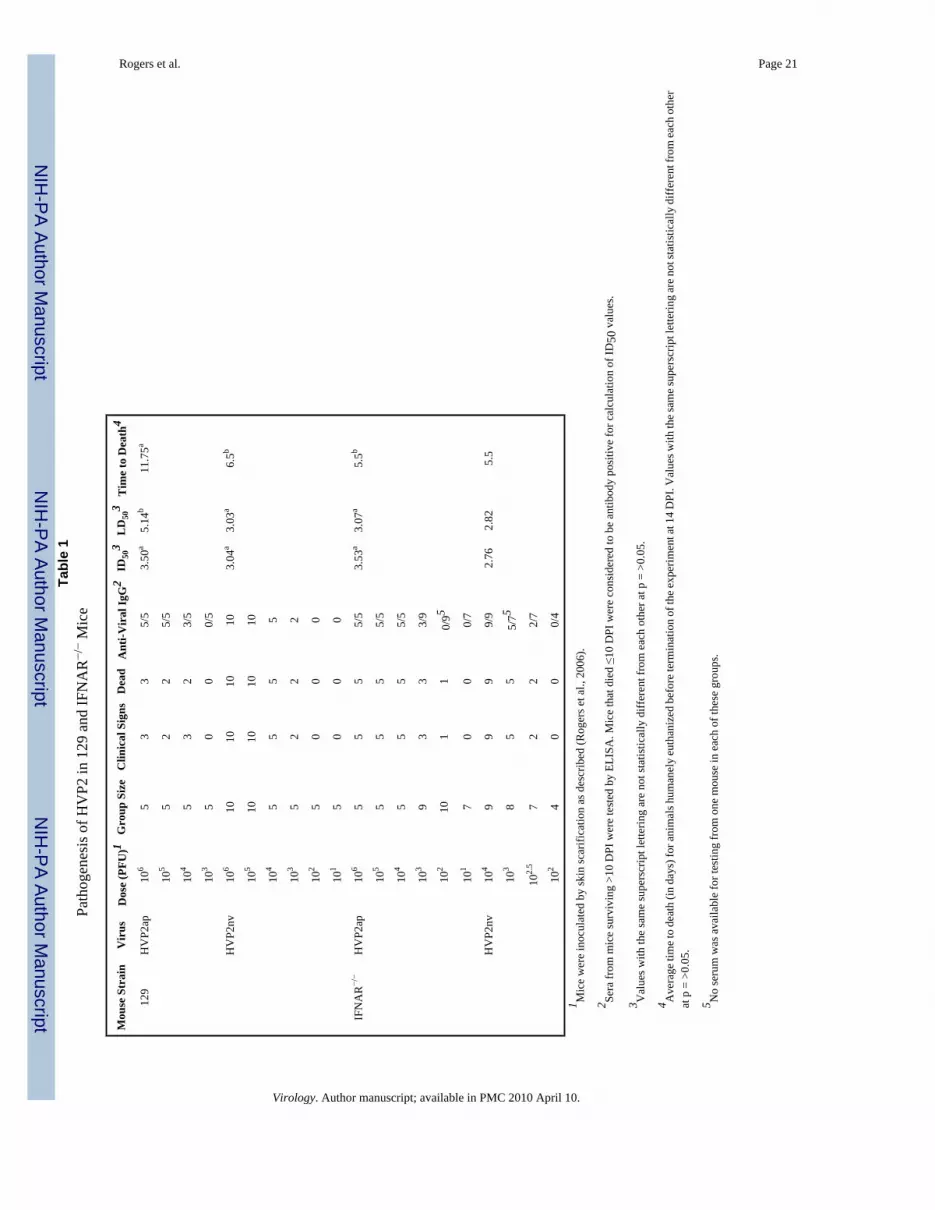
Based on the susceptibility of IFNAR−/− cells to infection by HVP2ap strains, IFNAR−/− miceshould be susceptible to HVP2ap infection. To test this, mice were by infected skin scarificationwith HVP2ap strain OU2-5 or HVP2nv strain OU1-76. The results of these experiments aresummarized in Table 1. The pathogenesis of HVP2ap and HVP2nv infections in 129 mice werethe essentially same as described previously for Balb/c mice (Rogers et al., 2006). ID50 andLD50 values for HVP2nv were not significantly different, while the ID50 value for HVP2apwas significantly less that the LD50. The LD50 for HVP2ap strain OU2-5 in 129 mice (5.14PFU) was also significantly lower than in Balb/c mice (>6.0 PFU). However, all 129 mice thatdied of HVP2ap infection did so at times far past the time at which mice die of acute CNSinfection (5–6 DPI). At necropsy, all HVP2ap infected 129 mice that died <14 DPI showedsigns of dehydration with distended bladders and impacted intestines. Histological examinationof tissues revealed pathology of the mural ganglia characterized by mild to moderate infiltrationof mononuclear cells. Immunohistochemical staining demonstrated the presence of viralantigen in these ganglia. These histological findings are similar to those previously reportedin the large intestines and urinary bladders of HVP2 and Saimirine herpesvirus 1 infected Balb/c mice (Breshears, Eberle, and Ritchey, 2001;Breshears, Eberle, and Ritchey, 2005;Ritchey etal., 2002).
The ID50 and LD50 of HVP2nv were not significantly different in IFNAR−/− and 129 mice (p> 0.05), indicating that the lack of the IFN-β receptor did not result in an increase in theneuropathogenicity of HVP2nv. In contrast, the LD50 for HVP2ap in IFNAR−/− mice was notsignificantly different from the LD50 for HVP2nv (p >0.05). Furthermore, HVP2ap infectedIFNAR−/− mice died within the same timeframe as HVP2nv infected mice. As seen for HVP2nvinfected mice, the LD50 and ID50 values for HVP2ap in IFNAR−/− mice were not significantlydifferent from one another (p > 0.05). Histologically, IFNAR−/− mice infected with eitherHVP2nv or HVPap and 129 mice infected with HVP2nv exhibited lesions having similarquality and intensity. Cutaneous lesions were characterized by abrupt epidermal necrosis withmultifocal ulceration and the exposed dermis overlain by a coagulum of fibrin and necroticcell debris. Scattered remaining epidermal keratinocytes as well as follicular epithelial cellsexhibited typical herpetic intranuclear inclusion bodies. The dermis contained a moderate tosevere, locally extensive inflammatory infiltrate composed of neutrophils admixed with fewermacrophages and lymphocytes. Spinal cords exhibit multifocal to diffuse vacuolation of theneuropil, usually worse on the ipsilateral side, with numerous scattered (small) neuronspredominantly in the ipsilateral dorsal grey horn exhibiting herpetic inclusion bodies. In lumbarsegments, neurons within dorsal root ganglia and other paraspinal ganglia were often acutelynecrotic or had intranuclear inclusion bodies. For the most part, although spinal cord lesionswere replete with inclusion bodies, inflammation was minimal to non-existent. Spinal cord andskin sections collected at 14 DPI from 129 mice infected with HVP2ap exhibited no significantmicroscopic lesions.
Rogers et al. Page 6
Virology. Author manuscript; available in PMC 2010 April 10.
NIH
-PA Author Manuscript
NIH
-PA Author Manuscript
NIH
-PA Author Manuscript

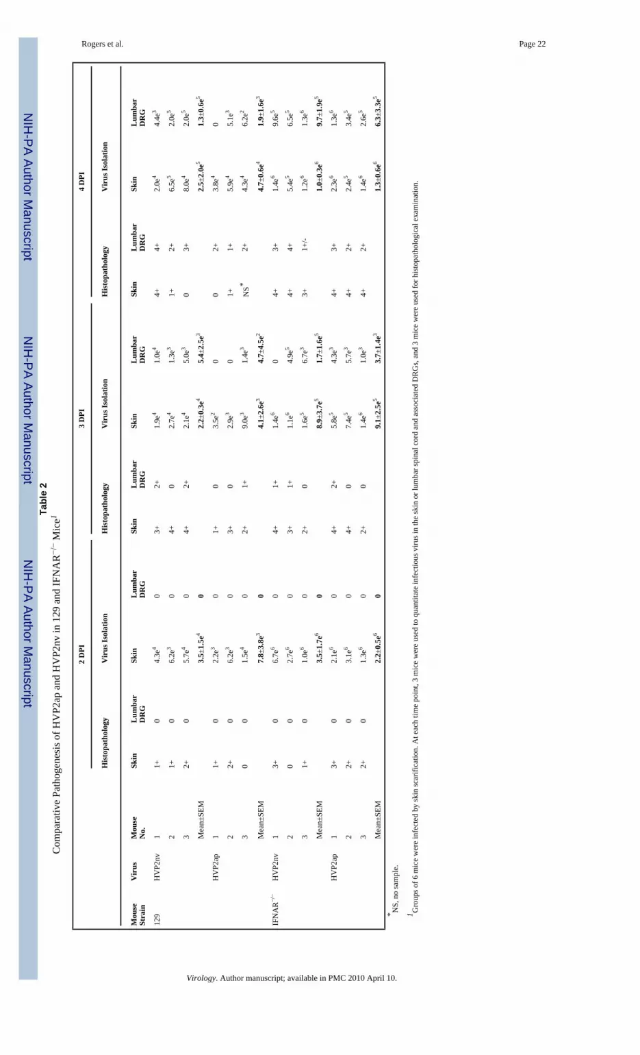
In Balb/c mice HVP2ap invades the CNS, but does so much less effectively than HVP2nv;HVP2ap takes longer to appear in the CNS, is present in the CNS in lesser amounts, and doesnot induce as strong an inflammatory response in the CNS (Rogers et al., 2006). However, thein vivo results above suggest that the kinetics of HVP2ap neuropathogenesis in IFNAR−/− miceshould be similar to that of HVP2nv. To test this, groups of six mice were infected and sacrificedat 2, 3 and 4 dpi, three being processed for histopathology and three for virus quantitation. Theresults are summarized in Table 2. Differences in titers of the two viruses in the skin of 129infected mice were statistically significant (p = >0.05) only at 3 DPI when HVP2ap titersdecrease more than HVP2nv titers. Although HVP2nv titers were generally higher thanHVP2ap titers, the differences at days 2 and 4 were not statistically significant due to the smallgroup size and variation among individual mice. However, comparison of virus titers in theskin at 2, 3 and 4 DPI revealed that while there was a significant increase in HVP2nv titerseach day, there was no statistically significant increase in HVP2ap titers from 2–4 DPI. Basedon histopathological examination, skin lesions were subjectively more severe in 129 miceinoculated with HVP2nv than HVP2ap, but this could reflect variability in sampling of theinoculation site rather than definitive differences in skin lesion severity. Thus, replication inthe skin of 129 mice at the site of inoculation was not dramatically different for the two HVP2subtypes up to 4 dpi.
When virus levels in the lumbar spinal cord and associated DRGs of 129 mice were compared,titers of HVP2nv were significantly higher than HVP2ap levels at 3 and 4 DPI (p = >0.05).Microscopic lesions in the CNS and DRGs were also more severe in HVP2nv infected 129mice, with larger numbers of necrotic neurons and neurons with intranuclear inclusion bodiesbeing present than were observed in HVP2ap infected mice. In the CNS, the varying severityof microscopic lesions between HVP2 subtypes was consistent with the observed differencein virus titers. These results indicate that in 129 mice HVP2nv is better able to invade and/orreplicate in the CNS than is HVP2ap.
Results were very different in IFNAR−/− mice. In the skin at the site of inoculation there wasnot a statistically significant increase in viral titers of either virus between 2 and 4 DPI, norwere titers of HVP2nv significantly different from those of HVP2ap at any time point. Whilelevels of HVP2ap and HVP2nv in the skin were equivalent in IFNAR−/− mice, there was asignificant difference in the amount of each virus present in the skin of 129 vs. IFNAR−/− miceat all time points. The same was true for virus levels in the CNS of IFNAR−/− mice: while titersof both HVP2ap and HVP2nv increased dramatically between 2 and 4 DPI, titers of the twovirus subtypes were not significantly different from each other. At each time point, microscopicskin lesions in the skin and CNS were of similar character and severity between the two virussubtypes. The lack of variation in lesion severity in IFNAR−/− mice is consistent with the lackof difference between tissue virus titers in IFNAR−/−mice. Thus, consistent with the equivalentLD50 values for the two HVP2 subtypes in IFNAR−/− mice (Table 1), there was no apparentdifference in the ability of HVP2ap and HVP2nv to replicate in the skin at the site of inoculationor to invade the CNS in IFNAR−/− mice.
Comparison of virus levels in tissues of 129 vs. IFNAR−/− mice revealed that the in the skin,titers of both viruses were significantly higher at all time points in IFNAR−/− mice (p = >0.05).When titers of virus in the CNS were compared, titers of HVP2nv at 3 and 4 DPI were notsignificantly different in the two mouse strains. However, CNS titers of HVP2ap weresignificantly higher (p = >0.05) in IFNAR−/− as compared to 129 mice at 3 and 4 DPI. Thus,the ability of HVP2ap to replicate locally in the skin to levels equivalent to that of HVP2nvappears to correlate with the ability of the virus to effectively invade the CNS.
In HSV, the vhs protein encoded by the UL41 gene has been shown to be important inabrogating the IFN-β response of cells following infection, presumably through the ability of
Rogers et al. Page 7
Virology. Author manuscript; available in PMC 2010 April 10.
NIH
-PA Author Manuscript
NIH
-PA Author Manuscript
NIH
-PA Author Manuscript

the vhs protein to degrade some cellular mRNAs (Kwong and Frenkel, 1987; Kwong andFrenkel, 1989; Murphy et al., 2003). The reduced pathogenicity of HSV UL41 deletion mutantsin mice (Smith, Ackland-Berglund, and Leib, 2000; Smith, Morrison, and Leib, 2001; Strelow,Smith, and Leib, 1997; Strelow and Leib, 1995; Suzutani et al., 2000) is similar to that ofHVP2ap strains, raising the possibility that differences in the UL41 gene might underlie thedichotomous neuropathogenicity of the two HVP2 subtypes. To address this possibility theUL41 ORF of four HVP2 strains (2 apathogenic & 2 neurovirulent) was sequenced and thepredicted amino acid sequences aligned to identify substitutions that correlated with theneurovirulence phenotype of the isolates (Figure 4). There were six amino acid residues thatshowed variation. Although two of the variations were unique to a single HVP2 isolate, fourcorrelated with the neurovirulence phenotype. None of these four substitution sites werelocated in a highly conserved region of the vhs polypeptide. Two were conservativesubstitutions (Ile ↔ Leu and Ala ↔ Thr) and two were non-conservative substitutions (bothLeu ↔ Pro). The two Leu ↔ Pro substitutions were located very near each other. Since prolineresidues induce turns in the polypeptides, it is possible that the Leu/Pro substitutions ‘offset’any effect via their close proximity in the peptide. Thus, there did not appear to be any majordifference in the vhs sequences of HVP2ap and HVP2nv isolates.
To experimentally assess the role of the UL41 gene in pathogenicity of HVP2 isolates, theUL41 ORF was deleted from a neurovirulent strain of HVP2 (designated Δ41) and thenreplaced with the UL41 ORF from either an apathogenic virus (Δ41C, a recombinant) or theparent neurovirulent virus (Δ41R, a revertant). Based on one-step growth curves, there wereno differences in the ability of any of these viruses to replicate in Vero cells (Figure 5A). Whiledegradation of host cell β-actin mRNA occurred in cells infected with any of the virusescontaining an intact UL41 ORF (wt, Δ41C & Δ41R), β-actin mRNA degradation was moreefficient in cells infected with the HVP2ap parental virus than in cells infected with HVP2nvparent virus, and β-actin mRNA degradation by the Δ41C recombinant and the Δ41R revertantreflected that of the parent virus their UL41 ORFs were derived from (Figure 5B). Therecombinant Δ41C replicated in PMDF cultures as efficiently as the HVP2nv parent and Δ41Rrevertant viruses, destroying the entire monolayer within 48 hrs. In contrast, virus lacking theUL41 ORF (Δ41) did not exhibit significant degradation of β-actin mRNA and grew muchslower in PMDFs than the other viruses. However, unlike HVP2ap plaques which ceased toexpand after 24 hrs and were eventually overgrown by uninfected cells (Rogers, Black, andEberle, 2007), HVP2nvΔ41 plaques continued to slowly enlarge in size up to 6 days PI whenthe experiment was terminated.
The ability of the UL41 mutants to prevent an IFN-β response was compared by assaying themedium from infected PMDF cultures at 24 hrs PI (Figure 5C). Cultures infected with theΔ41 mutant produced high levels of IFN-β typical of HVP2ap strains. Cultures infected withthe recombinant Δ41C produced low levels of IFN-β comparable to those produced by wtHVP2nv and Δ41R revertant infected cultures. These results indicate that while the vhs proteinis not responsible for the difference in the ability of HVP2ap and HVP2nv strains to preventan IFN-β response by the host cell, its absence does allow the host cell to mount an effectiveIFN-β response following infection.
Since the Δ41C recombinant virus grows as well as the parental HVP2nv virus in PMDFcultures and as virus replication in PMDF cultures correlates with the neurovirulence of HVP2isolates (Rogers, Black, and Eberle, 2007), the Δ41C recombinant should have aneurovirulence phenotype similar to the parental HVP2nv and Δ41R revertant viruses. To testthis, Balb/c mice were infected with each virus by skin scarification. The results aresummarized in Table 3. As expected, the LD50 of the revertant virus (Δ41R) was notsignificantly different from that of the parental HVP2nv virus. Most mice rapidly developedparesis of the ipsilateral hind foot by 5–6 days PI, followed by paralysis and euthanasia within
Rogers et al. Page 8
Virology. Author manuscript; available in PMC 2010 April 10.
NIH
-PA Author Manuscript
NIH
-PA Author Manuscript
NIH
-PA Author Manuscript
24 hrs. Mice infected with the same dose of the Δ41C recombinant virus followed a similarpathogenic progression of infection and time course of disease. Differences in the LD50 andmean time to death of the wt HVP2nv, Δ41C recombinant and Δ41R revertant viruses werenot statistically significant but were different from that of HVP2ap. Mice inoculated with theΔ41 deletion mutant exhibited a somewhat delayed development of paresis of the ipsilateralhind foot, and paresis did not always progress to full paralysis of the affected limb. As timeprogressed, skin lesions at the site of inoculation in Δ41 infected mice continued to developand spread. Despite the prolonged disease progression, the LD50 of the Δ41 deletion mutantwas not significantly different from that of the other HVP2nv viruses but was significantlylower than that of HVP2ap (p = >0.05). Thus, while deletion of the UL41 ORF did qualitativelyaffect the neuropathogenicity of HVP2nv, it did not severely reduce neuropathogenicity of thevirus as has been reported for HSV1 and HSV2 (Korom, Wylie, and Morrison, 2008; Strandand Leib, 2004; Strelow and Leib, 1995).
Histologically, skin and spinal cord lesions in mice infected with the UL41 mutants werequalitatively and quantitatively similar to those previously described in Balb/c mice infectedwith wt HVP2nv isolates. Briefly, there was epithelial necrosis and ulceration with herpeticinclusions present in sloughed epithelial cells in the crusts as well as in scattered remnantepidermal cells and follicular epithelial cells. The dermis exhibited moderate to severedisseminated mononuclear and neutrophilic dermatitis. Immunostaining revealed conspicuousHVP2 antigen confined to the epithelium. In the lumbar spinal cord, there was multifocal todisseminated vacuolation of the ipsilateral white matter in all funiculi, most usually severe inthe dorsal and lateral funiculus (Figure 6). There was also mild, mononuclear inflammation inthe ipsilateral dorsal grey horn and funiculus with numerous conspicuous herpetic inclusionswithin small neurons and glial cells. The only quantitative difference among the mutants wasthat extension of the inflammation to the brain only occurred in wt HVP2nv, Δ41C and Δ41R;there were no brain lesions observed in 33 brains examined from Δ41 infected mice, even inthose inoculated with high doses of virus.
DiscussionIn their natural host, acute infection by HSV and related primate herpesviruses is normallyfollowed by the establishment of a latent infection within the PNS that persists for the life ofthe host. In contrast to the usually mild, self-limiting infections that occur in the natural host,when these viruses infect a non-natural host they can produce severe, frequently fatal infectionsthat involve the CNS such as occurs when BV infects humans. The host and viral mechanismsthat allow these viruses to enter and spread via lytic infection within the CNS in a non-naturalhost rather than establishing a latent infection in the PNS as occurs in the natural host are largelyunknown. A more thorough understanding of virus/host interactions which determine theserious outcome of cross-species or zoonotic infections is critical.
With regards to pathogenicity, the HVP2nv-mouse model closely resembles human BVinfections. Although HVP2nv is not neurovirulent in its natural baboon host and only rarelycauses severe infections in baboons (Rogers et al., 2005), HVP2nv isolates are extremelyneuropathogenic in mice. In contrast, mice are able to very efficiently control infections bynaturally occurring HVP2ap isolates. Previous studies indicate that control of HVP2apinfections in mice begins locally at the site of inoculation and continues even within the CNS(Rogers et al., 2006). Since the pathogenesis of HVP2nv and HVP2ap infection in micediverges well before an adaptive immune response develops, innate immunity likely plays amajor role in effecting the divergent pathogenicity of the two HVP2 subtypes. IFN-β appearsto be involved in controlling HVP2ap infections since HVP2nv isolates do not induce and/orovercome an IFN-β response, and are thus not as susceptible to IFN-β in vitro as HVP2apisolates are (Rogers, Black, and Eberle, 2007). The experiments presented here extend these
Rogers et al. Page 9
Virology. Author manuscript; available in PMC 2010 April 10.
NIH
-PA Author Manuscript
NIH
-PA Author Manuscript
NIH
-PA Author Manuscript

observations and confirm a critical role for the type I IFN response in controlling HVP2apinfections in vivo.
Other investigators have observed that HSV1 and HSV2 UL41 mutants exhibit reducedneurovirulence in the mouse model, but in IFNAR−/− mice their neurovirulence is comparableto that of the wild-type virus (Leib et al., 1999; Murphy et al., 2003). Consistent with theseHSV studies, HVP2ap isolates are just as neuropathogenic as HVP2nv isolates in IFNAR−/−
mice as evidenced by similar LD50 values and similar levels of virus in the skin at early timesPI. The relationship between the ID50 and LD50 for HVP2ap isolates in IFNAR−/− mice alsoreflects that of HVP2nv isolates in 129 and Balb/c mice: the ID50 and LD50 values are notsignificantly different, indicating that when HVP2ap establishes an infection in IFNAR−/− miceit inexorably progresses to death of the host. Similarly, temporal progression of virus from theperipheral site of inoculation into the CNS is similar for both HVP2 subtypes in IFNAR−/−
mice. Since the level of virus in the lumbar spinal cord and associated DRGs represents theearliest point in the pathogenic process that is demonstrably different for HVP2ap and HVP2nvin wild-type 129 mice, it is likely that the ability of the host to delay and/or lessen entry ofvirus into the CNS plays a critical role in the dichotomous outcome of infection with the twoHVP2 subtypes. In contrast, studies with HSV have shown that replication of the virus at theinoculation site appears to be important in that this replication amplifies the amount of virusthereby increasing the likelihood of viral entry into unmyelinated nerve fibers present in theskin to provide access to the CNS (Cunningham et al., 2006; Mossman and Ashkar, 2005;Yamada et al., 1986). While differences in the amount of HVP2ap vs. HVP2nv in skin werestatistically significant only at 3 DPI, levels of HVP2ap were lower than those of HVP2nv at2 and 4 DPI. Given the small group size used (3 mice/group), it is possible that that were largergroup sizes used these differences may have been significant. If so, our results would beconsistent with the results of other investigators studying HSV pathogenesis which suggestthat reduction of virus replication at the site of inoculation is critical in determining the outcomeof the infection.
Another observation from these experiments was that the inability of the host to mount a typeI IFN response did not have a major effect on the temporal or spatial progression of HVP2nvinfection at early times PI. The only noticeable difference was that in IFNAR−/− mice highertiters of HVP2nv were present in skin at the inoculation site at 2 dpi than in 129 mice. However,HVP2ap titers at 2 dpi at the site of inoculation were also higher in the knock-out mice vs.wild-type indicating that higher viral titers at this time is a function of the mouse genotyperather than being peculiar to HVP2nv. This suggests that an intact type I IFN response doesnot interfere with the ability of HVP2nv to invade and replicate within the CNS. The highertiters of HVP2nv in skin of IFNAR−/− mice at 2 DPI suggests that the early IFN response atthe site of inoculation may nonetheless have a dampening effect on HVP2nv replication in theskin at very early times after infection. This would again be consistent with the hypothesis thatcontrolling the initial replication and amplification of virus at the site of inoculation is a criticalfactor in the ability of the host to prevent virus from gaining access to the CNS.
In previous experiments pre-treatment of PMDF cultures with recombinant murine IFN-β didnot effectively protect cells from HVP2nv infection, suggesting that it may be the induction ofthe IFN system rather than the production of type I IFNs which is important for controllingHVP2 infections (Rogers, Black and Eberle, 2007). In addition, while we have not detectedIFN-α induction by HVP2 in vitro, IFNAR loss would affect both IFN-β and IFN-α inducedpathways of antiviral immunity. As such, the importance of IFN-α in controlling HVP2infection in vivo can not be ruled out. While type I IFNs are important for the early, innateresponse to viral infection they also contribute to the antiviral immune response by stimulatingthe cytotoxic activity of natural killer cells, signaling for dendritic cell maturation, andpromoting various T-cell functions, including expansion of the memory population (Garcia-
Rogers et al. Page 10
Virology. Author manuscript; available in PMC 2010 April 10.
NIH
-PA Author Manuscript
NIH
-PA Author Manuscript
NIH
-PA Author Manuscript

Sastre and Biron, 2006). Further studies are needed to determine the exact IFN mechanism thatso efficiently controls HVP2ap while allowing CNS invasion by HVP2nv.
In HSV1 and HSV2 the UL41 gene encoding the vhs protein has been shown to play a majorrole in determining the neurovirulence of the virus. UL41 mutants of HSV replicate normallyin tissue culture but are impaired in their ability to invade the CNS from peripheral sites andare defective in their ability to spread within the CNS of mice (Korom, Wylie, and Morrison,2008; Smith, Ackland-Berglund, and Leib, 2000; Smith, Morrison, and Leib, 2001; Strelow,Smith, and Leib, 1997; Strelow and Leib, 1995). This is very similar to what is observed forapathogenic isolates of HVP2. However, replacement of the HVP2nv UL41 ORF with anHVP2ap UL41 ORF did not affect the ability of the virus to infect mice and invade the CNS,nor did it result in high levels of IFN-β production in infected PMDF cultures. Thisdemonstrates that the UL41 protein alone is not responsible for the disparate neurovirulenceof the two HVP2 subtypes.
Although the UL41 gene does not appear to underlie the neuropathogenic differences ofHVP2ap and HVP2nv isolates, it does appear to play a role in allowing HVP2 to prevent thehost IFN-β response. Deletion of the HVP2nv UL41 gene resulted in a strong IFN-β responsein infected PMDF cultures. Consistent with this, HVP2nvΔ41 replicated poorly in PMDFswhile HVP2ap abortively infects PMDFs. HVP2nvΔ41 also displayed reduced pathogenesisin mice much as described for HSV UL41 deletion mutants. While HVP2nvΔ41 can invadethe CNS, it does not produce the rapid and severe CNS disease observed in mice infected withwt HVP2nv. Since HVP2nvΔ41 infected PMDF cultures produce high levels of IFN-βcomparable to those induced by HVP2ap isolates, the UL41 gene does appear to be involvedin preventing or suppressing the IFN-β response. The recombinant HVP2nvΔ41C carrying anHVP2ap UL41 gene behaved similar to the wt HVP2nv in all respects, including suppressionof the host IFN-β response. This demonstrates that the vhs protein itself is not functionallydifferent in apathogenic and neurovirulent HVP2 isolates. This apparent inconsistency couldbe explained by the interaction of the vhs protein with some other viral factor(s) to suppressthe IFN-β response, this second factor being the one actually responsible for the disparatepathogenic phenotype of HVP2 isolates.
The interplay between large, complex herpesviruses and the intricate workings of the hostimmune system is understandably convoluted. In addition to vhs, α-herpesviruses encodenumerous other IFN antagonists which most likely interact with multiple IFN inductionpathways in the host. To further complicate matters, during HSV infection pathogenrecognition and subsequent type I IFN production has been shown to be both cell-type andtime-dependent (Rasmussen et al., 2007). While pDCs are responsible via Toll-like receptor 9for early production of IFN in response to HSV infection in vivo, macrophages and fibroblastsalso produce type I IFNs later in infection. The production of type I IFNs in these cell types isdependent on both viral entry and replication, and is ablated in cells unable to signal throughthe mitochondrial antiviral signaling (MAVS) protein pathway. Therefore, interference withboth the MAVS- and TLR9-dependent pathways at multiple times PI may be necessary forHVP2nv to ensure sufficient replication in the dermatome for entry into the CNS.
The fact that the LD50 for HVP2ap isolates in IFNAR−/− mice was the same as the LD50 forHVP2nv strains suggests that the type I IFN response may be the primary if not sole facet ofthe host innate response involved in the initial control of HVP2ap infection. The basis for thedivergent neuropathogenicity of HVP2nv vs. HVP2ap isolates in mice thus appears to lie inthe ability of the HVP2nv isolates to prevent and/or overcome the host type I IFN responseand/or downstream host effector mechanisms which are controlled and initiated via the type IIFN pathway. The UL41-encoded vhs protein gene does appear to be directly involved inpreventing the IFN-β response in that deletion of the UL41 gene from HVP2nv results in IFN-
Rogers et al. Page 11
Virology. Author manuscript; available in PMC 2010 April 10.
NIH
-PA Author Manuscript
NIH
-PA Author Manuscript
NIH
-PA Author Manuscript

β production following infection. However, since the HVP2nvΔ41 deletion mutant canreplicate in PMDF cells and mice despite the induction of an IFN-β response, the vhs proteindoes not appear to be the primary factor responsible for the ability of HVP2nv to overcomethe IFN-β response. It remains to be seen how HVP2nv isolates accomplish this and whyHVP2ap isolates are unable to do so.
AcknowledgmentsThis work was supported in part by grants P40 RR12317, R24 RR16556 and T35 RR07061-13 from the Public HealthService.
ReferencesBlack DH, Saliki JT, Eberle R. Development of a green fluorescent protein reporter cell line to reduce
biohazards associated with detection of infectious Cercopithecine herpesvirus 1 (monkey B virus) inclinical specimens. Compar Med 2002;52(6):534–42.
Brandt CR. The role of viral and host genes in corneal infection with herpes simplex virus type 1. ExptlEye Res 2005;80(5):607–21. [PubMed: 15862167]
Breshears MA, Eberle R, Ritchey JW. Characterization of gross and histological lesions in Balb/c miceexperimentally infected with Herpesvirus saimiri 1 (HVS1). J Compar Pathol 2001;125(1):25–33.
Breshears MA, Eberle R, Ritchey JW. Temporal progression of viral replication and gross and histologicallesions in Balb/c mice inoculated epidermally with Saimiriine herpesvirus 1 (SaHV-1). J Compar Path2005;133(2–3):103–13.
Cunningham AL, Diefenbach RJ, Miranda-Saksena M, Bosnjak L, Kim M, Jones C, Douglas MW. Thecycle of human herpes simplex virus infection: virus transport and immune control. J Infect Dis 194Suppl 2006;1:S11–8.
Dai J, Megjugorac NJ, Amrute SB, Fitzgerald-Bocarsly P. Regulation of IFN regulatory factor-7 andIFN-alpha production by enveloped virus and lipopolysaccharide in human plasmacytoid dendriticcells. J Immunol 2004;173(3):1535–48. [PubMed: 15265881]
Darnell JE, Kerr IM, Stark GR. Transcriptional activation in response to IFNs and other extracellularsignalling proteins. Science 1994;264:248–254.
Duerst RJ, Morrison LA. Herpes simplex virus 2 virion host shutoff protein interferes with type Iinterferon production and responsiveness. Virol 2004;322(1):158–67.
Eberle R, Black DH, Blewett EL, White GL. Prevalence of Herpesvirus papio 2 in baboons andidentification of immunogenic viral polypeptides. Lab Anim Sci 1997a;47(3):256–62. [PubMed:9241626]
Eberle R, Black DH, Lehenbauer TW, White GL. Shedding and transmission of baboon Herpesviruspapio 2 (HVP2) in a breeding colony. Lab Anim Sci 1998;48(1):23–28. [PubMed: 9517885]
Eberle R, Black DH, Lipper S, Hilliard JK. Herpesvirus papio 2, an SA8-like alpha-herpesvirus ofbaboons. Arch Virol 1995;140(3):529–45. [PubMed: 7733825]
Eberle R, Hilliard JK. The simian herpesviruses: a review. Infect Agents Dis 1995;4:55–70. [PubMed:7613729]
Eberle R, Tanamachi B, Black D, Blewett EL, Ali M, Openshaw H, Cantin EM. Genetic and functionalcomplementation of the HSV1 UL27 gene and gB glycoprotein by simian alpha-herpesvirushomologs. Arch Virol 1997b;142(4):721–36. [PubMed: 9170500]
Enquist LW, Husak PJ, Banfield BW, Smith GA. Infection and spread of alphaherpesviruses in thenervous system. Adv Virus Res 1998;51:237–347. [PubMed: 9891589]
Esclatine A, Taddeo B, Roizman B. The UL41 protein of herpes simplex virus mediates selectivestabilization or degradation of cellular mRNAs. PNAS 2004;101(52):18165–70. [PubMed:15596716]
Everly DN, Feng P, Mian IS, Read GS. mRNA degradation by the virion host shutoff (vhs) protein ofherpes simplex virus: genetic and biochemical evidence that vhs is a nuclease. J Virol 2002;76:8560–8571. [PubMed: 12163576]
Rogers et al. Page 12
Virology. Author manuscript; available in PMC 2010 April 10.
NIH
-PA Author Manuscript
NIH
-PA Author Manuscript
NIH
-PA Author Manuscript
Garcia-Sastre A, Biron CA. Type 1 interferons and the virus-host relationship: a lesson in detente. Science2006;312(5775):879–82. [PubMed: 16690858]
Goodbourn S, Didcock L, Randall RE. Interferons: cell signalling, immune modulation, antiviral responseand virus countermeasures. J Gen Virol 2000;81(Pt 10):2341–64. [PubMed: 10993923]
Huff JL, Barry PA. B-virus (Cercopithecine herpesvirus 1) infection in humans and macaques: potentialfor zoonotic disease. Emerg Infect Dis 2003;9:246–250. [PubMed: 12603998]
Karr BM, Read GS. The virion host shutoff function of herpes simplex virus degrades the 5′ end of atarget mRNA before the 3′ end. Virol 1999;264(1):195–204.
Keeble SA. B virus infection in monkeys. Ann NY Acad Science 1960;85:960–969.Korom M, Wylie KM, Morrison LA. Selective ablation of virion host shutoff protein RNase activity
attenuates herpes simplex virus 2 in mice. J Virol 2008;82(7):3642–53. [PubMed: 18234805]Kwong AD, Frenkel N. Herpes simplex virus-infected cells contain a function(s) that destabilizes both
host and viral mRNAs. PNAS 1987;84(7):1926–30. [PubMed: 3031658]Kwong AD, Frenkel N. The herpes simplex virus virion host shutoff function. J Virol 1989;63:4834–
4839. [PubMed: 2552156]Lam Q, Smibert CA, Koop KE, Lavery C, Capone JP, Weinheimer SP, Smiley. Herpes simplex virus
VP16 rescues viral mRNA from destruction by the virion host shutoff function. EMBO J1996;15:2575–2581. [PubMed: 8665865]
Lee HK, Lund JM, Ramanathan B, Mizushima N, Iwasaki A. Autophagy-dependent viral recognition byplasmacytoid dendritic cells. Science (New York, N Y) 2007;315(5817):1398–401.
Leib DA, Harrison TE, Laslo KM, Machalek MA, Moorman NJ, Virgin HW. Interferons regulate thephenotype of wild-type and mutant herpes simplex viruses In vivo. J Exp Med 1999;189:663–672.[PubMed: 9989981]
Loomis MR, O’Neill T, Bush M, Montali RJ. Fatal herpesvirus infection in patas monkeys and a blackand white colobus monkey. J Am Vet Med Assoc 1981;179:1236–1239. [PubMed: 6276349]
Mossman KL, Ashkar AA. Herpesviruses and the innate immune response. Viral Immunology 2005;18(2):267–81. [PubMed: 16035939]
Muller U, Steinhoff U, Reis LF, Hemmi S, Pavlovic J, Zinkernagel RM, Aguet M. Functional role oftype I and type II interferons in antiviral defense. Science 1994;264(5167):1918–21. [PubMed:8009221]
Murphy JA, Duerst RJ, Smith TJ, Morrison LA. Herpes simplex virus type 2 virion host shutoff proteinregulates alpha/beta interferon but not adaptive immune responses during primary infection in vivo.J Virol 2003;77(17):9337–45. [PubMed: 12915549]
Ohsawa K, Lehenbauer TW, Eberle R. Herpesvirus papio 2: a safer and sensitive alternative forserodiagnosis of B virus infection in macaque monkeys. Lab Anim Sci 1999;49:605–616. [PubMed:10638495]
Palmer AE. Herpesvirus simiae: historical perspective. J Med Primatol 1987;16:99–130. [PubMed:3035187]
Rasmussen SB, Sorensen LN, Malmgaard L, Ank N, Baines JD, Chen ZJ, Paludan SR. Type I interferonproduction during herpes simplex virus infection is controlled by cell-type-specific viral recognitionthrough Toll-like receptor 9, the mitochondrial antiviral signaling protein pathway, and novelrecognition systems. J Virol 2007;81(24):13315–24. [PubMed: 17913820]
Ritchey JW, Ealey KA, Payton M, Eberle R. Comparative pathology of infections with baboon andAfrican green monkey alpha-herpesviruses in mice. J Compar Pathol 2002;127:150–161.
Ritchey JW, Payton ME, Eberle R. Clinicopathological characterization of monkey B virus(Cercopithecine herpesvirus 1) infection in mice. J Compar Pathol 2005;132:202–217.
Rogers KM, Black DH, Eberle R. Primary mouse dermal fibroblast cell cultures as an in vitro modelsystem for the differential pathogenicity of cross-species herpesvirus papio 2 infections. Arch Virol2007;152(3):543–52. [PubMed: 17122896]
Rogers KM, Ealey KA, Ritchey JW, Black DH, Eberle R. Pathogenicity of different baboon Herpesviruspapio 2 isolates is characterized by either extreme neurovirulence or complete apathogenicity. J Virol2003;77:10731–10739. [PubMed: 14512523]
Rogers et al. Page 13
Virology. Author manuscript; available in PMC 2010 April 10.
NIH
-PA Author Manuscript
NIH
-PA Author Manuscript
NIH
-PA Author Manuscript

Rogers KM, Ritchey JW, Payton M, Black DH, Eberle R. Neuropathogenesis of Herpesvirus papio 2 inMice Parallels Cercopithecine herpesvirus 1 (B Virus) Infections in Humans. J Gen Virol2006;87:267–276. [PubMed: 16432011]
Rogers KM, Wolf RF, White GL, Eberle R. Experimental infection of baboons (Papio cynocephalusanubis) with apathogenic and neurovirulent subtypes of Herpesvirus papio 2. Compar Med 2005;55(5):425–430.
Rouse, BT.; Lopez, C. Immunobiology of herpes simplex virus infection. CRC Press; 1984.Sabin AB, Wright AM. Acute ascending myelitis following a monkey bite, with isolation of a virus
capable of reproducing the disease. J Expl Med 1934;59:115–136.Schmelter J, Knez J, Smiley JR, Capone JP. Identification and characterization of a small modular domain
in the herpes simplex virus host shutoff protein sufficient for Interaction with VP16. J Virol1996;70:2124–2131. [PubMed: 8642633]
Simons A, Nash AA. Zosteriform spread of herpes simplex virus as a model of recrudescence and its useto investigate the role of immune cells in prevention of recurrent disease. J Virol 1984;52:816–821.[PubMed: 6092713]
Smith TJ, Ackland-Berglund CE, Leib DA. Herpes simplex virus virion host shutoff (vhs) activity altersperiocular disease in mice. J Virol 2000;74:3598–3604. [PubMed: 10729135]
Smith TJ, Morrison LA, Leib DA. Pathogenesis of herpes simplex virus type 2 virion host shutoff (vhs)mutants. J Virol 2001;76:2054–2061. [PubMed: 11836383]
Strand SS, Leib DA. Role of the VP16-binding domain of vhs in viral growth, host shutoff activity, andpathogenesis. J Virol 2004;78(24):13562–72. [PubMed: 15564467]
Strelow L, Smith T, Leib D. The virion host shutoff function of herpes simplex virus type 1 plays a rolein corneal invasion and functions independently of the cell cycle. Virol 1997;231(1):28–34.
Strelow LI, Leib DA. Role of the viron host shutoff (vhs) of herpes simplex virus type 1 in latency andpathogenesis. J Virol 1995;69:6779–6786. [PubMed: 7474089]
Strelow LI, Leib DA. Analysis of conserved domains of UL41 of herpes simplex virus type 1 in virionhost shutoff and pathogenesis. J Virol 1996;70(8):5665–7. [PubMed: 8764085]
Suzutani T, Nagamine M, Shibaki T, Ogasawara M, Yoshida I, Daikoku T, Nishiyama Y, Azuma M. Therole of the UL41 gene of herpes simplex virus type 1 in evasion of non-specific host defencemechanisms during primary infection. J Gen Virol 2000;81(Pt 7):1763–71. [PubMed: 10859382]
Thompson SA, Hilliard JK, Kittel D, Lipper S, Giddens WE, Black DH, Eberle R. Retrospective analysisof an outbreak of B virus in a colony of DeBrazza’s monkeys (Cercopithecus neglectus). ComparMed 2000;50:649–657.
Troan BV, Perelygina L, Patrusheva I, van Wettere A, Hilliard J, Loomis M, De Voe R. NaturallyTransmitted Herpesvirus papio 2 Infection in a Black and White Colobus Monkey. JAVMA2007;231:1878–1883. [PubMed: 18081530]
Weeks BS, Ramchandran RS, Hopkins JJ, Friedman HM. Herpes simplex virus type-1 and -2pathogenesis is restricted by the epidermal basement membrane. Arch Virol 2000;145:385–396.[PubMed: 10752560]
Weigler BJ. Biology of B virus in macaque and human hosts: a review. Clin Infect Dis 1992;14:555–567. [PubMed: 1313312]
Whitely, RJ.; Hilliard, JK. Cercopithecine herpesvirus (B virus). In: Knipe, DM.; Howley, PM., editors.Fields Virology. Vol. 4. Lippincott Williams and Wilkins; Philadelphi, PA: 2001. p. 2835-2848.
Wolf RF, Rogers KM, Blewett EL, Fakhari F, Hill CA, Kosanke SD, White GL, Eberle R. A naturallyoccurring fatal case of Herpesvirus papio 2 pneumonia in an infant baboon (Papio cynocephalusanubis). Compar Med 2006;45(1):42–46.
Yamada M, Arao Y, Uro F, Nii S. Mechanisms of difference in pathogenicity between two variants of alaboratory strain of herpes simplex virus type 1. Microb Immunol 1986;30:1259–1270.
Rogers et al. Page 14
Virology. Author manuscript; available in PMC 2010 April 10.
NIH
-PA Author Manuscript
NIH
-PA Author Manuscript
NIH
-PA Author Manuscript

Figure 1. Plaque formation by HVP2 subtypes in BALB/c, 129 and IFNAR−/− PMDF culturesConfluent PMDF monolayers were inoculated at an MOI of 0.25 PFU/cell and photographedat 48 hrs PI. HVP2nv isolates produced complete CPE within 48 hrs in all three cell types.HVP2ap isolates produced discrete plaques by 24 hrs in Balb/c and 129 cell cultures, but thesefailed to enlarge with time. In contrast, HVP2ap isolates completely destroyed IFNAR−/−
cultures by 48 hr PI.
Rogers et al. Page 15
Virology. Author manuscript; available in PMC 2010 April 10.
NIH
-PA Author Manuscript
NIH
-PA Author Manuscript
NIH
-PA Author Manuscript

Figure 2. Replication of HVP2 subtypes in Balb/c, 129 and IFNAR−/− PMDF culturesConfluent Balb/c (light gray), 129 (dark gray) or IFNAR−/− (black) PMDF cultures wereinoculated with HVP2ap isolates A951 and OU2-5 or HVP2nv isolates X313 and OU1-76 atan MOI of 0.25 PFU/cell. At 48 hrs PI, infected cells were scraped into the media and infectiousvirus quantitated. There was no significant difference in the amount of HVP2nv produced inthe three cell lines while HVP2ap isolates produced significantly more infectious progeny virusin IFNAR−/− cells than in either Balb/c or 129 cells (as indicated by asterisks above the standarderror bars).
Rogers et al. Page 16
Virology. Author manuscript; available in PMC 2010 April 10.
NIH
-PA Author Manuscript
NIH
-PA Author Manuscript
NIH
-PA Author Manuscript

Figure 3. IFN-β production in HVP2ap and HVP2nv infected PMDF culturesPMDF cultures derived from three mouse strains were infected with HVP2nv strains OU1-76(black) and X313 (hatched) or HVP2ap strains OU2-5 (light gray) and A951 (dark gray) at anMOI of 1.0 PFU/cell. Media was collected at 24 hrs PI and IFN-β quantitated by ELISA. InBalb/c and 129 cells, the IFN-β concentration was significantly higher in HVP2ap infectedthan in HVP2nv infected cultures (as indicated by asterisks above the standard error bars);there was no significant difference in IFN-β produced in IFNAR−/− cultures infected with thetwo HVP2 subtypes.
Rogers et al. Page 17
Virology. Author manuscript; available in PMC 2010 April 10.
NIH
-PA Author Manuscript
NIH
-PA Author Manuscript
NIH
-PA Author Manuscript

Figure 4. Location of variable amino acid residues in the HVP2 UL41 (vhs) polypeptideAA sequences of four HVP2 strains (HVP2ap A951 & OU2-5 and HVP2nv OU1-76 & X313)were aligned to identify residues that varied among isolates. The sequences for HSV1 andHSV2 are included for reference. AA residues conserved in all sequences are indicated by lackof shading. Residues that vary between HSV and HVP2 are indicated by gray/black shading.The four potential HVP2 subtype-specific substitutions are indicated by a solid triangle belowthe sequences and the two isolate-specific substitutions are indicated by an open triangle belowthem.
Rogers et al. Page 18
Virology. Author manuscript; available in PMC 2010 April 10.
NIH
-PA Author Manuscript
NIH
-PA Author Manuscript
NIH
-PA Author Manuscript

Figure 5. Characterization of UL41 mutantsOne-step growth curves performed in Vero cells are shown in A. Viruses included the parentalHVP2nv (■——-■), HVP2nv Δ41 (▲………▲), HVP2nv Δ41C (□----□) and HVP2nv Δ41R(●——-●). Northern blot of β-actin mRNA in infected cells is shown in B. Total RNA wasisolated from cells at 4 hrs PI, 15 μg loaded in each lane, and northern blotting performed usingthe Vero cell β-actin gene as probe. IFN-β production in PMDF cultures is shown in C. Cultureswere infected with the indicated viruses at an MOI = 1 PFU/cell and the medium assayed forIFN-β at 24 hrs PI. IFN-β levels that were significantly different (p = >0.05) from the levelinduced by the parental HVP2nv virus are indicated by an asterisk above the standard errorbar.
Rogers et al. Page 19
Virology. Author manuscript; available in PMC 2010 April 10.
NIH
-PA Author Manuscript
NIH
-PA Author Manuscript
NIH
-PA Author Manuscript
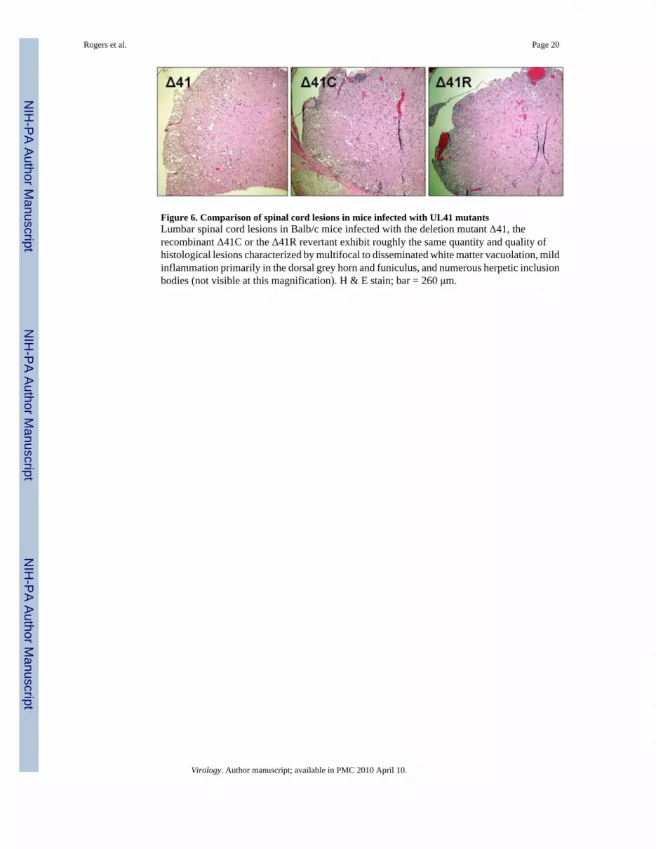
Figure 6. Comparison of spinal cord lesions in mice infected with UL41 mutantsLumbar spinal cord lesions in Balb/c mice infected with the deletion mutant Δ41, therecombinant Δ41C or the Δ41R revertant exhibit roughly the same quantity and quality ofhistological lesions characterized by multifocal to disseminated white matter vacuolation, mildinflammation primarily in the dorsal grey horn and funiculus, and numerous herpetic inclusionbodies (not visible at this magnification). H & E stain; bar = 260 μm.
Rogers et al. Page 20
Virology. Author manuscript; available in PMC 2010 April 10.
NIH
-PA Author Manuscript
NIH
-PA Author Manuscript
NIH
-PA Author Manuscript
NIH
-PA Author Manuscript
NIH
-PA Author Manuscript
NIH
-PA Author Manuscript
Rogers et al. Page 21Ta
ble
1Pa
thog
enes
is o
f HV
P2 in
129
and
IFN
AR−/−
Mic
e
Mou
se S
trai
nV
irus
Dos
e (P
FU)1
Gro
up S
ize
Clin
ical
Sig
nsD
ead
Ant
i-Vir
al Ig
G2
ID50
3L
D50
3T
ime
to D
eath
4
129
HV
P2ap
106
53
35/
53.
50a
5.14
b11
.75a
105
52
25/
5
104
53
23/
5
103
50
00/
5
HV
P2nv
106
1010
1010
3.04
a3.
03a
6.5b
105
1010
1010
104
55
55
103
52
22
102
50
00
101
50
00
IFN
AR−/−
HV
P2ap
106
55
55/
53.
53a
3.07
a5.
5b
105
55
55/
5
104
55
55/
5
103
93
33/
9
102
101
10/
95
101
70
00/
7
HV
P2nv
104
99
99/
92.
762.
825.
5
103
85
55/
75
102.
57
22
2/7
102
40
00/
41 M
ice
wer
e in
ocul
ated
by
skin
scar
ifica
tion
as d
escr
ibed
(Rog
ers e
t al.,
200
6).
2 Sera
from
mic
e su
rviv
ing
>10
DPI
wer
e te
sted
by
ELIS
A. M
ice
that
die
d ≤1
0 D
PI w
ere
cons
ider
ed to
be
antib
ody
posi
tive
for c
alcu
latio
n of
ID50
val
ues.
3 Val
ues w
ith th
e sa
me
supe
rscr
ipt l
ette
ring
are
not s
tatis
tical
ly d
iffer
ent f
rom
eac
h ot
her a
t p =
>0.
05.
4 Ave
rage
tim
e to
deat
h (in
day
s) fo
r ani
mal
s hum
anel
y eu
than
ized
bef
ore t
erm
inat
ion
of th
e exp
erim
ent a
t 14
DPI
. Val
ues w
ith th
e sam
e sup
ersc
ript l
ette
ring
are n
ot st
atis
tical
ly d
iffer
ent f
rom
each
oth
erat
p =
>0.
05.
5 No
seru
m w
as a
vaila
ble
for t
estin
g fr
om o
ne m
ouse
in e
ach
of th
ese
grou
ps.
Virology. Author manuscript; available in PMC 2010 April 10.
NIH
-PA Author Manuscript
NIH
-PA Author Manuscript
NIH
-PA Author Manuscript
Rogers et al. Page 22Ta
ble
2C
ompa
rativ
e Pa
thog
enes
is o
f HV
P2ap
and
HV
P2nv
in 1
29 a
nd IF
NA
R−/−
Mic
e1
2 D
PI3
DPI
4 D
PI
His
topa
thol
ogy
Vir
us Is
olat
ion
His
topa
thol
ogy
Vir
us Is
olat
ion
His
topa
thol
ogy
Vir
us Is
olat
ion
Mou
seSt
rain
Vir
usM
ouse
No.
Skin
Lum
bar
DR
GSk
inL
umba
rD
RG
Skin
Lum
bar
DR
GSk
inL
umba
rD
RG
Skin
Lum
bar
DR
GSk
inL
umba
rD
RG
129
HV
P2nv
11+
04.
3e4
03+
2+1.
9e4
1.0e
44+
4+2.
0e4
4.4e
3
21+
06.
2e3
04+
02.
7e4
1.3e
31+
2+6.
5e5
2.0e
5
32+
05.
7e4
04+
2+2.
1e4
5.0e
30
3+8.
0e4
2.0e
5
Mea
n±SE
M3.
5±1.
5e4
02.
2±0.
3e4
5.4±
2.5e
32.
5±2.
0e5
1.3±
0.6e
5
HV
P2ap
11+
02.
2e3
01+
03.
5e2
00
2+3.
8e4
0
22+
06.
2e3
03+
02.
9e3
01+
1+5.
9e4
5.1e
3
30
01.
5e4
02+
1+9.
0e3
1.4e
3N
S*2+
4.3e
46.
2e2
Mea
n±SE
M7.
8±3.
8e3
04.
1±2.
6e3
4.7±
4.5e
24.
7±0.
6e4
1.9±
1.6e
3
IFN
AR−/−
HV
P2nv
13+
06.
7e6
04+
1+1.
4e6
04+
3+1.
4e6
9.6e
5
20
02.
7e6
03+
1+1.
1e6
4.9e
54+
4+5.
4e5
6.5e
5
31+
01.
0e6
02+
01.
6e5
6.7e
33+
1+/-
1.2e
61.
3e6
Mea
n±SE
M3.
5±1.
7e6
08.
9±3.
7e5
1.7±
1.6e
51.
0±0.
3e6
9.7±
1.9e
5
HV
P2ap
13+
02.
1e6
04+
2+5.
8e5
4.3e
34+
3+2.
3e6
1.3e
6
22+
03.
1e6
04+
07.
4e5
5.7e
34+
2+2.
4e5
3.4e
5
32+
01.
3e6
02+
01.
4e6
1.0e
34+
2+1.
4e6
2.6e
5
Mea
n±SE
M2.
2±0.
5e6
09.
1±2.
5e5
3.7±
1.4e
31.
3±0.
6e6
6.3±
3.3e
5
* NS,
no
sam
ple.
1 Gro
ups o
f 6 m
ice
wer
e in
fect
ed b
y sk
in sc
arifi
catio
n. A
t eac
h tim
e po
int,
3 m
ice
wer
e us
ed to
qua
ntita
te in
fect
ious
viru
s in
the
skin
or l
umba
r spi
nal c
ord
and
asso
ciat
ed D
RG
s, an
d 3
mic
e w
ere
used
for h
isto
path
olog
ical
exa
min
atio
n.
Virology. Author manuscript; available in PMC 2010 April 10.

NIH
-PA Author Manuscript
NIH
-PA Author Manuscript
NIH
-PA Author Manuscript
Rogers et al. Page 23Ta
ble
3Pa
thog
enes
is o
f HV
P2 U
L41
Mut
ants
in B
alb/
c M
ice
Dos
e (P
FU)
OU
1-76
OU
2-5
Δ41
Δ41C
Δ41R
106
ND
21/
101
ND
ND
ND
105
8/8
0/8
10/1
010
/10
5/5
104
6/8
0/8
8/12
9/12
10/1
2
103
2/12
ND
1/11
1/12
3/12
102
0/8
ND
0/5
0/5
0/5
LD50
310
3.6
>106
103.
910
3.7
103.
4
1 Num
ber o
f mic
e th
at d
evel
oped
clin
ical
sign
s of f
atal
CN
S in
fect
ion
in e
ach
grou
p.
2 ND
, not
don
e
3 The
LD50
for H
VP2
ap O
U2-
5 w
as si
gnifi
cant
ly d
iffer
ent (
p =
>0.0
5) fr
om th
e LD
50 o
f all
othe
r viru
ses (
whi
ch w
ere
not s
igni
fican
tly d
iffer
ent f
rom
eac
h ot
her)
.
Virology. Author manuscript; available in PMC 2010 April 10.