Embed Size (px)
Citation preview

Review
Fluorescent Protein Approaches in AlphaHerpesvirus Research
Ian B. Hogue, Jens B. Bosse :, Esteban A. Engel, Julian Scherer, Jiun-Ruey Hu, Tony del Rio ;
and Lynn W. Enquist *
Received: 20 August 2015 ; Accepted: 14 October 2015 ; Published: 19 November 2015Academic Editor: Eric O. Freed
Department of Molecular Biology & Princeton Neuroscience Institute, Princeton University, Princeton,NJ 08544, USA; [email protected] (I.B.H.); [email protected] (J.B.B.);[email protected] (E.A.E.); [email protected] (J.S.); [email protected] (J.-R.H.);[email protected] (T.d.R.)* Correspondence: [email protected]; Tel.: +1-609-258-2664; Fax: +1-609-258-1035: Present address: Heinrich Pette Institute, Leibniz Institute for Experimental Virology,
Hamburg 20251, Germany; Present address: Boston Biomedical, Inc., Cambridge, MA 02139, USA
Abstract: In the nearly two decades since the popularization of green fluorescent protein (GFP),fluorescent protein-based methodologies have revolutionized molecular and cell biology, allowingus to literally see biological processes as never before. Naturally, this revolution has extendedto virology in general, and to the study of alpha herpesviruses in particular. In this review, weprovide a compendium of reported fluorescent protein fusions to herpes simplex virus 1 (HSV-1) andpseudorabies virus (PRV) structural proteins, discuss the underappreciated challenges of fluorescentprotein-based approaches in the context of a replicating virus, and describe general strategiesand best practices for creating new fluorescent fusions. We compare fluorescent protein methodsto alternative approaches, and review two instructive examples of the caveats associated withfluorescent protein fusions, including describing several improved fluorescent capsid fusions inPRV. Finally, we present our future perspectives on the types of powerful experiments these toolsnow offer.
Keywords: alpha herpesvirus; herpes simplex virus; HSV; pseudorabies virus; fluorescent protein;GFP; fluorescence microscopy; capsid
1. Introduction
In the nearly two decades since the popularization of green fluorescent protein (GFP), fluorescentprotein (FP)-based methodologies have revolutionized molecular and cell biology, allowing us toliterally see biological processes as never before. Naturally, this revolution has extended to virologyin general, and to the study of alpha herpesviruses in particular. In this review, we provide acompendium of reported FP fusions to herpes simplex virus 1 (HSV-1) and pseudorabies virus (PRV)structural proteins, discuss the particular challenges of FP-based approaches in the context of areplicating virus, describe general strategies and best practices for creating FP fusions, review twoinstructive examples of the caveats associated with FP fusions, and present our future perspectiveson the types of powerful experiments these tools now offer.
1.1. A Brief History of Fluorescent Proteins
GFP was first discovered in 1961 from Aequorea victoria jellyfish [1]. The GFP gene was clonedin 1992 [2], demonstrated to function as a transgene in other organisms in 1994 [3,4], and by 1995,
Viruses 2015, 7, 5933–5961; doi:10.3390/v7112915 www.mdpi.com/journal/viruses

Viruses 2015, 7, 5933–5961
many groups reported functional fusions between GFP and a variety of cellular proteins [5]. Anenhanced GFP (EGFP) was described in 1996, widely disseminated, and remains the most commonlyused “workhorse” FP today. EGFP contains mutations that increase expression in higher eukaryotes,improve protein folding, accelerate chromophore maturation, and shift its excitation maximum from395 nm (ultraviolet) to 488 nm (blue-green). Variants were also engineered creating blue, cyan,and yellow FPs. Subsequently, the mutation A206K was identified, which suppresses the weakdimerization of EGFP variants [6]. Monomeric versions containing this mutation are frequentlyidentified by a lowercase “m” prefix to the FP name (e.g., mEGFP). Many of the best FPs availabletoday are only a few amino acids different from the parental EGFP.
The first orange-red fluorescent protein was not identified until 1999 [7]. DsRed, discovered inDiscosoma sp. coral, was initially less useful than EGFP due to obligate tetramerization, aggregation,and very slow chromophore maturation. However, it did prove a fruitful precursor for the directedevolution of new FPs. A monomeric derivative, mRFP1 [8], was described in 2002 and became verypopular. Further optimization has produced a variety of monomeric yellow, orange, red, and far-redFPs, many named for colorful fruits [9]. For example, mCherry [9] is currently the most popularred FP.
In addition to these two original families, FPs have also been developed from many othermarine organisms. Some offer little advantage over more widely used and well-validated FPs, buta few do outperform EGFP and DsRed derivatives, as detailed below. Efforts are still underway toidentify better FPs, and improved variants are reported every year. However, aside from improvinggeneral-purpose FPs, a major focus is now on the creation of specialized FP variants. SpecializedFP variants include photo-activatable/switchable FPs [10,11], and “biosensors” designed to measuretheir local biochemical environment [12,13]. Examples of Ca2+ and pH biosensors are given inSection 1.2, Section 3.1.3, and Figure 1. Photo-activatable/switchable FPs are valuable for bothensemble studies (e.g., pulse-chase experiments using live-cell fluorescence microscopy), as well assingle-molecule approaches due to the ability to stochastically switch single FP molecules (e.g., PALMsuper-resolution microscopy). Current photo-activatable/switchable FPs are more fully reviewedelsewhere [14,15].
1.2. Alpha Herpesviruses Strains Expressing Fluorescent Proteins and Biosensors
The first recombinant alpha herpesvirus expressing a FP was PRV [16], followed shortly byothers, including HSV-1 [17], varicella-zoster virus (VZV) [18], and simian varicella virus [19]. Alphaherpesvirus strains expressing FPs are useful as cloning vectors for subsequent genetic manipulationsof the virus, and facilitate basic virological research by allowing quick and easy visualization of liveinfected cells and tissues without the need to destructively fix and stain samples (e.g., Figure 1A).
In addition to FPs, alpha herpesviruses can also be engineered to express a variety of othertransgenes to probe cellular and virological processes. In our experience, FP fusions to host proteinsare not typically expressed well from the viral genome [20]. However, it was recently reported thatsynthetic introns and optimizing codon usage to match that of the GC-rich viral genome can enhancetransgene expression in PRV [21].
In particular, PRV and HSV-1 strains expressing FPs have proven to be especially powerful andversatile tools in neuroanatomical research [22]. Alpha herpesviruses replicate and spread alongchains of synaptically-connected neurons, and are among the very few viruses capable of efficientlyspreading in both anterograde and retrograde directions. Unidirectional and non-replicating tracingstrains also exist. For example, PRV Bartha and its FP-expressing derivatives spread only retrograde,the HSV-1 strain H129 is biased towards anterograde spread in vivo [23], and strains have beendescribed that efficiently transduce neurons but do not replicate and spread [24]. See reference [22]for a list of available PRV-based neurotracing strains. PRV and HSV-1 strains have been engineeredto conditionally replicate and express a fluorescent protein in subpopulations of neurons (e.g.,Bartha2001 [25], and H129∆TK-TT [26]), or combinatorially express one of several different FPs
5934

Viruses 2015, 7, 5933–5961
(i.e., the “Brainbow” cassette [27]) under the control of Cre recombinase (see Section 2.3). Thesehighly-specialized neural circuit tracing strains allow the kind of complex multifactorial experimentsnecessary to finely map neural circuits [25,28,29]. In addition to standard FPs, PRV strains have alsobeen created expressing GCaMP Ca2+ biosensors, allowing researchers to simultaneously visualizeneural connectivity and activity [30,31] (e.g., Figure 1D).
Originally, the fluorescent transgenes were inserted by replacing or disrupting non-essentialviral genes, such as thymidine kinase, or glycoproteins gJ and gK in HSV-1 [17,32], and gG inPRV [16]. However, constructs have also been described in which the FP gene is inserted intonon-coding intergenic regions. Insertions between UL3-UL4, UL26-UL27, UL50-UL51, and US1-US2are all reported to be well tolerated in HSV-1 [33,34]. In each case, these intergenic regions existbetween two converging transcriptional units (i.e., the flanking viral genes are oriented “tail-to-tail”),reducing the chances that a transgene insertion will produce polar effects on the surrounding genes.
Viruses!2015,!7,!page–page!
3!
2.3).!These!highlyYspecialized!neural!circuit!tracing!strains!allow!the!kind!of!complex!multifactorial!experiments! necessary! to! finely!map! neural! circuits! [25,28,29].! In! addition! to! standard! FPs,! PRV!strains! have! also! been! created! expressing! GCaMP! Ca2+! biosensors,! allowing! researchers! to!simultaneously!visualize!neural!connectivity!and!activity![30,31]!(e.g.,!Figure!1D).!
Originally,! the! fluorescent! transgenes!were! inserted!by! replacing!or!disrupting!nonYessential!viral!genes,!such!as!thymidine!kinase,!or!glycoproteins!gJ!and!gK!in!HSVY1![17,32],!and!gG!in!PRV![16].!However,!constructs!have!also!been!described!in!which!the!FP!gene!is!inserted!into!nonYcoding!intergenic! regions.! Insertions! between! UL3YUL4,! UL26YUL27,! UL50YUL51,! and! US1YUS2! are! all!reported!to!be!well!tolerated!in!HSVY1![33,34].!In!each!case,!these!intergenic!regions!exist!between!two! converging! transcriptional! units! (i.e.,! the! flanking! viral! genes! are! oriented! “tailYtoYtail”),!reducing!the!chances!that!a!transgene!insertion!will!produce!polar!effects!on!the!surrounding!genes.!
!Figure/ 1.! Examples! of! alpha! herpesvirus! research! using! fluorescent! proteins! and! biosensors.! !(A)!Three!pseudorabies!virus!(PRV)!strains!expressing!mCherry!(red),!enhanced!yellow!fluorescent!protein! (EYFP;!depicted! in!green),!or!mCerulean! (cyan)! segregate! to!produce! singleYcolor!plaques!after!spread!from!neurons![35];!(B)!PRV!expressing!pHluorin,!a!pH!sensitive!FP!biosensor!(green),!and!mRFP1!fused!to!viral!capsid!protein!VP26!(red),!reveals!the!exocytosis!of!single!virus!particles![36];! (C)! PRV! expressing! mRFP1! fused! to! viral! capsid! protein! VP26! (red)! coYtransport! with!mCitrineYtagged! kinesinY3! microtubule! motors! (green)! in! neuronal! axons.! Kymograph! reveals!movement! of! individual! virus! particles! over! time! [37];! (D)! PRV! expressing! GCaMP3,! a! Ca2+!biosensor,!reveals!synchronized!firing!of!infected!neurons+in+vivo![31];!(E)!An!explanted!peripheral!nervous! system!ganglion! infected!with! a!mixture! of! three!PRV! strains! expressing!mCherry! (red),!EYFP! (green),! or! mCerulean! (cyan)! [38].! All! images! are! reproduced! with! permission! of! original!authors.!
1.3.+Fluorescent+Protein+Fusions+to+Viral+Proteins+in+HSVG1+and+PRV+
The!alpha!herpesviruses!have!been!studied!for!decades!using!classical!virological,!biochemical,!genetics,! and! electron!microscopy! approaches! [39].! These! types! of! studies! have! yielded! a! broad!understanding!of!virion!composition!and! the!major!steps!of! the!virus! replication!cycle.!However,!these!classical!methods!typically!lack!the!spatiotemporal!resolution!to!fully!understand!the!kinetics!and!dynamics!of!viral!processes.!FPYbased!techniques!fill!this!gap,!allowing!dynamic!viral!processes!to!be!imaged!over!a!time!course!in!live!infected!cells!(e.g.,!Figure!1B,C).!
The! first! reported!viral!proteinYFP!fusion!among!the!alpha!herpesviruses!was!EGFP!fused! to!the!HSVY1!tegument!protein!VP22,!described!by!Elliott!and!O’Hare!in!1997![40].!This!was!followed!shortly!by!an!EGFP!fusion!to!the!small!capsid!protein,!VP26,!described!by!Desai!and!Person!in!1998![41].!In!this!case,!the!authors!monitored!the!accumulation!of!capsid!proteins!in!the!nucleoplasm!and!cytoplasm!during! infection,!but!were!not!yet! able! to! resolve! individual! capsids.! Since! then,! there!
Figure 1. Examples of alpha herpesvirus research using fluorescent proteins and biosensors. (A) Threepseudorabies virus (PRV) strains expressing mCherry (red), enhanced yellow fluorescent protein(EYFP; depicted in green), or mCerulean (cyan) segregate to produce single-color plaques after spreadfrom neurons [35]; (B) PRV expressing pHluorin, a pH sensitive FP biosensor (green), and mRFP1fused to viral capsid protein VP26 (red), reveals the exocytosis of single virus particles [36]; (C)PRV expressing mRFP1 fused to viral capsid protein VP26 (red) co-transport with mCitrine-taggedkinesin-3 microtubule motors (green) in neuronal axons. Kymograph reveals movement of individualvirus particles over time [37]; (D) PRV expressing GCaMP3, a Ca2+ biosensor, reveals synchronizedfiring of infected neurons in vivo [31]; (E) An explanted peripheral nervous system ganglion infectedwith a mixture of three PRV strains expressing mCherry (red), EYFP (green), or mCerulean (cyan) [38].All images are reproduced with permission of original authors.
1.3. Fluorescent Protein Fusions to Viral Proteins in HSV-1 and PRV
The alpha herpesviruses have been studied for decades using classical virological, biochemical,genetics, and electron microscopy approaches [39]. These types of studies have yielded a broadunderstanding of virion composition and the major steps of the virus replication cycle. However,these classical methods typically lack the spatiotemporal resolution to fully understand the kineticsand dynamics of viral processes. FP-based techniques fill this gap, allowing dynamic viral processesto be imaged over a time course in live infected cells (e.g., Figure 1B,C).
The first reported viral protein-FP fusion among the alpha herpesviruses was EGFP fused tothe HSV-1 tegument protein VP22, described by Elliott and O’Hare in 1997 [40]. This was followedshortly by an EGFP fusion to the small capsid protein, VP26, described by Desai and Person in1998 [41]. In this case, the authors monitored the accumulation of capsid proteins in the nucleoplasmand cytoplasm during infection, but were not yet able to resolve individual capsids. Since then, therehave been many functional FP fusions reported, including fusions to envelope, tegument, and capsidstructural proteins (see Figure 2 and Table 1), as well as many non-structural proteins.
5935

Viruses 2015, 7, 5933–5961
Table 1. Fluorescent protein fusions to structural proteins in HSV-1 and PRV.
Gene/Protein Name Description/Function Structural Role Fusion LocationReferences
HSV-1 PRV
Capsid ProteinsUL17 capsid vertex specific component capsid C-terminal [42]UL25 capsid vertex specific component capsid internal [43] [44]
UL35 VP26 small capsid protein capsid N-terminal [41,45] [46]C-terminal [47]
Tegument Proteins
UL13 protein kinase tegument N-terminal [48]C-terminal [48]
UL16 interacts with UL21 tegument C-terminal [49] [50]
UL21 interacts with UL16N-terminal [51]C-terminal [52]b
UL31 nuclear egress tegument a N-terminal [53,54] [55]
UL36 VP1/2 large tegument protein tegument N-terminal [56]C-terminal [57] [44]
UL37 interacts with UL36 tegument N-terminal [57]C-terminal [58] [56]
UL41 VHS RNAase tegument N-terminal [59]
UL46 VP11/12 most abundant tegument protein tegument N-terminal [57]C-terminal [60] [50]
UL47 VP13/14 tegument N-terminal [61] [56]
UL48 VP16 transactivation of viral gene expression tegument N-terminal [62]C-terminal [57,62] [56]
UL49 VP22 secondary envelopment tegument N-terminal [57,63] [56,64]C-terminal [40,63] [64]
US1 ICP22 regulation of viral gene expression tegument C-terminal [65]
US3 protein kinase tegument N-terminal [66]C-terminal [67]b [48,68]
US10 unknown tegument C-terminal [69]c
ICP34.5 tegument N-terminal [70] *
5936

Viruses 2015, 7, 5933–5961
Table 1. Cont.
Gene/Protein Name Description/Function Structural Role Fusion LocationReferences
HSV-1 PRV
ICP0 (EP0)transactivation of viral gene expression,
E3 ubiquitin ligasetegument
N-terminal [71]C-terminal [72] [73]
internal [74]
ICP4 (IE180) transactivation of viral gene expression tegument N-terminal [75]C-terminal [76] [77]
Envelope Proteins
UL10 gM glycoprotein M envelope
N-terminal, intravirion [78] [78]C-terminal, intravirion [36,79]
internal, extravirion [36]UL11 lipid-anchored membrane protein envelope C-terminal [49] [49]UL20 multipass transmembrane protein envelope [80]
UL22 gH glycoprotein H, membrane fusion envelope N-terminal, extravirion [81]
UL27 gB glycoprotein B, receptor binding,membrane fusion
envelope N-terminal, extravirion [82,83]C-terminal, intravirion [84]
UL34 transmembrane protein, nuclear egress envelope a C-terminal [85]d
UL43 multipass transmembrane protein envelope C-terminal, intravirion [86]d
UL44 gC glycoprotein C, receptor binding envelope C-terminal, intravirion [87]UL49A gN glycoprotein N envelope C-terminal, intravirion [88]
UL51 lipid-anchored membrane protein envelope C-terminal [89]UL53 gK glycoprotein K envelope C-terminal, extravirion [90]d
US2 lipid-anchored membrane protein envelope N-terminal [91]
US6 gD glycoprotein D, receptor binding,membrane fusion envelope C-terminal, intravirion [92] [93]
US7 gI glycoprotein I, anterograde axonal transport envelope N-terminal, extravirion [94]
US8 gE glycoprotein E, anterograde axonal transport envelope N-terminal, extravirion [94]C-terminal, intravirion [95]
US9 transmembrane protein,anterograde axonal transport envelope N-terminal, intravirion [37] [79]
Notes: Viral proteins incorporated into virus particles determined by mass spec proteomics approaches [96,97]. a present in primary enveloped particles, but not mature envelopedparticles; b fusion reported in HSV-2; c fusion reported in Marek's disease virus; d fusion reported in equine herpesvirus 1; * PRV does not encode an ortholog of this gene/protein.
5937

Viruses 2015, 7, 5933–5961
Journal(2015,"volume,"page–page"
6!
"Figure+2."Structure"of" the"Alpha"Herpesvirus"particle." (A)"Virions"are"composed"of"an" icosahedral"capsid" containing" the" viral" genome," a" complex" and" pleomorphic" tegument," and" pleomorphic"envelope." See" Table" 1" for" a" complete" list" and" references" of" viral" structural" proteins" reported" to"tolerate"fluorescent"protein"fusion."(B,C)"Enhanced"green"fluorescent"protein"(EGFP)"fusions"to"viral"capsid"protein"UL25"can"be"detected"by"cryo"electron"microscopy"in"herpes"simplex"virus"1"(HSVK1)"and"pseudorabies"virus"(PRV)."Five"copies"of"UL25"are"bound"to"each"penton."Due"to"the"elongated"conformation" of" UL25" [98]," the" EGFP" moiety" (green)" appears" distal" to" the" penton" (P)," between"hexons" (H)." The" EGFP" density" thus" serves" as" a" convenient" mass" tag" to" aid" in" determining" the"location" of" particular" viral" proteins" on" the" capsid." (B)" A" 13.7" Å" icosahedral" reconstruction" of" an"HSVK1"nuclear"CKcapsid,"rendered"from"EMDBK1904,"originally"published"by"Conway(et(al."[99]."(C)"A" 23.2" Å" icosahedral" reconstruction" of" a" PRV" nuclear" CKcapsid," rendered" from" EMDBK5657,"originally"published"by"Homa(et(al."[100]."
2.+Unique+Challenges+of+Fluorescent+Protein=based+Approaches+in+Viruses+
2.1.(Structure(of(Virion(
The"mature" icosahedral" herpesvirus" capsid" is" composed" of" the" major" capsid" protein" (VP5),"triplex" proteins" (VP19C" and" VP23)," and" the" portal" protein" (UL6)." Due" to" strict" structural" and"stoichiometry"requirements,"FP"fusion"may"not"be"well"tolerated."Our"attempt"at"creating"FP"fusion"to"VP5"in"PRV"was"partially"successful:"a"VP5KmEGFP"fusion"is"tolerated"only"when"coKexpressed"with" wildKtype," unfused" VP5" [101]." The" small" capsid" protein" (VP26)" and" capsid" vertexKspecific"components" (UL25" and" UL17)" are" peripherally" associated" with" the" capsid" and" do" tolerate" FP"fusions."Tagging"VP26"is"currently"the"most"common"method"to"visualize"alpha"herpesvirus"capsids"in"fluorescence"microscopy."FPKtagged"VP26"incorporates"hundreds"of"copies,"producing"a"brightly"fluorescent" capsid;" however" incorporation" is" somewhat" variable" and" less" than" the" 900"molecules"predicted"to"be"on"a"wildKtype"capsid"[44]."In"addition,"VP26"fusions"cause"a"0.5K1"log"defect"in"virus"replication" (see"Figure"5)," and" reduce"neurovirulence( in(vivo( [102]." In" contrast,"FPKtagged"UL25" is"incorporated"with"a"more"strictly"defined"60"copies"per"capsid" (see"Figure"2),"and"does"not"cause"detectable"replication"or"pathogenicity"defects(in(vitro"or(in(vivo"[44,50,99,100]."UL17"is"also"reported"to" tolerate"FP" fusion,"but" appears" to" incorporate" about" 30%" fewer" copies" than"UL25" [42]." Finally,"
Figure 2. Structure of the Alpha Herpesvirus particle. (A) Virions are composed of an icosahedralcapsid containing the viral genome, a complex and pleomorphic tegument, and pleomorphicenvelope. See Table 1 for a complete list and references of viral structural proteins reported totolerate fluorescent protein fusion. (B,C) Enhanced green fluorescent protein (EGFP) fusions toviral capsid protein UL25 can be detected by cryo electron microscopy in herpes simplex virus 1(HSV-1) and pseudorabies virus (PRV). Five copies of UL25 are bound to each penton. Due to theelongated conformation of UL25 [98], the EGFP moiety (green) appears distal to the penton (P),between hexons (H). The EGFP density thus serves as a convenient mass tag to aid in determiningthe location of particular viral proteins on the capsid. (B) A 13.7 Å icosahedral reconstruction ofan HSV-1 nuclear C-capsid, rendered from EMDB-1904, originally published by Conway et al. [99].(C) A 23.2 Å icosahedral reconstruction of a PRV nuclear C-capsid, rendered from EMDB-5657,originally published by Homa et al. [100].
2. Unique Challenges of Fluorescent Protein-based Approaches in Viruses
2.1. Structure of Virion
The mature icosahedral herpesvirus capsid is composed of the major capsid protein (VP5),triplex proteins (VP19C and VP23), and the portal protein (UL6). Due to strict structural andstoichiometry requirements, FP fusion may not be well tolerated. Our attempt at creating FP fusion toVP5 in PRV was partially successful: a VP5-mEGFP fusion is tolerated only when co-expressed withwild-type, unfused VP5 [101]. The small capsid protein (VP26) and capsid vertex-specific components(UL25 and UL17) are peripherally associated with the capsid and do tolerate FP fusions. TaggingVP26 is currently the most common method to visualize alpha herpesvirus capsids in fluorescencemicroscopy. FP-tagged VP26 incorporates hundreds of copies, producing a brightly fluorescentcapsid; however incorporation is somewhat variable and less than the 900 molecules predicted to beon a wild-type capsid [44]. In addition, VP26 fusions cause a 0.5-1 log defect in virus replication (seeFigure 5), and reduce neurovirulence in vivo [102]. In contrast, FP-tagged UL25 is incorporated witha more strictly defined 60 copies per capsid (see Figure 2), and does not cause detectable replicationor pathogenicity defects in vitro or in vivo [44,50,99,100]. UL17 is also reported to tolerate FP fusion,but appears to incorporate about 30% fewer copies than UL25 [42]. Finally, there is an unpublishedreport of an FP fusion to the scaffold protease (VP24), which remains inside capsids after assembly,which may facilitate visualization of procapsids prior to VP26 or UL25 incorporation [103].
5938

Viruses 2015, 7, 5933–5961
In contrast to the capsid, most of the virion tegument and envelope are pleomorphic. While thislack of strict structure and stoichiometry may allow the virus to better tolerate FP fusions, variableincorporation of these proteins may produce misleading experimental results or unappreciatedmutant phenotypes (see Section 4 below for illustrative examples). See Figure 2 and Table 1 for alist of reported FP fusions to HSV-1 and PRV structural proteins.
2.2. Genome Constraints
Even if viral proteins and protein complexes can tolerate FP fusion, the genome structure maynot. Although not as compact as RNA viruses, alpha herpesvirus genomes contain many overlappinggenes. Promoter and other cis-acting elements often overlap transcribed and coding regions, and thereare many co-terminal transcripts in which multiple genes share the same polyadenylation signal. Forexample, the gG (US4) locus is a common place to insert transgenes into the PRV genome. Thislocus is transcribed as two co-terminal transcripts, the first expressing the US3 protein kinase, andthe second expressing gG. Transgenes inserted in place of gG are also transcribed in the 3’ UTR of theUS3 transcript, which has been shown in some cases to reduce US3 protein expression [104]. Suchpotentially disruptive polar effects are difficult to predict, as viral promoters and other cis-actingelements are not well identified.
Another potential problem is that FP sequences themselves may contain cryptic cis elements. Forexample, the original Aequorea GFP sequence contained a cryptic intron that limited its expression inplants [105], and Katushka (the ancestor of mKate2 and others) contains a cryptic splice donor thatcan cause unexpected RNA splicing in mammalian cells [106].
However, inadvertent effects of transgene insertion are not always disadvantageous. Asdiscussed more fully below, the original HSV-1 VP26 capsid fusion, reported by Desai et al. [41],contains an inadvertent deletion of an upstream promoter element. The resulting decrease in fusionprotein expression is actually beneficial for viral replication [45].
2.3. Evolvable and Highly Recombinogenic Genomes
Just like cellular life, viruses are replicating genetic entities that are subject to the pressures ofnatural selection; in short, they are evolvable. The virus genome can acquire point mutations bythe intrinsic error rate of its viral polymerase, environmental DNA damage, and interactions withthe host DNA repair machinery or intrinsic immune defense mechanisms (e.g., APOBEC [107,108]).In addition, the alpha herpesvirus genomes contain many repetitive sequences and are highlyrecombinogenic. It has been suggested that this ability to expand and contract repeats byhomologous recombination allows a virus population to more rapidly generate genetic diversityand more efficiently explore the fitness landscape, an idea referred to as the “genetic accordion”hypothesis [109]. With this relatively high recombination rate and very large population sizes,introducing a transgene that is disadvantageous in any way can rapidly select for reversions,deletions, and compensatory mutations.
Inserting multiple homologous FPs can lead to frequent recombination and exchange ofFP properties, a phenomenon previously described as “GFP walking” [110]. Given the highlyrecombinogenic nature of alpha herpesviruses, we previously predicted that this phenomenon wouldoccur more frequently in alpha herpesvirus genomes [111]. Here we quantify this phenomenon byconstructing an HSV-1 17syn+ mutant expressing the Brainbow 1.0L cassette [27], which containsmultiple homologous FPs and other repeated sequences. HSV-1 17syn+ is a non-syncytial variant ofGlasgow strain 17, and is commonly used as a laboratory wild-type HSV-1 strain [112]. In Vero cells,we measured the rate of intergenic non-Cre-mediated homologous recombination between EYFPand mCerulean, which share 97% nucleotide identity. To identify these recombinants, we inserted anuclear localization sequence (NLS) at the C-terminus of EYFP. As expected, yellow fluorescence wasobserved only in the nuclei of infected cells after Cre-mediated recombination (Figure 3). However,due to non-Cre-mediated homologous recombination, the NLS expression pattern was unexpectedly
5939

Viruses 2015, 7, 5933–5961
detected in 12% of virus clones expressing mCerulean. In a total of 200 mCerulean-expressing viralplaques, 24 exhibited nuclear localization of mCerulean.
Similar unexpected rearrangements can happen when intramolecular tandem dimer FPs, liketdTomato [9], are used in alpha herpesvirus genomes. tdTomato may recombine to produce dTomato,which forms intermolecular dimers. If tdTomato is fused to a viral protein of interest, recombinationmay produce unexpected and undesired dimerization of that viral protein.
When several FPs are cloned into a viral genome, the chances of undesired recombination,rearrangements, or deletions can be minimized by carefully choosing FP with low sequencehomology. Newer Brainbow 3.0 cassettes have partially solved some of the problems mentionedabove. Instead of homologous Aequorea-derived FPs, the Brainbow 3.0 cassettes express FPs chosento aggregate less in vivo, remain fluorescent after chemical fixation, and that are antigenically distinct.This latter property allows FPs to be distinguished by antibody staining when necessary, andalso reduces the risk of intergenic homologous recombination when inserted into viral or cellulargenomes [113].
Journal(2015,"volume,"page–page"
8!
recombination" (Figure" 3)."However," due" to" nonKCreKmediated"homologous" recombination," the"NLS"expression"pattern"was"unexpectedly"detected" in"12%"of"virus" clones" expressing"mCerulean." In"a"total"of"200"mCeruleanKexpressing"viral"plaques,"24"exhibited"nuclear"localization"of"mCerulean."
Similar"unexpected"rearrangements"can"happen"when"intramolecular" tandem"dimer"FPs," like"tdTomato" [9]," are" used" in" alpha" herpesvirus" genomes." tdTomato" may" recombine" to" produce"dTomato,"which" forms" intermolecular" dimers." If" tdTomato" is" fused" to" a" viral" protein" of" interest,"recombination"may"produce"unexpected"and"undesired"dimerization"of"that"viral"protein."
When" several" FPs" are" cloned" into" a" viral" genome," the" chances" of" undesired" recombination,"rearrangements," or" deletions" can" be" minimized" by" carefully" choosing" FP" with" low" sequence"homology."Newer" Brainbow" 3.0" cassettes" have" partially" solved" some" of" the" problems"mentioned"above."Instead"of"homologous"AequoreaKderived"FPs,"the"Brainbow"3.0"cassettes"express"FPs"chosen"to" aggregate" less( in( vivo," remain" fluorescent" after" chemical" fixation," and" that" are" antigenically"distinct."This" latter"property"allows"FPs"to"be"distinguished"by"antibody"staining"when"necessary,"and" also" reduces" the" risk" of" intergenic" homologous" recombination" when" inserted" into" viral" or"cellular"genomes"[113]."
"Figure+ 3." Recombination" between" homologous" fluorescent" proteins." (A)" Herpes" simplex" virus" 1"(HSVK1)"17syn+"strain"containing"the"Brainbow"1.0L"cassette"inserted"into"the"UL37/UL38"intergenic"region."The"original"construct"expresses"dTomato"(red)."Upon"coKexpression"with"Cre"recombinase,"siteKspecific" recombination" at" Lox" sites" produces" progeny" virus" genomes" that" express" either"mCerulean" (cyan)" or" enhanced" yellow" fluorescent" protein" (EYFP;" depicted" in" green)" fused" to" a"nuclear" localization" signal" (NLS)." Due" to" the" high" sequence" similarity" between" mCerulean" and"EYFP," nonKCre" homologous" recombination" can" transfer" the" NLS" to" mCerulean," producing" an"unexpected" phenotype." (B)" HSVK1" plaques" expressing" dTomato," mCerulean," or" EYFPKNLS." (C)"HSVK1"plaques"expressing"mCerulean,"EYFPKNLS,"or"unexpectedly,"mCeruleanKNLS."Boxed"regions"are" magnified" in" panels" FKH." (D)" HSVK1" plaques" expressing" EYFPKNLS," or" unexpectedly,"mCeruleanKNLS."Scale"bars"in"panels"BKD"are"250"nm."(E)"Cells"coKinfected"with"HSVK1"expressing"mCerulean" and"EYFPKNLS," illustrating" the"distinction" between" cytosolic" and"nuclear" localization."(F)"Cells"infected"with"HSVK1"expressing"cytosolic"mCerulean,"as"expected."(G)"Cells"infected"with"
Figure 3. Recombination between homologous fluorescent proteins. (A) Herpes simplex virus1 (HSV-1) 17syn+ strain containing the Brainbow 1.0L cassette inserted into the UL37/UL38intergenic region. The original construct expresses dTomato (red). Upon co-expression with Crerecombinase, site-specific recombination at Lox sites produces progeny virus genomes that expresseither mCerulean (cyan) or enhanced yellow fluorescent protein (EYFP; depicted in green) fused to anuclear localization signal (NLS). Due to the high sequence similarity between mCerulean and EYFP,non-Cre homologous recombination can transfer the NLS to mCerulean, producing an unexpectedphenotype. (B) HSV-1 plaques expressing dTomato, mCerulean, or EYFP-NLS. (C) HSV-1 plaquesexpressing mCerulean, EYFP-NLS, or unexpectedly, mCerulean-NLS. Boxed regions are magnified inpanels F-H. (D) HSV-1 plaques expressing EYFP-NLS, or unexpectedly, mCerulean-NLS. Scale barsin panels B-D are 250 µm. (E) Cells co-infected with HSV-1 expressing mCerulean and EYFP-NLS,illustrating the distinction between cytosolic and nuclear localization. (F) Cells infected with HSV-1expressing cytosolic mCerulean, as expected. (G) Cells infected with HSV-1 unexpectedly expressingmCerulean-NLS. (H) Cells infected with HSV-1 expressing EYFP-NLS, as expected. Scale bars inpanels E–H are 100 µm.
5940

Viruses 2015, 7, 5933–5961
3. How-To: General Strategies and Best Practices
3.1. Choice of Fluorescent Protein
The best general-purpose FPs are optimized to be bright, stable, and relatively insensitiveto their surroundings. The Nikon Imaging Center at UCSF provides an excellent overview(nic.ucsf.edu/FPvisualization/). These FPs are most appropriate for the types of experiments wherefluorescence intensity is assumed to be representative of the local concentration of the fusion proteinof interest. In contrast, some FPs are optimized for specialized purposes, such as FP biosensorsthat are designed to be more sensitive to their surroundings. Moreover, there are often trade-offsin FP optimization: highly optimizing one property may come at the detriment of other properties.Therefore, the newest and most highly optimized FP variants may not be the best, depending on theparticular experimental demands. In addition to general considerations, like excitation and emissionspectra, brightness, and photostability, below we highlight several FP properties that we have foundto be often overlooked, yet particularly important in the study of viruses.
3.1.1. Fluorescent Protein Dimerization
With some viral proteins present at many hundreds of copies per virion, high local FPconcentration can intensify problems associated with weak FP dimerization. For example, thereare reported to be ~450 copies of GFP-tagged tegument protein VP13/14 (UL47) per virion [50]. Ifthese molecules are evenly dispersed in the tegument volume, the local concentration of GFP wouldbe approximately 0.1 mM. This high local concentration is near the dissociation constant of EGFPderivatives that lack the monomerizing A206K mutation (Kd ~0.1 mM). Thus, within the confines ofthe virion, some FP variants may function predominantly as a dimer. This weak dimerization affinityhas indeed been reported to interfere with capsid assembly in HSV-1 [45], and we report a similarphenomenon in PRV, as discussed in Section 4.2.
3.1.2. Fluorescent Protein Maturation Time
Within a newly translated FP molecule, critical residues must undergo an oxidation reactionto form the fluorophore, a rate-limiting step that is 1–2 orders of magnitude slower than proteinfolding. Maturation rates reported in the literature are measured by a variety of methods; as a result,rates are not always directly comparable, and popular FP variants have a range of reported values.Nevertheless, maturation half-times reported in Table 2 provide an estimate of whether a FP maturesquickly. The blue, cyan, green, yellow, and far-red FPs, including most of the Aequorea-derived FPs,mature on the order of tens of minutes. The popular mRFP1 and mCherry also mature quickly, butother DsRed derivatives, like mOrange2, require many hours. While brighter and more photostablethan mRFP1 and mCherry, the newer red FPs TagRFP-T and FusionRed are reported to maturesomewhat more slowly (Table 2).
Compared to many other viruses, including most of the beta and gamma herpesviruses,the alpha herpesviruses have a relatively fast replication cycle. In HSV-1, immediate-early geneexpression peaks at 2–4 h post infection (hpi), early gene expression and DNA replication is detectableas early as 3 hpi [114], and progeny virus appears by 6 hpi in single-step replication assays. PRV iseven faster: using single-particle fluorescence microscopy techniques, we have observed the assemblyof capsids in the nucleus as early as 3.25 hpi and the exocytosis of enveloped virions at the cell surfaceas early as 4.5 hpi [36,115]. With such a fast replication cycle, the time it takes for a FP to mature andbecome fluorescent becomes very important.
5941

Viruses 2015, 7, 5933–5961
Table 2. Maturation time (t1/2) of selected fluorescent protein variants.
Fluorescent Protein t1/2 (min) References
BluemTagBFP2 12 [116]
CyanSCFP3A 82 [117]
mCerulean3, mTurquoise2 a [118]Green
wild-type Aequorea GFP 37–83 b [119,120]EGFP, mEGFP 10–65 b [120,121]
Emerald 8 [120]mNeonGreen <10 [122]
TagGFP2 18 [123]Yellow
Venus, mVenus 28–72 b [117,120]SYFP2 55 [117]OrangemKO2 108 [124]
mOrange2 270 [125]Red
DsRed ~600 [9]mRFP1 <60 [9,125]
mCherry 15–40 b [9,125,126]mStrawberry 50
TagRFP-T 100 [125]FusionRed 130 [127]
Far-RedmKate2 <20 [126]
mNeptune 35 [128]a similar kinetics as SCFP3A in vivo; b different values are reported in the literature based on different methods.
As an extreme example, the maturation half time of the ancestral DsRed FP is around 10 h, bywhich time the alpha herpesvirus replication cycle is nearly complete. In Figure 4, PK15 cells wereinfected with PRV strains expressing EGFP-tagged VP22 or DsRed-tagged VP22. The EGFP-taggedversion produced nuclear and cytosolic puncta by 4 hpi. In contrast, the slow maturation of DsRedis not well matched to time-course of virus infection: DsRed-VP22 is undetectable at 4 hpi, and onlybeginning to reveal intracellular puncta at 8 hpi, near the end of the replication cycle (Figure 4).Modern FP variants that mature in minutes are better suited to study early events in viral replicationthan those requiring hours.
5942

Viruses 2015, 7, 5933–5961
Journal(2015,"volume,"page–page"
11!
!Figure+4."SlowKmaturing"DsRed"does"not"accurately"reflect"viral"protein"expression."PK15"cells"were"infected" with" pseudorabies" virus" (PRV)" strains" expressing" EGFPKtagged" VP22" (left" column)," or"DsRedKtagged"VP22" (right"column),"and" imaged"at"4,"6,"and"8"hpi."Because"of" its" fast" fluorophore"maturation" (EGFP" t1/2" =" tens" of"minutes)," EGFPKtagged"VP22" can" be" readily" detected" by" confocal"microscopy"at"4"h"postKinfection"(hpi)."DsRedKtagged"VP22"is"not"efficiently"detected"throughout"the"course" of" infection" likely" due" to" slow" fluorophore" maturation" (DsRed" t1/2" =" ~10" h)." Scale" bars"represent"10"µm."
3.1.3."Fluorescent"Protein"pH"Sensitivity"
Most" intracellular"membranes" are" acidified" by" the" action" of" vacuolar"ATPases,"which"pump"protons"into"the"lumen"of"these"organelles."Cargo"of"the"secretory"pathway"experiences"a"gradient"of"pH,"from"near"neutral"in"the"ER,"moderately"acidified"in"the"Golgi,"to"most"acidic"in"postKGolgi"secretory"vesicles."Endocytic"cargo"is"also"exposed"to"a"pH"gradient,"from"moderately"acidic"early"and" recycling" endosomes" maturing" into" more" acidic" late" endosomes," to" most" acidic" in" the"lysosomes"(Table"3)."
Table+3."Typical"pH"of"intracellular"compartments."
Compartment+ pH+ References+Cytoplasm" 7.2–7.4" [129,130]"Nucleus" 7.4–7.8" [131,132]"
Secretory"Pathway" " "Endoplasmic"Reticulum" 7.2" [129]"
cisKGolgi" 6.7" [129]"transKGolgi" 6.0" [129]"
Secretory"Vesicles" 5.2–5.7" [129]"Endocytic"Pathway" " "
Early"and"Recycling"Endosomes" 6.3–6.5" [129]"Late"Endosomes" 6.0" [129]"
Lysosomes" 5.5" [129]"
Figure 4. Slow-maturing DsRed does not accurately reflect viral protein expression. PK15 cellswere infected with pseudorabies virus (PRV) strains expressing EGFP-tagged VP22 (left column), orDsRed-tagged VP22 (right column), and imaged at 4, 6, and 8 hpi. Because of its fast fluorophorematuration (EGFP t1/2 = tens of minutes), EGFP-tagged VP22 can be readily detected by confocalmicroscopy at 4 h post-infection (hpi). DsRed-tagged VP22 is not efficiently detected throughoutthe course of infection likely due to slow fluorophore maturation (DsRed t1/2 = ~10 h). Scale barsrepresent 10 µm.
3.1.3. Fluorescent Protein pH Sensitivity
Most intracellular membranes are acidified by the action of vacuolar ATPases, which pumpprotons into the lumen of these organelles. Cargo of the secretory pathway experiences a gradientof pH, from near neutral in the ER, moderately acidified in the Golgi, to most acidic in post-Golgisecretory vesicles. Endocytic cargo is also exposed to a pH gradient, from moderately acidic earlyand recycling endosomes maturing into more acidic late endosomes, to most acidic in the lysosomes(Table 3).
Table 3. Typical pH of intracellular compartments.
Compartment pH References
Cytoplasm 7.2–7.4 [129,130]Nucleus 7.4–7.8 [131,132]
Secretory PathwayEndoplasmic Reticulum 7.2 [129]
cis-Golgi 6.7 [129]trans-Golgi 6.0 [129]
Secretory Vesicles 5.2–5.7 [129]Endocytic PathwayEarly and Recycling
Endosomes 6.3–6.5 [129]
Late Endosomes 6.0 [129]Lysosomes 5.5 [129]
Herpesviruses can enter cells via endocytosis in some cell types, viral membrane proteins aresynthesized in the secretory pathway, and secondary envelopment and egress of virus particles
5943
Viruses 2015, 7, 5933–5961
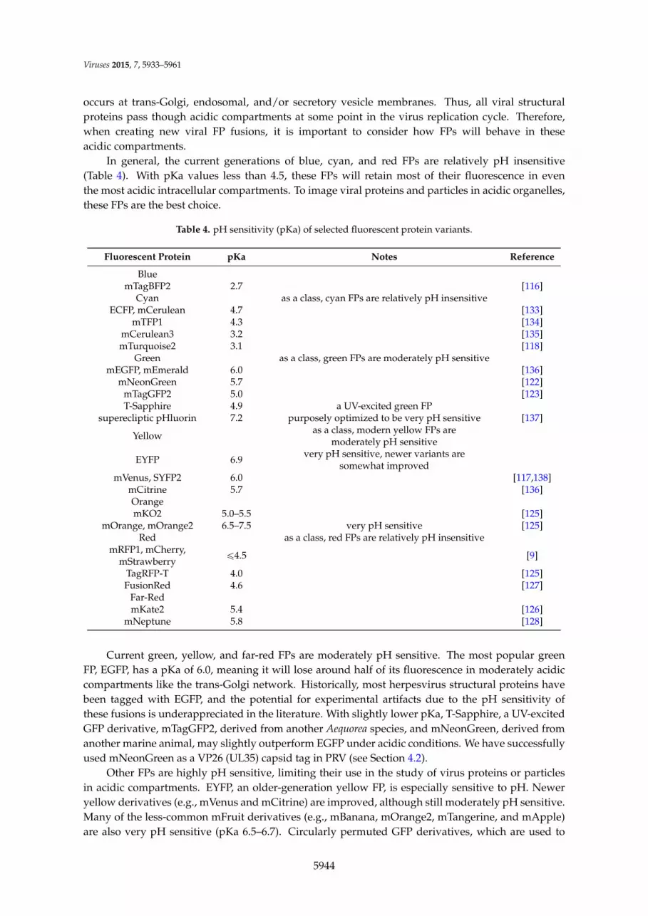
occurs at trans-Golgi, endosomal, and/or secretory vesicle membranes. Thus, all viral structuralproteins pass though acidic compartments at some point in the virus replication cycle. Therefore,when creating new viral FP fusions, it is important to consider how FPs will behave in theseacidic compartments.
In general, the current generations of blue, cyan, and red FPs are relatively pH insensitive(Table 4). With pKa values less than 4.5, these FPs will retain most of their fluorescence in eventhe most acidic intracellular compartments. To image viral proteins and particles in acidic organelles,these FPs are the best choice.
Table 4. pH sensitivity (pKa) of selected fluorescent protein variants.
Fluorescent Protein pKa Notes Reference
BluemTagBFP2 2.7 [116]
Cyan as a class, cyan FPs are relatively pH insensitiveECFP, mCerulean 4.7 [133]
mTFP1 4.3 [134]mCerulean3 3.2 [135]mTurquoise2 3.1 [118]
Green as a class, green FPs are moderately pH sensitivemEGFP, mEmerald 6.0 [136]
mNeonGreen 5.7 [122]mTagGFP2 5.0 [123]T-Sapphire 4.9 a UV-excited green FP
superecliptic pHluorin 7.2 purposely optimized to be very pH sensitive [137]
Yellow as a class, modern yellow FPs aremoderately pH sensitive
EYFP 6.9 very pH sensitive, newer variants aresomewhat improved
mVenus, SYFP2 6.0 [117,138]mCitrine 5.7 [136]OrangemKO2 5.0–5.5 [125]
mOrange, mOrange2 6.5–7.5 very pH sensitive [125]Red as a class, red FPs are relatively pH insensitive
mRFP1, mCherry,mStrawberry ď4.5 [9]
TagRFP-T 4.0 [125]FusionRed 4.6 [127]
Far-RedmKate2 5.4 [126]
mNeptune 5.8 [128]
Current green, yellow, and far-red FPs are moderately pH sensitive. The most popular greenFP, EGFP, has a pKa of 6.0, meaning it will lose around half of its fluorescence in moderately acidiccompartments like the trans-Golgi network. Historically, most herpesvirus structural proteins havebeen tagged with EGFP, and the potential for experimental artifacts due to the pH sensitivity ofthese fusions is underappreciated in the literature. With slightly lower pKa, T-Sapphire, a UV-excitedGFP derivative, mTagGFP2, derived from another Aequorea species, and mNeonGreen, derived fromanother marine animal, may slightly outperform EGFP under acidic conditions. We have successfullyused mNeonGreen as a VP26 (UL35) capsid tag in PRV (see Section 4.2).
Other FPs are highly pH sensitive, limiting their use in the study of virus proteins or particlesin acidic compartments. EYFP, an older-generation yellow FP, is especially sensitive to pH. Neweryellow derivatives (e.g., mVenus and mCitrine) are improved, although still moderately pH sensitive.Many of the less-common mFruit derivatives (e.g., mBanana, mOrange2, mTangerine, and mApple)are also very pH sensitive (pKa 6.5–6.7). Circularly permuted GFP derivatives, which are used to
5944

Viruses 2015, 7, 5933–5961
create FP biosensors, such as the GCaMP Ca2+ sensors, are also especially pH sensitive. The pHsensitivity of such FP biosensors has indeed led to experimental artifacts [139]. However, the pHsensitivity of FPs can also be exploited as an experimental tool. For example, EYFP has been used asa pH sensor to study retrovirus fusion and entry [140], and several GFP variants, named pHluorins,have been purposely optimized as pH sensors. With a pKa of 7.2, superecliptic pHluorin is stronglyquenched in the secretory pathway, but dequenched upon exocytosis, allowing researchers to imageindividual exocytosis events [137,141], including the egress of PRV particles from infected cells [36](Figure 1B).
3.2. Choice of Fluorescent Protein Insertion Site and Linker
Many of the published FP fusions in the alpha herpesviruses were created using laboriousrestriction digestion and ligation-based methods. This approach frequently produces arbitraryjunctions between FP and viral sequences determined by the presence of convenient restrictionsites. As a result, many existing FP fusions contain insertions of restriction sites or plasmid-derivedsequences, and deletions or duplications in flanking viral genomic sequences. Nevertheless,many viral FP fusions created this way worked very well, sometimes better than subsequentrationally-designed fusions. Today, improved methods, such as traceless bacterial artificialchromosome (BAC) recombination [142], and the ability to synthesize whole synthetic genes to order,provide greater flexibility to rationally design FP fusion proteins. Yet, even when detailed FP andviral protein structures are known, we are unable to accurately predict structure and function, socreating a fully functional FP fusion remains largely a process of trial and error. Nonetheless, severalgeneral considerations may improve the probability of creating a functional fusion.
Foremost, previous reports of functional FP fusions can be a valuable guide. Since approximately40 core genes are conserved throughout the herpesviruses, and even more are conserved within thealpha herpesviruses, a similar FP fusion may already exist in a related herpesvirus. Table 1 mayfacilitate this search.
To design a novel fusion, it is worth considering any known structural or functional motifs toavoid interfering with protein function. The UniProt Knowledgebase (www.uniprot.org) is a usefulrepository of this sort of information for both viral and host proteins. If any structure is known,inserting a FP into unstructured termini or loops may reduce the chances of the FP interfering withprotein structure. When no empirical data is available, structure prediction software can estimatesome features with varying degrees of certainty. For example, prediction of transmembrane domainsis relatively robust. Unstructured hinges or loops, possibly providing good FP insertion sites, mayflank such strongly predicted structural motifs. In addition, regions with an abundance of hydrophilicresidues are generally more likely to be exposed to the solvent on the surface of proteins, and thusmay be better insertion sites than buried hydrophobic regions.
Finally, consider whether to insert a linker peptide between the FP and protein of interest. Itis not always necessary to add linkers, as most FPs contain unstructured residues on their N- andC-termini that may provide adequate flexibility and spacing. However, when structural or stericconflicts do occur, adding additional flexible linker residues between the FP and gene of interestmay help. Linkers containing residues that are small (glycine, alanine) or polar (serine, threonine,asparagine) may favor the formation of unstructured flexible loops, and proline residues disfavor theformation or extension of alpha helices.
3.3. Choice of Excitation Light Sources
Excitation sources for fluorescence microscopy have changed dramatically in recent years,transitioning from arc lamps and gas lasers to light emitting diodes (LEDs), laser diodes, and diodepumped solid state (DPSS) lasers. These new sources have major advantages in efficiency, totalpower, and direct modulation; however, they also present unique challenges to integrate their usewith existing FPs.
5945

Viruses 2015, 7, 5933–5961
To excite green FPs, powerful DPSS lasers can replace argon gas lasers as a source for 488 nmlaser illumination; however, 488 nm DPSS lasers remain costly. 473 nm DPSS lasers are an inexpensivealternative, resulting in only slightly less excitation efficiency of EGFP and similar derivatives. Thereare currently few LEDs available in the 490 nm range, and their efficiency is inferior to the much morecommon 470 nm LEDs. As most LEDs have relatively broad emission spectra, 470 nm LEDs typicallywork well with commonly-used multiband excitation filters, and are a good alternative to arc lamps.
The demise of argon gas lasers presents a challenge for those using cyan and yellow FPs. Argongas lasers produce 454.6 nm and 514.5 nm laser lines, which efficiently excite cyan and yellow FPs,respectively. Now, microscopes typically use 405 nm laser diodes and 488 nm DPSS lasers, whichare both suboptimal for these FPs. This is unfortunate because some of the newest FPs in this range,like mTurquoise2 [118], are among the brightest and most monomeric FPs available. However, recentadvances have made laser diodes in the 445 and 515 nm range very cost-effective. Still, these arerarely integrated into commercial microscopes. On custom built systems, these laser lines can beadded relatively easily and inexpensively. Another big advantage of these diodes is that they canbe modulated directly, omitting the need for costly acousto-optical tunable filters (AOTF). Highpower LEDs centered around 450 nm and 505 nm are readily available, and are a good alternativeto arc lamps.
For red FPs, there are currently no widely-available laser diodes in the 560–590 nm range.Instead, 561 nm DPSS lasers are typically used on commercial microscopes. While this wavelengthis optimal for common chemical fluorophores, it is suboptimal for mCherry and other red FPs.However, 589 and 593 nm DPSS lasers do exist, and could be a better option for imaging red FPs.Very few LEDs exist in the 540–580 nm range, and most are phosphor-converted LEDs that producebroad spectra with low peak power. Amber LEDs centered around 590 nm are available; however,they are relatively weak, and older arc lamps generally out-perform LEDs when imaging red FPs.Future developments in diode technology will hopefully close this gap in this spectral region.
4. Case Studies: Discussion of Particular Fluorescent Protein Fusions
4.1. PRV Membrane Protein US9
Our laboratory has long been interested in the axonal transport and trans-neuronal spreadof alpha herpesviruses. While many viruses can invade the nervous system, for most it is anon-productive dead-end pathway. The alpha herpesviruses are among the very few that haveevolved to efficiently enter and productively exit the nervous system of their natural hosts.Accordingly, while many viruses are capable of entering at nerve termini and undergoing retrogradeaxonal transport post-entry, the alpha herpesviruses are among the very few capable of efficientanterograde axonal transport of replicated progeny [143].
The attenuated vaccine strain, PRV Bartha, provided our first toehold to begin to understand themolecular mechanisms of alpha herpesvirus anterograde axonal transport and spread. PRV Barthacontains a large, 3 kb deletion in the unique short (US) genomic region, disrupting viral proteins gI(US7), gE (US8), US9, and US2. Of these disrupted genes, US9 is essential for anterograde axonaltransport of progeny virions in infected neurons [144,145], and similar phenotypes exist in HSV-1.US9 is a small, 98 amino acid, viral membrane protein. The cytosolic N-terminal domain of US9contains many functional motifs, including phosphorylation sites [146], endocytic motifs [147], and itis thought to recruit the kinesin microtubule motor KIF1A (kinesin-3) [37] in concert with gE/gI [95].The transmembrane domain is located near the C-terminus, producing a very small, 3 amino acidextracellular domain with no known functional motifs.
Originally, our laboratory fused EGFP to the short C-terminal domain to avoid interfering withthe functional motifs of the cytosolic domain [148]. This US9-EGFP fusion protein was efficientlyincorporated into virus particles, and its intracellular transport and localization was indistinguishablefrom that of wild-type US9 [147,148]. Together, these observations suggested that the US9-EGFP
5946

Viruses 2015, 7, 5933–5961
fusion was indeed functional. However, an important caveat is that US9-null viruses do not havea detectable phenotype in non-neuronal cells. It was only once we constructed a recombinant virusexpressing this US9-EGFP fusion and assessed its neuronal spread in vitro and in vivo did its mutantphenotype become apparent: the US9-EGFP fusion protein is not functional in anterograde axonaltransport in neurons [149]. Thus, our experience with EGFP-tagged US9 serves as an instructiveexample of the major pitfalls associated with FP fusions: these are ultimately mutant alleles that mayproduce unexpected mutant phenotypes.
More recently, we described an FP fusion to the opposite N-terminal side, EGFP-US9. Virusesexpressing this fusion are capable of anterograde axonal transport and spread in neurons, this fusionallows us to visualize the co-transport of EGFP-US9 and capsids in axons [79], and it has provenespecially useful for immunoaffinity purification and proteomic identification of cellular factorsinvolved in viral transport [37].
4.2. HSV-1 and PRV Capsid Protein VP26
VP26, the small capsid protein of HSV-1 and PRV, was one of the first herpesvirus proteins tobe fused to a FP [41,46]. Since then, these capsid-tagged virus mutants have become a valuabletool to study virus particle transport in living cells. While VP26 is not essential in HSV-1 or PRV,there are differing reports on the effect of FP fusions to VP26. Some studies did not find anyeffect on virus replication kinetics [41,56], another reported moderate effects on cell-to-cell spreadand pathogenesis in vivo [44], and yet another reported severe replication defects and reduction inneuro-invasiveness in vivo, comparable to a VP26-null virus [102]. A recent report has shown thatsome FP-VP26 fusions in HSV-1 severely attenuate virus replication, while others do not [45]. Thereasons for these differences seem to be multifactorial, and here we will dissect what we believe areimportant factors that influence the viability of VP26 fusions.
4.2.1. Compensatory Mutations that May Affect FP-VP26 Expression
It has been shown that the original HSV-1 strain containing an EGFP-VP26 fusion harbors aninadvertent 65 bp deletion upstream from the VP26 start codon [45]. Nagel et al. took the effortto recreate the virus mutant as originally designed [41] using a BAC recombination approach, andsurprisingly found that this mutant was highly attenuated. Because the deleted region containstranscriptional enhancers [45], it is likely that this deletion is a compensatory mutation that causesreduced expression of the fusion protein.
PRV seems to show a similar phenomenon. The original PRV EGFP-VP26 mutant (PRVGS443, [46]) was constructed in Escherichia coli by BAC recombination. Upon reconstitution ofreplicating virus, mixed plaque phenotypes were observed, with an approximately equal numberof large and small plaques. Both phenotypes were stable during virus propagation; however,transfection of purified nucleocapsid DNA from these two populations yielded the surprising resultthat DNA from small plaque clones could again generate small and large plaques, while DNA fromlarge plaque clones (PRV GS443L) only produced large plaques. Subsequent restriction analysis ofnucleocapsid DNA revealed a SalI restriction fragment length polymorphism near the UL35 (VP26)gene [103]. We independently constructed VP26 fusions to mEGFP and mCherry (see Figure 5), andfound similar results, suggesting that some adaptive mutation near the FP-VP26 coding region isneeded to compensate for the defects caused by FP fusion. We sequenced the genome of severalrecombinants expressing FP-VP26 using next-generation sequencing methods, but were unable toclearly identify a putative compensatory mutation [150]. It is likely that the putative compensatorymutation is associated with expansion or contraction of a short sequence repeat region downstreamof the UL35 (VP26) gene. A previous study identified copy number variation between commonlaboratory strains of PRV within this short sequence repeat region, and the repeated sequencecontains a predicted binding site for the host transcriptional repressor CTCF [151]. Reconstitutionof replicating virus by transfection of BAC or nucleocapsid DNA might increase the frequency of
5947

Viruses 2015, 7, 5933–5961
recombination in this repeat region, which may modulate the expression of the FP-VP26 fusion. Itis possible that such variation explains the apparent differences in replication kinetics reported bydifferent laboratories [44,56,102]. Further investigation into these putative compensatory mutations,and, in particular, development of new sequencing methods that can better cope with highlyrepetitive and GC-rich alpha herpesvirus genomes are needed.
4.2.2. Fluorescent Protein Dimerization
Nagel et al. also found another piece of the puzzle. While the EGFP-VP26 HSV-1 mutant lackingthe 65 bp upstream deletion was highly attenuated, another mutant expressing ECFP on the samegenetic background only showed slight attenuation, even though ECFP and EGFP differ by only afew amino acids. Why did the ECFP recombinant grow while the EGFP recombinant could not? Thesolution seems to be that ECFP has a 30-fold higher dissociation constant (Kd ~3 mM) than EGFP(Kd ~0.1 mM), meaning that ECFP only dimerizes at a 30-fold higher concentration. This differencein dimerization affinity is a result of the N146I mutation present in ECFP, and newer cyan variantslike mTurquoise2 contain a similar N146F mutation. Consistent with this observation, another HSV-1recombinant expressing VP26 fused to mVenus also replicated well despite having a wild-type 65 bpupstream promoter region [45,82]. mVenus, like most newer Aequorea derivatives, contains the A206Kmutation that further increases the dissociation constant to 74 mM [6]. Together, both of these factors,compensatory mutations reducing FP-VP26 expression and FP variants with reduced dimerizationaffinity, appear to improve replication of virus recombinants expressing FP-VP26 fusions.
4.2.3. Assemblons
Both of these factors also affect intranuclear aggregation of capsid proteins. Nuclear aggregatesformed by capsid proteins were found in cells infected with wild-type alpha herpesviruses, evenbefore the first VP26 fusion was created. At that time it was believed that these were sites of capsidassembly, and these structures were dubbed “assemblons” [152]. However, Nagel et al. showed that“assemblons” are not sites of capsid assembly, but rather dead-end aggregations of capsid proteins,an idea also supported by earlier reports [153,154]. The early EGFP fusions to VP26 in HSV-1 showan especially strong disposition to form large “assemblons” earlier in infection [41,155], and thesize and number of these aggregates correlates with FP dimerization affinity, with EGFP fusionsmaking more aggregates than ECFP, mVenus, or mRFP1 fusions [45]. It therefore seems that FP-VP26expression level and FP dimerization propensity are two sides of the same coin: A more stronglydimerizing FP forms aggregates even at lower expression levels, while a more monomeric FP formsfewer aggregates at higher expression levels. In mouse cytomegalovirus, an inducibly and ectopicallyexpressed EGFP fusion to SCP (small capsid protein, the beta herpesvirus ortholog of VP26) acts asa very potent dominant negative [156], suggesting that the EGFP-SCP fusion can bind and sequesterwild-type capsid proteins, inhibiting capsid assembly. It is possible that FP-VP26 fusions in thealpha herpesviruses also act as dominant negatives in a less strict sense, by sequestering capsidproteins into nuclear aggregates. This idea was suggested by Nagel et al., and is supported by theiridentification of five different capsid proteins in the intranuclear aggregates. This would explainwhy EGFP-VP26 fusions, which form severe intranuclear aggregates also assemble relatively fewindividual intranuclear capsids [45] (Figure 5J–K).
The question remains, why are VP26 fusions so prone to induce protein aggregations? Capsidformation can be described as an irreversible phase separation. Typically, biological systems takeadvantage of phase separations by balancing at the “tipping point”, allowing rapid responses to smallconcentration changes. Capsid formation might be similar such that capsids rapidly assemble as soonas a critical capsid protein concentration is reached. Adding a dimerizing FP to capsid proteins mighttip the balance, resulting in premature aggregation of capsid proteins.
5948

Viruses 2015, 7, 5933–5961
4.2.4. Description and Characterization of New VP26 Fusions in PRV
We generated a palette of different FP-VP26 fusions in PRV to identify those most suitable forlive cell imaging (Figure 5A). FP coding sequences were inserted by homologous recombinationbetween codons 2–3 of the PRV UL35 (VP26) gene, as previously described [64]. PRV GS443L ([46],and see above), PRV 959 [157], PRV 180 [64], PRV 543 [158], and PRV 950 [36] were previouslydescribed. Newer FP variants were chosen based on the criteria discussed in Section 3 (e.g., mEGFP,mNeonGreen, mCherry, and mTurquoise2), and were compared to older variants (e.g., EGFP, mRFP1,and ECFP). We found that all VP26 fusions caused an ~10-fold defect in single-step virus replication(Figure 5B), and slightly reduced plaque sizes (Figure 5C). Using identical imaging parameters(within each spectral class), we found that newer FP variants are generally brighter than theirpredecessors (e.g., PRV 959 is brighter than PRV GS443L, and PRV 950 is brighter than PRV 543;however, PRV 180 is similar to PRV 960) (Figure 5C).
Journal(2015,"volume,"page–page"
17!
as" soon" as" a" critical" capsid" protein" concentration" is" reached." Adding" a" dimerizing" FP" to" capsid"proteins"might"tip"the"balance,"resulting"in"premature"aggregation"of"capsid"proteins."
4.2.4."Description"and"Characterization"of"New"VP26"Fusions"in"PRV"
We"generated"a"palette"of"different"FPKVP26"fusions"in"PRV"to"identify"those"most"suitable"for"live" cell" imaging" (Figure" 5A)." FP" coding" sequences"were" inserted" by" homologous" recombination"between"codons"2–3"of"the"PRV"UL35"(VP26)"gene,"as"previously"described"[64]."PRV"GS443L"([46],"and" see" above)," PRV" 959" [157]," PRV" 180" [64]," PRV" 543" [158]," and" PRV" 950" [36]"were" previously"described."Newer"FP"variants"were"chosen"based"on"the"criteria"discussed"in"Section"3"(e.g.,"mEGFP,"mNeonGreen," mCherry," and" mTurquoise2)," and" were" compared" to" older" variants" (e.g.," EGFP,"mRFP1,"and"ECFP)."We"found"that"all"VP26"fusions"caused"an"~10Kfold"defect" in"singleKstep"virus"replication" (Figure" 5B)," and" slightly" reduced" plaque" sizes" (Figure" 5C)." Using" identical" imaging"parameters"(within"each"spectral"class),"we"found"that"newer"FP"variants"are"generally"brighter"than"their"predecessors"(e.g.,"PRV"959"is"brighter"than"PRV"GS443L,"and"PRV"950"is"brighter"than"PRV"543;"however,"PRV"180"is"similar"to"PRV"960)"(Figure"5C)."
"Figure+5."Fluorescent"protein"fusions"to"VP26,"the"small"capsid"protein,"in"pseudorabies"virus"(PRV)."(A)" Fluorescent" protein" coding" sequences" were" inserted" by" homologous" recombination" between"codons" 2–3" of" the" PRV"UL35" (VP26)" gene." (B)" SingleKstep" virus" replication." Parallel" cell" cultures"were" infected" in" triplicate" with" the" indicated" viruses," cells" and" supernatants" were" harvested" at"indicated"times,"and"infectious"virus"titer"was"measured"by"plaque"assay."Recombinants"have"~1"log"defect" in"endKpoint"virus"titer"relative"to"parental"PRV"Becker." (C)"Comparison"of"plaque"size"and"fluorescence" intensity."PK15"cells"were" infected"with" the" indicated"viruses,"and"one"representative"plaque" is"shown."Note" that"plaque"sizes"of"recombinants"are"slightly"smaller" than"that"of"parental"PRV"Becker."Newer"FP"variants"are"generally"brighter"than"their"predecessors:"PRV"959"is"brighter"than"PRV"GS443L,"and"PRV"950"is"brighter"than"PRV"543,"but"PRV"180"is"similar"to"PRV"960."(D–K)"Capsid"assembly"and"nuclear" aggregate" formation."PtK2" cells" (ATCC"CCLK56)"were" infected"with"the" indicated" viruses," fixed" at" 6" h" postKinfection," and" imaged" by" confocal" microscopy." (D–I)"Maximum" intensity" projection" of" representative" nuclei" containing" individual" fluorescent" PRV"capsids" (smallest"puncta)"and" large"capsid"protein"aggregates" (i.e.,"“assemblons”)."Scale"bars"are"5"nm."(J–K)"Quantification"of"intranuclear"capsids"and"aggregates."More"than"100"cells"infected"with"each"indicated"virus"were"manually"scored."(J)"Intranuclear"aggregates"were"scored"as"follows:"+++,"very"large"aggregates,"as"in"panel"D;"++,"midKsized"aggregates,"as"in"panels"EKH;"+,"small"aggregates,"as" in" panel" I;" –," no" aggregates." (K)" Intranuclear" capsid" concentration"was" scored" as" follows:" +++,"
Figure 5. Fluorescent protein fusions to VP26, the small capsid protein, in pseudorabies virus (PRV).(A) Fluorescent protein coding sequences were inserted by homologous recombination betweencodons 2–3 of the PRV UL35 (VP26) gene. (B) Single-step virus replication. Parallel cell cultures wereinfected in triplicate with the indicated viruses, cells and supernatants were harvested at indicatedtimes, and infectious virus titer was measured by plaque assay. Recombinants have ~1 log defect inend-point virus titer relative to parental PRV Becker. (C) Comparison of plaque size and fluorescenceintensity. PK15 cells were infected with the indicated viruses, and one representative plaque is shown.Note that plaque sizes of recombinants are slightly smaller than that of parental PRV Becker. NewerFP variants are generally brighter than their predecessors: PRV 959 is brighter than PRV GS443L,and PRV 950 is brighter than PRV 543, but PRV 180 is similar to PRV 960. (D–K) Capsid assemblyand nuclear aggregate formation. PtK2 cells (ATCC CCL-56) were infected with the indicatedviruses, fixed at 6 h post-infection, and imaged by confocal microscopy. (D–I) Maximum intensityprojection of representative nuclei containing individual fluorescent PRV capsids (smallest puncta)and large capsid protein aggregates (i.e., “assemblons”). Scale bars are 5 µm. (J–K) Quantificationof intranuclear capsids and aggregates. More than 100 cells infected with each indicated virus weremanually scored. (J) Intranuclear aggregates were scored as follows: +++, very large aggregates, as inpanel D; ++, mid-sized aggregates, as in panels E-H; +, small aggregates, as in panel I; –, no aggregates.(K) Intranuclear capsid concentration was scored as follows: +++, dense, as in panels G-I; ++, manyindividual capsids, as in panel F; +, few individual capsids, as in panels D–E.
We next imaged cells by spinning disk confocal microscopy at 6 h post-infection to determinethe extent of capsid assembly and aggregate formation. As described above, and in agreement withHSV-1 VP26 fusions [45], monomeric FPs produce generally less aggregation and more individual
5949

Viruses 2015, 7, 5933–5961
intranuclear capsids (Figure 5D–K). By these criteria, the mTurquoise2 fusion appears to perform thebest, and mRFP1 and mCherry fusions perform similarly well (Figure 5G–K). mEGFP, containing themonomerizing A206K mutation, appears to be a modest improvement over EGFP (Figure 5 D–E,J–K);however, mNeonGreen, which not derived from Aequorea, appears to be the best green capsid tag todate (Figure 5F,J–K). In particular, the combination of brightness, photostability, efficient excitationby powerful and inexpensive 470 nm LEDs makes the mNeonGreen-VP26 fusion especially suitedfor tracking capsid transport in neurons [157].
5. Comparison to Immunofluorescence
First described in 1942, the method of detecting and localizing antigens in cells ortissues using fluorescent antibodies has proven invaluable in molecular and cell biology [159].Immunofluorescence was first used to visualize VZV infected tissues and cells in 1954 [160], and HSVin 1956 [161]. However, to visualize intracellular antigens, immunofluorescence protocols requiredestructive fixation and permeabilization steps, typically accomplished by aldehyde crosslinkingof proteins, protein precipitation and lipid extraction by organic solvents, and/or lipid extractionusing detergents. Variability in fixation and permeabilization can easily introduce artifacts, rangingfrom over-extraction leading to loss or relocalization of proteins of interest, to under-permeabilizationleading to poor antibody accessibility [162].
Due to their complex architecture, including lipid envelope and thick tegument layer,immunofluorescence artifacts may be particularly problematic when visualizing assembled alphaherpesvirus particles. In one study, HSV-1 particles incorporating an mRFP1-VP26 capsid tagwere adhered to a coverslip, fixed, permeabilized, and immunostained to detect the major capsidprotein VP5. Naked capsids purified from the nuclei of infected cells were readily stainedby anti-VP5 antibodies; however, fewer than half of assembled virions released from infectedcells and immunostained in situ were detected by immunofluorescence [163]. Importantly,immunofluorescence detection was restored when assembled virions were purified or concentratedby ultracentrifugation [163,164]. Others have observed a time-dependent structural reorganizationthat alters the sensitivity of the virion tegument to detergent extraction [165]. In light of theseobservations, it seems likely that age and mechanical forces on virus particles can affect theirpermeability during immunostaining procedures. This inefficiency of immunofluorescence is likelycompounded inside infected cells, where additional membrane layers and cellular proteins mayimpede permeability. Indeed, inefficient immunodetection of HSV-1 virions inside infected cells hasbeen reported [166].
There is currently a debate in the alpha herpesvirus literature whether nascent virus particlesare fully assembled prior to long distance anterograde transport in axons (the “married model”), or ifcapsids and membranes are transported separately before envelopment at distal sites (the “separatemodel”). The details of this debate are discussed elsewhere [167–169], but if immunostaining offully-assembled virus particles inside axons is inefficient, this could lead to an underestimation offully-assembled virions transporting in axons.
It is clear that immunofluorescence and FP-based approaches have different strengths andweaknesses. Immunofluorescence approaches suffer from variability in immunostaining, but havethe advantage of detecting native protein at normal expression levels. Plus there already exists awealth of well-characterized antibodies targeting viral and cellular proteins. However, while FPfusions may produce unexpected mutant phenotypes, they do enable a dynamic view in live cells,and enable a wide range of specialized techniques to better dissect complex multistep viral processes.
5950

Viruses 2015, 7, 5933–5961
6. Beyond Traditional Fluorescence Microscopy
6.1. Fluorescent Protein Fusions as Proteomics Probes
Most viral and cellular biological processes are mediated by protein-protein interactions andprotein complexes. Immunoaffinity purification coupled to mass spectrometry provides a powerfulapproach to isolate and characterize protein complexes and identify protein interaction networks.Although immunoaffinity purification can be performed using antibodies raised against viralproteins themselves, the alpha herpesviruses express a multitude of viral proteins, and antibodiesproduced using standard methods may not be of sufficient specificity and affinity for effectiveimmunoisolation. In addition, antibodies against specific proteins may interfere with protein-proteininteractions of interest. Alternatively, genetically fusing epitope tags to viral proteins canfacilitate immunoaffinity purification of a variety of viral proteins using a single well-characterizedhigh-affinity antibody. Peptide epitope tags are highly effective [170]; however, FPs have the distinctadvantage of serving as both an epitope tag for immunoaffinity purification and a fluorescentprobe for live-cell fluorescence microscopy. Combining these methods allows the identification ofprotein-protein interactions and provides a richer understanding of the spatiotemporal dynamicsof these protein-protein interactions [171,172]. As described above, this approach allowed ourlaboratory to identify interaction between PRV US9 and the cellular kinesin-3 motor KIF1A, andconnect this motor recruitment event to the dynamic axonal transport of virus particles [37].
6.2. Flow Virometry
Cell sorting was established over 50 years ago [173], modified to allow for fluorescence–assistedsorting (FACS) [174], and is now used in a wide array of biomedical research. Over the years, sizerestrictions were improved to reliably sort bacterial cells [175] and subcellular eukaryotic organelles(fluorescence-assisted organelle sorting, FAOS), including mitochondria, lysosomes, endosomes, andexosomes [176–179]. Importantly, FAOS operates in the size range of larger virions (>100 nm) andcan be adapted for virus research. Pioneering work in the field of “flow virometry” has greatlyimproved the throughput and sensitivity of large-scale virus detection and discovery. Modern FACSequipment, improved sheath water filtration, and more sensitive optics now allow the detection ofviral particles less than 60 nm [180]. Detection of individual virus particles by FACS can principallybe done through two methods: One method uses the light scattering properties of the particle. Earlyreports indicated that viruses with distinct morphologies (e.g., T2 phage) can be identified solelybased on their light scattering [181]. The other method uses fluorescent dyes, or a combinationof light scattering and fluorescent dyes. Fluorescent labeling has been used to reliably detect andquantify viruses like HSV, CMV, adenovirus, influenza A, RSV, rubella, coronavirus, dengue, andparainfluenza [182,183]. In many cases, non-specific nucleic acid and protein-binding fluorescentdyes are used to label virus particles, but some reports also describe the use of specific antibodiesto detect HIV-1, poliovirus, dengue virus [184–186]. These fluorescent labeling methods may affectparticle infectivity, which is of minor concern if virometry is used only to detect and measurevirus particles. However, if virus particles need to be sorted retaining infectivity, for example, tomeasure infectivity of different subpopulations, FP fusions might be a more suitable choice. As aproof-of-principle, the Lippé laboratory analyzed HSV-1 capsids containing EGFP-VP26 [187], andmore recently sorted infectious HSV-1 virions containing EGFP-tagged tegument proteins [188]. Justas FACS analysis provides a population distribution of cellular properties rather than a bulk average,“flow virometry” will allow the alpha herpesvirus field to better understand the variability withinsubpopulations of virus particles.
6.3. Herpes Past the Diffraction Barrier
For over a century, light microscopy resolution was limited by the Abbe diffraction barrier [189],which means in practice that structures closer than ~200 nm cannot be resolved. However, new
5951

Viruses 2015, 7, 5933–5961
super-resolution fluorescence microscopy methods have been developed, pushing these limits tounder 20 nm. This allows more precise localization and discrimination of delicate cellular structures,providing greater insights into biological processes (reviewed in [190]). Virologists have nowbegun to exploit the potential of super-resolution microscopy, the progress of which is reviewedin detail elsewhere [191–193]. While some popular super-resolution techniques like stochasticoptical reconstruction microscopy (STORM) and stimulated emission depletion (STED) rely onchemical fluorophores, other methods, like photoactivated localization microscopy (PALM) andstructured illumination microscopy (SIM) are compatible with FPs. PALM does not require chemicalfixation, permeabilization, and immunostaining, so samples can be analyzed without many of thecaveats described above (see Section 5). This method might, for example, be useful to understandthe dense network of proteins in the herpesvirus tegument, or virus-related cellular structureslike “assemblons”. The first steps have been taken in this direction, measuring the asymmetricdistribution of viral structural proteins in PRV particles using a related method [50], and radialdistribution of tegument proteins in HSV-1 particles using STORM [194].
Most super-resolution techniques, however, are limited by very slow acquisition rates. SIMis presently the only method that can deliver super-resolution data at high enough frame rates tofollow virus dynamics in infected cells. However, in our experience, most FP-tagged virus mutantswere incompatible with this method due to rapid photobleaching [195]. Further developments areneeded to optimize FP brightness and photostability, and optimize the speed and sensitivity ofsuper-resolution techniques to allow a super-resolution view of the dynamic virus replication cycle.
Acknowledgments: We thank Orkide O. Koyuncu, Matthew P. Taylor, and members of the Enquist lab forcritically reading the manuscript. We acknowledge support from National Institutes of Health grants R01NS033506, R01 NS060699, and P40 RR018604 (L.W.E.) and postdoctoral fellowship F32 GM112337 (J.S.). I.B.H. issupported by postdoctoral fellowship PF-13-050-01- MPC from the American Cancer Society. J.B.B. is supportedby DFG fellowship BO 4158/1-1.
Conflicts of Interest: The authors declare no conflict of interest. The funding sponsors had no role in the designof the study, in the collection, analyses, or interpretation of data, in the writing of the manuscript, or in thedecision to publish the results.
References
1. Shimomura, O. The discovery of aequorin and green fluorescent protein. J. Microsc. 2005, 217, 3–15.[CrossRef] [PubMed]
2. Prasher, D.C.; Eckenrode, V.K.; Ward, W.W.; Prendergast, F.G.; Cormier, M.J. Primary structure of theAequorea victoria green-fluorescent protein. Gene 1992, 111, 229–233. [CrossRef]
3. Chalfie, M.; Tu, Y.; Euskirchen, G.; Ward, W.W.; Prasher, D.C. Green fluorescent protein as a marker for geneexpression. Science 1994, 263, 802–805. [CrossRef] [PubMed]
4. Inouye, S.; Tsuji, F.I. Aequorea green fluorescent protein. Expression of the gene and fluorescencecharacteristics of the recombinant protein. FEBS Lett. 1994, 341, 277–280. [CrossRef]
5. Cubitt, A.B.; Heim, R.; Adams, S.R.; Boyd, A.E.; Gross, L.A.; Tsien, R.Y. Understanding, improving andusing green fluorescent proteins. Trends Biochem. Sci. 1995, 20, 448–455. [CrossRef]
6. Zacharias, D.A.; Violin, J.D.; Newton, A.C.; Tsien, R.Y. Partitioning of Lipid-Modified Monomeric GFPs intoMembrane Microdomains of Live Cells. Sci. Signal. 2002, 296, 913. [CrossRef] [PubMed]
7. Matz, M.V.; Fradkov, A.F.; Labas, Y.A.; Savitsky, A.P.; Zaraisky, A.G.; Markelov, M.L.; Lukyanov, S.A.Fluorescent proteins from nonbioluminescent Anthozoa species. Nat. Biotechnol. 1999, 17, 969–973.[CrossRef] [PubMed]
8. Campbell, R.E.; Tour, O.; Palmer, A.E.; Steinbach, P.A.; Baird, G.S.; Zacharias, D.A.; Tsien, R.Y. A monomericred fluorescent protein. Proc. Natl. Acad. Sci. USA 2002, 99, 7877–7882. [CrossRef] [PubMed]
9. Shaner, N.C.; Campbell, R.E.; Steinbach, P.A.; Giepmans, B.N.G.; Palmer, A.E.; Tsien, R.Y. Improvedmonomeric red, orange and yellow fluorescent proteins derived from Discosoma sp. red fluorescentprotein. Nat. Biotechnol. 2004, 22, 1567–1572. [CrossRef] [PubMed]
10. Patterson, G.H. Photoactivation and imaging of optical highlighter fluorescent proteins. In Current ProtocolsCytometry; John Wiley & Sons, Inc.: Hoboken, NJ, USA, 2011; Chapter 12, Unit 12.23. [CrossRef]
5952

Viruses 2015, 7, 5933–5961
11. Nienhaus, K.; Ulrich Nienhaus, G. Fluorescent proteins for live-cell imaging with super-resolution. Chem.Soc. Rev. 2013, 43, 1086–1106. [CrossRef] [PubMed]
12. Tantama, M.; Hung, Y.P.; Yellen, G. Optogenetic reporters: Fluorescent protein-based genetically encodedindicators of signaling and metabolism in the brain. Prog. Brain Res. 2012, 196, 235–263. [PubMed]
13. Bizzarri, R.; Serresi, M.; Luin, S.; Beltram, F. Green fluorescent protein based pH indicators for in vivo use:a review. Anal. Bioanal Chem. 2008, 393, 1107–1122. [CrossRef] [PubMed]
14. Adam, V.; Berardozzi, R.; Byrdin, M.; Bourgeois, D. Phototransformable fluorescent proteins: Futurechallenges. Curr. Opin. Chem. Biol. 2014, 20, 92–102. [CrossRef] [PubMed]
15. Adam, V. Phototransformable fluorescent proteins: Which one for which application? Histochem. Cell Biol.2014, 142, 19–41. [CrossRef] [PubMed]
16. Jöns, A.; Mettenleiter, T.C. Green fluorescent protein expressed by recombinant pseudorabies virus as anin vivo marker for viral replication. J. Virol. Methods 1997, 66, 283–292. [CrossRef]
17. Foster, T.P.; Rybachuk, G.V.; Kousoulas, K.G. Expression of the enhanced green fluorescent protein byherpes simplex virus type 1 (HSV-1) as an in vitro or in vivo marker for virus entry and replication. J. Virol.Methods 1998, 75, 151–160. [CrossRef]
18. Zerboni, L.; Sommer, M.; Ware, C.F.; Arvin, A.M. Varicella-Zoster Virus Infection of a Human CD4-PositiveT-Cell Line. Virology 2000, 270, 278–285. [CrossRef] [PubMed]
19. Mahalingam, R.; Wellish, M.; White, T.; Soike, K.; Cohrs, R.; Kleinschmidt-Demasters, B.K.; Gilden, D.H.Infectious simian varicella virus expressing the green fluorescent protein. J. Neurovirol. 1998, 4, 438–444.[CrossRef] [PubMed]
20. Hogue, I.B.; Enquist, L.W.; Princeton University, Princeton, NJ, USA. Unpublished work. 2011.21. Klingbeil, K.; Lange, E.; Teifke, J.P.; Mettenleiter, T.C.; Fuchs, W. Immunization of pigs with an attenuated
pseudorabies virus recombinant expressing the hemagglutinin of pandemic swine origin H1N1 influenzaA virus. J. Gen. Virol. 2014, 95 (Pt 4), 948–959. [CrossRef] [PubMed]
22. Card, J.P.; Enquist, L.W. Use and Visualization of Neuroanatomical Viral Transneuronal Tracers. InVisualization Techniques; Humana Press: New York, NY, USA, 2012; Volume 70, pp. 225–268.
23. Wojaczynski, G.J.; Engel, E.A.; Steren, K.E.; Enquist, L.W.; Patrick Card, J. The neuroinvasive profiles ofH129 (herpes simplex virus type 1) recombinants with putative anterograde-only transneuronal spreadproperties. Brain Struct. Funct. 2014, 220, 1395–1420. [CrossRef] [PubMed]
24. Wu, B.W.; Engel, E.A.; Enquist, L.W. Characterization of a replication-incompetent pseudorabies virusmutant lacking the sole immediate early gene IE180. MBio 2014, 5, e01850. [CrossRef] [PubMed]
25. DeFalco, J.; Tomishima, M.; Liu, H.; Zhao, C.; Cai, X.; Marth, J.D.; Enquist, L.; Friedman, J.M. Virus-assistedmapping of neural inputs to a feeding center in the hypothalamus. Science 2001, 291, 2608–2613. [CrossRef][PubMed]
26. Lo, L.; Anderson, D.J. A Cre-dependent, anterograde transsynaptic viral tracer for mapping outputpathways of genetically marked neurons. Neuron 2011, 72, 938–950. [CrossRef] [PubMed]
27. Livet, J.; Weissman, T.A.; Kang, H.; Draft, R.W.; Lu, J.; Bennis, R.A.; Sanes, J.R.; Lichtman, J.W. Transgenicstrategies for combinatorial expression of fluorescent proteins in the nervous system. Nature 2007, 450,56–62. [CrossRef] [PubMed]
28. Card, J.P.; Kobiler, O.; Ludmir, E.B.; Desai, V.; Sved, A.F.; Enquist, L.W. A dual infection pseudorabies virusconditional reporter approach to identify projections to collateralized neurons in complex neural circuits.PLoS ONE 2011, 6, e21141. [CrossRef] [PubMed]
29. Card, J.P.; Kobiler, O.; McCambridge, J.; Ebdlahad, S.; Shan, Z.; Raizada, M.K.; Sved, A.F.; Enquist, L.W.Microdissection of neural networks by conditional reporter expression from a Brainbow herpesvirus. Proc.Natl. Acad. Sci. USA 2011, 108, 3377–3382. [CrossRef] [PubMed]
30. Sun, X.R.; Badura, A.; Pacheco, D.A.; Lynch, L.A.; Schneider, E.R.; Taylor, M.P.; Hogue, I.B.; Enquist, L.W.;Murthy, M.; Wang, S.S.-H. Fast GCaMPs for improved tracking of neuronal activity. Nat. Commun. 2013, 4,2170. [CrossRef] [PubMed]
31. Granstedt, A.E.; Bosse, J.B.; Thiberge, S.Y.; Enquist, L.W. In vivo imaging of alphaherpesvirus infectionreveals synchronized activity dependent on axonal sorting of viral proteins. Proc. Natl. Acad. Sci. USA2013, 110, E3516–E3525. [CrossRef] [PubMed]
5953

Viruses 2015, 7, 5933–5961
32. Thompson, R.L.; Shieh, M.T.; Sawtell, N.M. Analysis of herpes simplex virus ICP0 promoter function insensory neurons during acute infection, establishment of latency, and reactivation in vivo. J. Virol. 2003, 77,12319–12330. [CrossRef] [PubMed]
33. Morimoto, T.; Arii, J.; Akashi, H.; Kawaguchi, Y. Identification of multiple sites suitable for insertion offoreign genes in herpes simplex virus genomes. Microbiol. Immunol. 2009, 53, 155–161. [CrossRef] [PubMed]
34. Balliet, J.W.; Kushnir, A.S.; Schaffer, P.A. Construction and characterization of a herpes simplex virus typeI recombinant expressing green fluorescent protein: Acute phase replication and reactivation in mice.Virology 2007, 361, 372–383. [CrossRef] [PubMed]
35. Taylor, M.P.; Kobiler, O.; Enquist, L.W. Alphaherpesvirus axon-to-cell spread involves limited viriontransmission. Proc. Natl. Acad. Sci. USA 2012, 109, 17046–17051. [CrossRef] [PubMed]
36. Hogue, I.B.; Bosse, J.B.; Hu, J.-R.; Thiberge, S.Y.; Enquist, L.W. Cellular mechanisms of alpha herpesvirusegress: live cell fluorescence microscopy of pseudorabies virus exocytosis. PLoS Pathog. 2014, 10, e1004535.[CrossRef] [PubMed]
37. Kramer, T.; Greco, T.M.; Taylor, M.P.; Ambrosini, A.E.; Cristea, I.M.; Enquist, L.W. Kinesin-3 mediatesaxonal sorting and directional transport of alphaherpesvirus particles in neurons. Cell Host Microbe 2012,12, 806–814. [CrossRef] [PubMed]
38. Granstedt, A.E.; Enquist, L.W.; Princeton University, Princeton, NJ, USA. Unpublished work. 2012.39. Smith, G.A. Herpesvirus Transport to the Nervous System and Back Again. Annu. Rev. Microbiol. 2012, 66,
153–176. [CrossRef] [PubMed]40. Elliott, G.; O'Hare, P. Intercellular trafficking and protein delivery by a herpesvirus structural protein. Cell
1997, 88, 223–233. [CrossRef]41. Desai, P.; Person, S. Incorporation of the green fluorescent protein into the herpes simplex virus type 1
capsid. J. Virol. 1998, 72, 7563–7568. [PubMed]42. Toropova, K.; Huffman, J.B.; Homa, F.L.; Conway, J.F. The herpes simplex virus 1 UL17 protein is the second
constituent of the capsid vertex-specific component required for DNA packaging and retention. J. Virol.2011, 85, 7513–7522. [CrossRef] [PubMed]
43. Cockrell, S.K.; Sanchez, M.E.; Erazo, A.; Homa, F.L. Role of the UL25 protein in herpes simplex virus DNAencapsidation. J. Virol. 2009, 83, 47–57. [CrossRef] [PubMed]
44. Bohannon, K.P.; Sollars, P.J.; Pickard, G.E.; Smith, G.A. Fusion of a fluorescent protein to the pUL25 minorcapsid protein of pseudorabies virus allows live-cell capsid imaging with negligible impact on infection.J. Gen. Virol. 2011, 93, 124–129. [CrossRef] [PubMed]
45. Nagel, C.-H.; Döhner, K.; Binz, A.; Bauerfeind, R.; Sodeik, B. Improper Tagging of the Non-Essential SmallCapsid Protein VP26 Impairs Nuclear Capsid Egress of Herpes Simplex Virus. PLoS ONE 2012, 7, e44177.[CrossRef] [PubMed]
46. Smith, G.A.; Gross, S.P.; Enquist, L.W. Herpesviruses use bidirectional fast-axonal transport to spread insensory neurons. Proc. Natl. Acad. Sci. USA 2001, 98, 3466–3470. [CrossRef] [PubMed]
47. Hogue, I.B.; Wilson, J.J.; Enquist, L.W.; Princeton University, Princeton, NJ, USA. Unpublished work. 2015.48. Coller, K.E.; Smith, G.A. Two viral kinases are required for sustained long distance axon transport of a
neuroinvasive herpesvirus. Traffic 2008, 9, 1458–1470. [CrossRef] [PubMed]49. Loomis, J.S.; Courtney, R.J.; Wills, J.W. Binding partners for the UL11 tegument protein of herpes simplex
virus type 1. J. Virol. 2003, 77, 11417–11424. [CrossRef] [PubMed]50. Bohannon, K.P.; Jun, Y.; Gross, S.P.; Smith, G.A. Differential protein partitioning within the herpesvirus
tegument and envelope underlies a complex and variable virion architecture. Proc. Natl. Acad. Sci. USA2013, 110, E1613–E1620. [CrossRef] [PubMed]
51. Harper, A.L.; Meckes, D.G.; Marsh, J.A.; Ward, M.D.; Yeh, P.-C.; Baird, N.L.; Wilson, C.B.; Semmes, O.J.;Wills, J.W. Interaction domains of the UL16 and UL21 tegument proteins of herpes simplex virus. J. Virol.2010, 84, 2963–2971. [CrossRef] [PubMed]
52. Le Sage, V.; Jung, M.; Alter, J.D.; Wills, E.G.; Johnston, S.M.; Kawaguchi, Y.; Baines, J.D.; Banfield, B.W.The herpes simplex virus 2 UL21 protein is essential for virus propagation. J. Virol. 2013, 87, 5904–5915.[CrossRef] [PubMed]
53. Lorenz, M.; Vollmer, B.; Unsay, J.D.; Klupp, B.G.; García-Sáez, A.J.; Mettenleiter, T.C.; Antonin, W. A singleherpesvirus protein can mediate vesicle formation in the nuclear envelope. J. Biol. Chem. 2015, 290,6962–6974. [CrossRef] [PubMed]
5954

Viruses 2015, 7, 5933–5961
54. Guan, Y.; Guo, L.; Yang, E.; Liao, Y.; Liu, L.; Che, Y.; Zhang, Y.; Wang, L.; Wang, J.; Li, Q. HSV-1 nucleocapsidegress mediated by UL31 in association with UL34 is impeded by cellular transmembrane protein 140.Virology 2014, 464–465, 1–10. [CrossRef] [PubMed]
55. Leelawong, M.; Guo, D.; Smith, G.A. A physical link between the pseudorabies virus capsid and the nuclearegress complex. J. Virol. 2011, 85, 11675–11684. [CrossRef] [PubMed]
56. Luxton, G.W.G.; Haverlock, S.; Coller, K.E.; Antinone, S.E.; Pincetic, A.; Smith, G.A. Targeting ofherpesvirus capsid transport in axons is coupled to association with specific sets of tegument proteins.Proc. Natl. Acad. Sci. USA 2005, 102, 5832–5837. [CrossRef] [PubMed]
57. Antinone, S.E.; Smith, G.A. Retrograde axon transport of herpes simplex virus and pseudorabies virus: Alive-cell comparative analysis. J. Virol. 2010, 84, 1504–1512. [CrossRef] [PubMed]
58. Saksena, M.M.; Wakisaka, H.; Tijono, B.; Boadle, R.A.; Rixon, F.; Takahashi, H.; Cunningham, A.L. Herpessimplex virus type 1 accumulation, envelopment, and exit in growth cones and varicosities in mid-distalregions of axons. J. Virol. 2006, 80, 3592–3606. [CrossRef] [PubMed]
59. Zenner, H.L.; Mauricio, R.; Banting, G.; Crump, C.M. Herpes simplex virus 1 counteracts tetherin restrictionvia its virion host shutoff activity. J. Virol. 2013, 87, 13115–13123. [CrossRef] [PubMed]
60. Willard, M. Rapid directional translocations in virus replication. J. Virol. 2002, 76, 5220–5232. [CrossRef][PubMed]
61. Donnelly, M.; Elliott, G. Fluorescent tagging of herpes simplex virus tegument protein VP13/14 in virusinfection. J. Virol. 2001, 75, 2575–2583. [CrossRef] [PubMed]
62. La Boissière, S.; Izeta, A.; Malcomber, S.; O'Hare, P. Compartmentalization of VP16 in cells infected withrecombinant herpes simplex virus expressing VP16-green fluorescent protein fusion proteins. J. Virol. 2004,78, 8002–8014. [CrossRef] [PubMed]
63. Xue, X.; Huang, J.; Wang, H. The study of the intercellular trafficking of the fusion proteins of herpessimplex virus protein VP22. PLoS ONE 2014, 9, e100840. [CrossRef] [PubMed]
64. Del Rio, T.; Ch'ng, T.H.; Flood, E.A.; Gross, S.P.; Enquist, L.W. Heterogeneity of a fluorescent tegumentcomponent in single pseudorabies virus virions and enveloped axonal assemblies. J. Virol. 2005, 79,3903–3919. [CrossRef] [PubMed]
65. Cun, W.; Hong, M.; Liu, L.-D.; Dong, C.-H.; Luo, J.; Li, Q.-H. Structural and functional characterizationof herpes simplex virus 1 immediate-early protein infected-cell protein 22. J. Biochem. 2006, 140, 67–73.[CrossRef] [PubMed]
66. Kato, A.; Tanaka, M.; Yamamoto, M.; Asai, R.; Sata, T.; Nishiyama, Y.; Kawaguchi, Y. Identification ofa physiological phosphorylation site of the herpes simplex virus 1-encoded protein kinase Us3 whichregulates its optimal catalytic activity in vitro and influences its function in infected cells. J. Virol. 2008,82, 6172–6189. [CrossRef] [PubMed]
67. Finnen, R.L.; Roy, B.B.; Zhang, H.; Banfield, B.W. Analysis of filamentous process induction and nuclearlocalization properties of the HSV-2 serine/threonine kinase Us3. Virology 2010, 397, 23–33. [CrossRef][PubMed]
68. Calton, C.M.; Randall, J.A.; Adkins, M.W.; Banfield, B.W. The pseudorabies virus serine/threonine kinaseUs3 contains mitochondrial, nuclear and membrane localization signals. Virus Genes 2004, 29, 131–145.[CrossRef] [PubMed]
69. Mao, W.; Kim, T.; Cheng, H.H. Visualization of Marek's disease virus in vitro using enhanced greenfluorescent protein fused with US10. Virus Genes 2013, 47, 181–183. [CrossRef] [PubMed]
70. Harland, J.; Dunn, P.; Cameron, E.; Conner, J.; Brown, S.M. The herpes simplex virus (HSV) protein ICP34.5is a virion component that forms a DNA-binding complex with proliferating cell nuclear antigen and HSVreplication proteins. J. Neurovirol. 2003, 9, 477–488. [CrossRef] [PubMed]
71. Lomonte, P.; Everett, R.D. Herpes simplex virus type 1 immediate-early protein Vmw110 inhibitsprogression of cells through mitosis and from G(1) into S phase of the cell cycle. J. Virol. 1999, 73, 9456–9467.[PubMed]
72. Gu, H.; Poon, A.P.; Roizman, B. During its nuclear phase the multifunctional regulatory protein ICP0undergoes proteolytic cleavage characteristic of polyproteins. Proc. Natl. Acad. Sci. USA 2009, 106,19132–19137. [CrossRef] [PubMed]
73. Paulus, C.; Enquist, L.W.; Princeton University, Princeton, NJ, USA. Unpublished work. 2005.
5955

Viruses 2015, 7, 5933–5961
74. Liu, M.; Schmidt, E.E.; Halford, W.P. ICP0 dismantles microtubule networks in herpes simplexvirus-infected cells. PLoS ONE 2010, 5, e10975. [CrossRef] [PubMed]
75. Everett, R.D.; Sourvinos, G.; Orr, A. Recruitment of herpes simplex virus type 1 transcriptional regulatoryprotein ICP4 into foci juxtaposed to ND10 in live, infected cells. J. Virol. 2003, 77, 3680–3689. [CrossRef][PubMed]
76. Simpson, S.A.; Manchak, M.D.; Hager, E.J.; Krummenacher, C.; Whitbeck, J.C.; Levin, M.J.; Freed, C.R.;Wilcox, C.L.; Cohen, G.H.; Eisenberg, R.J.; et al. Nectin-1/HveC Mediates herpes simplex virus type 1 entryinto primary human sensory neurons and fibroblasts. J. Neurovirol. 2005, 11, 208–218. [CrossRef] [PubMed]
77. Dukhovny, A.; Sloutskin, A.; Markus, A.; Yee, M.B.; Kinchington, P.R.; Goldstein, R.S. Varicella-zoster virusinfects human embryonic stem cell-derived neurons and neurospheres but not pluripotent embryonic stemcells or early progenitors. J. Virol. 2012, 86, 3211–3218. [CrossRef] [PubMed]
78. Crump, C.M.; Bruun, B.; Bell, S.; Pomeranz, L.E.; Minson, T.; Browne, H.M. Alphaherpesvirus glycoproteinM causes the relocalization of plasma membrane proteins. J. Gen. Virol. 2004, 85, 3517–3527. [CrossRef][PubMed]
79. Taylor, M.P.; Kramer, T.; Lyman, M.G.; Kratchmarov, R.; Enquist, L.W. Visualization of an alphaherpesvirusmembrane protein that is essential for anterograde axonal spread of infection in neurons. MBio 2012, 3.[CrossRef] [PubMed]
80. Lau, S.-Y.K.; Crump, C.M. HSV-1 gM and the gK/pUL20 complex are important for the localization of gDand gH/L to viral assembly sites. Viruses 2015, 7, 915–938. [CrossRef] [PubMed]
81. Lorentzen, E.U.; Eing, B.R.; Hafezi, W.; Manservigi, R.; Kühn, J.E. Replication-competent herpes simplexvirus type 1 mutant expressing an autofluorescent glycoprotein H fusion protein. Intervirology 2001, 44,232–242. [CrossRef] [PubMed]
82. Sugimoto, K.; Uema, M.; Sagara, H.; Tanaka, M.; Sata, T.; Hashimoto, Y.; Kawaguchi, Y. Simultaneoustracking of capsid, tegument, and envelope protein localization in living cells infected with triplyfluorescent herpes simplex virus 1. J. Virol. 2008, 82, 5198–5211. [CrossRef] [PubMed]
83. Potel, C.; Kaelin, K.; Gautier, I.; Lebon, P.; Coppey, J.; Rozenberg, F. Incorporation of green fluorescentprotein into the essential envelope glycoprotein B of herpes simplex virus type 1. J. Virol. Methods 2002,105, 13–23. [CrossRef]
84. Potel, C.; Kaelin, K.; Danglot, L.; Triller, A.; Vannier, C.; Rozenberg, F. Herpes simplex virus type 1glycoprotein B sorting in hippocampal neurons. J. Gen. Virol. 2003, 84, 2613–2624. [CrossRef] [PubMed]
85. Neubauer, A.; Rudolph, J.; Brandmüller, C.; Just, F.T.; Osterrieder, N. The equine herpesvirus 1 UL34 geneproduct is involved in an early step in virus egress and can be efficiently replaced by a UL34-GFP fusionprotein. Virology 2002, 300, 189–204. [CrossRef] [PubMed]
86. Huang, T.; Ma, G.; Osterrieder, N. Equine Herpesvirus 1 Multiply Inserted Transmembrane Protein pUL43Cooperates with pUL56 in Downregulation of Cell Surface Major Histocompatibility Complex Class I.J. Virol. 2015, 89, 6251–6263. [CrossRef] [PubMed]
87. Goheen, M. Construction and Characterization of Pseudorabies Virus Mutants Expressing gC-GFP. Master'sThesis, Princeton University, Princeton, NJ, USA, 2007.
88. El Kasmi, I.; Lippé, R. Herpes simplex virus 1 gN partners with gM to modulate the viral fusion machinery.J. Virol. 2015, 89, 2313–2323.
89. Koshizuka, T.; Kawaguchi, Y.; Nozawa, N.; Mori, I.; Nishiyama, Y. Herpes simplex virus protein UL11 butnot UL51 is associated with lipid rafts. Virus Genes 2007, 35, 571–575. [CrossRef] [PubMed]
90. Neubauer, A.; Osterrieder, N. Equine herpesvirus type 1 (EHV-1) glycoprotein K is required for efficientcell-to-cell spread and virus egress. Virology 2004, 329, 18–32. [CrossRef] [PubMed]
91. Le Sage, V.; Banfield, B.W. Dysregulation of autophagy in murine fibroblasts resistant to HSV-1 infection.PLoS ONE 2012, 7, e42636. [CrossRef] [PubMed]
92. Milne, R.S.B.; Nicola, A.V.; Whitbeck, J.C.; Eisenberg, R.J.; Cohen, G.H. Glycoprotein D receptor-dependent,low-pH-independent endocytic entry of herpes simplex virus type 1. J. Virol. 2005, 79, 6655–6663.[CrossRef] [PubMed]
93. Antinone, S.E.; Smith, G.A. Two modes of herpesvirus trafficking in neurons: Membrane acquisition directsmotion. 2006, 80, 11235–11240. [CrossRef] [PubMed]
94. Tirabassi, R.S.; Enquist, L.W. Role of the pseudorabies virus gI cytoplasmic domain in neuroinvasion,virulence, and posttranslational N-linked glycosylation. J. Virol. 2000, 74, 3505–3516. [CrossRef] [PubMed]
5956

Viruses 2015, 7, 5933–5961
95. Kratchmarov, R.; Kramer, T.; Greco, T.M.; Taylor, M.P.; Ch'ng, T.H.; Cristea, I.M.; Enquist, L.W.Glycoproteins gE and gI are required for efficient KIF1A-dependent anterograde axonal transport ofalphaherpesvirus particles in neurons. J. Virol. 2013, 87, 9431–9440. [CrossRef] [PubMed]
96. Loret, S.; Guay, G.; Lippé, R. Comprehensive characterization of extracellular herpes simplex virus type 1virions. J. Virol. 2008, 82, 8605–8618. [CrossRef] [PubMed]
97. Kramer, T.; Greco, T.M.; Enquist, L.W.; Cristea, I.M. Proteomic characterization of pseudorabies virusextracellular virions. J. Virol. 2011, 85, 6427–6441. [CrossRef] [PubMed]
98. Dai, X.; Gong, D.; Wu, T.-T.; Sun, R.; Zhou, Z.H. Organization of capsid-associated tegument componentsin Kaposi's sarcoma-associated herpesvirus. J. Virol. 2014, 88, 12694–12702. [CrossRef] [PubMed]
99. Conway, J.F.; Cockrell, S.K.; Copeland, A.M.; Newcomb, W.W.; Brown, J.C.; Homa, F.L. Labeling andlocalization of the herpes simplex virus capsid protein UL25 and its interaction with the two triplexesclosest to the penton. J. Mol. Biol. 2010, 397, 575–586. [CrossRef] [PubMed]
100. Homa, F.L.; Huffman, J.B.; Toropova, K.; Lopez, H.R.; Makhov, A.M.; Conway, J.F. Structure of thepseudorabies virus capsid: Comparison with herpes simplex virus type 1 and differential binding ofessential minor proteins. J. Mol. Biol. 2013, 425, 3415–3428. [CrossRef] [PubMed]
101. Bosse, J.B.; Enquist, L.W.; Princeton University, Princeton, NJ, USA. Unpublished work. 2013.102. Krautwald, M.; Maresch, C.; Klupp, B.G.; Fuchs, W.; Mettenleiter, T.C. Deletion or green fluorescent protein
tagging of the pUL35 capsid component of pseudorabies virus impairs virus replication in cell culture andneuroinvasion in mice. J. Gen. Virol. 2008, 89, 1346–1351. [CrossRef] [PubMed]
103. Smith, G.A.; Northwestern University, Chicago, IL. Personal communication, 2015.104. Demmin, G.L.; Clase, A.C.; Randall, J.A.; Enquist, L.W.; Banfield, B.W. Insertions in the gG gene of
pseudorabies virus reduce expression of the upstream Us3 protein and inhibit cell-to-cell spread of virusinfection. J. Virol. 2001, 75, 10856–10869. [CrossRef] [PubMed]
105. Haseloff, J.; Siemering, K.R.; Prasher, D.C.; Hodge, S. Removal of a cryptic intron and subcellularlocalization of green fluorescent protein are required to mark transgenic Arabidopsis plants brightly. Proc.Natl. Acad. Sci. USA 1997, 94, 2122–2127. [CrossRef] [PubMed]
106. Gurskaya, N.G.; Staroverov, D.B.; Fradkov, A.F.; Lukyanov, K.A. The coding region of far-red fluorescentprotein Katushka contains a strong donor splice site. Russ. J. Bioorg. Chem. 2011, 37, 380–382. [CrossRef]
107. Gee, P.; Ando, Y.; Kitayama, H.; Yamamoto, S.P.; Kanemura, Y.; Ebina, H.; Kawaguchi, Y.; Koyanagi, Y.APOBEC1-mediated editing and attenuation of herpes simplex virus 1 DNA indicate that neurons have anantiviral role during herpes simplex encephalitis. J. Virol. 2011, 85, 9726–9736. [CrossRef] [PubMed]
108. Suspène, R.; Aynaud, M.-M.; Koch, S.; Pasdeloup, D.; Labetoulle, M.; Gaertner, B.; Vartanian, J.-P.;Meyerhans, A.; Wain-Hobson, S. Genetic editing of herpes simplex virus 1 and Epstein-Barr herpesvirusgenomes by human APOBEC3 cytidine deaminases in culture and in vivo. J. Virol. 2011, 85, 7594–7602.[CrossRef] [PubMed]
109. Elde, N.C.; Child, S.J.; Eickbush, M.T.; Kitzman, J.O.; Rogers, K.S.; Shendure, J.; Geballe, A.P.; Malik, H.S.Poxviruses deploy genomic accordions to adapt rapidly against host antiviral defenses. Cell 2012, 150,831–841. [CrossRef] [PubMed]
110. Bestvater, F.; Knoch, T.A.; Langowski, J.; Spiess, E. Construct conversions caused by simultaneousco-transfection: "GFP-walking". BioTechniques 2002, 32, 844–854. [PubMed]
111. Smith, G.A.; Banfield, B.W. The development and use of alpha-herpesviruses-expressing fluorescentproteins. In Alpha Herpesviruses: Molecular and Cellular Biology; Sandri-Goldin, R.M., Ed.; Caister AcademicPress: Wymondham, UK, 2006; pp. 205–218.
112. Brown, S.M.; Ritchie, D.A.; Subak-Sharpe, J.H. Genetic studies with herpes simplex virus type 1.The isolation of temperature-sensitive mutants, their arrangement into complementation groups andrecombination analysis leading to a linkage map. J. Gen. Virol. 1973, 18, 329–346. [CrossRef] [PubMed]
113. Cai, D.; Cohen, K.B.; Luo, T.; Lichtman, J.W.; Sanes, J.R. Improved tools for the Brainbow toolbox. Nat.Methods 2013, 10, 540–547. [CrossRef] [PubMed]
114. Boehmer, P.E.; Lehman, I.R. Herpes simplex virus DNA replication. Annu. Rev. Biochem. 1997, 66, 347–384.[CrossRef] [PubMed]
115. Bosse, J.B.; Hogue, I.B.; Feric, M.; Thiberge, S.Y.; Sodeik, B.; Brangwynne, C.P.; Enquist, L.W. Remodelingnuclear architecture allows efficient transport of herpesvirus capsids by diffusion. Proc. Natl. Acad. Sci.USA 2015, 112, E5725–E5733. [CrossRef] [PubMed]
5957

Viruses 2015, 7, 5933–5961
116. Subach, O.M.; Cranfill, P.J.; Davidson, M.W.; Verkhusha, V.V. An Enhanced Monomeric Blue FluorescentProtein with the High Chemical Stability of the Chromophore. PLoS ONE 2011, 6, e28674. [CrossRef][PubMed]
117. Kremers, G.-J.; Goedhart, J.; van Munster, E.B.; Gadella, T.W.J. Cyan and yellow super fluorescent proteinswith improved brightness, protein folding, and FRET Förster radius. Biochemistry 2006, 45, 6570–6580.[CrossRef] [PubMed]
118. Goedhart, J.; von Stetten, D.; Noirclerc-Savoye, M.; Lelimousin, M.; Joosen, L.; Hink, M.A.; van Weeren, L.;Gadella, T.W.J.; Royant, A. Structure-guided evolution of cyan fluorescent proteins towards a quantumyield of 93%. Nat. Commun. 2012, 3. [CrossRef] [PubMed]
119. Heim, R.; Cubitt, A.B.; Tsien, R.Y. Improved green fluorescence. Nature 1995, 373, 663–664. [CrossRef][PubMed]
120. Iizuka, R.; Yamagishi-Shirasaki, M.; Funatsu, T. Kinetic study of de novo chromophore maturation offluorescent proteins. Anal. Biochem. 2011, 414, 173–178. [CrossRef] [PubMed]
121. Evdokimov, A.G.; Pokross, M.E.; Egorov, N.S.; Zaraisky, A.G.; Yampolsky, I.V.; Merzlyak, E.M.;Shkoporov, A.N.; Sander, I.; Lukyanov, K.A.; Chudakov, D.M. Structural basis for the fast maturation ofArthropoda green fluorescent protein. EMBO Rep. 2006, 7, 1006–1012. [CrossRef] [PubMed]
122. Shaner, N.C.; Lambert, G.G.; Chammas, A.; Ni, Y.; Cranfill, P.J.; Baird, M.A.; Sell, B.R.; Allen, J.R.;Day, R.N.; Israelsson, M.; et al. A bright monomeric green fluorescent protein derived from Branchiostomalanceolatum. Nat. Methods 2013, 10, 407–409. [CrossRef] [PubMed]
123. Subach, O.M.; Gundorov, I.S.; Yoshimura, M.; Subach, F.V.; Zhang, J.; Grüenwald, D.; Souslova, E.A.;Chudakov, D.M.; Verkhusha, V.V. Conversion of Red Fluorescent Protein into a Bright Blue Probe. Chem.Biol. 2008, 15, 1116–1124. [CrossRef] [PubMed]
124. Strack, R.L.; Keenan, R.J.; Glick, B.S. Noncytotoxic DsRed Derivatives for Whole-Cell Labeling. In Methodsin Molecular Biology; Methods in Molecular Biology; Humana Press: Totowa, NJ, USA, 2010; Volume 699,pp. 355–370.
125. Shaner, N.C.; Lin, M.Z.; McKeown, M.R.; Steinbach, P.A.; Hazelwood, K.L.; Davidson, M.W.; Tsien, R.Y.Improving the photostability of bright monomeric orange and red fluorescent proteins. Nat. Methods 2008,5, 545–551. [CrossRef] [PubMed]
126. Shcherbo, D.; Murphy, C.S.; Ermakova, G.V.; Solovieva, E.A.; Chepurnykh, T.V.; Shcheglov, A.S.;Verkhusha, V.V.; Pletnev, V.Z.; Hazelwood, K.L.; Roche, P.M.; et al. Far-red fluorescent tags for proteinimaging in living tissues. Biochem. J. 2009, 418, 567. [CrossRef] [PubMed]
127. Shemiakina, I.I.; Ermakova, G.V.; Cranfill, P.J.; Baird, M.A.; Evans, R.A.; Souslova, E.A.; Staroverov, D.B.;Gorokhovatsky, A.Y.; Putintseva, E.V.; Gorodnicheva, T.V.; et al. A monomeric red fluorescent protein withlow cytotoxicity. Nat. Commun. 2012, 3. [CrossRef] [PubMed]
128. Lin, M.Z.; McKeown, M.R.; Ng, H.-L.; Aguilera, T.A.; Shaner, N.C.; Campbell, R.E.; Adams, S.R.;Gross, L.A.; Ma, W.; Alber, T.; et al. Autofluorescent Proteins with Excitation in the Optical Window forIntravital Imaging in Mammals. Chem. Biol. 2009, 16, 1169–1179. [CrossRef] [PubMed]
129. Paroutis, P.; Touret, N.; Grinstein, S. The pH of the secretory pathway: Measurement, determinants, andregulation. Physiology (Bethesda) 2004, 19, 207–215. [CrossRef] [PubMed]
130. Llopis, J.; McCaffery, J.M.; Miyawaki, A.; Farquhar, M.G.; Tsien, R.Y. Measurement of cytosolic,mitochondrial, and Golgi pH in single living cells with green fluorescent proteins. Proc. Natl. Acad. Sci.USA 1998, 95, 6803–6808. [CrossRef] [PubMed]
131. Seksek, O.; Bolard, J. Nuclear pH gradient in mammalian cells revealed by laser microspectrofluorimetry.J. Cell Sci. 1996, 109 (Pt 1), 257–262. [PubMed]
132. Benink, H.; McDougall, M.; Klaubert, D.; Los, G. Direct pH measurements by using subcellular targeting of5(and 6-) carboxyseminaphthorhodafluor in mammalian cells. BioTechniques 2009, 47, 769–774. [CrossRef][PubMed]
133. Rizzo, M.A.; Springer, G.H.; Granada, B.; Piston, D.W. An improved cyan fluorescent protein variant usefulfor FRET. Nat. Biotechnol. 2004, 22, 445–449. [CrossRef] [PubMed]
134. Ai, H.-W.; Henderson, J.N.; Remington, S.J.; Campbell, R.E. Directed evolution of a monomeric, bright andphotostable version of Clavularia cyan fluorescent protein: Structural characterization and applications influorescence imaging. Biochem. J. 2006, 400, 531–540. [CrossRef] [PubMed]
5958

Viruses 2015, 7, 5933–5961
135. Markwardt, M.L.; Kremers, G.-J.; Kraft, C.A.; Ray, K.; Cranfill, P.J.C.; Wilson, K.A.; Day, R.N.; Wachter, R.M.;Davidson, M.W.; Rizzo, M.A. An Improved Cerulean Fluorescent Protein with Enhanced Brightness andReduced Reversible Photoswitching. PLoS ONE 2011, 6, e17896. [CrossRef] [PubMed]
136. Shaner, N.C.; Steinbach, P.A.; Tsien, R.Y. A guide to choosing fluorescent proteins. Nat. Methods 2005, 2,905–909. [CrossRef] [PubMed]
137. Sankaranarayanan, S.; de Angelis, D.; Rothman, J.E.; Ryan, T.A. The Use of pHluorins for OpticalMeasurements of Presynaptic Activity. Biophys. J. 2000, 79, 2199–2208. [CrossRef]
138. Nagai, T.; Ibata, K.; Park, E.S.; Kubota, M.; Mikoshiba, K.; Miyawaki, A. A variant of yellow fluorescentprotein with fast and efficient maturation for cell-biological applications. Nat. Biotechnol. 2002, 20, 87–90.[CrossRef] [PubMed]
139. Schwarzländer, M.; Logan, D.C.; Fricker, M.D.; Sweetlove, L.J. The circularly permuted yellow fluorescentprotein cpYFP that has been used as a superoxide probe is highly responsive to pH but not superoxidein mitochondria: implications for the existence of superoxide “flashes”. Biochem. J. 2011, 437, 381–387.[CrossRef] [PubMed]
140. Melikyan, G.B.; Barnard, R.J.O.; Abrahamyan, L.G.; Mothes, W.; Young, J.A.T. Imaging individual retroviralfusion events: from hemifusion to pore formation and growth. Proc. Natl. Acad. Sci. USA 2005, 102,8728–8733. [CrossRef] [PubMed]
141. Miesenböck, G.; de Angelis, D.A.; Rothman, J.E. Visualizing secretion and synaptic transmission withpH-sensitive green fluorescent proteins. Nature 1998, 394, 192–195. [CrossRef] [PubMed]
142. Wagner, M.; Ruzsics, Z.; Koszinowski, U.H. Herpesvirus genetics has come of age. Trends Microbiol. 2002,10, 318–324. [CrossRef]
143. Koyuncu, O.O.; Hogue, I.B.; Enquist, L.W. Virus infections in the nervous system. Cell Host Microbe 2013,13, 379–393. [CrossRef] [PubMed]
144. Lyman, M.G.; Feierbach, B.; Curanovic, D.; Bisher, M.; Enquist, L.W. Pseudorabies virus Us9 directs axonalsorting of viral capsids. J. Virol. 2007, 81, 11363–11371. [CrossRef] [PubMed]
145. Tomishima, M.J.; Enquist, L.W. A conserved alpha-herpesvirus protein necessary for axonal localization ofviral membrane proteins. J. Cell Biol. 2001, 154, 741–752. [CrossRef] [PubMed]
146. Kratchmarov, R.; Taylor, M.P.; Enquist, L.W. Role of Us9 phosphorylation in axonal sorting and anterogradetransport of pseudorabies virus. PLoS ONE 2013, 8, e58776. [CrossRef] [PubMed]
147. Brideau, A.D.; del Rio, T.; Wolffe, E.J.; Enquist, L.W. Intracellular trafficking and localization of thepseudorabies virus Us9 type II envelope protein to host and viral membranes. J. Virol. 1999, 73, 4372–4384.[PubMed]
148. Brideau, A.D.; Banfield, B.W.; Enquist, L.W. The Us9 gene product of pseudorabies virus, analphaherpesvirus, is a phosphorylated, tail-anchored type II membrane protein. J. Virol. 1998, 72,4560–4570. [PubMed]
149. Lyman, M.G.; Curanovic, D.; Brideau, A.D.; Enquist, L.W. Fusion of enhanced green fluorescent protein tothe pseudorabies virus axonal sorting protein Us9 blocks anterograde spread of infection in mammalianneurons. J. Virol. 2008, 82, 10308–10311. [CrossRef] [PubMed]
150. Szpara, M.L.; Bosse, J.B.; Enquist, L.W.; Princeton University, Princeton, NJ, USA. Unpublished work. 2012.151. Szpara, M.L.; Tafuri, Y.R.; Parsons, L.; Shamim, S.R.; Verstrepen, K.J.; Legendre, M.; Enquist, L.W. A Wide
Extent of Inter-Strain Diversity in Virulent and Vaccine Strains of Alphaherpesviruses. PLoS Pathog. 2011,7, e1002282. [CrossRef] [PubMed]
152. Ward, P.L.; Ogle, W.O.; Roizman, B. Assemblons: nuclear structures defined by aggregation of immaturecapsids and some tegument proteins of herpes simplex virus 1. J. Virol. 1996, 70, 4623–4631. [PubMed]
153. Lamberti, C.; Weller, S.K. The herpes simplex virus type 1 cleavage/packaging protein, UL32, is involvedin efficient localization of capsids to replication compartments. J. Virol. 1998, 72, 2463–2473. [PubMed]
154. De Bruyn Kops, A.; Uprichard, S.L.; Chen, M.; Knipe, D.M. Comparison of the Intranuclear Distributions ofHerpes Simplex Virus Proteins Involved in Various Viral Functions. Virology 1998, 252, 162–178. [CrossRef][PubMed]
155. Forest, T.; Barnard, S.; Baines, J.D. Active intranuclear movement of herpesvirus capsids. Nat. Cell Biol.2005, 7, 429–431. [CrossRef] [PubMed]
156. Rupp, B.; Ruzsics, Z.; Sacher, T.; Koszinowski, U.H. Conditional cytomegalovirus replication in vitro andin vivo. J. Virol. 2005, 79, 486–494. [CrossRef] [PubMed]
5959

Viruses 2015, 7, 5933–5961
157. Koyuncu, O.O.; Song, R.; Greco, T.M.; Cristea, I.M.; Enquist, L.W. The number of alphaherpesvirus particlesinfecting axons and the axonal protein repertoire determines the outcome of neuronal infection. MBio 2015,6. [CrossRef] [PubMed]
158. Kobiler, O.; Brodersen, P.; Taylor, M.P.; Ludmir, E.B.; Enquist, L.W. Herpesvirus replication compartmentsoriginate with single incoming viral genomes. MBio 2011, 2, e00278-11. [CrossRef] [PubMed]
159. Coons, A.H.; Creech, H.J.; Jones, R.N.; Berliner, E. The demonstration of pneumococcal antigen in tissuesby the use of fluorescent antibody. J. Immunol. 1942, 45, 3159–3170.
160. Weller, T.H.; Coons, A.H. Fluorescent antibody studies with agents of varicella and herpes zosterpropagated in vitro. Proc. Soc. Exp. Biol. Med. 1954, 86, 789–794. [CrossRef] [PubMed]
161. Lebrun, J. Cellular localization of Herpes simplex virus by means of fluorescent antibody. Virology 1956, 2,496–510. [CrossRef]
162. Schnell, U.; Dijk, F.; Sjollema, K.A.; Giepmans, B.N.G. Immunolabeling artifacts and the need for live-cellimaging. Nat. Methods 2012, 9, 152–158. [CrossRef] [PubMed]
163. Antinone, S.E.; Zaichick, S.V.; Smith, G.A. Resolving the assembly state of herpes simplex virus during axontransport by live-cell imaging. J. Virol. 2010, 84, 13019–13030. [CrossRef] [PubMed]
164. Wisner, T.W.; Sugimoto, K.; Howard, P.W.; Kawaguchi, Y.; Johnson, D.C. Anterograde transport of herpessimplex virus capsids in neurons by both separate and married mechanisms. J. Virol. 2011, 85, 5919–5928.[CrossRef] [PubMed]
165. Newcomb, W.W.; Brown, J.C. Time-dependent transformation of the herpesvirus tegument. J. Virol. 2009,83, 8082–8089. [CrossRef] [PubMed]
166. Nagel, C.-H.; Döhner, K.; Fathollahy, M.; Strive, T.; Borst, E.M.; Messerle, M.; Sodeik, B. Nuclear egress andenvelopment of herpes simplex virus capsids analyzed with dual-color fluorescence HSV1(17+). J. Virol.2008, 82, 3109–3124. [CrossRef] [PubMed]
167. Diefenbach, R.J.; Miranda-Saksena, M.; Douglas, M.W.; Cunningham, A.L. Transport and egress of herpessimplex virus in neurons. Rev. Med. Virol. 2008, 18, 35–51. [CrossRef] [PubMed]
168. Kratchmarov, R.; Taylor, M.P.; Enquist, L.W. Making the case: Married versus separate models ofalphaherpes virus anterograde transport in axons. Rev. Med. Virol. 2012, 22, 378–391. [CrossRef] [PubMed]
169. Cunningham, A.; Miranda-Saksena, M.; Diefenbach, R.; Johnson, D. Letter in response to: Making the case:Married versus Separate models of alphaherpes virus anterograde transport in axons. Rev. Med. Virol. 2013,23, 414–418. [CrossRef] [PubMed]
170. Einhauer, A.; Jungbauer, A. The FLAG peptide, a versatile fusion tag for the purification of recombinantproteins. J. Biochem. Biophys. Methods 2001, 49, 455–465. [CrossRef]
171. Engel, E.A.; Song, R.; Koyuncu, O.O.; Enquist, L.W. Investigating the biology of alpha herpesviruses withMS-based proteomics. Proteomics 2015, 15, 1943–1956. [CrossRef] [PubMed]
172. Cristea, I.M.; Williams, R.; Chait, B.T.; Rout, M.P. Fluorescent proteins as proteomic probes. Mol. Cell.Proteom. 2005, 4, 1933–1941. [CrossRef] [PubMed]
173. Fulwyler, M.J. Electronic separation of biological cells by volume. Science 1965, 150, 910–911. [CrossRef][PubMed]
174. Hulett, H.R.; Bonner, W.A.; Barrett, J.; Herzenberg, L.A. Cell sorting: automated separation of mammaliancells as a function of intracellular fluorescence. Science 1969, 166, 747–749. [CrossRef]
175. Bailey, J.E.; Fazel-Makjlessi, J.; McQuitty, D.N.; Lee, Y.N.; Allred, J.C.; Oro, J.A. Characterization of bacterialgrowth by means of flow microfluorometry. Science 1977, 198, 1175–1176. [CrossRef] [PubMed]
176. Cavelier, L.; Johannisson, A.; Gyllensten, U. Analysis of mtDNA copy number and composition of singlemitochondrial particles using flow cytometry and PCR. Exp. Cell Res. 2000, 259, 79–85. [CrossRef] [PubMed]
177. Rajotte, D.; Stearns, C.D.; Kabcenell, A.K. Isolation of mast cell secretory lysosomes using flow cytometry.Cytom. A 2003, 55, 94–101. [CrossRef] [PubMed]
178. Murphy, R.F. Analysis and isolation of endocytic vesicles by flow cytometry and sorting: demonstration ofthree kinetically distinct compartments involved in fluid-phase endocytosis. Proc. Natl. Acad. Sci. USA1985, 82, 8523–8526. [CrossRef] [PubMed]
179. Nolte-'t Hoen, E.N.M.; van der Vlist, E.J.; Aalberts, M.; Mertens, H.C.H.; Bosch, B.J.; Bartelink, W.;Mastrobattista, E.; van Gaal, E.V.B.; Stoorvogel, W.; Arkesteijn, G.J.A.; et al. Quantitative and qualitativeflow cytometric analysis of nanosized cell-derived membrane vesicles. Nanomedicine 2012, 8, 712–720.[CrossRef] [PubMed]
5960

Viruses 2015, 7, 5933–5961
180. Steen, H.B. Flow cytometer for measurement of the light scattering of viral and other submicroscopicparticles. Cytometry A 2004, 57, 94–99. [CrossRef] [PubMed]
181. Hercher, M.; Mueller, W.; Shapiro, H.M. Detection and discrimination of individual viruses by flowcytometry. J. Histochem. Cytochem. 1979, 27, 350–352. [CrossRef] [PubMed]
182. Brussaard, C.P.; Marie, D.; Bratbak, G. Flow cytometric detection of viruses. J. Virol. Methods 2000, 85,175–182. [CrossRef]
183. Stepp, P.C.; Ranno, K.A.; Ferris, M.M. New Method for Rapid Virus Quantification. Genet. Eng. Biotechnol.News 2010, 30, 24.
184. Arakelyan, A.; Fitzgerald, W.; Margolis, L.; Grivel, J.-C. Nanoparticle-based flow virometry for the analysisof individual virions. J. Clin. Invest. 2013, 123, 3716–3727. [CrossRef] [PubMed]
185. Sivaraman, D.; Yeh, H.-Y.; Mulchandani, A.; Yates, M.V.; Chen, W. Use of flow cytometry for rapid,quantitative detection of poliovirus-infected cells via TAT peptide-delivered molecular beacons. Appl.Environ. Microbiol. 2013, 79, 696–700. [CrossRef] [PubMed]
186. Kao, C.L.; Wu, M.C.; Chiu, Y.H.; Lin, J.L.; Wu, Y.C.; Yueh, Y.Y.; Chen, L.K.; Shaio, M.F.; King, C.C. Flowcytometry compared with indirect immunofluorescence for rapid detection of dengue virus type 1 afteramplification in tissue culture. J. Clin. Microbiol. 2001, 39, 3672–3677. [CrossRef] [PubMed]
187. Loret, S.; El Bilali, N.; Lippé, R. Analysis of herpes simplex virus type I nuclear particles by flow cytometry.Cytom. A 2012, 81, 950–959. [CrossRef] [PubMed]
188. Lippé, R.; University of Montreal, Montreal, QC, Canada. Personal communication, 2015.189. Abbe, E. Beiträge zur Theorie des Mikroskops und der mikroskopischen Wahrnehmung. Archiv für
mikroskopische Anatomie 1873, 9, 413–418. [CrossRef]190. Schermelleh, L.; Heintzmann, R.; Leonhardt, H. A guide to super-resolution fluorescence microscopy. J.
Cell Biol. 2010, 190, 165–175. [CrossRef] [PubMed]191. Müller, B.; Heilemann, M. Shedding new light on viruses: Super-resolution microscopy for studying human
immunodeficiency virus. Trends Microbiol. 2013, 21, 522–533. [CrossRef] [PubMed]192. Grove, J. Super-resolution microscopy: A virus' eye view of the cell. Viruses 2014, 6, 1365–1378. [CrossRef]
[PubMed]193. Colberg-Poley, A.M.; Patterson, G.H.; Salka, K.; Bhuvanendran, S.; Yang, D.; Jaiswal, J.K. Superresolution
imaging of viral protein trafficking. Med. Microbiol. Immunol. 2015, 204, 449–460. [CrossRef] [PubMed]194. Laine, R.F.; Albecka, A.; van de Linde, S.; Rees, E.J.; Crump, C.M.; Kaminski, C.F. Structural analysis
of herpes simplex virus by optical super-resolution imaging. Nat. Commun. 2015, 6, 5980. [CrossRef][PubMed]
195. Bosse, J.B.; Grange, M.; Enquist, L.W.; Schermelleh, L.; Grünewald, K.; Oxford University, Oxford, UK.Unpublished work. 2013.
© 2015 by the authors; licensee MDPI, Basel, Switzerland. This article is an openaccess article distributed under the terms and conditions of the Creative Commons byAttribution (CC-BY) license (http://creativecommons.org/licenses/by/4.0/).
5961