Embed Size (px)
Citation preview

RESEARCH ARTICLE
Adenovirus and Herpesvirus Diversity in Free-Ranging Great Apes in the Sangha Region ofthe Republic of CongoTracie A. Seimon1,3*‡, Sarah H. Olson2,4‡, Kerry Jo Lee3, Gail Rosen3, Alain Ondzie2,Kenneth Cameron2, Patricia Reed2, Simon J. Anthony3, Damien O. Joly2¤,Denise McAloose1, W. Ian Lipkin3
1 Zoological Health Program, Wildlife Conservation Society, Bronx, New York, United States of America, 2Wildlife Health and Health Policy Program, Wildlife Conservation Society, Bronx, New York, United States ofAmerica, 3 Center for Infection and Immunity, Columbia University, New York, New York, United States ofAmerica, 4 Center for Sustainability and the Global Environment, University of Wisconsin, Madison,Wisconsin, United States of America
¤ Current address: Metabiota Inc., Nanaimo, BC, Canada‡ These authors contributed equally to this work* [email protected]
AbstractInfectious diseases have caused die-offs in both free-ranging gorillas and chimpanzees.
Understanding pathogen diversity and disease ecology is therefore critical for conserving
these endangered animals. To determine viral diversity in free-ranging, non-habituated go-
rillas and chimpanzees in the Republic of Congo, genetic testing was performed on great-
ape fecal samples collected near Odzala-Kokoua National Park. Samples were analyzed to
determine ape species, identify individuals in the population, and to test for the presence of
herpesviruses, adenoviruses, poxviruses, bocaviruses, flaviviruses, paramyxoviruses, co-
ronaviruses, filoviruses, and simian immunodeficiency virus (SIV). We identified 19 DNA vi-
ruses representing two viral families, Herpesviridae and Adenoviridae, of which three
herpesviruses had not been previously described. Co-detections of multiple herpesviruses
and/or adenoviruses were present in both gorillas and chimpanzees. Cytomegalovirus
(CMV) and lymphocryptovirus (LCV) were found primarily in the context of co-association
with each other and adenoviruses. Using viral discovery curves for herpesviruses and ade-
noviruses, the total viral richness in the sample population of gorillas and chimpanzees was
estimated to be a minimum of 23 viruses, corresponding to a detection rate of 83%. These
findings represent the first description of DNA viral diversity in feces from free-ranging goril-
las and chimpanzees in or near the Odzala-Kokoua National Park and form a basis for un-
derstanding the types of viruses circulating among great apes in this region.
PLOS ONE | DOI:10.1371/journal.pone.0118543 March 17, 2015 1 / 18
OPEN ACCESS
Citation: Seimon TA, Olson SH, Lee KJ, Rosen G,Ondzie A, Cameron K, et al. (2015) Adenovirus andHerpesvirus Diversity in Free-Ranging Great Apes inthe Sangha Region of the Republic of Congo. PLoSONE 10(3): e0118543. doi:10.1371/journal.pone.0118543
Academic Editor: Eric J Kremer, French NationalCentre for Scientific Research, FRANCE
Received: November 4, 2014
Accepted: January 20, 2015
Published: March 17, 2015
Copyright: © 2015 Seimon et al. This is an openaccess article distributed under the terms of theCreative Commons Attribution License, which permitsunrestricted use, distribution, and reproduction in anymedium, provided the original author and source arecredited.
Data Availability Statement: All relevant data arewithin the paper and its Supporting Information files,and available through Genbank, and the sequencealignment for Figs. 1 and 2 have been uploaded asS1 and S2 Dataset files.
Funding: This study was made possible by thegenerous support of the American people through theUnited States Agency for International Development(USAID) Emerging Pandemic Threats program,PREDICT project to WIL and DJ; from the NationalInstitute of Health Centers of Excellence forTranslational Research (CETR) grant No.

IntroductionWith the exception of humans, all members of the family Hominidae, which includes a total of6 species and 11 subspecies of chimpanzees, apes, and orangutans, are listed as endangered orcritically endangered by the International Union for the Conservation of Nature (IUCN)(http://www.iucnredlist.org/). Human activities, notably hunting, habitat destruction and deg-radation from mining, agriculture and other extractive practices, are the most significant his-torical and ongoing factors driving population declines and local extinctions. Infectiousdisease, in particular Ebola virus disease, has more recently been recognized as an additionalsignificant conservation threat that has caused periodic outbreaks with mortality as high as90% in some populations [1].
Overlap of human and non-human great ape habitats provides opportunity for transmis-sion of pathogens between these closely related species. Approximately 60% of emerging infec-tious diseases in humans are zoonotic (have a non-human origin) and of these, approximately70% come from wildlife [2,3]. Malaria, Ebola, and HIV are among the most globally importantand economically challenging infectious diseases in humans; all had their origin in a non-human primate host [4–8]. Conversely, wildlife also face challenges through increased contactand subsequent disease transmission from humans, as has been documented in outbreaks andmortality due to polio in chimpanzees and respiratory tract diseases, including influenza andmetapneumovirus infections, in chimpanzees and gorillas [9–11].
Despite a developing understanding of the ecology and significance of pathogens and thediseases they cause in primates, it is estimated that only half of all micro- and macro-organismshave been identified even in the most well-studied non-human primate species [12]. This gapin our understanding of both the normal microbial flora and presence and significance of path-ogens in non-human great apes has relevance both for these animals and humans, as intraspe-cies and zoonotic or zooanthroponotic transmission can have devastating effects in naïvepopulations in either group [13,14].
Here we report the first broad pathogen analysis of fecal samples obtained from free-rangingcentral chimpanzees (Pan troglodytes troglodytes) and western lowland gorillas (Gorilla gorillagorilla) from within or bordering Odzala-Kokoua National Park (OKNP), Republic of Congo;an area containing significant numbers and the highest density of these two species of greatapes in Central Africa [7,15].
Materials and Methods
Ethics statementFecal samples from free-ranging wild western lowland gorillas (Gorilla gorilla gorilla) and cen-tral chimpanzees (Pan troglodytes troglodytes) were collected from the environment. Collec-tions were conducted by permission of the Congolese Ministry of Scientific Research (permitNos. 003/MRS/DGRST/DMAST and No. 014/MRS/DGRST/DMAST) and in accordance withthe American Society of Primatologists' Principles for the Ethical Treatment of Non-humanPrimates: (https://www.asp.org/society/resolutions/EthicalTreatmentOfNonHumanPrimates.cfm). The non-invasive nature of sample collection negated the need for further ethical review.
Sample collection and storageA total of 35 reconnaissance walk surveys for fecal collection were conducted between 2006and 2010 in 13 locations in (n = 1) or to the east (n = 12) of the OKNP, and feces were collectedfrom 12 locations. National Route 2 is a major north-south road that services several populatedhuman communities and bisects the western portion of the study zone. The areas surveyed
Adenoviruses and Herpesviruses in Free-Ranging Great Apes
PLOS ONE | DOI:10.1371/journal.pone.0118543 March 17, 2015 2 / 18
1U19AI109761 to WIL, from the United States Fishand the Wildlife (USFWS) Great Ape ConservationFund, the Neu Foundation, Mr. and Mrs. Bradley L.Goldberg to KC and TR, and donors supporting theWildlife Conservation Society Zoological and WildlifeHealth and Health Policy Programs to TS. Thefunders had no role in study design, data collectionand analysis, decision to publish, or preparation ofthe manuscript.
Competing Interests: The authors have declaredthat no competing interests exist.

represent varying levels of shared habitat use by humans and non-human primates. Species oforigin for each fecal sample was provisionally determined by morphological analysis of fecesusing previously described methods [16]. Feces were preserved in RNAlater and held at ambi-ent temperature for 130 to 1,679 days (mean storage duration = 796 days) prior to shipmentfrom the Republic of Congo to the United States for diagnostic testing. All appropriate exportand import permits were received prior to shipment. Upon arrival in the US, samples werestored at −80C until processed.
Viral testingTotal nucleic acid was extracted (QIAamp DNA stool mini kit; Qiagen Inc.; Valencia CA,USA) from 0.5 mL packed volume of pelleted feces and quantified (Nanodrop ND-1000 Spec-trophotometer; Nanodrop Technologies; Wilmington, DE, USA) for all samples and RNAquality (Agilent Technologies Bioanalyzer; Santa Clara, CA, USA) was determined for a subset.PCR amplification was performed using one or more sets of primers (Eurofins MWGOperon;Huntsville, AL, USA) for each target pathogen and the Qiagen One-Step RT-PCR kit for RNAviruses (Qiagen Inc.; Valencia, CA, USA) or Amplitaq 360 Mastermix for DNA viruses (LifeTechnologies; Grand Island, NY, USA). Fecal samples were screened for the following genesand viruses using consensus and/or nested PCR as described elsewhere: DNA polymerase orglycoprotein B for herpesviruses [17–20], DNA polymerase for adenoviruses [21], L gene for fi-loviruses [22,23], L gene (large polymerase) for paramyxoviruses [23], RNA-dependent RNApolymerase for coronaviruses [24,25], NS5 gene for flavivirus [26], polymerase for orthopox-viruses [27], NS1 gene for bocavirus [28], and polymerase for SIV [29,30]. Amplified PCRproducts of appropriate molecular weight were purified using ExoSAP-IT (Affymetrix; SantaClara, CA, USA) or gel purified (Qiagen MinElute Gel Extraction Kit; Qiagen Inc.; Valencia,CA, USA). Products were directly sequenced or subcloned into plasmid vectors (TOPO TAcloning kit; Life Technologies; Grand Island, NY, USA) and sequenced in the forward and re-verse directions using an ABI 3730x/DNA analyzer for capillary electrophoresis and fluores-cent dye terminator detection (Genewiz Inc., South Plainfield, NJ, USA)
DNA barcoding and microsatellite analysisSamples were genotyped through PCR amplification of a 450–500 basepair (bp) D loop regionof mitochondrial DNA [31]. In some samples, an additional 386 bp portion of the mitochon-drial 12S gene was used to confirm the species [32]. PCR products were directly sequencing(Genewiz; South Plainfield, NJ) and DNA was analyzed using Geneious (v.5.6.3 software creat-ed by Biomatters; available from http://www.geneious.com). To control for resampling fromthe same individual, microsatellite analysis through capillary electrophoresis was performedusing the following 8 loci: D18s536, D4s243, D10s676, D9s922, D2S1326, D2S1333, D4S1627,and D9S905. Microsatellite loci were analyzed in Geneious and compared in Cervus 3.0 (devel-oped by Field Genetics, London, UK). Samples identified from the same individual were testedfor viruses individually and the combined viral PCR results were pooled as the representativefor the diversity within that individual.
Phylogenetic analysisNucleotide sequences were trimmed and aligned against each other and with available se-quences in GenBank (GenBank, National Center for Biotechnology Information. http://www.ncbi.nlm.nih.gov) using the Geneious alignment tool (Geneious Pro 5.1.7 software; BiomattersLTD. Auckland, NZ) and Bayesian analysis was performed (MrBayes 3.1 plugin in GeneiousPro using gamma distributed rate variation using a general HKY85 substitution model [21]).
Adenoviruses and Herpesviruses in Free-Ranging Great Apes
PLOS ONE | DOI:10.1371/journal.pone.0118543 March 17, 2015 3 / 18

The first 25% of a 1,100,000 chain length was discarded as burn-in, and four heated chainswere run with a sub-sampling frequency of 200 [33]. Trees were finalized and labeled (FigTreev1.3.1 software, 2006–2009, Andrew Rambaut; Institute of Evolutionary Biology, University ofEdinburgh. http://tree.bio.ed.ac.uk/). For adenovirus analysis, DNA sequences were analyzedby BLASTn and binned into subgroups based on sequence homology of greater than 98% nu-cleotide identity with closely related simian and human strains in GenBank. Sub-groups of sim-ian adenoviruses were assigned to denote sequences that could not be phylogeneticallyseparated from closely related strains because they were>99% identical to two or moresimian adenoviruses.
Statistical analysisBinomial logistic regression was performed in R (version 3.0.2, 2014) to determine significantstatistical differences in virus prevalence between chimpanzees and gorillas that adjusted forstorage conditions using a z-score of ambient storage days [34]. When significant (p<0.05),odds ratios were calculated. Similar to Anthony et al, 2013 [35], rarefaction curves, nonpara-metric viral richness estimates (Chao2, Jack1 and ICE), and the Chao2 95% confidence intervalbased on 50 randomizations with R packages vegan and fossil were also performed. Univariatelinear regression was used to describe the relationship of virus discovery with ambientstorage duration.
ResultsFecal samples were collected between 2006 and 2010 from 12 of the 13 sites east of OKNP(Fig. 1). Feces were identified to species, based both on morphological characteristics and ge-netic analysis of mitochondrial D loop gene. Individual apes were identified through microsat-ellite markers. Great ape species was confirmed in all of the samples. Four of 166 samples wereof human origin and were eliminated from the study. In the remaining 162 samples, duplicateswere received from 3 individuals, for a total sample population of 159 individuals. Morphologicassignment to ape species, which was confirmed by genetic testing, was correct for 147 individ-uals (92.5%). Genetic testing confirmed sample collection from 136 (85.5%) western lowlandgorillas and 23 central chimpanzees.
Fecal samples were tested for the presence of herpesvirus, adenovirus, poxvirus, and boca-virus DNA and flavivirus, filovirus, paramyxovirus, coronavirus, and SIV RNA by PCR. Onlyherpesviruses and adenoviruses (Table 1 and S1 Table) were detected. BLASTn analysis resultsare shown in S2 Table. Overall, 57.9% (92 of 159) individual primates were positive for one ormore virus; 31.4% (50 of 159) were positive for two or more viruses. Twenty-six samples con-tained more than one adenovirus, 19 contained more than one herpesvirus, and 27 containedboth adenoviruses and herpesviruses (S1 Table). No RNA viruses and no viruses of knownhuman origin were detected. Failure to detect RNA viruses may reflect degradation due to sub-optimal long-term storage in RNALater, as samples were stored for extended periods at ambi-ent temperature rather than frozen; the latter being the vendor recommendation for long term(>4 weeks) storage. Indeed, when tested, RNA concentrations were variable and often at lowlevels, ranging from 3–300 ng/μL in the undiluted total nucleic acid extracts. No intact 18S or28S rRNA was detected by Bioanalyzer analysis. Accordingly, we focus in this report on theDNA viruses.
Adenoviruses and Herpesviruses in Free-Ranging Great Apes
PLOS ONE | DOI:10.1371/journal.pone.0118543 March 17, 2015 4 / 18

Fig 1. Location of sites and phylogenetic tree of herpesvirus lineages found in gorillas and chimpanzees. A. Map of the Republic of Congo showingthe location of Odzala-Kokoua National Park (arrow) and the study area (yellow box). B. Shows the 12 sites where the samples were collected. Blue dots
Adenoviruses and Herpesviruses in Free-Ranging Great Apes
PLOS ONE | DOI:10.1371/journal.pone.0118543 March 17, 2015 5 / 18
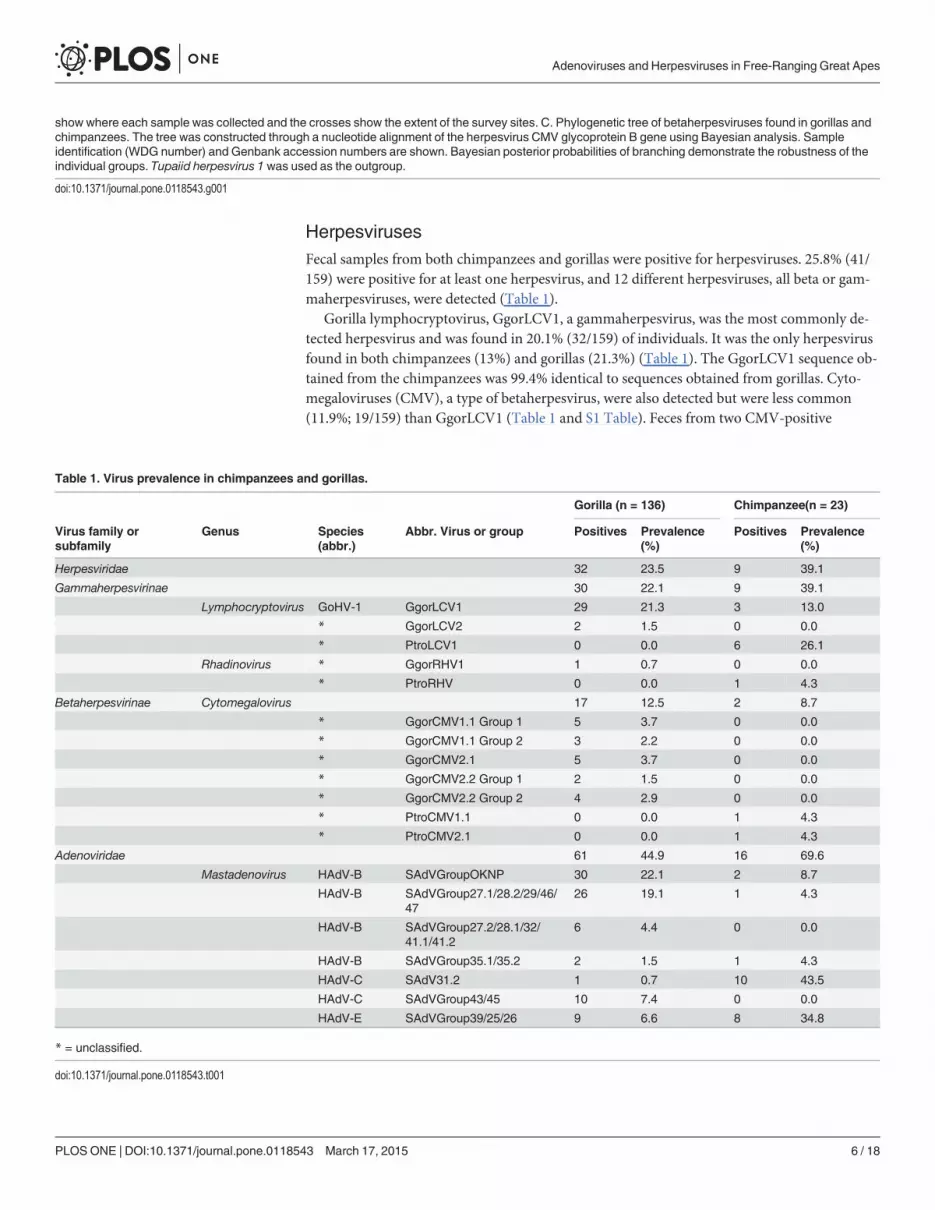
HerpesvirusesFecal samples from both chimpanzees and gorillas were positive for herpesviruses. 25.8% (41/159) were positive for at least one herpesvirus, and 12 different herpesviruses, all beta or gam-maherpesviruses, were detected (Table 1).
Gorilla lymphocryptovirus, GgorLCV1, a gammaherpesvirus, was the most commonly de-tected herpesvirus and was found in 20.1% (32/159) of individuals. It was the only herpesvirusfound in both chimpanzees (13%) and gorillas (21.3%) (Table 1). The GgorLCV1 sequence ob-tained from the chimpanzees was 99.4% identical to sequences obtained from gorillas. Cyto-megaloviruses (CMV), a type of betaherpesvirus, were also detected but were less common(11.9%; 19/159) than GgorLCV1 (Table 1 and S1 Table). Feces from two CMV-positive
show where each sample was collected and the crosses show the extent of the survey sites. C. Phylogenetic tree of betaherpesviruses found in gorillas andchimpanzees. The tree was constructed through a nucleotide alignment of the herpesvirus CMV glycoprotein B gene using Bayesian analysis. Sampleidentification (WDG number) and Genbank accession numbers are shown. Bayesian posterior probabilities of branching demonstrate the robustness of theindividual groups. Tupaiid herpesvirus 1 was used as the outgroup.
doi:10.1371/journal.pone.0118543.g001
Table 1. Virus prevalence in chimpanzees and gorillas.
Gorilla (n = 136) Chimpanzee(n = 23)
Virus family orsubfamily
Genus Species(abbr.)
Abbr. Virus or group Positives Prevalence(%)
Positives Prevalence(%)
Herpesviridae 32 23.5 9 39.1
Gammaherpesvirinae 30 22.1 9 39.1
Lymphocryptovirus GoHV-1 GgorLCV1 29 21.3 3 13.0
* GgorLCV2 2 1.5 0 0.0
* PtroLCV1 0 0.0 6 26.1
Rhadinovirus * GgorRHV1 1 0.7 0 0.0
* PtroRHV 0 0.0 1 4.3
Betaherpesvirinae Cytomegalovirus 17 12.5 2 8.7
* GgorCMV1.1 Group 1 5 3.7 0 0.0
* GgorCMV1.1 Group 2 3 2.2 0 0.0
* GgorCMV2.1 5 3.7 0 0.0
* GgorCMV2.2 Group 1 2 1.5 0 0.0
* GgorCMV2.2 Group 2 4 2.9 0 0.0
* PtroCMV1.1 0 0.0 1 4.3
* PtroCMV2.1 0 0.0 1 4.3
Adenoviridae 61 44.9 16 69.6
Mastadenovirus HAdV-B SAdVGroupOKNP 30 22.1 2 8.7
HAdV-B SAdVGroup27.1/28.2/29/46/47
26 19.1 1 4.3
HAdV-B SAdVGroup27.2/28.1/32/41.1/41.2
6 4.4 0 0.0
HAdV-B SAdVGroup35.1/35.2 2 1.5 1 4.3
HAdV-C SAdV31.2 1 0.7 10 43.5
HAdV-C SAdVGroup43/45 10 7.4 0 0.0
HAdV-E SAdVGroup39/25/26 9 6.6 8 34.8
* = unclassified.
doi:10.1371/journal.pone.0118543.t001
Adenoviruses and Herpesviruses in Free-Ranging Great Apes
PLOS ONE | DOI:10.1371/journal.pone.0118543 March 17, 2015 6 / 18

gorillas, WDG61 and WDG39, each contained two different CMVs (S1 Table). Previous workhas shown that within the CMVs, two chimpanzee and gorilla co-speciation clades (CG1 andCG2) exist, which contain closely related gorilla and chimpanzee viruses [18]. Nine individualswere positive for CMV in the CG1 clade. GgorCMV1.1 was identified in eight individual goril-las; phylogenetic analysis assigned these viruses into two groups within the CG1 clade(GgorCMV1.1 Group 1 (n = 4) and a separate group that we have called GgorCMV1.1 Group2 (n = 4)) (Fig. 1). A ninth individual was positive for Pan troglodytes CMV (PtroCMV1.1), an-other cytomegalovirus in the CG1 clade. CMVs in the CG2 clade were detected in 12 individualgorillas and were assigned to four groups, GgorCMV2.2 Group 1 (n = 2) and a second groupthat has not previously been described that we have called GgorCMV2.2 Group 2 (n = 4),GgorCMV2.1 (n = 5), and PtroCMV2.1 (n = 1) (Fig. 1).
AdenovirusesAdenoviruses were present in feces from 48.4% (77/159) of individuals; 69.6% (16/23) of thechimpanzees and 44.9% (61/136) of the gorillas were positive (Table 1). Gorilla and chimpan-zee adenoviruses clustered within one of seven adenovirus groups in the HAdV-B, HAdV-C,or HAdV-E clades (Fig. 2). Of these, identified members in five groups were found in bothchimpanzees and gorillas (Table 1). A previously unidentified group, which we named SimianAdenovirus B Group OKNP (SAdVGroupOKNP) was the most common adenovirus groupand was detected in 20.1% (32/159) of tested fecal samples, and all but two positives were fromgorillas (Table 1). Phylogenetically, SAdVGroupOKNP clustered with B2 human adenovirusesbut shared less than 90% nucleotide identity with all other known human and simian se-quences from HAdV-B. Two groups of viruses, SAdV31.2 and SAdVGroup 43/45, members ofadenovirus HAdV-C, were also identified. There was a significantly greater occurrence ofSAdV31.2 in chimpanzee feces (OR = 95[95% CI = 11, 810]) than in gorilla feces, butSAdVGroup 43/45 was only found in gorillas. Lastly, a group belonging to HAdV-E was alsoidentified. This group, SAdVGroup 39/35/26 E, was also more likely to be found in chimpan-zees than gorillas (OR = 6.5[95% CI = 2.1, 20]). While SAdV31.2 and SAdVGroup 39/35/26 Ewere found to be significantly more prevalent in chimpanzees, adenovirus B groups (SAdV-GroupOKNP, SAdVGroup27.1/28.2/29/46/47, SAdVGroup27.2/28.1/32/41.1/41.2, orSAdVGroup35.1/35.2) were statistically more prevalent in gorillas (OR = 2.4[95% CI = 1.3,14]). Adenovirus co-detection, where more than one type of adenovirus was detected, was alsonot statistically different among chimpanzees and gorillas, and occurred in 26.0% (6/23) ofchimpanzees and 14.7% (20/136) of gorillas.
Multiple viral sequences were detected in feces from 31.4% (50/159) of individuals. 27.9%(38/136) of the gorilla and 52.2% (12/23) of the chimpanzee fecal samples contained more thanone virus. The distribution of positive results per individual, ranked from one to five virusesfound, is shown in Fig. 3A. The data from two positive individuals that were resampled are pre-sented in S1 Table. The remaining third individual (WDG93 and WDG95) was negative for allviruses tested. Matrix analysis comparing the presence of each of the 19 different viruses de-tected within individuals was performed (Fig. 3B). In individuals positive for more than onevirus, GgorLCV1 and SAdVGroupOKNP was the most common virus combination detected(Fig. 3B, dark maroon cell). In the 12 individuals positive for both of these viruses, one wasfrom a chimpanzee and 11 were from gorillas (S1 Table). SAdVGroup 27.1/28.2/29/46/47 andSAdVGroupOKNP virus combinations were also relatively common and found in 10 gorillas.Other combinations of viruses were also seen. 94.7% (18/19) of the CMV-positive individualsand 84.6% (33/39) of the LCV-positive fecal samples from individual apes contained anotheradenovirus or herpesvirus (S1 Table). LCV’s were found in combination with all 19 detected
Adenoviruses and Herpesviruses in Free-Ranging Great Apes
PLOS ONE | DOI:10.1371/journal.pone.0118543 March 17, 2015 7 / 18

Adenoviruses and Herpesviruses in Free-Ranging Great Apes
PLOS ONE | DOI:10.1371/journal.pone.0118543 March 17, 2015 8 / 18

virus or viral groups. Overall, co-detection of HAdV-B or LCV was found in 52.6% (10/19) or89.5% (17/19) respectively of the CMV-positive individuals.
Estimating viral richnessIn chimpanzees and gorillas, the estimated number of viruses or viral groups present in oursample population was 23 [95%CI = 20, 26] (Fig. 4). Our result shows that when estimating theviral richness for both herpesviruses and adenoviruses, the Chao 2 estimator began to plateauat 100 individuals, and was stable by 125. We estimate that we captured 83% (19/23) of thetotal viruses or viral groups in our study population of chimpanzees and gorillas. Looking atbetaherpesviruses alone, viral richness was estimated to be nine, of which 78% (7/9) were cap-tured in our study. For gammaherpesviruses, viral richness was estimated to be seven, of which71% (5/7) were captured in our study. Because we could not differentiate between all the strainsand had to bin our adenovirus results into closely related groups, we were unable to estimatethe true viral richness for individual adenovirus strains and instead estimate the viral richnessof these subgroups. For example, the viral richness of adenoviruses in this population was esti-mated to be seven groups, of which 100% were detected. However, this is an underestimate ofthe total number of actual strains present. Also, it is important to point out that we analyzedone gene for adenovirus, the polymerase gene, which is highly conserved. We did not comparethe less conserved hexon gene [21]. We therefore cannot rule out the co-presence of other ade-novirus serotypes belonging to the same species in our study. Additional the sequences fromthis study were obtained through cloning of the PCR product and sequencing multiple clones,which has the potential to underestimate viral diversity based on the number of times the PCRwas repeated and the number of clones that were sequenced. We did not test the viral richnessof individual ape species because our sample size for chimpanzees was small.
Because ultralow temperatures are recommended for long term storage of samples in RNALater and our samples were stored at ambient temperatures (>4°C) for extended periods, testresult data were analyzed to determine if the number of viruses retrieved was associated withsample storage duration. Using univariate linear regression on all data combined, a statisticallysignificant negative association (p<0.05) was found where decreasing numbers of viruses wererecovered as sample storage duration increased. Ambient storage duration of 1,000 days orgreater reduced the mean number of detected viruses by 1.2 or more (S1 Fig.). Our results sug-gest that if samples are immediately tested (no ambient storage and processing immediatelyafter collection) detection of an average of 2.0 viruses/sample would be expected. We did notdetect a significant association between the total amount of nucleic acid recovered and ambientstorage time (data not shown). Because the number of viruses detected per sample declinedwith prolonged storage time, the probability of virus detection was not uniform across all sam-ples, and therefore our overall data may represent an underestimation of viral prevalence.However, there was no statistically significant negative correlation with viral detection and am-bient temperature if we narrowed our samples to those stored between 130 and 750 days (S1Fig.). Viral discovery curves were therefore repeated with only those samples stored for lessthan 750 days. In this analysis, the estimated viral richness for betaherpesvirus and adenovi-ruses was similar to that for all samples combined (S2 Fig.). However, the Chao2 estimator
Fig 2. Phylogenetic tree of adenovirus lineages found in gorillas and chimpanzees. The tree was constructed through a nucleotide alignment of theadenovirus polymerase gene using Bayesian analysis. Sample identification (WDG number) and Genbank accession numbers are shown. New AdVsequences are indicated in green for gorillas, and chimpanzees in blue. The colored vertical bars indicate the names assigned to each virus group used in theanalysis. The colored boxes denote adenovirus species clades. Tree shrew AdV-1was used as the outgroup. All SAdVGroupOKNP sequences are availablein GenBank under the following accession numbers: KJ780330-KJ780358.
doi:10.1371/journal.pone.0118543.g002
Adenoviruses and Herpesviruses in Free-Ranging Great Apes
PLOS ONE | DOI:10.1371/journal.pone.0118543 March 17, 2015 9 / 18

Adenoviruses and Herpesviruses in Free-Ranging Great Apes
PLOS ONE | DOI:10.1371/journal.pone.0118543 March 17, 2015 10 / 18

failed to stabilize for total viruses and gammaherpesvirus. Taken together, these results suggestthat our estimate of viral richness of adenovirus subgroups and betaherpesvirus has not beeninfluenced by ambient storage, however gammaherpesvirus viral richness may have been un-derestimated in our total estimation of viral richness of 23 viruses.
DiscussionIn order to understand natural patterns of health and disease and to monitor populations offree-ranging chimpanzees and gorillas for pathogen presence or emergence, baseline informa-tion on the natural viral diversity in these populations is needed. The goals of this study weretherefore to investigate the diversity of viruses in free-ranging gorillas and chimpanzees fromthe Sangha region of the Republic of Congo and estimate viral richness in these populationsthrough pathogen detection in fecal samples. We detected 19 viruses or viral groups, includinga previously undescribed group of HAdV-B and 2 groups of cytomegaloviruses. Based on viraldiversity curves, we estimate that we detected*80% of the viruses or viral groups within theAdenoviridae and Herpesviridae families that are circulating in western lowland gorillas andchimpanzees in the Sangha region near OKNP. Our estimation of herpesvirus and adenovirusrichness in these two ape populations, although limited to a single region, provides importantinformation about the types of viruses that circulate in these species and this population. Thisestimate does not apply to the viral richness that could be determined in ape populations else-where in the world or in captivity, but our results can provide opportunities for future compar-ison. Our study also does not address novel viruses that can only be detected using unbiasedhigh throughput sequencing methods.
Adenoviruses are double-stranded DNA viruses that can infect a wide range of vertebratehosts and cause a variety of diseases including mild to life-threatening respiratory and gastroin-testinal disease. Human and non-human primates shed adenoviruses in their stool [21,36–38].In one study, adenovirus DNA was identified in 40% of stool samples from wild chimpanzeesin Cameroon, and the Democratic Republic of Congo (DRC), and captive gorillas maintainedin zoos with a much higher prevalence of shedding (36–100%) in the latter [21]. Herein we re-port prevalence statistics, but because of potential deterioration issues with the samples theseare assumed to be an underestimate of the true value in our sampled population. In our studyregion in the Republic of Congo, adenovirus shedding was also common in free-ranging greatapes, being present in fecal samples from 68% of chimpanzees and 45% of gorillas. Results inour chimpanzee study population were higher than in the previous Cameroon/DRC study.This could be due to differences in sample storage conditions, population density, seasonality,or geographic region, but is less likely due to laboratory differences, since both studies used thesame PCR assay [21]. Of the three adenovirus species detected in the current study (HAdV-B,HAdV-C, and HAdV-E, all were found in both chimpanzees and gorillas. Previous studieshave only detected HAdV-E in chimpanzees, bonobos and humans [21,37]; thus, our findingsof HAdV-E in both gorillas and chimpanzees support the possibility for an earlier cross-speciestransmission event in this population. Adenoviruses are thought to co-evolve with their hostspecies and usually do not cross the species barrier [39–41]. However, genetic and serologicalstudies support the concept of cross-species adenovirus transmission between non-human
Fig 3. Viral co-detection in chimpanzee and gorilla fecal samples. A. Graph depicting the type and number of viruses detected in fecal samples fromindividual great apes. Gorillas are highlighted in green and chimpanzees are highlighted in blue. B. Matrix analysis showing all 19 detected viruses or viralgroups and the number of pair-wise combinations with each other. On the left side of the matrix is a heat map showing the number of individuals positive forco-detection of two viruses as is indicated in the red color key. For example, feces from 12 individuals, highlighted in maroon (>8 samples detected with thatvirus combination), were positive for both GgorLCV1 and SAdVGroupOKNP. The upper right half of the matrix (mirror image of lower left) are the number ofindividuals that tested positive for co-detection, with colors indicating which species, or if both were affected.
doi:10.1371/journal.pone.0118543.g003
Adenoviruses and Herpesviruses in Free-Ranging Great Apes
PLOS ONE | DOI:10.1371/journal.pone.0118543 March 17, 2015 11 / 18

Fig 4. Estimating viral richness of herpesviruses and adenoviruses in great apes of the Sangha region. A. We calculated the total viral richness inchimpanzees and gorillas associated with the Adenoviridae and Herpesviridae families. ICE and Jackknife estimators are shown as orange and grey curvesrespectively. The refraction curve (black) shows the number of virus groups or viruses as a function of the number of samples, and the collector curve (red)shows the accumulation of viruses found with increasing sample size. The Chao2 estimator (blue) shows the number of viruses that are estimated in the apepopulation with 95% confidence intervals indicated by the brackets. The horizontal bar shows the number of viruses estimated in the analysis. In B, C, and Dseparate richness analyses were done for the Herpesviridae subfamilies (Betaherpesvirinae andGammaherpesvirinae) and Adenoviridae.
doi:10.1371/journal.pone.0118543.g004
Adenoviruses and Herpesviruses in Free-Ranging Great Apes
PLOS ONE | DOI:10.1371/journal.pone.0118543 March 17, 2015 12 / 18

primates and humans [37,42,43]. Additional surveys are warranted to better understand thelikelihood of cross-species transmission events in primates and determine if recombinationand generation of hybrid adenoviruses, which have developed in captive settings [21], couldoccur in the wild. This type of information would promote a better understanding of the riskfor zooanthroponotic transmission, and whether adenoviruses in wild gorillas and chimpan-zees are a risk factor for disease emergence in humans in situations where humans are workingclosely with captive primates or living in communities that border free-ranging gorilla orchimpanzee habitat.
Detection of fragments from multiple different adenoviruses in gorilla and chimpanzeefeces was a significant finding in our study. Feces from 16.4% (26/159) of individuals, six chim-panzees and 20 gorillas, contained more than one type of adenovirus from seven differentgroups of three different species. This adds significantly to our understanding of the differentadenoviruses to which gorilla and chimpanzees are exposed in or east of OKNP. Adenoviruscoinfection associated with illness has been well documented in humans. Human adenovirusaccount for about 8% of clinically relevant viral disease globally, are divided into 52 serotypesfrom seven species [44,45]. About 50% of the 52 identified human adenovirus serotypes areknown to cause illness [46]. Adenovirus infections, such as HAdV-14 (B2) cause respiratorydisease and pneumonia [47]. Other adenoviral disease, including conjunctivitis, hepatitis anddiarrhea [48], are described in children, immunocompromised people, and in patients withmultiple co-infections [46]. Two recent studies showed a high rate of adenoviral co-infectionwith HAdV-B and E in humans with acute respiratory disease [44,49]. In the great apes de-scribed herein, adenoviral co-detections included combinations of HAdV-B/C and HAdV-B/E.In contrast to humans, very little is known about the pathogenicity of adenoviruses or the sig-nificance of their co-infections in great apes and other non-human primates [21,38,43,50].Given current limits in our understanding of the ecology, including cross species transmission,our results underscore the importance of developing a more comprehensive understanding ofadenoviral infections in great apes.
Herpesviruses are divided into three distinct subfamilies known as Alphaherpesvirinae,Betaherpesvirinae, and Gammaherpesvirinae, based on biological and molecular properties andgenomic sequence divergence [51]. Cytomegaloviruses (CMVs) and lymphocryptoviruses(LCVs) are members of the Beta- and Gammaherpesvirinae subfamilies respectively, that infectmammals, human and non-human primates. Although host-virus co-divergence is thought tobe the primary mode of herpesvirus evolution, cross-species transmission events have beenknown to occur. For example, humans infected with macaque simplex virus (Herpes B virus)can develop severe illness or death [52], and simian CMV has been shown to infect humancells [53,54].
Because of the close evolutionary relationship between great apes and humans, herpesvi-ruses of apes are of particular interest for public health and those that impact great apes directlyare important for species conservation. Several new CMVs have recently been characterized inboth chimpanzees and gorillas [18]. These discoveries have led to the identification of two newco-speciation clades of CMV (CG1 and CG2) that are thought to have evolved through a hori-zontal transmission event between chimpanzees and gorillas 6–8 million years ago [18]. DNAfragments from seven CMVs were identified in the non-human great ape feces from this study.These included two previously undescribed CMVs that we designated GgorCMV1.1 Group 2and GgorCMV2.2 Group 2, within the CG1 and CG2 clades, respectively. All of the CMV vi-ruses detected in our study were found in feces from either gorillas or chimpanzees (no detec-tion of the same viral fragment in both species). GgorLCV1, a gammaherpesvirus, was the onlyherpesvirus found in both gorillas and chimpanzees.
Adenoviruses and Herpesviruses in Free-Ranging Great Apes
PLOS ONE | DOI:10.1371/journal.pone.0118543 March 17, 2015 13 / 18

Epstein-Barr virus (EBV, human lymphocryptovirus) and CMV are the most commoncause of infectious mononucleosis (IM) and illnesses resembling IM in children, and can leadto severe fatigue, fever, pharyngitis, lymphadenopathy and recurrent microbial infection[55,56]. Co-infections of these two herpesvirus in the context of other viral infections, such asan additional herpes or adenovirus, can worsen symptoms and illness [55, 57], and reactivationof CMV has been suggested to be a factor leading to exacerbation of disease induced by otherviral agents [18]. In the current study, 95% of the CMV-positive and 85% of the LCV-positiveape fecal samples contained more than one virus, either another herpesvirus and/or adenovi-rus, which suggests that CMV and LCV are primarily shedding into the feces in the context ofthese other viruses. Because CMV/LCV/adenovirus co-association is so tightly correlated inour study, pre-screening of ape feces for these viruses could be used to identify animals orgroups of animals with co-infections. Evaluating these animals for disease outcomes may be auseful strategy to determine if viral co-infection reflects similar outcomes as seen in humans.
Understanding the diversity of pathogens to which endangered gorillas and chimpanzeesare exposed is important in defining health risks that impact population dynamics and conser-vation efforts. Our results provide initial information about the presence and diversity of ade-noviruses and herpesviruses in and near OKNP to which wild gorillas and chimpanzees, aswell as humans or other species that live in the region, are exposed. Limitations of the currentstudy are a likely underestimation of viral diversity in the sample ape population due to fecaldegradation of samples stored for long term that resulted in reduced DNA and RNA viral de-tection. Subsequent studies across this and other regions in Africa, that incorporate unbiasedsequencing technologies, will be valuable for estimating the true viral diversity in great ape spe-cies as a whole, not just at the population level. In addition, continuous monitoring of these en-dangered species is necessary to understand the expression and outcomes of infection, co-infection, and cross-species transmission; all crucial steps for defining health-related conserva-tion threats and guiding conservation management actions.
Supporting InformationS1 Dataset. FASTA file. DNA sequence alignment of herpesvirus glycoprotein B sequencesused for phylogenetic analysis in Fig. 1.(FASTA)
S2 Dataset. FASTA file. DNA sequence alignment of adenovirus DNA polymerase sequencesused for phylogenetic analysis in Fig. 2.(FASTA)
S1 Fig. Association of viral detection and days stored at ambient temperature.A. Univariate linear regression showing a statistically significant negative association (p<0.05)with the number of viruses recovered and the number of days the samples were stored at ambi-ent temperature. Ambient storage duration of 1,000 days or greater reduced the mean numberof viruses detected by 1.2 or more. B. Univariate linear regression showing no statistically sig-nificant negative association with the virus count and the number of days the samples werestored at ambient temperature when analyzing samples stored up to 750 days.(TIFF)
S2 Fig. Viral richness in samples stored at ambient temperature for less time. Estimatedviral richness curves for chimpanzees and gorillas associated with the Adenoviridae and Her-pesviridae families repeated as in Fig. 4, but with only samples stored for less than 750 days.(TIFF)
Adenoviruses and Herpesviruses in Free-Ranging Great Apes
PLOS ONE | DOI:10.1371/journal.pone.0118543 March 17, 2015 14 / 18

S1 Table. Summary of positive results. Positive PCR results obtained for each individualgrouped by Gammaherpesvirinae, Betaherpevirinae and Adenoviridae subfamilies or family,the total number of viruses detected for each individual, and type of animal each sample wasderived from.(DOCX)
S2 Table. BLASTn results of viral alignments with those found in GenBank. National Cen-ter for Biotechnology Information: http://www.blast.ncbi.nlm.nih.gov/Blast.cgi.(DOCX)
AcknowledgmentsDisclaimer: The contents are the responsibility of the authors and do not necessarily reflect theviews of USAID, USFWS or the United States Government.
Ministry of Sustainable Development and Forest Economy, Republic of Congo; Ministry ofScientific Research, Republic of Congo. Beatrice Hahn, M.D., University of Pennsylvania (forsharing expertise and protocols for microsatellite typing).
Author ContributionsConceived and designed the experiments: TAS SHO PR KC DMWIL. Performed the experi-ments: TAS SHO AO PR KC GR KJL. Analyzed the data: TAS SHO KJL GR SJA. Contributedreagents/materials/analysis tools: AO PR KCWIL. Wrote the paper: TAS SHO KC PR DM KJLSJAWIL. Contributed intellectual advice: SJA DJ.
References1. Bermejo M, Rodríguez-Teijeiro J, Illera G, Barroso A, Vilà C, Walsh P. Ebola outbreak killed 5000 goril-
las. Science. 2006; 314: 1564. PMID: 17158318
2. Jones K, Patel N, Levy M, Storeygard A, Balk D, Gittleman J, et al. Global trends in emerging infectiousdiseases. Nature. 2008; 451: 990–993. doi: 10.1038/nature06536 PMID: 18288193
3. KareshWB, Dobson A, Lloyd-Smith JO, Lubroth J, Dixon MA, Bennett M, et al. Ecology of zoonoses:natural and unnatural histories. The Lancet. 2012; 380: 1136–1145.
4. Liu W, Li Y, Learn G, Rudicell R, Robertson J, Keele B, et al. Origin of the humanmalaria parasite Plas-modium falciparum in gorillas. Nature. 2010; 467: 420–425. doi: 10.1038/nature09442 PMID:20864995
5. Prugnolle F, Durand P, Neel C, Ollomo B, Ayala F, Arnathau C, et al. African great apes are naturalhosts of multiple related malaria species, including Plasmodium falciparum. Proc Natl Acad Sci USA.2010; 107: 1458–1463. doi: 10.1073/pnas.0914440107 PMID: 20133889
6. Olson S, Reed P, Cameron K, Ssebide B, Johnson C, Morse S, et al. Dead or alive: animal samplingduring Ebola hemorrhagic fever outbreaks in humans. Emerg Health Threats J. 2012 5: 3402/ehtj.v3405i3400.9134.
7. Reed P, Mulangu S, Cameron K, Ondzie A, Joly D, Bermejo M, et al. A new approach for monitoringebolavirus in wild great apes. PLOS Negl Trop Dis. 2014; 8: e3143. doi: 10.1371/journal.pntd.0003143PMID: 25232832
8. Wolfe ND, Dunavan CP, Diamond J. Origins of major human infectious diseases. Nature. 2007; 447:279–283. PMID: 17507975
9. Kaur T, Singh J, Tong S, Humphrey C, Clevenger D, TanW, et al. Descriptive epidemiology of fatal re-spiratory outbreaks and detection of a human-related metapneumovirus in wild chimpanzees (Pan trog-lodytes) at Mahale Mountains National Park, Western Tanzania. Am J Primatol. 2008; 70: 755–765.doi: 10.1002/ajp.20565 PMID: 18548512
10. Palacios G, Lowenstine LJ, Cranfield MR, Gilardi KV, Spelman L, Lukasik-BraumM, et al. Humanmetapneumovirus infection in wild mountain gorillas, Rwanda. Emerg Infect Dis. 2011; 17: 711–713.doi: 10.3201/eid1704.100883 PMID: 21470468
11. Ryan S andWalsh P. Consequences of non-intervention for infectious disease in African great apes.PLOS One. 2011; 6: e29030. doi: 29010.21371/journal.pone.0029030 PMID: 22216162
Adenoviruses and Herpesviruses in Free-Ranging Great Apes
PLOS ONE | DOI:10.1371/journal.pone.0118543 March 17, 2015 15 / 18

12. Cooper N and Nunn CL. Identifying future zoonotic disease threats: Where are the gaps in our under-standing of primate infectious diseases? Evolution, Medicine, and Public Health. 2013: 27–36.
13. Oates JF, Tutin CEG, Humle T, Wilson ML, Baillie J, Balmforth Z, et al. Pan troglodytes. In: IUCN 2013.IUCN Red List of Threatened Species.
14. Walsh PD, Tutin CEG, Oates JF, Baillie J, Maisels F, Stokes E, et al.Gorilla gorilla. In: IUCN 2013.IUCN Red List of Threatened Species.
15. Devos C, Sanz C, Morgan D, Onononga J, Laporte N, Huynen MC. Comparing ape densities and habi-tats in northern Congo: surveys of sympatric gorillas and chimpanzees in the Odzala and Ndoki re-gions. Am J Primatol. 2008; May: 439–451.
16. Lilly AA, Mehlman PT, Doran D. Intestinal parasites in gorillas, chimpanzees, and humans at MondikaResearch Site, Dzanga-Ndoki National Park, Central African Republic. International Journal of Evolu-tionary Biology. 2002; 23: 555–573.
17. Chmielewicz B, Goltz M, Ehlers B. Detection and multigenic characterization of a novel gammaherpes-virus in goats. Virus Res. 2001; 75: 87–94. PMID: 11311431
18. Leendertz F, Deckers M, SchemppW, Lankester F, Boesch C, Mugisha L. et al. Novel cytomegalovi-ruses in free-ranging and captive great apes: phylogenetic evidence for bidirectional horizontal trans-mission. J Gen Virol. 2009; 90: 2386–2394. doi: 10.1099/vir.0.011866-0 PMID: 19553394
19. VanDevanter D, Warrener P, Bennett L, Schultz E, Coulter S, Garber RL, et al. Detection and analysisof diverse herpesviral species by consensus primer PCR. J Clin Microbiol. 1996; 34: 1666–1671.PMID: 8784566
20. Prepens S, Kreuzer K, Leendertz F, Nitsche A, Ehlers B. Discovery of herpesviruses in multi-infectedprimates using locked nucleic acids (LNA) and a bigenic PCR approach. Virology Journal. 2007; 4: 1–14. PMID: 17204159
21. Roy S, Vandenberghe L, Kryazhimskiy S, Grant R, Calcedo R, Yuan X, et al. Isolation and characteriza-tion of adenoviruses persistently shed from the gastrointestinal tract of non-human primates. PLOSPathog. 2009; 5: e1000503. doi: 10.1371/journal.ppat.1000503 PMID: 19578438
22. Zhai J, Palacios G, Towner J, Jabado O, Kapoor V, Venter M, et al. Rapid molecular strategy for filovi-rus detection and characterization. J Clin Microbiol. 2007; 45: 224–226. PMID: 17079496
23. Tong S, Chern S, Li Y, Pallansch M, Anderson L. Sensitive and broadly reactive reverse transcription-PCR assays to detect novel paramyxoviruses. J Clin Microbiol. 2008; 46: 2652–2658. doi: 10.1128/JCM.00192-08 PMID: 18579717
24. Quan P, Firth C, Street C, Henriquez J, Petrosov A, Tashmukhamedova A, et al. Identification of a se-vere acute respiratory syndrome coronavirus-like virus in a leaf-nosed bat in Nigeria. mBio. 2010; 1:e00208–00210. doi: 10.1128/mBio.00208-10 PMID: 21063474
25. Watanabe S, Masangkay J, Nagata N, Morikawa S, Mizutani T, Fukushi S, et al. Bat coronaviruses andexperimental infection of bats, the Philippines. Emerg Infect Dis. 2010; 16: 1217–1223. doi: 10.3201/eid1608.100208 PMID: 20678314
26. Moureau G, Temmam S, Gonzalez J, Charrel R, Grard G, de Lamballerie X. A real-time RT-PCRmeth-od for the universal detection and identification of flaviviruses. Vector Borne Zoonotic Dis. 2007; 4:467–477. PMID: 18020965
27. Bracht A, Brudek R, Ewing R, Manire C, Burek K, Rosa C, et al. Genetic identification of novel poxvi-ruses of cetaceans and pinnipeds. Arch Virol. 2006; 3: 423–438. PMID: 16328132
28. Kapoor A, Mehta N, Esper F, Poljsak-Prijatelj M, Quan P, Qaisar N, et al. Identification and characteri-zation of a new bocavirus species in gorillas. PLOSOne. 2010; 5: e11948. doi: 10.1371/journal.pone.0011948 PMID: 20668709
29. Clewley J, Lewis J, Brown D, Gadsby E. A novel simian immunodeficiency virus (SIVdrl) pol sequencefrom the drill monkey,Mandrillus leucophaeus. J Virol. 1998; 72: 10305–10309. PMID: 9811781
30. Takehisa J, Kraus M, Ayouba A, Bailes E, Van Heuverswyn F, Decker J, et al. Origin and biology of sim-ian immunodeficiency virus in wild-living western gorillas. J Virol. 2009; 83: 1635–1648. doi: 10.1128/JVI.02311-08 PMID: 19073717
31. Wroblewski EE, Murray CM, Keele BF, Schumacher-Stankey JC, Hahn BH, Pusey AE. Male domi-nance rank and reproductive success in chimpanzees, Pan troglodytes schweinfurthii. Animal Behav-ior. 2009; 77: 873–885. PMID: 19498952
32. Kocher TD, ThomasWK, Meyer A, Edwards SV, Pääbo S, Villablanca FX, et al. Dynamics of mitochon-drial DNA evolution in animals: Amplification and sequencing with conserved primers. Proc Natl AcadSci USA.1989; 86: 6196–6200. PMID: 2762322
33. Huelsenbeck JP, Ronquist F. MRBAYES: Bayesian inference of phylogeny. Bioinformatics. 2001; 17:754–755. PMID: 11524383
Adenoviruses and Herpesviruses in Free-Ranging Great Apes
PLOS ONE | DOI:10.1371/journal.pone.0118543 March 17, 2015 16 / 18

34. R Development Core Team (2011) R: A language and environment for statistical computing. R Founda-tion for Statistical Computing. Available: http://www.R-project.org/ Vienna, Austria.
35. Anthony SJ, Epstein JH, Murray KA, Navarrete-Macias I, Zambrana-Torrelio CM, Solovyov A, et al. Astrategy to estimate unknown viral diversity in mammals. mBio. 2013; 4: e00598–00513. doi: 10.1128/mBio.00598-13 PMID: 24003179
36. Noble RT, Allen SM, Blackwood AD, ChuW, Jiang SC, Lovelace GL, et al. Use of viral pathogens andindicators to differentiate between human and non-human fecal contamination in a microbial sourcetracking comparison study. J Water Health Dec. 2003; 1: 195–207. PMID: 15382724
37. Wevers D, Metzger S, Babweteera F, Bieberbach M, Boesch C, Cameron K, et al. Novel Adenovirusesin Wild Primates: A High Level of Genetic Diversity and Evidence of Zoonotic Transmissions. J Virol.2011; 85: 10774. doi: 10.1128/JVI.00810-11 PMID: 21835802
38. Wevers D, Leendertz F, Scuda N, Boesch C, Robbins M, Head J, et al. A novel adenovirus of Westernlowland gorillas (Gorilla gorilla gorilla). Virol J. 2010; 7: doi: 10.1186/1743-1422X-1187-1303
39. Benkö M and Harrach B. Molecular evolution of adenoviruses. Curr Top Microbiol Immunol. 2003; 272:3–35. PMID: 12747545
40. Davison A, Benkö M, Harrach B. Genetic content and evolution of adenoviruses. J Gen Virol. 2003. 84:2895–2908. PMID: 14573794
41. Benkö M, Harrach B, Kremer E. Do nonhuman primate or bat adenoviruses pose a risk for humanhealth? Future Microbiol. 2014; 9: 269–272. doi: 10.2217/fmb.13.170 PMID: 24762301
42. Chen E, Yagi S, Kelly K, Mendoza S, Maninger N, Rosenthal A, et al. Cross-species transmission of anovel adenovirus associated with a fulminant pneumonia outbreak in a NewWorld monkey colony.PLOS Pathog. 2011; 7: e1002155. doi: 10.1371/journal.ppat.1002155 PMID: 21779173
43. Chiu C, Yagi S, Lu X, Yu G, Chen E, Liu M, et al. A novel adenovirus species associated with an acuterespiratory outbreak in a baboon colony and evidence of coincident human infection. mBio. 2013; 4:e00084–00013. doi: 10.1128/mBio.00084-13 PMID: 23592261
44. Vora GJ, Lin B, Gratwick K, Meador C, Hansen C, Tibbetts C, et al. Co-infections of adenovirus speciesin previously vaccinated patients. Emerg Infect Dis. 2006; 12:921–930. PMID: 16707047
45. Virus taxonomy: classification and nomenclature of viruses: Ninth Report of theInternational Committeeon Taxonomy of Viruses. Ed: King A.M.Q., Adams M.J., Carstens E.B. and Lefkowitz E.J. San Diego:Elsevier. AdenoviridaeChapter. 2012. pp. 125–141.
46. Gray G, McCarthy T, Lebeck M, Schnurr D, Russell K, Kajon AE et al. Genotype prevalence and riskfactors for severe clinical adenovirus infection, United States 2004–2006. Clin Infect Dis. 2007; 45:1120–1131. PMID: 17918073
47. Lewis P, Schmidt M, Lu X, Erdman D, Campbell M, Thomas A, et al. A community-based outbreak ofsevere respiratory illness caused by human adenovirus serotype 14. J Infect Dis. 2009; 199: 1427–1434. doi: 10.1086/598521 PMID: 19351259
48. Ruuskanen O, Meurman O, Akusjarvi G. Adenoviruses. In: Richman DDWR, Hayden FG, eds., editor.Clinical Virology. New York: Churchill Livingstone.1997. pp. 1355.
49. Wang SL, Chi CY, Kuo PH, Tsai HP, Wang SM, Liu CC, et al. High-incidence of human adenoviral co-infections in taiwan. PLOS One. 2013; 8: e75208. doi: 75210.71371/journal.pone.0075208 PMID:24073254
50. Duncan M, Cranfield M, Torano H, Kuete H, Lee G, Glenn A, et al. Adenoviruses isolated from wild go-rillas are closely related to human species C viruses. Virology. 2013; 444: 119–123. doi: 110.1016/j.virol.2013.1005.1041 PMID: 23806387
51. Davison A. Evolution of the herpesviruses. Vet Microbiol. 2002; 86: 69–88. PMID: 11888691
52. Wertheim J, Smith M, Smith D, Scheffler K, Kosakovsky Pond S. Evolutionary origins of human herpessimplex viruses 1 and 2. Mol Biol Evol. 2014; 31: 2356–2364. doi: 10.1093/molbev/msu185 PMID:24916030
53. Lilja A, Shenk T. Efficient replication of rhesus cytomegalovirus variants in multiple rhesus and humancell types. Proc Natl Acad Sci USA. 2008; 105: 19950–19955. doi: 10.1073/pnas.0811063106 PMID:19064925
54. Michaels M, Alcendor D, St George K, Rinaldo C, Ehrlich G, Becich MJ, et al. Distinguishing babooncytomegalovirus from human cytomegalovirus: importance for xenotransplantation. J Infect Dis. 1997;176: 1476–1483. PMID: 9395357
55. Müller R, Ditzenb A, Hillea K, Stichlinga M, Ehrichtc R, Illmer T, et al. Detection of herpesvirus and ade-novirus co-infections with diagnostic DNA-microarrays. Journal of Virological Methods. 2009; 155:161–166. doi: 10.1016/j.jviromet.2008.10.014 PMID: 19022297
Adenoviruses and Herpesviruses in Free-Ranging Great Apes
PLOS ONE | DOI:10.1371/journal.pone.0118543 March 17, 2015 17 / 18

56. Wang X, Yang K, Wei C, Huang Y, Zhao D. Coinfection with EBV/CMV and other respiratory agents inchildren with suspected infectious mononucleosis. Virol J. 2010; 7: doi: 10.1186/1743-1422X-1187-1247
57. Coaquette A, Bourgeois A, Dirand C, Varin A, ChenW, Herbein G. Mixed cytomegalovirus glycoproteinB genotypes in immunocompromised patients. Clin Infect Dis. 2004; 39: 155–161. PMID: 15307021
Adenoviruses and Herpesviruses in Free-Ranging Great Apes
PLOS ONE | DOI:10.1371/journal.pone.0118543 March 17, 2015 18 / 18


