Embed Size (px)
Citation preview

VIROLOGY 222, 1–13 (1996)ARTICLE NO. 0392
Comparison of the EBNA1 Proteins of Epstein–Barr Virus and HerpesvirusPapio in Sequence and Function
JOHN L. YATES,1 SARAH M. CAMIOLO, SAYED ALI, and ANGELA YING
Department of Human Genetics, Roswell Park Cancer Institute, Elm and Carlton Streets, Buffalo, New York 14263
Received March 5, 1996; accepted May 31, 1996
The EBNA1 protein of Epstein–Barr virus (EBV) supports replication and maintenance of the circularized viral chromosomein cells that are latently infected. We have isolated, sequenced, and functionally characterized the EBNA1 gene of herpesviruspapio (HVP), an EBV-like virus that infects baboons. The amino acid sequences of EBNA1 of HVP and EBV are 56% identical,if the difference in the length of the glycine and alanine containing repetitive region, which is much shorter for HVP EBNA1,is omitted for the calculation. The key structural features of the DNA-binding/dimerization domain (the carboxyl-terminaldomain) appear to have been conserved, as have amino acids in the two regions thought to be most critical for DNA binding.Most of the salient features of the amino-terminal two-thirds of EBNA1 (the amino-terminal domain), including a dearth ofsequences predictive of alpha-helical or beta-sheet structures, are shared by the two sequences, although numerous gapsin this region were needed for alignment of the sequences. The amino-terminal fifty amino acids of EBNA1 of both EBVand HVP weakly resemble the amino terminus of rat ribosomal protein S2. Plasmids carrying oriP of either virus replicatedstably in mammalian cells and supported efficient outgrowth of colonies under selection when supported by EBNA1 fromeither virus, although with each oriP there was a noticeable preference for EBNA1 to be from the same virus. HVP EBNA1was less effective than EBV EBNA1 at activating the enhancer function of EBV oriP and under certain conditions was lesseffective than EBV EBNA1 at supporting maintenance of plasmids carrying EBV oriP. Results obtained with hybrid EBNA1molecules indicated that differences in the amino-terminal and carboxyl-terminal domains, respectively, are primarily respon-sible for the differences in transcriptional activation and plasmid maintenance, respectively. The results showed that changeswithin EBNA1 can differentially alter its transcriptional and replicational activities. q 1996 Academic Press, Inc.
INTRODUCTION sential elements and each uses multiple EBNA1 bindingsites to function in distinct ways (Reisman et al., 1985;
Like other members of the herpesvirus family, Ep- Chittenden et al., 1989). At one end of oriP, four EBNA1stein–Barr virus establishes life-long, usually asymptom- binding sites are arranged suitably to serve as an originatic, infections of people (reviewed by Miller, 1990). Cen- of bidirectional DNA replication, or OBR (also called thetral to the long-term maintenance of EBV infections is dyad symmetry region) (Wysokenski and Yates, 1989;the latent infection of B cells (reviewed by Klein, 1994; Gahn and Schildkraut, 1989; Harrison et al., 1994). At thesee also Qu and Rowe, 1992; Tierney et al., 1994; Miya- other end of oriP, a family of 30-bp repeats (FR) con-shita et al., 1995; Chen et al., 1995). In one stage of latent taining as many as 20 EBNA1 binding sites activatesinfection of B cells, a small subset of EBV genes are initiation of replication at the OBR (Reisman et al., 1985;expressed and together cause the cells to proliferate Wysokenski and Yates, 1989) as well as performing two(reviewed by Kieff and Liebowitz, 1990). The induced pro- additional functions. In the presence of EBNA1, the FRliferation expands the latent infection, but this requires of oriP permits plasmids carrying it to be retained forthat the viral genome be replicated during latency and prolonged periods after transfection into cells in the ap-passed on to progeny cells. parent absence of replication, in some manner pre-
Latently infecting EBV genomes are replicated once venting the rapid elimination of transfected plasmids thatper cell cycle in synchrony with cellular DNA replication otherwise occurs (Reisman et al., 1985; Krysan et al.,(Adams, 1987). The EBV-encoded nuclear protein, 1989; Middleton and Sugden, 1994). Finally, the FR is anEBNA1, and an 1800-bp region of the EBV chromosome, EBNA1-dependent enhancer of transcription which canoriP, are sufficient to provide such stable and regulated activate heterologous promoters as well as latency pro-replication to recombinant plasmids introduced into hu- moters located several kilobase pairs away on the EBVman cell lines (Yates et al., 1984, 1985; Lupton and Lev- genome (Reisman and Sugden, 1986; Sugden and War-ine, 1985; Yates and Guan, 1991). OriP includes two es- ren, 1989; Gahn and Sugden, 1995). Each of the functions
of EBNA1 is presumed to involve interactions betweenEBNA1 and cellular factors that remain to be identified.1 To whom correspondence and reprint requests should be ad-
dressed. E-mail: [email protected]. (For a review, see Yates, 1996.)
10042-6822/96 $18.00Copyright q 1996 by Academic Press, Inc.All rights of reproduction in any form reserved.
AID VY 8032 / 6a1b$$$141 07-07-96 19:10:34 vira AP: Virology
2 YATES ET AL.
Herpesvirus papio (HVP), which infects baboons, is viruses and the HindIII sites on the plasmid and located1.1 kb 3* of the EBNA1 ORF on HVP DNA. The hybridsimilar to EBV biologically and genetically (Falk et al.,
1976; Gerber et al., 1977; Heller and Kieff, 1981; Lee et EBNA1 ORFs, E-P(437) and E-P(438), were constructedusing an ApaI site that is present at codon 437 of theal., 1981). OriP of HVP has been isolated and character-
ized and, although somewhat different than EBV oriP, it EBV EBNA1 gene and at the homologous position of HVPand using either an AvrII site at the 5* end or a HindIIIhas a similar bipartite functional organization and func-
tions in cells infected by EBV (Pesano and Pagano, 1986; site at the 3* end to replace either half of the EBV EBNA1ORF with HVP DNA on a plasmid, p277, which carriesLoeb et al., 1990). Here we report the sequence of the
HVP homologue of EBNA1 and characterization of its the EBNA1 ORF and has no other ApaI site; the hybridORFs were then transferred to p205H using AvrII andactivities in conjunction with oriP of HVP and EBV.HindIII. P-E(457) was constructed using an SfiI site at thejunction position of HVP DNA and incorporating an SfiIMATERIALS AND METHODSsite into the end of a PCR-amplified 3* half of the EBV
Cloning and sequencing the HVP EBNA1 gene EBNA1 gene using the primer, 5*-ACCCCGGGGCCA-GGGTGGCCGAGGCAGGCGC. (Restriction sites areThe HVP-infected baboon B-cell line, 594S (Rabin etunderlined.) Similarly, to construct P-E(487), an SstI siteal., 1977), was kindly provided by Dr. Joe Pagano at theat the junction in EBV DNA was incorporated at the 3*University of North Carolina. The cells were expanded inend of the 5* half of the HVP EBNA1 gene using the PCR3 liters of RPMI 1640 medium supplemented with 9%primer, 5*-GGACAGTAGAGCTCGCAGGCTCTG. P-E(548)fetal bovine serum. DNA was isolated from cells usingand the reciprocal junction at this position were madethe Hirt method (Hirt, 1967), which gave a several-foldby incorporating a KpnI site into the EBV end, in eachenrichment of HVP DNA over cellular DNA in the super-case using primer, 5*-CCTGGTACCGGCCCGCAACCTGnatant fraction. The DNA was digested with EcoRI andor 5*-GCCGGTACCAGGGGCCATTCCAAGG, respec-then sedimented through a 10–40% sucrose density gra-tively. The amplified gene halves were transferred todient (Maniatis et al., 1982). DNA fragments larger thanp205H or to the appropriate derivative using the incorpo-approximately 9 kb were used to generate a bacterio-rated restriction sites and either AvrII (5*) or HindIII (3*)phage l library using EcoRI-digested EMBL4 vector andflanking sites.Gigapack II packaging extract, both purchased from Stra-
The source of HVP oriP was pKan2:EcoRI-K (Pesanotagene. Recombinant phage carrying the 9.2-kb EcoRI Gand Pagano, 1986), kindly provided by Dr. Joe Pagano.fragment of HVP were identified by probing plaque-liftsThe sequence of HVP EcoRI K DNA was kindly providedusing a plasmid carrying the 8.0-kb EBV SalI F fragmentby Dan Loeb (McArdle Laboratory, University of Wiscon-(Buell et al., 1981). A SalI restriction site, apparently corre-sin). Using this plasmid, DNA between an ApaI site closesponding to the SalI site at 105,296 of B95-8, permittedto oriP and a vector ClaI site were deleted, thus placingsubcloning the HVP EcoRI G fragment as 3.6- and 5.5-the vector HindIII site adjacent to HVP sequences, lesskb fragments between the EcoRI and SalI sites of pBlue-than 200 bp from the left boundary of oriP. From thisscript-KS/ (Stratagene). Nested deletions were madeplasmid, HVP oriP with an additional 750 bp flankingfrom each end of the 5.5-kb subclone using specific re-DNA to its right was excised using HindIII and EcoRVstriction sites or by digestion with Bal31 or using exo-and inserted between the HindIII and NarI sites of p205H,nuclease III and S1 (Maniatis et al., 1982). Sequencingreplacing EBV oriP and the 500 bp of EBV sequences thatwas performed on both strands from double-strandedflank it. The FR region of HVP oriP was highly unstable intemplates (Kraft et al., 1988) at the RPCI Biopolymer Facil-the context of p205H if carried in the recA0 Escherichiaity using an Applied Biosystems Model 373A DNA se-coli strain, DH1, but the plasmid could be maintainedquencer. Wisconsin Package software package wasstably, although primarily as a dimer, in the strain, SUREused for computational analysis (Program Manual for the(recB0, recJ0, sbcC201) from Stratagene. To substituteWisconsin Package, Version 8, September 1994, Genet-the OBR of EBV oriP with that of HVP oriP, 143 bp of HVPics Computer Group, 575 Science Drive, Madison, WIoriP, 3079–3222, was amplified using primers, 5*-53711).GCGATATCCCTTTAAATGTAAAGGTAAGCATTTGGC-TTCTT and 5*-GCAGTTAACCTTACCCGGATAGCAAAT-Plasmids usedACTAC, which attach EcoRV and HpaI sites to the leftand right ends, respectively. This DNA was cut with theAlterations of p205 (Yates et al., 1985) were as follows.
A unique NsiI site located a few hundred bp 3* of the two enzymes and used to replace the 140 bp betweenthe EcoRV and HpaI sites of EBV oriP of p205H and ofEBNA1 gene and adjacent to oriP was converted to a
HindIII site by addition of a synthetic linker, creating its derivative carrying HVP EBNA1.For transient assays of replication and enhancer acti-p205H. The EBV EBNA1 coding region was then replaced
by the homologous region from HVP using a conserved vation, the EBNA1 gene of HVP and hybrid EBNA1 geneswere each used to replace the EBV EBNA1 gene of p367BstEII site that is upstream of the EBNA1 ORF of both
AID VY 8032 / 6a1b$$$142 07-07-96 19:10:34 vira AP: Virology
3EBNA1 OF HERPESVIRUS PAPIO
(between flanking BstEII and HindIII sites), which uses third of the cells was transferred to 10-cm dishes thefollowing day and cells were harvested using Reporterthe RSV LTR to direct transcription of the EBNA1 ORF
(Yates and Camiolo, 1988). Lysis Buffer (Promega) 60–72 hr following transfection.Luciferase activity was measured using an assay reagentmixture from Promega and a Berthold Lumat luminom-Transfection assayseter. CAT activity was measured as described previously(Gorman et al., 1982).Adherent cell lines were grown in Iscove’s Modified
Dulbecco’s Medium (IMDM) supplemented with 9% fetalbovine serum, penicillin, and streptomycin (Gibco-BRL). RESULTS143, a human oestosarcoma-derived cell line, CRFK, a
The nucleotide sequence of 3222 bp of HVP DNAfeline kidney epithelial cell line, V79-4, a Chinese ham-
including the EBNA1 ORFster lung fibroblast-like cell line, and D17, a dog oesteo-genic sarcoma cell line, are available from the American The 9.2-kb EcoRI G fragment of HVP, which was known
to correspond to the region of EBV DNA containing theType Culture Collection. For stable transfection assays,subconfluent 6-cm dishes of cells were each transfected EBNA1 gene (Lee et al., 1981; Heller and Kieff, 1981),
was isolated as a recombinant bacteriophage l clonewith 2.5 mg plasmid DNA by the calcium-phosphate co-precipitation method (Graham and Van der Eb, 1973), by isolating plaques that hybridized with the SalI F frag-
ment of EBV DNA. Subsequent sequencing from theshocking cells with 20% glycerol:80% IMDM for 2 min 4 –5 hr later (Frost and Williams, 1978). The following day, EcoRI ends showed that the two ends correspond to
positions 101,673 and 111,332 of EBV strain B95-8, wherecells were washed with phosphate-buffered saline/1 mMEDTA, trypsined, and a fraction of them (1/2 for CRFK, 1/ near matches to the EcoRI recognition sequence occur.
The nucleotide sequence was determined for 3222 bp5 or 1/10 for 143) were replated in 6-cm dishes. (D17cells were not replated before selection.) Beginning 1.5 – from the ‘‘right-hand’’ end, corresponding to EBV nucleo-
tides 107,622 through 111,337 and including the EBNA12 days after transfection, cells were cultured in the pres-ence of hygromycin B at concentrations of 275 mg/ml for coding region (Genbank Accession No. U23857).
All genes known to exist in the homologous region of143, CRFK, and V79 cells and 190 mg/ml for D17 cells.When cells from hygromycin B-resistant colonies had the EBV nucleotide sequence were found in the HVP
sequence (Table 1). There are no large discontinuitiesgrown to confluence, cells from the different transfec-tions were split appropriately so that all could be ex- in the HVP sequence relative to the EBV sequence except
at the IR3 repeats. This array of repeating trinucleotides,panded to confluence in 10-cm dishes and harvestedwithin a few days of each other. Plasmid DNAs were which encodes the glycine and alanine containing repeti-
tive part of EBNA1, was found for HVP to be 165 bp long,recovered from cells in the supernatant fractions of Hirtextracts (Hirt, 1967) as previously described (Wysokenski or only one-fourth as long as with most EBV strains. The
length of the IR3-like repeats in the cloned HVP DNAand Yates, 1989). One-third or one-fourth of the DNAobtained from one 10-cm dish was electrophoresed un- was compared by Southern analysis to the length of this
region of the HVP genomes carried in 594S cells, in ordercut through 0.7% agarose gels in the absence of ethidiumbromide, the DNA was transferred to nylon membranes, to determine whether recombination within the repeats
might have reduced the length of the repetitive arrayand the membranes were probed with random-prime-labeled pHyg (Sugden et al., 1985), which overlaps during propagation of the recombinant phage or plasmid.
No difference in size was discernible, within a resolutionequally with all of the plasmids under study.For transient replication assays, each 6-cm dish of of 50 bp (data not shown).
Within the EBNA1 open reading frame (ORF), the nucle-cells received 2.5 mg of p367 or a derivative with a differ-ent EBNA1 gene. Two-thirds of the cells from each dish otide sequences of HVP and EBV are 63% identical, if
the difference in the length of the IR3 arrays is excludedwere replated into a 10-cm dish the day after transfection,allowed to grow to confluence, and harvested for small from the calculation. The nucleotide sequences flanking
the EBNA1 ORF are more highly conserved for the twoDNAs by the Hirt method. One-third of the DNA obtainedin the Hirt supernatant fraction was digested with 10 U viruses, being 85% identical, on average. Correspond-
ingly, comparison of the encoded amino acid sequencesof BamHI, 5 U of DpnI, and 0.5 mg RNase A for 4 hrand then assayed by gel electrophoresis and Southern revealed a lower degree of conservation for EBNA1 than
for the viral proteins encoded by the neighboring ORFs.analysis.To assay activation of the oriP enhancer, 6-cm dishes As shown in Table 1 and Fig. 1, EBNA1 of EBV and HVP
are 56% identical (ignoring the difference in the lengthsof cells received 2.5 mg of an EBNA1-expressing plasmid(p367 or a derivative), 1.0 mg of a tk-luciferase reporter of the glycine, alanine repeats) and require a large num-
ber of gaps for alignment. In contrast, the ORFs of theplasmid containing or lacking the FR of oriP (Middletonand Sugden, 1992), and 0.5 mg of pRSV-CAT (Gorman et neighboring genes are 67–95% identical between the
two viruses, without any gaps needed for optimal align-al., 1982) to monitor relative transfection efficiency. One-
AID VY 8032 / 6a1b$$$142 07-07-96 19:10:34 vira AP: Virology

4 YATES ET AL.
TABLE 1
HVP ORFs Sequenced and Comparison to Homologous EBV ORFs
HVP ORF Regulatory Length % Identity NumberHomologous EBV ORF locationa class (aa) with EBV of gaps
BRRF2 C terminus 1–294 Late Last 98 67 0BKRF1 (EBNA1) 330–1760 Latent 476 56b 13BKRF2 1843–2259 Late 138 80 0BKRF3 (uracil DNA glycosylase) 2241–3008 Unknown 256 95 0BKRF4 N terminus 3019–3222 Early First 68 74 0
a Here each ORF is defined to begin at a presumptive initiator methionine codon.b For EBNA1, the percentage identity was calculated while neglecting the difference in length between the glycine and alanine containing repeating
regions of the two proteins. The alignment is shown in Fig. 1.
ment. Therefore, the sequence of the EBNA1 gene, which and of Garnier, Osguthorpe, and Robson were used forboth EBNA1 sequences, and the results were compared.is expressed during latent infection, has been conserved
significantly less well than the sequences of the flanking An agreement of predicted secondary structure in a re-gion where several amino acid substitutions have oc-genes, which are expressed during lytic development.curred favors the existence and importance of the pre-
Comparison of the EBNA1 amino acid sequences of dicted structure. As indicated in Fig. 1, concordant pre-EBV and HVP dictions for alpha and beta structures were all within the
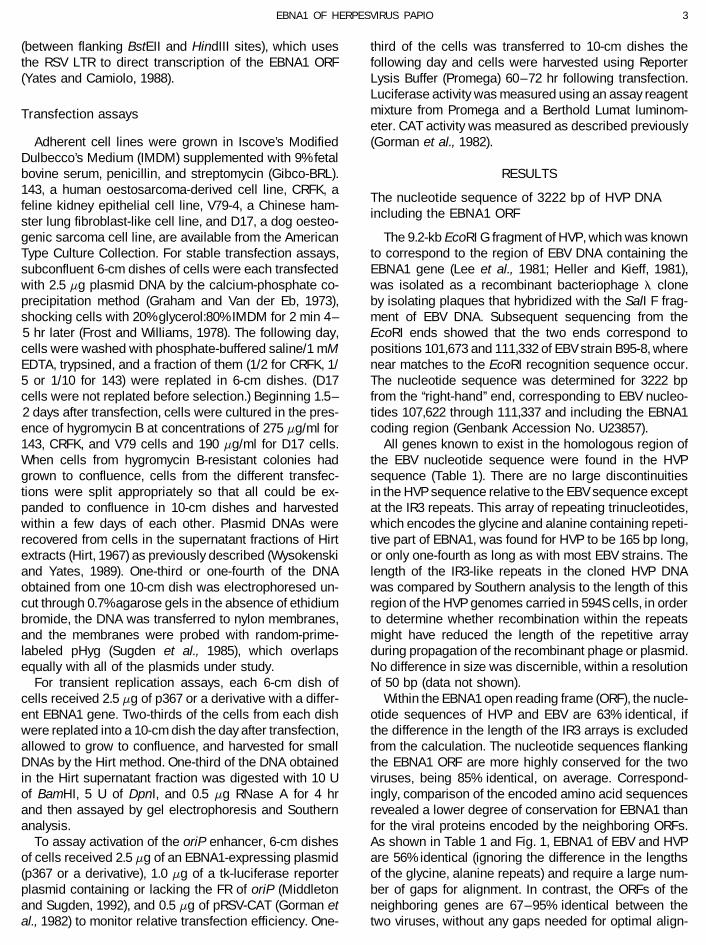
carboxyl-terminal one-third of EBNA1, all but one withinThe salient features of the EBNA1 sequence of EBVthe DNA-binding/dimerization domain. Within the regionare all apparent in the sequence of HVP EBNA1, althoughbetween 470 and 607, for which a crystal structure hasnumerous gaps were needed for alignment of the amino-been determined (Bochkarev et al., 1995), concordantterminal halves of the proteins (Fig. 1). A basic aminopredictions of secondary structure in every case identi-terminus, including a glycine–arginine dipeptide repeat,fied secondary structures that were observed by crystal-precedes a repetitious array of mostly glycine and ala-lography (Helix 1, Beta 1, Helix 3, and perhaps Beta 3 innine beginning at position 95. The glycine and alanineFig. 1). The two-thirds portion of EBNA1 that is amino-containing repetitive part of HVP EBNA1, in addition toterminal to the DNA-binding/dimerization domain isbeing only one-fourth as long as the corresponding partnearly devoid of sequences that appear favorable toof EBV EBNA1, also differs by containing serine and byforming alpha-helical or beta-strand secondary struc-being composed of a perfect repeat of a heptapeptide,tures. Within the amino-terminal domain, predicted alphaGGSGAGA, in contrast to the rather irregular array ofand beta secondary structures were confined to a shorttripeptides that form this part of EBV EBNA1. Followingregion between 411 and 436 of EBV EBNA1, and therethe glycine, alanine repeats, a highly basic region, richwere no matches of predicted structures in this region.in arginine and glycine, ends with a nuclear localizationIt should be noted, however, that just as the algorithmssequence that is nearly identical for the two viruses.failed to predict Helix 2 and Beta 2 within the DNA-bind-Although three gaps were needed for optimal alignmenting/dimerization domain, alpha-helical or beta-sheetof this basic region, this part of EBNA1 is very similar instructures might yet exist within the amino-terminal do-amino acid composition, net charge, and length for themain of EBNA1.two viruses. For both viruses, a cluster of serines follows
the nuclear localization sequence, but for HVP EBNA1Comparison of HVP and EBV EBNA1 in supporting
eight additional serines are included within the next 15oriP functions
amino acids. Following a dissimilar region of 34 or 45amino acids, an acidic, proline-rich region approximately To begin functional studies of HVP EBNA1, we altered
the hygromycin B-selectable plasmid, p205 (Yates et al.,30 amino acids in length is present in both sequences,with about two-thirds sequence identity. Within the DNA 1985), by replacing either the EBV EBNA1 gene or EBV
oriP or both, with homologous DNA from HVP. The plas-binding/dimerization domain, approximately from 458 to617 of EBV EBNA1 (Ambinder et al., 1991), the known mids were used to transfect human 143 cells, which were
then cultured in the presence of hygromycin B. All fourstructural and functional elements appear to have beenconserved (see below). Both EBNA1s have very similar, combinations of EBNA1 and oriP from EBV and HVP al-
lowed efficient transformation to hygromycin B resis-highly acidic carboxyl termini which are identical for theirfinal 9 amino acids. tance, yielding 600–1000 colonies per microgram of
DNA, compared to one-tenth as many with a similar plas-To infer potential secondary structure within theEBNA1 polypeptides, the algorithms of Chou and Fasman mid, pHEBo (Sugden et al., 1985), lacking an EBNA1
AID VY 8032 / 6a1b$$$143 07-07-96 19:10:34 vira AP: Virology
5EBNA1 OF HERPESVIRUS PAPIO
FIG. 1. Comparison of amino acid sequences of EBNA1 of EBV and HVP, with gaps introduced for optimal alignment. The sequences werealigned using the computer program, GAP. The glycine, alanine repeats for each protein (90–324 for EBV) are in italics. Note that 120 amino acidsfrom these repeats of EBV EBNA1 have been skipped. A functional nuclear localization sequence (Ambinder et al., 1991) is underlined in the EBVsequence and indicated by NLS. The positions of the three helical regions and the four beta strands determined from the crystal structure of theEBNA1 fragment, 470–607, (Bochkarev et al., 1995) are indicated with lines over the EBV sequence. Regions favorable to forming alpha-helical orbeta-strand secondary structures were predicted using the algorithms of Chou and Fasman (C-F) and of Garnier, Osguthorpe, and Robson (G-O-R). Positions predicted by both programs to form these secondary structures are indicated by a or b, respectively, above or below each sequence.Positions that favor alpha or beta structures according to G-O-R or strongly favor a structure according to C-F, but not according to both algorithms,are indicated by a or b, for alpha or beta, respectively.
gene. However, the different plasmids produced colonies tion and required cumulative splits equaling 1/60 beforecells were harvested on Day 17; with the similar plasmidthat grew at noticeably different rates under selection.
p205, in which both oriP and EBNA1 are from EBV, al- expressing EBNA1 of HVP, colonies reached confluenceby the 10th day after transfection and could be split onlylowed colonies to grow significantly faster than the simi-
lar plasmid containing oriP of EBV and EBNA1 of HVP. 1/10 before harvest on Day 18.) With HVP oriP, HVPEBNA1 appeared to be slightly better than EBV EBNA1(For example, colonies in duplicate plates that had re-
ceived p205 grew to confluence by 8 days after transfec- at supporting colony growth; however, the plasmid hav-
AID VY 8032 / 6a1b$$$143 07-07-96 19:10:34 vira AP: Virology

6 YATES ET AL.
ing both EBNA1 and oriP of HVP was not as effective asp205, in which both elements are from EBV.
To determine whether the preference of each virus’sEBNA1 for its own oriP might be related to structuraldifferences at the OBR of oriP, 144 bp of DNA containingthe OBR of HVP oriP was inserted between EcoRV andHpaI sites of EBV oriP, replacing 140 bp containing theOBR of EBV oriP. Plasmids carrying this hybrid oriP couldsupport rapid growth of drug-resistant colonies in 143cells whether supported by EBNA1 of EBV or of HVP andwere almost as effective as p205. The plasmid havingthe hybrid oriP and EBNA1 of HVP was noticeably moreeffective than the plasmid having both oriP and EBNA1from HVP. These comparisons show that, while EBNA1of each virus has some preference for its own OBR oforiP, the remainder of oriP from EBV, including the familyof repeats, is more effective in the presence of eitherEBNA1 at supporting growth of drug-resistant coloniesusing these plasmids.
To examine how efficiently the plasmids were main-tained as the cells were propagated under selection,
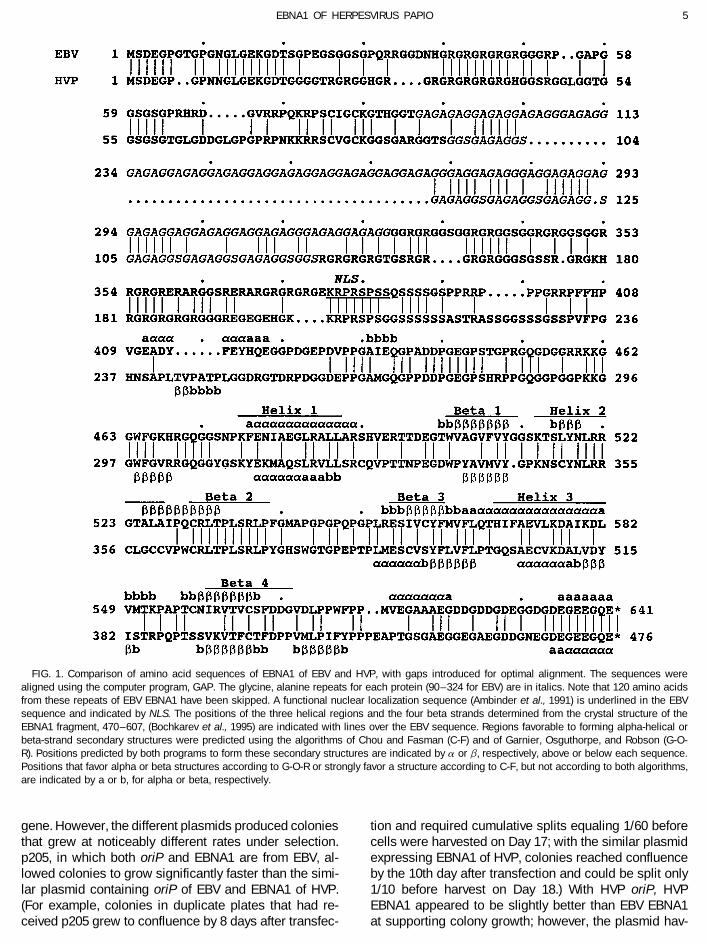
FIG. 2. Maintenance under drug selection of plasmids carrying oriPcolonies from duplicate transfections with each plasmidof EBV or HVP and expressing EBNA1 of EBV or HVP. Duplicate cultures
were allowed to grow to confluence, and then the cul- of 143 cells (upper panel) were transfected with derivatives of p205tures were split and passaged in selective medium until carrying oriP and the EBNA1 gene from EBV (E) or from HVP (P) in
different combinations, as indicated, and grown under selection untilthe 17th or 18th day after transfection. Plasmid DNA was17–18 days following transfection. Free plasmid DNAs were then ex-then extracted from the cells and detected by Southerntracted, electrophoresed through a 0.7% agarose gel, and detected byanalysis. Despite their differences in supporting growthSouthern analysis. ‘‘E/P’’ refers to the hybrid oriP (see text); ‘‘-’’ repre-
of cells under selection, the six plasmids were found to sents use pHEBo, which lacks an EBNA1 gene. The bracket labeledhave been maintained at comparable levels, at multiple ‘‘Ch’’ marks the position of the bulk of linear, fragmented chromosomal
DNA present in the Hirt supernatant fractions. The lower panel repre-copies per cell (Fig. 2, upper panel). A complicating factorsents a similar experiment using D17 cells, from which plasmids werein the analysis was the strong tendency of the plasmidsharvested at 19–22 days posttransfection. In lanes ‘‘Std’’, 50 and 250that carried HVP oriP to form dimers and larger circularpg of p205 were loaded, representing 0.7 and 3.4 plasmid molecules
concatamers during propagation in E. coli, so that only extracted per cell.about 20% of each plasmid preparation was monomeric,although there appeared to be no deletions or other alter-ations at HVP oriP or elsewhere within these plasmids. mids such as p205 in cell lines derived from a dog (D17)
and from a cat (CRFK) as well as from primates, whileIn human cells, the monomeric circular form was notmaintained as well as the concatameric forms when EBV cell lines derived from rodents have consistently been
found to be nonpermissive for replication (Yates et al.,EBNA1 was used, while the monomeric form was main-tained, in addition to the concatamers, when HVP EBNA1 1985; Wysokenski and Yates, 1989; Clemens and Carl-
son, 1989). p205 and its derivative containing the EBNA1was used. In situations where replication or segregationof a plasmid is less than fully efficient, dimers or larger gene of HVP were tested in dog (D17), cat (CRFK), and
hamster (V79) cell lines. Both plasmids were maintainedconcatamers of the plasmid will be maintained more ef-fectively than the monomeric form (e.g., see Yates and at similar copy levels under selection in CRFK cells, with
HVP EBNA1 resulting is somewhat slower rates ofCamiolo, 1988; Brun et al., 1995). This suggests that HVPEBNA1 supports the replication and/or maintenance growth, and neither plasmid could replicate in hamster
V79 cells (data not shown). With D17 cells, HVP EBNA1functions of HVP oriP somewhat more effectively in 143cells than does EBV EBNA1. resulted in a slower rate of growth under selection and
also a lower number of plasmid molecules that wereGiven that EBNA1 of EBV and EBNA1 of HVP differ at44% of their amino acids, it seemed reasonable to expect maintained per cell in the selected populations, about
25% of the level obtained with EBV EBNA1 in the experi-that they might differ in their ability to support replicationin some mammalian host species with which they did ment of Fig. 2 (lower panel) and about 10% of the EBV
EBNA1 level in the experiment of Fig. 3B (lower panel).not evolve and, thus, in which the possibility of productiveinteractions between EBNA1 and host factors would in- In D17 cells, EBV EBNA1 was also more effective than
HVP EBNA1 at supporting selective maintenance of theclude an element of chance. EBV EBNA1 has beenshown to support the replication of oriP-carrying plas- plasmids containing the hybrid oriP, in which the OBR
AID VY 8032 / 6a1b$$$143 07-07-96 19:10:34 vira AP: Virology

7EBNA1 OF HERPESVIRUS PAPIO
of repeats of oriP, when positioned similarly on a plasmidrelative to this promoter, is known to enhance its activityby 20-fold or more (Reisman and Sugden, 1986). There-fore, weaker enhancer activation by HVP EBNA1 couldbe responsible for the observed slower rate of growthunder selection. A plasmid expressing EBV EBNA1, buthaving a derivative of oriP lacking the OBR so that repli-cation would be blocked, was used in a comparison withp205 and its derivative that expresses HVP EBNA1. TheOBR-deleted plasmid expressing EBV EBNA1 gave colo-nies that grew much more rapidly for the first 10 to 14days under selection (before aborting) than colonies ob-tained using the oriP-containing plasmid expressing HVPEBNA1. This comparison indicated that inefficient repli-cation by the HVP EBNA1-supported plasmid could notby itself account for the slower initial growth rate of thecells under selection; either less effective plasmid main-tenance and/or less efficient expression of the hygro-mycin B-resistance gene must be at least partly responsi-ble. Activation of the enhancer of EBV oriP was measuredfor each EBNA1 using a reporter plasmid that expressesthe luciferase gene from the HSV thymidine kinase pro-moter. In such assays using 143 cells, HVP EBNA1 wasless than half as effective as EBV EBNA1 (Table 2), andsimilar results were obtained using D17 cells (data notshown). The relative efficiency of replication of plasmidscarrying oriP of EBV and either EBNA1 were also mea-sured after transient transfection of cells. In human 143
TABLE 2FIG. 3. Long-term maintenance of plasmids carrying oriP of EBV
Enhancer Activation by EBV EBNA1, HVP EBNA1,and expressing hybrid EBNA1 genes. See the Fig. 2 legend for theand Hybrid EBNA1experimental design and an explanation of symbols. The designations
of hybrid EBNA1 molecules are explained in the text. (A) Human 143Relativecells and dog D17 cells were grown under selection for 24–26 days
EBNA1 FR of EBV oriP luciferase activityaposttransfection and 33–39 days posttransfection, respectively. As astandard (Std.), 100 pg of p205 was used, representing 1.4 plasmid
Experiment 1molecules per cell. For 143 cells transfected with the plasmid express-ing P-E(438), DNA from only half the number of cells was analyzed as
EBV 0 3.6was analyzed for the other samples. (B) DNA was isolated from 143EBV / 163cells 18 days after transfection and from D17 cells 21–25 days afterHVP 0 2.5transfection. Lanes ‘‘Stds’’ received 100 and 30 pg of p205, representingHVP / 721.4 and 0.4 molecules per cell.
Experiment 2
was from HVP oriP (Fig. 2, lower panel). The plasmids EBV (dl56)b / 0.9, 1.1carrying HVP oriP were maintained in D17 cells equally EBV / 251, 273
HVP / 95, 108well by either EBNA1 (Fig. 2, lower panel), as though theE-P(437) / 241, 274preference for HVP EBNA1 by HVP oriP was balancedP-E(438) / 66, 61by the greater ability of EBV EBNA1 to support plasmid
maintenance in D17 cells. a For the first experiment, tk reporter-luciferase plasmids containingThe slower rate of growth under selection of cells car- or lacking the family of repeats of EBV oriP (Middleton and Sugden,
1992) were mixed with an EBNA1-expressing plasmid and with pRSV-rying plasmids supported by HVP EBNA1 could result,CAT (Gorman et al., 1982) to cotransfect 143 cells. Ratios of luciferasetheoretically, from a reduced efficiency of replication oractivity to CAT activity are given in arbitrary units. The CAT-expressingof plasmid retention by the cells. However, plasmidsplasmid was not used in the second experiment, for which values from
such as p205 use the relatively weak thymidine kinase duplicate transfections are given.gene promoter from herpes simplex virus to direct tran- b The deletion, dl56 (Yates and Camiolo, 1988), eliminates EBNA1’s
DNA binding activity.scription of the hygromycin B-resistance gene; the family
AID VY 8032 / 6a1b$$$143 07-07-96 19:10:34 vira AP: Virology

8 YATES ET AL.
initially as well as EBV EBNA1, but proliferation ceasedafter 2 weeks and most colonies slowly died, consistentwith a failure of the plasmid to replicate. Free monomericor dimeric plasmid could not be detected in the resistantpopulations of cells that emerged (Fig. 3A).
The activities of hybrid EBNA1 proteins, P-E(438), P-E(457), and P-E(487), having the amino terminus fromHVP and the carboxyl terminus from EBV appeared tobe altered in the opposite way. These hybrid EBNA1supported slow growth of colonies under selection, simi-lar to HVP EBNA1, yet plasmids were present at levelsthat were at least as high or higher than those obtainedwith EBV EBNA1 (Figs. 3A and 3B). (Note that in Fig. 3A,FIG. 4. Replicational activities of different EBNA1 molecules in tran-lanes P–E(438) for 143 cells, DNA from only half as manysiently transfected cells. Human 143 cells or dog D17 cells were
transfected in duplicate with derivatives of p367, which carry EBV oriP cells was loaded as with the remaining samples. Thisand express EBNA1 of EBV or HVP or a hybrid EBNA1, as designated was done to minimize distortion resulting from chromo-in the previous figures. A deletion in the EBNA1 gene, dl56 (Yates and somal DNA that was not effectively removed from oneCamiolo, 1988), that abolishes DNA binding was used as a control
of the samples.) Plasmid copy levels were particularly(lanes ‘‘E0’’). DNA was extracted from cells 3.5 days after transfection,elevated in D17 cells with two of the hybrids, P-E(438)digested with BamHI to linearize the plasmids and with DpnI to degrade
unreplicated plasmid DNA, and analyzed by the Southern method (see and P-E(457). An EBNA1 hybrid in which amino acidsMaterials and Methods). To verify digestion by DpnI, 2 ng of p367 438–486 of EBV EBNA1 were replaced with the homolo-DNA was mixed with DNA extracted from mock-transfected cells and gous region of HVP EBNA1, E-P-E(437,487), appeared toanalyzed in parallel (lane ‘‘Co.’’). Lanes ‘‘M’’ at the left and right received
function identically to EBV EBNA1 in 143 cells and in30 and 60 pg, respectively, of linear p367 DNA.D17 cells (Fig. 3B). These results imply that the region ofEBNA1 that allows more rapid growth of colonies underselection and more efficient enhancer activation iscells and in dog D17 cells, EBV and HVP EBNA1 sup-amino-terminal to position 438; the region that allowsported similar levels of plasmid replication, indicated byefficient replication from EBV oriP in D17 cells is car-the amount of full-length DpnI-resistant plasmid detectedboxyl-terminal to position 487.by Southern analysis (Fig. 4).
The abilities of E-P(437) and P-E(438) to support thereplicational and enhancer functions of EBV oriP wereHybrid EBNA1 proteins with altered activitiesmeasured in transiently transfected cells. In the en-hancer activation assays, E-P(437) was as active as EBVTo investigate what regions of the EBNA1 proteins
were responsible for their differences, hybrid genes were EBNA1, while P-E(438) was somewhat weaker than HVPEBNA1 (Table 2), consistent with the relative abilities ofconstructed to generate hybrid EBNA1 proteins, with the
amino-terminal domain originating from one virus and the hybrid proteins to support growth of drug-resistantcolonies. In transient replication assays using 143 cells,the carboxyl-terminal domain from the other. The gene
fusions were made using restriction sites that were pres- the hybrid EBNA1 proteins could not be distinguishedfrom EBV EBNA1 (Fig. 4). Any differences in the replica-ent in at least one of the genes, and in every case the
precise alignment shown in Fig. 1 was preserved. For tion activities of these proteins that might account for thealtered plasmid copy levels obtained under selection areexample, the hybrid designated E-P(437) contains co-
dons 1–437 of EBV EBNA1 joined to 272–476 of HVP too small to be detected after just a few cell divisions.In D17 cells, however, the activities of the hybrid EBNA1sEBNA1, and was constructed using an ApaI site present
in both genes. P-E(438) represents the reciprocal hybrid, differed enough to be detected easily. E-P(437) sup-ported only one-fifth the level of replication that was ob-with the number in parentheses always designating the
codon on the EBV side of the junction. Each hybrid tained with EBV EBNA1; this activity was barely detect-able in the assay. In contrast, P-E(438) gave elevatedEBNA1 gene was used to replace the EBV EBNA1 gene
of p205 to allow testing in combination with EBV oriP. E- levels of transient replication in D17 cells.In an attempt to localize the region within the carboxyl-P(437) supported rapid growth of colonies under selec-
tion in 143 cells, and in this respect was similar or identi- terminal domain of EBNA1 that allows efficient replica-tion of EBV oriP in D17 cells, three different hybrids werecal to EBV EBNA1 and unlike HVP EBNA1, which pro-
duced slower growth under selection. Surprisingly, the made containing a junction before or after amino acid546 or 548 of EBV EBNA1, within the ‘‘proline loop’’ be-expanded cultures of cells were found to carry signifi-
cantly lower levels of plasmid than were maintained with tween beta strands 2 and 3 (Bochkarev et al., 1995).None of the hybrids exhibited any EBNA1 activity in theEBV EBNA1 (Fig. 3A). When tested in D17 cells, E-P(437)
supported the rapid growth of drug-resistant colonies selective plasmid maintenance assays. The efficiencies
AID VY 8032 / 6a1b$$$143 07-07-96 19:10:34 vira AP: Virology

9EBNA1 OF HERPESVIRUS PAPIO
of colony formation were as low as that of the control sequence between EBV and HVP supports the notionadvanced from crystallography that its unusual structureplasmid lacking the EBNA1 gene, and free plasmids
could not be detected in the drug-resistant cells that is crucial for proper tertiary folding (Bochkarev et al.,1995). Another region of interest is the ‘‘recognition helix’’emerged (Fig. 3B). The amino-terminal half of the DNA-
binding/dimerization domain of either EBNA1 is thus in- (Helix 2 in Fig. 1), named by analogy to its structuralcounterpart within papillomavirus E2 (Bochkarev et al.,compatible with the carboxyl-terminal half of this domain
of the other EBNA1. 1995). Several amino acids of this helical region werelisted as candidates for making base-specific contactswith DNA: K514, T515, Y518, N519, R521, and R522. OfDISCUSSIONthese, only T515 is not identical in HVP EBNA1.
We isolated and studied the EBNA1 gene of HVP with Apart from the recognition helix and the second betathe expectation that differences and similarities between strand, the extent of sequence identity between EBV andit and EBV EBNA1 might reveal functionally important HVP is only 52% within the remaining regions of alpharegions of the proteins. The comparison appears to con- or beta secondary structure in the DNA-binding/dimeriza-firm the importance of certain regions of EBNA1 to which tion domain and only 41% for the sequences connectingfunctions have been assigned, but it also raises ques- these regions. The functional studies, as well as the pre-tions as to why some regions of the proteins have been dictions of secondary structure summarized in Fig. 1,particularly well conserved. This sequence comparison imply that the two EBNA1 proteins adopt very similarshould aid future studies that employ site-directed muta- structures, despite significant divergence of sequence.genesis to analyze EBNA1’s functions. Some specific ex- However, we found that the amino-terminal half of theamples are considered below. DNA-binding/dimerization domain from either virus is in-
compatible with the carboxyl-terminal half of the domainThe DNA-binding/dimerization domainfrom the other virus, when joined at the ‘‘proline loop’’that connects the second and third beta strands. ThisThe best understood part of EBNA1 is its DNA-binding/
dimerization domain, which extends approximately from indicates that some of the nonconserved amino acidsare only tolerated in the context of the protein in whichamino acid 458 to 611–617 near the carboxyl terminus
(Ambinder et al., 1991). This functional domain can be they occur.The proline loop of EBNA1 was suggested as a likelyconsidered to consist of a core, encompassing the four
beta strands shown in Fig. 1, which supports dimerization region to form functional contacts with cellular proteins(Bochkarev et al., 1995), and it is conceivable that thisand weak binding to DNA, and an amino-terminal exten-
sion which strengthens DNA binding (Ambinder et al., loop was rendered nonfunctional in the hybrid proteinsmentioned above. However, neither possibility is likely.1991; Chen et al., 1993, 1994). A crystal structure was
determined for the central core, revealing a beta-barrel The first half of this loop of HVP EBNA1 has severalnonconservative differences. Moreover, replacement ofstructure similar to that of the papillomavirus E2 protein
(Bochkarev et al., 1995). central part of the loop spanning three of the prolines(547–551) with nonconservative substitutions had no no-Within the DNA-binding/dimerization domain, two
short regions have been particularly well conserved. One ticeable effect on the activity of EBV EBNA1 (J.L.Y. andS.M.C., unpublished data).highly conserved block includes a 12-of-14 match begin-
ning at 460 of EBV EBNA1, at the beginning of the DNA-binding domain. A synthetic peptide composed of amino The amino-terminal domainacids 458–478 of EBNA1 was shown to bind to DNAnonspecifically when dimerized, and several mutations Alignment of the amino-terminal two-thirds of the two
EBNA1 sequences requires a large number of gaps, indi-within this region of EBNA1 were shown to reduce oreffectively eliminate EBNA1’s DNA-binding activity (Chen cating significant structural differences between the two
proteins in this region. This is consistent with the notionet al., 1994). In that study, all substitution mutations withinthe conserved block that interfered with DNA binding that the functions of this region of EBNA1 lack the kind
of structural requirements that apply to the DNA-binding/changed amino acids that EBV and HVP have in common,while both of two harmless double substitutions, 467 – dimerization domain, an idea that is supported by experi-
mental data. This broad region of EBNA1 is required for468 and 474–475, were at nonconserved positions. Theother highly conserved region of the DNA-binding/dimer- activation of replication and transcriptional enhancement
from oriP (Yates and Camiolo, 1988; Polvino-Bodnar andization domain, a 12-of-14 match beginning at position529 of EBV EBNA1, includes the second beta strand in Schaffer, 1992), for FR-specific plasmid retention in cells
(Middleton and Sugden, 1994), and for the physical prop-which all 10 amino acids are identical for the two viruses.This beta strand has an unusual structure that is also erty of linking together the two elements of oriP (Gold-
smith et al., 1993; Mackey et al., 1995). At least threepresent in the corresponding beta strand of the papillo-mavirus E2 protein, and the absolute conservation of its regions of EBNA1, between positions 54 and 391 and
AID VY 8032 / 6a1b$$$144 07-07-96 19:10:34 vira AP: Virology

10 YATES ET AL.
FIG. 5. Comparison of the amino-terminal sequences of the rat ribosomal protein S2, EBV EBNA1, and HVP EBNA1. Beneath the rat S2 sequence,vertical lines indicate identity with both EBNA1 proteins, while ‘‘e’’ and ‘‘p’’ indicate identity between S2 and either EBV EBNA1 (e) or HVP EBNA1(p). ‘‘:’’ indicates similarly charged amino acids. The first 53 amino acids of S2 share 47% identity with EBV EBNA1 and 53% identity with HVP EBNA1.Each occurrence of RGG is underlined.
interrupted by the glycine, alanine repeats, contribute in et al., 1994), a property for which a biological functionhas yet to be discovered.an additive way to the DNA-linking activity (Mackey et al.,
1995). Most of the amino-terminal two-thirds of EBNA1,Functional differences between EBNA1sexcluding the glycine, alanine repeats, contributes toof EBV and HVPtranscriptional and replicational activities, although no
single region is essential for activity, suggesting againEBNA1 of either EBV or HVP was shown to support
that several regions contribute additively (Yates andthe functions of oriP from either virus, indicating that the
Camiolo, 1988; J.L.Y. and S.M.C., unpublished). Suchbiological functions of these viral components and their
functional independence of these parts of the amino-molecular mechanisms have largely been conserved.
terminal domain suggests that they are unlikely to func-However, EBNA1 of EBV and HVP did not function identi-
tion interdependently as a highly organized structure.cally when assayed using either oriP, and this should be
Despite the large number of gaps in the alignment, considered when comparing their amino acid se-the most salient features of the amino-terminal part of quences.EBNA1 have been conserved between the two viruses HVP EBNA1 was observed to be only one-half to one-(see Results). After alignment, the extent of sequence fourth as effective as EBV EBNA1 at activating the FRidentity is as high for this region of the protein as it is enhancer of EBV oriP, and experiments using hybridfor the carboxyl-terminal portion. In some regions, the EBNA1 proteins indicated that differences in the amino-conservation of sequence cannot be explained by what terminal two-thirds of the EBNA1 proteins are responsi-is known about function. For two regions that have been ble for the difference. This is consistent with the previousparticularly well-conserved, the amino-terminal 20 amino finding that amino-terminal domain plays an essentialacids and amino acids 423 through 454, deletions that role in transcriptional activation by EBNA1 (Yates andremoved most of the sequences were found to cause Camiolo, 1988). These results also imply that the involve-only minimal impairment of EBNA1’s known activities ment of amino-terminal domain in transcriptional activa-(Yates and Camiolo, 1988). tion is not limited to the indirect role of targeting to pro-
An interesting feature of the amino terminus of EBNA1 tein to the nucleus, as suggested by one study (Ambinderis that it bears a weak resemblance to the amino termi- et al., 1991). In other studies we have shown that certainnus of the rat ribosomal protein, S2 (Suzuki et al., 1991), large deletions within the amino-terminal domain abolishas shown in Fig. 5. Including HVP EBNA1 in the compari- transcriptional activation and replication activity but doson lends some credence to the notion that the amino not impair nuclear localization or prevent auto-repressionterminus of EBNA1 might have been acquired from ribo- of the EBV EBNA1 promoter, Qp (J.L.Y. and S.M.C., unpub-somal protein S2 during evolution (the human and rat S2 lished data).sequences are probably nearly identical (Suzuki et al., HVP EBNA1 was also less effective at maintaining a1991)). For example, preceding the alternating glycines plasmid carrying EBV oriP in cells under selection, re-and arginines that begin at position 40 of EBV EBNA1, sulting in fewer plasmid molecules maintained per cellan insertion of four amino acids is present in EBV EBNA1 in dog D17 cells. Such a difference, although less inrelative to HVP EBNA1; however, HVP EBNA1 and S2 magnitude, was sometimes observed using human 143sequences align in this region without a gap. In fact, cells (data not shown). Differences between the carboxyl-each EBNA1 is more similar to S2 at the 12 to 16 amino terminal domains of the EBNA1 proteins were found toacids preceding the glycine–arginine repeats than they be responsible for this difference. A possible reason forare to each other. On both sides of the glycine–arginine this result could be that HVP EBNA1 cannot interact asrepeats, all three sequences contain ‘‘RGG’’ motifs, which effectively with EBV oriP, in particular at the OBR, as EBVare common to RNA-binding proteins (Kiledjian and Drey- EBNA1 does; the functional components of oriP of thefuss, 1992), although these motifs do not align among two viruses differ, e.g., in the number of EBNA1 bindingthe three sequences. This region is suspected of contrib- sites they contain and in other ways (Loeb et al., 1990;
discussed by Yates, 1996). Another possible factor influ-uting to EBNA1’s nonspecific affinity for RNA (Snudden
AID VY 8032 / 6a1b$$$144 07-07-96 19:10:34 vira AP: Virology

11EBNA1 OF HERPESVIRUS PAPIO
encing these results could be differences in the ways that longer periods of time than are genes that support lyticinfection (discussed by Wrightham et al., 1995). The gly-the two EBNA1s interact with host factors. The results of
a deletional analysis of EBNA1 led to the hypothesis cine and alanine containing repeats of EBNA1 appear tobe responsible for preventing epitopes within EBNA1that a host-specific factor, that is needed for initiation of
replication at oriP, interacts with the carboxyl-terminal from being presented by class I HLA for recognition cyto-toxic T lymphocytes (Levitskaya et al., 1995). But if thisdomain of EBNA1 (Yates and Camiolo, 1988).
Hybrid EBNA1 molecules having with the amino-termi- escape mechanism is less than perfect and if EBNA1 isexpressed for the duration of latent infection, as somenal domain from HVP and the carboxyl-terminal domain
from EBV (P-E hybrids) are noteable because while acti- data suggest (Tierny et al., 1994; Chen et al., 1995), signif-icant pressure could exist to eliminate epitopes of CTL.vation of the FR enhancer was significantly weaker than
with EBV EBNA1, support of oriP-dependent replication The frequency and nature of intrastrain differences withinthe carboxyl-terminal domain of EBNA1 seem to supporteither was not different or was more efficient than with
EBV EBNA1. In our previous study, deletions introduced this possibility (Wrightham et al., 1995). HVP EBNA1 con-tains a different kind of glycine-alanine repeat array, onethroughout the EBNA1 gene invariably reduced both rep-
licational and transcriptional activities, at least measur- that is only about one-fourth as long as the array withinEBNA1 of most EBV strains, and is composed of a hep-ably, if they reduced one of the activities, suggesting
mechanistic steps in common to both replicational and tameric repeat unit that includes serine. It will be interest-ing to learn whether this shorter, serine-containing arraytranscriptional activation by EBNA1 (Yates and Camiolo,
1988). Both domains of EBNA1 are clearly required for functions in the same way as the much longer repeatarray of EBV EBNA1.both activities, the carboxyl-terminal domain for specific
binding to DNA, and the amino-terminal domain, to sup-port EBNA1–EBNA1 interactions, presumably. The re- ACKNOWLEDGMENTSsults with the P-E hybrids indicate that activation of tran-
We thank Dr. Joe Pagano and Dr. Dan Loeb for providing us withscription and activation of replication by EBNA1 need not the HVP reagents and DNA sequence, respectively. This work wasbe linked and, indeed, might be competing activities. supported by Grant CA43122 from the N.I.H.
Some evidence suggests that transcription from EBV-derived plasmids can limit the number of plasmids that REFERENCESreplicate and reduce the number that are maintained by
Adams, A. (1987). Replication of latent Epstein–Barr virus genomes incells over several generations. Replication of EBV-de- Raji cells. J. Virol. 61, 1743–1746.rived plasmids was shown to decrease in relation to the Ambinder, R. F., Mullen, M. A., Chang, Y.-N., Hayward, G. S., and Hay-
ward, S. D. (1991). Functional domains of Epstein–Barr virus nuclearstrength of transcription from promoters placed on theantigen EBNA-1. J. Virol. 65, 1466–1478.plasmids (Haase et al., 1994), and blocking transcription
Bochkarev, A., Barwell, J. A., Pfuetzner, R. A., Furey, W., Edwards,from an oriP-replicated plasmid by methylating cytosinesA. M., and Frappier, L. (1995). Crystal structure of the DNA-binding
at CG sites was found to increase plasmid copy levels domain of the Epstein–Barr virus origin-binding protein EBNA1. Celland stability (Hsieh, 1994). Consistent with these obser- 83, 39–46.
Brun, C., Dubey, D., and Huberman, J. A. (1995). pDblet, a stable autono-vations, P-E hybrid EBNA1 might allow more efficientmously replicating shuttle vector for Schizosaccharomyces pombe.plasmid replication or maintenance because it is lessGene 164, 173–177.effective in activating transcription from the plasmid,
Buell, G. N., Reisman, D., Kintner, C., Crouse, G., and Sugden, B. (1981).thereby interfering less with replication and/or mainte- Cloning overlapping DNA fragments from the B95-8 strain of Ep-nance. stein–Barr virus reveals a site of homology to the internal repetition.
J. Virol. 40, 977–982.Chen, M.-R., Middeldorp, J. M., and Hayward, S. D. (1993). SeparationProtein sequence divergence and cytotoxic immunity
of the complex DNA binding domain of EBNA-1 into DNA recognitionand dimerization subdomains of novel structure. J. Virol. 67, 4875–
It seems worth noting that the EBNA1 proteins of HVP 4885.and EBV, which function during latent infection, have di- Chen, M.-R., Zong, J., and Hayward, S. D. (1994). Delineation of a 16
amino acid sequence that forms a core DNA recognition motif in theverged in sequence significantly more than four proteinsEpstein–Barr virus EBNA-1 protein. Virology 205, 486–495.of neighboring lytic phase genes that were also se-
Chen, F., Zou, J.-Z., Renzo, L., Wingerg, G., Hu, L.-F., Klein, E., Klein, G.,quenced (Table 1). EBNA2, which like EBNA1, functionsand Ernberg, I. (1995). A subpopulation of normal B cells latently
during latent infection, shares only about 36% sequence infected with Epstein–Barr virus resembles Burkitt Lymphoma cellsidentity with EBV EBNA2, with several gaps in the se- in expressing EBNA-1 but not EBNA-2 or LMP1. J. Virol. 69, 3752–
3758.quence alignment (Ling et al., 1993). A possible reasonChittenden, T., Lupton, S., and Levine, A. J. (1989). Functional limits ofgiven for more frequent intrastrain and intertypic se-
oriP, the Epstein–Barr virus plasmid origin of replication. J. Virol. 63,quence divergence within genes that encode proteins3016–3025.
expressed during latency is that these genes might be Clemens, H. L., and Carlson, J. O. (1989). Regulated expression of theunder greater pressure to eliminate epitopes of cytotoxic feline panleukopenia virus P38 promoter on extrachromosomal FPV/
EBV chimeric plasmids. J. Virol. 63, 2737–2745.immunity because they are expressed in the host for
AID VY 8032 / 6a1b$$$144 07-07-96 19:10:34 vira AP: Virology

12 YATES ET AL.
Falk, L., Deinhardt, F., Nonoyama, M., Wolfe, L. G., Bergholz, C., Lapin, virus Papio: DNA sequence and enhancer function. J. Virol. 64, 2876–2883.B., Yakovleva, L., Agrba, V., Henle, G., and Henle, W. (1976). Proper-
ties of a baboon lymphotropic herpesvirus related to Epstein–Barr Lupton, S., and Levine, A. J. (1985). Mapping genetic elements of Ep-virus. Int. J. Cancer 18, 798– 807. stein–Barr virus that facilitate extrachromosomal persistence of Ep-
Frost, E., and Williams, J. (1978). Mapping temperature-sensitive and stein–Barr virus derived plasmids in human cells. Mol. Cell. Biol. 5,host-range mutations of adenovirus type 5 by marker rescue. Virology 2533–2542.91, 39–50. Mackey, D., Middleton, T., and Sugden, B. (1995). Multiple regions of
Gahn, T. A., and Schildkraut, C. L. (1989). The Epstein–Barr virus origin EBNA1 can link DNAs. J. Virol. 69, 6199–6208.of plasmid replication, oriP, contains both the initiation and termina- Maniatis, T., Fritsch, E. F., and Sambrook, J. (1982). ‘‘Molecular Cloning:tion sites of DNA replication. Cell 58, 527–535. A Laboratory Manual,’’ Cold Spring Harbor Laboratory Press, Cold
Gahn, T. A., and Sugden, B. (1995). An EBNA-1-dependent enhancer Spring Harbor, New York.acts from a distance of 10 kilobase pairs to increase expression of Middleton, T., and Sugden, B. (1992). A chimera of EBNA1 and thethe Epstein–Barr virus LMP gene. J. Virol. 69, 2633–2636. estrogen receptor activates transcription but not replication. J. Virol.
Gerber, P., Kalter, S. S., Schidlovsky, G., Petrson, W. D., and Daniel, 66, 1795–1798.M. D. (1977). Biologic and antigenic characteristics of Epstein–Barr Middleton, T., and Sugden, B. (1994). Retention of plasmid DNA invirus-related herpesviruses of chimpanzees and baboons. Int. J. Can- mammalian cells is enhanced by binding of the Epstein–Barr viruscer 20, 448–459. replication protein EBNA1. J. Virol. 68, 4067–4071.
Goldsmith, K., Bendell, L., and Frappier, L. (1993). Identification of Miller, G. (1990). Epstein–Barr virus, biology, pathogenesis, and medi-EBNA1 amino acid sequences required for the interaction of the cal aspects. In ‘‘Virology’’ (B. N. Fields et al., Eds.), pp. 1921–1957.functional elements of the Epstein–Barr virus latent origin of DNA Raven Press, New York.replication. J. Virol. 67, 3418 – 3426. Miyashita, E. M., Yang, B., Lam, K. M., Crawford, D. H., and Thorley-
Gorman, C. M., Merlino, G. T., Willingham, M. C., Pastan, I., and Howard, Lawson, D. A. (1995). A novel form of Epstein–Barr virus latency inB. H. (1982). The Rous sarcoma virus long terminal repeat is a strong normal B cells in vivo. Cell 80, 593–601.promoter when introduced into a variety of eukaryotic cells by DNA- Pesano, R. L., and Pagano, J. S. (1986). Herpesvirus papio contains amediated transfection. Proc. Natl. Acad. Sci. USA 79, 6777–6781. plasmid origin of replication that acts in cis interspecies with an
Graham, F. L., and Van der Eb, A. J. (1973). A new technique for the Epstein–Barr virus trans-acting function. J. Virol. 60, 1159–1162.assay of infectivity of human adenovirus 5 DNA. Virology 52, 456– Polvino-Bodnar, M., and Schaffer, P. A. (1992). DNA binding activity is467. required for EBNA 1-dependent transcriptional activation and DNA
Haase, S. B., Heinzel, S. S., and Calos, M. P. (1994). Transcription replication. Virology 187, 591–603.inhibits the replication of autonomously replicating plasmids in hu-
Qu, L., and Rowe, D. T. (1992). Epstein–Barr virus latent gene expres-man cells. Mol. Cell. Biol. 14, 2516–2524.
sion in uncultured peripheral blood lymphocytes. J. Virol. 66, 3715–Harrison, S., Fisenne, K., and Hearing, J. (1994). Sequence requirements
3721.of the Epstein–Barr virus latent origin of DNA replication. J. Virol. 68,
Rabin, H., Neubauer, R. H., Hopkins, R. F., Dzhikidze, E. K., Shevtsova,1913–1925.Z. V., and Lapin, B. A. (1977). Transforming activity and antigenicityHeller, M., and Kieff, E. (1981). Colinearity between the DNAs of Ep-of an Epstein–Barr-like virus from lymphoblastoid cell lines of ba-stein–Barr virus and herpesvirus papio. J. Virol. 37, 821–826.boons with lymphoid disease. Intervirology 8, 240–249.Hirt, B. (1967). Selective extraction of polyoma DNA from infected
Reisman, D., and Sugden, B. (1986). Trans-activation of an Epstein–mouse cell cultures. J. Mol. Biol. 26, 365–369.Barr viral transcriptional enhancer by the Epstein–Barr viral nuclearHsieh, C.-L. (1994). Dependence of transcriptional repression on CpGantigen 1. Mol. Cell. Biol. 6, 3838–3846.methylation density. Mol. Cell. Biol. 14, 5487–5494.
Reisman, D., Yates, J. L., and Sugden, B. (1985). A putative origin ofKieff, E., and Liebowitz, D. (1990). Epstein–Barr Virus and its replication.replication of plasmids derived from Epstein–Barr virus is composedIn ‘‘Virology’’ (B. N. Fields, D. M. Knipe, et al., Eds.), pp. 1889–1920.of two cis-acting components. Mol. Cell. Biol. 5, 1822–1832.Raven Press, New York.
Snudden, D. K., Hearing, J., Smith, P. R., Grasser, F. A., and Griffin,Kiledjian, M., and Dreyfuss, G. (1992). Primary structure and bindingB. E. (1994). EBNA-1, the major nuclear antigen of Epstein–Barr virus,activity of the hnRNP U protein: Binding RNA through RGG box.resembles ‘RGG’ RNA binding proteins. EMBO J. 13, 4840–4847.EMBO J. 11, 2655–2664.
Sugden, B., Marsh, K., and Yates, J. L. (1985). A Vector that replicatesKlein, G. (1994). Epstein–Barr virus strategy in normal and neoplasticas a plasmid and can be efficiently selected in B-lymphoblasts trans-B cells. Cell 77, 791–793.formed by Epstein–Barr virus. Mol. Cell. Biol. 5, 410–413.Kraft, R., Tardiff, J., Krauter, K. S., and Leinwand, L. A. (1988). Using
Sugden, B., and Warren, N. (1989). A promoter of Epstein-Barr virusmini-prep plasmid DNA for sequencing double stranded templateswhich can function during latent infection can be trans-activated bywith Sequenase. BioTechniques 6, 544–546.EBNA1, a viral protein required for DNA replication during latentKrysan, P. J., Haase, S. B., and Calos, M. P. (1989). Isolation of humaninfection. J. Virol. 63, 2644–2649.sequences that replicate autonomously in human cells. Mol. Cell.
Suzuki, K., Olvera, J., and Wool, I. G. (1991). Primary structure of ratBiol. 9, 1026–1033.ribosomal protein S2. J. Biol. Chem. 266, 20007–20010.Lee, Y. S., Tanaka, A., Lau, R. H., Nonoyama, M., and Rabin, H. (1981).
Tierney, R. J., Steven, N., Young, L. S., and Rickinson, A. B. (1994).Linkage map of the fragments of herpesvirus papio DNA. J. Virol. 37,Epstein–Barr virus latency in blood mononuclear cells: Analysis of710–720.viral gene transcription during primary infection and in the carrierLevitskaya, J., Coram, M., Levitsky, V., Imreh, S., Steigerwald-Mullen,state. J. Virol. 68, 7374–7385.P. M., Klein, G., Kurilla, M. G., and Masucci, M. G. (1995). Inhibition
Wrightham, M. N., Stewart, J. P., Nusrat, J. J., Pepper, S. D. V., Sample,of antigen processing by the internal repeat region of the Epstein-C., Rooney, C. M., and Arrand, J. R. (1995). Antigenic and sequenceBarr virus nuclear antigen-1. Nature 375, 685–658.variation in the C-terminal domain of the Epstein-Barr virus nuclearLing, P. D., Ryon, J. J., and Hayward, S. D. (1993). EBNA-2 of herpesvirusantigen EBNA-1. Virology 208, 521–530.papio diverges significantly from the type A and type B EBNA-2
proteins of Epstein–Barr virus but retains an efficient transactivation Wysokenski, D. A., and Yates, J. L. (1989). Multiple EBNA1-binding sitesare required to form an EBNA1-dependent enhancer and to activatedomain with a conserved hydrophobic motif. J. Virol. 67, 2990–3003.
Loeb, D., Schwab, D., Sung, N., Pesano, R. L., Sexton, C. J., Hutchison, a minimal replicative origin within oriP of Epstein-Barr virus. J. Virol.63, 2657–2666.C., and Pagano, J. S. (1990). Plasmid origin of replication of herpes
AID VY 8032 / 6a1b$$$145 07-07-96 19:10:34 vira AP: Virology

13EBNA1 OF HERPESVIRUS PAPIO
Yates, J. L. (1996). Epstein–Barr virus DNA replication. In ‘‘Eukaryotic replicate only once per cell cycle and are not amplified after entryinto cells. J. Virol. 65, 483–488.DNA Replication’’ (M. DePamphilis, Ed.). Cold Spring Harbor Labora-
tory Press, Cold Spring Harbor, New York, in press. Yates, J. L., Warren, N., Reisman, D., and Sugden, B. (1984). A cis-actingelement from the Epstein–Barr viral genome that permits stable repli-Yates, J. L., and Camiolo, S. M. (1988). Dissection of DNA replication and
enhancer activation function of Epstein–Barr virus nuclear antigen 1. cation of recombinant plasmids in latently infected cells. Proc. Natl.Acad. Sci. USA 81, 3806–3810.In ‘‘Cancer Cells: Eukaryotic DNA Replication’’ (B. Stillman and T.
Kelley, Eds.), Vol. 6, pp. 197 – 205. Cold Spring Harbor Laboratory, Yates, J. L., Warren, N., and Sugden, B. (1985). Stable replication ofplasmids derived from Epstein–Barr virus in various mammalianCold Spring Harbor, NY.
Yates, J. L., and Guan, N. (1991). Epstein–Barr virus-derived plasmids cells. Nature 313, 812–815.
AID VY 8032 / 6a1b$$$145 07-07-96 19:10:34 vira AP: Virology