Embed Size (px)
Citation preview

FEBS Letters 582 (2008) 949–955
Activation of the farnesoid X receptor represses PCSK9 expressionin human hepatocytes
Cedric Langhia,b, Cedric Le Maya,b, Sanae Kourimatea,b, Sandrine Carond,e,f, Bart Staelsd,e,f,Michel Krempfa,b,c, Philippe Costeta,b, Bertrand Carioua,b,c,*
a INSERM, U915, CHU Hotel Dieu, 3�emeNORD, Nantes F-44000, Franceb Universite de Nantes, Faculte de Medecine, l�Institut du Thorax, Nantes F-44000, France
c CHU Nantes, Clinique d�Endocrinologie, Maladies Metaboliques et Nutrition, l�Institut du Thorax, Nantes F-44000, Franced Institut Pasteur de Lille, Departement d�Atherosclerose, Lille F-59019, France
e INSERM, U545, Lille F-59019, Francef Universite de Lille 2, Faculte de Pharmacie, Faculte de Medecine, Lille F-59006, France
Received 22 December 2007; revised 12 February 2008; accepted 17 February 2008
Available online 25 February 2008
Edited by Robert Barouki
Abstract The purpose of this study was to determine whetherbile acids (BAs) modulate hepatic pro-protein convertase subtil-isin/kexin 9 (PCSK9) gene expression. Immortalized humanhepatocytes were treated with various BAs. Chenodeoxycholicacid (CDCA) treatment specifically decreased both PCSK9mRNA and protein contents. Moreover, activation of the BA-activated farnesoid X receptor (FXR) by its synthetic specificagonist GW4064 also decreased PCSK9 expression. Of func-tional relevance, coadministration of CDCA counteracted thestatin-induced PCSK9 expression, leading to a potentiation ofLDL receptor activity. This study suggests that a transcriptionalrepression of PCSK9 by CDCA or FXR agonists may potentiatethe hypolipidemic effect of statins.� 2008 Federation of European Biochemical Societies. Publishedby Elsevier B.V. All rights reserved.
Keywords: PCSK9; Bile acid; FXR; Statin; LDL-cholesterol
1. Introduction
Pro-protein convertase subtilisin/kexin 9 (PCSK9) has re-
cently emerged as a central player in the regulation of choles-
terol homeostasis [1]. In addition to mutations affecting the
LDL-receptor (LDLr) and apolipoprotein B, ‘‘gain-of-func-
tion’’ PCSK9 mutations lead to an increase of plasma LDL-
cholesterol (LDL-c) levels and premature atherosclerosis
[1,2]. In contrast, ‘‘loss-of-function’’ mutations are associated
with low levels of LDL-c and confer protection against cardio-
vascular disease [3]. PCSK9 is primarily expressed in the liver
and the intestine. PCSK9 inhibits the LDLr activity in a post-
transcriptional manner [1]. Recent data suggest that, once
Abbreviations: BA, bile acid; CA, cholic acid; CDCA, chenodeoxy-cholic acid; DCA, deoxycholic acid; FXR, farnesoid X receptor; IHH,immortalized human hepatocytes; LA, lithocholic acid; LDL-c, lowdensity lipoprotein cholesterol; LDLr, low density lipoprotein recep-tor; PCSK9, pro-protein convertase subtilisin/kexin 9; PXR, pregnaneX receptor; UDCA, ursodeoxycholic acid
*Corresponding author. Address: INSERM, U915, CHU Hotel Dieu,3�emeNORD, Nantes F-44000, France. Fax: +33 (0) 2 40 28 75 44.E-mail address: [email protected] (B. Cariou).
0014-5793/$34.00 � 2008 Federation of European Biochemical Societies. Pu
doi:10.1016/j.febslet.2008.02.038
cleaved, secreted PCSK9 acts as a chaperone and promotes
the intracellular degradation of the LDLr by interfering with
its recycling to the plasma membrane [4,5].
Various positive and negative regulatory pathways of
PCSK9 have been identified. The hypocholesterolemic drugs
statins were shown to increase PCSK9 expression, a pathway
which exerts a break on their efficiency [6,7]. In accordance
with this negative feedback pathway, PCSK9-deficient mice
[8] and patients bearing non sense mutations for PCSK9 [9]
are more responsive to statins. Our laboratory characterized
the PCSK9 promoter and showed that PCSK9 is also up-reg-
ulated by insulin as well as by the Liver X Receptor agonist
T0901317 via SREBP-1c [10]. In an opposite way, fenofibrate
decreases hepatic PCSK9 expression in a PPARa-dependent
manner [11].
Bile acids (BAs) are liver-synthesized cholesterol-derivatives
that represent the major route for removal of excess cholesterol
from the body. Besides their role as detergents, it has now been
clearly demonstrated that BAs also exert signalling activities
and regulate gene expression in a variety of tissues, including
liver and intestine, at least partly through the activation of
the farnesoid X receptor (FXR), a member of the nuclear
receptor superfamily of ligand-activated transcription factors
[12,13]. In addition to FXR, BAs can also activate other nucle-
ar receptors such as PXR (pregnane X receptor), CAR (consti-
tutive androstane receptor) and vitamin D receptor [14,15].
Here, we investigated whether BAs can modulate the expres-
sion of PCSK9. We found that chenodeoxycholic acid
(CDCA) specifically represses PCSK9 expression in immortal-
ized human hepatocytes, thereby potentiating the activity of
the LDLr in response to statins.
2. Materials and methods
2.1. ChemicalsBAs (CDCA, UDCA, DCA, CA, LA), pravastatin, rifampicin and
actinomycin D were purchased from Sigma (France). GW4064 waskindly provided by Genfit (Loos, France).
2.2. Cell cultureImmortalized human hepatocytes were cultured on collagen-coated
flasks in William�s E medium in the presence of a 10% fetal calf serum(FCS). HepG2 cells were cultured in DMEM containing 10% FCS and1% glutamine. The cells were exposed to various treatments in the
blished by Elsevier B.V. All rights reserved.

950 C. Langhi et al. / FEBS Letters 582 (2008) 949–955
presence of a 5% lipoprotein deficient serum (LPDS) unless notified.For PCSK9 secretion analysis, cells were incubated in 1 ml of mediumwithout FCS and LPDS. Treatment toxicity was assessed by quantifi-cation of lactate dehydrogenase activity in cell medium using theRoche cytotoxicity detection kit (Roche Diagnostics, Indianapolis,USA).
2.3. Western blotsProteins were analysed by Western blot as described elsewhere [10],
using a polyclonal rabbit IgG directed against the CRSRHLAGAS-QELQ peptide (Neosystem, Strasbourg, France), an epitope of theC-terminal domain of human and mouse PCSK9, or with the mono-clonal anti b-actin AC-15 antibody (Sigma). For secretion analysis,proteins from 400 ll of cell culture media were precipitated with ace-tone.
2.4. RNA extraction and real time PCRRNA extraction and real time quantitative PCR (Q-PCR) was per-
formed as previously described [10], using the following primers:LDLR: AAGGCTGTCCCCCCAAGA forward, CGAACTGCC-
GAGAGATGCA reverse; PCSK9: ACGTGGCTGGCATTGCAforward, AAGTGGATCAGTCTCTGCCTCAA reverse; 18S: AAG-
A
mR
NA
rela
tive
leve
ls
UGT2B4/1
DMSOCDCA
U
0.0
1.0
2.0
3.0 ***
0.0
0.5
1.0
1.5
2.0
2.5
DMSOCDCA
UDCADCA
CA
PCSK9/18S
***
***
PCSK9/18S UGT2B4/18S
0.2
0.4
0.6
0.8
1.0
1.2
DMSO CDCA50 M
*** 1
2
3
4
5
DMSO CDCA50 M
***
00.0
mR
NA
rela
tive
leve
ls
C
B
10DMSO
UGT2B4/1
0.0
1.0
2.0
3.0
4.0
5.0
6.0
*
mR
NA
rela
tive
leve
ls
0.2
0.4
0.6
0.8
1.0
1.2
1.4
0.0
CDCA (µM)10 25 50 100DMSO
PCSK9/18S
***
****
µ µ
Fig. 1. CDCA reduces PCSK9 mRNA levels in human hepatocytes cell linQ-PCR. (A) IHH, cells were incubated for 48 h with 50 lM CDCA, CA,increasing concentrations (as indicated) of CDCA for 48 h. (C) HepG2 cells wto 18S mRNA and are expressed (means ± S.E.M.; n = 5) relative to thossignificant differences compared to vehicle-treated cells are indicated (***P <
TCCCTGCCCTTTGTACACA forward, CGATCCGAGGGCCTC-ACTA reverse; UGT2B4: CAACCAGTGAAGCCCCTTGA forward,GAAGGTGCTTGGCTCCTTTATG reverse; SHP: CTCTTCCT-GCTTGGGTTGGC forward, GCACATCGGGGTTGAAGAGGreverse; CYP3A4: CTCTTCACCGTGACCCAAAGTACT forward,AGCAAACCTCATGCCAATGC reverse.
2.5. Isolation and radiolabelling of LDLHuman LDL (d 1.019–1.063 g/ml) was isolated from plasma of
healthy normolipaemic fasting donors by isopycnic preparative ultra-centrifugation using a discontinuous KBr density gradient [16]. Iso-lated LDL was dialysed at least 36 h at 4 �C against PBS pH 7.4.Radiolabelling procedure was performed according to the iodogen�
method modified by Fraker et al. [17].
2.6. Binding of 125I-labelled LDLIHH cells were incubated for 4 h at 4 �C with 10 lg/ml of 125I-
labelled LDL in 250 ll of William�s LPDS 5% containing 4% fattyacid-free BSA and 50 mM Hepes, pH 7.4. Non-specific binding wasdetermined by the addition of 0.5 mg/ml unlabelled LDL. At the endof the incubation period, the cells were washed three times with 1 mlof D-PBS containing 1% BSA then three times with 1 ml of D-PBS.
8S
DCADCA
CA
CDCA ( M)25 50 100
8S
***
****
µ µ
*
0.0
2.0
4.0
6.0
8.0LDLr/18S
CDCA ( M)10 25 50 100DMSO
******
**
0.0
0.5
1.0
1.5
2.0
2.5
DMSOCDCA
UDCADCA
CA
LDLr/18S
*** ***
es. PCSK9, UGT2B4 and LDLr mRNA contents were measured byUDCA, DCA or vehicle (DMSO). (B) IHH cells were treated withere treated with 50 lM CDCA for 48 h. Values are normalized relativee of vehicle-treated cells, which are arbitrarily set at 1. Statistically0.001; **P < 0.01; and *P < 0.05).
C. Langhi et al. / FEBS Letters 582 (2008) 949–955 951
The cells were solubilized in 1 ml of 1 M NaOH, the protein contentwas determined using BSA as a standard, and the radioactivity wasmeasured (1480 Wizard 300 Automatic Gamma Counter, Wallac, Wal-tham, Massachusetts, USA). The measured radioactivity was normal-ized per milligram of cell protein, the specific binding was calculated bysubtracting the non-specific binding of 125I-labelled LDL from the to-tal binding.
2.7. StatisticsResults are representative of at least two independent experiments,
with at least triplicates in each experiment. Statistical significancewas analyzed using an unpaired Student�s t-test. The values ofP < 0.05 were considered significant.
0.5
1.0
1.5
2.0
2.5
3.0
DMSO CDCA50 M
Prava Prava/CDCA
mR
NA
rela
tive
leve
ls
***
***
**
A
DMSO CDCA
actin
PCSK9
B
C
1 ± 0.22 0.35 ± 0.1*
1 ± 0.10 0.23 ± 0.29**
DMSO Prava 5 µM
1
2
3
4
5
*
I125 -
LDLr
elat
ive
spec
ificb
indi
ng
PCSK9/18S
Pro-PCSK9
PCSK9*
µ 5 Mµ
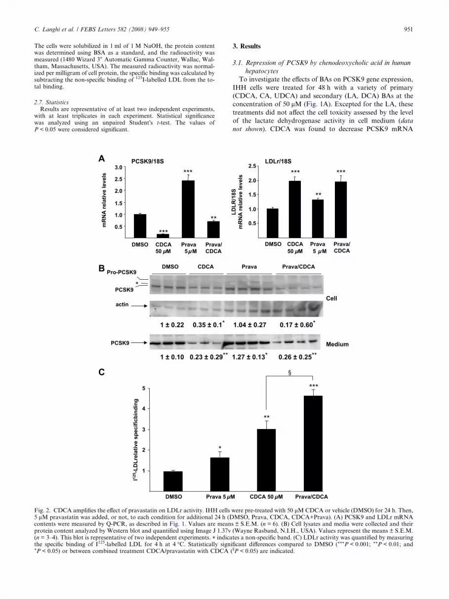
Fig. 2. CDCA amplifies the effect of pravastatin on LDLr activity. IHH cells5 lM pravastatin was added, or not, to each condition for additional 24 h (Dcontents were measured by Q-PCR, as described in Fig. 1. Values are meanprotein content analyzed by Western blot and quantified using Image J 1.37v(n = 3–4). This blot is representative of two independent experiments. * indicathe specific binding of I125-labelled LDL for 4 h at 4 �C. Statistically signi*P < 0.05) or between combined treatment CDCA/pravastatin with CDCA (
3. Results
3.1. Repression of PCSK9 by chenodeoxycholic acid in human
hepatocytes
To investigate the effects of BAs on PCSK9 gene expression,
IHH cells were treated for 48 h with a variety of primary
(CDCA, CA, UDCA) and secondary (LA, DCA) BAs at the
concentration of 50 lM (Fig. 1A). Excepted for the LA, these
treatments did not affect the cell toxicity assessed by the level
of the lactate dehydrogenase activity in cell medium (data
not shown). CDCA was found to decrease PCSK9 mRNA
0.5
1.0
1.5
2.0
2.5
DMSO CDCA Prava Prava/CDCA
LDLR
/18S
mR
NA
rela
tive
leve
ls
**
*** ***
Prava Prava/CDCA
Cell
Medium
1.04 ± 0.27 0.17 ± 0.60*
1.27 ± 0.13* 0.26 ± 0.25**
CDCA 50 µM Prava/CDCA
**
***
§
LDLr/18S
50 Mµ 5 Mµ
were pre-treated with 50 lM CDCA or vehicle (DMSO) for 24 h. Then,MSO, Prava, CDCA, CDCA+Prava). (A) PCSK9 and LDLr mRNA
s ± S.E.M. (n = 6). (B) Cell lysates and media were collected and their(Wayne Rasband, N.I.H., USA). Values represent the means ± S.E.M.tes a non-specific band. (C) LDLr activity was quantified by measuringficant differences compared to DMSO (***P < 0.001; **P < 0.01; and§P < 0.05) are indicated.
952 C. Langhi et al. / FEBS Letters 582 (2008) 949–955
levels (�59%, P < 0.001). In contrast, DCA increased PCSK9
gene expression (+76%, P < 0.001), whereas CA and UDCA
had no effect. Since CDCA is the more potent natural agonist
of FXR [18], the expression of the FXR target-gene UGT2B4
[19] was measured under the same conditions. As expected,
CDCA was the sole BA which significantly increased UGT2B4
mRNA levels (Fig. 1A). The effect of BAs treatment on LDLr
expression was also investigated in IHH cells. In accordance
with previous results in HepG2 cells [20], both CDCA and
DCA increased LDLr mRNA levels (+104%, P < 0.001 and
+109%, P < 0.001, respectively) (Fig. 1A). Notably, a strong
induction of LDLr mRNA levels was observed for the higher
dose of CDCA (+544% with 100 lM, P < 0.001) (Fig. 1B).
Conversely, a significant and dose-dependent repression of
PCSK9 mRNA levels was observed in IHH cells treated with
various doses of CDCA for 48 h (Fig. 1B). Moreover, this
repression of PCSK9 also occurred in HepG2 cells treated with
50 lM CDCA (�87%, P < 0.001) (Fig. 1C). Taken together,
these results demonstrate that CDCA is a new repressor of
PCSK9 gene expression in human hepatocytes.
3.2. CDCA amplifies the effect of pravastatin on LDLr activity
To assess whether PCSK9 upregulation by statins could be
affected by CDCA, we first exposed IHH cells to 50 lM
CDCA or DMSO during 24 h before adding or not 5 lM prav-
astatin for an additional 24 h. As expected [6], 24 h treatment
with pravastatin alone increased PCSK9 mRNA levels
(+141%, P < 0.001). Interestingly, pretreatment with CDCA
abolished the induction of PCSK9 gene expression in response
to pravastatin. The statin only slightly increased LDLr gene
expression (+32%, P < 0.01), as described elsewhere [6]. The
combination of both drugs did not further increase LDLr
mRNA levels compared to CDCA treatment alone
(Fig. 2A). In accordance to the mRNA variations, CDCA
strongly reduced the quantity of both intracellular and secreted
PCSK9 protein content, even in the presence of pravastatin
(Fig. 2B). To further investigate whether CDCA functionally
influences LDLr activity, IHH cells were treated for 48 h with
50 lM CDCA and the surface binding of 125I-labelled human
LDL was assayed (Fig. 2C). Both pravastatin and CDCA
increased the LDLr activity (+72%, P < 0.05 and +204%,
P < 0.001 vs. DMSO, respectively). Interestingly, when a com-
bined treatment with CDCA and pravastatin was performed, a
mR
NA
rela
tive
leve
ls
PCSK9/18S
0.2
0.4
0.6
0.8
1.0
1.2
0 1 2 3 4 5 6 7 8 9
DMSO + act D 5 µg/ml
CDCA + act D 5 µg/ml
Actinomycin D treatment (hours)
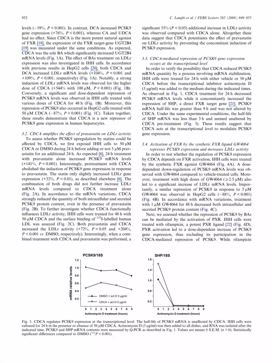
Fig. 3. CDCA regulates PCSK9 expression at the transcriptional level. Thcultured for 24 h in the presence or absence of 50 lM CDCA. Actinomycin Dindicated time. PCSK9 and SHP mRNA contents were measured by Q-PCRsignificant differences compared to DMSO (***P < 0.001).
significant 55% (P < 0.05) additional increase in LDLr activity
was observed compared with CDCA alone. Altogether these
data suggest that CDCA potentiates the effect of pravastatin
on LDLr activity by preventing the concomitant induction of
PCSK9 expression.
3.3. CDCA-mediated repression of PCSK9 gene expression
occurs at the transcriptional level
In order to verify the possibility that CDCA reduced PCSK9
mRNA quantity by a process involving mRNA stabilization,
IHH cells were treated for 24 h with either vehicle or 50 lM
CDCA before the transcriptional inhibitor actinomycin D
(5 lg/ml) was added to the medium during the indicated times.
As observed in Fig. 1, CDCA treatment for 24 h decreased
PCSK9 mRNA levels while it concomitantly increased the
expression of SHP, a direct FXR target gene [21]. PCSK9
mRNA half-life was greater than 9 h and was not altered by
CDCA. Under the same experimental conditions, the half-life
of SHP mRNA was less than 3 h and seemed unaltered by
CDCA pre-treatment (Fig. 3). These results suggest that
CDCA acts at the transcriptional level to modulate PCSK9
gene expression.
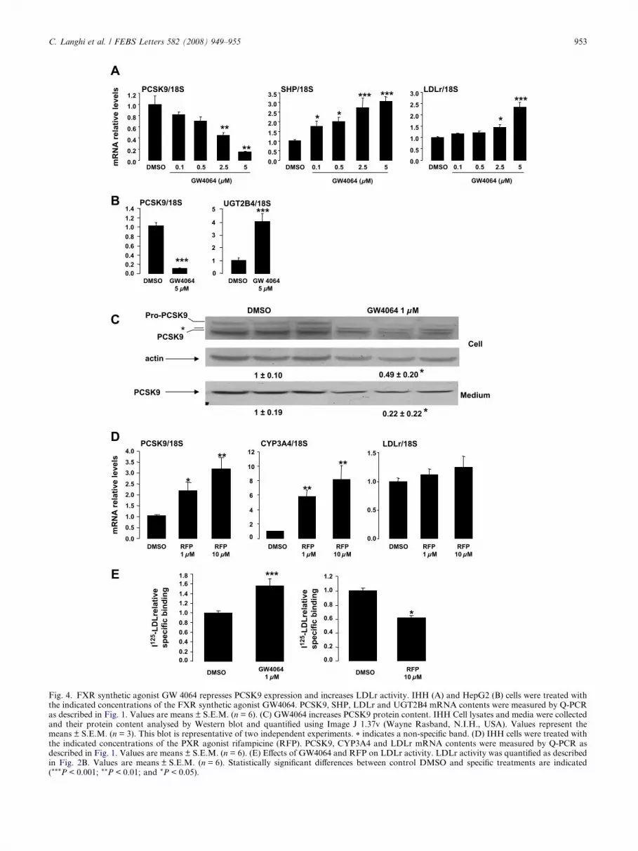
3.4. Activation of FXR by the synthetic FXR ligand GW4064
represses PCSK9 expression and increases LDLr activity
In order to test whether the regulation of PCSK9 expression
by CDCA depends on FXR activation, IHH cells were treated
by the synthetic FXR agonist GW4064 (Fig. 4A). A dose-
dependent down-regulation of PCSK9 mRNA levels was ob-
served with GW4064 compared to vehicle-treated cells. More-
over, treatment with high doses of GW4064 (P2.5 lM) also
led to a significant increase of LDLr mRNA levels. Impor-
tantly, a similar repression of PCSK9 in response to 5 lM
GW4064 was observed in HepG2 cells (�88%, P < 0.001)
(Fig. 4B). In accordance with mRNA variations, treatment
with 1 lM GW4064 for 48 h decreased both intracellular and
secreted PCSK9 protein content (Fig. 4C).
Next, we assessed whether the repression of PCSK9 by BAs
can be mediated by the activation of PXR. IHH cells were
treated with rifampicin, a potent PXR ligand [22] (Fig. 4D),
PXR activation led to a dose-dependent increase of PCSK9
gene expression, thus excluding its participation in the
CDCA-mediated repression of PCSK9. While rifampicin
SHP/18S
1.4
1.8
0 1 2 3 4 5 6 7 8 9
0.2
0.6
1.0
Actinomycin D treatment (hours)
e half-life of PCSK9 mRNA is unaffected by CDCA. IHH cells were(5 lg/ml) was then added to all dishes, and RNA was isolated after theas described in Fig. 1. Values are means ± S.E.M. (n = 6). Statistically
1 µ µ
APCSK9/18S SHP/18S LDLr/18S
DMSO 0.1 0.5 2.5 5
0.20.40.60.81.01.2
0.0 0.00.51.01.52.02.53.03.5
DMSO 0.1 0.5 2.5 5mR
NA
rela
tive
leve
ls
0.00.5
1.01.52.0
2.53.0
DMSO 0.1 0.5 2.5 5
*
**** *
*** ***
**
**
GW4064 (µM) GW4064 (µM) GW4064 (µM)
C
actin
PCSK9
DMSO GW4064 1 µM
Cell
Medium
1 ± 0.10 0.49 ± 0.20
1 ± 0.19 0.22 ± 0.22
*
Pro-PCSK9
PCSK9*
*
CYP3A4/18SPCSK9/18S
mR
NA
rela
tive
leve
ls
DMSO RFP1 µ µ µ µ µ µM
RFP10 M
DMSO RFP1 M
RFP10 M
0.51.01.52.02.53.03.54.0
*
**
0.0
2
4
6
8
10
12
**
**
0
LDLr/18S
0.0
0.5
1.0
1.5
DMSO RFP1 M
RFP10 M
D
0.20.40.60.81.01.21.41.6
DMSO GW4064M
***
I125
-LD
Lrel
ativ
esp
ecifi
c bi
ndin
g
I125
-LD
Lrel
ativ
esp
ecifi
c bi
ndin
g
0.0
DMSO RFP10 M
0.2
0.4
0.6
0.8
1.0
1.2
0.0
*
1.8E
DMSO GW4064
0.20.40.60.81.01.21.4
0.0
PCSK9/18S
DMSO GW 40645 µM5 µM
1
2
3
4
5
0
UGT2B4/18S***
***
B
Fig. 4. FXR synthetic agonist GW 4064 represses PCSK9 expression and increases LDLr activity. IHH (A) and HepG2 (B) cells were treated withthe indicated concentrations of the FXR synthetic agonist GW4064. PCSK9, SHP, LDLr and UGT2B4 mRNA contents were measured by Q-PCRas described in Fig. 1. Values are means ± S.E.M. (n = 6). (C) GW4064 increases PCSK9 protein content. IHH Cell lysates and media were collectedand their protein content analysed by Western blot and quantified using Image J 1.37v (Wayne Rasband, N.I.H., USA). Values represent themeans ± S.E.M. (n = 3). This blot is representative of two independent experiments. * indicates a non-specific band. (D) IHH cells were treated withthe indicated concentrations of the PXR agonist rifampicine (RFP). PCSK9, CYP3A4 and LDLr mRNA contents were measured by Q-PCR asdescribed in Fig. 1. Values are means ± S.E.M. (n = 6). (E) Effects of GW4064 and RFP on LDLr activity. LDLr activity was quantified as describedin Fig. 2B. Values are means ± S.E.M. (n = 6). Statistically significant differences between control DMSO and specific treatments are indicated(***P < 0.001; **P < 0.01; and *P < 0.05).
C. Langhi et al. / FEBS Letters 582 (2008) 949–955 953

954 C. Langhi et al. / FEBS Letters 582 (2008) 949–955
strongly induced mRNA levels of the PXR target gene
Cyp3A4, it did not alter LDLr mRNA levels.
Of functional relevance, 1 lM GW4064 significantly in-
creased the LDLr activity (+60%, P < 0.001). In correlation
with the PXR-mediated induction of PCSK9, LDLr activity
was conversely reduced by 30% (P < 0.05) in response to
10 lM rifampicin (Fig. 4E). These results suggest that FXR
activation may contribute to the CDCA-mediated repression
of PCSK9 in IHH cells.
4. Discussion
Human genetic studies indicate that inhibiting PCSK9 is a
very promising strategy to lower LDL-c [3]. One way to reach
this goal is to decrease the synthesis of PCSK9. Recently, the
administration of an anti-sense oligonucleotide inhibitor tar-
geting PCSK9 in high-fat fed mice led to a 38% decrease of
the plasma LDL-c concentrations [23]. An alternative strategy
is to directly inhibit PCSK9 transcription. In the current study,
we demonstrated for the first time that CDCA is a new repres-
sor of PCSK9 gene expression in human hepatocytes. Of func-
tional relevance, the down-regulation of PCSK9 mRNA levels
by CDCA treatment is associated with an increased LDLr
activity. More importantly, CDCA is able to counterbalance
the positive regulation of PCSK9 by pravastatin and therefore
potentiates its stimulating effect on the LDLr activity. CDCA
increases LDLr mRNA levels [20], an effect that should also
contribute to stimulate LDLr activity. In our cellular model,
however, the potentiation of LDLr activity in response to
CDCA and pravastatin combination treatment occurs in the
absence of a further increase of LDLr mRNA levels. This find-
ing validates the working hypothesis that coadministration of
a PCSK9 inhibitor may amplify the hypolipidemic effect of
statins.
Amongst the BAs we tested, only the most potent FXR ago-
nist CDCA repressed PCSK9 expression, indicating that the
regulatory pathway might be specific for this nuclear receptor.
In accordance with this hypothesis, we found that the specific
synthetic agonist GW4064 reduces PCSK9 mRNA and protein
contents. Moreover, we demonstrate for the first time that
GW4064 increases the LDLr activity in vitro. On the other
hand, a contribution of the other signalling pathways induced
by the CDCA in the repression of PCSK9 was excluded. Acti-
vation of PXR leads indeed to an increased PCSK9 expression
and a subsequent decrease of LDLr activity. In addition, treat-
ment with DCA, a more potent ligand than CDCA for the G
protein-coupled cell-surface BA receptor TGR5 [24], moder-
ately increased PCSK9 expression. CDCA has been shown
to increase LDLr mRNA stability by inducing mitogen acti-
vated protein (MAP) kinase signalling pathways, particularly
extracellular-regulated kinases (ERK) 1/2 [20]. Inhibition of
ERK1-2 signalling pathway by the specific inhibitor U0126
significantly reduced PCSK9 mRNA levels (data not shown).
These results indicate that neither the activation of PXR,
TGR5 nor ERK1–2 is involved in the CDCA-mediated repres-
sion of PCSK9.
To definitively confirm that CDCA-mediated repression of
PCSK9 is FXR-dependent, a FXR-silencing gene experiment
using siRNA was performed. Despite a highly significant de-
crease of FXR mRNA levels (�80%, P = 0.001) and the abo-
lition of CDCA-induced SHP gene expression, the repression
of PCSK9 still occurred in FXR siRNA transfected cells (Sup-
plementary Fig. S1). Therefore, this RNAi mediated approach
failed to abolish the repressive potency of the FXR pathway.
Thus, in addition to FXR activation, it can not be formally ex-
cluded that additional BAs-mediated signalling pathways can
govern CDCA-mediated PCSK9 repression.
In addition, we cannot use the FXR-deficient mouse model
since the CDCA-mediated PCSK9 repression was not retrieved
in primary mouse hepatocytes (data not shown). Such a specie-
specific regulation in response to FXR was already observed
for many genes involved in lipid metabolism as PPARa [25],
hepatic lipase [26] and syndecan-1 [27]. While actinomycin D
experiments indicate that CDCA acts at the transcriptional
level for repressing PCSK9, additional studies are needed to
precisely assess the molecular mechanism by which CDCA
and GW4064 repress PCSK9 expression.
Very recently, Nilsonn et al. demonstrated that a 3-weeks
treatment with CDCA reduces LDLr mRNA levels in the liver
of subjects who underwent a cholecystectomy for gallstone dis-
ease, while PCSK9 mRNA levels were not altered [28]. Con-
versely, a 3-weeks treatment with cholestyramine, a BA
sequestrant, increased LDLr and PCSK9 mRNA levels by
65% and 70%, respectively. Although this study reinforces
the hypothesis for a cross-talk between BAs and PCSK9, it re-
mains difficult to reconcile these in vivo findings with our
in vitro data, as well as with previously published results dem-
onstrating that CDCA induces LDLr gene expression in vitro
[20,29]. The reason for this discrepancy remains unclear. How-
ever, it should be noticed that CDCA treatment in these
patients failed to modulate the expression of hepatic FXR-
target genes such as ApoCIII and ApoAI [28].
FXR appears as a promising target to treat dyslipidemia
[12]. CDCA has been shown to reduce triglyceride and
cholesterol levels in the fructose fed hamster model of
metabolic syndrome and combined dyslipidemia [30]. Similar
results were recently reported with a new synthetic FXR ago-
nist in several rodent models of dyslipidemia [31], and phase I
studies in human are in progress (ClinicalTrials.gov Identifier:
NCT00499629 and NCT00509756). Our present results suggest
that part of the hypolipidemic effects of FXR agonists might
be mediated by PCSK9 repression.
Acknowledgements: This work was supported by Grants from theAgence Nationale de la Recherche (No. PPV06217NSA and ANR-06-PHYSIO-027-01, Project R06510NS), Laboratoires Pierre Fabre,and the EU Grant Hepadip (No. 018734). C. Langhi is a recipient ofa fellowship from the Nouvelle Societe Francaise d�Atherosclerose.C Le May is supported by a Grant from the Fondation pour laRecherche Medicale. S. Kourimate is supported by a Grant fromRegion Pays de la Loire and Philippe Costet is titular of a Contratd�interface INSERM – CHU de Nantes.
We gratefully acknowledge Lucie Arnaud and Anne-Laure Jarnouxfor expert Technical Assistance. We thank Dr. K.E. Berge for provid-ing us TK-Renilla plasmid.
Appendix A. Supplementary data
Supplementary data associated with this article can be
found, in the online version, at doi:10.1016/j.febslet.
2008.02.038.

C. Langhi et al. / FEBS Letters 582 (2008) 949–955 955
References
[1] Horton, J.D., Cohen, J.C. and Hobbs, H.H. (2007) Molecularbiology of PCSK9: its role in LDL metabolism. Trends Biochem.Sci. 32, 71–77.
[2] Abifadel, M., Varret, M., Rabes, J.P., Allard, D., Ouguerram, K.,Devillers, M., Cruaud, C., Benjannet, S., Wickham, L., Erlich, D.,Derre, A., Villeger, L., Farnier, M., Beucler, I., Bruckert, E.,Chambaz, J., Chanu, B., Lecerf, J.M., Luc, G., Moulin, P.,Weissenbach, J., Prat, A., Krempf, M., Junien, C., Seidah, N.G.and Boileau, C. (2003) Mutations in PCSK9 cause autosomaldominant hypercholesterolemia. Nat. Genet. 34, 154–156.
[3] Cohen, J.C., Boerwinkle, E., Mosley Jr., T.H. and Hobbs, H.H.(2006) Sequence variations in PCSK9, low LDL, and protectionagainst coronary heart disease. New Engl. J. Med. 354, 1264–1272.
[4] McNutt, M.C., Lagace, T.A. and Horton, J.D. (2007) Catalyticactivity is not required for secreted PCSK9 to reduce low densitylipoprotein receptors in HepG2 cells. J. Biol. Chem. 282, 20799–20803.
[5] Zhang, D.W., Lagace, T.A., Garuti, R., Zhao, Z., McDonald, M.,Horton, J.D., Cohen, J.C. and Hobbs, H.H. (2007) Binding ofproprotein convertase subtilisin/kexin type 9 to epidermal growthfactor-like repeat A of low density lipoprotein receptor decreasesreceptor recycling and increases degradation. J. Biol. Chem. 282,18602–18612.
[6] Dubuc, G., Chamberland, A., Wassef, H., Davignon, J., Seidah,N.G., Bernier, L. and Prat, A. (2004) Statins upregulate PCSK9,the gene encoding the proprotein convertase neural apoptosis-regulated convertase-1 implicated in familial hypercholesterol-emia. Arterioscler. Thromb. Vasc. Biol. 24, 1454–1459.
[7] Maxwell, K.N., Soccio, R.E., Duncan, E.M., Sehayek, E. andBreslow, J.L. (2003) Novel putative SREBP and LXR target genesidentified by microarray analysis in liver of cholesterol-fed mice. J.Lipid Res. 44, 2109–2119.
[8] Rashid, S., Curtis, D.E., Garuti, R., Anderson, N.N., Bashma-kov, Y., Ho, Y.K., Hammer, R.E., Moon, Y.A. and Horton, J.D.(2005) Decreased plasma cholesterol and hypersensitivity tostatins in mice lacking Pcsk9. Proc. Natl. Acad. Sci. USA 102,5374–5379.
[9] Berge, K.E., Ose, L. and Leren, T.P. (2006) Missense mutations inthe PCSK9 gene are associated with hypocholesterolemia andpossibly increased response to statin therapy. Arterioscler.Thromb. Vasc. Biol. 26, 1094–1100.
[10] Costet, P., Cariou, B., Lambert, G., Lalanne, F., Lardeux, B.,Jarnoux, A.L., Grefhorst, A., Staels, B. and Krempf, M. (2006)Hepatic PCSK9 expression is regulated by nutritional status viainsulin and sterol regulatory element-binding protein 1c. J. Biol.Chem. 281, 6211–6218.
[11] Lambert, G., Jarnoux, A.L., Pineau, T., Pape, O., Chetiveaux,M., Laboisse, C., Krempf, M. and Costet, P. (2006) Fastinginduces hyperlipidemia in mice overexpressing proprotein con-vertase subtilisin kexin type 9: lack of modulation of very-low-density lipoprotein hepatic output by the low-density lipoproteinreceptor. Endocrinology 147, 4985–4995.
[12] Cariou, B. and Staels, B. (2007) FXR: a promising targetfor the metabolic syndrome? Trends Pharmacol. Sci. 28, 236–243.
[13] Lee, F.Y., Lee, H., Hubbert, M.L., Edwards, P.A. and Zhang, Y.(2006) FXR, a multipurpose nuclear receptor. Trends Biochem.Sci. 31, 572–580.
[14] Staudinger, J.L., Goodwin, B., Jones, S.A., Hawkins-Brown, D.,MacKenzie, K.I., LaTour, A., Liu, Y., Klaassen, C.D., Brown,K.K., Reinhard, J., Willson, T.M., Koller, B.H. and Kliewer, S.A.(2001) The nuclear receptor PXR is a lithocholic acid sensor thatprotects against liver toxicity. Proc. Natl. Acad. Sci. USA 98,3369–3374.
[15] Zollner, G., Marschall, H.U., Wagner, M. and Trauner, M.(2006) Role of nuclear receptors in the adaptive response to bileacids and cholestasis: pathogenetic and therapeutic consider-ations. Mol. Pharm. 3, 231–251.
[16] Chapman, M.J., Goldstein, S., Lagrange, D. and Laplaud, P.M.(1981) A density gradient ultracentrifugal procedure for theisolation of the major lipoprotein classes from human serum. J.Lipid Res. 22, 339–358.
[17] Fraker, P.J. and Speck Jr., J.C. (1978) Protein and cell membraneiodinations with a sparingly soluble chloroamide, 1,3,4,6-tetra-chloro-3a,6a-diphrenylglycoluril. Biochem. Biophys. Res. Com-mun. 80, 849–857.
[18] Makishima, M., Okamoto, A.Y., Repa, J.J., Tu, H., Learned,R.M., Luk, A., Hull, M.V., Lustig, K.D., Mangelsdorf, D.J. andShan, B. (1999) Identification of a nuclear receptor for bile acids.Science 284, 1362–1365.
[19] Barbier, O., Torra, I.P., Sirvent, A., Claudel, T., Blanquart, C.,Duran-Sandoval, D., Kuipers, F., Kosykh, V., Fruchart, J.C. andStaels, B. (2003) FXR induces the UGT2B4 enzyme in hepato-cytes: a potential mechanism of negative feedback control of FXRactivity. Gastroenterology 124, 1926–1940.
[20] Nakahara, M., Fujii, H., Maloney, P.R., Shimizu, M. and Sato,R. (2002) Bile acids enhance low density lipoprotein receptor geneexpression via a MAPK cascade-mediated stabilization ofmRNA. J. Biol. Chem. 277, 37229–37234.
[21] Wang, L., Lee, Y.K., Bundman, D., Han, Y., Thevananther, S.,Kim, C.S., Chua, S.S., Wei, P., Heyman, R.A., Karin, M. andMoore, D.D. (2002) Redundant pathways for negative feedbackregulation of bile acid production. Dev. Cell 2, 721–731.
[22] Moore, L.B., Maglich, J.M., McKee, D.D., Wisely, B., Willson,T.M., Kliewer, S.A., Lambert, M.H. and Moore, J.T. (2002)Pregnane X receptor (PXR), constitutive androstane receptor(CAR), and benzoate X receptor (BXR) define three pharmaco-logically distinct classes of nuclear receptors. Mol. Endocrinol. 16,977–986.
[23] Graham, M.J., Lemonidis, K.M., Whipple, C.P., Subramaniam,A., Monia, B.P., Crooke, S.T. and Crooke, R.M. (2007) Antisenseinhibition of proprotein convertase subtilisin/kexin type 9 reducesserum LDL in hyperlipidemic mice. J. Lipid Res. 48, 763–767.
[24] Kawamata, Y., Fujii, R., Hosoya, M., Harada, M., Yoshida, H.,Miwa, M., Fukusumi, S., Habata, Y., Itoh, T., Shintani, Y.,Hinuma, S., Fujisawa, Y. and Fujino, M. (2003) A G protein-coupled receptor responsive to bile acids. J. Biol. Chem. 278,9435–9440.
[25] Pineda, T.I., Claudel, T., Duval, C., Kosykh, V., Fruchart, J.C.and Staels, B. (2003) Bile acids induce the expression of thehuman peroxisome proliferator-activated receptor alpha gene viaactivation of the farnesoid X receptor. Mol. Endocrinol. 17, 259–272.
[26] Sirvent, A., Verhoeven, A.J., Jansen, H., Kosykh, V., Darteil,R.J., Hum, D.W., Fruchart, J.C. and Staels, B. (2004) FarnesoidX receptor represses hepatic lipase gene expression. J. Lipid Res.45, 2110–2115.
[27] Anisfeld, A.M., Kast-Woelbern, H.R., Meyer, M.E., Jones, S.A.,Zhang, Y., Williams, K.J., Willson, T. and Edwards, P.A. (2003)Syndecan-1 expression is regulated in an isoform-specific mannerby the farnesoid-X receptor. J. Biol. Chem. 278, 20420–20428.
[28] Nilsson, L.M., Abrahamsson, A., Sahlin, S., Gustafsson, U.,Angelin, B., Parini, P. and Einarsson, C. (2007) Bile acids andlipoprotein metabolism: effects of cholestyramine and chenode-oxycholic acid on human hepatic mRNA expression. Biochem.Biophys. Res. Commun. 357, 707–711.
[29] Taniguchi, T., Chen, J. and Cooper, A.D. (1994) Regulation ofcholesterol 7 alpha-hydroxylase gene expression in Hep-G2 cells.Effect of serum, bile salts, and coordinate and noncoordinateregulation with other sterol-responsive genes. J. Biol. Chem. 269,10071–10078.
[30] Bilz, S., Samuel, V., Morino, K., Savage, D., Choi, C.S. andShulman, G.I. (2006) Activation of the farnesoid X receptorimproves lipid metabolism in combined hyperlipidemic hamsters.Am. J. Physiol. Endocrinol. Metab. 290, E716–E722.
[31] Evans, M.J., Mahaney, P.E., Zhang, S., Gantan, E., Borges-Marcucci, L.A., Lai, K., Wang, S. and Harnish, D.C. (2007) Asynthetic farnesoid X receptor agonist protects against diet-induced dyslipidemia. Circulation. (Abstract) AHA07L1_584.