Embed Size (px)
Citation preview

A Novel Corepressor, BCoR-L1, Represses Transcriptionthrough an Interaction with CtBP*
Received for publication, January 9, 2007, and in revised form, March 20, 2007, Published, JBC Papers in Press, March 20, 2007, DOI 10.1074/jbc.M700246200
Julia K. Pagan‡§, Jeremy Arnold‡, Kim J. Hanchard‡, Raman Kumar¶, Tiziana Bruno�, Mathew J. K. Jones‡§,Derek J. Richard‡, Alistair Forrest**, Amanda Spurdle‡, Eric Verdin‡‡, Merlin Crossley§§, Maurizio Fanciulli�,Georgia Chenevix-Trench‡, David B. Young‡1, and Kum Kum Khanna‡1,2
From the ‡Queensland Institute of Medical Research, 300 Herston Road, Herston 4029, Queensland, Australia, §School of Medicine,Central Clinical Division, University of Queensland, Royal Brisbane Hospital, Herston 4029, Queensland, Australia, ¶Breast CancerGenetics Group, Hanson Institute, Institute of Medical and Veterinary Science, Frome Road, Adelaide, South Australia 5000, Australia,�Regina Elena Cancer Institute, Via delle Messi d’Oro 156, 00158 Rome, Italy, **Institute for Molecular Bioscience, University ofQueensland, Brisbane, Queensland 4072, Australia, ‡‡Gladstone Institute of Virology and Immunology, University ofCalifornia, San Francisco, California 94158, and the §§School of Molecular and Microbial Biosciences, G08, University ofSydney, New South Wales 2006, Australia
Corepressors play a crucial role in negative gene regulationand are defective in several diseases. BCoR is a corepressorfor the BCL6 repressor protein. Here we describe and func-tionally characterize BCoR-L1, a homolog of BCoR. Whentethered to a heterologous promoter, BCoR-L1 is capable ofstrong repression. Like other corepressors, BCoR-L1 associ-ates with histone deacetylase (HDAC) activity. Specifically,BCoR-L1 coprecipitates with the Class II HDACs, HDAC4,HDAC5, and HDAC7, suggesting that they are involved in itsrole as a transcriptional repressor. BCoR-L1 also interactswith the CtBP corepressor through a CtBP-interacting motifin its amino terminus. Abrogation of the CtBP binding sitewithin BCoR-L1 partially relieves BCoR-L1-mediated tran-scriptional repression. Furthermore, BCoR-L1 is located onthe E-cadherin promoter, a known CtBP-regulated promoter,and represses the E-cadherin promoter activity in a reporterassay. The inhibition of BCoR-L1 expression by RNA-medi-ated interference results in derepression of E-cadherin incells that do not normally express E-cadherin, indicating thatBCoR-L1 contributes to the repression of an authenticendogenous CtBP target.
The overall transcriptional output of a cell depends on thecoordinated silencing or activation of gene expression by theaction of DNA-bound transcriptional repressors or activa-tors (1). Coregulator proteins, including coactivators andcorepressors, interact with DNA-bound transcription fac-tors to mediate gene activation or repression, respectively,by recruiting multiprotein catalytic complexes that regulatechromatin structure and thus the transcription of targetgenes.
Although the role of coactivator proteins in transcriptionalregulation is well established, the equally important role ofcorepressor proteins in gene regulation has become apparentonly relatively recently. There are many corepressor proteins,recruited to a wide range of transcriptional silencers. They reg-ulate many processes, including differentiation, proliferation,apoptosis, and the cell cycle (2). The aberrant function of core-pressors can lead to developmental defects and disease, sincegenes that should be turned off are instead aberrantlyexpressed, or “derepressed” (3). Alternately, overactive repres-sion leads to enhanced silencing and has been reported in sev-eral types of leukemia, involving gene fusions to transcriptionalsilencers (4). Increased corepressor binding to these fusion pro-teins leads to inappropriate repression of target genes impor-tant for normal cellular differentiation.Corepressors act within multiprotein complexes contain-
ing DNA-binding proteins, histone deacetylases, methyl-CpG-binding proteins, nucleosomal histones, and the basaltranscriptional machinery. The same corepressor can befound in different corepression complexes, and multiplecorepressors can be used by individual silencers. Corepres-sors are thought to bridge the interaction between DNA-bound transcriptional repressors and the chromatin-modi-fying enzymes that mediate repression. They typically act inmultiple ways, including via the targeted modification ofchromatin structure, nucleosomal remodeling, and seques-tration of the basal transcription machinery as well as byinhibiting trans-activation (2).Lysine acetylation of the histone tails is a major modification
associated with transcriptional activation, and in contrast,deacetylation is associated with transcriptional repression. It isthought that the acetylation of the lysine residues of the corehistone tails neutralizes the positive charge on the lysine resi-due, thereby resulting in a more open structure of chromatinbecause of the decreased affinity between the histone and theDNA (5, 6). Chromatin is therefore more accessible to tran-scription factors and other proteins. Conversely, the deacetyla-tion of histone substrates results in chromatin condensationand inhibits the accessibility of transcription factors and otherproteins to the DNA.
* This work was supported by Australian Research Council, National Healthand Medical Research Council of Australia and the Queensland CancerFund. The costs of publication of this article were defrayed in part by thepayment of page charges. This article must therefore be hereby marked“advertisement” in accordance with 18 U.S.C. Section 1734 solely to indi-cate this fact.
1 These authors contributed equally to this work.2 To whom correspondence should be addressed. Tel.: 61-7-3362-0338; Fax:
61-7-3362-0105.
THE JOURNAL OF BIOLOGICAL CHEMISTRY VOL. 282, NO. 20, pp. 15248 –15257, May 18, 2007© 2007 by The American Society for Biochemistry and Molecular Biology, Inc. Printed in the U.S.A.
15248 JOURNAL OF BIOLOGICAL CHEMISTRY VOLUME 282 • NUMBER 20 • MAY 18, 2007
at Mem
orial Sloane-K
ettering Cancer C
enter, on January 23, 2013w
ww
.jbc.orgD
ownloaded from

The acetylation reaction is a reversible process, catalyzed bythe opposing activity of histone acetyltransferases and deacety-lases (HDACs)3 in vivo. There are several classes of histoneacetyltransferases, each employing different mechanisms ofcatalysis (7). Likewise, there are three main classes of HDACsgrouped according to homology to their counterparts in yeast,their subcellular localization, and enzymatic activities (8, 9).Class I (HDAC1, -2, -3, and -8; homologous to yeast Rpd3) areubiquitously expressed and are primarily localized in thenucleus. Class II (HDAC4, -5, -6, -7, -9, and -10; homologous toHda1) are expressed in a tissue-specific manner and shuttlebetween the nucleus and cytoplasm (10). Class III (Sirt1, -2, -3,-4, -5, -6, and -7; similar to Sir2) use a different mode of catal-ysis, relying on NAD� (11). Most targets are histones, althoughother proteins are deacetylated as well, including �-tubulin(12). All known repression complexes employHDACs tomedi-ate their repression.CtBP is also a critical component of many transcriptional
repression complexes (reviewed inRef. (13).Microarray studiesfrom CtBP knock-out mice implicate CtBP in the repression ofgenes involved in apoptosis and in the epithelial-to-mesenchy-mal transition (18, 19). To mediate its repression, it recruitsenzymes involved in transcription repression, such as methy-lases and deacetylases, to the sequence-specific DNA-bindingproteins via a conserved PXDLS CtBP interaction motif in theCtBP-interacting proteins (14, 15). Recently, it has been shownthat many histone acetyltransferases contain the PXDLS motifand that CtBP inhibits histone acetylation by blocking access ofnuclear histone acetyltransferases to their target (16, 17).Interestingly, CtBP structure is similar to a subfamily of
NAD�-dependent dehydrogenases, and biochemical andstructural studies have recently demonstrated that CtBP pos-sesses dehydrogenase activity (20–22). CtBP bindsNADHwitha higher affinity than NAD� (22), although this has been con-troversial (20). CtBP function is regulated by the relative levelsof NAD� and NADH within the nucleus, suggesting a role forCtBP in sensing the redox state of the cell and regulating tran-scription accordingly (22, 23). Current models suggest thatincreases inNADH levels promoteCtBPdimerization,which inturn increases the interaction between CtBP and proteins con-taining the PXDLS CtBP-recruitment motif.We have identified a novel PXDLS-containing protein desig-
nated as BCoR-L1 in the GenBankTM data base. BCoR-L1 isrelated to the BCoR, a transcriptional corepressor that potenti-ates BCL6 repression in reporter assays (24). BCL6 is a tran-scription factor that is required for germinal center formationand is linked to lymphomagenesis (25).We find that, like BCoR,BCoR-L1 functions as a corepressor when tethered to DNA.BCoR-L1 interacts withClass IIHDACs,HDAC4,HDAC5, andHDAC7, suggesting that they are involved in its function astranscriptional corepressor. It mediates its repression throughrecruitment of the CtBP corepressor protein and affects the
repression of at least one CtBP target, the tumor suppressorprotein, E-cadherin.
MATERIALS AND METHODS
Cloning of Full-length BCoR-L1 and Other Plasmids—Full-length BCoR-L1 (BCoR-L1a) and�Exon9 BCoR-L1 (BCoR-L1)cDNAs were produced in two stages. The C terminus ofBCoR-L1 was PCR-cloned using Pfu polymerase from cDNAderived from the normal ovarian epithelial cell line (HOSE17.1). Primers used for this PCRwere designed from the in silicopredictions of the mRNA and later from known sequence. ThecDNA was amplified in several overlapping fragments, whichwere cloned into the pPCR-Script vector (Stratagene) and laterassembled into a single sequence using appropriate restrictionenzyme sites. The N terminus of BCoR-L1 was provided by apartial cDNA clone (bp 60–4449, FLJ00190), which was a gen-erous gift of Kasuza Institute (Chiba, Japan). The first 60 bp ofBCoR-L1 were incorporated into primers, which were used toamplify the N terminus of BCoR-L1 from the cDNA clone andthen subcloned into the existing C terminus of BCoR-L1. Theentire coding sequences for BCoR-L1 were then subcloned intopEGFP-C1 (Clontech) and pFLAG-CMV-2 (Sigma) parentplasmids using SalI and BamHI sites.GST-BCoR-L1a-(1328–1785) vector for antibody produc-
tion was constructed by cloning BCoR-L1 sequence (aminoacids 1328–1785) in framewith pGEX-5X-1. The pSUPER vec-tor system (26) (a gift ofDr. ReuvenAgami;NetherlandsCancerInstitute, Amsterdam) was used to create the siRNA constructto knock-down BCoR-L1 levels “pSUPER-BcoR-L1.” The fol-lowing sequencewas cloned into the pSUPERvector to producepSUPER-BCoR-L1: forward (FWD), 5�-GATCCCCCGTGGC-AGAGGCTGAGGGCTTCAAGAGAGCCCTCAGCCTCTG-CCACGTTTTTGGAAA-3�; reverse (RVS), 5�-AGCTTTTC-CAAAAACGTGGCAGAGGCTGAGGGCTCTCTTGAA-GCCCTCAGCCTCTGCCACGGGG-3�. This constructgenerated siRNAs targeting the following 21-nucleotidesequence: 5�-CGTGGCAGAGGCTGAGGGCT-3� (comple-mentary to nucleotides 312–331 within BCoR-L1 cDNA). ThepSUPER-GFP plasmid has been described previously (27). Gal4DNA-binding domain (Gal4-DBD) vectors were constructedby replacing GFP from pEGFP vectors (Clontech) with theGal4-DBD from pGBT9 (Clontech). BCoR-L1 was cloned as aGal4-DBD fusion protein using SalI and BamHI sites. BCoR-L1fragments were cloned as Gal4-DBD fusion proteins usingBamHI and EcoRI sites incorporated into primers designed toamplify the fragments from plasmid. The Gal4-DBD-BCoR-L1�CtBP binding site mutant (PLDLS to PLASS) was createdusing the QuikChangeTM site-directed mutagenesis method(Stratagene). FLAG-tagged expression vectors forHDAC4 to -7(28, 29), the pGL3-hE-cad (E-Cad-Luc) (15), and Myc-taggedCtBP1 expression vectors (30) have been described previously(31, 32). The GFP-BCoR plasmid was subcloned from Myc-BCoR (kindly provided by Vivian Bardwell, University of Min-nesota, Minneapolis, MN).Northern Analysis—The probe for Northern analysis was
prepared from RT-PCR products derived from the aminoterminus of BCoR-L1 amplified from the HOSE 17.1 ovariancell line using the following primer sequences: forward, 5�-
3 The abbreviations used are: HDAC, histone deacetylase; siRNA, small inter-fering RNA; DBD, DNA-binding domain; GFP, green fluorescent protein; RT,reverse transcription; Luc, luciferase; TK, thymidine kinase; E-Cad, E-cad-herin; WT, wild type; BCoR, BCL6 corepressor; BCoR-L1, BCoR-like 1.
A Novel CtBP-interacting Corepressor
MAY 18, 2007 • VOLUME 282 • NUMBER 20 JOURNAL OF BIOLOGICAL CHEMISTRY 15249
at Mem
orial Sloane-K
ettering Cancer C
enter, on January 23, 2013w
ww
.jbc.orgD
ownloaded from

GTGCACAACTGGACCAGTTCTGACCG-3�; reverse, 5�-GAGTCAGAGATGAGCGTGGGCACTG-3�. Probes were[�-32P]dCTP-labeled using the MegaPrime kit (AmershamBiosciences). Hybridization to a Clontech humanMultiple Tis-sue Northern blot II membrane was carried out for 2 h inExpressHyb solution (Clontech) at 65 °C, followed by a stand-ard washing procedure.Cell Culture and Transfections—Cells were maintained at
37 °C in a 5% CO2 incubator and grown in RPMI 1640 mediumsupplemented with 10% Serum Supreme, 1% L-glutamine, 100units/ml penicillin, and 100 units/ml streptomycin. Transfec-tions of cells were carried out using either electroporation orlipofection. For electroporation, 293T cells in exponentialgrowth were pelleted and resuspended at 106 cells/300 �l incomplete media and then electroporated at room temperaturewith 7 �g of DNA in a 0.4-cm cuvette (Interpath). Electropora-tion was performed with a BTXTM820 electroporator (Gen-etronics Inc.) at 260 Vwith a time constant of 10ms. HeLa cellsgrowing in dishes were transfected using LipofectamineTM2000 reagent (Invitrogen) according to the manufacturer’sinstructions.Luciferase Assays—293T cells were grown in 24-well dishes
and were transfected using LipofectamineTM 2000. ThepRL-TK plasmid (Promega) was cotransfected as a control fortransfection efficiency. The amount of DNA per well was keptconstant using an empty expression vector (pCMV; Strat-agene). Cellswere grown for 48hprior to harvesting usingPassiveLysis Buffer (Promega, Annandale, Australia). The lysate wasassayed for luciferase activity using the Dual-LuciferaseTMreporter assay system (Promega), according to themanufacturer’srecommendations. Readings were taken using a TD-20/20 Lumi-nometer (TurnerDesigns). Results shown are from representativeexperiments. The error bars represent one S.D.Antibodies and BCoR-L1 Antibody Production—Polyclonal
antibodies were produced in rabbits following standard injec-tion protocols. Rabbits were immunized with GST-BCoR-L1a-(1328–1785) fusion protein. An �-BCoR-L1 peptide antibodywas raised against the sequence EERRAPLSDEESTTGD byOpen Biosystems. The following antibodies were used: rabbitanti-GFP (Molecular Probes), mouse monoclonal anti-FLAG(Sigma), mouse monoclonal anti-Myc (Cell Signaling Technol-ogy), ImmunoPure Recomb Protein A/G HRP conjugate(Molecular Probes), and rabbit anti-Gal4-DBD (Santa CruzBiotechnology, Inc., Santa Cruz, CA).ChIP Analysis and RT-PCR—ChIP analysis of the E-cadherin
promoter was performed according to Shi et al. (15). The PCRprimers used to amplify the E-cadherin promoter were as follows:forward, 5�-TAGCCTGGCGTGGTGGTGTGCACCTG-3�;reverse, 5�-GTGCGTGGCTGCAGCCAGGTGAGCC-3�. Thefollowing primers were used for quantitative PCR detectionof BCoR-L1 levels: forward, 5�-GACCGACATCCTGAACA-TCC-3�; reverse, 5�-ATAGGACAGCAGGAGCCAGA-3�.The primers used to detect E-cadherin levels were as follows:forward, 5�-GAAAATCTGAAAGCGGCTGAT-3�; reverse,5�-GCCCCATTCGTTCAAGT-3�.Lysate Preparation, Coimmunoprecipitation, Immunoblot-
ting, and Antibodies—Cell extracts were prepared by lysis inuniversal immunoprecipitation buffer (50 mM Tris-HCl (pH
7.4), 150 mM NaCl, 2 mM EDTA, 25 mM �-glycerophosphate,0.2% Triton X-100, and 0.3% Nonidet P-40), supplementedwith 25mM sodium fluoride, 25 mM sodium orthovanadate, 0.1mM phenylmethylsulfonyl fluoride, and a mixture of proteaseinhibitors (Roche Applied Science). For coimmunoprecipita-tion experiments, protein samples were precleared by incuba-tionwith proteinG-Sepharose beads (Sigma) for 30min at 4 °C.Supernatants (2mg)were incubatedwith 2�g of antibody over-night. Immune complexes were collected with proteinG-Sepharose beads. Beads were washed four times with lysisbuffer, eluted using Laemmli sample buffer, and analyzed byimmunoblotting with specific antibodies.Immunofluorescence Staining—Cells grown on coverslips
were fixed in 4% paraformadehyde in phosphate-bufferedsaline and permeabilized in 0.2% Triton X-100 in phosphate-buffered saline. Coverslips were blocked with 0.5% bovineserum albumin in phosphate-buffered saline and then incu-bated with primary antibodies diluted in blocking solution for1 h at room temperature, followed by AlexaFluor-546-conju-gated secondary antibodies (Molecular Probes). Cells werecounterstained with 4�,6-diamidino-2-phenylindole (Molecu-lar Probes). Coverslips were mounted on glass slides withMowiol (Calbiochem) containing 0.6% diazobicyclo-octane(Sigma). Fluorescence was visualized using a Zeiss Axioskop20microscope, Zeiss AxioCamMRc digital camera, andMRGrab1.0 software. Dual confocal images were collected sequentiallyusing a 635 objective on Leica TCS SP2 confocal fluorescentmicroscope system using a 488-nm argon laser for GFP and543-nmhelium-neon laser to excite theAlexaFluor-546.Confocalhardware was driven by a work station running Leica confocalimaging software. FigureswereprocessedusingAdobePhotoshop6.0.
FIGURE 1. Schematic representation of BcoR-L1. BCoR-L1 exists as at leasttwo isoforms. An additional exon, designated exon 9 (�Ex9), is present inBCoR-L1a. The recognizable domains and motifs are indicated. BCoR-L1 con-tains a nuclear localization signal (NLS) and tandem ankyrin repeats (ANK). Italso contains two LXXLL motifs known to recruit nuclear receptor coregula-tors. The proteins that share sequence similarity with BCoR-L1 are repre-sented under BCoR-L1. The numbers represent percentage identity/percent-age homology between BcoR-L1 amino acid sequence and its homologs. Theregion of homology between BCoR-L1 and BARD1 spans 431 amino acids.The region of homology with RRP1 spans 351 amino acids. There are threeregions of homology between BCoR and BCoR-L1: one small amino-terminalregion and two substantial regions spanning 600 amino acids and 336 aminoacids within the central region and carboxyl terminus, respectively.
A Novel CtBP-interacting Corepressor
15250 JOURNAL OF BIOLOGICAL CHEMISTRY VOLUME 282 • NUMBER 20 • MAY 18, 2007
at Mem
orial Sloane-K
ettering Cancer C
enter, on January 23, 2013w
ww
.jbc.orgD
ownloaded from

RESULTS
A Novel BCoR-related Protein—BCoR-L1 (BCoR-like 1) wasisolated as a partial cDNA using full-length BRCA1 as bait in ayeast two-hybrid screen with a testis cDNA library. The basisand significance of the interaction with BRCA1 is currently
under investigation and is not discussed here. One set of posi-tive clones encoded the partial cDNA of a hypothetical protein(GenBankTM number NM_019294, BCL6 corepressor-like 1;function unknown), expressed from the X chromosome(Xq25–26.1). The entire BCoR-L1 transcript contains 14 exonsspliced from �75 kb of genomic DNA. The original yeast two-hybrid clone contained an additional exon (exon 9) not presentin the composite sequence on GenBankTM and represents analternatively spliced form of BCoR-L1 mRNA, which we callBCoR-L1a. The complete cDNA sequence of full-lengthBCoR-L1 (BCoR-L1a) and BCoR-L1 �Exon 9 (BCoR-L1;NM_019294) was generated in mammalian expression vectors(see “Materials and Methods”).The open reading frame of BCoR-L1 cDNA encodes a pro-
tein of 1711 amino acids containing a putative bipartite nuclearlocalization signal (NLS) and tandem ankyrin repeats (ANK)(Fig. 1). BCoR-L1a is 1785 amino acids long. Neither BCoR-L1nor BCoR has identifiable DNA-binding domains. BCoR-L1contains a PXDLS motif, involved in binding the CtBP core-pressor (33, 34). BCoR-L1 also contains two LXXLL nuclearreceptor recruitmentmotifs found in coregulator proteins (35).The amino acid sequence shares homology with several pro-teins involved in chromatin remodeling, transcription, andrepair of DNA damage. More specifically, BCoR-L1 is homolo-gous to BCoR, a transcriptional corepressor for the BCL6 tran-scriptional repressor (24). Additionally, part of the BCoR-L1sequence shares homology with the predominant BRCA1-in-teracting protein, BARD1 (36). A region of BCoR-L1 is relatedto the Drosophila recombination repair protein (dRRP1), arepair endonuclease. Although BCoR-L1 lacks the RRP1 nucle-ase domain, it shares homology with the RRP1 domain thatcatalyzes strand transfer during homologous recombination(37, 38). A high degree of sequence conservation exists betweenhuman BCoR-L1 and mouse and rat orthologues (80 and 77%amino acid identity, respectively). We have not been able toidentify any BCoR-L1 orthologues in lower eukaryotes.Tissue Expression of BCoR-L1—Northern blot analysis using
the multiple-tissue Northern II filter (Clontech) demonstratedthat BCoR-L1 is expressed at low levels in many tissues. The
FIGURE 2. Tissue expression of BCoR-L1. A, Northern blot analysis ofBCoR-L1 expression using a Clontech membrane. B, HeLa cells were trans-fected with FLAG-tagged BCoR-L1a or BCoR-L1. Lysates from the transfectedand untransfected cells were separated on SDS-polyacrylamide gels and sub-jected to immunoblotting using �-BCoR-L1 antibody (top panel) and anti-FLAG antibody (bottom panel). C, 293T cells were transfected with pSUPER-GFP or pSUPER-BCoR-L1 and left for 72 h before harvesting. Lysates wereseparated by SDS-PAGE and subjected to immunoblotting using the peptide�-BCoR-L1 antibody.
FIGURE 3. Localization of BCoR-L1 and BCoR. HeLa cells were transfectedwith GFP-BCoR-L1 or GFP-BCoR. Cells were fixed in paraformaldehyde 24 hlater and stained with 4�,6-diamidino-2-phenylindole (DAPI) to mark the cellnucleus.
A Novel CtBP-interacting Corepressor
MAY 18, 2007 • VOLUME 282 • NUMBER 20 JOURNAL OF BIOLOGICAL CHEMISTRY 15251
at Mem
orial Sloane-K
ettering Cancer C
enter, on January 23, 2013w
ww
.jbc.orgD
ownloaded from
highest level of expression was observed in testis and prostate.Medium levels of expression were also seen in peripheral bloodlymphocytes and spleen (Fig. 2A).A rabbit polyclonal antibody was raised against recombinant
GST-BCoR-L1a (aa 1328–1785). The specificity of the anti-body for immunoblot analysis was tested against recombinantFLAG-tagged BCoR-L1a and BCoR-L1 in protein extracts pre-pared fromHeLa cells. The antibody recognized a bandmigrat-
ing through SDS-polyacrylamidegels at a size of�200 kDa and comi-grating with exogenously expressedFLAG-tagged BCoR-L1 (Fig. 2B).This result provides evidence thatendogenous BCoR-L1 is predomi-nantly encoded from the BCoR-L1�Exon 9 isoform, which was alsothe predominant isoform detectedby RT-PCR analysis (results notshown). Furthermore, this bandwassignificantly diminished in inten-sity when cells were depleted ofBCoR-L1 by siRNA, further demon-strating the specificity of BCoR-L1antibody (Fig. 2C).BCoR-L1 Is a Nuclear Protein—
Since the amino acid sequence ofBCoR-L1 contains a classical bipar-tite nuclear localization signal andits homolog BCoR is a nuclear pro-tein, we expected BCoR-L1 to benuclear. To confirm this, BCoR-L1was cloned as a GFP fusion proteinand transfected into HeLa cells, andits localization was examined byepifluorescence microscopy. GFP-BCoR-L1 localized exclusively inthe nucleus and was distributed in aheterogeneous subnuclear patternof dots (Fig. 3). Between 5 and 30bright dots were scattered through-out the nucleus of each cell. Inter-estingly, its pattern of localizationdiffered from that of GFP-BCoR.Although a substantial proportionof GFP-BCoR-L1 was present in thenucleosol, BCoR was exclusivelyfound in many speckle-like dots of aconsistent size. On the other hand,GFP-BCoR-L1 localization washighly heterogeneous, with cells dis-playing dots of various dimensionsand number, suggesting thatBCoR-L1 localization could be reg-ulated through the cell cycle.BCoR-L1 Is a Strong Transcrip-
tional Repressor—We next exam-ined whether BCoR-L1 is able toregulate transcription, given its high
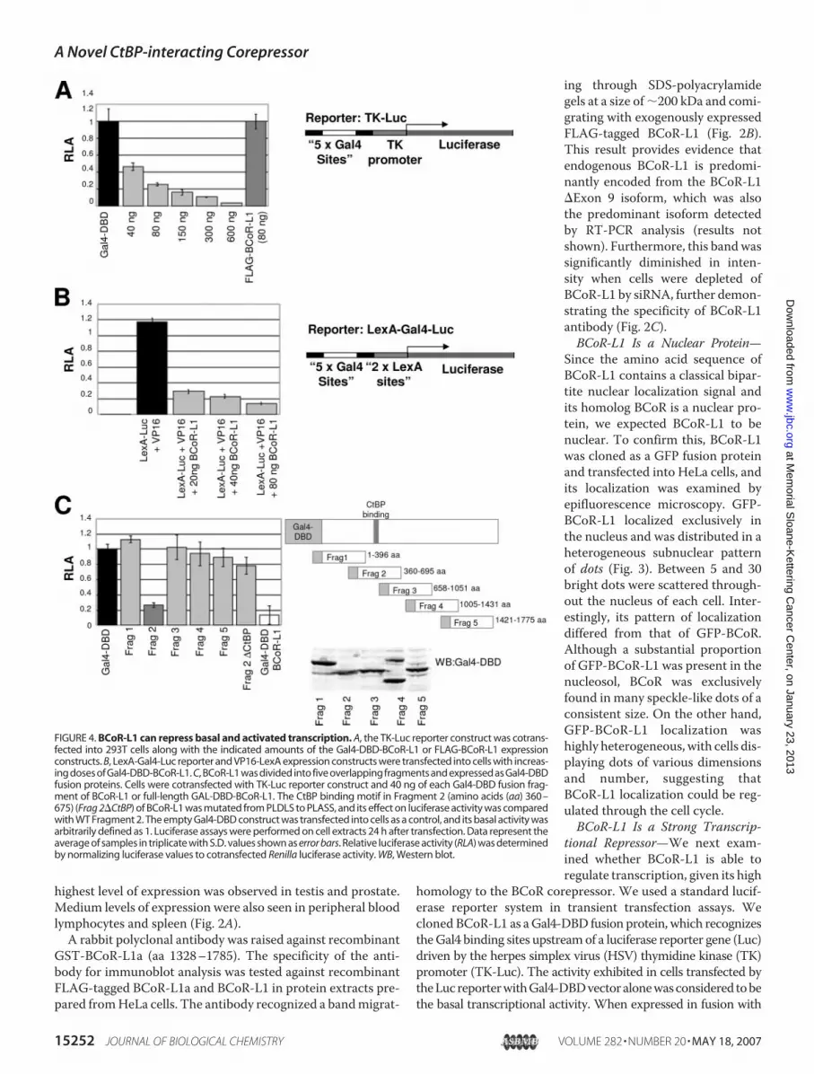
homology to the BCoR corepressor. We used a standard lucif-erase reporter system in transient transfection assays. Wecloned BCoR-L1 as aGal4-DBD fusion protein, which recognizestheGal4 binding sites upstreamof a luciferase reporter gene (Luc)driven by the herpes simplex virus (HSV) thymidine kinase (TK)promoter (TK-Luc). The activity exhibited in cells transfected bytheLucreporterwithGal4-DBDvectoralonewasconsidered tobethe basal transcriptional activity. When expressed in fusion with
FIGURE 4. BCoR-L1 can repress basal and activated transcription. A, the TK-Luc reporter construct was cotrans-fected into 293T cells along with the indicated amounts of the Gal4-DBD-BCoR-L1 or FLAG-BCoR-L1 expressionconstructs. B, LexA-Gal4-Luc reporter and VP16-LexA expression constructs were transfected into cells with increas-ing doses of Gal4-DBD-BCoR-L1. C, BCoR-L1 was divided into five overlapping fragments and expressed as Gal4-DBDfusion proteins. Cells were cotransfected with TK-Luc reporter construct and 40 ng of each Gal4-DBD fusion frag-ment of BCoR-L1 or full-length GAL-DBD-BCoR-L1. The CtBP binding motif in Fragment 2 (amino acids (aa) 360–675) (Frag 2�CtBP) of BCoR-L1 was mutated from PLDLS to PLASS, and its effect on luciferase activity was comparedwith WT Fragment 2. The empty Gal4-DBD construct was transfected into cells as a control, and its basal activity wasarbitrarily defined as 1. Luciferase assays were performed on cell extracts 24 h after transfection. Data represent theaverage of samples in triplicate with S.D. values shown as error bars. Relative luciferase activity (RLA) was determinedby normalizing luciferase values to cotransfected Renilla luciferase activity. WB, Western blot.
A Novel CtBP-interacting Corepressor
15252 JOURNAL OF BIOLOGICAL CHEMISTRY VOLUME 282 • NUMBER 20 • MAY 18, 2007
at Mem
orial Sloane-K
ettering Cancer C
enter, on January 23, 2013w
ww
.jbc.orgD
ownloaded from

the Gal4-DNA binding domain (Gal4-DBD), BCoR-L1 repressedexpression of the Luc reporter gene in a dose-dependentmanner.Comparable expression of FLAG-tagged BCoR-L1, which is nottethered to the Gal4 sites, did not repress luciferase activity, dem-onstrating that BCoR-L1 is able to repress basal transcription onlywhen physically tethered to a heterologous promoter (Fig. 4A).
To determine whether BCoR-L1 was able to repress acti-vated transcription, Gal4-DBD-BCoR-L1 was cotransfectedwith VP16-LexA, a strong transcriptional activator. In thisassay, the LexA-VP16 transcriptional activator drives a LexA-responsive promoter, and the reporter construct encodes theluciferase gene with the Gal4-binding sites upstream of twoLexA binding sites (39). LexA-VP16 activated the LexA-lucif-erase reporter gene�300 fold. Gal4-DBD-BCoR-L1was able toreduce VP16-mediated transactivation significantly, in a dose-dependent manner (Fig. 4B), thereby demonstrating thatBCoR-L1 can repress activated transcription.To identify the regions in BCoR-L1 that mediate repression,
we dividedBCoR-L1 cDNA into five overlapping fragments andexpressed them as Gal4-DBD fusion proteins. Each fragmentexpressed a protein of the expected size.When these fragmentswere tested for repression in the luciferase assay, Fragment 2(amino acids 360–675) of BCoR-L1 was sufficient for maxi-mum repression, indicating that the repressive activity ofBCoR-L1 is mediated through this domain. The amino acidsequence of Fragment 2 (amino acids 360–675) of BCoR-L1contains a classical CtBP1/2 binding motif, PXDLS. Therefore,to test the hypothesis that CtBP is involved in repressionthrough this domain, the CtBP recruitment motif was mutatedfrom PLDLS to PLASS, and its effect on repressor function wascomparedwith that ofwild-type Fragment 2. Themutated frag-ment was no longer capable of efficient repression (Fig. 4C).CtBP Is Required for BCoR-L1 Repression—We next studied
the interaction of the full-length BCoR-L1 with CtBP1 usingcoimmunoprecipitation experiments. Myc-CtBP1 specifi-cally coprecipitated with GFP-BCoR-L1. Similarly, GFP-BCoR-L1 coprecipitated withMyc-CtBP1 (Fig. 5A). After theconsensus CtBP-binding site in BCoR-L1 was mutated fromPLDLS (WT-BCoR-L1) to PLASS (�CtBP-BCoR-L1), �CtBP-BCoR-L1 was no longer capable of interacting with CtBP inreciprocal coimmunoprecipitation experiments, demonstrat-ing that BCoR-L1 recruits CtBP via a classical CtBP-bindingmotif. In addition, mutation of the CtBP consensus bindingmotif in full-length Gal4-DBD-BCoR-L1 (from PLDLS toPLASS) significantly impaired repression at the Gal4-respon-sive promoter in a dose-dependent manner (Fig. 5B). Ourresults indicate that recruitment of CtBP is one mechanismemployed by BCoR-L1 to achieve its repression.BCoR-L1 Associates with Class II HDACs—The mechanism
most commonly employed by repressors involves the recruit-ment of HDACs that remove acetyl groups from the terminaltails of histones. To investigate whether BCoR-L1 is associatedwith specific HDACs, we coexpressed GFP-BCoR-L1 and theFLAG-tagged mammalian expression constructs encoding thehuman Class I and Class II HDACs (HDAC1 to -7). Apart fromHDAC6, which was exclusively cytoplasmic, as has been previ-ously reported (40), each HDAC was observed in the nucleus,with various levels also present in the cytoplasm. GFP-
BCoR-L1 colocalized with the class II HDACs, HDAC4,HDAC5, andHDAC7,within dots of various dimensionswithinthe nucleus (Fig. 6A). Interestingly, we saw a striking redistri-bution of BCoR-L1 into large subnuclear “patches” with thecoexpression of FLAG-HDAC5. We did not observe the colo-calization of GFP-BCoR-L1 with any class I HDACs (HDAC1,-2, and -3) (HDAC2 shown) or with cytoplasmic HDAC6.Coimmunoprecipitation experiments using FLAG-taggedHDAC constructs confirmed the interactions betweenBCoR-L1 and the individual HDACs, HDAC4, -5, and -7 (Fig.6B). BCoR-L1 did not coprecipitate with Class I HDACs (notshown). Furthermore, the interaction between Class II HDACsand BCoR-L1 was not facilitated through CtBP, because�CtBP-BCoR-L1 interacted equivalently with these HDACs.Overall, the colocalization and coprecipitation of BCoR-L1with Class II HDACs suggests that BCoR-L1 function relies onclass II HDAC activity.BCoR-L1 Resides on the E-cadherin Promoter—E-cadherin
is a well characterized target of CtBP in vivo. CtBP associates
FIGURE 5. Transcriptional repression by BCoR-L1 is partially mediatedthrough CtBP. A, mutation of the CtBP binding site in BCoR-L1 abolishes theinteraction between CtBP and BCoR-L1. WT-GFP-BCoR-L1 or �CtBP-GFP-BCoR-L1 was cotransfected with Myc-CtBP1 into 293T cells. Reciprocal immu-noprecipitations using Myc or GFP antibodies were performed, followed byWestern blotting as shown. B, mutation of the CtBP binding site withinBCoR-L1 attenuates repression by BCoR-L1. 293T cells were cotransfectedwith the Gal4-TK-Luc reporter and either WT-Gal4-DBD-BCoR-L1 or �CtBP-Gal4-DBD-BCoR-L1, and the relative luciferase activity (RLA) was assayed.
A Novel CtBP-interacting Corepressor
MAY 18, 2007 • VOLUME 282 • NUMBER 20 JOURNAL OF BIOLOGICAL CHEMISTRY 15253
at Mem
orial Sloane-K
ettering Cancer C
enter, on January 23, 2013w
ww
.jbc.orgD
ownloaded from

with the E-cadherin promoter topromote its repression, andaccordingly, down-regulation ofCtBP results in derepression ofE-cadherin transcription in cellsthat do not normally expressE-cadherin (15, 18, 22, 41). Weperformed chromatin immuno-precipitation assays to evaluatewhether BCoR-L1 associates withthe same region of the E-cadherinpromoter as CtBP. As previouslyshown, Myc-CtBP physically asso-ciated with the endogenous E-cad-herin promoter. GFP-BCoR-L1 alsoassociated with the E-cadherin pro-moter, indicating that BCoR-L1 andCtBP share E-cadherin as a com-mon target (Fig. 7A). Next, we usedan E-cadherin promoter luciferasereporter gene (E-Cad-Luc) (�427 to�53; containing the full-lengthE-Box) to determine if BCoR-L1was able to repress transcriptionfrom the E-cadherin promoter.
293T cells were cotransfected with E-Cad-Luc and either WT-BCoR-L1 or�CtBP-BCoR-L1, and luciferase activity wasmeas-ured. WT-BCoR-L1 was able to significantly suppress lucifer-ase activity driven by the E-Cad promoter. �CtBP-BCoR-L1was not able to reduce E-Cad promoter activity to the sameextent as WT-BCoR-L1, suggesting that CtBP is partiallyrequired for BCoR-L1-mediated repression of E-cadherin (Fig.7B). Next, we used siRNA to reduce BCoR-L1 levels and testedwhether the reduction of BCoR-L1 decreases the repression ofE-cadherin transcription in E-Cad-negative U2OS cells. Weused the pSUPER vector system (26) to direct the synthesis ofsiRNAs to specifically knock down BcoR-L1 levels. We meas-ured E-cadherin transcript levels by RT-PCR in U2OS cellstransfected with pSUPER-BCoR-L1 (depleted of BCoR-L1) orpSUPER-GFP (as control). In these experiments, BCoR-L1 lev-els were reduced to �20%, as determined by RT-PCR (data notshown). A reduction of expression of BCoR-L1 resulted in an�2.5-fold increase in the transcript levels of E-cadherin (Fig.7C). Taken together, these data indicate that BCoR-L1 isdirectly involved in the repression of E-cadherin, an authenticCtBP target gene.
DISCUSSION
BCoR-L1 fits the definition of a corepressor protein in that,although it lacks a DNA-binding domain, it has a portablerepression domain and is capable of repression when recruitedto promoters. It exhibits a number of similarities with BCoR.They are both large proteins and in fact only differ in size by 10amino acids (BCoR is 1721 amino acids) containing ankyrinrepeats of unknown function, although presumably they areprotein-protein interaction modules (42). BCoR was identifiedas a corepressor of the BCL6 transcriptional repressor (24).Unlike BCoR, BCoR-L1 does not interact with BCL6 or poten-
FIGURE 6. BCoR-L1 interacts with Class II HDACs. A, HeLa cells were cotransfected with GFP-BCoR-L1 (green)and each of the mammalian FLAG-tagged HDAC constructs. Cells were stained with the �-FLAG antibody (red)and viewed using confocal microscopy. B, 293T cells were cotransfected with individual FLAG-tagged Class IIHDACs and either Gal4-WT-BCoR-L1 or Gal4-�CtBP-BCoR-L1. Extracts were immunoprecipitated (IP) with theanti-FLAG antibody and immunoblotted (IB) using anti-Gal4-DBD antibody. Input is one-twentieth of the totalimmunoprecipitate.
FIGURE 7. BCoR-L1 contributes to the repression of E-cadherin. A, ChIPanalysis of the E-cadherin promoter. Myc-CtBP1 or GFP-BCoR-L1 was precip-itated from U20S cells, and coprecipitating chromatin was subjected to PCRto amplify the E-cadherin promoter. B, 293T cells were cotransfected withE-Cad-Luciferase and either Gal4-WT-BCoR-L1 or Gal4-�CtBP-BCoR-L1. Lucif-erase activity was monitored 24 h later. C, U2OS cells were transfected withpSUPER-BCoR-L1 (to deplete BCoR-L1 levels) or pSUPER-GFP (as control). RNAwas taken 72 h later, and the levels of E-cadherin transcript were measured byquantitative PCR. Results shown are the average of three separate experi-ments repeated in duplicate.
A Novel CtBP-interacting Corepressor
15254 JOURNAL OF BIOLOGICAL CHEMISTRY VOLUME 282 • NUMBER 20 • MAY 18, 2007
at Mem
orial Sloane-K
ettering Cancer C
enter, on January 23, 2013w
ww
.jbc.orgD
ownloaded from

tiate BCL6-mediated repression (data not shown), providingevidence that BCoR-L1 and BCoR may have distinct functionsin human cells. However, BCoR is likely to have roles independ-ent of BCL6, given its ubiquitous expression in tissues whereBCL6 is not expressed. Furthermore, we found that BCoR-L1 isexpressed atmuch higher levels in the hormone-responsive tis-sues, prostate and testis, than in other tissues.Typically, corepressors recruitmultiple cofactors involved in
chromatin remodeling, histone deacetylation, or basal tran-scription to mediate concerted transcriptional silencingthrough multiple repression pathways. We have shown thatBCoR-L1 achieves its repression in at least two ways, via aninteraction with the CtBP corepressor and possibly via Class IIHDACs. Elimination of the CtBP-binding site within BCoR-L1partially relieves BCoR-L1-mediated transcriptional repres-sion, demonstrating that BCoR-L1 repression is mediatedthrough CtBP. Since CtBP has been shown to be regulated byfluctuating levels of NADH in the cell, this raises the possibilitythat BCoR-L1 is regulated similarly (20, 22, 23).Recently, the BCoR complexwas shown to contain Polycomb
group (PcG) proteins (NSPC1, RING1, RNF2, and RYBP) and aJmjC domain histone H3 K36 demethylase, which is able toremovemethyl groups from lysine (43). Another complex asso-ciated with CtBP is the LSD1-CoREST complex, capable ofdemethylatingH3-K4within nucleosomes (15, 44, 45). It will beinteresting to addresswhether BCoR-L1 interacts with histone-demethylating enzymes or Polycomb group proteins in thesame fashion as BCoR and CtBP.Here, we have shown that BCoR-L1 is involved in the repres-
sion of E-cadherin, a known CtBP target. This repression par-tially requires CtBP. A number of transcriptional repressors areknown to regulate E-cadherin expression, including Snail (46),Slug (47, 48), Twist (49), and ZEB/�EF1 (50, 51), and it is pos-sible that BCoR-L1 might function together with these repres-sors, or as part of a separate as yet unknown complex. Further-more, it might be of great value to determine if BCoR-L1represses the expression of other CtBP-regulated genes andpossibly those involved in promoting apoptosis (18, 19).E-cadherin is critical to maintain normal epithelial cell con-
tact (52), and down-regulation of E-cadherin is seen in a largepercentage of carcinomas or borderline tumors (53, 54). SinceBCoR-L1/CtBP represses E-cadherin, interfering with BCoR-L1/CtBP functionmight prevent loss of the epithelial state. Thiswould be the reverse of the phenotype induced by pinin/DRS,which binds CtBP and relieves its repression of the E-cadherinpromoter (55, 56). Interestingly, CtBP binding to the E-cad-herin promoter is induced by elevations in free NADH. Thisredox-regulated repression of E-cadherin has been postulatedto be involved in increasing tumor cell migration (41). It is con-ceivable, therefore, that interfering with CtBP and/or BCoR-L1might inhibit tumor metastasis.CtBP recruits Class II HDACs via consensus CtBP-binding
motifs within their amino termini (30). However, CtBP is notrequired for the interaction between BCoR-L1 and Class IIHDACs, since our �CtBP-BCoR-L1 was able to interact withthe Class II HDACs. It is possible that the class II HDACs inter-act with the ankyrin repeats of BCoR-L1 directly, since theyinteract with the ankyrin repeat of certain proteins, ANKRA1
(ankyrin-repeat family A protein) and ANKRA2 (57, 58).BCoR-L1 is able to partially repress transcription independ-ently of CtBP, suggesting a contribution from HDACs or someother mechanism. This observation also fits with the principlethat generally, the contributions of all of the separate compo-nents of any repressive complex act in an additive fashion toachieve full repression.Only a relatively small number of transcription factors are
known to interact with Class II HDACs, although interactionshave been reported between Class I HDACs and many tran-scription factors. The colocalization and coprecipitation ofBCoR-L1 with Class II HDACs strongly, although indirectly,suggest that BCoR-L1 function is linked to that of Class IIHDACs.Most research has focused on the association betweenClass II HDACs and members of theMEF2 (myocyte enhancerfactor-2) family of MADS-box transcription factors via theiramino-terminal extensions, which results in transcriptionalrepression (59–61). The MEF2 family regulate genes involvedin myogenesis and accordingly class II HDACs inhibit patho-logic cardiac hypertrophy by modulating MEF2 transcriptionfactor activity (62, 63). Class II HDACs are highly expressed inheart, skeletal muscle, and brain, in contrast to the Class IHDACs, which have a more ubiquitous expression (64). Atpresent, we do not know the relative expression of BCoR-L1 inheart, muscle, and brain, since these tissues were not repre-sented on our Northern blot. In the future, it will be of interestto elucidate if BCoR-L1 plays a role in myogenesis or generallyin Class II HDAC function.We have not been able to identify of the nature of the sub-
nuclear foci containing BCoR-L1. Notably, BCoR-L1 and BCoRlocalize in distinct compartments, suggesting that they havedivergent functions. Many proteins involved in transcriptionalregulation localize to specific compartments within thenucleus. It is unknown whether these subnuclear foci are sitesof active gene silencing or are required to recruit repressorsaway from sites of active transcription. Class II HDACs localizeto distinct nuclear bodies within the cell nucleus, although asyet the function and biological significance of these dots isunknown (10). Corepressors, such as SMRT and NCoR, areconcentrated in these nuclear foci, sometimes called deacety-lase bodies, with the HDACs (65). One remarkable observationis the redistribution of BCoR-L1 to large HDAC5 “patches” byoverexpression of HDAC5. Interestingly, expression of thecorepressor, NCoR, leads to recruitment of HDAC5 intointranuclear bodies (65). The BCoR-L1-HDAC5 relocationworks in the opposite direction; rather than BCoR-L1 recruit-ing HDAC5, our data demonstrate that HDAC5 is responsiblefor the relocalization of BCoR-L1. Another similar example isseen in the redistribution of the NCoR by the PIT-1 transcrip-tion factor (67).It is possible that BCoR-L1 is a substrate of theHDACs, since
there is evidence of nonhistone substrates, including p53 (68,69) and �-tubulin (40). Acetylation of a lysine residue adjacentto the CtBP recruitment motif in E1A protein has been shownto modulate its interaction with CtBP (34). Interestingly,BCoR-L1 sequence also contains this flanking lysine residue,raising the possibility that its interaction with CtBP is similarlyregulated by acetylation.
A Novel CtBP-interacting Corepressor
MAY 18, 2007 • VOLUME 282 • NUMBER 20 JOURNAL OF BIOLOGICAL CHEMISTRY 15255
at Mem
orial Sloane-K
ettering Cancer C
enter, on January 23, 2013w
ww
.jbc.orgD
ownloaded from
The importance of active transcriptional repression is high-lighted by the fact that aberrant gene silencing is linked to arange of diseases, including developmental diseases and can-cers (70, 71). Recently, mutations in BCoR have been linkedwith the developmental abnormality oculofaciocardiodentalsyndrome (66). This developmental syndrome has been linkedto two loci, MAA1 (Xq27) and MAA2 (Xp11, the BCoR locus).Intriguingly, mutations in BCoR were found inMAA2 families.Coincidentally, MAA1 maps extraordinarily close to theBCoR-L1 locus. There is thus the possibility that aberrantBCoR-L1 function may contribute to this disease.
Acknowledgment—We gratefully acknowledge Vivian Bardwell (Uni-versity ofMinnesota,Minneapolis, MN) for providing theMyc-taggedBCoR cDNA clone.
REFERENCES1. Levine, M., and Tjian, R. (2003) Nature 424, 147–1512. Jepsen, K., and Rosenfeld, M. G. (2002) J. Cell Sci. 115, 689–6983. Gabellini, D., Tupler, R., and Green, M. R. (2003) Curr. Opin. Genet. Dev.
13, 239–2454. Look, A. T. (1997) Science 278, 1059–10645. Struhl, K. (1998) Genes Dev. 12, 599–6066. Gorisch, S. M., Wachsmuth, M., Toth, K. F., Lichter, P., and Rippe, K.
(2005) J. Cell Sci. 118, 5825–58347. Marmorstein, R., and Roth, S. Y. (2001) Curr. Opin. Genet. Dev. 11,
155–1618. Bolden, J. E., Peart, M. J., and Johnstone, R. W. (2006) Nat. Rev. Drug.
Discov. 5, 769–7849. Grozinger, C. M., and Schreiber, S. L. (2002) Chem. Biol. 9, 3–1610. Yang, X. J., and Gregoire, S. (2005)Mol. Cell. Biol. 25, 2873–288411. Gao, L., Cueto, M. A., Asselbergs, F., and Atadja, P. (2002) J. Biol. Chem.
277, 25748–2575512. Glozak, M. A., Sengupta, N., Zhang, X., and Seto, E. (2005) Gene (Amst.)
363, 15–2313. Chinnadurai, G. (2002)Mol. Cell 9, 213–22414. Chinnadurai, G. (2003) BioEssays 25, 9–1215. Shi, Y., Sawada, J., Sui, G., Affar el, B., Whetstine, J. R., Lan, F., Ogawa, H.,
Luke, M. P., and Nakatani, Y. (2003) Nature 422, 735–73816. Kim, J. H., Cho, E. J., Kim, S. T., and Youn, H. D. (2005) Nat. Struct. Mol.
Biol. 12, 423–42817. Zhao, L. J., Subramanian, T., Zhou, Y., and Chinnadurai, G. (2006) J. Biol.
Chem. 281, 4183–418918. Grooteclaes, M. L., and Frisch, S. M. (2000) Oncogene 19, 3823–382819. Grooteclaes,M., Deveraux, Q., Hildebrand, J., Zhang, Q., Goodman, R. H.,
and Frisch, S. M. (2003) Proc. Natl. Acad. Sci. U. S. A. 100, 4568–457320. Kumar, V., Carlson, J. E., Ohgi, K. A., Edwards, T. A., Rose, D. W., Es-
calante, C. R., Rosenfeld, M. G., and Aggarwal, A. K. (2002)Mol. Cell 10,857–869
21. Thio, S. S., Bonventre, J. V., and Hsu, S. I. (2004) Nucleic Acids Res. 32,1836–1847
22. Zhang, Q., Piston, D. W., and Goodman, R. H. (2002) Science 295,1895–1897
23. Fjeld, C. C., Birdsong,W. T., and Goodman, R. H. (2003) Proc. Natl. Acad.Sci. U. S. A. 100, 9202–9207
24. Huynh, K. D., Fischle,W., Verdin, E., and Bardwell, V. J. (2000)Genes Dev.14, 1810–1823
25. Jardin, F., and Sahota, S. S. (2005) Hematology 10, 115–12926. Brummelkamp, T. R., Bernards, R., and Agami, R. (2002) Science 296,
550–55327. Fabbro, M., Savage, K., Hobson, K., Deans, A. J., Powell, S. N., McArthur,
G. A., and Khanna, K. K. (2004) J. Biol. Chem. 279, 31251–3125828. Parra, M., Kasler, H., McKinsey, T. A., Olson, E. N., and Verdin, E. (2005)
J. Biol. Chem. 280, 13762–13770
29. Grozinger, C. M., Hassig, C. A., and Schreiber, S. L. (1999) Proc. Natl.Acad. Sci. U. S. A. 96, 4868–4873
30. Zhang, C. L., McKinsey, T. A., Lu, J. R., and Olson, E. N. (2001) J. Biol.Chem. 276, 35–39
31. Scully, R., Ganesan, S., Brown,M., De Caprio, J. A., Cannistra, S. A., Feun-teun, J., Schnitt, S., and Livingston, D. M. (1996) Science 272, 123–126
32. Scully, R., Chen, J., Plug, A., Xiao, Y., Weaver, D., Feunteun, J., Ashley, T.,and Livingston, D. M. (1997) Cell 88, 265–275
33. Schaeper, U., Subramanian, T., Lim, L., Boyd, J. M., and Chinnadurai, G.(1998) J. Biol. Chem. 273, 8549–8552
34. Zhang, Q., Yao, H., Vo, N., and Goodman, R. H. (2000) Proc. Natl. Acad.Sci. U. S. A. 97, 14323–14328
35. Plevin, M. J., Mills, M. M., and Ikura, M. (2005) Trends Biochem. Sci. 30,66–69
36. Wu, L. C., Wang, Z. W., Tsan, J. T., Spillman, M. A., Phung, A., Xu, X. L.,Yang,M. C., Hwang, L. Y., Bowcock, A.M., and Baer, R. (1996)Nat. Genet.14, 430–440
37. Sander,M., Lowenhaupt, K., Lane,W. S., andRich, A. (1991)Nucleic AcidsRes. 19, 4523–4529
38. Sander, M., Lowenhaupt, K., and Rich, A. (1991) Proc. Natl. Acad. Sci.U. S. A. 88, 6780–6784
39. Zhou, X., Richon, V. M., Rifkind, R. A., andMarks, P. A. (2000) Proc. Natl.Acad. Sci. U. S. A. 97, 1056–1061
40. Hubbert, C., Guardiola, A., Shao, R., Kawaguchi, Y., Ito, A., Nixon, A.,Yoshida, M., Wang, X. F., and Yao, T. P. (2002) Nature 417, 455–458
41. Zhang, Q., Wang, S. Y., Nottke, A. C., Rocheleau, J. V., Piston, D. W., andGoodman, R. H. (2006) Proc. Natl. Acad. Sci. U. S. A. 103, 9029–9033
42. Sedgwick, S. G., and Smerdon, S. J. (1999) Trends Biochem. Sci. 24,311–316
43. Gearhart, M. D., Corcoran, C. M., Wamstad, J. A., and Bardwell, V. J.(2006)Mol. Cell. Biol. 26, 6880–6889
44. Lee,M. G.,Wynder, C., Cooch, N., and Shiekhattar, R. (2005)Nature 437,432–435
45. Shi, Y. J.,Matson, C., Lan, F., Iwase, S., Baba, T., and Shi, Y. (2005)Mol. Cell19, 857–864
46. Cano, A., Perez-Moreno,M. A., Rodrigo, I., Locascio, A., Blanco,M. J., delBarrio, M. G., Portillo, F., and Nieto, M. A. (2000)Nat. Cell Biol. 2, 76–83
47. Bolos, V., Peinado, H., Perez-Moreno,M. A., Fraga,M. F., Esteller,M., andCano, A. (2003) J. Cell Sci. 116, 499–511
48. Hajra, K. M., Chen, D. Y., and Fearon, E. R. (2002) Cancer Res. 62,1613–1618
49. Yang, J., Mani, S. A., Donaher, J. L., Ramaswamy, S., Itzykson, R. A., Come,C., Savagner, P., Gitelman, I., Richardson, A., and Weinberg, R. A. (2004)Cell 117, 927–939
50. Comijn, J., Berx, G., Vermassen, P., Verschueren, K., van Grunsven, L.,Bruyneel, E.,Mareel,M., Huylebroeck, D., and van Roy, F. (2001)Mol. Cell7, 1267–1278
51. Peinado, H., Portillo, F., and Cano, A. (2004) Int. J. Dev. Biol. 48, 365–37552. Hajra, K. M., and Fearon, E. R. (2002) Genes Chromosomes Cancer 34,
255–26853. Thiery, J. P. (2003) Curr. Opin. Cell Biol. 15, 740–74654. Takeichi, M. (1993) Curr. Opin. Cell Biol. 5, 806–81155. Joo, J. H., Alpatov, R., Munguba, G. C., Jackson, M. R., Hunt, M. E., and
Sugrue, S. P. (2005)Mol. Vis. 11, 133–14256. Alpatov, R., Munguba, G. C., Caton, P., Joo, J. H., Shi, Y., Shi, Y., Hunt,
M. E., and Sugrue, S. P. (2004)Mol. Cell. Biol. 24, 10223–1023557. Wang, A. H., Gregoire, S., Zika, E., Xiao, L., Li, C. S., Li, H., Wright, K. L.,
Ting, J. P., and Yang, X. J. (2005) J. Biol. Chem. 280, 29117–2912758. McKinsey, T. A., Kuwahara, K., Bezprozvannaya, S., and Olson, E. N.
(2006)Mol. Biol. Cell 17, 438–44759. Miska, E. A., Karlsson, C., Langley, E., Nielsen, S. J., Pines, J., and Kouzar-
ides, T. (1999) EMBO J. 18, 5099–510760. Sparrow, D. B., Miska, E. A., Langley, E., Reynaud-Deonauth, S., Kotecha,
S., Towers, N., Spohr, G., Kouzarides, T., and Mohun, T. J. (1999) EMBOJ. 18, 5085–5098
61. Lu, J., McKinsey, T. A., Nicol, R. L., and Olson, E. N. (2000) Proc. Natl.Acad. Sci. U. S. A. 97, 4070–4075
62. Chang, S., McKinsey, T. A., Zhang, C. L., Richardson, J. A., Hill, J. A., and
A Novel CtBP-interacting Corepressor
15256 JOURNAL OF BIOLOGICAL CHEMISTRY VOLUME 282 • NUMBER 20 • MAY 18, 2007
at Mem
orial Sloane-K
ettering Cancer C
enter, on January 23, 2013w
ww
.jbc.orgD
ownloaded from

Olson, E. N. (2004)Mol. Cell. Biol. 24, 8467–847663. Zhang, C. L.,McKinsey, T. A., Chang, S., Antos, C. L., Hill, J. A., andOlson,
E. N. (2002) Cell 110, 479–48864. Verdin, E., Dequiedt, F., and Kasler, H. G. (2003) Trends Genet. 19,
286–29365. Downes, M., Ordentlich, P., Kao, H. Y., Alvarez, J. G., and Evans, R. M.
(2000) Proc. Natl. Acad. Sci. U. S. A. 97, 10330–1033566. Ng, D., Thakker, N., Corcoran, C. M., Donnai, D., Perveen, R., Schneider,
A., Hadley, D. W., Tifft, C., Zhang, L., Wilkie, A. O., van der Smagt, J. J.,Gorlin, R. J., Burgess, S.M., Bardwell, V. J., Black,G.C., andBiesecker, L.G.
(2004) Nat. Genet. 36, 411–41667. Voss, T. C., Demarco, I. A., Booker, C. F., and Day, R. N. (2005) J. Cell Sci.
118, 3277–328868. Vaziri, H., Dessain, S. K., Ng Eaton, E., Imai, S. I., Frye, R. A., Pandita, T. K.,
Guarente, L., and Weinberg, R. A. (2001) Cell 107, 149–15969. Luo, J., Li, M., Tang, Y., Laszkowska, M., Roeder, R. G., and Gu,W. (2004)
Proc. Natl. Acad. Sci. U. S. A. 101, 2259–226470. Gabellini, D., Green, M. R., and Tupler, R. (2004) Curr. Opin. Genet. Dev.
14, 301–30771. Gabellini, D., Green, M. R., and Tupler, R. (2002) Cell 110, 339–348
A Novel CtBP-interacting Corepressor
MAY 18, 2007 • VOLUME 282 • NUMBER 20 JOURNAL OF BIOLOGICAL CHEMISTRY 15257
at Mem
orial Sloane-K
ettering Cancer C
enter, on January 23, 2013w
ww
.jbc.orgD
ownloaded from