Embed Size (px)
Citation preview

A NOVAL DIGITAL IMPRESSION AND MAXILLOMANDIBULAR RELATIONSHIP WORKFLOW FOR FULL-ARCH IMPLANT-SUPPORTED PROSTHESIS Ahmed, Walaa *, Verhaeghe, Tyler; Mccullagh, Anthony The University of British Columbia Prosthodontics Vancouver, Bc, Canada Keywords: digital workflow,impression,implant-supported prosthesis Case Presentation: Intraoral scanners are being increasingly used as a replacement for conventional impressions, including the capturing of edentulous arches, although this may be clinically challenging. Paucity in the literature investigating intraoral scanning for fabricating multiple implant supported full-arch restorations remains, particularly with regards to in vivo studies and the establishment of a definitive scan protocol. This technique describes the fabrication and use of a custom scanning device to scaffold an intraoral full-arch scan of at least 6 implant scan bodies in the edentulous maxilla while simultaneously capturing the maxillomandibular relationship for the purpose of establishing a digital workflow to fabricate a maxillary full-arch implant supported prosthesis. This technique provides the intraoral scanner with anatomical landmarks for improving the likelihood of scanning efficiency and offers a possible scan protocol which may help to improve the accuracy of the scan.
NOVEL NANO-SIZED HYDROXYAPATITE COATING FOR DENTAL IMPLANTS FOR OSSEOINTEGRATION AND OSTEOGENIC GENE EXPRESSION Ajeeb, Zainab *, Kenji Inoue1, Hironori Kasai, Kentaro Imamura, Yoichiro Taguchi, Lukasz Witek, Paulo G. Coelho, and Seiichi Yamano New York University College of Dentistry Department of Prosthodontics New York, Ny, United States Purpose/Aim: Nanostructures applied to dental implants have been suggested to contribute to a higher grade of

osseointegration. However, the effect of nanostructure modification of dental implants on molecular osteogenesis is still unclear, especially in the early period of bone healing. The hypothesis for this project is that a novel implant surface coated with nano-sized (20 nm) hydroxyapatite (Nano) will positively effect gene expression patterns during initial bone-implant integration as well as accelerate osseointegration in comparison to implants with conventional surfaces, a double acid etched (DAE). In particular, the primary focus was on the early stages of bone healing, as during the first two weeks the dynamics of bone healing around implants coated with nano-hydroxyapatite, currently are inadequately understood. In this study, we evaluated the effect of Nano on gene expression related to osteogenic activity and nano mechanical properties compared to DAE and none-treated control at 1 and 2 weeks after implant placement in a rodent femur model. Materials and Methods: Nano displayed significantly higher osteogenic gene expression of type I collagen, osteocalcin and osterix in comparison to DAE and control at 1 and 2 weeks, indicating progressive mineralization of newly formed bone (p < 0.05). Nanoindentation analysis revealed that Nano presented significantly higher-rank hardness and ranks elastic modulus compared to DAE and control at 1 week (p < 0.05). Histomorphometric analysis showed that there is no significant differences in bone-to-implant contact and bone area fraction occupancy levels between the Nano and DAE. Results: Demonstrated that the treated Nano surface positively and significantly induced osteogenic gene expression compared to DAE and control groups in the early phases of bone healing. Degree of mineralization (hardness and elastic modulus) of the bone in the proximity of the implant was enhanced for Nano at the early time point. Conclusions: This study demonstrated that nano-sized (20 nm) hydroxyapatite significantly induced osteogenic gene expression and osseointegration compared to a double acid etched and control groups in the early phases of bone healing. This surface has the potential to have a positive impact on osseointegration.
ELECTROMYOGRAPHY METHOD IN A FULL MOUTH REHABILITATION WITH VERTICAL DIMENSION LOSS Alava, Angela *, Alava, Angela Universidad Nacional Autónoma De México División De Estudios De Posgrado E Investigación En Odontología Universidad Nacional Autónoma De Mexico Ciudad De México, Ciudad De México, Mexico Case Presentation: Recovering the vertical dimension is a necessary procedure during the oral rehabilitation treatment to achieve a functional occlusal scheme in the patient, the use of electromyography as an auxiliary method element in this treatment is necessary to obtain an operational safety and occlusal stability. ELECTROMYOGRAPHY METHOD IN A FULL MOUTH REHABILITATION WITH VERTICAL DIMENSION LOSS Objective: to evaluate the usefulness of electromyography as an auxiliary method to assess the changes obtained in the restoration of the vertical dimension Challenge: Patient with fatigue and occlusal instability and loss of vertical dimension. Clinical case: 60-year-old patient with instability and severe muscular fatigue due to the preparations of total crowns in the maxilla. Diagnosis treatment plan: First stage: Evaluation of the electromyographic parameter of the patient with the previous provisional, in which an irregular parameter associated with severe fatigue is observed, which made it difficult to evaluate the vertical dimension. Second stage: The vertical dimension was restored with the development of provisionals and regular electromyographic parameters were observed. Third stage: definitive stage, the muscular stability of the rehabilitation of the vertical dimension was observed. Conclusion: electromyography is an element of easy application as a diagnostic and treatment aid to measure occlusal and muscular stability especially in severe cases of loss of vertical dimension.
A CASE REPORT OF FABRICATING CUSTOM CROWN FOR PRESS-FIT IMPLANT AND PRESS-FIT ABUTMENT

Alghamdi, Reem *, Sawada, Mitsuhiro; Hengjeerajaras, Parnward; Manasse, Maryse; Cho, Sang Choon. New York University College of Dentistry A Ashman Department of Periodontology and Implant Dentistry New York, Ny, United States Keywords: Custom Crown, Press-Fit Implant, Press-Fit Abutment Case Presentation: Dental implant treatment is a dependable therapy for single or multiple teeth replacement and fully edentulous rehabilitation. Treatment outcomes are highly successful with great patient satisfaction. A 60-year-old female patient presented to our implant clinic with press-fit implant that was placed in area of tooth #29 over 30 years with one-piece press-fit custom abutment. The implant (4.0x14mm) was well-integrated in the bone with no signs and symptoms, and the patient didn’t want to remove the implant for the high risk of bone fracture. The patient complained about the crown that kept falling off. Flap was opened and the press-fit abutment was prepared and the margin was dropped to gain more height that is critical for the retention of the future cement-retained crown. Anti-rotational groove was created. In this poster, intra-oral technique is presented for fabricating custom cement-retained crown over press-fit abutment and press-fit implant.
IMMEDIATE IMPLANT PROVISIONALIZATION TECHNIQUES, WHEN AND HOW Alodadi, Abdullah *, Alkhurays, Mohammad Armed Forces Hospital Southern Region, Msd Dental Department Khamis Mushait, Southern Region, Saudi Arabia Keywords: Immediate Provisionalization,Socket Shield Case Presentation: Abstract: In the last few decades, Implant Dentistry has been a widely accepted treatment modality. During this period of development, its concepts and techniques have undergone tremendous changes. At first, the only surgical protocol that considered reproducible and predictable is the one involving two-stage surgery. Later, the one-stage surgical protocol became accepted. However, the first review of the literature on immediate loading was published in 1998. Since then, immediate loading has become one of the most relevant topics in implant dentistry. For years, dental implants have been loaded immediately with varying degrees of success. With the evolvement of the biological and biomechanical understanding of immediate loading and with the implementation of Cone Beam Computed tomography and implant guided surgery, recent clinical investigations shows similar clinical outcome to the conventional implant loading protocol. The purpose of this poster is to review the concept of immediate implant loading and to present different provisionalization techniques.

FRACTURE LOAD COMPARISON BETWEEN 3D PRINTED AND MILLED PROVISIONAL RESTORATION Alsaeid, Waleed *, Zhivago, Paul; Choi, Mijin New York University Prosthodontics Department New York City, Ny, United States Purpose/Aim: The objective of this study was to measure and compare the fracture load of a four-unit provisional prosthesis fabricated with 3D printing (SprintRay, Los Angeles, CA) and milled technology (Roland, Irvine, CA) from digitally scanned files. Materials and Methods: A metal master model with prepared abutments on teeth #28 and 31, and missing teeth #29 and #30 for a four unit FPD was obtained. The model was scanned using a Trios scanner (3Shape, Copenhagen, Denmark). The provisional prosthesis was designed using an open source CAD software (ZBrush, Pixologic, Los Angeles, CA). Forty-five samples were fabricated which were then separated into three groups of fifteen samples each.?Group 1 samples were 3D printed using NextDent crown and bridge (C&B) material (NextDent, AV Soesterberg, Netherlands). Group 2 samples were 3D printed using NextDent C&B MFH micro-filled hybrid material.?Group 3 (control group) samples

that were milled using polymethylmethacrylate (PMMA). ?A three-point bending test (Instron, Norwood, MA) was used to fracture the samples. The fracture load was recorded for each sample. Results: Mean values and standard deviations have been obtained for each group. Group 1 (364.4+-66.5 Ncm), Group 2 (226+-21 Ncm), Group 3 (183.5+-21 Ncm) Conclusions: The recorded fracture load showed that groups 1 and 2 had a higher fracture load. Further studies are needed to determine if 3D printing can be used as a viable alternative to milling PMMA.
THE USE OF MESHMIXER AS A TOOL FOR 3D RENDERING IN DENTISTRY - A PRACTICAL GUIDE Alsulaimi, Abdulrahman *, Mccrea, Kyle / Kiat-Amnuay, Sudarat The University of Texas - Health Sciences Center at Houston Advanced Education In General Dentistry Houston, Tx, United States Case Presentation: Clinical Challenges: The main goal of a removable prosthesis is to have a functional, natural and pleasing appearance; different dental practitioners have their vision and technique for their final smile. Lab communication is an essential step in developing a diagnostic wax-up and final prosthesis. This step can be time-consuming and tedious, especially when cases are sent out to independent dental labs. Multiple studies have indicated that dental laboratories agree on a lack of communication by the dentists that is reflected by the work authorization forms and can affect the quality of prosthesis, thus resulting in patient frustration due to longer waiting-time and increased number of office visits. Purpose: The purpose of this poster is to provide a visual step-by-step practical guide for general dentists in practices that do not have in-house labs and are utilizing 3D scanners. Meshmixer is a free 3D rendering and printing software tool available for both Windows and macOS. Some of the functions of the software, according to meshmixer.com, are 3D Sculpting and Surface Stamping, Precise 3D Positioning with Pivots, Automatic Alignment of Surfaces, 3D Measurements and, Stability & Thickness Analysis. In this guide, we will be describing dental applications related to removable prostheses design using Meshmixer for macOS. Clinical Implications: Time efficiency is an imperative factor, especially in the private sector, where it could be a decisive determinant in choosing the impression systems, labs, and turnover. Digital impressions have many advantages over conventional impressions. Relevant examples of these advantages are 1. There is no dimensional change or susceptibility to damage from handling. 2. No trays, tray adhesives, and impression material dispensing systems are needed. 3. Lower cost from the restricted shelf-life of impression materials. 4. Electronic transport of the dental impression to the lab. This includes better communication with the lab leading to a more efficient process, reducing both cost and effort in the long term with no cross-infection risk.
ALTERNATIVE TECHNIQUE TO ACHIEVE ESTHETIC IMPLANT RESTORATION IN THE ANTERIOR MAXILLA Alzahrani, Saeed *, Hansakul, Phurin; Leung, Martin Leung; Bloom, Mitchell J; Cho, Sang Choon. New York University College of Dentistry A Ashman Department of Periodontology and Implant Dentistry New York, Ny, United States Keywords: bone regeneration techniques, intra-osseous defect Case Presentation: Implant therapy has become an important part of dentistry. Successful placement of a dental implant

requires adequate surrounding bone. In cases where there is an inadequate quantity of bone, the bone volume can be increased by bone augmentation, followed by implant placement. Despite all the advancements in bone regeneration techniques, the outcome in many cases is not highly predictable. The morphology of a defect at the implant site has been reported to be a critical factor for the success of bone augmentation. Similar to the extraction socket or maxillary sinus cavity, defect surrounded by bony walls is called an intra-osseous defect, and this type of defect is known to yield a highly successful regeneration due to good blood supply and being well contained. In contrast, an extra-osseous defect with less bony walls is known to be less predictable in its bone augmentation procedure. This technique helps to convert an extra-osseous defect into an intra-osseous defect. Using this technique can significantly enhance the results of guided tissue regeneration, and enable a more successful and predictable implant placement in a wider scope of cases. Moreover, clinician can expand the existing atrophic ridge to a prosthetically oriented implant position. The purpose of this presentation is to document the surgical steps of this technique leading to a prosthetically oriented implant position.
PATIENT CLASS II SKELETAL REHABILITATED WITH PROSTHESIS ON IMPLANTS IN BOTH MAXILARS Apreza, Gisela *, Pereza Martinez Enrique Benemerita Universidad Autonoma De Puebla Master In Oral Rehabilitation Puebla , Puebla , Mexico Keywords: IMPLANTS,CLASS II, PROSTHESIS Case Presentation: PATIENT CLASS II SKELETAL REHABILITATED WITH PROSTHESIS ON IMPLANTS IN BOTH MAXILARS Background The loss of dental organs at present has increased considerably, and this brings as consequences alterations, phonetic, facial, aesthetic and functional for the patient, however with the advance of technology we can now restore the harmony of the mouth with the help of Implant placement strategically using digital surgical planning, also allowing us to place a prosthesis on the same day as the surgery. Case report A 61-year-old female patient, class II skeletal, has mismatched prostheses, edentulous spaces, and mobility in multiple dental organs. The reason for consultation is complete rehabilitation of both jaws, a rehabilitation on implants of both arches is proposed to return functionality and aesthetics. Using digital tomography, digital planning is performed for the placement of 4 implants in the lower arch, and 6 in the upper arch with immediate loading in the maxilla. Discussion The guided surgery facilitates the operative and surgical times when placing multiple implants, obtaining more predictable results and with this an adequate rehabilitation for the patient, if it is possible to obtain an adequate torque of the implants an immediate load can be made providing the patient greater comfort and aesthetics, after surgery. Conclusion Guided surgery turns out to be an indispensable step when it comes to placing multiple implants in patients with several edentulous spaces, since it allows you a better planning of surgery and rehabilitation. In this case, successful results were obtained in the placement of the implants in both arches, returning the patient the functionality and aesthetics she expected. Clinical implications The patient presented a skeletal class 2, so the space between both jaws had to be compensated to obtain an adequate occlusion, and that this did not affect the placement of the implants, the lack of bone in some areas of the bone had to be taken into account maxillary and take into account the distribution of the implants.

DIGITAL PLANNING AND GUIDED IMPLANT SURGERY IN FULL ARCH INMEDIATE LOAD. Arellano, Juan *, Franco, Guillermo & Pérez, Enrique Benemérita Universidad Autónoma De Puebla Rehabilitación Oral Puebla, Puebla, Mexico Keywords: IMPLANTS, FULL ARCH, DIGITAL PLANNING Case Presentation: BACKGROUND: In 1979 Lederman, published the first cases where an overdenture was placed over four implants the same day of the surgical intervention. In the initial protocols of Branemark of 1985, immediate loading implants (ILI) was contraindicated. It was claimed that dental implants needed 3 to 6 months to generate direct, functional and structural connection to the bone. Throughout history, implant placement and loading protocols have been analyzed separately from one another. Different factors influence in which technique use for each case. CASE REPORT: 85-year-old male patient attend to the BUAP Oral Rehabilitation clinic for reasons of function and aesthetics. The patient refers to being dissatisfied with the general condition of his mouth. Despite the considerable amount of bone loss and unwillingness to undergo any major bone graft procedures, the patient demanded fixed prostheses. Four implants were planned in the Blue Sky Bio Dental Implant System, two angulated implants at the posterior sites (Alpha Bio Neo 4.20X13mm.) and two vertical in the anterior site (Alpha Bio Neo 3.75X13mm.) for the maxilla as a All – on – 4 protocol. After the planation a full restricted surgical guide was designed and printed for the surgery of the upper arch. On the day of the intervention, two selective extractions were first performed, so that our surgical guide would passively go down, but remain dento supported. The four implants were placed with a kit of restrictive drills and then the multi-unit attachments were placed at 30 degrees in the posterior sector and at 17 degrees in the anterior sector, to be able to place the provisional titanium attachments and fix the intermediate prosthesis with the correct vertical dimension and occlusion. DISCUSSION: Although the conventional protocol for the placement of fixed implant prosthesis is composed of more than one stage, more and more professionals and patients require less invasive, rapid and predictable treatments. Therefore, developing and incorporating digital planning techniques, restrictive surgical guides and immediate loading are essential to solve the demands of today. CONCLUSION: Guided surgery allows less invasive treatments and with more predictable results. It reduces surgical risks, assists in the planning and execution of surgery and rehabilitation.
IMPACT OF SOCIAL DETERMINANTS AND MEDICAL CONDITIONS TO EDENTULISM IN INDONESIA'S AGING POPULATION Arini, Mustika *, Maula, Ahmad Watsiq; Sukotjo, Cortino; Yuan, Judy University of Illinois at Chicago

School of Public Health; College of Dentistry Chicago, Il, United States Keywords: Social determinant, medical status, aging Purpose/Aim: Indonesia is a country located in Southeast Asia which is the fourth largest population in the world with a total population of 265 million people in 2018 (Bappenas, 2013). By the year 2050, it is estimated that the elderly in Indonesia will reach 21.1 % of total population (Age in Asia, 2019). A study which was conducted to see the self-reported prevalence of edentulism in Indonesia based on the national community-based survey among people aged 50 years and above showed that the prevalence of edentulism in Indonesia was 7.2 % in 2014/2015 (Pengpid & Peltzer, 2018). However, there are only limited studies related to edentulism and its associated factors related to the social determinant of health and medical condition reported in Indonesia. Thus, this study would like to fill the gap to give more understanding about this problem Materials and Methods: Indonesian Family Life Survey (IFLS) 5 data used in this study. This survey conducted by RAND Institution and Survey Meter in Indonesia on 2014/2015. The study sample was aging people aged 65 years old and above. Social determinants related variables used in this study were education level, poverty level by using per capita expenditure measurement, social activities, and urban or rural status. All chronic diseases interviewed in this survey included in this study. The outcome of this study was the edentulism status of the subject. Results: The total sample included in this study was 2598 individuals. The prevalence of the edentulism of the people aged 65 years old and above was 17.40 %. From the bivariate analysis, social determinants associated with edentulism were lower education (prevalence ratio =1.99) and low per-capita expenditure (prevalence ration =1.17). Moreover, the medical conditions associated with edentulism were hypertension (prevalence ratio=1.25), smoking (prevalence ratio=1.20). Conclusions: The edentulism in the aging population in Indonesia was associated with low education, low per-capita expenditure, hypertension, and smoking. This finding might be useful for further improvement of the dental health system in Indonesia to consider not only the medical condition but also the social determinant of health.
EFFECTS OF RADIOGRAPHIC CONDITION OF CBCT ON THE IMAGE REGISTRATION FOR COMPUTER GUIDED IMPLANT SURGERY Bae, Soo Hyun *, Choi, Yong-Do, Lee, Du-Hyeong University or Institution: Kyungpook National University Department: Department of Prosthodontics, School of Dentistry Daegu, , Korea (South) Keywords: Image registration,accuracy,computer guided implant surgery Purpose/Aim: The purpose of the study is to draw the optimal radiographic condition of come-beam computed tomography (CBCT) for computer guided implant surgery by controlling factors of tube voltage, tube current, and voxel size, and to evaluate the effect of proficiency of software operator. Materials and Methods: A human mandible was prepared for this experiment. The radiographic tomographic data of the mandible were taken using a CBCT with combinations of radiographic factors: tube voltage (60, 80, 100 kVp), tube current (4, 6, 8 mA), and voxel size (0.1, 0.2, 0.3 mm). The total number of experimental CBCT combination groups was 27. The CBCT images obtained were converted to standard tessellation language(STL) format in computer software. Optical surface scan images were acquired using a lab-based scanner . The radiographic and scan files obtained under each condition were then merged in implant design software. The registration process was performed by four by four participants that consisted of two proficient operators and two non-proficient operators. The accuracy of image registration at each condition and by different operator was calculated by comparing the 3D position of the matched scan image and a reference image. The reference image was created in the correct orientation by directly converting the CBCT image to STL file. The bucco-lingual and mesio-distal cross-sectional views were obtained at the both premolar, and the registration discrepancy was measured in the occlusal area (buccal cusp, lingual cusp, and central fossa). All data were visualized using box plot and line graphs, and statistical

significance was calculated using Independent t test and analysis of variance (ANOVA) in SPSS software. The significant level was set at .05.
Results: In general, the tube voltage and ampere radiographic factors showed no correlation in the accuracy of image registration. The operators with proficiency in computer image registration presented smaller matching discrepancy than the operators with non-proficiency. The registration discrepancy increased as the voxel size increased in the left premolar area; whereas no trend was found in the right premolar area. Conclusions: Within the limitation of this study, radiographic condition factors of CBCT, radiation dose and image slice thickness, did not affect the accuracy of the image registration in computer software for computer-guided implant surgery. Experienced operator resulted in smaller and more consistent image matching error in the various conditions of CBCT taking.
RESTORING VERTICLE DIMENSION OF OCCLUSION WITH A MAXILLARY OVERDENTURE OPPOSING MANDIBULAR FULL ARCH ZIRCONIA RESTORATIONS Bergmann, Andrew *, Sanchez-Lara, Alejandro Eastman Institute for Oral Health Prosthodontics Rochester , Ny, United States Case Presentation: There are several treatment options when it comes to restoring a partially edentulous patient. The ability to maintain as much of the patient’s natural dentition has a significant impact on the long term prognosis, patient satisfaction, and eventual success of the case. Deciding on which treatment option is best suited for a patient depends on several factors, such as, the condition and prognosis of the remaining teeth, the esthetic demands of the patient, and the ability of the patient to tolerate a removable prostheses. When removable complete dentures are either supported by teeth or implants, the functional capacity of the patient is greatly improved. Masticatory forces are significantly enhanced as well as the patient’s ability to speak and function efficiently. In this case, a maxillary overdenture was fabricated to oppose the patient’s natural mandibular dentition. The objective of this case was to enhance the patient’s esthetics and restore the proper vertical dimension of occlusion. The maxillary overdenture was supported with the patient’s natural teeth instead of dental implants. This was possible due to fact that the vital tissue in the maxillary teeth had significantly receded with age. Opposing the overdenture was the patient’s natural dentition which was restored with full coverage zirconia restorations. A successful result was achieved with this case as the correct vertical dimension of occlusion was established and the patient was satisfied with the esthetics. Although there is little research regarding the long term prognosis of maxillary overdentures, their ability to improve patient satisfaction and chewing efficiency is something that should be considered when restoring a partially edentulous patient.
TWO MATERIALS, WITH THE SAME OBJECTIVE: TOTAL REHABILITATION - CASE REPORT Bojorquez, Daniela *, Lugo Ancona, Pedro. Cortes Carrillo, David. Peñaloza Cuevas, Ricardo Universidad Autónoma De Yucatán Posgrado De Odontología Restauradora Merida, Yucatan, Mexico Keywords: disilicate, metal porcelain, rehabilitation Case Presentation: INTRODUCTION:

Aesthetic restorative dentistry consists in planning the morphological, functional, periodontal and aesthetic restoration to generate a dentofacial harmony. The use of different materials to achieve the objective will consist of the patient and the case we are going to rehabilitate. CASE REPORT: A 57-year-old female patient goes to the postgraduate clinic for assessment, since she wants to "have her big and beautiful teeth." The patient presented: TMJ problems, gingival asymmetries, poor restorations and dental organs in poor condition. Tx plan: 1. Stabilization splint 2. wax up and provisionalization 3. gingivectomies and gingivoplasty 4. Preparation for porcelain metal and metal free crowns 5. occlusal splint. Discussion: The use of auxiliary devices for greater adaptability and functional improvement with respect to the TMJ is essential in any treatment, in which the materials for the rehabilitation are also involved, making a selection that meets the needs, however it is necessary to use a single material or can we combine them? Conclusion: Metal-free restorations such as disilicate withstand a load of 320-450 MPa and metal porcelain restorations 600-800 MPa. According to Okeson, women generate a chewing force of 35.8 to 44.9 kg. Therefore a rehabilitation in the later sector of porcelain metal and a disilicate in the previous sector is accepted. However, it is important not to forget that at the end of a treatment we must complement it with a protective assistant such as the occlusal splint, providing greater durability of the rehabilitation.
RESTORING FOUR UNFAVORABLY MAXILLARY IMPLANTS CHALLENGE: A CLINICAL CASE REPORT. Cabadas Reyes, Alexa Paola *, Navarro Bori, Enrique * Viales Sosa José * Szalay Ríos Enrique * Unam Restorative Advance Dentistry Postgraduate Program Mexico City, Mexico City, Mexico Case Presentation: Implant supported fixed prostheses full mouth rehabilitation is one of the common challenging treatments. Important factors as, aesthetic and function should be considered. In improper alignment and position implants cases, modified prosthetic design with consideration of biomechanical principles should be incorporated to overcome the limitations. Divergent angle implants, represents a hole challenge for the prosthetic rehabilitation. The main factors to consider are: how many implants, as well as their lengths, diameter and distribution, inter maxillary distance, bone quality, maxillary shape and opposing arch. A 64 years old male patient was referred to the UNAM Restorative Advance Dentistry postgraduate program. Oral examination and orthopantomography radiograph, revealed four maxillary implants and their corresponding trans mucosal abutments, which were placed about 7 years ago. On initial examination, the patient was wearing a fractured implant- supported fixed complete denture that was removed. Bone quality and quantity was examined through CBCT; no loss of peri- implant bone was observed. Implants position and divergence were evaluated clinically by placing impression copings. All implants presented unfavorable angulations with divergence axes between them, also arch implants distribution was determined as unfavorable. Peri implant tissues were evaluated: probing depth no more than 3 mm and no bleeding on probe. After a patient´s condition comprehensive analysis, two viable options were considered for prosthetic rehabilitation to ensure a biomechanical advantage, enable better force balance and avoid potential overloading of implants.

1. Screwed retained intraoral welded framework 2. Two implant- supported milled bars To compensate implants unfavorable divergence trans mucosal abutments can be used. The determination of the treatment plan would be guided by the patient´s decision as of the cost of considered options could be increased.
PROSTHETIC ALTERNATIVES WITHOUT DENTAL IMPLANTS TO PATIENTS TREATED WITH BISPHOSPHONATES Campos, Mirihely *, Torres Ulises, Carillo Hugo Universidad Latinoamericana Ula Campus Valle Ciudad De Mexico, Benito Juarez, Mexico Keywords: bisphosphonates,attachments,overdentures Case Presentation: Background Nowadays it is increasingly common for patients taking bisphosphonates to go to the dental clinic to recover their oral health. Bisphosphonates are a group of medications used to treat bone problems such as osteopenia or osteoporosis. Bisphosphonates act by reducing the activity of osteoclasts and, therefore, decreasing bone turnover or removal of old bone to try to maintain their bone strength. Although this treatment helps patients with bone problems, it is also a limitation when performing surgeries that involve bone remodeling. Thanks to technological advances, the use of dental implants in all prosthetic treatments is usual. However, patients who use medications such as bisphosphonates will have a high risk of healing problems due to the alteration in bone metabolism caused by these medications. Case report 82-year-old male patient comes to the dental clinic referring dental mobility and disagreement with his prostheses, as background the patient refers to being diabetic and having taken bisphosphonate a few months ago. It is important to mention that in the following visits the patient reported having lost some of the teeth due to the mobility they presented. After making the complete diagnosis, it is decided with the patient that the treatment plan to follow is the placement of a bilateral removable with attachments together with ferulized ceramic metal crowns on the remaining teeth in the maxilla and the placement of two domes on the lower canines to place an overdenture. Discussion For this type of cases where patients are not candidates for implants, we must have prosthetic alternatives that, although they are no longer our first choice, they represent a very good option for these patients because they help to recover dental health in a less invasive way Conclusion After having performed a combination of these treatments together with ferulized ceramic metal crowns, a total rehabilitation was achieved in the patient obtaining an aesthetic and functional result.

PROSTHETIC MANAGEMENT DENTAL IMPLANTS WITH FUNCTIONAL AND ESTHETIC COMPROMISE Castro Del Razo, Hector Javier *, Quintero Sifuentes Roberto, Ayala Hernandez Aline Unam Advanced Restorative Dentistry Postgraduate Program México , Coyoacan , Mexico
Keywords: dental implant,prosthetic
Case Presentation: Implant rehabilitation must accomplish functional, esthetic and prosthetic aims, considering patient expectations and needs. One of the factors that mostly affects the restorative outcomes is the hard and soft tissues deficiencies at the future implant site. The restorative professional will have to deal with unfavorable conditions by performing a thorough diagnostic assessment.
A 37-year-old female patient was referred to the Advanced Restorative Dentistry postgraduate program, presenting a 4-unit fixed prosthesis supported by two implants located at the upper lateral incisors area. Clinically and radiographically, no signs of peri-implantitis were noted. An atrophic anterior ridge and an inadequate prosthetic emergence profile were observed, resulting in an unnatural aspect from a lateral view.
An alveolar ridge augmentation was performed to increase the horizontal dimension, using a bone allograft-xenograft combination along a collagen barrier membrane fixed with cyanoacrylate glue and a coronally advanced flap. During the 15-day postoperative period, the patient used the previous prosthesis as a temporary restoration, avoiding it to contact the regenerated site. One month after the surgical procedure, several modifications were made with a new heat-cured acrylic resin temporary restoration in order to achieve an appropriate emergence profile and an ovate pontic design for soft tissue management. Following a six-month healing period, a 4-unit implant-supported prosthesis with a zirconia structure will be placed as the final restoration.

EFFECTS OF FIDUCIAL MARKERS DISTRIBUTION ON THE IMAGE REGISTRATION ACCURACY IN COMPUTER-GUIDED IMPLANT SURGERY Choi, Yong-Do *, Mai, Hang-Nga; Mai, Hai Yen; Lee, Du-Hyeong Kyungpook National University Department of Prosthodontics, School of Dentistry Daegu, , Korea (South) Keywords: Image registration,Fiducial marker,Distribution Purpose/Aim: Image registration of the optical intraoral scan to computed tomography image is essential for computer-guided implant surgery. The remaining teeth, which are considered to be congruent structures observed in the scan and radiographic images, are used to perform the image registration. The purpose of this study was to evaluate the effects of the distribution of matching fiducial points on the accuracy of the image matching. Materials and Methods: A partially edentulous model that has three anterior remaining teeth was prepared. Two microscrews were inserted in the posterior edentulous areas on both sides, and computed tomography and surface scan data were obtained. Three groups were set according to the distribution of the image matching points used: localized distribution (LD), unilateral distribution (UD), and bilateral distribution (BD). Fifteen graduate students performed the registration process in each group using the same image matching method. The accuracy of image registration was evaluated by measuring the geometric discrepancies between the radiographic and registered scan images in the anterior, middle, and posterior regions. One-way and two-way analysis of variance with the Tukey HSD post hoc test were used for statistical analysis (?=.05) Results: In general, the matching discrepancy was lowest in the BD group, followed by the UD and LD groups (P<.001). In the regional analysis, the matching error tended to increase as the measurement region moved farther from the matching points. The distribution of the matching points and measurement regions had a statistical interaction in the accuracy of image matching. Conclusions: The accuracy of image registration of the surface scan to the computed tomography is affected by the matching point distribution that can be improved by placing artificial markers in the edentulous areas.
A NEW FIXED RESTORATIVE TREATMENT OPTIONS FOR MALPOSED IMPLANTS Choi, Weon Jae * New York University College of Dentistry Prosthodontics New York, Ny, United States Case Presentation: The purpose of this presentation is to review fixed restorative treatment options for malposed implants. A literature review of traditional methods and a case report using the LOCATOR F-Tx® system (Zest Dental Solutions. Carlsbad, CA) is included. A 67- year-old male patient presented to the department of prosthodontic with Kennedy class I partial edentulism of the mandible with an implant retained partial overdenture on severely malposed implants. Due to the severity of the misalignments, the existing implant-retained RPD was fabricated with locators on custom bar-shaped abutments that resulted in failure of the prosthesis. Other relevant clinical conditions included excessive space between the distal abutment teeth and the implants that necessitated a mesial cantilever design. Traditionally, implant misalignments are managed with multiunit abutments, custom abutments or a substructure. But, these prosthetic options do not allow for complete correction of the angulations for severely misaligned implants. Recently, the LOCATOR F-Tx® system was introduced for correction of misalignments up to 40 degrees. Also, the LOCATOR F-Tx® system allows for correction of angulations in all directions since there is 360 degrees rotational component. For the presented patient’s treatment, the LOCATOR F-Tx® system was used to fabricate partial implant supported fixed prosthesis on severely misaligned implants.

DIFFERENTIAL DIAGNOSIS USING CBCT SCAN, VERSUS PERIAPICAL RADIOGRAPHS IN THE ASSESSMENT OF PERI-IMPLANT BONE DEFECTS Colín Blas, Rafael Amaury *, Ríos Salay, Enrique. Martínez Hernández, Rosalía Universidad Nacional Autónoma De México (Unam) Advanced Restorative Dentistry Ciudad De México, Ciudad De México, Mexico Keywords: CBCT,radiograph,diagnosis Case Presentation: A multidisciplinary diagnostic approach is fundamental for implant and tooth-supported prosthetic treatment. Diagnostic imaging aids are essential to determine the nature of a disease. These elements contribute to a better decision-making process to accomplish a predictable treatment plan. The Cone Beam Computed Tomography (CBCT) analysis is considered a supplementary modality in instances where intraoral and extraoral two-dimensional imaging techniques do not provide fulfilling diagnostic information. Radiation doses from conventional imaging are less than those required for a CBCT scan. The main indications for performing a CBCT scan are implant planning and impacted teeth assessment. Regarding the necessary radiographic information to achieve a diagnosis and implant planning, bone volume, structure and density are required. Osseous topography and the relationship with critical anatomic structures must be thoroughly assessed. Peri-implantitis diagnosis is based on clinical parameters and radiographic findings, making it more accessible in Dentistry; nevertheless, an underestimation of bone loss is possible. According to an in vitro study, periapical radiographs were superior in finding peri-implant bone defects compared with three-dimensional CBCT images. One of the features that limits CBCT to achieve a proper diagnosis, is its association with artifacts that modify the radiation beam around metals. The patient’s slight movement during a CBCT scan causes artifacts, which requires to expose the patient to more radiation. Thus, it is questionable if CBCT is the indicated technique to assess peri-implant surrounding structures. Case presentation A 74-year-old, apparently healthy male came to the Advanced Restorative Dentistry Program, seeking a complete restorative treatment. The clinical evaluation revealed implant-supported crowns in the 24 and 25 area teeth. As for the radiographic analysis, 2 mm bone crest loss of approximately was observed at both implant sites. Peri-implant probing depth values of ? 5 mm were identified. After performing the CBCT assessment for implant placement 16, 26 and 36 teeth areas, hypodense zones were identified at the peri-implant bone of 24 and 25 in the cross-section and axial images. The latter raised doubts about the diagnosis and prosthetic treatment plan that was previously accomplished with the use of intraoral and extraoral radiographs. Having performed a second digital radiographic assessment, clinically re-evaluated the patient and executed a thorough literature review, a diagnosis of healthy peri-implant tissues was determined. Regarding the treatment plan, ill-fitting crowns were removed and new implant-supported restorations were placed in 24 and 25 teeth zones..
FRAGMENTS REATTACHMENT AND POST REATTACHMEN BEVEL Cruz, Alba Eugenia *, Candelaria Cruz Alba Eugenia Universidad Autonoma De Yucatán Merida, Yucatán, Mexico Keywords: fixation of fragments, dental trauma and adhesion Case Presentation: FRAGMENTS REATTACHMENT AND POST REATTACHMEN BEVEL INTRODUCTION: Trauma to the oral region occurs frequently and comprises 5% of all injuries for which people seek treatment. In preschool age, the figure reaches 18% of all injuries, with crown fractures and dislocations occurring most frequently. An adequate treatment plan after an injury is important for a good prognosis. CASE REPORT A 12-year-old boy with moderate hypotonic ataxic type cerebral palsy, went to the specialty clinic of Restorative Dentistry of

the Universidad Autonomous University of Yucatan for the restoration of central incisors with dental trauma. Crown-root fractre without out pulp exposure and crown-root fracture with pulp exposure. After the evaluation of diagnostic elements: Fragments, photos, study models, and x-rays. Offered was: Placement of fragments by means of adhesion.
DISCUSSION: Many techniques have been developed to restore fractured crowns, such as porcelain veneers, indirect inlays or full veneer crowns; however, such treatments require substantial sacrifice of tooth structure. The development of resin composite materials has made it possible to have a more conservative approach to the treatment of these lesions, if the fractured tooth fragment is available, coupling is possible.
CONCLUSION: The wear of the incisal margin, which coincides with that of adjacent teeth known as post-reinsertion beveling, offers several advantages, such as better esthetic results and longer lasting. Whenever possible, fragment relocation is always the best option. This is a "minimally invasive" procedure and will ensure greater resistance to fragment dislocation.
ORAL REHABILITATION OF A YOUNG PATIENT WITH DENTINOGENESIS IMPERFECTA Dipalma, David *, Carranza, Gabriella Eastman Institute for Oral Health Prosthodontics Rochester, Ny, United States
Keywords: implant, dentinogenesis imperfecta
Case Presentation: Background An 18 year old patient presented to the prosthodontics department for treatment for rampant decay caused by an initially unknown syndrome. The patient was referred for genetic testing where he obtained a medical diagnosis of dentinogenesis imperfecta.
Technique/Case Report/Discussion Due to the non-restorability of the majority of teeth and multiple periapical pathologies, after ensuring the patient was completed skeletal growth it was decided to perform full mouth extractions and plan for full arch implant restorations following healing and immediate dentures. The dentures were duplicated in radiopaque material and a CBCT was utilized for the placement of implants. Due to poor quality and quantity of bone, implant placement was initially not ideal however was sufficient to provide fixed provisionals to allow for stability during subsequent guided bone regeneration of the ridge. A digital approach was utilized to fabricate full arch fixed provisionals and allowed for the minimization of clinical visits due to the patient's college schedule and 3.5 hour driving distance.
Conclusion and Clinical Implications This patient's quality of life was significantly improved through his first year in college while undergoing complete oral rehabilitation through a collaborative approach with the periodontics department and utilizing a CAD/CAM technology for his restorations.
USE OF UNAM COMPUTERIZED OCCLUSAL ANALYZER WITH T-SCAN SENSOR TO IDENTIFY INTEROCCLUSAL RELATIONSHIPS: CASE REPORT. Flores Posada, Denis *, Fernandez, Arturo Unam Advanced Odontology Restaurative Cdmx, , Mexico

Case Presentation: Diverse outcomes such as esthetics, speech and function are traced during treatment planning. Functional requirements emphasize the need of enhancing the occlusal contact areas and surfaces, as well as of improving the masticatory performance.
Different methods are used to graphically and visually identify occlusal contacts, but not in a quantitative and objective manner. To achieve the latter, occlusal metal foil and articulating paper are commonly employed. Therefore, it is convenient to take advantage of spearhead technology such as the Computerized Occlusal Analyzer, which allows to obtain real values of the occlusal force exerted by the patient. This technology also enables to locate the areas, amount and magnitude of occlusal contacts, which are conveniently stored in a digital file.
A 63-year-old patient presented to the Restorative Dentistry department. The occlusal evaluation demonstrated occlusal interferences and premature contacts generated by a crown restoration on tooth 16, which lacked an appropriate marginal seal and a marked contact area with the antagonist tooth 36. Five occlusal records were performed with the Computerized Occlusal Analyzer to find the quantitative values. The average load value at the right side was of 40.73 kg and at the left side of 27.64 kg in a time frame of 6 seconds, generating a total load of 68.42 kg. The main objective was to achieve maximum intercuspation and a balanced occlusion.
VIRTUAL PLANNING, AND COMPUTER-GUIDED IMPLANT SURGERY WITH REDUCTION GUIDE, IMPLANT PLACEMENT GUIDE AND IMMEDIATE PROSTHESIS Foncerrada, Rosa Maria *, Perez Martinez Enrique, Franco Romero Guillermo Benemerita Universidad Autonoma De Puebla (Buap) Puebla, Puebla, Mexico
Keywords: digital planning, implant-supported fixed dental prosthesis with immediate loading,
Case Presentation: A 71-year-old patient who presents with prostheses and mismatched crowns in the lower jaw, presents the absence of multiple Od. The reason for consultation is placement of fixed prostheses on implants. Using a CT scan and model scanning, the diagnosis and digital planning are started. After this, corresponding extractions, regularization of the flange, placement of Miss implants in anterior 4.2 x 11.5mm, posterior 4.2 x 13 mm and of the prosthesis with immediate loading at the same appointment are performed. All surgery was totally guided with totally restrictive guides and fixation pins for orientation. A digital planning with restrictive guides and placement of prostheses with immediate loading was achieved.
MAXILLARY REHABILITATION WITH CROWN LENGTHENING, DENTAL IMPLANTS AND PFZ CROWNS ON NATURAL TEETH Frantz, Kristen *, Hosney, Sherif; Rapoport, Tal; Frias, Vladimir University of Rochester, Eastman Institute for Oral Health Prosthodontics Rochester, Ny, United States
Case Presentation: Introduction: Multidisciplinary treatment in dentistry can allow us to provide the best treatment options for our patients. A 30-year-old female presented to the Prosthodontic and Periodontic Departments with chief complaint that she "doesn't like her smile or the look or color of her teeth." Case: A 30-year-old patient presented with carious and broken teeth #5, 11, missing teeth #12, 13, and composite veneers #6-11. The surgical treatment plan included: crown lengthening maxillary anterior with later extraction and pontic site development #7, extraction #5 with delayed implant placement, extraction #11 with immediate implant placement, and conventional implant

placement #12, 13. The prosthetic treatment plan included preparation of teeth #3, 4, 6, 8-10 for porcelain-fused-to-zirconia crowns. After several months of healing following the surgical phase and preparation of the natural teeth, a conventional final impression was taken with PVS light and heavy body. Final restorations on implants #5, 11, 12, 13 were custom abutments with zirconia crowns. On natural teeth, porcelain-fused-to-zirconia crowns were delivered. The interdisciplinary treatment led to successful patient outcomes. The patient was extremely happy with her smile and esthetics. Conclusion and implications: Collaboration between the Periodontal and Prosthodontic departments allowed this patient to receive prosthetically-driven implant placement, surgical crown lengthening according to the future tooth length and position, and esthetic restorations, addressing her chief complaint. Porcelain fused to zirconia crowns allowed the patient to have the strength of zirconia material combined with the esthetics of porcelain in the maxillary anterior. Sharing knowledge between disciplines allows us to provide the highest level of care to our patients.
ANTIBIOTICS USED IN DENTAL OFFICE – AN EVIDENCE-BASED REVIEW Gajendran Sarah, Sangeetha *, Sudarat Kiat-Amnuay, Dds, Ms, Fcap, Famp University of Texas School of Dentistry, Houston, Tx Advanced Education In General Dentistry Houston, Tx, United States
Purpose/Aim: CLINICAL CHALLENGE: Dental specialists and general dentists are the third highest prescribers of antibiotics in all outpatient settings in the US. Recent reports from 2017-2019 suggested that 30-85% of dental antibiotic prescriptions are “not indicated or suboptimal.” Over prescribing of antibiotic may probably contribute to large harms and provide negligible benefits. PURPOSE The purpose of this presentation is to review current evidence-based guidelines to assist clinicians in determining the appropriate use of antibiotics in dental office.
Materials and Methods: The review will include the articles focusing on the current guidelines and recommendations of antibiotics used in dentistry
Results: Antibiotics are commonly used in dental practice. It has been estimated that 10% of antibiotics are prescribed for dental infections. The incidence of bacteremia is high in dental extractions, periodontal procedures such as surgery, scaling and root planning, dental implant placement and avulsion of teeth. In addition, antibiotics are prescribed in dental practice for the following dental conditions such as acute odontogenic infections such as necrotizing ulcerative gingivitis and abscess ; non-odontogenic infections includes specific infections of the oral cavity (tuberculosis, syphilis, leprosy), and nonspecific infections of the mucosal membranes, muscles and fascia, salivary glands and bone; as prophylaxis of focal infections in patients at risk (endocarditis and joint prostheses) and in immunocompromised patients when the signs and symptoms of infections are evident in the oral cavity
Conclusions: Invasive dental procedures should be preceded with antibiotic prophylaxis. However, consultation with the physicians is required before dental treatment in patients with organ transplant and pregnancy. To acquire best dental outcome prescribing accurate dosage and special caution is required while treating patient to avoid complications in the dental office
FLOW INJECTION TECHNIQUE: ALTERNATIVE TREATMENT FOR DENTAL EROSION

Galaz Cordero, Laura Sofía *, Lugo Ancona Pedro, Cárdenas Erosa Rubén Y Peñaloza Cuevas Ricardo Universidad Autónoma De Yucatán Especialidad En Odontología Restauradora Mérida, Yucatán, Mexico
Keywords: Flow Injection Technique, Composite, Eroded Dentition
Case Presentation: Female patient of 56 years old presents slight dental wear on multiple dental organs, it is decided to use flow injection technique to increase the vertical dimension and as a funtional prototype. A waxing was performed through which a transparent silicone matrix was obtained, perforations were made in the matrix, after the coating of the adjacent teeth with Teflon and the adhesion protocol, the matrix was placed in the arch and the composite was injected , then the matrix was removed and the surpluses were cut. This procedure was done for each unit. At the end of both arches, the occlusion was polished and assessed.
FULL-ARCH IMPLANT PROSTHESIS FOR 20-YEAR OLD WITH EHLER-DANLOS SYNDROME USING STRAUMANN® SMILE IN A BOX Genetti, Loren *, Chochlidakis, Konstantinos Eastman Institute for Oral Health, University of Rochester Prosthodontics Rochester, Ny, United States
Case Presentation: A 20-year old female patient presented with a full dentition of compromised teeth with severe decay and enamel hypoplasia. All the teeth were deemed hopeless and treatment was planned for full mouth implant retained fixed prostheses. The digital treatment planning and manufacturing service Straumann® Smile in a Box was chosen to aid in her surgical and prosthetic treatment. Conventional means were used to create an ideal tooth set up for the patient by way of denture teeth. The set-up denture was scanned by CBCT extra-orally and intra-orally using radiopaque markers to orient the teeth in the digital planning software. The data including clinical photographs were sent to the company for implant planning. A conference call with a shared screen allowed the resident, prosthetic faculty, surgeon, and software technician to simultaneously evaluate, plan, and approve the designs for the surgical guides and provisional prostheses. All components were fabricated and shipped to the doctors for surgery. This streamlined approach to implant treatment planning allows for a convenient, simple, and predictable treatment process combining digital workflow and traditional prosthodontic values.
FULL MOUTH IMPLANT SUPPORTED REHABILITATION: A CASE REPORT Ginsburg, Shari *, Thalji, Ghadeer University of Illinois at Chicago, College of Dentistry Advanced Prosthodontics Chicago, Il, United States
Case Presentation: Background: This case reports on a 30-year old Caucasian male who presented with complete edentulism due to a history of rampant caries. Upon presentation, patient did not have any prostheses and felt that his absence of teeth was hindering him from finding a job.
Materials and methods: Comprehensive evaluation was completed including assessment for midline, incisal edge, occlusal plane, buccal corridors and occlusal vertical dimension. The wax-up was merged with his CBCT for implant planning. Maxillary right lateral window sinus

augmentation and anterior guided bone regeneration along with the placement with six mandibular implants using a digitally fabricated surgical guide were completed. Immediate provisionalization for the mandibular arch was done following surgery. Maxillary implants were placed after six months of healing. The provisional prostheses were digitally scanned and used to fabricate milled PMMA prototypes. The final restorations were milled using zirconia with minimal layering involving the facial surfaces. Discussion: A full mouth rehabilitation was completed successfully and an improved quality of life was reported by the patient. Conclusion: Digital workflow allowed for accurate implant planning and better communication with the laboratory for fabrication of full mouth implant supported zirconia fixed partial dentures. Clinical Implications: Full mouth implant supported screw retained zirconia FDPs is a great way to restore an edentulous maxilla or mandible, particularly for young patients with adequate bone height.
PLACEMENT OF POST-EXTRACTION IMPLANTS IN THE POSTERIOR SECTOR: A PROSTHETIC APPROACH. Gómez, Itzel Lian *, , Cárdenas Alfaro Jose Luis, Lugo Ancona Pedro Ernesto, Cardenas Erosa Ruben Universidad Autonoma De Yucatán Odontología Restauradora Merida , Yucatan , Mexico Keywords: post-extracción, planning, implants. Case Presentation: INTRODUCTION The success of osseointegration and the development of Oral Implantology radically changed the surgical and prosthodontic treatment plan of partial rehabilitations, being an alternative to conventional prostheses. Nowadays, the evolution of implants in the treatment of the surface as well as in the prosthetic components transformed implantology into one of the first treatment options when it comes to restoring function and aesthetics. CLINICAL CASE DESCRIPTION A 46 year old female patient, apparently healthy present to the clinic with the chief complaint of his smile and function. After the evaluation of diagnostic tools, observed presence off deficient restorations in 45 and 47 with unfavorable prognosis. The treatment offered was: placement of post-extraction implants in the respective areas wich will be subsequently rehabilitated with fixed metal-porcelain restorations. DISCUSSION When patients present for treatment, we must ensure the long-term function and stability of the associated restorations. The management of 4 risk categories affecting oral health (periodontics, biomechanics, dentofacial function and characteristics) must be evaluated to reduce or eliminate potential risks and provide an appropriate treatment plan for each patient. Requirements for the placement of immediate intact buccal table implants > 1 mm, absence of active lesions, sufficient bone volume at the extraction site will also be taken into account. When rehabilitating patients, the choice of connection, diameter, length and type of implant is a dilemma, in which the preference for an internal connection is given by the ability to ensure better stability in our prosthetic structures. CONCLUSION The success of an implant-supported rehabilitation is based on following certain fundamental criteria, so teamwork and proper interaction are of paramount importance.

PERIO- PROSTHO MANAGEMENT OF A COMPLEX CASE COMBINATION OF DIFFERENT MATERIALS AND PAPILLA RECONSTRUCTION Gomez Meda, Ramon *, Esquivel, Jonathan Ponferrada, León, Spain Keywords: papilla reconstruction, dental materials,esthetic area Case Presentation: A middle age non-smoking patient without medical problems presents to the office seeking an esthetic solution for her anterior teeth. She describes them as very long on the right side. Her dental history includes implant placement on the right canine and later incisor positions. The reduced distance between both implants compromised the quantity of bone. As a result of the inter-implant distance the soft tissue position receded and showed a lack of papilla and a asymmetrical vertical defect which altered the harmony of the smile. We kept the implants, removed the abutments and performed a connective tissue grafting procedure in order to enhance the biotype and increase the tissue volume under the papilla. This option was selected because of two main advantages: CTG’s with a tunnelling approach are predictable, reduce risks, complications and future scars. No signs of periimplantitis were observed. Even though the implants were placed buccally, they could be preserved reducing the morbidity, number of surgeries, timing and cost to the patient.
PREDICTABLE ESTHETIC IN FULL ARCH IMPLANT RESTORATION Gonzalez De La Torre, Eduardo *, Kukiratirat, Tanatcha; Tsitsimelis, Dimitrios; Natour, Mazen; Cho, Sang Choon.

New York University College of Dentistry A Ashman Department of Periodontology and Implant Dentistry New York, Ny, United States Keywords: Dental implants, full arch, incisive canal Case Presentation: Dental implants have been documented to be highly successful and a predictable option to restore partial or complete edentulism. There are three options to treat patients that require the transition to a full arch implant-supported restoration: serial extraction, immediate implant placement with immediate loading or immediate dentures, these treatment options present different benefits and limitations. Several authors have described the use of an incisive canal implant as an alternative treatment option to consider when rehabilitating patients with an atrophic maxilla using implant-supported prostheses. The bone remaining around the nasopalatine canal usually provides enough support to enhance the biomechanics of the implant-supported prosthesis allowing a favorable anteroposterior spread of the occlusal load.
COMPLETE REHABILITATION IN THE BRUXIST PATIENT Gonzalez Soto, Adriana *, Ulises Torres Universidad Latinoamericana Protesis Ciudad De México , Ciudad De México , Mexico Case Presentation: Bruxist female patient, with poor occlusal scheme. The patient refers to joint pain, mentioning at the nigh she grinds her teeth, since she is always under a lot of stress Mismatched restorations are removed, placed provisionally for 6 months. New prostheses are made restoring the oclusal plane. At the end occlusal splint is placed.
CLINICAL APPLICATIONS OF 3-D PRINTING IN IMPLANT DENTISTRY Hansakul, Phurin *, Alzahrani, Saeed; Barsoum, Adam; Yu, Yc Paul; Cho, Sang-Choon. New York University College of Dentistry A. Ashman Department of Periodontology and Implant Dentistry New York, Ny, United States Keywords: 3D Printing, Implant Dentistry, Clinical Applications Case Presentation: An appropriate simulation-based training model can be invaluable for the education of new surgical techniques. 3D-printed models provide an accurate anatomical morphology of a specific patient's edentulous ridge. Due to extreme resorption patterns of the alveolar ridge, a need for advanced surgical procedures has become common. By understanding the mechanical properties of bone, the printable resin can be altered to more realistically simulate the actual surgery. The implications of a surgical simulation model that provides more accurate tactile sensation and similar mechanical properties to that of in vivo bone is invaluable for training surgeons. This can help experienced surgeons simulate more challenging surgery when treating cases, which have limited treatment options. An accurate surgical simulation can increase the predictability of a procedure, reducing risk and patient complications intra-operatively.

SILVER NANO PARTICLES DEPOSITED ON COLLAGE MEMBRANE VIA ATOMIC LAYER DEPOSITION: CHARACTERIZATION AND BIOCOMPABILITY. Hashemi Astaneh, Sarah *, Sukotjo, Cortino, Takoudis, Christos University of Illinois at Chicago Chemical Engineering, Restorative Dentistry, Bioengineering Chicago, Il, United States Keywords: Atomic Layer Deposition, Silver, Collagen Purpose/Aim: Silver is a well-established anti-bacterial material and can enhance the property of the implant surface against bacteria. Collagen is being used for bone grafting in dental industry. In current work, surface functionalization and modification of collagen with silver was done to improve the performance of this dental material. Materials and Methods: To optimize the deposition process, first, atomic layer deposition (ALD) was used to deposit thin films of particulated silver on Si wafer. ALD is a well-known vapor phase thin film deposition technique which yields to conformal coating of the targeted film on the substrate. Ag(fod) (Pet3) from Strem Chemicals, inc. was used as a silver precursor and dimethyl amineborane from Sigma Aldrich® was used as a co-reactant for silver deposition and kept at 96 °C and 52 °C respectively. ALD reactor pressure and temperature were kept at 500 mtorr and 115 °C, respectively. The growth and composition of the silver on top of collagen were characterized with X-ray photoelectron spectroscopy (XPS) and scanning electron microscope (SEM). In this work, assays employing 3-(4,5-dimethylthiazol-2-yl)-2,5 diphenyltetrazolium bromide (MTT), gingival cells were cultured and used as a model cell line to study the biocompatibility of silver coated samples. Results: SEM results showed uniform and conformal particulated silver coating on collagen. XPS survey spectrum showed corresponding peaks of silver. The sharpness of the asymmetric Ag 3d peaks at ~368 eV confirmed the formation of metallic Ag(0). Cell culture test results showed that these samples had not negative effect on gingival cells and are suitable for upcoming antibacterial tests. Conclusions: XPS and SEM characterization results showed that silver was successfully deposited on collagen membrane. MTT gingival cell culture test showed that deposited silver had no negative effect on viability of the cells. As future work we intend to test the antibacterial activity of these silver coated collagen samples.
MULTIDISCIPLINARY TREATMENT OF A PATIENT WITH CLASS III MALOCCLUSION: A MINIMAL INVASIVE TECHNIQUE Hernández Rivera, Lourdes Paola *, Pérez Martínez ,Enrique. Rodríguez Hernández, Karla. Romero Franco, Guillermo Benemérita Universidad Autónoma De Puebla (Buap) Oral Rehabilitation Puebla, Puebla, Mexico Keywords: Class III,adhesion,malocclusion Case Presentation: Background:Patients with a severe Class III malocclusion usually try to compensate the discrepancy between maxilla and mandible and the result is a generalized wear of the crowns. The treatment by choice is orthognathic surgery and after that, it is necessary to restore the wear with minimally invasive techniques in order to improve the anatomy and the occlusion using materials that offers both aesthetics and strength. Purpose/Clinical implications:To show the multidisciplinary treatment of a patient with a severe Class III malocclusion with a minimally invasive technique. Case Report: A 47-year-old man went to the dental clinic with a severe Class III malocclusion, dental erosion and diastemas and he wanted to improve his aesthetics and function. The patient underwent orthognathic surgery and when he finished the orthodontic treatment, he was referred to the department of oral rehabilitation. It was decided to preserve the dental enamel with vertical

preparations and overlays using as definitive material, lithium disilicate that offers aesthetics and good strength. Teeth (4-12, 21) were prepare for lithium disilicate crowns and (1,13,14,15, 18,19, 20) overlays. The dental preparations were calibrated in order to preserve enamel and the provisionalization was made with bisacrylic resin. After two weeks, definitive crowns and overlays were cemented with dual cure resin luting cement. Occlusal guard was left for protection of the restorations. Discussion: In this case report, the teeth were prepare preserving the enamel to achieve proper adhesion and to have a conservative approach. This provides excellent results and preserves the structure of the tooth. The use of a wax up decides how much is needed to be prepare in contrast to conventional technique. Conclusion: Prosthodontic treatment after an orthognathic surgery is an excellent option to improve aesthetics and function since many cases it is not able to achieve the final result only with the surgery. Also, it is important to take care of the remanent structure of the teeth and prepare with minimally invasive techniques.
DIGITAL ANALYSIS OF THE DIMENSIONAL CHANGE OF IRREVERSIBLE HYDROCOLLOID IMPRESSION MATERIAL WITH VARYING STORAGE TIMES Ibrahem, Fatemah * New York University Advanced Education Program In Prosthodontics New York, Ny, United States Purpose/Aim: The purpose of this study is to digitally measure the amount of dimensional change in irreversible hydrocolloid (alginate) impression material (Dentsply Sirona, York, PA) resulting from varying storage times. Materials and Methods: One type V die stone control cast was used to make 25 alginate impressions using perforated stock trays. The impressions were then wrapped in a wet paper towel and stored in sealed Zip-lock (SC Johnson, Racine, WI) plastic bags (100% relative humidity) at room temperature. 25 impressions were randomly divided into five different groups. The impressions were then poured at five different times designated as group A-immediately, group B-at 1 hour, group C-at 1 day, group D-at 3 days and group E-at 7 days. All casts were scanned with a digital 3D desktop scanner (3Shape, Copenhagen, Denmark) and saved as electronic stereolithography (.stl) files. Each .stl of the scanned casts were superimposed on the .stl of the control cast using Geomagic Control X software (3D Systems, Rock Hill, SC). Three points were selected and superimposed: two points over the right and left second molars and one point anteriorly. The discrepancies between files were analyzed with color maps and quantified. The tolerance was set at 0.01mm. Results: The mean differences in control and testing groups were calculated which ranged in group A from 0.033 to 0.089mm, group B 0.033 to 0.062mm, group C 0.047 to 0.083mm, group D 0.021 to 0.090mm and group E 0.0028mm to -0.049 Conclusions: The color maps indicated increased dimensional change with increased storage time up to 1 day. After 3 days shrinkage up to 139 was noticed.
DENTAL IMPLANT REHABILITATION IN PATIENT WITH THE HISTORY OF MANDIBULAR RESECTION AND HIGH-DOSE RADIATION THERAPY Imani Emadi, Mohammad *, Rodriguez Alcantara, Julio; Piermatti, Jack Nova Southeastern University Prosthodontics Dallas, Tx, United States Keywords: Radiation,implant,cancer

Case Presentation: Background: Dental implants play an essential role in the therapy of patients receiving radiation of the head and neck region, improving their quality of life by allowing proper retention of removable prostheses, reconstruction of defects, and a reduction in the overload of vulnerable soft tissues. Case Report: Patient M.W (55-year-old-female) presented with a chief complaint of compromised function due to the left mandibular resection following Adenoid Cystic Carcinoma of the left sub mandibular salivary gland in 2010. This case presented multiple problems which complicated treatment: 1. Significant size discrepancy between maxilla and mandible following left mandibular resection. 2. Radiation therapy of 5500cGy to tumor bed and mandible. 3. Dry mouth and radiation caries on the lower teeth. 4. Limited jaw opening, 23mm. Patient had removable mandibular partial denture and she could barely use it which resulted in weight loss and malnourished appearance. Patient had maxillary natural dentition from #4 to 13. Upon obtaining full medical history and consult with her oncologist, it was cleared that the patient went through the Intensity-modulated radiation therapy (MIRT), and only the left side of the mandible posterior to #19 was affected by radiation. Considering all the limitations, implant-supported over-denture with bar attachment on 4 implants was the treatment of choice after extraction of the remaining mandibular teeth. The treatment started with the extraction of retained root #31 and implant placement on #30. After 4 weeks of follow up, the rest of the mandibular dentition was extracted and immediate implant was placed on #23, 26, 28 with bone graft/membrane. After 12 weeks of healing, final impression was made to fabricate the bar attachment. Teeth try-in was done and final prosthesis was delivered. Discussion: Patients undergoing oral cancer and mandibular resection are faced with complex issues related to dental rehabilitation. These cases can be helped significantly though the use of dental implants, however, if the jaw has been exposed to high-dose radiation therapy, treatment can be risky. Patients expect the finest treatment available and it is the clinician’s responsibility to offer a sophisticated level of care even in the most demanding circumstances. Conclusion: Patient was extremely happy with the results since she would be able to gain weight after a while. There was a significant improvement in her speech and appearance. Clinical implication: 1. Properly diagnose and treatment plan cases for surgical intervention following radiation therapy. 2. Considerations in rehabilitation of the resected mandible. 3. Concepts in fabrication and modification of the dental implant bar/overdenture.
NASOALVEOLAR MOLDING: NAM AT 25 YEARS Ip, Charmaine *, Brecht, Lawrence and Choi, Mijin New York University College of Dentistry Jonathan and Maxine Ferencz Advanced Education Program In Prosthodontics New York, Ny, United States Keywords: Nasoalveolar molding Purpose/Aim: The purpose of this presentation is to define NAM, address its controversial topics to allowing for a clear understanding of the current concept of NAM. Materials and Methods: A literature search was made through PUBMED and Google Scholar search engine. Keywords: “nasoalveolar molding” and “presurgical infant orthopedic device” were searched in the title and abstract from articles published from 1990-2019. Inclusion search criteria were to include randomized control (RCT), retrospective (RT), case control (CC), prospective (PT) and cohort (C) studies with 10 or more patients. Results: The ideal time to initiate NAM is within 1 month of birth in order to optimize the malleability of tissues and cartilage.

A nasal stent is added when the cleft segments are 5mm apart. Gingivoperiosteoplasty (GPP) is performed if appropriate to close the cleft gap. In studies comparing UCLP and BCLP repairs with and without NAM, it was shown that NAM (1) improves nasal base and dome symmetry (2) reduces the alveolar and lip gaps (3) lengthens the columella (4) normalizes anatomy thereby reducing the extent of the surgery dissection (5) may reduce need for future surgical revisions and pre-adolescent bone grafting (6) prevents maxillary arch collapse and (7) improves appearance of lip scar. Current controversies and critiques of NAM are: (1) More long-term studies are needed in order to confirm that that NAM does not increase the incidence of maxillary hypoplasia. (2) “The burden of care” associated with NAM is a major criticism of NAM.(3) There is a lack of standardized protocol for NAM in the community. (4) There are widely varying protocols for NAM described in the literature. Recently technological advances have allowed for 3D imaging of patients and casts that allows for the partial automation of NAM that may simplify the process, reduce cost of treatment, and allows for greater access to NAM care. Conclusions: Even with 25 years of clinical application, more studies are needed to determine the long term effects of NAM. This poses a challenge due to the paucity of patients seen by NAM treatment centers. Establishment of a standardized NAM protocol is crucial for data gathering and will allow for more robust studies that will pool of data from multiple NAM treatment centers. NAM at 25 still remains controversial, however NAM imparts overwhelmingly positive effects when employed properly.
CROSSBITE CORRECTION WITH PROSTHODONTICS Koujan, Amir *, Dr Wen-Chou Wu University of Alabama at Birmingham Prosthodontics Birmingham, Al, United States Case Presentation: Abstract: A 68 years old male patient, anterior crossbite with worn dentition and some dental interferences, visited the department of prosthodontics at UAB School of Dentistry seeking functional and aesthetic treatment for his dental condition. He was prosthodontically rehabilitated by increasing VDO and using crowns and bridges made of monolithic zirconia in the posterior region and facially veneered zirconia crowns in the anterior region. Materials and Methods: We started by taking records and making digital smile design using DSD software. Patient got interested in the result and a wax up was made following the digital guidelines. A mock up was made using PVS matrix. The patient loved the result. A plan was made and it included: 1-Extraction #5 2- Enameloplasty 22,23,24 3- Composite veneers #25,26,27,28 4- Crowns #2, 7, 8, 9, 10, 11, 12,,14,15 5- Bridges 3 to 6, 18 to 20 Treatment is sequenced as following: 1- Enameloplasty #22, 23, 24. 2- Composite veneers #24, 25, 26, 27. 3- Teeth Preparation # 2,3, 6,7,8,9,10,11,12,14,15,18, 20. 4- Temporization of teeth on the new vertical dimension. 5- Evaluation of the new VDO. 6- Fabricating definitive crowns #3,4,5,6,7,8,9,10,11 7- Fabricating definitive crowns #2,12,14,15. 8- Follow ups Conclusion: We tried in this case to prosthodontically rehabilitate the patient’s dental condition by increasing the vertical dimension of occlusion and using crowns and bridge work.

TREATMENT OR REFERRAL? ASSESSING FULL MOUTH REHABILITATION OF SEVERALLY WORN DENTITION. Labbe, Austin *, Kiat-Amnuay, Sudarat Dds Ms, Facp, Faamp Ut Health at Houston Aegd Houston, Tx, United States Keywords: VDO,attrition,rehabilitation Case Presentation: Clinical Challenges: Full mouth rehabilitation of severely worn dentition presents a multitude of challenges for practitioners of all experience levels. Though these cases are traditionally reserved for specialists, general practitioners have grown bolder and consequently less likely to refer. Successful management and rehabilitation of severally worn dentition is exceptionally rewarding to the practitioner and the patient alike. Alternatively, improper management of these cases often results in lasting harm to the patient and erosion of patient trust, both at a significant financial cost. Although each patients presentation is distinctly unique from one another, predictably restoring proper functional efficiency and biological esthetics as a general dentist is feasible if the proper steps are utilized to select an appropriate case. Purpose: The purpose of this case study is to present a stepwise method of assessing the rehabilitation of severely worn dentition with or without the loss of vertical dimension of occlusion. Application of this method is applied to the full mouth rehabilitation of a 63 year old male in order to decide if pursuing treatment is appropriate and predictable. Clinical Implications: The inherent multistep nature of full mouth rehabilitation necessitate a stepwise approach to diagnosis, evaluation, and record taking to achieve a successful outcome. Errors introduced in early stages of treatment precipitate costly and potentially catastrophic missteps moving forward. The steps demonstrated in this case study can serve as a guide for the general practitioner when making the critical decision between treatment and referral to a specialist.

TREATMENT OF MALPOSITIONED IMPLANTS Li, Tzu Ying *, Patel, Viraj; Reis, Natacha; Bloom, Mitchell J; Cho, Sang Choon New York University College of Dentistry A Ashman Department of Periodontology and Implant Dentistry New York, Ny, United States Keywords: implant malposition, osseointegration Case Presentation: Failure of osseointegrated implants can occur due to several etiologies, including implant fracture, implant malpositioning, and periimplantitis. Techniques available for the removal of failed implants include counter-ratchet techniques; reverse screw techniques; and the use of piezo tips, high-speed burs, and trephines for bone removal around implants. The removal of failed, nonmobile implants requires careful consideration of the least invasive technique that can be used in a given situation as well as the post-removal site.
DENTAL MANAGEMENT OF CANCER TREATMENT-INDUCED ORAL COMPLICATIONS IN DENTAL PRACTICE Liu, Shimu *, Sudarat Kiat-Amnuay, Dds, Ms, Facp, Faamp University of Texas Health Science Center at Houston School of Dentistry Aegd Houston, Tx, United States Purpose/Aim: The purpose of this review is to summarize common oral complications in patients who have undergone cancer treatment and the corresponding pre-treatment and post-treatment dental management in a dental practice setting. Materials and Methods: This presentation is based off PDQ cancer information summaries and a systemic review. Primary focus of the discussion is on oral complications that have direct impacts on dental management, including xerostomia, oral mucositis, dental caries and osteoradionecrosis. Results: Dental clearance before cancer therapy is vital and can significantly help to reduce the risk and severity of oral complications. The protocols for pretreatment oral care involves restoration of carious teeth, extraction of hopeless teeth and dental prophylaxis. For post-treatment dental care, management centers around xerostomia induced post-radiation caries and ill-fitting removal prostheses and risk of osteonecrosis. Lifelong fluoride therapy plays essential role in prevention of caries, alongside with use of chlorhexidine rinses and saliva substitutes. Dental restorations and reconstruction of dentures are still effective for disease control and improvement of function. For any invasive surgical procedures including extraction, close evaluation of radiation dose and field size from cancer treatment needs to be done due to increased risk of osteoradionecrosis and oral infections, which potentially lead to major complications. Conclusions: A multidisciplinary approach to dental management is critical for cancer patients before and after cancer therapy. Depending on the medical complexity, dental treatment planning, prioritization and timing might need to be modified. Pre-treatment dental clearance and long-term follow up oral care during and after cancer treatment can help to achieve preventive and therapeutic outcomes for the oral complications.
FULL MOUTH RESTORATION IN A PATIENT WITH OCCLUSAL TRAUMA AND NON-CARIOUS LESIONS.

López Cobos, Angélica *, José Luis Alfaro Cárdenas; Pedro Ernesto Lugo Ancona. Celia Mendiburu Zavala Universidad Autónoma De Yucatán. Posgrado De Odontología Restauradora, Mérida, , Mexico Keywords: Abfraction, dental wear, Adhesive restoration. Case Presentation: Backround: A traumatic occlusion may translate into the loss of dental structure with a non-carious etiology, such a as dental wear (abrasive dental injury) or the phenomenon of abfraction. It is crucial to identify the causes involved in this process to be able to build a predictable treatment plan that will translate into a long-term restoration, that combines both functional and esthetic requirements. CASE REPORT DIAGNOSIS Male patient of 53 years old, the patient refers being a smoker and being under treatment for Diabetes. Presented to our dental practice for dental treatment. We identified the presence of multiple cervical abfraction lesions, Oclusal wear, tooth decay and periodontal inflammation due to poor hygiene. No proper prosthetic space. Treatment Plan • Periodontal treatment • Impresions and mounting models • Wax up • Assesment of oclusal plane trough provitionals • Creation of posterior occlusion at an modified VDO • Reestablishment of final anterior guidance • Provitional stage and adjustments • Final adhesive restorations DISCUSSION: Nowadays restorative denstistry has a constant challenge with patients with complex cases. It is proven that trough a comprehensive approach that takes into consideration the ehiology has a better outcome. There are many hyphotesis such as erosive potential of acids to weak the thin cervical enamel; between tooth brusihing with abrasive tooth pastes and in the other hand as abfraction usually occurs in middle age and increased in size as patients grow older. Indeed, none of these theories has been fully approved yet. CONCLUSION: The correct diagnoses of occlusal trauma and a cracked tooth may be easily overlooked if the relevant signs and symptoms are not recognized and the appropriate diagnostic tests are not performed. It should be recognized that both diagnoses may even be identified in the same tooth at the same time, as occlusal trauma can initiate structural fatigue and abfraction. Proper diagnosis leads to proper management. Clinical Implications: Success can be accomplished through careful clinical examination focusing on the etiological factors and past medical history of the patient. Multiple restorative materials are used nowadays to restore such defects. The choice among these materials depend a great extent on the patient esthetic demand and the experience of the dental practitioners.
MANAGEMENT OF INVASIVE EXTERNAL CERVICAL ROOT RESORPTION OF THE TEETH IN HIGHLY ESTHETIC ZONE Maketone, Panpicha * University of Rochester Prosthodontics Rochester, Ny, United States Keywords: implant,esthetic,provisionalization Case Presentation: Several complications could occur following dental traumatic injuries including invasive external cervical root resorption (ICRR) even if its etiology seems to be multifactorial. Often times, patients with ICRR are asymptomatic so that many cases are diagnosed incidentally on radiographs with advanced lesion. As a consequence, extraction would be recommended in such cases. Dental implants represent a predictable and long-term successful fixed treatment option to replace one or more missing teeth with a fixed restoration. In the esthetic zone, careful treatment planning is essential in order to achieve a long-term success. Immediate provisionalization should be also considered. This case report is to demonstrate a severe ICRR on tooth #8 with inadequate labial bone thickness treated with extraction and immediate removable provisionalization, early implant placement with Guided bone regeneration (GBR). Fixed provisional provided in purposed for soft tissue molding before the definitive restoration delivered as well as maintain phonation, appearance, and psychological well-being. Case selection and treatment planning individually is the key of success to provide patients with an ultimate holistic care.
OVERCOMING CHALLENGES IN THE ANTERIOR ESTHETIC AREA Malamou, Charikleia *, Kotsaildi Elli-Anna, Kahn Julian, Tsigarida Alexandra, Chochlidakis Konstantinos, Ercoli Carlo Eastman Institute for Oral Health, University of Rochester Prosthodontics Rochester, Ny, United States Keywords: Challenges, Anterior, Esthetic Case Presentation: Background: Tooth extractions often lead to hard and soft tissue deformities which may compromise the rehabilitation with implant-supported fixed partial dentures (FDP). The objective of this case report is to describe the clinical steps of reconstructing the anterior maxilla prior to implant placement as well as restoring esthetics, phonetics and function with provisional milled polymethyl methacrylate (PMMA) and zirconia layered as definitive prosthesis. Technique: A 55-year-old male presented with missing #8,9,10. Uneven level of the mucogingival junction, decreased vestibular depth, high frenum attachment and decreased keratinized tissue width were observed. Cone beam computed tomography (CBCT) revealed inadequate horizontal bone width for implant placement. The mucogingival deformities were addressed with apically positioned flap (APF), and free connective tissue graft (FCTG). Horizontal ridge augmentation was carried out with the use of particulated autograft, anorganic bovine bone-derived mineral and bilayered resorbable collagen barrier membranes. After 6 months, two implants were placed on sites #8,10. Provisional milled PMMA implant-supported FPD followed by zirconia layered FPD delivered. Discussion: Gain in keratinized tissue width of 2.5 mm, improvement of soft tissue contours and good tissue color match were achieved. Follow-up CBCT showed gain of horizontal bone width of 2-3 mm with bone augmentation. The anterior maxilla was successfully restored with an esthetic and functionally stable implant-supported FDP. Conclusions / Clinical Implications: Treatment of mucogingival and horizontal ridge deformities with the use of APF, FCTG and horizontal ridge augmentation in combination with zirconia layered FPD may lead to esthetic and stable rehabilitation of the edentulous anterior maxilla.

PERSONALIZED REMOVABLE ANTERIOR PROSTHESIS FOR PEDIATRIC PATIENT DUE TO PREMATURE LOSS OF TEETH. CASE REPORT. Manzanero Gamboa, Regina *, Alfaro Cárdenas José Luis, Lugo Ancona Pedro, Mendiburu Zavala Celia Universidad Autónoma De Yucatán Especialidad En Odontología Restauradora Mérida, Yucatán, Mexico
Keywords: REMOVABLE,ANTERIOR,PROSTHESIS
Case Presentation: 11-year-old female patient, with absence of dental organs 11, 12, 21 and 22 due to dental trauma, she had a motorcycle accident with her dad 3 months ago and lost her teeth at the moment, currently the patient does not present any type of prosthesis and wants to have her teeth again. The objective of the present case was to preserve the support tissue so that the functions of chewing, swallowing and phonation are not altered; provide a dentofacial harmonic aesthetic which will prevent the extrusion of the opposing teeth into the edentulous space in order to maintain and improve occlusion for a subsequent fixed treatment.
ORAL REHABILITATION WITH MAXILAR IMPLANT SCREW-RETAINED PROSTHESIS OPPOSING MANDIBULAR NATURAL DENTITION: A CASE REPORT. Martín Del Campo, Bryan *, Franco Romero, Guillermo; Pérez Martínez, Enrique Benemérita Universidad Autónoma De Puebla Oral Rehabilitation Aguascalientes, Aguascalientes, Mexico
Keywords: Material selection, All on four, Guided surgery
Case Presentation: Background Usually the maxilla is the weaker of the two arches, predominantly due to less dense bone. From a biomechanical perspective, an implant-restored premaxilla is often the weakest section compared with the other regions of the mouth. Group function or mutually protected occlusion with shallow anterior guidance is recommended when opposing natural dentition or a full-arch fixed prosthesis. There should be no working side and balancing contact on the cantilever. Purpose/ Clinical Implications Present the fabrication of a maxilar implant retained hybrid prosthesis opposing a mandibular natural dentition. Case Report A 64-year-old woman was referred to the Department of Oral Rehabilitation- Benemérita Universidad Autónoma de Puebla requiring a fixed prosthetic rehabilitation in the upper jaw. The patient had been previously treated in private practice. Guided implant planning was performed using CBCT, and computer-assisted implant treatment planning software Blue Sky Bio (Libertyville, IL, USA) was used to create the virtual template, and Any Cubic Photon S 3D printer (USA), to create the 3D template. Implants were placed through the drill keys of the surgical template in the planning anatomic sites, two tilted and two axial (Straumann BLT SLActive® 3.3x 16mm, 4.1x 16 mm,4.1x16 mm and BL 4.1x14mm) achieving an insertion torque of 35 to 45?Nmc. The provisional prosthesis (traditional acrylic resin) was positioned in the mouth using the remaining teeth allowing good implant osseointegration and tissue regeneration. After 6 months, angled abutments were use (Straumann SRA 17º for anterior and 30º for posterior implants). It was used conventional loading for decreasing the risk of implant failure. Discussion For a square maxillary arch form (most favorable), implants may be placed in the canine and posterior position. Porcelain fused to metal fixed restoration was selected for final rehabilitation because it requires 12 mm or more of space from

the head of the implant to the opposing dentition Conclusion Material selection for full arch implant supported restorations is multifactorial with a wide range of considerations for clinical success. Design of frameworks, manufacturing processes utilized, and clinical parameters are just a few of these considerations
NATURAL TOOTH AS SURGICAL GUIDE FOR INMEDIATE IMPLANT PLACEMENT: CASE REPORT. Martínez, Rodrigo *, Soto, Victor; Hernández, Alinne Hernández Universidad Nacional Autónoma De México Odontología Restauradora Avanzada Ciudad De México, , Mexico
Keywords: implant, guide implant, immediate implant
Case Presentation: Natural tooth as surgical guide for immediate implant placement: Case report. Immediate implants have several advantages: less quantity of surgeries, shorter time treatment, less bone lose. However, the technique has certain complications and must contemplated the tooth morphology, the implant angulation, primary stability, periodontal phenotype and the management of the soft tissue. These considerations can consider the use of additional material and regeneration techniques. In the restorative field, the immediate load and the prosthetic protocol must be viewed before the procedures and their trans and post-surgical variants. The aim was planning description for immediate implant using natural tooth as surgical guide. CASE REPORT A 38-years-old female patient with systemic diagnosis of hypertriglyceridemia under treatment, was treated in the Advanced Restorative Dentistry postgraduate program. The intraoral exploration was observed the tooth 5 at the place of the first upper right premolar for the orthodontic treatment, the crown of the natural tooth was loosed for deep caries and the root rest has periodontal pocket and previously root canal treatment, non-restorative tooth. The surgery planning is performed, the type and diameter por the implant (C1 MIS 3.75x11.5mm) was selected in base to the tomography and wax up. The surgical procedure was performed under local anesthesia, the milling protocol using the localization of the natural tooth. The wear within the canal to a length of 13mm, the reference was the cervical third of the root. All tooth was extraction and then the implant was placement, the gaps was regenerated with xenograft and sutured with a collatape over the surgical site. DISCUSSION The survival rate of the immediate placement of implants according to Atieh et al. in the follow-up of 1,013 implants, during a period of 12 to 133 months, it was 93.9% to 100%. CONCLUSION The natural tooth as surgical guide for immediate implant placement offers tridimensional orientation. Nevertheless, this protocol not exempt the planification using tomography as diagnosis tool in the placement of immediate, early and late implants.
DIGITAL WORK FLOW IN ESTHETIC ZONE REHABILITATION WHIT MINIMAL TEETH PREPARATION Mauricio Méndez, Rosalia Yanin *, Torres Porras Ulises Universidad Latinoamericana Campus Valle Protesis Bucal Ciudad De México , , Mexico
Case Presentation: The evolution of digital dentistry allows to develop a workflow from diagnosis, to the elaboration of the restoration in a simple, dynamic, efficient and predictable way, reducing the margin of error in the communication between patient, laboratory and clinician to reach the Success in the result of the treatment.
The present clinical case aims to use a complete digital flow from the diagnosis with DSD, milling of mock-up, provisional and veneers with minimal wear for the rehabilitation of the upper anterior segment. The clinical case of a 57-year-old Female Patient, who attends the postgraduate oral prosthesis, for rehabilitation with veneers of

the previous sector, presents spaces between teeths in 11-12, 11-21, 21-22. The treatment plan consists of placing veneers with minimal preparation. With the help of digital tools, we design and manufacture lithium disilicate veneers in 13,12,11,21,22,23, using a fully digital workflow, three appointments were required to perform the rehabilitation, it began with a photographic protocol, which was used to perform the DSD and thus be able to send the design to the laboratory to make the waxing, which overlaps with the patient's intraoral scan. In the laboratory, the waxing and mock-up model was milled, the muckup test was done on the second appointment and the preparations in this case would be carilas with minimal preparation, the povisionals that were also milled were sneaked. On the third date the veneers were cemented.
Technology and materials are optimized thanks to digital dentistry. In this clinical case report, we design and manufacture lithium disilicate veneers using a fully digital workflow, from diagnosis to milling. The chosen technique provides the opportunity to manufacture milled temporary restorations based on the DSD that facilitate the results. proposed from the beginning of treatment to our patients
MINIMALLY INVASIVE ADHESIVE OCLUSAL REHABILITATION Morales Mijangos, Concepción De Fátima *, Lugo Ancona, Pedro. Mendiburu Zavala, Celia. Cardenas Erosa, Ruben Universidad Autónoma De Yucatán Especialización En Odontologia Restauradora Mérida, Yucatán, Mexico
Keywords: MINIMALLY INVASIVE, ADHESIVE, REHABILITATION
Case Presentation: INTRODUCTION In modern restorative dentistry, the development of adhesive procedures has led to an important cultural and methodological revolution. Similarly, the evolution of restorative materials and adhesive systems has influenced the approach to restore posterior teeth, significantly modifying the treatment plan. The need for adhesive restorations of posterior teeth is not only linked to aesthetic purposes, but also to biological and economic principles; as well as the possible biomechanical strengthening of the remaining dental structure.
CASE DESCRIPTION (DIAGNOSIS AND TREATMENT PLAN) A 36-year-old female patient goes to the Restorative Clinic to change her restorations in poor condition, referring pain in the area of posterior teeth all the time. A clinical history and clinical examination were performed. Diagnosis: absence of OD 1.4, 2.4, 3.4, 4.4 extracted for orthodontic reasons, loss of OD 3.5 due to tooth decay. OD 1.7, 2.6,2.7, 3.6,4.7 have broad, poorly adjusted and filtered encrustations. OD 3.7 and 4.6 have endodontics and mismatched metal-ceramic crowns. OD 2.1, 2.2 and 2.3 show wear on the incisal edge. Photographs, periapical radiographs, study models were taken. Treatment plan: performing a diagnostic waxing, occlusal adjustment, preparation of OD 1.7,2.6,2.7,3.6 and 4.7 for overlay and
table tops. Removal of metal-ceramic crowns in OD 3.7 and 4.7 change for zirconia crowns, impression, laboratory, placement of definitive restorations. Flow injections techniques for OD 1.1,1.2,1.3,2.1,2.2,2.3. Occlusal guard.
DISCUSSION The clinical advantages of the anatomical preparation design are: improving the quality of adhesion, optimizing the cutting of the enamel prisms and increasing the available enamel surface; minimize dentin exposure; maximizing the preservation of hard tissue, the cavity is designed for cementation with reinforced composite resins, improved flow and removal of excess material; the optimization of aesthetic integration due to the design of the inclined plane, which allows a better mix in the transition area between the tooth and the restoration. These preparation principles can be used effectively for all adhesive cemented restorations, both in accordance with traditional and new concepts.
CONCLUSION
The conservative spirit must permeate all procedures. The preservation of healthy tissue not only dental, but also pulp and periodontal, has become the priority. With this approach, indirect adhesive restorations are indicated in large cavities associated with cusp coverage with absent or reduced amounts of cervical enamel, therefore, implement the new principles of cavity preparation based on morphological considerations in terms of geometry and structure, They allow defining a new line for prosthetic treatments, in favor of a more conservative approach.
INTERCHANGEABILITY OF THE IVOCLAR STRATOS 300 SEMI-ADJUSTABLE ARTICULATOR Mutnuri, Venkata Mohan * New York University College of Dentistry Prosthodontics New York , Ny, United States
Purpose/Aim: The objective of this study is to determine the interchangeability of the Ivoclar Vivadent Stratos 300 semi adjustable articulator. The advent of “interchangeable” articulators has eliminated the need to transport articulators to the lab.
Materials and Methods: In this laboratory study 11 articulators were assessed, and one served as a control. An impact resistant acrylic maxillary study cast was articulated to the control which was then transferred to every articulator along with the incisal guide pin. A total of six points were selected (CMP 1 through CMP 6) as follows: two fixed points each on the right and left first molar and canine. The fifth is on the upper member of the articulator and the sixth is the midpoint between the maxillary central incisors. All the articulators were digitally scanned with a FaroArm (Faro, Park Lake Mary, FL) and Geomagic Control X software (3D Systems, Rock Hill, SC) was used to assess the scans through color coding. The level of discrepancy represented by the gap distance, was measured by superimposing scans of the samples on to the control between the reference points.
Results: The gap distance (mm) in control and samples were calculated which ranged in CMP1 from 0.0655 to -0.0838, CMP2 0.0707 to -0.0814, CMP3 0.1145 to -0.1939, CMP4 0.1055 to - 0.0989, CMP5 -0.0969 to -0.0033, CMP6 0.1636 to 0.0423.
Conclusions: There were differences in the molar and the cuspal positions between the control and sample articulators.
DIGITAL FULL MOUTH REHABILITION FOR A SEVERELY WORN DENTITION PATIENT Navarrete Azcorra, Kevin Rodrigo *, Cardenas Vermont, Rubén; Lugo Ancona, Pedro; Cortes Carrillo, David Universidad Autonoma De Yucatán Odontología Restauradora Merida, Yucatán, Mexico

Keywords: Digitalrehabilitation, worndentition, adhesivedentistry
Case Presentation: DIAGNOSIS: 56-year-old male patient, gingival inflammation, generalized tooth wear (the vertical dimension has decreased due the severe attrition) and maladaptive restorations are observed (some restorations violate the biological space). Tooth 46 shows a root decay. TREATMENT PLAN: periodontal cleaning and detachment, multiple periodontal lengthening and extraction of tooth 46. The protest phase consists in conventional rehabilitation (crowns) for those teeth that have been prepared and the rest of teeth by adhesive rehabilitation (onlays).
DIGITAL FLOW FOR FIXED PROSTHODONTIC REHABILITATION IN ESTHETIC ZONE. A CASE REPORT. Nikellis, Theocharis *, Carranza, Gabriella . Vlachopoulos ,Epameinondas . Ercoli, Carlo University of Rochester Institute of Oral Health Prosthodontics Rochester, Ny, United States
Keywords: Digital,Fixed,Esthetics
Case Presentation: Objectives: Digital dentistry is continuously developing and has a big impact on our daily practice. While the field of dentistry is advancing, the combination of the conventional and digital world is still an option. In this case, a conventional wax-up was translated to a digital one-piece milled temporary prostheses, a guide for crown lengthening and dental implant placement, and a simulation for final rehabilitation of the maxillary arch.
Methods: A 72-year-old patient presented to the Department of Prosthodontics with the chief complaint “I don’t like my smile.” Full coverage crowns, composite resin bondings and attrition were present in the anterior maxilla. The patient was also missing tooth #13, which was planned for dental implant replacement. Removal of previous prosthetic rehabilitations were performed. A one-piece milled temporary polymethylmethacrylate provisional was designed digitally and delivered. According to the wax-up, crown lengthening was performed and the patient underwent eight weeks of healing. Finalization of the preparations and digital impression were conducted. The digital impression consisted of the provisional restoration and the preparations which were generated as STL files. These two files were merged in order to fabricate the final restorations. Lithium disilicate full coverage crowns with veneered feldspathic porcelain and a screw-retained crown for the implant were fabricated. Cementation with resin base cement was performed and occlusion was checked. A nightguard was provided to the patient to avoid chipping through parafunctional movements. Finishing and polishing were performed, and occlusion and phonetics were checked. Post-delivery instructions were given to the patient.
Results: The maxillary rehabilitation was successfully completed. An esthetic smile was created digitally. The patient was extremely satisfied with the esthetics and functionality of the restoration.
Conclusions: Digital dentistry is an alternative approach to treatment that strives to enhance clinical prosthodontic outcomes.The selected treatment plan is highly recommended due to the functionality, esthetic outcome, stability, and the patient’s satisfaction.
PROSTHETIC MANAGEMENT OF PERI-IMPLANT TISSUES WITH DIGITAL SURGICAL GUIDE Ornelas Montiel, Claudia *, Torres Porras, Ulises Universidad Latinoamericana

Postgraduate Prosthesis Ciudad De México, Ciudad De México, Mexico Keywords: implant, guided surgery, tissue management Case Presentation: One of the greatets challenges of prosthetic dentistry is the rehabilitation of the anterior area due biomimetics in this area involving special cares of many factors, in implants cases, tissue management and position after extraction will be critical for a good adaptation of the restoration. This case consisted in the rehabilitation of the upper teeth with the placement and rehabilitation of an implant in the area of the 21 with guided surgery, regeneration and immediate provisionalization to the extraction and crown on the 11 tooth. After the bone integration period of the implant, the definitive restorations were performed with cad-cam designs in zirconia-ceramics, obtaining adequate, predictable and satisfactory results for the patient. Among the clinical implications it was found that the patient had loss of papillae due to overbuilt restorations, which was improved with the adaptation of the provisional ones, the aesthetic was reserved by the proportions of both centers. One of the biggest challenges at the crowns of the anterior teeth is the position of the access hole which is the achievement at the height of the angle this was for the placement of the implant that was completely guided. Our conclusion is that the use of digital tools allows us to control more easily the factors that involve the rehabilitation of the tissues management.
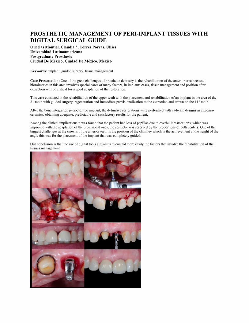
PROSTHETIC MANAGEMENT OF PERI-IMPLANT TISSUES WITH DIGITAL SURGICAL GUIDE Ornelas Montiel, Claudia *, Torres Porras, Ulises Universidad Latinoamericana Postgraduate Prosthesis Ciudad De México, Ciudad De México, Mexico Keywords: implant, guided surgery, tissue management Case Presentation: One of the great challenges of prosthetic dentistry is the rehabilitation of the anterior area because biomimetics in this area involves special cares of many factors, in implants cases, tissue management and position after extraction will be critical for a good adaptation of the restoration. This case consisted in the rehabilitation of the upper teeth with the placement and rehabilitation of an implant in the area of the 21 tooth with guided surgery, regeneration and immediate provisionalization to the extraction and crown on the 11° tooth. After the bone integration period of the implant, the definitive restorations were performed with cad-cam designs in zirconia-ceramics, obtaining adequate, predictable and satisfactory results for the patient. Among the clinical implications it was found that the patient had loss of papillae due to overbuilt restorations, which was improved with the adaptation of the provisional ones, the aesthetic was reserved by the proportions of both centers. One of the biggest challenges at the crowns of the anterior teeth is the position of the chimney which is the achievement at the height of the angle this was for the placement of the implant that was completely guided. Our conclusion is that the use of digital tools allows us to control more easily the factors that involve the rehabilitation of the tissues management.

UPPER FULL ARCH REHABILITATION WITH HYBRID MAXILAR PROSTHESIS: PROTESICALLY GUIDED WITH CAD-CAD Ortiz, Daniel *, Lugo, Pedro Mendiburu, Celia Cortes, David Universidad Autónoma De Yucatán Odontología Restauradora Mérida, Yucatán, Mexico
Keywords: CAD-CAM, hybrid maxilar prosthesis
Case Presentation: Male patient of 70 years old partially edentulous is assessed at the clinic for a upper full arch implant rehabilitation, after the clinical assessment, the following treatment plan was stablish: endodontic treatment of the remaining teeth (1.3, 2.3) to remove the clinical crown leaving 2mm of it supragingival for root maintaining for further immediate implant placement after extraction on the zone, fabrication of a conventional total upper denture to stablish esthetics, phonetics, function and prosthetic space, both sinus elevation for implant placement in the zone, six implant placement aided by a digital printed surgical guide stablished by the previous elaborated conventional denture and final rehabilitation with a hybrid prosthesis.
SINGLE IMPLANT PLACEMENT WITH THE NAVIDENT SYSTEM , DIGITAL WORKFLOW Pérez, César *, Lugo Ancona, Pedro; Mendiburu Zavala , Celia; Peñaloza Cuevas, Ricardo Universidad Autónoma De Yucatán Restorative Dentistry Monclova, Coahuila, Mexico
Keywords: Implantology,digital,navident
Case Presentation: 54 year old female patient with controlled hypertension, and history of sinusitis, ASA type II, presented to the clinic with edentulism of the dental organ 2.6. due to prior extraction with alveolar preservation due to infection. With the use of the navident system, a BTI wide platform implant 5.0 mm in diameter and 6.5 mm in length was planned and placed. For this, study models were previously taken and the waxing was scanned to obtain an STL file and in the NAVIDENT software match the DICOM file of the CBCT tomography.

COMPARISON OUTCOMES OF DIFFERENT METHODS FOR TREATMENT OF TEMPOROMANDIBULAR DISORDERS Raddad, Ahmed *, Ananthan, Sowmya. Heir, Gary 1- Boston University. 2- Rutgers School of Dental Medicine1- Department of Restorative Sciences and Biomaterials. 2- Diagnostic Sciences, Center of Orofacial Pain and Temporomandibular DisordersBoston, Ma, United States
Keywords: Temporomandibular Disorders, Temporomandibular Joint, Muscles.
Purpose/Aim: This study aims to compare the outcome of whether combining occlusal splint therapy with home care instructions is better than occlusal splint used alone.
Materials and Methods: The research design is a prospective, randomized control study. The subjects were diagnosed with related TMD according to the DC/TMD diagnostic criteria. The subjects divided to 2 groups: 1-group1 (Occlusal splint therapy alone) 2- Group 2 (Combined therapy group). Study instruments:1-VAS 2-Rescue medication 3- Symptom questionnaire form,4- DC/TMD exam form 5-TMD home care instructions.
Results: The mean reported muscle and joint pain duration calculated for occlusal splint group (86 ± 102) months,duration for muscle and joint pain for the subjects in the combined group care was (55± 56.6)months ranging from 3-190 months. 50% of the subjects were using rescue medication in the first visit, while the third visit the usage of rescue medication increased to75%. During the first visit in the occlusal splint therapy group the mean pain free mouth opening was 36.7±9.5 mm,In the third visit for the same group the mean pain free mouth opening was calculated to be 41.75±9.44 mm.During the first visit in the combination group the mean pain free mouth opening was calculated to be 34±6.63 mm. In the third visit for the same group the mean pain free mouth opening was calculated to be 41±5.29 mm.
Conclusions: In this study, two methods i.e. occlusal splint and combination therapy (occlusal splint along with home care instructions and exercises) for Temporomandibular Disorders patients were compared. Participants were distributed into two groups. Each group was provided different therapy. The results of the study show that there was improvement in the free mouth opening, pain intensity level (VAS), and muscle sites pain in both groups, while the improvement in the combination group showed a statistically significant improvement. Regarding the muscle and joint sites pain treatment outcomes were questionable. As there were no way to find the actual improvement of the muscles and joint sites pain cause of the yes/no answers, scaled tool should be used in future studies. The addition of home care instructions and exercises to the occlusal splint showed more improvement results in all outcome measurements.
ENDOCROWN: A REVIEW Rezaei, Kiana *, Kiat-Amnuay,Sudarat Uthealth School of Dentistry Advanced Education In General Dentistry (Aegd) Houston, , Tx, United States
Keywords: Endocrown, Molars, Endodontically treated
Case Presentation: Clinical Challenges: Restoration of the significantly damaged posterior tooth structure following caries removal and root canal therapy is a challenge for a restorative dentist. Full coverage restoration following post and/or core build up is the most favorable method to regain endodontically treated tooth certain functionality and strength. Yet, root fracture, thinning of the root canal walls during post space preparation and limitation of using post in narrow canal and calcified canals are important matters that weaken the tooth

even further and create restrictions. Endocrown has functional longevity and has become a promising alternative in functional recovery of endodontically treated molar teeth.
Purpose: The purpose of this review is to determine whether the computer-aided design/computer-aided manufacturing endocrown restorations would be less invasive option with similar or higher success rate on compromised endodontically treated molar teeth than post/core-retained indirect conventional restorations.
Clinical Implications: Endocrown presents a conservative approach for mechanical restoration of nonvital posterior teeth. It shows functional longevity and has become a reliable alternative in the functional recovery of endodontically treated molar teeth when proper design and adhesion protocols are executed.
PROSTHETIC PLANNING FROM BONE REGENERATION Robles, Marco *, Zarate Aída Lucía Universidad Latinoamericana Protesis Bucal Culiacan, Sinaloa, Mexico
Case Presentation: A 37-year-old female patient goes to the university, multiple extractions of the upper jaw are planned for the placement of an immediate prosthesis, prior to a regeneration with an autologous graft.
RESTORING FUNCTION AND ESTHETICS IN A FULL MOUTH CASE Romeo, Davide *, N.A. Eastman Institute for Oral Health Department of Prosthodontics Rochster, Ny, United States
Case Presentation: A 68-year-old woman, no smoker, came to the clinic with a specific chief complaint “ I would like to improve my chewing function and my dental esthetics”. Her medical history revealed high blood pressure, but no systemic contraindications for implant treatment. She wanted to have fixed prostheses, specifically individual crowns. Clinical and radiographic examination evidenced no active periodontal disease and partially edentulous arches. She has a low smile line with extended buccal corridors. In the maxilla, the only remaining tech were #6, 7, 8, and 9 with PFM crowns. In the lower jaw, residual dentition included teeth from #21 to 28, all presenting full coverage crowns with the exception of both canines. Patient was informed about possible treatment modalities with fixed prosthesis and she decided to keep her residual teeth and replace missing teeth with dental restorations. For the maxillary arch, sinus augmentation and ridge augmentation were done before implant placement and restorations. Residual teeth received new crowns. In the mandible, implant placement with ridge augmentation was performed bilaterally and residual teeth received a combination of crowns and veneers. A hard night guard was delivered after the final restorations.
PLACEMENT OF INTRACORONARY ATTACHMENTS : CASE REPORT Salazar Cortazar, Jessica *, Lugo Ancona Pedro, Zavala Mendiburu Elena, Cardenas Erosa Ruben, Cortes Carrillo David. Universidad Autonoma De Yucatan

Odontologia Restauradora Mexico (Mex), Yucatán, Mexico
Case Presentation: INTRODUCTION: The overdentures could be defined with a total or removable prosthesis that completely covers the occlusal surface, can be supported by remnant teeth, roots and / or osseointegrated implants. The use of attachments in overdentures provide orientation, retention, stability and comfort for the patient. The most important objective in the selection of the attachments is the way in which the force of these devices is transferred through the adjacent pillars and structures.
CLINICAL REPORT: DIAGNOSIS
Female patient of 60 years of age, is referred to the postgraduate of Restorative Dentistry for presenting absence of superior dental organs.
TREATMENT PLAN: - Diagnostic Elements: Study Models, Radiographs and Photographs.- Treatment: Endodontics, lower overdenture using O.D 3.3 4.3 and 4.5 as pillars with semiprecision intraradicular attachments(Locator) and in the upper part a conventional total prosthesis.
IMPLANT SUPPORTED APPROACH OF AN ECTODERMAL DYSPLASIA PATIENT WITH GENERAL ANODONTIA: CASE REPORT Sarmiento Rosales, Zulia Paulette *, Pérez Martínez, Enrique; Franco Romero, Guillermo Benemérita Universidad Autónoma De Puebla (Buap) Master In Oral Rehabilitation Puebla, Puebla, Mexico
Keywords: Ectodermal Dysplasia,Hybrid metal-acrylic prosthesis,Implant Supported Restoration
Case Presentation: BACKGROUND Ectodermal dysplasia is a genetic disorder that involves congenital birth defects of two or more ectodermal structures. It is characterized by anhidrosis, hypodontia, and hypotrichosis. The clinical signs are brittle nails, sparse hair, deficiency of sweat glands, dry skin, dental malformation, reduced alveolar bone growth, anodontia, altered tooth eruption, widely spaced teeth, poorly shaped teeth, often with reduced width, and salivary hypofunction. The resulting lack of alveolar bone formation and regular oral and craniofacial development leads to significant diagnostic and management challenges. TECHNIQUE/CASE REPORT 20-year-old patient diagnosed with Ectodermal Dysplasia who has received dental control since he was 6 years old at the Pediatric clinic of the Benemérita Universidad Autónoma de Puebla (BUAP). During his time of attendance, dentures were made and changed according to his growth stages, and subsequently referred to the Oral Rehabilitation department. His chief complaint was to change his upper and lower dentures. The patient only wears the upper denture because the lower one frequently caused him pain and forms him sores making it difficult to chew appropriately. At the intraoral exam complete anodontia and knife edged alveolar ridges were observed, palatal mucosa had papilliform lesions and the overall mucosa presented lack of saliva secretion. In the upper maxilla had scarce bone volume so a conventional denture was proposed as treatment and hybrid metal-acrylic Implant supported prosthesis following the all on four protocol in the lower maxilla. DISCUSSION Ectodermal dysplasia patients represent a challenge during the dental treatment, specially in complete anodontia, since the dimension of the alveolar ridges is reduced. Implant supported prosthesis are the ideal solution in cases where implants can be placed and if there is complete anodontia or the existing teeth aren´t in the appropriate conditions or distribution to accomplish the rehabilitation. It´s of clinical relevance to control the salivation using artificial saliva to preserve the health of the soft tissues and provide the right conditions for the implants and prosthesis. CONCLUSION Combining traditional and implant supported restoration is a viable treatment option when the overall conditions of the patient aren´t favorable to make either of them. It is important to go through a correct diagnose through several clinical studies in other to achieve a suitable result. CLINICAL IMPLICATIONS Rehabilitating patients with congenital anodontia might be a challenge, however, with the new surgical and prosthetic techniques, restoring aesthetics, occlusion, masticatory function and speech can improve their quality of life and self-esteem.

COMPREHENSIVE TREATMENT OF A PATIENT WITH DANDY-WALKER SYNDROME. – A CASE REPORT Schwartz, David *, Hosney, Sherif; Chochlidakis, Konstantinos; Ercoli, Carlo Eastman Institute for Oral Health - University of Rochester Prosthodontics Rochester, Ny, United States
Case Presentation: Objective: The purpose of this case report is to present oral rehabilitation using implants and prosthesis for a patient with Dandy-Walker Syndrome (DWS). Few published studies have reported the clinical dento-facial manifestations as well as treatments of patients with DWS. Case: A 21 yo female presented with medical history of DWS and Cervical Fusion Syndrome (CFS). The patient had remaining dentirion extracted in the operating room under general anesthesia. Two implants were placed in the mandible with no complications. A maxillary complete denture and mandibular overdenture was fabricated. Implant stability and esthetics were satisfactory. Conclusion: This clinical case report shows the comprehensive treatment of a patient with Dandy-Walker Syndrome. We were able to achieve a method for rehabilitation that improves the function and esthetics as well as the overall quality of life.
CALCIUM-ALUMINATE GLASS IONOMER HYBRID CEMENTS: A REVIEW Skelton, Park *, Sudarat Kiat-Amnuay, Dds, Ms, Fcacp, Faamp University of Texas As Houston School of Dentistry, Aegd Houston, , United States
Keywords: Calcium-Aluminate, Dental, Cements
Purpose/Aim: The purpose of this review is to highlight the new bioactive calcium-aluminate, glassionomer hybrid cements which release calcium and may precipitate hydroxyapatite in vivo. This has potential to progressively close restorative microgaps, one of the contributing factors in recurrent decay.
Materials and Methods: This presentation reviews five studies which focus on calcium aluminate (CA) containing cements such as Ceramir C&B and the unique properties that CA imparts. The normal qualities of the cements such as setting time, working time, film thickness and ease of use were measured. In addition, studies measured the ability of these cements to form new hydroxyapatite and also inhibit S. Mutans growth in vitro.
Results: The calcium aluminate (CA) containing cement named Ceramir C&B was given the highest rating for marginal staining, marginal integrity, absence of recurrent decay and cleanup (p value<.05). The studies confirmed that CA containing cements in vitro will contribute to the deposition of new hydroxyapatite (HA) when the correct in vivo-simulating medium is used. The precipitate's identity was confirmed to be HA via scanning electron microscopy, x-ray photoelectron spectroscopy, grazing incidence electron diffraction. One study confirmed that CA containing cements can completely occlude an artificial tooth-cement microgap of width 50µm to 120µm within 24-48 hours (p value <.05). A separate study showed that CA containing cements or CA/Glass ionomer hybrids may contribute to an anticariogenic affect more strongly than glass ionomer cement alone (p value <.05).
Conclusions: Calcium aluminate containing cements appear to offer the potential unique qualities such as microgap occlusion with new hydroxyapatite formation and increased anticariogenicity in vitro. CA-Glass Ionomer hybrid cements such as Ceramir C&B have been rated highly for their handling qualities and should perhaps be considered among one of the top choices for luting cement options.

DIGITAL WORKFLOW FOR A COMPLETE REHABILITATION Torres, Jimena *, Lugo, Pedro Mendiburu, Celia Cortés, David Universidad Autónoma De Yucatán Odontología Restauradora Merida, Yucatan, Mexico
Case Presentation: Male patient of 71 years old is referred to the postgraduate of restorative dentistry for presenting pain and mobility en superior dental organs. The treatment plan was to re-establish the patient's occlusal scheme, masticatory function and improve the aesthetic appearance with fixed prostheses and implants supported with monolithic zirconia crowns milled with CAD/ CAM through digital planning.
DIAGNOSIS AND TREATMENT PLAN IN ALTERED PASSIVE ERUPTION (PAE) Torres, Alejandra *, Soria, Diana. Quintero, Jose Universidad Nacional Autónoma De México División De Estudios De Posgrado E Investigación (Depei) En Odontología,Unam Ciudad De México, Cdmx, Mexico
Case Presentation: Abstract Background: Altered passive eruption (APE) is described as a condition in which the gingival margin during the final stage of dental eruption does not conclude its physiological migration until the enamel-cement junction. It also contributes as an etiological factor for the "gummy smile" and compromises the aesthetic appreciation of patients. Case report: Female patient who does not like her smile and refers the absence of tooth 8 was diagnosed with altered passive eruption type 1 B. Clinical implications: The treatment plan consists of aesthetic crown lengthening with osteotomy to reduce gingival exposure and to allow tissues preparation for be prosthetic rehabilitated, guided bone regeneration following of dental implant placement and prosthetic rehabilitation with lithium disilicate restorations. Discussion: Authors report that the factors influencing the amount of coronal displacement of the marginal tissue appear to be related to the patient's periodontal phenotype, being greater in the thick phenotype. Several authors suggest the surgical removal of 3 mm periodontal support (Ingber 1977), 2.5 to 3.5 mm (Palomo 1978), 4 mm (Rosenberg 1980).Conclusion: Altered passive eruption demands the elaboration of a well supported periodontal and prosthetic diagnosis because periodontal conditions are the key of an optimal colocation of any aesthetic restoration, creating an ideal prosthetic-periodontal relationship.
A TECHNIQUE TO LOCALIZE SCREW ACCESS HOLE IN CEMENT-RETAINED IMPLANT RESTORATION Wangsrimongkol, Buddhapoom *, Shyu, Chih-Hao; Alghamdi, Reem; Scolnick, Jeffrey; Cho, Sang Choon. New York University College of Dentistry A Ashman Department of Periodontology and Implant Dentistry New York, Ny, United States
Keywords: cemented crowns, screw access, screw loosening
Case Presentation: Dental implant treatment is a dependable therapy for single or multiple teeth replacements and fully edentulous rehabilitation. Treatment outcomes are highly successful with great patient satisfaction. However, complications can arise after long term function. Abutment screw loosening is one of the most common challenges and can lead to additional financial costs for the patient and the clinician if not properly managed. This is especially the case for cement-retained implant

restorations as the screw access is difficult to locate and aggressive procedures are necessary to expose the screw access, resulting in crown replacement when proper localization techniques are not employed. In this poster, novel intra-oral and extra-oral techniques are presented for locating abutment screw access in cemented restorations. The purpose is to minimize the damage inflicted onto cemented crowns, allowing the restorations to be reused after screw tightening.
PRESCRIBING POST-OPERATIVE ANALGESIA: AVOIDING COMPLICATIONS AND DRUG INTERACTIONS. AN UPDATE FOR A PROSTHODONTICS-CENTERED PRACTICE. Wells, Judson *, Kiat-Amnuay, Sudarat, Dds, Ms, Facp, Faamp University of Texas Health Science Center at Houston School of Dentistry, Aegd Houston, Tx, United States
Keywords: Analgesia, Dental, Post-operative
Case Presentation: Clinical Challenges: As dentists change prescribing habits in accordance with ADA and CDC recommendations on opioids, they must be aware of potential pitfalls to avoid with pharmacotherapy. NSAIDs and Acetaminophen are not without risks. An understanding of absolute and relative contraindications and drug-drug interactions is essential to mitigate iatrogenic harm when selecting a suitable drug.
Purpose: The purpose of this presentation is to provide a straightforward and concise update on post-operative pain management. The focus is awareness of potential complications and interactions of common analgesics.
Clinical Implications: This informs a structured decision-making process laid out for prescribers, allowing them to safely and effectively manage pain throughout prosthodontic treatment.