Embed Size (px)
Citation preview

Inspired by knowledge
PROTOCOLS
MICROBIOLOGICAL ANALYSIS
IN THE FOOD INDUSTRY
ALL PROCEDURES ACCORDING TO ISO REGULATIONS

2
How important are the reference methods in food control?Many of the foods that do not undergo a microbiological analysis become a health risk, because they can cause a wide range of diseases. Diarrhoeal diseases, for example, are the leading cause of death in children and the second in adults, and in many cases they are related to the intake of a contaminated food. That is why it is necessary to establish microbiological criteria to ensure the safety of food and protect the health of the consumer.
The development of criteria for the microbiological control of foods implies, among other things, fixing the analytical method to be used to ensure compliance with these criteria. These methods, developed by organizations such as ISO or CEN, can be considered as reference methods, and their implementation and application in the laboratory can often be difficult.
Currently, the strategy in the development and update of these ISO methods aims at improving them through the simplification and incorporation of new technologies, so that they can be routinely used in food control laboratories.
The use of standardized methods for conducting microbiological analyses is a tool that allows laboratories to use internationally recognized testing methods, thus contributing to the production of comparable results, without sacrificing reliability and ensuring the quality and safety of the foods subject to control.
That is why it is increasingly common for these standardized methods to be taken as a reference by accreditation bodies as well as to establish microbiological criteria (EU Regulations 2073/2005 and 1441/2007) where the international standards developed by the ISO and CEN Committees are considered reference methods.
At Condalab we want to help the different quality control laboratories by making available the full range of culture media under ISO formulations as well as their different analysis procedures.

3
Index
MESOPHILIC MICROORGANISMS COUNT ________________________________________ 05 Procedure as defined by ISO 4833:2013
MOLD AND YEAST COUNT _______________________ 06 Procedure as defined by ISO 21527:2008
DETECTION OF SALMONELLA SPP _________________ 07 Procedure as defined by ISO 6579:2017
DETECTION AND ENUMERATION OF LISTERIA SPP ___________________________________ 09 Procedure as defined by ISO 11290:2017
DETECTION AND ENUMERATION OF CAMPYLOBACTER SPP ___________________________ 11 Procedure as defined by ISO 10272:201
DETECTION OF CRONOBACTER SPP _______________ 13 Procedure as defined by ISO 22964:2017
PRESUMPTIVE BACILLUS CEREUS COUNT ________________________ 14 Procedure as defined by ISO 7932:2004
DETECTION AND ENUMERATION OF ENTEROBACTERIAS ______________________________ 15 Procedure as defined by ISO 21528:2017
CLOSTRIDIUM PERFRINGENS COUNT ________________________________________ 16 Procedure as defined by ISO 7937:2004

4
DETECTION AND COUNT OF COAGULASE-POSITIVE STAPHYLOCOCCI ___________ 17 Procedure as defined by ISO 6888
DETECTION OF PATHOGENIC YERSINIA ENTEROCOLITICA _______________________ 19 Procedure as defined by ISO 10273:2017
DETECTION AND ENUMERATION OF SHIGELLA SPP __________________________________ 20 Procedure as defined by ISO 21567:2004
DETECTION AND ENUMERATION OF ESCHERICHIA COLI ______________________________ 21 Procedure as defined by ISO 7251:2005 / ISO 16649:2015
TOTAL COLIFORM COUNT ________________________________________ 23 Procedure as defined by ISO 4832:2006
DETECTION OF ESCHERICHIA COLI O157 __________ 24 Procedure as defined by ISO 16654:2001
DETECTION OF VIBRIO SPP _______________________ 25 Procedure as defined by ISO 21872:2007
DETECTION AND ENUMERATION OF OTHER MICROORGANISMS IN THE FOOD INDUSTRY _________________________ 26 Procedure as defined by ISO 15213:2003 / ISO 13720:2010 / ISO 6611:2004 / ISO 15214:1998

5
Mesophilic Microorganisms countProcedure as defined by ISO 4833:2013
Introduction
Bacteria are considered the main cause of diseases that have the consumption of contaminated food as their root cause. When foods show variations or a change in colour, texture or consistency, it is a sign of deterioration and possible contamination. To grow, bacteria need:
• Nutrients such as carbon, nitrogen, hydrogen and phosphorus (among others) that are available in food.
• Water as a fundamental element, because in its absence, bacterial growth stops.
• A suitable pH, oscillating around a neutral value (6-7). At a lower or higher pH, bacterial growth stops.
The analysis of this group of bacteria includes all microorganisms capable of developing in the presence of oxygen at a temperature between 20 °C and 45 °C. The count of aerobic mesophilic microorganisms, under the established conditions, estimates the total microflora of the product but without specifying and identifying the type of microorganism.
Bibliography
AFSA, 2001. Evaluation of microbiological methods for detection and for enumeration of microbiological contaminants in food. Final report Contract SMT4/CT96 2098. Coordination by Agence Française de Securité Sanitarie des Aliments. AFSSA, France, February 2001.
ISO 4833-1:2013. Microbiology of the food chain – Horizontal method for the enumeration of microorganisms. Part 1: Colony-count at 30 degrees C by the pour plate technique.
ISO 4833-2:2013. Microbiology of the food chain – Horizontal method for the enumeration of microorganisms. Part 2: Colony-count at 30 degrees C by the surface plate technique.
INITIAL SUSPENSION
Refer to the ISO 6887-1 specific to the product that is analyzed
COUNTING MEDIUM
1 ml sample diluted in Standard Methods Agar (PCA) (CAT. 1056) Incubation: 30 ºC ± 1 ºC // 72 ± 3 h
For the analysis of dairy products, the culture medium should carry 1 g/l of skimmed milk powder: Standard Methods Agar
with Powdered Milk (CAT. 1033)
NOTE: *For this standard, two methods can be distinguished: Pour plate (4833-1) and spread plate (4833-2) techniques. Both sections of the standard establish the same working protocol but warn that, for certain matrices, different results may be obtained according to the section followed.
Method

6
Mold and Yeast countProcedure as defined by ISO 21527 1:2008
Introduction
Foods that have fungi on their surface may have toxins in their interior, constituting a risk of allergic reactions. When fungi appear on the surface of a food, their roots have already invaded a significant portion of the product. Fungi live on plant or animal matter and contain spores that can be transported by air, water or insects.
Fungi are found mostly in foods such as fruit, vegetables, moist bread or cheeses. Different types can be distinguished depending on their behaviour. Aspergillus or Penicillium are some of the fungi most frequently found. The filaments in their structure, the hyphae, produce enzymes that can break down the hardest molecules. Under suitable conditions (warm temperatures and humidity), fungi produce mycotoxins, substances with the capacity to cause diseases and that appear mainly in cereals and nuts.
Bibliography
VALERIE T., MICHAEL T.S., PHILIP B., MISLIVEC, HERBERT A.K. AND RUTH B. BAM: Yeast, Molds and Mycotoxins. US FOOD AND DRUG ADMINISTRATION.
ISO 21527-1:2008. Microbiology of the food chain – Horizontal method for the enumeration of yeast and molds. Part 1: Colony count technique in products with water activity greater than 0,95.
ISO 21527-2:2008. Microbiology of the food chain – Horizontal method for the enumeration of yeast and molds. Part 1: Colony count technique in products with water activity less than or equal 0,95.
INITIAL SUSPENSION
Except for preparations of specific samples (ISO 6887-1), dilution in Buffered Peptone Water (CAT. 1402) is recommended
COUNTING MEDIUM
0.1 ml sample diluted in Rose Bengal Agar + Cholamphenicol + Dichloran (DRBC Agar) (CAT. 1160) Incubation: 25 ºC ± 1 ºC // 5 days
PRODUCTS WITH WATER ACTIVITY > 0,95 (ISO 21527-1)
INITIAL SUSPENSION
Refer to the ISO 6887-1 standard specific to the product to be analysed. Except for preparations involving specific samples, dilution in Buffered Peptone Water (CAT. 1402) is recommended
COUNTING MEDIUM
0.1 ml sample diluted in Dichloran Glycerol Agar (DG 18) (CAT. 1161) / Incubation: 25 ºC ± 1 ºC / 5 – 7 days
PRODUCTS WITH WATER ACTIVITY ≤ 0,95 (ISO 21527-2)

7
Detection of Salmonella sppProcedure as defined by ISO 6579:2017
Introduction
Salmonella is a very diverse family of microorganisms, including about 2,300 different serotypes. It is estimated that this family of bacteria is the cause of millions of hospitalizations in the world (1.2 million in the USA alone), but this number would increase up to 30 times if undiagnosed infections were taken into account, as they usually cause a normal diarrhoeal process.
This group of bacteria is gram-negative (since they belong to the large group of Enterobacteriaceae), facultative anaerobic, and motile by means of flagella. They are glucose fermenters, but they are not lactose fermenters and do not produce urease.
Salmonella is present in the intestines of people and animals, and in raw meat, poultry, eggs, and unpasteurised milk. The optimal temperature for growth of this bacteria is between 30 ºC and 37 ºC; therefore, it is especially important to adopt preventative measures in the warmest months of the year, which is when the risk grows considerably.
Bibliography
AFSA, 2001. Evaluation of microbiological methods for detection and for enumeration of microbiological contaminants in food. Final report Contract SMT4/CT96 2098. Coordination by Agence Française de Securité Sanitarie des Aliments. AFSSA, France, February 2001.
ISO 6579-1:2017. Microbiology of the food chain – Horizontal Method for detection and enumeration and Serotyping of Salmonella - Part 1: Detection of Salmonella spp.
ISO 6887-1:2017. Microbiology of the food chain – Preparation on test samples, initial suspension and decimal dilutions for microbiological examination – Part 1: General rules for the preparation of the initial suspension and decimal dilutions.

8
ISOLATION IN NON-SELECTIVE AGAR
Nutrient Agar (CAT. 1060) or Nutrient Agar with Sodium Chloride (CAT. 1355) / Incubation: 34-38 ºC – 24 ± 3 h
NOTE:
*According to Appendix D of ISO 6579-1:2017, the procedure for the detection of enteric subspecies (S. typhi and S. paratyphi) should be as follows:
1.- Selenite Cystine Broth (CAT. 1220) as selective enrichment
2.- As selective secondary medium, select Bismuth Sulfite Agar (Wilson Blair) (CAT. 1011)
SELECTIVE ISOLATION
XLD Agar (Xylose Lysine Desoxycholate Agar) (CAT. 1274) Incubation: 41.5 ºC ± 1 ºC – 24 ± 3 h
SELECTIVE ISOLATION
As a secondary medium, select among the following:
Salmonella Shigella Agar (SS Agar) (CAT.1064)
Hektoen Enteric Agar (CAT. 1030)
Brilliant Green Agar Modified (CAT. 1143)
Bismuth Sulfite Agar (Wilson Blair) (CAT. 1011)
Salmonella Chromogenic Agar (CAT. 1122)
For result reading and suspect colony selection, refer to the technical data sheet of each product. Next, select a single suspect colony for spreading in non-selective culture medium. If the result
is negative, test 4 suspect colonies previously marked
CONFIRMATION
Complete a biochemical and serological test on the grown colonies in the non-selective agar.
The biochemical test shall consist of:• Triple Sugar Iron Agar (TSI) (CAT. 1172)• Christensen's Urea Agar (CAT. 2000)• Lysine Decarboxylase Medium (CAT. 1176)• ß-galactosidase (optional)• Indole reaction (optional)
PRE-ENRICHMENT
25 g sample + 225 ml of Buffered Peptone Water (CAT. 1402) / Incubation: 34 ºC to 38 ºC – 18 ± 2 h
For environmental samples of the primary production phase or of faeces, spread directly over Agar MSRV
SELECTIVE ENRICHMENT
0.1 ml + 10 ml Rappaport Soy Broth (Vassiliadis) (CAT. 1174) or Modified Semisolid Rappaport Vassiliadis Medium (MSRV) (CAT. 1376) Incubation: 41.5 ºC ± 1 ºC – 24 ± 3 h
Warning: When incubating the MSRV plate, do not invert the plate
SELECTIVE ENRICHMENT
1 ml + 10 ml Muller Kaufmann Broth with brilliant Green and Novobiocin (MKTTN) (CAT. 1173) Incubation: 37 ºC ± 1 ºC – 24 ± 3h
Method
9
Detection and enumeration of Listeria sppProcedure as defined by ISO 11290:2017
Introduction
Gram-positive bacteria with basophil morphology belong to the genus Listeria. It is a genus composed of only 6 species, with Listeria monocytogenes being the most important since it is a pathogenic species present in foods suitable for human consumption.
L. monocytogenes causes listeriosis (food poisoning with a mortality rate close to 30%). These bacteria are gram-positive, very small in size, catalase-positive, and non-sporulating. They are facultative anaerobic, capable of growing in a wide range of temperatures (1 ºC to 45 ºC) and in high salt concentrations. Additionally, it is a flagellated microorganism and, as such, it is motile, which further increases its pathogenicity.
The microorganism is widely distributed in nature and it is very common to find it in food processing plants, where its survival capacity in such a wide range of temperatures makes it, perhaps, one of the key microorganisms to control in the industry.
Bibliography
KATHARIOUS S. Listeria monocytogenes virulence and pathogenicity, a food safety perspective. J. Food Prot. 2002; 65: 1881-1829.
ISO 11290-1. Microbiology of the food chain – Horizontal Method for detection and enumeration of Listeria monocytogenes and Listeria spp - Part 1: Detection Method.
ISO 11290-2. Microbiology of the food chain – Horizontal Method for detection and enumeration of Listeria monocytogenes and Listeria spp - Part 2: Enumeration Method.

10
SELECTIVE ISOLATION
Listeria Chromogenic Agar according to Listeria Chromogenic Agar according to Ottaviani and Agosti (ALOA) (CAT. 1345) and Supplements (CAT.6031 + CAT. 6040) / Incubation: 37 ºC ± 1 ºC
Listeria monocytogenes suspect colony reading in ALOA Agar: Greenish-blue with opaque halo (also consider those without
halo for Listeria spp.)
ISOLATION IN NON-SELECTIVE AGAR
Choose between: Blood Agar Nº2 (CAT. 1328) or TSYEA Agar (Tryptone Soy Yeast Extract Agar) (CAT. 1398) / Incubation: 37 ºC ± 1
L. MONOCYTOGENES CONFIRMATION• Microscopy
• Hemolytic Activity – Blood Agar Nº2 (CAT. 1328)
• Carbohydrates Utilization Broth (CAT. 1342)
• Catalase (optional)
• Motility (optional)
• CAMP test (optional)
LISTERIA SPP CONFIRMATION• Microscopy
• Catalase
• Motility (optional)
• Voges-Proskauer Reaction (optional)
INITIAL SUSPENSION
x g/ml sample + 9 x g/ml of diluent according to ISO 6887
In the event that the enumeration is performed on the same sample as the detection, Listeria Half-Fraser Broth (CAT. 1183)
and Listeria Fraser Broth (CAT. 1182) should be used in the same proportions as indicated above
2 - ENUMERATION METHOD (ISO 11290-2)
L. MONOCYTOGENES CONFIRMATION• Microscopy
• Hemolytic Activity – Blood Agar Nº2 (CAT. 1328)
• Carbohydrates Utilization Broth (CAT. 1342)
• Catalase (optional)
• Motility (optional)
• CAMP test (optional)
LISTERIA SPP CONFIRMATION• Microscopy
• Catalase
• Motility (optional)
• Voges-Proskauer Reaction (optional)
SECONDARY SELECTIVE ENRICHMENT
0.1 ml of primary culture + 10 ml of Listeria Fraser Broth (CAT. 1182 + CAT. 6050) Incubation: 37 ºC ± 1 ºC – 24 ± 2 h
ISOLATION IN NON-SELECTIVE AGAR
Choose between: Nutrient Agar (CAT. 1060), Nutrient Agar with Sodium Chloride (CAT. 1355), Blood Agar Nº2 (CAT. 1328), TSYEA Agar (Tryptone Soy Yeast Extract Agar) (CAT. 1398) or TSYEB Broth (Tryptone Soy Yeast Extract Broth) (CAT. 1339) / Incubation: According to specification. Refer to relevant technical data sheet
1 - DETECTION METHOD (ISO 11290-1)
PRIMARY SELECTIVE ENRICHMENT
25 g/ml sample + 225 ml of Listeria Half-Fraser Broth (CAT. 1183 + CAT. 6050) Incubation: 30 ºC ± 1 ºC – 24 + 2 h
You could then go directly to spread directly on the ALOA Selective Agar without secondary selective enrichment
SELECTIVE ISOLATION
Listeria Chromogenic Agar according to Ottaviani and Agosti (ALOA) (CAT. 1345) and Supplements (6031 + 6040) Incubation: 37 ºC ± 1 ºC – 24/48 ± 2h
SELECTIVE ISOLATION
As secondary medium, select among the following: Listeria Oxford Agar (CAT. 1133 + 6003) Listeria Palcam Agar (CAT. 1141 + 6004)
Reading of Listeria monocytogenes suspect colonies in ALOA agar: Greenish-blue colonies with opaque halo (also consider those without a halo for Listeria spp). For colony reading in
secondary agar, temperatures and incubation times, refer to the relevant technical data sheets

11
Detection and enumeration of Campylobacter sppProcedure as defined by ISO 10272:2017
Introduction
Microorganisms of the genus Campylobacter are gram-negative bacilli with a comma or curved spiral shape, motile by means of a unipolar or bipolar flagellum and microaerophiles.
They are thermophilic bacteria with optimal growth at temperatures between 42-43 ºC. Additionally, they are the most frequent cause of gastroenteritis in humans.
The World Health Organization (WHO 2011) considers that, at a global level, Campylobacter is one of the primary causes of food-transmitted diarrhoeal diseases. The majority of cases of campilobacteriosis are associated with food consumption, mainly poultry; therefore, these bacteria represent a top-priority in food safety.
Bibliography
AESAN (2012). Informe del Comité Científico de la Agencia Española de Seguri dad Alimentaria y Nutrición (AESAN) con relación a las medidas de control para reducir la presencia de Campylobacter spp. en carne fresca de aves (pollo). Revista del Comité Científico, Nº 16: 21-55.
OMS (2011) Campylobacter. Nota descriptiva núm. 255.Octubre de 2011.
UNE-EN-ISO 10272-1:2017. Microbiology of the food chain – Horizontal Method for detection and enumeration of Campylobacter spp. Part 1: Detection Method.
UNE-EN-ISO 10272-2:2017. Microbiology of the food chain – Horizontal Method for detection and enumeration of Campylobacter spp. Part 2: Colony Count Technique.
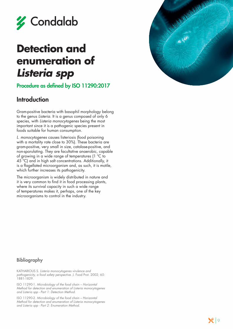
PRESUMPTIVE ISOLATION
Campylobacter Agar Blood Free (CCDA) (CAT. 1129 + CAT. 6053) Incubation in microaerobic atmosphere: 41.5 ºC – 44h ± 2 h
In turn, spread in secondary selective medium under the same conditions as incubation. One of the ISO-compliant culture media
is Campylobacter Agar (Preston) (CAT. 1131 + CAT. 6019)
RESULT READING
Typical colonies appear greyish, often showing a shiny metallic tone. They are usually flat with a humid aspect, and have a tendency to disseminate. Other morphologies could also be present
CONFIRMATION
Execute the following on the suspect colonies grown in CCDA Agar:• Spreading in Columbia Agar (CAT. 1104) Incubation
in microaerobic atmosphere at 41.5 ºC – 24/48 h • Examination of morphology and motility under the
microscope: Motile, flagellated and curved bacilli• Study of aerobic growth at 25 ºC: Negative growth
in Columbia Agar (CAT. 1104) under aerobic conditions during 44± 4 h
• Oxidase Activity (+)
SELECTIVE ENRICHMENT
10 ml/g sample + 90 ml of Bolton Selective Enrichment Broth (CAT. 1441 + CAT. 6070) / Incubation in microaerobic atmosphere: 37 ºC – 4 h / 6h then 41.5 ºC – 44h ± 2 h
1 - DETECTION BY ENRICHMENT: Samples with low concentration of Campylobacter and low accompanying microflora levels (e.g. cooked or frozen products)
12
PRESUMPTIVE ISOLATION
Campylobacter Agar Blood Free (CCDA) (CAT. 1129 + CAT. 6053) Incubation in microaerobic atmosphere: 41.5 ºC – 44 h ± 2 h
RESULT READING
Typical colonies appear greyish, often showing a shiny metallic tone. They are usually flat with a humid aspect, and have a tendency to disseminate. Other morphologies could also be present
RESULT READING
Typical colonies appear greyish, often showing a shiny metallic tone. They are usually flat with a humid aspect, and have a tendency to disseminate. Other morphologies could also be present
CONFIRMATION
Execute the following on the suspect colonies grown in CCDA Agar:
• Spreading in Columbia Agar (CAT. 1104) / Incubation in microaerobic atmosphere at 41.5 ºC – 24/48 h
• Examination of morphology and motility under the microscope: Motile, flagellated and curved bacilli
• Study of aerobic growth at 25 ºC: Negative growth in Columbia Agar (CAT. 1104) under aerobic conditions during 44± 4 h
• Oxidase Activity (+)
SELECTIVE ENRICHMENT
10 ml/g sample + 90 ml of Campylobacter Preston Broth (CAT. 2166 + CAT. 6081) / Incubation in microaerobic atmosphere: 41.5 ºC – 44 h ± 2 h
2 - DETECTION BY ENRICHMENT: samples with low concentrations of Campylobacter and high accompanying microflora levels (e.g. meat or raw milk)
PRESUMPTIVE ISOLATION
Campylobacter Agar Blood Free (CCDA) (CAT. 1129 + CAT. 6053) Incubation in microaerobic atmosphere: 41.5 ºC – 44 h ± 2 h
In turn, spread in secondary selective medium under the same conditions as incubation. One of the ISO-
compliant culture media is Campylobacter Agar (Preston) (CAT. 1131 + CAT. 6019)
3 - DETECTION BY DIRECT SPREADING: Samples with a high concentrations of Campylobacter (e.g. faeces, raw poultry)

13
Detection of Cronobacter sppProcedure as defined by ISO 22964:2017
Introduction
Bacteria belonging to the Cronobacter genus are gram-negative, facultative anaerobic, oxidase- negative and catalase-positive bacilli. They are usually motile and have the capacity to produce a variety of acids coming from multiple different carbohydrates. In fact, such attribute is used in the method confirmation stage.
The best known type of this genus is the Cronobacter sakazakii. Such type is considered nowadays an emerging pathogen that causes severe meningitis in infants. These processes may result in a percentage of mortality ranging between 40-80 %.
Bibliography
Fiedemann M., Enterobacter sakazakii in food and beverage (other than infant formula and milk powder). Int. J. Food Microbiol. 2007; 116:1-10.
ISO 22964:2017. Microbiology of the food chain. Horizontal method for the detection of Cronobacter spp.
Center of Disease Control and Prevention. Enterobacter sakazakii infections associated with the use of powered infant formula. JAMA, 2002; 287:2204 - 2205.
INITIAL SUSPENSION
10 ml/g sample + 90 ml of Buffered Peptone Water (CAT. 1402) / Incubation: 38 ºC – 24 ± 2 h
SELECTIVE ENRICHMENT
0.1 ml pre-enriched sample + 10 ml of Cronobacter Selective Broth (CSB) (CAT. 2143) Incubation: 41.5 ºC – 24 ± 2 h
PRESUMPTIVE SELECTIVE ISOLATION
Chromogenic Cronobacter Isolation Agar (CCI) (CAT. 1446) / Incubation: 41.5 ºC – 24 ± 2 h
ISOLATION IN NON-SELECTIVE AGAR
Streaking in Trypticasein Soy Agar (TSA) (CAT. 1068) Incubation: 34-38 ºC - 21 ± 3 h
BIOCHEMICAL CONFIRMATION
• Oxidase assay (-) • Enzyme α-glucosidase assay• Lysine Decarboxylase Broth (-) (CAT. 1208)• L-Ornithine Decarboxylation Medium (CAT. 2149)• Carbohydrate Fermentation Medium • Methyl Red (optional)• Voges-Proskauer (optional)
RESULT READINGThe suspect colonies of Cronobacter sakazakii are small in size (1-3 mm) showing a blue or greenish blue colour
The white, white with a green centre, grey or black colonies, as well as the yellow or red pigmented colonies, do not
correspond to the Cronobacter genus type
Method

14
Presumptive Bacillus Cereus countProcedure as defined by ISO 7932:2004
Introduction
Bacillus cereus is a facultative anaerobic, gram-positive bacillus, former of non-deforming spores. It is also catalase-positive, and fermenter of glucose and sucrose, and also of salicin and glycerol. In addition, it produces lecithinase that can be used as a marker to establish its presence.
This microorganism is widely distributed and can be found in various foods, mainly in dehydrated products such as cookies, soups, cereals, and tea. It grows in a wide range of temperatures, which makes these bacteria very resistant to the processing of food. If we add to this characteristic their spore-forming capacity, these bacteria become of critical analysis in the food industry.
Bibliography
MOSSELl, D.A.A. KOOPMAN, M.J, JONGERIUS, E. Enumeration of Bacillus cereus in Foods. 1967. Appli. Micobiol., 1 5; 650-653.
ISO 7932:2014. Microbiology of the food chain – Horizontal method for enumeration of presumptive Bacillus cereus. Colony Count technique at 30 ºC.
ISO 6887-1:2017. Microbiology of the food chain – Preparation on test samples, initial suspension and decimal dilutions for microbiological examination – Part 1: General rules for the preparation of the initial suspension and decimal dilutions.
INITIAL SUSPENSION
Refer to the ISO 6887-1 standard specific to the product to be analysed
CONFIRMATION
Hemolytic activity – Blood Agar Nº2 (CAT. 1328) Incubation: 30 ºC // 24 ± 2 h
SELECTIVE ISOLATION
Bacillus Cereus Selective Agar (MYP) (CAT. 1343) and Supplements (CAT. 6021 + CAT. 5152) / Incubation: 30 ºC // 18 – 48 h
The suspect colonies of the presumptive Bacillus cereus show a pink colour and are surrounded by a precipitate
If the plates are overgrowned and well isolated colonies cannot be selected, a purification in Bacillus Cereus Selective
Agar (MYP) (CAT. 1343) of 5 presumptive colonies should be carried out
Method

15
Detection and enumeration of EnterobacteriaProcedure as defined by ISO 21528:2017
Introduction
The family of the Enterobacteriaceae includes around 30 types of genus formed by 100 different bacterial species. This group is characterized for being gram-negative bacteria mainly with the morphology of bacilli, although cocci and pleomorphic forms can also be found. The members of this group belong to the microbiota of the intestine although they can also be isolated in other human organs, plants, and animals.
The total count of enterobacteria is used as a marker of faecal contamination and of good manufacturing practices. Therefore, it is an element that indicates the quality of processed food products. A high count of this marker would indicate a deficient manufacturing process or a possible later contamination of the end product, implying a hygienic-sanitary risk for the consumer.
Bibliography
MOSSELl, D.A.A. Media for Enterobacteriaceae. 1985. Int. J. Food. Micobiol. 2:27-35
ISO 21528-1:2017. Microbiology of the food chain – Horizontal method for the detection and enumeration of Enterobacteriaceae. Part 1: Detection of Enterobacteriaceae.
iSO 21528-2:2017. Microbiology of the food chain – Horizontal method for the detection and enumeration of Enterobacteriaceae. Part 2: Colony-count technique.
INITIAL SUSPENSION
10 ml/g sample + 90 ml of Buffered Peptone Water (CAT. 1402) / Incubation: 38 ºC – 18 ± 2 h
RESULT READING
The suspect colonies of Enterobacteriaceae display a reddish pink or purple colour and could also show halos of precipitation
If more than one morphology of colony is present, select one of each type for a non-selective isolation in the following stage. If expected colonies do not appear, select those of whitish colour
for confirmation
PRESUMPTIVE SELECTIVE ISOLATION
Violet Red Bile Agar with Glucose (VRBG) (CAT. 1092) / Incubation: 37 ºC – 24 ± 2 h
ISOLATION IN NON-SELECTIVE AGAR
Streaking in Nutrient Agar with Sodium Chloride (CAT. 1355) / Incubation: 37 ºC – 24 ± 2 h
CONFIRMATION• Oxidase assay (-) • Fermentation of glucose: Tubes including 1 cm of
Glucose OF Medium (CAT. 2150) and creation of an anaerobic atmosphere with sterile mineral oil (+)
Method

16
Clostridium Perfringens count Procedure as defined by ISO 7937:2004
Introduction
Clostridium perfringens is an anaerobic, gram-positive bacillus, although it is also aerotolerant in particular occasions. It is also one of the bacterial pathogens more widely distributed in the atmosphere thanks to its capacity to form spores, and it is commonly found in the intestinal microflora of humans and animals.
These bacteria can be detected in a wide range of raw foods as a result of land contamination or presence of faeces. They can be found in raw meat, fish, dehydrated soups and sauces, milk, gelatine, pasta, flour, soybean, raw vegetables, and spices.
Food poisoning caused by C. perfringens (due to the release of endotoxins) is commonly associated with cooked meat dishes, poultry, dry or pre-cooked food, and, less frequently, vegetables. In the case of heat-resisting strains, the heat of the cooking process provides the necessary thermal shock for the activation and germination of spores. Additionally, such cooking reduces the level of oxygen providing and optimal environment for the growth of vegetative cells.
Bibliography
ISO 7937:2004. Microbiology of food and animal feeding stuffs – Horizontal method for the enumeration of Clostridium perfringens – Colony-count technique.
INITIAL SUSPENSION
Refer to the relevant ISO 6887 or 8261 standard for the product under analysis
RESULT READING
Select a number fewer than 150 colonies for the count
SELECTIVE ISOLATION
T.S.C. Agar (Tryptose Sulfite Cycloserine) (CAT. 1029 + CAT. 6020) Incubation in anaerobiosis: 37 ºC – 20 ± 2 h
CONFIRMATION
Choose one of the two techniques described below:
METHOD A
Colony purification not required
• Thioglycollate Medium (CAT. 1533). Incubation in anaerobiosis 37 ºC – 18/ 24h
• Lactose Sulfite Broth (CAT. 1009) Incubation in anaerobiosis 46 ºC – 18/24h
METHOD B
Requires well-isolated characteristic colonies, otherwise, spread 5 characteristic colonies in Thioglycollate Medium (CAT. 1533)
• Nitrate Motility Medium (CAT. 1565) Incubation in anaerobiosis 37 ºC – 24h
• Gelatin Lactose Medium (CAT. 1526) Incubation in anaerobiosis at 46 ºC – 18/24h
RESULT READING
Those characteristic colonies with production of gas and the presence of a black precipitate are considered positive
RESULT READING
Non-motile colonies, intense nitrate reducers, lactose fermenters and gelatine liquefying colonies are considered positive
Method
17
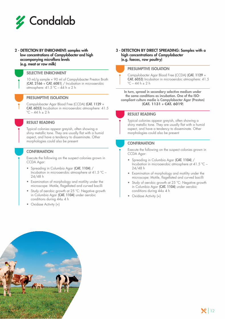
Detection and count of Coagulase-Positive Staphylococci Procedure as defined by ISO 6888:2003
IntroductionThe staphylococci are gram-positive, catalase- positive, immotile cocci that grow in conditions of aerobiosis. Staphylococcus genus includes about 30 species, among which S. aureus, S. saprophyticus and S. epidermidis stand out.
The most pathogenic of them is Staphylococcus aureus, which typically causes skin infections, although it can also cause pneumonia, endocarditis, and osteomyelitis. In general, they are associated with the formation of abscesses. Some strains produce toxins that cause gastroenteritis, scalded skin syndrome and toxic shock syndrome.
Another characteristic that increases the pathogenicity of the staphylococci is their capacity to coagulate blood thanks to the production of coagulases. S. aureus coagulase positive is among the most ubiquitous and dangerous pathogens for humans, both for its virulence and its capacity to develop resistance to antibiotics.
This ubiquitousness together with its virulence, makes its detection in food of utmost importance to guarantee consumer safety.
BibliographyISO 6888-1:1999. Microbiology of food and animal feeding stuffs – Horizontal method for the enumeration of coagulase-positive staphylococci (Staphylococcus aureus and other species). Part 1: Technique using Baird-Parker agar medium.
ISO 6888-2:1999/2003. Microbiology of food and animal feeding stuffs – Horizontal method for the enumeration of coagulase-positive staphylococci (Staphylococcus aureus and other species). Part 2: Technique using rabbit plasma fibrinogen agar medium.
ISO 6888-3:2003. Microbiology of food and animal feeding stuffs – Horizontal method for the enumeration of coagulase-positive staphylococci (Staphylococcus aureus and other species). Part 3: Detection and MPN technique for low numbers.
PRESUMPTIVE SELECTIVE ISOLATION
Baird Parker Agar (CAT. 1100 + CAT. 5129) / Incubation: 35 or 37 ºC – 24 ± 2 h / Reincubation after marking the characteristic colonies: 35 or 37 ºC – 24 ± 2 h
RESULT READING
The characteristic colonies are black or grey, shiny and convex, surrounded by a clear area with possible presence of an opalescent ring
CONFIRMATION
• Spread on Brain Heart Infusion Broth (CAT. 1331) Incubation 35 or 37 ºC – 24 ± 2 h
• Coagulase test (+)
INITIAL SUSPENSION
Refer to the relevant ISO 6887-1 standard specific for the product to be analysed
ENUMERATION: PROCEDURE AS DEFINED BY ISO 6888-1

18
SELECTIVE ISOLATION
Baird Parker Agar (CAT. 1319 + CAT. 6024) Incubation: 35 or 37 ºC – 24 ± 2 h / Reincubation after marking the characteristic colonies: 35 or 37 ºC – 24 ± 2 h
RESULT READING
The characteristic colonies are black, grey or white, surrounded by a halo of precipitation indicating coagulase activity
SELECTIVE ISOLATION
Baird Parker Agar (CAT. 1100 + CAT. 5129) or Baird Parker Agar + RPF (CAT. 1319 + CAT. 6024) Incubation: 37 ºC – 24 ± 2 h and 48 ± 2 h
CONFIRMATION
Not required, since coagulase activity is detectedwith RPF
CONFIRMATION
The Baird-Parker method is only used for the count:• Spread on Brain Heart Infusion Broth (CAT. 1331)
Incubation 35 or 37 ºC – 24 ± 2 h • Coagulase Test (+)
INITIAL SUSPENSION
Refer to the relevant ISO 6887-1 standard specific for the product to be analysed
ENUMERATION: PROCEDURE AS DEFINED BY ISO 6888-2
INITIAL SUSPENSION
Refer to the relevant ISO 6887 or 8261 standard for the product under analysis
DETECTION AND MPN: PROCEDURE AS DEFINED BY ISO 6888-3
ENRICHMENT
0.1 ml initial suspension + 9 ml Giolitti-Cantoni Broth (CAT. 1287) Incubation: 37 ºC – 24 ± 2 h
ENRICHMENT
10 ml initial suspension + 10 ml Giolitti-Cantoni Broth double concentration (CAT. 1287) Incubation: 37 ºC – 24 ± 2 h

19
Detection of pathogenic Yersinia EnterocoliticaProcedure as defined by ISO 10273:2017
Introduction
Yersinia enterocolitica is a gram-negative, oxidase- negative, non-sporulating and facultative anaerobic type of bacteria. This type of bacteria is capable of growing in a wide range of temperatures (from -1 ºC to 40 ºC) and presents a capsule with antifagocytic factors, thus increasing its pathogenicity.
Its capacity to multiply in foods at low temperatures, as well as in vacuum packaged products, are the main reasons for it to be a major concern within the food industry. The majority of breakouts detected are associated with the consumption of meat, milk and unpasteurized dairy products.
Although there are many animal-derived foods that may be carriers of Y. enterocolitica, porcine livestock gets infected more than any other animal species.
Bibliography
TENNANT S.H, GRANT T.H and ROBINS-BROWNE R.M. Pathogenicity of Yersinia enterocolitica biotype 1A.FEMS Inmun. Medical Microbiol. 2003, 38 pp 127-137.
BOTTONE E.J, Yersinia enterocolitica. Overview and epidemiologic correlates. Microbes Infetc. 1999, 1 (4) pp. 323-333.
ISO 10273:2017. Microbiology of the food chain – Horizontal method for the detection of pathogenic Yersinia enterocolitica.
RESULT READING
The characteristic colonies are small and circular. They present a central zone with a well-defined, dark red border. Around them, a translucent or transparent zone can be observed
SELECTIVE ISOLATION
Yersinia Selective Agar (CIN) (CAT. 1126 + CAT. 6033) Incubation: 30 ºC ± 1 ºC – 24 ± 2 h
DETERMINATION OF PATHOGENIC SPECIES
• Urease test (+): (CAT. 2000)• Bile Esculin Agar (-): (CAT.1031)• Plasmid pYV (+)• Detection of pyrazinamidase (-)
BIOCHEMICAL CONFIRMATION
• Lysine Decarboxylase Broth (-): (CAT. 1208)• Arginine dihydrolase (-)• Phenylalanine Agar: (CAT. 1040)• Fermentation of carbohydrates:
Sucrose (+) Sorbitol (+) Ramnosa (-) Melibious (-)
• Simmons Citrate Agar (-): (CAT. 1014)
PRIMARY SELECTIVE ENRICHMENT
X ml/g sample + 9X ml of Sorbitol Peptone Broth and Bile Salts (PSB)(CAT. 1298) Incubation: 25 ºC ± 1 ºC – 44 ± 4h
SECONDARY SELECTIVE ENRICHMENT
10 ml initial suspension in PSB Broth + 90 ml of Minerals Modified Glutamate Broth (MMBG) (CAT. 1365 + CAT 6051) Incubation: 25 ºC ± 1 ºC – 44 ± 4h
Alkaline treatment (4.5 ml KOH) during 20 ± 5S
Method

20
Detection and enumeration of Shigella sppProcedure as defined by ISO 21567:2004
Introduction
Shigella is a genus of immotile, gram-negative, non-sporulating, facultative anaerobic bacillus, belonging to the Enterobacteriaceae family. They present reduced biochemical activity with cytochrome-oxidase negative activity and glucose fermentation without gas production.
It is a highly enteroinvasive type of bacteria: Its habitat is the colon and its principal reservoir is human beings, although it has also been isolated in other higher primates. Shigella species are very sensitive to changes in temperature and to unfavourable environmental conditions. However, they are able to tolerate low pH, therefore they are one of the few bacteria that can survive the acidity of the stomach and then colonize the digestive tract. This ability, combined with the fact that they are infectious in low doses, contributes to their pathogenicity.
Contamination of foods with Shigella may result from direct or indirect contact with faecal matter from infected persons, through infected water, pests (flies), or from lack of hygiene and proper handling techniques during food preparation.
Bibliography
PASCUAL ANDERSON, Mª R. (1992) Microbiología Alimentaria. Díaz de Santos, S.A. Madrid.
ATLAS, R.M., L.C. PARK (1993) Handbook of Microbiological Mediafor the examination of Food. CRC Press Inc.Boca Ratón.
ISO 21567:2004. Microbiology of food and animal feeding stuffs – Horizontal method for the detection of Shigella spp.
RESULT READING
To identify characteristic colonies of Shigella, refer to the relevant technical data sheets of each culture medium
SELECTIVE ISOLATION (low selectivity)
Macconkey Agar (CAT. 1052) Incubation: 37 ± 1 ºC – 20/24 h
1
SELECTIVE ISOLATION (medium selectivity)
XLD Agar (Xylose Lysine Desoxycholate Agar) (CAT. 1274) Incubation: 37 ± 1 ºC – 20/24 h
2
SELECTIVE ISOLATION (high selectivity)
Hektoen Enteric Agar (CAT. 1030) Incubation: 37 ± 1 ºC – 20/24 h
3
PURIFICATION COLONIES
Spread in Nutrient Agar (CAT. 1060) Incubation: 37 ± 1 ºC – 20/24 h
SEROLOGICAL IDENTIFICATION
• Antigen differentiation• Agglutination tests
BIOCHEMICAL CONFIRMATION
• Triple Sugar Iron Agar (TSI) (Refer to reading information): (CAT. 1172)
• Semisolid Nutrient Agar (-): (CAT. 2046)• Urea Agar (-): (CAT. 2000)• Lysine Decarboxylase Medium (-): (CAT. 1176)• L-ornithine decarboxylation (+/- according to spp)• Tryptophan Culture Broth (+/- according to spp):
(CAT. 1237)• ß-galactosidase Detection (+/- according to spp)• Sugar Fermentation (according to spp)• Acetate Differential: (CAT. 1192)
SELECTIVE ENRICHMENT
X ml/g sample + 9X ml of Shigella Broth (CAT. 2078) Incubation in anaerobiosis: 41,5 ± 1 ºC – 16/20 h
Method
21
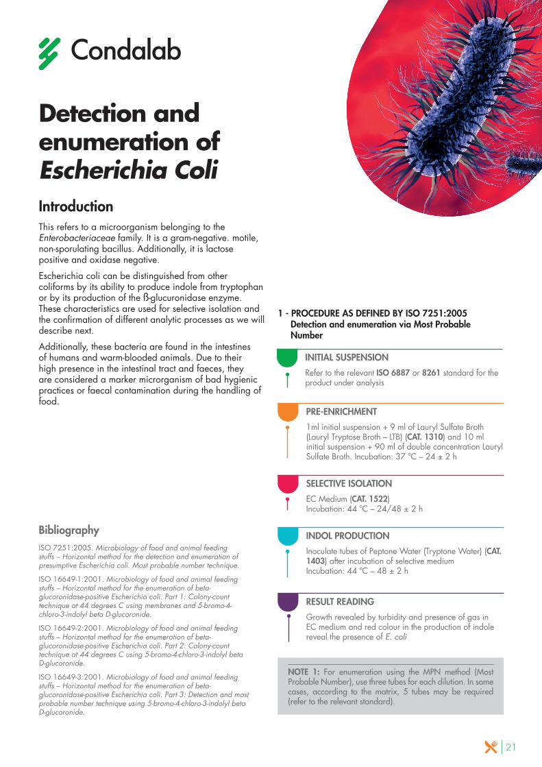
Detection and enumeration of Escherichia Coli IntroductionThis refers to a microorganism belonging to the Enterobacteriaceae family. It is a gram-negative. motile, non-sporulating bacillus. Additionally, it is lactose positive and oxidase negative.
Escherichia coli can be distinguished from other coliforms by its ability to produce indole from tryptophan or by its production of the ß-glucuronidase enzyme. These characteristics are used for selective isolation and the confirmation of different analytic processes as we will describe next.
Additionally, these bacteria are found in the intestines of humans and warm-blooded animals. Due to their high presence in the intestinal tract and faeces, they are considered a marker microrganism of bad hygienic practices or faecal contamination during the handling of food.
BibliographyISO 7251:2005. Microbiology of food and animal feeding stuffs – Horizontal method for the detection and enumeration of presumptive Escherichia coli. Most probable number technique.
ISO 16649-1:2001. Microbiology of food and animal feeding stuffs – Horizontal method for the enumeration of beta-glucoronidase-positive Escherichia coli. Part 1: Colony-count technique at 44 degrees C using membranes and 5-bromo-4-chloro-3-indolyl beta D-glucoronide.
ISO 16649-2:2001. Microbiology of food and animal feeding stuffs – Horizontal method for the enumeration of beta-glucoronidase-positive Escherichia coli. Part 2: Colony-count technique at 44 degrees C using 5-bromo-4-chloro-3-indolyl beta D-glucoronide.
ISO 16649-3:2001. Microbiology of food and animal feeding stuffs – Horizontal method for the enumeration of beta-glucoronidase-positive Escherichia coli. Part 3: Detection and most probable number technique using 5-bromo-4-chloro-3-indolyl beta D-glucoronide.
PRE-ENRICHMENT
1ml initial suspension + 9 ml of Lauryl Sulfate Broth (Lauryl Tryptose Broth – LTB) (CAT. 1310) and 10 ml initial suspension + 90 ml of double concentration Lauryl Sulfate Broth. Incubation: 37 ºC – 24 ± 2 h
SELECTIVE ISOLATION
EC Medium (CAT. 1522) Incubation: 44 ºC – 24/48 ± 2 h
INDOL PRODUCTION
Inoculate tubes of Peptone Water (Tryptone Water) (CAT. 1403) after incubation of selective medium Incubation: 44 ºC – 48 ± 2 h
RESULT READING
Growth revealed by turbidity and presence of gas in EC medium and red colour in the production of indole reveal the presence of E. coli
NOTE 1: For enumeration using the MPN method (Most Probable Number), use three tubes for each dilution. In some cases, according to the matrix, 5 tubes may be required (refer to the relevant standard).
INITIAL SUSPENSION
Refer to the relevant ISO 6887 or 8261 standard for the product under analysis
1 - PROCEDURE AS DEFINED BY ISO 7251:2005 Detection and enumeration via Most Probable Number

22
SELECTIVE ENRICHMENT
On a membrane on MMG Agar plates, add 1ml of the initial suspension. Dry for 15 minutes at room temperature. / Incubation: 37 ºC – 4 ± 1 h
SELECTIVE ISOLATION
Transfer membrane to TBX Chromogenic Agar (Tryptone Bile X-Glucuronide) (CAT. 1151) Incubatión: 44 ºC – 18/24 h
SELECTIVE ISOLATION
TBX Chromogenic Agar (Tryptone Bile X-Glucuronide) (CAT. 1151) / Incubation: 44 ºC – 18/24 h
NOTE 2: The technician should decide between part 16649-1 and 16649-2 of the standard, keeping in mind that the first method is developed for when the sample exhibits cells with a high stress index.
SELECTIVE ENRICHMENT
Add a proportion of the sample/initial suspension in Minerals Modified Glutamate Broth (MMBG) (CAT. 1365) at single (1:9) and double (1:1) concentrations. / Incubation: 37 ± 1 ºC – 24 ± 2 h
SELECTIVE ISOLATION
Spread in TBX Chromogenic Agar (Tryptone Bile X-Glucuronide) (CAT. 1151) Incubation: 44 ºC – 22 ± 2 h
NOTE 3: For enumeration according to the MPN (Most Probable Number) method, use 3 tubes for each dilution, which will later be used for the MPN. In some cases, according to the matrix, 5 tubes may be required (refer to the relevant standard).
INITIAL SUSPENSION
Refer to ISO 6887-1 standard or the standard relevant for the product to be analysed
2 - PROCEDURE AS DEFINED BY ISO 16649-1:2001 Colony count at 44 ºC using membranes
INITIAL SUSPENSION
Refer to ISO 6887-1 standard or the relevant standard for the product to be analysed
3 - PROCEDURE AS DEFINED BY ISO 16649-2:2001 Colony count at 44 ºC
INITIAL SUSPENSION
Refer to ISO 6887-1 standard or the relevant standard for the product to be analysed
4 - PROCEDURE AS DEFINED BY ISO 16649-3:2015 Detection and counting through the MPN technique

23
Count Total coliforms Procedure as defined by ISO 4832:2006
Introduction
Total coliforms are lactose-positive enterobacteria, and they constitute a group which is defined more by the tests used for their isolation than by taxonomic criteria. They belong to the Enterobacteriaceae family, and are characterised by their ability to ferment lactose with gas and acid production, more or less quickly, in a 48-hour period and with an incubation temperature between 30-37 ºC.
They are gram-negative, facultative aerobic and anaerobic, non-sporulating bacilli. From the "coliform" group, they form various types of genus: Escherichia, Enterobacter, Klebsiella, Citrobacter, etc. They are found in the intestines of humans and animals, as well as other environments, such as water, soil, plants, egg shells, etc.
Given that it is difficult to distinguish between existing coliforms and new contaminations, it is accepted that all appearances of coliforms are new contaminations unless proven otherwise.
Bibliography
COWELL and MORISETTI. J. Sci. Food Agric. 20, 1969, pp. 573
ISO 48321:2006. Microbiology of food and animal feeding – Horizontal method for the detection and enumeration of coliforms – Most probable number technique
ISO 4832:2006. Microbiology of food and animal feeding – Horizontal method for the enumeration of coliforms – Colony-count technique.
INITIAL SUSPENSION
Refer to the ISO 6887 or 8261 standard relevant to the product to be analysed
RESULT READING
Select plates with fewer than 150 CFU for enumeration. The characteristic colonies have a purple colour, occasionally surrounded by a reddish halo of precipitation
SELECTIVE ISOLATION
1ml of initial suspension in Violet Red Bile Agar with Lactose (VRBL) (CAT. 1093) Incubation: 30/37 ºC – 24 ± 2 h
CONFIRMATION
For atypical colonies, inoculate in brilliant green bile Brilliant Green Bile Broth 2% (CAT. 1228). Incubation: 30/37 ºC – 24 ± 2 h
Method

24
Detection of Escherichia Coli O157Procedure as defined by ISO 16654:2001
Introduction
The serotype O157 of Escherichia coli is rod-shaped and gram-negative. The "O" in the name refers to the antigen present in the cell wall (somatic antigen) that these bacteria present. It is a enterohaemorrhagic strain and causes food poisoning due to the production of a cytotoxic enterotoxin called verotoxin.
The highest incidence of the illness has been associated with the consumption of undercooked ground beef. Interpersonal contact within the family and in nursery schools is also a significant route of transmission. The infection can also occur after drinking raw milk and after swimming in or drinking water contaminated by contact with animal excrement, faecal water or sewage.
Bibliography
ZADIK P.M, CHAPMAN P.A and SIDDONS C.A. J. Med. Microbiol., 39, 1993, pp. 155-158
DOYLE M.P. and SCHOENI J.L. Appl. Environ. Microbiol., 53, 1987, pp 2394-2396
ISO 16654:2001. Microbiology of food and animal feeding stuffs – Horizontal method for the detection of Escherichia coli O157
ENRICHMENT
X g/ml sample + 9Xml of Trypticasein Soy Broth Modified with Novobiocin (mTSB) (CAT. 1292). Homogenisation and incubation 6 h and then 41.5 ºC – 12/18 h
IMMUNOMAGNETIC SEPARATION (IMS)
For 6 hours and later, after 12/18 h of incubation
PURIFICATION COLONIES
Spread in Nutrient Agar (CAT. 1060) Incubation: 37 ºC – 18/24 h
BIOCHEMICAL CONFIRMATION
Tryptophan Culture Broth (CAT. 1237) + Kovac's Reagent (CAT. 5205)
SEROLOGICAL IDENTIFICATION
Only for those colonies which are indole-positive
SELECTIVE ISOLATION
50µl of re-suspended magnetic particles in Macconkey Agar with Sorbitol (CT-SMAC) (CAT. 1099 + CAT. 6064) Incubation: 37 ºC – 18/24 h
SELECTIVE ISOLATION
50µl of re-suspended magnetic particles in chromogenic E. coli O157:H7 Cromogenic Agar (CAT. 1588 + CAT. 6064) Incubation: 37 ºC – 18/24 h
Method

25
Detection of Vibrio sppProcedure as defined by ISO 21872:2007
Introduction
Bacteria from the Vibrio genus are gram-negative bacilli, with comma-shaped cells. Vibrio is oxidase positive, facultative anaerobic, and does not form spores. All species from the genus are motile, generally with a unique polar flagellum.
Various Vibrio species are pathogenic, causing digestive tract illnesses, especially V. cholerae, the agent which causes cholera; V. parahaemolyticus which causes self-limiting inflammatory diarrhea, and V. vulnificus, which is transmitted by the intake of shellfish.
Vibrio infection originates in most cases by consumption of raw or undercooked shellfish. These bacteria naturally grows in marine environments, whether in salt water or estuaries, where there is a mix of fresh and salt water. This aquatic presence makes the fishing products the foods more related with the species in the Vibrio genus.
Bibliography
ISO 21872-1. Microbiology of the food chain – Horizontal method for the Vibrio spp. – Part 1: Detection of patoentially enteropathogenic Vibrio parahaemolyticus, Vibrio cholerae and Vibrio vulnificus.
PRIMARY ENRICHMENT
25 g/ml sample + 225 ml of Buffered Peptone Water (CAT. 2155) Incubation (fresh product): 41.5 ºC ± 1 – 6 ± 1 h Incubation (others): 37 ºC ± 1 – 6 ± 1 h
SECONDARY ENRICHMENT
1 ml sample + 10 ml Buffered Peptone Water (CAT. 2155) / Incubation (V. chlorae and V. parahemolyticus) 41.5 ºC ± 1 – 6 ± 1 h. Incubation (V. vulnificus) 37 ºC ± 1 – 6 ± 1 h
BIOCHEMICAL CONFIRMATION
• Decarboxylase L-Lysine in saline medium• Arginine Dihydrolase in saline medium• ß-galactosidase detection• Indole detection• Halotolerance
For result reading, refer to the relevant standard
SELECTIVE ISOLATION
1 µl of culture incubated in TCBS Agar (CAT. 1074) Incubation: 37 ºC ± 1 – 24 h ± 3 h
The standard also includes a secondary selective medium to be selected by the laboratory
Method
26
Detection and enumeration of other microorganisms in the food industry
Bibliography
ISO 15213:2003. Microbiology of food and animal feeding stuffs – Horizontal method for the enumeration of sulfite-reducing bacteria growing under anaerobic conditions.
ISO 13720:2010. Meat and meat products. Enumeration of presumptive Pseudomonas spp.
ISO 6611:2004//IDF 94. Milk and milk products – Enumeration of colony-forming units of yeast and/or molds – Colony-count technique at 25 ºC.
SELECTIVE ISOLATION
1 ml initial dilution on Iron Sulfite Agar (CAT. 1559) Incubation for thermophilic bacteria: 50 ºC ± 1 – 24/48 h Incubation for mesophilic bacteria: 37 ºC ± 1 – 24/48 h
SELECTIVE ISOLATION
0,1 ml initial dilution on Pseudomonas CFC Agar (CAT. 1356 + CAT. 6036) Incubation: 25 ºC ± 1 – 44 h ± 4 h
SELECTIVE ISOLATION
1 ml of initial dilution on OGA Medium (Oxytetracycline Glucose Agar) (OGYE) (CAT. 1527 + CAT. 6018) Incubation: 25 ºC ± 1 – 5 days
The standard also includes the Chloramphenicol Agar (YGC Agar) (CAT. 1301) as a medium for selective isolation
SELECTIVE ISOLATION
1 ml initial dilution on MRS Agar Low pH (CAT. 1433) Incubation: 30 ºC - 72 h ± 3 h
BIOCHEMICAL CONFIRMATION
• Breathing Test• Sporulation Test
BIOCHEMICAL CONFIRMATION
• Oxidase test (+)
INITIAL SUSPENSION
Refer to ISO 6887 or 8261 standards relevant according to the product to be analysed (proportion 1:9)
SULPHITE REDUCING BACTERIA COUNT IN ANAEROBIC CONDITIONS (ISO 15213:2003)
INITIAL SUSPENSION
Refer to the relevant ISO 6887-1 standard specific for the product to be analysed
LACTIC BACTERIA COUNT (ISO 15214:1998)
INITIAL SUSPENSION
Refer to ISO 8261 standard relevant to the product to be analysed
MOLD AND YEAST COUNT IN DAIRY PRODUCTS (ISO 6611:2004)
INITIAL SUSPENSION
Refer to the relevant ISO 6887 or 8261 standard for the product under analysis
ALLEGED PSEUDOMONAS SPP IN MEAT AND MEAT PRODUCTS (ISO 15213:2003)

[email protected] | www.condalab.com