Embed Size (px)
Citation preview
MICROBIOLOGICALPR O B LE M S
IN FOOD PR E SE R V A T IO N
BY IR R A D IA T IO N
PROCEEDINGS OF A PANEL.
VIENNA. 27 JUNE-1 JULY
1966ORGANIZED BY THE
JOINT F A O /IA EA DIVISION OF ATOMIC ENERGY
IN FOOD AND AGRICULTURE
FW °
7W к? r
I N T E R NA T I 0 N A L A T O M I G E N E R G Y A G E N C Y, V I E N N A , 1967


MICROBIOLOGICAL PROBLEMS IN FOOD PRESERVATION BY IRRADIATION

The fo llow in g States are Mem bers o f the International A tom ic Energy Agency:
AFG H ANISTAN
ALBANIA
ALGERIA
ARGENTINA
AUSTRALIA
AUSTRIA
BELGIUM
BOLIVIA
. BRAZIL
BULGARIA
BURMA
BYELORUSSIAN SOVIET
SOCIALIST REPUBLIC
CAM BODIA
CAMEROON
C A N AD A
CEYLON
CHILE
CH IN A
COLOMBIA
CONGO, DEMOCRATIC
REPUBLIC OF
CO STA RICA
CUBA
CYPRUS
CZECHOSLOVAK SOCIALIST
REPUBLIC
DENMARK
D O M IN IC AN REPUBLIC
ECUADOR
EL SALVADOR
ETHIOPIA
FINLAND
FRANCE
GABON
GERM ANY, FEDERAL
REPUBLIC OF
GHANA
GREECE
GU ATEM ALA
H A IT I
HOLY SEE
HUNGARY
ICELAND
IN D IA
INDONESIA
IRAN
IRAQ
ISRAEL
IT A L Y
IVO RY CO AST
JAM AICA
JAPAN
JORDAN
KENYA
KOREA, REPUBLIC OF
K U W AIT
LEBANON
LIBERIA
LIBYA
LUXEMBOURG
M ADAGASCAR
M ALI
MEXICO
M ONACO
MOROCCO
NETHERLANDS
NEW ZEALAND
NICARAG U A
NIGERIA
NORW AY
PAKISTAN
PANAM A
PARAGUAY
PERU
PHILIPPINES
POLAND
PORTUGAL
ROM ANIA
SAUDI ARABIA
SENEGAL
SIERRA LEONE
SINGAPORE
SOUTH AFRICA
SPAIN
SUDAN
SWEDEN
SWITZERLAND
SYRIAN ARAB REPUBLIC
TH AILAND
TU NISIA
TURKEY
UKRAIN IAN SOVIET SOCIALIST
REPUBLIC
UNION OF SOVIET SOCIALIST
REPUBLICS
UNITED ARAB REPUBLIC
UNITED KINGDOM OF GREAT
BRITAIN AND NORTHERN IRELAND
UNITED STATES OF AMERICA
URUGUAY
VENEZUELA
V IE T -N A M
YUGOSLAVIA
The A gen cy 's Statute was approved on 26 October 1956 by the Conference on the Statute o f the
IAEA held at United Nations Headquarters, New York ; it entered into force on 29 July 1957. The
Headquarters o f the A gency are situated in V ienna. Its principal ob jec t ive is " to accelera te and enlarge
the contribution o f a tom ic energy to peace, health and prosperity throughout the w o r ld ".
Printed by the IAEA in Austria
Novem ber 1967
PANEL PROCEEDINGS SERIES
MICROBIOLOGICAL PROBLEMS IN FOOD PRESERVATION
BY IRRADIATION
REPORT OF A PANE L ON MICROBIOLOGICAL PROBLEMS
IN FOOD PRESERVATION BY IRRADIATION ORGANIZED BY THE
JOINT FAO/IAEA DIVISION OF ATOMIC ENERGY IN FOOD AND AGRICULTURE
AND HELD IN VIENNA, 27 JUNE - 1 JULY 1966
IN T E R N A T IO N A L A TO M IC E N E R G Y A G E N C Y V IE N N A , 1967

MICROBIOLOGICAL PROBLEMS IN FOOD PRESERVATION BY IRRADIATION
(Panel Proceedings Series)
ABSTRACT. Proceedings o f a panel organized by the Joint FAO/IAEA Division o f A tom ic Energy in
Food and Agriculture and held in Vienna, 27 June - 1 July 1966. The m eeting was attended by 14 experts
from 9 countries and one international organization.
Fourteen papers were given and the topics included, among others, the inactivation o f micro-organisms
in seafood, studies on Clostridium botulinum, foot-and-mouth disease virus and salmonellas, and the effects
o f various additives and com bination treatments. The conclusions and recommendations o f the panel are
included.
A l l papers are in English with an abstract; the conclusions and recommendations are also in English.
(148 p p . , 16 x 24 cm paper-bound, 93 figures)
(1967) Price: US $3^00; £1 .1 .2
M IC R O B IO LO G IC AL PR O B LE M S IN FOOD PR E SE R V A TIO N B Y IR R A D IA T IO N
IA E A , V IE N N A , 1967 STl/PUB/ 168
FOREWORD
Irradiation is a technique that may increasingly be employed to help preserve the w o r ld 's food supplies. Some countries have a lready given public-health clearance fo r particu lar irrad iated foodstuffs, and pilot and semi-industrial irradiation plants have already been established or are under construction. W ide-spread industrial application is lik e ly in the not too distant future. However, there are s till problems to be solved; some of these are m icrob iological.
A Panel on M icrob iologica l Problem s in Food Preservation by Irrad iation was organized by the Joint FAO /IAEA D ivision o f A tom ic Energy in Food and Agricu lture on 27 June to Г July 1966. A detailed evaluation was made o f research and development needs in radicidation ( i . e . destroying m icro-organism s harmful to human health), in radurization ( i .e . extending the shelf life of perishable foods by reducing the spoilage m icro-organism s in it ), in the elimination of viruses and in the inactivation of preformed toxins. The Panel also considered the unification and standardization o f exp er imental methodology.
Recommendations were drawn up for the D irectors General of the Food and Agriculture Organization of the United Nations and of the International Atomic Energy Agency on how these two organizations could best fulfil their ro les in this field . It was considered important to continue sponsoring and co-ordinating research.. Establishing an international pilot and demonstration plant was thought essential for progress in development work, especially on radicidation.
Experts on rad io- and food m icrob io logy and a representative o f the World Health Organization attended the meeting. The proceedings contains the contributions of the members of the Panel together with the general conclusions and recommendations.
EDITORIAL NOTE
The papers and discussions incorporated in the proceedings published by the International A tom ic Energy Agency are edited by the Agency 's ed ito ria l staff to the extent considered necessary fo r the reader's assistance. The views expressed and the genera l style adopted rem ain, however, the respons ib ility o f the named authors o r participants.
F o r the sake o f speed o f publication the present Proceedings have been printed by composition typing and photo-offset lithography. Within the l im itations im posed by this method, every e ffo rt has been made to maintain a high ed itoria l standard; in particu lar, the units and symbols employed are to the fu llest practicable extent those standardized o r recommended by the competent international sc ien tific bodies.
The affilia tions o f authors are those given at the tim e o f nom ination.The use in these Proceedings o f particu la r designations o f countries o r
te rr ito r ie s does not im ply any judgement by the Agency as. to the legal statue o f such countries o r te rr ito r ie s , o f th e ir authorities and institutions o r o f the delim itation o f their boundaries.
The mention o f specific companies o r o f their products o r brand-names does not im ply any endorsement o r recommendation on the part o f the In ternational A tom ic Energy Agency.

CONTENTS
The inactivation of infection and intoxication m icro-organism sby irradiation in seafood (PL-199/9) ................................................ 1Dorothy J. Quinn, A. W. Anderson and J. F . Dyer
Salmonella radicidation of dry mixed feeds and feed ingredients(PL-199/2) ........................................................ ..................................... 15D .A .A . M ossel
The effect of ionizing radiation on Cl. botulinum spores (PL-199/16) 27N. N. Masokhina-Porshnyakova and G. V. Ladukhina
Toxin production by Cl. botulinum type E in fish (P L -1 9 9 / 1 5 )........ 37G. Hobbs
Radiosensitivity of type E botulinus toxin and its protection byproteins, nucleic acids and some related substances (PL-199/1) . 45T. Miura, S. Sakaguchi, G. Sakaguchi and K. Miyaki
Radiation resistance of botulinal toxins (PL-199/13) ........................ 55T. A. Roberts
Resistance of Aflatoxin to chemical and biological changes bygamma irradiation (P L -1 9 9 / 1 7 )................................ ................... . 57K. Miyaki, K. Aibara, T. M iura
. X -ray inactivation of foot-and-mouth disease, virus and vesicu larstomatitis virus in aqueous media (P L -1 9 9 / 4 )................................ 65C. D. Johnson
Gamma radiation fo r steriliz in g the carcasses of foot-and-mouthdisease virus infected animals (P L -1 9 9 / 3 )...................................... 77B. Baldelli
The effects of additives on radiation-resistance of C l. botulinum inmeat (P L -1 9 9 /1 0 )....................................................7. ........................ 87A. W. Anderson, D. A . Corlett, J r . , and K. L . Krabbenhoft
Combination treatment of spores of Cl. botulinum with heat plusradiation (PL-199/12) .......................................................................... 99N. Grecz, J. Upadhyay, T. C. Tang and C. A. L in
E ffects of heating and gamma radiation on the inhibition ofbacterial spores by curing agents (P L - 1 9 9 / l l ) ................ ............. 115T. A. Roberts
Reduction of radiation dose requirements of foods by additives(PL-199/8) .............................................................................................. 123J. Farkas, I. K iss and E . Andrássy
The effect of ionizing radiation and antioxidant treatment on the quality and storage life of vacuum-packed trout at 0°C(PL-199/6) .................................................. ................................. ......... 133P . Hansen and В. V. Jorgensen
Summaries, conclusions and recommendations of the Panel ............ 139L is t of Panel M em bers.............................................................................. 147


THE INACTIVATION OF INFECTION AND INTOXICATION MICRO-ORGANISMS BY IRRADIATION IN SEAFOOD
DOROTHY J. QUINN, A .W . ANDERSON AND J.F. DYER OREGON STATE UNIVERSITY,
. CORVALLIS, OREGON,UNITED STATES OF AMERICA
Abstract
THE IN A C T IV AT IO N OF INFECTION AND IN TO X ICATIO N MICRO-ORGANISMS BY IRRADIATION IN
SEAFOOD. Studies have been made to determine the lim its o f tolerance to gamma radiation for 16 cultures
in both broth and sterilized crabmeat. The cultures include 7 species o f Salmonella, 3 species o f Shigella
and one species each o f Neisseria, Mycobacterium , Escherichia, Proteus, Streptococcus and Staphylococcus.
A ll the cultures showed a near linear death curve when irradiated in broth (Hartsell’ s). However, when the
above cultures were irradiated in crabmeat, the Salmonella and Shigella exhibited in varying degrees a rapid
non-linear decline in v iab ility with respect to the radiation doses. This decline tended to becom e very
gradual as the irradiation dose increased, showing a distinct ’ tailing e ffe c t ’ o f small numbers o f increasing doses at the end o f the dose-survival curve. The most pronounced ’ ta iling ' o ff was shown by Staphylococcus
aureus, an organism in which survival could be observed even after subjecting inoculated crabmeat to over 2.0 megarad. It was also observed with several o f the above-named species (Salm onella) that i f the in
oculated crabmeat was diluted with water (from 1 to 10 fo ld ) and then irradiated, the tailing e ffe c t gradually
disappeared as the dilution was increased, suggesting some protective properties possessed by the crabmeat.
More recent evidence would indicate that shrimp meat is sim ilar to crab.
INTRODUCTION
Supplying man with food is becoming an increasing problem because of the rapidly expanding population and growing desire of the developing nations fo r a greater varie ty o f nutrients the year around. Scientists are constantly improving the old and developing new methods to meet this expanding demand. Most o f the underdeveloped nations of the world have neither the technological background nor the developed technology in food processing that the industrial nations of the world enjoy. These underdeveloped nations can best apply methods that can be used without the development of the complex and expensive prim ary or satellite industries, luxuries which the highly industrialized nations now enjoy. The irra d iation pasteurization and sterilization o f foods, already past the p re lim inary experimental stages, show prom ise fo r such future application, especia lly in the seafood industries.
An important aspect of the future application o f irradiation to the preservation o f foods is that o f ensuring that food made available to the public is free o f pathogens. A working dose sufficient to free a food from pathogens requires that one keep in mind those factors which may influence the resistance o f a m icro-organism . Among the more important of these is the radiating.menstruum. Studies have shown m icro-organism s to be more resistant when irrid iated in a medium such as nutrient broth than in one such as a buffer o r d istilled water [1 ]. As the chemical complexity of the growth environment is increased there appears to be a concomitant increase in the protective effect o f the medium. Thus, it is possible that
11

2 QUINN e t a l.
a m icro-organism , when irradiated in a new menstruum, w ill exhibit an entirely different dose survival curve, necessitating changes in the working dose required fo r each type o f food.
The intent of the present report is to compare the realtive resistances and to observe resulting survival patterns in different menstrua using non-sporeform ing food intoxication and infection m icro-organism s as- sociàted with seafoods. Previous experiments [2] were designed to observe the effect o f added water in influencing the 'tailing phenomena' observed when m icro-organism s are irradiated in some seafoods.
M ATER IAL AND METHODS
M icro-organism s selected represent species known to be pathogenic or non-pathogenic members o f various genera containing pathogenic species. Salmonella paratyphi and Salmonella wichita were obtained from the culture collection maintained at the Communicable Disease Center (CDC), Atlanta, Georgia.The following species were obtained from the collection maintained in the Department of M icrobiology, Oregon State University, Corvallis, and were originally obtained from the American Type Culture Collection (ATCC ), Bethesda, Maryland: Salmonella typhi, Salmonella paratyphi A , Salmonella choleraesuis, Salmonella enteritidis,' Salmonella pullo rum, Shigella dysenteriae. Shigella paradysenteriae, Shigella sonnei, Escherich iacoli. Streptococcus faeca lis , Proteus vulgaris, neisseria, catarrhalis, and Microbacterium smegmatis. Streptococcus pyogenes was obtained from the local hospital, having been isolated from a streptococcus infection, and Staphylococcus aureus was obtained from an actual food poisoning outbreak and was shown to be coagulative positive and salt tolerant. H artsell's broth o f the follow ing composition was used as the recovery and enrichment medium: tryptone, 5.0 g; proteose peptone (D ifco); 5.0 g; sodium chloride, 5.0 g; veal infusion, 100 ml; and distilled water to make one litre. The pH was adjusted so that after autoclaving at 121°C fo r 20 min, it was 7.2. H artsell's agar was prepared by adding20.0 g of agar to 1 litre o f H artsell's broth. To maintain uniform population fo r each of the cultures used fo r inoculation, an inoculum from the pure culture was transferred to 15 ml of H artsell's broth and incubated for 24 h at 35°C. From the above culture, 1 m l was inoculated into 99 m l and again incubated as above. Two flasks were inoculated in a proportion sim ilar to that above with cells from the latter culture. The resulting cultures were transferred s im ilarly into four flasks, incubated fo r 24 h and pooled, providing 400 m l of the suspended 24-h old cells . The population of the pooled cells was determined by measuring the optical density with a spectrophotometer calibrated against ce ll counts. The desired population was obtained by dilution and by varying the amount added to each sample.
The inoculated samples w ere irradiated in a cobalt-60 irrad ia tor located at Oregon State University, Corvallis . The source was composed o f 12 cobalt-60 rods, containing radioactivity equal to approximately 3 600 Ci. The dosimetry in the high flux chamber was determined by the F rick e ferru s sulphate method, and was 8.13 X 10⁵ ± 0.36 rad/h on the29 January 1964, Corrections fo r decay were made after each exposure.
The H artsell's broth samples were prepared as follows. Quantities of 2 and 5 ml from the final ce ll concentration, ( 107 cells/m m ) were

PL-199/9 3
transferred into screw cap tubes, (1.0 cm X 5.0 cm ). The tubes were packed and sealed into No. 2 cans and maintained at a low temperature (3 - 5°C) p r io r to irradiation. A ll samples o f the same test m aterial were replicated 5 times and each plated in trip licate. Low count dilutions were plated in 6-in. P e tr i dishes. Irradiated control samples were treated s im ilary in each experiment.
The crabmeat was' obtained from a com m ercial firm as sterile Dungeness crabmeat (Cancer m egester). packaged in No. \ lb flat tins in quantities of 150 g, each containing water, salt, and c itric acid. The oysters (Crassostrea gigas) and shrimp (Pandalus jardoni) were obtained from the com m ercia l distributor as a fresh raw product. The crabmeat cans were opened and the contents were transferred asceptically into No. 202 cans in a transfer chamber equipped with a germ icidal ultraviolet lamp. Just before irradiation the crabmeat was broken into sm all pieces and 17 g transferred to glass via ls (1.5 in. in diameter and 2.5 in. in height). One ml o f approximately 1.7 X 10⁷ cells was added to each via l and mixed with a sterile glass rod to distribute the cells throughout the crabmeat. The samples were maintained at 5°C until irradiated. The fresh oysters and shrimp were homogenized in a blender fo r five minutes; 17 g were transferred to vials, inoculated as above fo r crab meat and maintained at 5°C fo r irradiation. Twenty sample via ls w ere irradiated simultaneously around the inside periphery o f the high flux chamber in a specially designed holder.
The irradiated H artsell's broth samples were plated in H artsell's agar using appropriate dilutions with peptone water. The plates were incubated at 35°C fo r 48 h or until colonies were readily countable. Ir ra d iated food samples w ere transferred asceptically in the transfer chamber to 51 m l o f s terile H artse ll's broth (1 to 3 dilution). These w ere re fr ig e rated until plating. Adequate dilutions'were made w ith .sterile peptone water, and the plating was done with the appropriate media. The plates - w ere incubated at 35°C fo r 48 h or until colonies were readily visib le.At high irradiation doses where few or no survivors were anticipated when plated on H artsell's agar, the samples were asceptically transferred to 100 ml of H artsell's broth and incubated at 35°C for a minimum of 5 d.. A fte r incubation a loopful was spread on d ifferen tia l or selective (SS agar, bismuth sulphate agar, and trip le iron agar) media fo r a qualitative determination of survivors. A ll low-count samples w ere plated on 6-in. plates in order to use a la rger plating sample (usually 10 m l) and to m inim ize the difficu lties associated with recognizing the colonies from the sample debris. Irradiated controls o f all samples were prepared as for the regu lar samples. These were used to determine the radiation concentration o f cells .
RESULTS AND DISCUSSION
The elim ination o f salmonelae and other intoxication and in fector organisms is s im ilar in process to pasteurization since the irradiation doses used are in the same range. However, the aim in addition to increasing storage life is also to remove a group of organisms which is particu larly undesirable in the product. Vegetative bacteria are m oderately sensitive to irradiation, and doses around 0.5 to 0.8 Mrad w ill
4 QUINN e t a l .
usually affect a seven decimal reduction o f most infectious and intoxicating non-sporeform ing m icro-organism s occurring in seafoods.
Most work to this date has been concerned with rem oval of Salmonella from non-perishable products [2-5J in which no multiplication of survivors occurs. However, investigators examining a perishable product must consider the survival and growth o f pathogenic and spoilage m ic ro organisms. A dose which might inactivate the salmonellae and shigella would be ineffective against sporeform ing organisms and certain ly would not destroy all the staphlococci or the streptococci which, upon storage under favourable conditions o f tem perature,: could vegetate and lead to trouble.
Some idea o f the magnitude o f dose required may be obtained by ir ra d iation o f naturally contaminated m aterial. However, since the number of pathogenic organisms is usually low, it has been found useful to inoculate the product with large numbers, about 106/g, from pure cultures to study their inactivation. These organisms show an exponential death rate to a certain point in some substrates. However, in more complex food m aterial, a tailing effect indicative of greater survival than anticipated from exponential death is found. Thus, the D value cannot be applied with as much confidence. The logM of the number of survivors is usually plotted against dose and a line is drawn between the points. This curve is usually used to obtain the D value i f the tailing effect does not occur. However, due to this tailing effect, it is im practical to extrapolate .directly to obtain a value which would completely inactivate the organisms present.
In this work, it was found im practical to incubate the organisms d irectly in the food itse lf. Prelim inary experiments have shown that organisms inoculated d irectly into food irradiated resulted in a one-log lower .recovery than i f the same organisms were inoculated into a fresh food product (personal observation). Because of the effect o f the irradiated media on organisms, it was considered expedient to transfer the samples to a rtific ia l media in order to recover a greater number of ce lls able to multiply after irradiation. Each result shown in the tables is the observation made on five replications and with each replicant being plated in trip licate with the lower dilutions being plated in 6-in. plates instead of the usual 4-in. recommended and used in usual bacteriological techniques.
. When experiments o f this nature are conducted, environmental conditions such as temperature, oxygen and ce ll concentration w ere maintained constant, and procedures were standardized for the consistency necessary fo r valid comparison of data.
The following conditions were maintained throughout the investigations:(1) The same approximate in itial number of cells per g was maintained
fo r each organism(2) Contamination was carefu lly avoided throughout all phases o f each
experiment(3) Norm al atmospheric conditions were maintained throughout the
investigation period(4) The samples were maintained at low temperatures (5 ± 1°C) at all
times except during the irradiation (ambient)(5) The same facility fo r irradiation was used throughout the study(6) The methodology was maintained constant for.the entire
investigation.

PL-199/9 5
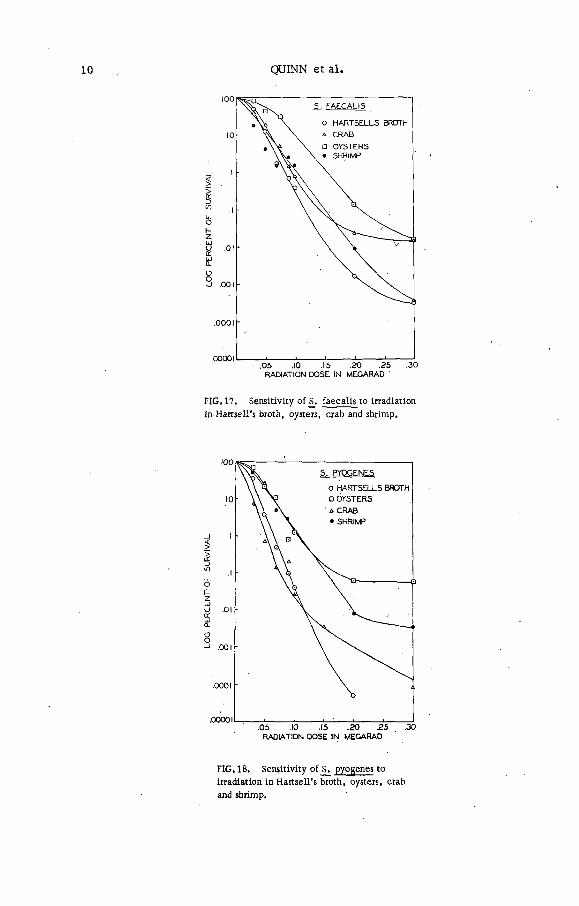
Three species o f Shigella were studied, Shigella d.ysenteriae, Shigella paradysenteriae and Shigella sonnei. The most resistant in all substrates including H artsell's broth was S. dysenteriae (Figs. 1-4). However, in none o f the substrates w ere any organisms recovered at 0.25 Mrad. It should also be noted that the irradiation resistance was not too different in any of the substrates including H artsell's broth. In all cases, ir ra d iation inactivation occurred between 0.10 and 0.25 Mrad (F igs 1-4).S. d.ysenteriae showed a slight tailing effect in all three substrates (F igs 1,3).
The Salmonella (F igs 5-12), with the exception of Salmonella enteritidis, were much more resistant to irradiation inactivation than the Shigella. The Shigella appear comparable to E_. co li [6 j while, the Salmonella approach the cocci in their irradiation resistance. It was also observed among the Salmonella that_S_. typhosa, S. paratyphi A . , S. paratyphi B . , _S. w ichita and S. choleraesuis were the most resistant in any of the other substrates (i. e. crab, oyster, shrimps) than they were in H artsell's broth (F igs 6, 7, 9, 11, 12). By the use o f the plating techniques described, the above-named organisms could be recovered at0.3 Mrad on all the substrates used. However, if the enrichment techniques as described in the methods above were used, then S. typhosa,S. pullo rum, _S. paratyphi A, S. paratyphi B, and_S. wichita could be recovered up to 0.9 Mrad. S. enteritidis appeared to be the most ir ra d iation sensitive of the group studied (F igs 5, 10). In general, oysters and shrimp offered the most protection. From the information shown a marked tailing effect was observed fo r most of the Salmonella species in all the substrates tested. The only exception being S. enteritidis (F igs 5, 10). .
25. PARADYSENTERIAE
O HARTSELLS BROTH a CRAB □ OYSTERS
- A
.05 .10 .15 .20 .25 .30RADIATION DOSE IN MEGARAD
-5.05 .10 .15 .20 .25 .30RADIATION DOSE IN MEGARAD
FIG. 1. Sensitivity o f S. dysenteriae,S. paradysenteriae and S. sonnei to irradiation
in Hartsell's broth.
F IG .2 . Sensitivity o f S. paradysenteriae
to irradiation in H artsell’ s broth, crab and
oysters.

6 QUINN e t a l.
FIG. 3.
F IG .4 Sensitivity o f S. sonnei to irradiation in Hartsell’ s broth, crab and oysters.

PL-199/9 7
FIG. 5. Sensitivity o f S. choleraesu is,
^ paratyphi A ,_S . pullorum , S. w ich ita ,
en teritid is , £ . typhosa and S. paratyphi В
to irradiation in Hartsell's broth.
RADIATION DOSE IN MEGARAD
FIG .6, Sensitivity of_S. choleraesuis to
irradiation in Hartsell’ s broth, oysters, crab and shrimp.
RADIATION DOSE IN MEGARAD
FIG .7. Sensitivity o f S. paratyphi A ,to
irradiation in Hartseil’ s broth, oysters, crab
and shrimp.
RADIATION DOSE IN MEGARAD
FIG. 8. Sensitivity o f S. pullorum to irradiation
in Hartsell’s broth, oysters, crab and shrimp.

8 QUINN e t a l.
i
-2
-4
S. WICHITA
HARTSELLS BROTH
. CRAB j OYSTERS
. SHRIMP
.05 .10 15 .20 .25
RADIATION DOSE IN MEGARAD
FIG. 9. Sensitivity o f S. wichita to irradiation in
Hartsell's broth, oysters, crab and shrimp.
4 > a: z>
RADIATION DOSE IN MEGARAD
FIG. Ю. Sensitivity o f S. enteritidis to
irradiation in Hartsell’ s broth, oysters and crab.
RADIATION DOSE IN MEGARAD
FIG. 11. Sensitivity o f S. typhosa to irradiation
in Hartsell's broth, oysters and crab.
RADIATION DOSE IN MEGARAD
FIG. 12. Sensitivity o f S. paratyphi В to
irradiation in Hartsell’ s broth, oysters and crab.
The results obtained are in general agreement with other investigators, ranging from a low fo r S. enteritidis o f 0.10 to 0.30 Mrad in any of the substrates to a high fo r S. typhosa from 0.4-1 Mrad depending upon the substrates [3,4, 7 ). Thus the salmonelas amongst the pathogens hold a

PL-199/9 9
RADIATION DOSE IN MEGARAD
FIG. 13. Sensitivity o f E. co li to irradiation
in Hartsell’ s broth, crab and oysters.FIG. 14. Sensitivity o f P. vulgaris to irradiation in Hartsell's broth, crab and oysters.
FIG, 15. Sensitivity o f E. c o li, P. vulgaris,
_N. catarrhalis and M_. smegmatis to irradiation
in Hartsell’s broth.
RADIATION DOSE IN MEGARAD
FIG. 16. Sensitivity o f S. faeca lis , S. pyogenes,
S. aureus to irradiation in Hartsell's broth.
position in some respect sim ilar to Cl. botulinum amongst the toxic m icro organisms. The usual non-infectious o r non-toxic m icro-organism s used as controls in heat-resistance tests cannot be used as controls in irra d iation tests with any confidence.
QUINN e t a l .
FIG . 17. Sensitiv ity o f j>. fa eca lis to irradiation
in H arw ell's broth, oysters, crab and shrimp.
RADIATION DOSE IN MEGARAD
FIG. 18. Sensitiv ity o f pyogenes to
irradiation in H artsell’ s broth, oysters, crab
and shrimp.

PL-199/9 11
RADIATION DOSE IN MEGARAD
FIG. 19. Sensitivity o f S. aureus to irradiation in Hartsell's broth, oysters and crab.
Escherichia coli and Proteus vulgaris were comparable in resistance to the Shigella. Both were more resistant in oysters than they were in H artsell's broth (F igs 13, 14). The results are comparable to those obtained by others fo r E. co li. N isseria catarrhalis and Mycobacterium smegmatis, because o f the difficulty in culturing, were tested only in H artsell's broth (F ig . 15). Th eir resistance was comparable to theSalm onella.
The most resistant o f all the m icro-organism s tested w ere the staphlococci, and streptococci (F igs 16-19).
Streptococcus faecalis was recoved by plating technique at 0.5 Mrad without difficulty (F ig . 6), while Streptococcus pyogenes and Staphylococcus aureus could be recovered by the plating technique between 0.3 (F ig . 16) and 0.5 Mrad. By the enrichment procedure, Staphylococcus aureus was recovered at 1.8 Mrad repeatedly, while no d ifficu lties were experienced in recovering S. faecalis and S. pyogenes above 1 Mrad. In all media a marked tailing effect (F igs 16-19) could be observed as w ell as a slight 'hump' at the curve origin . This hump could be due to the ce ll 1 aggregation characteristic of these bacteria.
Table I shows a range of D values based on the survival-curve methods. It is obvious that no one value fits all substrates. However, a range of values taking into account the tailing effect can be determined. It is realized that the end point method as used by Stumbo [8] or Schmidt [9J would have been better fo r covering a w ider range of inactivation. However, this work is still in progress and it is anticipated that further work w ill also include such information.

T A B L E I. . D V A L U E S O F B A C T E R IA IN SEAFOODS AND H A R T S E L L 'S BR O TH C A L C U L A T E D B Y TH E S U R V IV A L CU RVE M ETH O D
Organism
D va lue
(M rad)
Hartsell's broth Oysters Crabm eat Shrimp
0.012 0 .035 0 .014
0.010 0 .0 2 0.010 -
0.020 0.025 0.027 -
0.020 0.026 0.022 -
0.035 0.040 0.035 -
0.050 0 .100 0.075 0.075
0.045 0 .105 0.050 0.075
0.080 0.150 0.080 0.190
0.025 0 .035 0.035 0.075
0.055 0 .0 75 0.055 0.075
0.025 0 .05 0.025 -
0.025 0.075 0.05 0.085
0.055 0.085 0.100 -
0.045 0.075 0.087 0.100
0 .04 0.075 0.100 -
Escerichia c o l i
Proteus vu lgaris
Sh ige lla sonnei
Sh ige lla paradysenteriae
Sh ige lla dysenteriae
Streptococcus fa eca lis
Streptococcus pyogenes
S taphylococcus aureus
Salm onella pullorum
Salm onella choleraesu is
Salm onella en teritid is
Salm onella paratyphi A
Salm onella paratyphi В
Salm onella w ich ita
Salm onella typhosa
PL-199/9 13
R E F E R E N C E S
[1 ] BRIDGES, B.A., HORNE, T . , The influence o f environmental factors on the m icrobicidal e ffec t o f
ionising radiations, J. appl. Bact. 22 (1959) 96.
[2] DYER, J .K ., ANDERSON, A .W ., DUTIYABODHI, P ., Radiation survival o f food pathogens in com plex media, Appl. M icrob io l. Ы (1966) 92.
[3 ] PROCTOR, B .E ., JOSLYN, R .P ., NICKERSON, J .T .R ., LOCKHART, E .E ., Elimination o f Salmonella
in whole egg powder by cathode ray irradiation o f egg magma prior to drying, Food T ec h ., Champaign7 (1953) 291.
[4 ] NICKERSON, J. T . R ., CHARM, S .E ., BROGLE, R .C ., LOCKHART, E .E ., PROCTOR, B .E .,LINEWEAVER, H ., Use ot h igh-voltage cathode rays to destroy bacteria o f the Salmonella group
in liquid and frozen egg white solids, Food T ec h ., Champaign 11 (1957) 159.
[5 ] BROGLE, R .C ., NICKERSON, J .T .R ., PROCTOR, B.E., PYNE, A . , CAMPBELL, C . , CHARM, S .,
Use o f h igh-voltage cathode rays to destroy bacteria o f the Salmonella group in whole egg solids,
egg yolk solids, and frozen egg yolk, Food Res. 22 (1957) 572.
[6 ] ALPER, T . , GILLIES, N .E ., Restoration o f Escherichia co li strain В after irradiation; its dependence
on suboptimal growth conditions, J. gen. M icrob iol. 18 (1958) 461.
[7 ] MOSSEL, D .A .A . , The destruction o f Salmonella bacteria in refrigerated liquid whole egg by gamma
irradiation, Int. J. appl. Rad. Isotopes £ (1960) 109.
[8] STUMBO, C .R ., A technique for studying resistance o f bacterial spores to temperature in the higher
range, Food. T ech ., Champaign 2 (1948) 22S.[9 ] SCHMIDT, C .F . "Therm al resistance o f m icro-organisms", Chap.32 in Antiseptics, Disinfectants.
Fungicides and Sterilization (REDDISH, G .F ., Ed.) Lea and Febiger, Philadelphia (1954).

SALMONELLA RADICIDATION OF DRY MIXED FEEDS AND FEED INGREDIENTS
D . A . A . MOSSEL
CENTRAL IN S T IT U T E FOR N U T R IT IO N A N D FOOD. RESEARCH T N O ,
Z E IS T , THE NETHERLANDS
A N D
SA N M ARCOS U N IV ER SITY ,
LIM A , PERU
Abstract
SALMONELLA RADICIDATION OF DRY MIXED FEEDS AND FEED INGREDIENTS. Feed components
contaminated with salmonellae act as vehicles in the transmission o f these bacteria to slaughter animals and
hence to meat and poultry. Term inal decontamination o f ingredients or m ixed feed seems required because
sanitary improvements in processing, bagging and storage do not always appear e ffe c t iv e in considerably
reducing salmonella contamination rates.
Experiments were carried out to assay the decontamination e ffec t o f pelle tiza tion o f mixed feed.
Enumeration o f enterobacteriaceae was used as the analytical criterion. It appeared that a temperature
over 80°C generally led to f iv e dec im al reductions in enterobacteriaceae counts; however, also currently
used lower temperatures may bring about two dec im al reductions only. Follow ing earlier experiments with
fish m eal, range finding tests on the decontamination o f m ixed feed with 60Co gamma rays were also
performed. To ach ieve f iv e dec im al reductions in the counts o f the most resistant enterobacteriaceae which
were encountered about 0.5 Mrad was required; survival curves were generally not linear, so that 'overall
e ffe c t iv e dose' had to be used as a parameter. Feeding experiments with rats, using 35% fish m eal
irradiated at 0.8 Mrad in the d iet for two years, demonstrated that neither losses o f nutritive value nor
the occurrence o f orally toxic factors is effected by such an irradiation treatment.
It is recommended that p ilot plant tests be carried out. In these tests an attempt should be made to combine improved sanitation and p e lle tiz in g with a term inal radiation treatment o f the bagged m aterial
w ith the lowest dose required. Such tests should preferably be carried out in suitable areas o f countries
lik e Peru or Chile. A b rief outline is given o f the development work and training o f scientific and technical staff that should be carried out during the installation o f such a p ilot plant.
1. INTRODUCTION
It has been established beyond any doubt that quite a few feed ingredients, prepared in some of the most productive areas of the world, are more or less frequently contaminated with enteropathogenic and enterotoxinogenic organisms. Such contaminated products act as veh icles in the transmission of certain zoonoses, which in terfere with animal health and production of wholesome foods of animal origin. Contaminated feed ingredients may also sometimes entail quite serious exportation problems with economic consequences that may not be considered at all trifling.
Because radiation decontamination definitely o ffers a chance of overcom ing such problems, it was thought worthwhile to review those perspectives and study some aspects experimentally.
15

16 MOSSEL
2. INDICATIONS FOR RADIATION DECONTAMINATION(RADICIDATION) IN GENERAL
The use of ionizing radiation in food processing for m icrobiological purposes has been, and w ill continue to be hampered by the fact that, unlike heat processing, one cannot always, for reasons of acceptability of the final product, apply the dose of m icrobicidal energy required to render the product entirely safe. Therefore, possibilities for development of the surviving flo ra are of great importance, wherever radiation processing of foods is considered. In the so-called radappertization treatments [1] the risks of a surviving m icro flora have been almost elim inated [2-4] ; however, in what is called radurization of foods [1], they continue to present very serious problems [5-7].
In contrast to this, the radiation treatments under review here involve less bacteriological dangers. For our purpose, it is intended to apply radiation to proteinaceous mixed feed ingredients, such as fish meal, cotton seed meal and soya meal, or mixed feeds. The dose to be used fo r the elimination of pathogenic organisms, particu larly salmonellae, is of the order of 0.6 ± 0.3 Mrad [8-12] and therefore far from ster ilizes the commodities; nevertheless, the surviving organisms do not present ve ry much of a problem in this instance, as they occur in a medium with a water activity [13] of the order of 0.45 [11] which does not perm it bacterial proliferation.
In addition to the absence of contra-indications to salmonella radicidation, some essential technological advantages in this mode of processing exist. Due to the high penetrating power of many types of ionizing radiation, this type of m icrobicidal energy can be applied to the already packaged commodity, which precludes recontamination of the processed m ateria l. A lso, at the leve l of energy absorption required for rad ic idation almost no radiochemical changes and virtually no heat dissipation occur, so that the products are maintained chem ically and physically in their original state.
3. GENERAL ASPECTS OF THE DECONTAMINATION,OFANIM AL FEEDS
Some feed ingredients of b iological origin are nowadays obtained by modern manufacturing techniques such, as dehj^dration, extraction, toasting or combinations of such processes which exert a certain bactericidal effect. Certain products of this class, such as meat, blood, bone and feather meals are even deliberately heat-decontaminated before drying, to elim inate other pathogenic agents which sometimes occur in the raw m aterials used in their preparation. The occurrence in such materials of salmonellae and occasionally also of other pathogenic organisms must therefore be due to post-process recontamination, as has been frequently confirmed experimentally [14-18]. Hence, sanitary improvements in currently used processing and bagging methods can, in principle, lead to v irtually salm onella-free commodities, as we have c learly demonstrated [19]. Where this cannot yet be easily achieved, e.g. in certain developing areas, radiation decontamination is an attractive possibility since all that has to be done is to irradiate the properly bagged m aterial with doses of the order of 0.6 Mrad [11].

PL-199/2 17
However, a word of caution seems to be appropriate here. A great deal of attention has recently been paid to the decontamination of fish meal. This was probably because salmonellae were not expected to occur in anything other than products of warm-blooded animal origin and perhaps also in view of the considerable increase in the last decennia of the use of fish m eal in mixed feeds for various meat animals in northern countries. A country like Peru, e . g . , exported only some30 000 tons of fish m eal in 1956, but no less than 700 000 tons in 1961 [20].
This has sometimes diverted attention from the occurrence of salmonellae in other mixed feed ingredients, such as cotton seed flour, where they may be present in- higher initial numbers than in fish meal, but particu larly where enterobacteriaceae might die o ff tw ice as slowly as in fish meal (Table I) and may hence reach our production animals in higher numbers. These observations have led to the idea that the most practicable way of protecting the feeds of meat animals might be a term inal decontamination of the mixed feed. The adjective practicable should be especia lly underlined in this context because, from the point o f view of International Health Protection, decontamination of every single item in the country of production would obviously be much more attractive.
However this may be, term inal treatment of mixed feeds is c e r tainly going to be a point of interest to the feed industry. C learly this can be done by an ionizing radiation treatment using the dose indicated ea rlie r as suitable for feed ingredients. But irradiation has to face,
T A B L E I. SPO NTANEO US RED U C TIO N IN NUM BERS OF V IA B L E C E L L S OF A N A T U R A L M IXED F L O R A O F Salm onella oranienburg AND E nterobacter STRAINS IN FISH M E A L AND IN C O TTO N SEED FLO U R OF TH E SAM E a w = c. 0.40
Period o f storage
at 18 ± 1 "C
(weeks)
Enterobacteriaceae
(counts/1 g )
fish m ea l cotton seed flour
0 0. 2 X 10⁵ 0. 2 X 10⁶
2 0 .1 X 105 0. 1 X 10⁶
6 0 .2 X 104 0. 9 X 10⁵
10 0 .1 X 104 0. 3 X 10⁵
14 0. 5 X 10³ 0. 4 X 10⁵
20 0 .2 x 103 0. 3 X 10⁵
Approx. number o f d ec im a l
reductions in 20 weeks2
S ligh tly г
less than
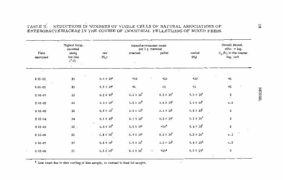
T A B L E IÍ. RED U CTIO NS IN NUM BERS OF V IA B L E C E L L S O F N A T U R A L ASSOCIATIONS OF E N T E R O B A C T E R IA C E A E IN THE COURSE OF IN D U S TR IA L P E L L E T IZ IN G OF M IXED FEEDS
Plant
exam ined
H ighest tem p,
recorded
a long
the lin e
( ° Q
raw
(N o )
E n terobacteriaceae count per 1 g m a ter ia l
steam ed p e lle t coo led
(N f )
O vera ll d ecoa t.
e f f ic . = lo g .
N 0 / N f to the nearest
lo g . unit
В 01-01 83 o. 6 x i o 6 <10 <10 <10 <5
В 01-02 80 0. 2 X 10⁶ <1 <1 <1 <5
U 01-01 52 0. 2 X 10⁶ 0. 5 X 10s 0. 2 X 10⁴ 0. 3 X ' 104 2
U 01-02 60 0. 4 X 10⁷ 0. 5 X 10⁶ 0. 8 X 10⁴ 1. 0 X 104 c . 3
U 01-03 59 0. 2 X 10⁷ 0. 2 X 10⁶ o . i x i ÿ 0 .2 X 1Cf 2
U 01-04 64 o. i x io 6 0. 7 X 10⁵ 0. 2 X 10⁴ o . 1 x i o 4 2
U 01-05 66 ■ 0. 4 X 10S 0 .2 X 105 <10a 0. 4 X 10³ 3
U 01-06 51 0. 8 X 10S 0 .9 X 104 0. 2 X 10⁴ 0 .2 X 105 c . 1
U 01-07 57 0. 8 X 10s l . o x io 5 0 .2 X 103 0 .4 X 10“ c . 2
U 01-08 67 o. 3 x i o s 0. 5 X 10⁵ <10a 0 .2 X 103 3
a Low count due to slow c o o lin g o f l in e sam ple, in contrast to fin a l lo t sam ple.
MO
SSEL

PL-199/2 19
particu larly in this instance, competition from pelleting with steam [21, 22]. As shown by the results of our line studies summarized in Table II, pelleting may indeed under certain technological circumstances have a most im pressive decontamination side-effect, v iz . up to six decimal reductions in enterobacteriaceae counts. Not a ll industrially used pelleting plants are so effective though, as is also shown in Table II; in fact, sometimes the reduction of enterobacteriaceae is so weak that it can only be called marginal when considered against the requirements dictated by the excess ively heterogeneous distribution of salmonellae over the raw m aterial [23]. The re lative m erits of gamma irradiation and pelleting for the decontamination of mixed feeds or feed ingredients w ill, therefore, u ltimately have to be determined by a carefu l com parison of data on their practicability and cost obtained from a series o f suitable pilot plant experiments.
The results of further studies of the feasib ility of salmonella radicidation of feeds are presented below.
4. M ICROBIOLOGICAL INVESTIGATION OF THERADICIDATION OF AN IM AL FEEDS
As a prelim inary step, the influence exerted by counting the surviving organisms o f the enterobacteriaceae group as such, i. e. without a previous resuscitation treatment [24-28], was studied. In this investigation resuscitation was confirmed to be occasionally required in the examination of dry foods in general (Table III, first part). However, irradiation at a dose of 1.0 Mrad did not seem to provoke in enterobacteriaceae additional sub-lethal lesions that prohibited applying immediate plating or enrichment methods, using media containing the conventional triphenyl methane dyes and bile salts [29](c f Table III, second part). These results were in agreement with ea rlie r observations regarding lack of a decisive influence o f the composition of culture media on the recovery of irradiated bacterial ce lls [30-32].
Next, range finding tests were carried out with a mixed feed of the type which is currently used in The Netherlands. Its gross composition was: corn 25%, barley 23%, m ilo corn 16%, oats 10%, wheat bran 10%,fish meal 8%, soya 4%, and lucerne meal 4%. The analytical criterion chosen for efficient decontamination was the absence of a ll enterobacteriaceae, i. e. besides salmonellae other lactose negative types and coliform bacteria, in a representative number of samples of the order of 10 g [33]. The reason for choosing this criterion is that only in this way can one guarantee with an acceptable margin of safety the absence of salmonellae in consignments treated under practical conditions, given their extrem ely heterogeneous distribution in the raw m aterial [23].
Pigmented, lactose negative, anaerogenic strains of the genus Enterobacter were found as sporadic survivors when the effective dose of about 0.6 Mrad established ea rlie r had.been applied to these feeds. This appeared to be partly due to rather high initial numbers of these organisms in the raw m aterials used. This, in turn, was caused by a higher in itial survival rate of these organisms in a dry environment

20 MOSSEL
T A B L E III. S IG N IF IC AN T B E N E F IC IA L E F F E C T S OF RESU SC ITATIO N , W HEN USE IS M ADE OF SOLID P L A T IN G M E D IA (Instances amongst a tota l o f 125 exam inations)
Without W ith
Substrate prior resuscitation on T D Y M -a ga r
(counts/1 g) (counts/1 g)
Cocoa <10 0. 9 X 10⁴
Dehydrated w ho le egg , 1 <10 0. 2 X 10⁴
D ehydrated w ho le egg , 2 <10 0 .1 X 104
Dehydrated w ho le egg , 3 <10 0 .8 X 104
M ixed feed , 1 0 .1 X 103 0. 9 X 10³
M ixed feed , 2 0. 2 X 10³ 3. 0 X 103
M ixed fee d , 3 0. 4 X 10⁴ 2. 0 X 10*
M ixed feed , 4 0 .1 X 103 0 .4 X 103
P elle ts , 1 0. 2 X 10³ 0. 4 X 10⁴
M ixed feed , N o . l , irr. 0. 4 Mrad 0 .1 X 103 0. 6 X 10³
M ixed feed , N o . 2, irr. 0. 2 Mrad 0 .5 X 106 2. 0 X 106
M ixed feed , N o . 2, irr. 0. 5 Mrad 0 .8 X 102 4. 0 X 102
M ixed feed . N o . 2, irr. 0. 6 M iad 0 ,1 X 102 0. 7 X 10²
M ixed feed , N o . 3, irr. 0 .1 Mrad 0. 5 X 10³ 2. 0 X 103
M ixed feed , N o. 4, irr. 0. 2 Mrad 0 .1 X 104 0. 4 X 10⁴
M ixed feed , N o . 5, irr. 0. 4 Mrad 0. 1 X 10⁴ 0. 4 X 10⁴
When app ly ing this technique resuscitation gave h igher results in 13% o f the
samples exam ined.
In an ea r lie r in vestigation w ith residual counts < 10 /g, necessitating a M PN -procedure,
higher counts a fter resuscitation w ere found in 2V’¡o o f the samples [29 ].
than of salmonellae (Table IV ). M oreover, the intrinsic radiation resistance of these organisms was found to be slightly higher than that of salmonellae. These facts together made it necessary to increase the radiation dose to a median value of 0.8 Mrad and a strict maximum of1.0 Mrad. .

PL-199/2 2 1
T A B L E IV . RED U C TIO N IN NUM BERS O F V IA B L E E N TE R O B A C T E R IA C E A E OF TW O D IF F E R E N T T Y P E S OF STRAINS IN M IXED FEED OF aw = 0.62
Strain
0
Period o f storage
(days at 18 ± 1 ° Q
1 5
Salm , b inza 0 .2 X 106 0 .1 x 104 0 .2 X 103
Salm . oranienburg 0 . 3 x 101 0. 6 X 10⁵ 0 . 5 x 10⁴
Enterobacter, strain P 3 0 .1 X 108 0 . 2 x 10⁶ 0. 2 X 10®
Enterobacter, strain P 4 0. 5 X 10s 0 .5 X 107 0. 2 X 10⁶
The technical detáils of the definitive pilot plant test were as follows: The mixed feed was packed in 12.5 kg quantities in 80 Polythene bags which were placed in rectangular cardboard boxes. These were given a dose of 1.0 ± 0.03 Mrad at a dose rate of approximately 3 Mrad/h from a 60Co source of approximately 5X 10⁵ Ci.
The initial enterobacteriaceae count of the mixed feed varied between 0 .3 and 1.0 X 1 0 s /g. To assess the efficacy of decontamination, 3 0
irradiated samples of 100 g each were taken from 30 different boxes, chosen strictly at random. From each of these samples 50 g were inoculated into 7 50 m l of EE broth, which, in previous investigations had been shown to recover virtually all viable cells of enterobacteriaceae occurring in dried feeds [29, 33]. A fter 24 hours incubation at 3 5°C a loopfull of these enrichment cultures was streaked onto plates of vio let red bile glucose agar [33] and incubated for a further 24 hours at 35°C.Only in two out of the 30 samples examined was a positive culture obtained. According to the results of studies on the distribution of enterobacteriaceae over feed ingredients [23], this result means that less than 5% of the boxes of irradiated mixed feed contained enterobacteriaceae. The initial leve l of contamination of the meal subjected to the irradiation treatment was probably no higher than 1 Salmonella per gram [21, 11] and, as we established, about 105 enterobacteriaceae per gram; hence, the probability of salmonellae occurring in the irradiated m aterial is of the order of 105 times lower than the-above figure obtained from the statistical evaluation. Such a low leve l of probable residual contamination is c learly quite satisfactory.
22 MOSSEL
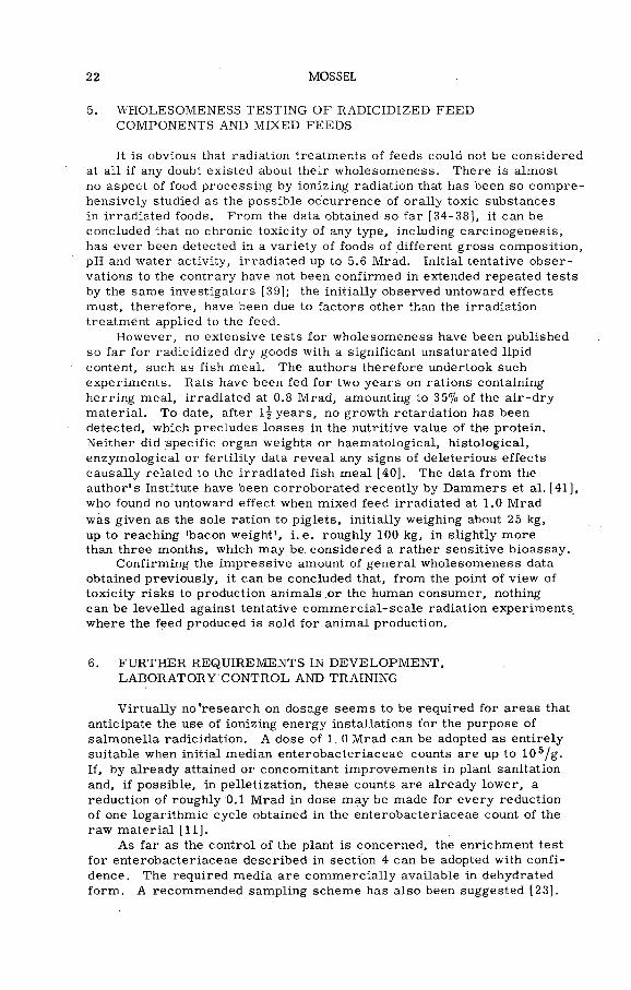
5. WHOLESOMENESS TESTING OF RADICIDIZED FEEDCOMPONENTS AND MIXED FEEDS
It is obvious that radiation treatments of feeds could not be considered at a ll if any doubt existed about their wholesomeness. There is almost no aspect of food processing by ionizing radiation that has been so com prehensively studied as the possible occurrence of ora lly toxic substances in irradiated foods. From the data obtained so far [34-38], it can be concluded that no chronic toxicity of any type, including carcinogenesis, has ever been detected in a varie ty of foods of different gross composition, pH and water activity, irradiated up to 5.6 Mrad. Initial tentative observations to the contrary have not been confirmed in extended repeated tests by the same investigators [39]; the in itia lly observed untoward effects must, therefore, have been due to factors other than the irradiation treatment applied to the feed.
However, no extensive tests for wholesomeness have been published so far for radicidized dry goods with a significant unsaturated lipid content, such as fish meal. The authors therefore undertook such experiments. Rats have been fed for two years on rations containing herring meal, irradiated at 0.8 Mrad, amounting to 35% of the a ir-d ry m aterial. To date, after i f years, no growth retardation has been detected, which precludes losses in the nutritive value of the protein. Neither did specific organ weights or haematological, h istological, enzym ological or fe rtility data revea l any signs of deleterious effects causally related to the irradiated fish m eal [40]. The data from the author's Institute have been corroborated recently by Dammers et al. [41], who found no untoward effect when mixed feed irradiated at 1.0 Mrad was given as the sole ration to piglets, in itially weighing about 25 kg, up to reaching 'bacon weight', i .e . roughly 100 kg, in slightly more than three months, which may be. considered a rather sensitive bioassay.
Confirming the im pressive amount of general wholesomeness data obtained previously, it can be concluded that, from the point of view of toxicity risks to production animals .or the human consumer, nothing can be leve lled against tentative com m ercia l-scale radiation experiments where the feed produced is sold for animal production.
6. FURTHER REQUIREMENTS IN DEVELOPM ENT,LABO RATO RY CONTROL AND TRAINING
Virtually no’research on dosage seems to be required for areas that anticipate the use of ionizing energy installations for the purpose of salmonella radicidation. A dose of 1. 0 Mrad can be adopted as entirely suitable when initial median enterobacteriaceae counts are up to 105/g.If, by already attained or concomitant improvements in plant sanitation and, if possible, in pelletization, these counts are already lower, a reduction o f roughly 0.1 Mrad in dose may be made for every reduction o f one logarithm ic cycle obtained in the enterobacteriaceae count of the raw m aterial [11].
As far as the control of the plant is concerned, the enrichment test fo r enterobacteriaceae described in section 4 can be adopted with confidence. The required media are com m ercia lly available in dehydrated form . A recommended sampling scheme has also been suggested [23].

PL-199/2 23
In areas where pilot plants fo r feed irradiation might be advantageously located, for example in Peru and/or Chile, some introductory training and extension work in the radiation m icrobiology of food and feed might be required. Form al post-doctorate courses and one to tw o-year training periods fo r young university graduates would be quite helpful in providing the required scientific o fficers. Some less extensive training at the technical le ve l would also be most useful. Finally, information services, at the request of interested industrial c irc les and as extension activities of the irradiation centre itself, would be most helpful in making the regional feed manufacturing industries acquainted with the possib ilities o f ionizing radiation as a tool in feed technology.
As to the location of such radiation centres, these could be most advantageously placed on University campuses which have already good programm es in nuclear physics and general m icrobiology, or are found w illing to start such teaching and research activities at the required academic level.
A C K N O W L E D G E M E N T S
The experimental work reported in this paper was carried out under Contract No. 199/RB with the International Atomic Energy Agency.
A number o f colleagues participated in the execution of some of the phases of the research reported in this paper; their participation constituted a more esséntial part of the investigations than would be implied by a m ere acknowledgement. M. de Proost, Research Centre for Atomic Energy, Mol, Belgium, provided dosim etric services for almost five years in the gam m a-cell dose range finding tests. S. Jefferson and F. J. Ley, United Kingdom Atomic Energy Authority, Wantage, England, supervised the pilot plant tests with animal feeds. E .H . Kampelmacher and M. van Schothorst, National Institute of Public Health, Utrecht, The Netherlands, actively co-operated in the latter experiments. A. P. de Groot, V. J. Feron and P. T il, Central Institute for Nutrition and Food Research, TNO, Zeist, The Netherlands, authorized the publication o f previously unpublished data on the wholesomeness of irradiated fish meal.
R E F E R E N C E S
[1 ] GORESLINE, H. E ., INGRAM, М ., MACUCH, P ., M OCQUOT, G ., MOSSEL, D .A .A . , NIVEN, C .F . ,THATCHER, F. S ., Ten tative classification o f food irradiation processes w ith m icrob iological
objectives, Nature, Lond. 204 (1964) 237..
[2 ] ROBERTS, T . A . , INGRAM, M . , Radiation resistance o f spores o f Clostridium species in aqueous suspension, J.Food Sci. 30 (1965) 879.
[3 ] ANELLIS, A . , GRECZ, N . , HUBER, D. A . . BERKO W ITZ , D . , SCHNEIDER, M. D . , SIMON, M . ,
Radiation sterilization o f bacon for m ilitary feeding, Appl. M icrobiol. 13 (1965) 37.
[4 ] GREENBERG, R. A . , BLADEL, В. O ., ZINGELM ANN, W .J ., Radiation injury o f Clostridium botulinum
spores in cured meat, Appl. M icrob iol. 13 (1965) 743.
[51 SCHMIDT, C. F . , LECHOWICH, R. V . . NANK, W. K . , Radiation resistance o f spores o f type E
Clostridium as related to extension o f the refrigerated storage l i fe o f foods, J.Food Sci. 27 (1962) 85.
[6 J ABRAHAMSSON, K . , de SILVA, N. N . , MOLIN, N . . Toxin production by Clostridium botulinum
type E in vacuum-packed, irradiated fresh fish in relation to changes o f the associated micro flora,Can. J. M icrobiol. _11 (1965) 523.
24• y
MOSSEL
[7 ] CANN, D. C . , WILSON, B .B ., HOBBS, G ., SHEW AN, J. M . , The growth and toxin production o f
Clostridium botulinum type E in certain vacuum packed fish, J. appl. Bacteriol. 28 (1965) 431.
[8 ] COMER, A. G . , ANDERSON, G. W . , GARRARD, E. H ., Gamma irradiation o f Salmonella species.
in frozen whole egg, Can. J. M icrobiol. 9 (1963) 321.L9] LEY, F .J ., FREEMAN, B. M . , HOBBS, B .C ., The use o f gamma radiation for the elim ination o f
Salm onellae from various foods, J. Hyg. 61 (1963) 515.[10] LICCIARDELLO, J. J ., Effect o f temperature on radiosensitivity o f Salmonella typhimurium, J.Food
Sci. 29 (1964) 469.[11] MOSSEL, D. A . A . , de GROOT, A . P., "T h e use o f pasteurizing doses o f gamma radiation for the
destruction o f Salm onellae and other Enterobacteriaceae in some foods o f low water activity.Radiation preservation o f foods” , Proc. Int. Conf. Boston. Mass. 1964. Publ. 1273, Nat. Acad. S c i . ,
Wash., D .C . (1965) 233.[12] DYER, J. K . , ANDERSON, A. W . , DUTIYABODHI, P ., Radiation survival o f food pathogens in com plex
m edia, Appl. M icrobiol. 14 (1966) 92.[13] SCOTT, W .J ., Water relations o f food spoilage microorganisms, Adv. Food Res. 7 (1957) 83.
[ 14] RUTQVIST, L . , Vorkommen von Salm onella in Futterm itteln vegetabilischenUrsprunges, Zentr.
Veterinaermed. 8 (1961) 1016.
[15] VAN DER SCHAAF, A . , Salmonellose onder de werking van de Veewet, Tijdschi.D iergeneesk. 87 (1962) 976.
[16] CLISE, J. D . , SWECKER, E. E ., Salm onellae from animal byproducts, Public Health Rept. Wash. 8£ (1965) 899.
[17] MAGWOOD, S .E ., FUNG, J ., BYRNE, J. L . , Studies on Salmonella contamination o f environment and
product o f rendering plants. Avian Diseases 9 (1965) 302.
[18] MOSSEL, D. A. A . , Les pays tropicaux com m e sources de matières premières contaminées par des
Salm onella; situation actuelle et moyens de correction, Bull. Soc. Pathol. Exot. 58 (1965) 687.
[19] ENGEL, C . , MOSSEL, D. A. A . , Onderzoekingen naar de bruto- en nettodecontaminatiewaarde van
mengvoedercomponenten, uitgevoerd in opdracht van het Productschap voor Veevoeder, Reports,
Central Institute for Nutrition Research 1891 (1965), 2135 (1966).
[20] SHEW AN, J. M . , HOLMES, N. E ., "Production and international trade in fish m eals ",Radiation Control o f
Salm onellae in Food and Feed Products, Techn ica l Reports Series No. 22, IAEA, Vienna (1963) 121.
[21] Salm onella organisms in animal feeding stuffs, Report o f a working party o f the Public Health Lab. Service. Monthly Bull. Min. Health, London 20 (1961) 73.
[22] CRANE, F. M . , HANSEN, М ., Salm onella in feedstuffs, Feedstuffs 37 45 (1965) 49.[23] VAN SCHOTHORST, M . , MOSSEL, D. A. A . , KAMPELMACHER, E. H ., DRION, E.F. , The
estimation o f the hygienic quality o f feed components using an Enterobacteriaceae enrichment assay, Zentr. Veterinaermed. 13. (1966) 273.
[24] GUNDERSON, M. F., ROSE, K. D . , Survival o f bacteria in a pre-cooked fresh-frozen food,
Food Res. 13 (1948) 254.[25] HARTSELL, S. E. t The longevity and behaviour o f pathogenic bacteria in frozen foods: the influence
o f plating media, Am. J. Public Health 41 (1951) 1072.[26] ALLEN, L. A . , PASLEY, S. M . , PIERCE, M. S .F . , Conditions affecting the growth o f Bacterium co li
on b ile salts media. Enumeration o f this organism in polluted waters, J. gen. M icrob iol. ]_ (1952) 257.
[27] LABOTS, H . , The behaviour o f sub-lethally heated Escherichia co li in m ilk and other media,
X V Int. Dairy Congress 3 <1959) 1355.
[28] SINSKEY, T .J. , McINTOSH, A. H . , PABLO, I. S ., SILVERMAN, G .J ., GOLDBLITH, S. A . ,Considerations in the recovery o f microorganisms from freeze-dried foods, Health Lab. Sci. _1
(1964) 297.'[29] MOSSEL, D. A. A . , JONGERIUS, E ., KOOPMAN, M .J ., Sur la nécessité d'une reviv ification préalable
pour le dénombrement des Enterobacteriaceae dans les aliments déshydratés, irradiés ou non, Ann.
Inst. Pasteur L ille 16 (1965) 119.[30] DAVYDOFF, S , , Influence des facteurs nutritifs sur la survie de Serratia indica après irradiation,
Compt. Acad. Sci. Paris 243 (1956) 1683.
[31] FREEMAN, B. M . , BRIDGES, B. A . , Suitability o f various plating media for counting bacteriaafter exposure to gamma radiation, Int. J. appl. Radiat. Isotopes £ (1960) 136.
[32] MOSSEL, D. A . A . , The destruction o f Salm onella bacteria in refrigerated liquid whole egg with
gamma radiation Int. J. appl. Radiat. Isotopes _9 (1960) 109.
[33] MOSSEL, D. A. A . , Eine m it dem Salmonella-Nachweis kommensurable Untersuchung von Lebens-
und Futtermitteln auf Enterobacteriaceae, Arch. Lebensmittelhyg. 15 (1964) 169.

PL-199/2 25
[34 ] RAÏCA, N . , "Data on wholesomeness studies. A progress report", Radiation preservation o f Foods,
Publ. 1273 Nat. Acad. Sci. -Nat. Res. Council, Washington D. C. (1965) 185.[ 35 ] RADOMSKI, J .L ., DEICHMANN, W. B. , AUSTIN, B. S ., MacDONALD, W .E ., BERNAL, E .,
A study o f the possible carcinogenecity o f irradiated foods, Tox ico l, appl. Pharmacol. T_ (1965)
122.[36] RADOMSKI, J. L . , DEICHMANN, W .B ., AU STIN , B .S ., MacDONALD, W. E ., Chronic toxicity
studies on irradiated bee f stew and evaporated m ilk , T ox ico l, appl. Pharmacol. 1_ (1965) 113.
[3 7 ] KENNEDY, T . S . , Studies on the nutritional value o f foods treated with gamma-radiation.
II. Effects on the protein in some animal feeds, egg and wheat, J. Sci. Food Agr. 16 (1965) 433.
[38] SHILLINGER, Y . I . , KACH KO VA, V. G . , M AGANOVA, N .B ., Influence produced on the canine
organism by meat food products gam ma-irradiated in radiopasteurization doses, Vopr. Pitaniya 24
(1965) 19.[39 ] THOMPSON, S .W ., HUNT, R .D ., FERRELL, J ., JENKINS, E .D ., MONSEN, H . , Histopathology o f
m ice fed irradiated foods, J. Nutr. 87 (1965) 274.
[ 40] GROOT, A . P . , TILL, H. P . , FERON, V, J ., MOSSEL, D. A. A . , Evaluation o f the wholesomeness
o f Salmonella radLcidized fish m eal, using rats as experimental animals (in preparation).
[41] DAMMERS, J ., KAMPELMACHER, E. H ., EDEL, W ., VAN SCHOTHORST, М ., "E ffect o f
decontamination o f feed mixtures by heat treatment and gamma radiation on growth and feed conversion in fattening pigs", Food Irradiation, IAEA, Vienna (1966) 159.

THE EFFECT OF IONIZING RADIATION ON Cl. botulinum SPORES
N . N . M A SO K H IN A -P O R SH N Y A K O V A A N D G . V . L A D U K H IN A
A L L -U N IO N RESEARCH IN ST IT U T E FOR TH E C A N N IN G A N D
VEGETABLE DRYING IN D U S T R Y ,
M O SC O W , USSR
Abstract
THE EFFECT OF IO NIZING RADIATION ON C l. botulinum SPORES. In the investigations on the
radioresistance o f the spores o f C l. botulinum the authors made use o f type A cultures (isolated from a
sample o f fresh carrot) and type В cultures (isolated from canned pork). Ten strains were used. The ex
periments were carried out in phosphate buffer solutions at different pH values and with foodstuffs (green
peas, m eat). The experimental results were obtained on the basis o f a direct calculation o f the colonies
fo llow ing incubation at 28eC in a casein-fungus-medium containing agar. The results were processed with the usual statistical analysis technique. The optimum period for storing the irradiated samples before adding
them to the nutritional medium was two weeks. In addition to cultures characterized by a radioresistance
curve with a ’ shoulder*, cases were observed where the radioresistance curve was exponential from the
start. In a fa iily large number o f cases (65%) another type o f curve was obtained (m ain ly in the case
o f re lative ly low spore concentration). The results obtained do not always correspond with current theories
on the e ffect o f radiation on the c e ll. The exponential pan o f the radioresistance curves was clearly
manifest in cases where the irradiated spores were in a neutral buffer solution. The value o f D for
suspensions irradiated and then stored in a buffer at pH 3.63 dropped by approximately 1/3 in comparison
with the value for spore suspensions irradiated in a buffer at pH 7 .0 - 6 .9 and 4 .6 - 4 .5 . The radioresistance
o f the cultures in meat and in green peas was lower than in the neutral buffer. In almost a ll experiments
ye t another part o f the curve was to be discerned, namely the 'ta i l ', for which no mathematical relation has as yet been established.
In the course o f the experiments the e ffect o f single and fractional irradiation in a C l. botulinum
culture suspended in a neutral buffer solution was determined and a linear relation between the D value and the duration o f the intervals between irradiations was found.
The resistance of spores o f Clostridium botulinum to ionizing radiation has been reflected in the survival curves published by some authors [1-12] . The curves indicate the dependence of the logarithm of the number of surviving spores upon the dose. They are characterized by the D value (the dose necessary for a ten-fold decrease of the number of cells).. For Cl. botulinum spores the D value varies in relation to the strain, chemical composition and physical state of the foods. The maximum D value (strain A -33) is 0. 37 Mrad. The survival curves described in the literature are o f a different shape. This latter can be used to establish regu larities o f the radiation effect on the cells , whereas the slope of the curve can be applied to calculate the steriliz in g doses.
To revea l the shape of survival curves, the effect of ionizing radiation was studied on ten strains o f Cl. botulinum, type A (isolated from fresh carrot) and type В (isolated from canned pork). The native liquid (the casein medium) o f type A cultures had a minimum lethal dose (MLD) o f 100 and that of type В cultures had 10 MLD. Spores w ere obtained by growing the culture on the casein medium at 35°C. Sporulation was observed under*the phase-contrast m icroscope. Spores w ere isolated
27

28 MASOKHINA-PORSHNYAKOVA and LADUKHINA
from the medium by means of washing after 60 to 70% of cells had produced spores. This was carried out by repeated centrifugation in the neutral phosphate buffer solution (pH 6. 8- 7.0). The suspension was heated at 80°C for 20 min and kept at 4°C. The suspension titre was estimated by its inoculation on the casein agaric medium with sodium thioglicolate. Irradiation was carried out with 1 ml of the suspension or 10 ml of the foods placed in cotton-sealed 'b iological tubes (15 X 140 mm). Simultaneously five samples underwent irradiation at the same dose.
A cobalt-60 radiation source composed of two plates was used to provide the dose rate o f 0.8 Mrad/sec. The dose absorbed varied within the range of + 8%. Irradiation was perform ed at 18 to 21°C.
The results w ere analysed by a d irect count of colonies in the casein agaric medium with sodium thioglicolate following their incubation at 28°C. The results were treated by common statistical methods.
Inoculated foods were analysed im m ediately after irradiation, while the suspension of irradiated spores was kept at room temperature for a fortnight. This procedure is very useful fo r revealing cells that have retained their viability and recovered after irradiation. In one of the experiments, storage o f the samples o f Cl. botulinum spores at 22°C for two weeks resulted in an increase of the D value and L value (the dose corresponding to the shoulder of the survival curve) from 0. 16 to 0. 18 Mrad and 0. 6 to 0. 8 Mrad, respective ly (F ig . 1).
FIG-1. Radioresistance change o f C l. botulinum irradiated spores follow ing a two-week storage.
As is w ell known, survival curves consist of three phases: (1) the initial phase at which the ce ll number remains practically unaltered, this is the 'shou lder' of the curve; (2) the main phase that represents an exponential part at which cells die o ff as a logarithmic function of the dose; and (3) the term inal phase, the 'ta il'o f the curve composed of
C-IMMEDIATELY AFTER IRRADIATION D«0.16 Mrad
0 - 2 WEEKS AFTER IRRADIATION
D .0 .18 Mrad L ■ 0.8 Mrad
0.5 1.0 1.5 2.0 Mrad
IRRADIATION DOSE

PL-199/16 29
TABLE I. RADIORESISTANCE OF Cl. botulinum SPORES IN THE NEUTRAL PHOSPHATE BUFFER SOLUTION
Index
o f
culture
Concentration
o f suspension
irradiated
(num ber o f
spores/m i)
L va lue
(M rad )
D va lue
(M rad )O ccurrence o f the ta il
B-40 3 .0 7 x 10⁴ 1.04. 0 .3 1 Absent up to 2 Mrad
B-40 6 .0 X 1 0 5 0 .40 0.32 Begins at 1 .4 Mrad
B-40 1 .2 X 1 0 6 0 .4 4 0 .3 3 Absent up to 2 Mrad
B-28 7 .4 X 1 0 6 0 .33 0.32 Begins at 1. 6 Mrad
B-28 2 .6 X 1 0 7 0 .26 0 .2 4 Begins at 1. 8 Mrad
B-4 1 .0 X 1 0 7 0.37 0 .2 4 Absent up to 2 Mrad
B-4 4 .0 x 107 0.22 0.27
В-49 1 .6 X 107 0 .3 0 .2 3 Begins at 1. 8 Mrad .
B-49 2 .3 X 1 0 7 0 .2 0 .2 9 Absent up to 2 Mrad
B-16 2 .9 X 1 0 7 0 .29 0.27 Absent up to 2 Mrad
B-16 1 .4 X 109 0 .0 0 .2 3 Absent up to 2 Mrad
B-41 2 .6 X 1 0 7 0 .0 0 .2 5 Begins at 1. 8 Mrad
В-42 4 .0 X 1 0 7 0 .0 0 .2 5 Absent up to 2 .2 M rad
В-42 3 .2 X 1 0 8 0 .29 0 .2 1 Absent up to 2 .2 Mrad
A -4/2 2 .6 X 1 0 7 0 .45 • 0 .25 Absent up to 2 .3 Mrad
A -4/2 3 .0 X 107 0.05 0 .3 Absent up to 2 .3 M rad
A-4/2 2 .2 X 1 0 7 0 .0 0 .2 6 Absent up to 2 Mrad
single cells of particularly high radioresistance. The values characterizing survival curves o f Cl. botulinum spores as experimentally determined are presented in Table I.
Our experiments revealed cultures whose survival curve had a shoulder (F ig . 2(a)) as well as those whose survival curve immediately acquired the exponential shape (F ig. 2(b)). The latter was peculiar to high concentrations of irradiated cells (108 - 109). Of particular interest was another shape of the curve found in a significant number of cases (65%) at re la tive ly low concentrations of spores ( 104- 106) (F ig . 2(c)). At the initial radiation stage (up to 0. 2 Mrad) those cultures exhibited an increased amount of cells found in the inoculations. The degree at which the number of cells increases depends on the initial concentration of spores in the irradiated suspension. This can be seen c learly in Fig. 3.
The size of the shoulder varies within the range of 0 to 1. 04 Mrad, the same culture sometimes showing this difference. The L/D ratio is in the main 1.1 to 1.5.

30 MASOKHINA-PORSHNYAKOVA and LADUKHINA
IRRADIATION DOSE (cO
FIG. 2. The in itia l part o f the survival curve o f C l. botulinum: (a ) strain B-4; (b ) strain B-16;
(c ) strain B-49.
IRRADIATION D O SE (o í)
F1G.3. Effect o f the concentration o f C l. botulinum B-40 spores on the post-irradiation activation.
The results obtained are often at variance with modern concepts on the radiation effect upon the cell. This is evidently accounted for by the complexity of physical processes and chemical reactions occurring in cells during radiation, reparation and reactivation.
The development of the theory of the biological effect of radiation needs further study with regard to in itial ce ll reactions to irradiation. Irradiation at doses of 0. 2 Mrad should be applied with great care to foods where Cl. botulinum may develop. This necessitates a thorough analysis o f the species composition of the m icro flora of the irradiated

log
N
PL-199/16
FIG .4 . Survival curve o f C l. botulinum B-40 at pH 7 .0 and pH 4. 6.
F IG .5. Effect o f pH on radioresistance o f C l. botulinum A-4/2.
MASOKHINA-PORSHNYAKOVA and LADUKHINA
IRRADIATION DOSE
FIG. 6. Radioresistance of Cl. botulinum A-4/2 spores in beef.
FIG. 7. Radioresistance o f C l. botulinum spores in green peas.
PL-199/16 33
food, taking into consideration the possibility of survival and activation of Cl. botulinum.
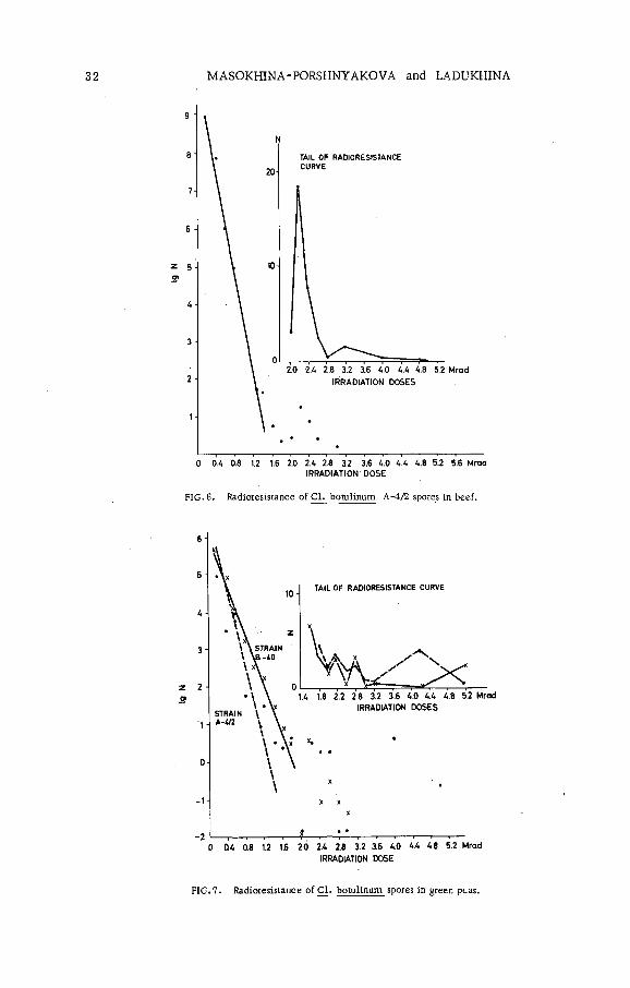
The exponential part of survival curves was distinctly observed during spore irradiation in the neutral phosphate buffer solution. The D value of the same suspension prepared at different times varied within the range of 0. 02 to 0. 08 Mrad. The D value remained unaltered for the same suspension of spores irradiated in the buffer with pH 7. 0-6. 9 and pH 4. 6 - 4. 5. The D values decreased by approximately one-third when the spores were irradiated and kept in the buffer with pH 3. 63 after irradiation (F igs 4,5 ). The culture radioresistance in beef (F ig . 6) and green peas (F ig. 7) was also lower than that in the neutral buffer solution.
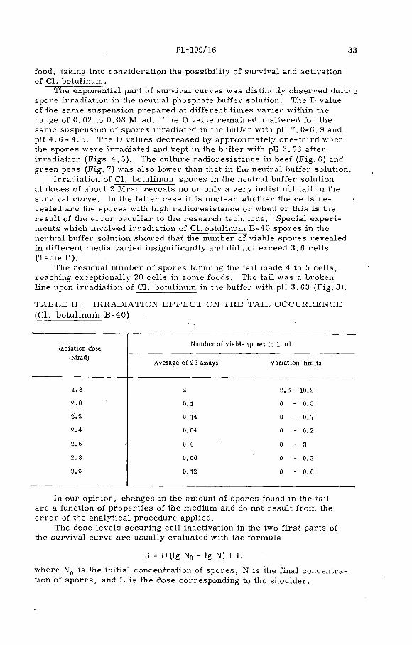
Irradiation of Cl. botulinum spores in the neutral buffer solution at doses of about 2 Mrad reveals no or only a very indistinct ta il in the survival curve. In the latter case it is unclear whether the cells r e vealed are the spores with high radioresistance or whether this is the result of the e rro r peculiar to the research technique. Special experiments which involved irradiation of Cl.botulinum B-40 spores in the neutral buffer solution showed that the number of viable spores revealed in different media varied insignificantly and did not exceed 3. 6 cells (Table II).
The residual number of spores form ing the tail made 4 to 5 cells , reaching exceptionally 20 cells in some foods. The tail was a broken line upon irradiation o f Cl. botulinum in the buffer with pH 3. 63 (F ig. 8).
T A B L E II. IR R A D IA T IO N E F F E C T ON THE T A IL OCCURRENCE (C l. botulinum B-40)
Radiation dose (Mrad)
Number of viable spores in 1 m l
Average of 25 assays Variation lim its
1 .8 2 3 .6 - 10.2
2 .0 0 .1 0 - 0 .5
2 .2 0.14 0 - 0 .4
2 .4 0.04 0 - 0 .2
2 . G 0 .6 0 - 3
2 .8 0.06 0 - 0 .3
3 .0 0.12 0 - 0 .6
In our opinion, changes in the amount of spores found in the tail are a function of properties of the medium and do not result from the e rro r of the analytical procedure applied.
The dose leve ls securing ce ll inactivation in the two firs t parts of the survival curve are usually evaluated with the formula
S = D (Ig N0 - lg N) + L
where N Q is the initial concentration o f spores, N is the final concentration of spores, and L is the dose corresponding to the shoulder.

34 MASOKHINA'PORSHNYAKOVA and LADUKHINA
FIG. 8. Radioresistance of Ç1. botulinum B-40 spores in buffer solution pH 3.63.
INTERVALS BETWEEN IRRADIATIONS ( h )
FIG.9. The D-value change upon the fractionated irradiation of Cl.botulinum B-27 spores.
This raises the problem of how to regard the cells occurring in the tail. It is obvious that this can be solved only on the basis of factual m aterial accumulated with respect to the relationship between single

PL-199/16 35
viable spores, sanitary quality of foods and occurrence of toxins in the latter.
The investigators hold differing opinions on the effect o f fractionated irradiation upon the ce ll survival. The effect of single and fractionated exposures to irradiation was studied on the Cl. botulinum culture suspended in the neutral buffer solution. Culture B-27 was exposed to fractionated irradiation in three experimental runs with intervals of 5, 15 and 28 hours. In every run the exposure was stopped after a dose of 0. 5 Mrad. The results obtained w ere as follows:
Interval duration (h) D value (Mrad)
0 0.235 0. 29
15 0.3828 0.55
The D value is in linear dependence upon the duration of the interval (F ig . 9). A significant increase in the D value during fractionated irra d iation can be accounted for by cell reparation.
It has been shown for some m icro-organism s, including Cl. botulinum, that some time must elapse before the maximum number of cells in irradiated cultures can be determined. The duration of this delay depends on the dose and increases as the dose increases. This may explain a decrease in the slope of survival curves with an increase in the interval between exposures.
R E F E R E N C E S
[1] DENNY, C .B ., BOHRER, W . , Food Res. 24 1 (1959) 44-50.[2] DNARKAR, S .D ., J. Food Science 29 5 (1964) 641-43.[3] GRECZ, N. et al. , Appl. Microbiol. _13 4 (1965) 527-36.[4] INGRAM, M. , THORNLEY, M .J. , Appl. Bacteriol. 24 (1961) 1.[5] MORGAN, B.H ., READ, J .M ., Food Res. 19 4 (1954) 357-65.[6] PRATT, G .B ., WHEATON, E. . Food Res. 24 1 (1959) 51-56.[7]* SCHMIDT, C. F . , NAUK, W .K ., Food Res. 25 (1960) 321-27.[8] SCHMIDT, C .F ., Int. J. appl. Radiat. Isotopes 14 (1963) 1.[9] WHEATON, E. et al. , J. biochem. microbiol. Tech. Engng 2 1 (1960) 1-8.
[10] WHEATON, E. et al. , J. Food Sci. 26 4 (1961) 345-50.[11] WHEATON, E. , PRATT, G .B ., J, Food Sci. 21 4 (1962) 327-34.[12] ROBERTS, T .A ., INGRAM, М ., J. appl. Bacteriol, 28 (1965) 125-38.


TOXIN PRODUCTION BY Clostridium botulinum TYPE E IN FISH*
G. HOBBSTORRY RESEARCH STATION,ABERDEEN, SCOTLAND,UNITED KINGDOM
Abstract
TOXIN PRODUCTION BY Clostridium botulinum TYPE E IN FISH. The incidence of Clostridium botulinum in fishery products in Britain and the North Sea is very low. The rate of toxin production was determined in various types of packaged fish which were inoculated with 1 0 1-10s spores of Cl. botulinum type E per 100 g of fish, at temperatures from 5 to 20“C. Radiation pasteurization at 0.3 Mrad resulted' in an extension of the storage life of the fish but had little effect on the rate of toxin production. In fish irradiated at this level it is possible for toxin to develop in about the same time as it becomes organoleptically unacceptable, in some cases before this stage. Whether toxin is produced before the fish becomes unacceptable or not depends on several factors, the most important being level of contamination and storage temperature. The results presented indicate that as the level of contamination and the storage temperature are decreased, the 'safety margin' between fish becoming unacceptable before development of toxin increases.
The occurrence o f Clostridium botulinum type E in fish and fishery products in certain parts of the world has been known fo r some tim e [1-4]. In fact the m ajority o f outbreaks o f this type of botulism have occurred as a result of the ingestion of fish o r fishery products. M ore recently a considerable e ffort has been made to determine the extent to which this organism is present in fish and the aquatic environment. In the USA surveys have shown that it is prevalent in samples of bottom deposits, fish and other marine animals on both the Pacific and Atlantic coasts, the Gulf of Mexico and the Great Lakes [5 -8 ]. The high incidence in Scandinavian waters, reported by Johannsen in 1963 [9] has been confirm ed and the incidence in the North Sea and British fishery products reported as very low [10, 11].
The ability of this organism to grow and produce toxin at low tem peratures has also been known fo r some tim e [12-14] . This has also been confirm ed on many occasions since.
The full significance of these facts was only realized recently, after severa l outbreaks of type E botulism due to the consumption o f vacuum packed, hot-smoked fish in the USA [15-17] . Whilst subsequent data showed that in some of these outbreaks the fish were not in fact vacuum packed, nevertheless the process o f vacuum packaging was imm ediately brought under suspicion,, since it did maintain the fish under anaerobic conditions. Whilst the circumstantial evidence of the outbreaks in the USA indicates that vacuum packaging may have been a m ajor contributory factor, evidence from Johannsen [13] indicates that if a product w ill support the growth o f Cl. botulinum type E, vacuum packaging is of little
* The work described in this paper was carried out as part of the programme of the Torry Research Station.
37
38 HOBBS
significance. We have ourselves shown that toxin is produced in approxim ately the same time with a given fish and inoculum size whether it is vacuum-packed or not. The results obtained by Abrahamsson et al. [18] to some extent resolve this apparent anomaly. They found that the rate of toxin production in packs incubated at 20°C was higher in inoculated vacuum-packed herring fille ts than in s im ilar packs sealed without vacuum. The lowest rate was in unsealed packs. A fter 72 h however, a ll three sets had the same amount of toxin present.
The increasing use o f packaging, along with the potential use of heat and irradiation pasteurization, which tend to favour the survival o f spore- form ing bacteria at the expense of spoilage organisms, leaves us with the possibility o f an increased hazard of type E botulism from fish and fishery products.
With a process such as irradiation pasteurization, the risk of toxin production must be at least no greater than with the unirradiated product.It is therefore necessary to establish what relationships exist between toxin production and the development of spoilage odours and flavours. In this connection it is important to bear in mind that spoilage as defined by a trained taste panel does not necessarily mean the same to a housewife. From the evidence available in recent outbreaks it would appear that the incriminated fish would have been considered spoiled by many people.
RESULTS
Our investigations o f the occurrence of type E in Britain have shown a much low er incidence than that reported in the USA or in Sweden. That our methods are not at fault has been discounted since we ourselves have confirmed the incidence reported by others in Scandinavia and parts of the USA. In our firs t report on this matter [11] we indicated no isolations , o f type E from fish in Britain. Since then we have in fact found a small number o f positive samples. At least some of these were from fish known to be o f Scandinavian origin . The number of positive samples found, however, is so sm all that their significance is difficult to assess.
A fter showing that type E occurs in certain fish products it is equally important then to determine i f those products w ill support growth and toxin production, and to define the conditions under which this w ill occur. Data available from either outbreaks or experimental inoculation, on which fishery products w ill support growth and toxin production are summarized in Table I.
In our own inoculation experiments we have, in common with other workers, employed re lative ly large inocula. In only a few experiments have we employed inocula of 1-10 spores per gram.
Toxin production in herring, kippers, smoked haddock and scallops at 10 and 20°C after inoculation with 5X 10⁶ viable organisms per gram is shown in F ig . 1. The effect of inoculum size on toxin production in herring incubated at 5, 10 and 20°C is given in F ig . 2.
When considering irradiation pasteurization a number of factors have to be taken into consideration. The irradiation dose is at the moment governed by organoleptic considerations. From data previously published by ourselves and other workers it is evident that the maximum dose possible without the introduction o f undesirable organoleptic changes is in
PL-199/15 39
T A B L E I. FISH KNOW N TO S U P PO R T PRO D U C TIO N OF C lostrid ium botulinum T Y P E E TO XtN
Marine sources Fresh water sources
Fresh Processed Fresh Processed
Herring b Herring*3 Trout*3 Salmon3 ' b
Se bastes Haddock Chub Salmon eggs*3
Cod Kipper Perch Whitefish chubs3’ b
Plaice Sprats3 > b Carp*3 Ciscoes3’ *3
Halibut Tunaa ,b
Scallops Sea bass*1
Soleb
Mackerel*3
Sardine*3
a Com m ercial product, k Responsible for outbreak of botulism.
SCALLOPS
SMOKEDHADDOCK
KIPPER
herring
1 2 3 A 5 6 7 8 9 Ю 1112Days storage at 2Cf
SMOKEDHAODOCK-.
KIPPER
HERRING
i, 8 12 16 20 Й 28 32 36 Days storage at 10°
|~~~]No toxic samples T
flflf 1 in 2 samples toxic
Duplicate samples toxic
FIG. 1. Toxin production in fish after inoculation with 5 x 10⁷ viab le organisms per pack.

40 HOBBS
A 8 12 16 20 2A 28 32 36 Days s to ra g e a t 10°
--------------------g
4 8 12 16 20 24 28 32 36Days s to rag e at 5°
No to x ic sam p les□v//A 1 in 2 sam p les toxic
El D uplicate sam p les toxic
FIG.2. Toxin production by Clostridium botulinum type E in herring.
the region of 0. 3 Mrad. The exact dose w ill depend to some extent on the type of fish and on whether it has been processed or not. For this reason our own irradiation experiments have been carried out at a dose leve l of0. 3 Mrad. Since the rate of toxin production is to some extent dependent on the size of inoculum used, we fee l it important to compare toxin production in irradiated and control fish where the number of spores present in the two lots of fish is the same after irradiation.
The irradiation process w ill alter, the composition of the fish to some extent, and also the spores. It is therefore desirable to design the experiments' in such a way that the fish is irradiated with the spores already present. Some of our recent data in fact suggests that irradiated spores tend to produce toxin more slowly than unirradiated spores, whilst irra d iation of herring tends to render it more suitable for growth and/or toxin production.
With these requirements fo r the design of experiments, it is obviously necessary to know what the rate of destruction of type E spores is in fish.In some of our ea rlie r work we assumed a destruction of 99. 9% of the spores present. In the light of m ore recent work [19] it appears in fact that 10% survived the irradiation. Since then we have repeated this experiment using herring, cod and haddock. The results o f this experiment are presented in F ig . 3. In the case o f the samples receiving the highest inoculum leve l, titrations'were made to determine the rate of toxin production more precisely . The amounts of toxin produced in the fish differed widely. In the case of herring more toxin was produced in the irradiated herring than in unirradiated herring. M oreover, the rate of toxin production in the form er was about twice that in the latter. In cod there was little difference between the rate o f toxin production. In the case of haddock there was either little difference between the titres in irradiated and unirradiated samples or the titres in the irradiated haddock were lower than in the unirradiated fish.

PL-199/15 41
HERRING
О
Ir r a d ia te d
U n irrad ia ted
Ü 3 6 9 12 15 18 21£3I HADDOCK
U n irra d ia te d
I r r a d ia te d
'U n ir ra d ia te d
Ir r a d ia te d
3 6 9 12 15 16 21S t o r a g e ( d a y s )
□ N o to x ic s a m p le s
S S o m e to x in d e t e c t e d
■ D u p l ic a te s a m p le s h igh ly to x ic
FIG.3. Comparison of toxin production in irradiated and unirradiated fish during storage at 10°C.
These results also illustrate the advisability of testing both trypsinized and untreated samples. In the ea r lie r stages of incubation, activation of toxin was observed. In the la ter stages, however, it appears that the toxin was already fo r the most part in the active state, and trypsinization in fact reduced the titres .
The effect o f inoculum size is also apparent and it is pertinent to note that in a ll samples except the irradiated haddock, toxin was regu larly produced with inoculum leve ls as low as 1 spore/g.
DISCUSSION
From the data available on the occurrence o f Cl. botulinum type E in fish and fishery products, it is evident that any general considerations o f radiation pasteurization must take into account the like ly presence of this organism, and in addition the fact that the doses of radiation envisaged are not lik e ly to reduce the numbers o f Cl. botulinum type E spores by more than 90%. An unknown factor in this work at the moment is the leve l of natural contamination in fish. A figure of 0.17 spores/g was given by Nickerson [20], and a few experiments of our own indicate numbers of one spore in 16-200 g of fish. In bottom deposit samples from an area of high incidence, figures of 50-60 spores/g were found. If these figures are a true estimate then the data presented on rate of toxin production includes a considerable safety margin with fish stored below 10°C.
42 HOBBS
At temperatures above 5°C we have presented data which show that in some fish at least, toxin is produced m ore rapidly after irradiation.A lso the time at which some of the samples become toxic is about the same as they become organoleptically unaccpetable. Since we have, fo r obvious reasons, not tasted fish inoculated with Cl. botulinum, the information we have is derived from tasting other fish. However, on the basis of taste panel assessment, cod becomes unacceptable after about 3 days at 10°C. Irradiation at 0.3 Mrad would be expected to extend this tim e to 6-9 days, and toxin was firs t detected in cod in 5-6 days, whether it was irradiated o r not, a fter inoculation with 105 spores/g. At the lowest inoculum leve l of 10 spores/g, toxin was produced in 19 days, which is much longer than the expected storage tim e. S im ilar considerations apply to herring since although toxin was produced somewhat quicker than in cod, the expected storage time would also be shorter. In the case of haddock the expected storage time is the same as cod, toxin production occurred at about the same tim e as in cod with the higher inoculum levels, but in the irradiated fish with 1 spore/g no toxin was detected even after 19 days storage.
Whether toxin is produced before the fish become unacceptable w ill therefore depend on severa l factors; the most important of.these are the leve l of contamination and the temperature of storage. As the leve l of contamination and the storage temperature are decreased the 'safety m argin 1 between fish becoming unacceptable and development of toxin in creases.
In many cases the effect of irradiation is to extend this 'safety m argin ', but in some of our results the opposite has occurred and in some there appears to be no effect.
In considering these results a number of factors have to be taken into account. F irstly , we have encountered appreciable variation between individual fish and even m ore so between different batches of fish. It is evident from a comparison o f our previous data and that presented here that from one batch of fish to another, the differences in tim e for toxin to develop are considerable. In herring inoculated with 10 spores/g, the number of days before toxin is detected at 10°C has varied between 12 and 24. Our data does show that the fish to fish variation in a given batch is not so great, and from one experiment to another the effects of such things as inoculum size and irradiation are reproducible.
To try and answer the question "does irradiated fish develop toxin quicker than unirradiated fish?" is perhaps somewhat premature and a categorical answer may in fact not be possible. It can, however, be stated that irradiated fish can become toxic before the appropriate unirradiated controls. In F ig . 3 we showed that with an inoculum of 105
spores the irradiated herring became toxic quicker than the unirradiated herring and that the rate of toxin production in the form er was tw ice that in the latter. At lower inoculum leve ls, however, this was not true; in fact in certain cases, such as inoculation with 1 0 3 spores, the opposite was true. A s im ilar situation exists with the other types of fish. A ■ further factor which is difficult to take into account is the fact that there is a considerable variation in what different people consider unacceptable as regards fish.
At 5°C there is in our experience a variation in the time required fo r toxin to develop depending on which strain of type E is used; n evertheless we have not so fa r observed toxin production in herring at this

PL-199/15 43
temperature in less than 30 days, which is beyond the expected storage tim e of irradiation pasteurized fish.
The main conclusion to be drawn from the available evidence is that provided the storage temperature o f fish can be kept below 5°C until it is consumed, there would appear to be little, if any, danger of accumulation o f type E toxin before the fish has reached an advanced state of spoilage.It is perhaps unfortunate that in current practice this cannot be assumed. The most difficult point at which to apply any kind of control is at the end of the distribution chain, after the fish has been purchased by the housewife. At least one of the recent outbreaks in the USA appears to have resulted from mishandling at precisely this point.
In conclusion, the recent report by Walls [21] of extrem ely rapid toxin production at low temperatures by type F require careful consideration. Whilst type F is not known to be common in fish and fishery products, two out of the three reported isolations are from fish and marine bottom deposits.
R E F E R E N C E S
[1] ANDO, Y ., MOUE, K ., Studies on growth and toxin production of Clostridium botulinum type E in fish products. Bull. Jap. Soc. Scient. Fish. 23 7 (1957) 8.
[2] DOLMAN, C. E ., Type E botulism; a hazard of the North, Arctic 13 (1960) 4.[3] DOLMAN, C .E ., IIDA, H ., Type E botulism; its epidemiology, prevention and specific treatment,
Can. J. publ. Hlth. 54(1963) 293.[4] JOHANNSEN, A ., Clostridium botulinum type E in foods and the environment generally,' J. appl.
Bact. 28 (1965) 90.[5] CRAIG, J .M ., PILCHER, K .S ., Ecological survey of Clostridium botulinum type E in the Pacific
Northwest, Bact. proc. 1965 p. 1.[6] EKLUND, M. W ., POYSKI, F. T . , "The significance of Clostridium botulinum type E in the appli
cation of radiation-pasteurization process to Pacific crab meat and flounder", Radiation Pasteurization of Foods, USAEC, fifth Ann. Contractors Meeting (1965) 73.
[7] WARD, B .Q ., CARROLL, B. J . , GARRETT, E. S. (1965), "Study of Clostridium botulinum type E", 'Radiation Pasteurization of Foods. USAEC fifth ann. Contractors Meeting (1965) 124.
[8] BOTT, T .L ., DEFFNER, J. S ., ivlcCOY, E ., FOSTER, E. M ., Occurrence of Clostridium botulinum type E in fish of the Great Lakes, Bact. proc. 1965, p. 1.
[9] JOHANNSEN, A ., Clostridium botulinum in Sweden and adjacent water, J. appl. Bact. 26 (1963) 43.[10] CANN, D .C ., WILSON, В. B., HOBBS, G ., SHEWAN, J.M ., JOHANNSEN. A ., The incidence of
Clostridium botulinum type E in fish and bottom deposits in the North Sea and off the coast of Scandinavia, J. appl. Bact. 28 (1965) 426.
[11] HOBBS, G ., CANN, D .C ., WILSON, B.B ., SHEWAN, J.M ., The incidence of organisms of the genus Clostridium in vacuum packed fish in the United Kingdom, J. appl. Bact. 28 (1965 ) 2 65.
[12] DOLMAN, C .E ., CHANG, H ., KERR, D. E ., SHEARER, A .R ., Fish-borne and type E botulism,Can. J. publ. Hlth. 46 (1950) 135.
[13] JOHANNSEN, A ., Environmental conditions for the growth and toxin formation of Clostridium botulinum, with particular reference to the behaviour in vacuum packed foods, Svenska Institutet forKonservingsforskning, Goteborg, Sweden, No. 100 (1961).
[14] SCHMIDT, C .F ., LECHOWICH, R .V ., FOLINAZZO, J .F ., Growth and toxin production of type E Clostridium botulinum below 40°F, J. food Sci. 26_6 (1961) 626.
[15] ANON, Botulism, Morbidity Mortality Weekly Rep. 12 (1963) 345.[16] ANON, Botulism outbreak from smoked white fish, Food Technol. Champaign 18 (1964) 71.[17] KAUTTER, D. A .. Clostridium botulinum type E in smoked fish, J. food Sci. 29 (1964) 843.[18] ABRAHAMSSON, K ., de SILVA, N. N ., MOLIN, N .. Toxin production by Clostridium botulinum
type E in vacuum-packed, irradiated fresh fish in relation to changes of the associated microflora,Can. J. Microbiol, (in press).
0 9 ] ROBERTS, T. A ., INGRAM, M .. The resistance of spores of Clostridium botulinum type E to heat and radiation, J. appl. Bact. £8 (1965) 125.

44 HOBBS
[20] NICKERSON, J .T . , "The effect of gamma rays on haddock ¿nd clams inoculated with Clostridium botulinum type E"t Radiation Pasteurization of Foods, USAEC fifth ann. Contractors Meeting (1965) 107.
[21] WALLS, N. W ., "Physiological studies on Clostridium botulinum type F", Radiation Pasteurization of Food, USAEC fifth ann. Contractors Meeting (1965) 81.

RADIOSENSITIVITY OF TYPE E BOTULINUS TOXIN AND ITS PROTECTION BY PROTEINS, NUCLEIC ACIDS AND SOME RELATED SUBSTANCES
T. MIURA, S. SAKAGUCHI, G. SAKAGUCHI AND K. MIYAKI DEPARTMENT OF FOOD RESEARCH,
. NATIONAL INSTITUTE OF HEALTH,SHINAGAWA-KU, TO KYO , JAPAN
Abstract
RADIOSENSITIVITY OF TYPE E BOTULINUS TOXIN AND ITS PROTECTION BY PROTEINS, NUCLEIC ACIDS AND SOME RELATED SUBSTANCES. Three preparations of different purities of type E botulinus toxin containing toxicity or potential toxicity of about 50 000 LD50 per ml were irradiated with cobalt-60 in 0.05M acetate or 0.2M phosphate buffer, pH 6.0 . The D values were about 2.1 Mrad for the cell suspension, about 0.21 Mrad for the cell extract and about 0.04 Mrad for the purified preparation. Tryptic activation did not change the,radiosensitivity of the toxin except for the cell suspension. Serum albumin, casein, desoxyribonucleic acid and ribonucleic acid protected the purified and activated toxin against radiation detoxification; sugars or ascorbic acid showed little or no protection. The same extent of protection was afforded by sulphur-containing, aromatic or some heterocyclic amino acids and also by purines. Pre-irradiation of amino acid solutions with 7.7 Mrad did not appreciably change the protecting effect on type E toxin, except for the irradiated lysine which was shown to be protective in some experiments. Some amino acid derivatives, methionine sulphoxide, aminoethyl mercaptan, and cadaverine, were shown to be protective to different extents.
INTRODUCTION
Much work has been perform ed on destruction of spores of Clostridium botulinum by ionizing radiation; but little information is available on the radiosensitivity of botulinus toxins. The present investigation was carried out to explain why doses as large as several megarads are required to inactivate botulinus toxins in foods [1-4]. Attempts were made to analyse the radiation destruction of type E toxin and the protection afforded by proteins, nucleic acids and some related substances.
M ATERIALS AND METHODS
Toxin m aterials: Three preparations of type E toxin of different purities with and without tryptic activation were used: (a) a suspension of washed cells of C l. botulinum type E, strain 35396, the German canned sprats strain isolated by Hazen [5]; (b) a cell extract in which the precursor was in the form of ribonucleo-protein; and (c) a m aterial partially purified by the methods described elsewhere [ 6 ]. The m aterial described under (c) possessed a sedimentation constant (S 2 0 .w) of approximately 11.5 before and after tryptic activation. Each m aterial was diluted in 0. 05M acetate or 0. 2M phosphate buffer, pH 6.0, so as to contain about 50 000 mouse L D 5o/ml. Protein concentrations of the m aterials were: 152/ug/ml fo r (a); 140 Mg/ml fo r (b); and 21 /ug/ml for (c).
Irradiation: 2-m l samples in glass dishes (3.1 cmX 1. 6 cm) were irra d iated with a 260-Ci and la ter a 550-Ci cobalt-60 source with dose rates of
45
46 MIURA e t a l.
>.Ô
Dose (M rad)
FIG .l. Detoxification of precursor and the trypsin-activated toxin of different purities of Cl. botulinum type E by irradiation with 60Co y-rays.
0.023.Mrad/h and 0.025 Mrad/h, respective ly. A ll irradiations were perform ed in a ir at ambient temperature (4-28°C ) and atmospheric pressure.
Titration of toxin potencies: A ll the m aterials were diluted 2-fold seria lly with 0. 05M phosphate buffer, pH 6 . 0, containing gelatin at 0. 2%. Four or more mice were injected intraperitoneally with each dilution.
RESULTS
1. Radiosensitivitv of type E toxin of different purities
The inactivation curves of type E toxin of different purities at pH 6 . 0 are illustrated in F ig . 1. The purer the material, the sm aller the D value About 1. 7 Mrad was required to inactivate 90% of the potential toxicity of the ce ll suspension, about 0. 21 Mrad for the cell extract, and only about0. 04 Mrad for the purified precursor. The tryptic activation of the cell suspension increased the sensitivity, but did not change the sensitivity of the cell extract or the purified precursor.
2. Influence of various substances on the radiosensitivity of the purified toxin
The small radiation dosage needed fo r inactivation of the purified precursor or toxin indicated the possible protection afforded by various

PL -199/1 47
T A B L E I. P R O T E C T I O N O F T Y P E E B O T U L IN U S TO X IN(21 Цg / m l , pH 6 .0 ) A G A IN ST D E T O X IF IC A T IO N BY0. 1 M r a d O F 7 -R A Y
Substance Concentration
Ш 50/ т !
I С T
Albumin 2 .4 m g/m l 25800 - 55 000 10 - 1810 10200 - 32 400
Casein 2 .4 m g/m l 32 700 - 55 000 10 - 1810 41400 - 68 200
RNA 1 .0 m g/m l 34400 - 46 000 < 1 0 - 20 6 960 - 12100
DNA 1. 0 m g /m l 34400 - 46 000 <10 - 20 12 600 - 14200
Glucose 1.0 m g/m l 81900 29 1080
Ribose 1. 0 m g /m l 81900 29 1520
Sucrose 1 .0 m g/m l 81900 29 • 828
Starch 1. 0 m g/m l 81900 29 20
I = In itia l; C = without the substance; T = with the substance.
constituents in the cruder m aterials. To analyse the protection, proteins, nucleic acids, sugars, etc. at different concentrations were added to a solution of trypsin-activated toxin and the mixture irradiated with 0. 1 Mrad. The toxin-potencies were determined and compared with those before and after irradiation without the added substance (Table I).
Serum' albumin, milk' casein, yeast ribonucleic acid, and herring- sperm desoxyribonucleic acid showed a marked protection, while sugar, such as ribose, glucose, sucrose or starch, showed little or no protection."
3. Influence of amino acids, purines and pyrim idines on the radiosensitiv ity of the purified toxin
Purines, adenine and guanine, at a concentration of 500 jug/ml, showed a marked protection; whereas pyrim idines, uracil and thymine, showed litt le protection (Table II).
Each of 19 amino acids at a final concentration of 10' 3 M was added to the toxin solution. Seven amino acids, histidine, phenylalanine, tyrosine, tryptophan, cysteine, cystine, and methionine, showed a marked protection of type E toxin against radiation detoxification; leucine and isoleucine little protection; and the other 10 amino acids litt le or no protection (Table III).

48 MIURA e t a l .
T A B L E II. P R O T E C T IO N O F T Y P E E B O T U L IN U S TOXIN(21 /u g /m l , pH 6 .0 ) A G A IN ST D E T O X IF IC A T IO N BY0. 1 M r a d O F y -R A Y
Substance Concentration
LD50 /m l
I С T
Adenine 500 )Jg/ml 34400 20 - 40 14 200 - 25 800
Guanine 500 pg/m l 47800 40 12 000
Uracil 500 /Jg/ml 34 400 20 ' 368
Thymine 500 (Jg/ml 34400 40 970
1 = In itial; C~ without the substance; T = with the substance.
The D value of the purified toxin solution, about 0. 04 Mrad, increased roughly 10 times to 0. 35 Mrad by adding albumin at 600 /ug/ml or cysteine at 1.3X 10"3M (F ig . 2). The concentration of cysteine corresponded to four tim es that of cysteine-cystine residues in the albumin solution.
4. Influence of pre-irrad iated amino acids and some of amino acidderivatives upon radiosensitivity of the purified toxin
Aqueous solutions of some amino acids at a concentration of 1 X 10~2M were pre-irrad iated with a dose of 7.7 Mrad. One volume of each irra d iated amino acid solution was added to 9 volumes of the toxin solution and the mixture was tested fo r protection in the same way. As Table IV shows, the irradiated histidine, phenylalanine, cysteine and methionine showed sim ilar protection to that of the corresponding non-treated amino acids; the other seven amino acids which had been shown to be non-protective had litt le or no effect. The irradiated lysine manifested an appreciable protection in some experiments, but no protection in other experiments.
Paper chromatography of the 7.7-M rad irradiated methionine located a spot identical with methionine sulphoxide. A sample of methionine sulphoxide showed a little protection at a concentration of 1 X 10"3 ]V[; but at 5X lO13 M the protection corresponded to that afforded by 1 X 10~3M methionine. Aminoethyl mercaptan afforded protection to'the same extent as equimolar cystine. It is of interest that cadaverine, a decarboxylated product of lysine, showed more protection than lysine.
5. A numerical expression of the protection by different substances
Table V summarizes the protection afforded by various substances tested. The protection by albumin and casein at a concentration of
PL -199/1 49
T A B L E III . P R O T E C T I O N O F T Y P E E B O T U L IN U S TOXIN(21 ¡ j g / m l , pH 6 .0 ) A G A IN ST D E T O X IF IC A T IO N BY 0. 1 M r a d O F7 -R A Y
Substance Concentration
LD60/ml
I С Т
Arginine 1 x 1СГ3 M 58 000 320 2 560
Histidine 1 x 10‘3M 58 000 - 67 500 320 - 2560 14 500 - 29 000
Lysine 1.x 1(Г3М 67 500 - 81900 21 - 2560 189 - 3 220
Aspartic acid 1 x 1(Г3М 81900 21 25
Glutamic acid 1 x 10‘3M 81900 21 57
Phenylalanine 1 x 1СГ3М 81900 21 - 107 8 520 - 24200
Tyrosine 1 x 10‘3М 57 800 20 17 700
Oxyproline 1 х 1<Г3М 58 000 320 742
Tryptophan 1 х 1(Г3М 51400 20 14 500
Cysteine 1 х 1<Г3М 51400 - 67 500 20 - 2560 15 000 - 57 700
Cystine 1 X 10'3М 51400 20 14 500
Methionine 1 х 1(Г3.М 53 000 - 58 000 20 - 422 17100 - 25 700 '
Serine 1 х 1(Г3М 57 800 20 21
Threonine 1 X 1<Г3М 57 800 20 ■ 21
Leucine . 1 х 1<Г3М 57 800 ; 20' 1280
Isoleucine 1 х 10'3М 57 800 20 1810
Valine 1 X 10'3М 81900 21 124 . ,
Alanine 1 х 1(Г3М 58 000 320 874
Glycine 1 х 10'3М 58 000 320 494
I = Initial; С = without the substance; T = with the substance.
50 MIURA e t a l.
к
Dose (Mrad)
Fig. 2. Protection of type E botulinus toxin (21 fjg/ml) by albumin (600/ugAnl) or cysteine (1.3 X 10~3M) against y-ray irradiation.
2.4 mg/ml was 18.6-214%. The per-cent protection by casein was always la rger than that by albumin and sometimes la rger than 100%. This fact suggests that irradiation with' a dose of 0. 1 Mrad in the presence of casein potentiated the toxicity.
Protein molecules were not indispensable to protection, but certain amino acids substituted fo r the proteins. The protection by the 7 amino acids, histidine, phenylalanine, tyrosine, tryptophan, cysteine, cystine, and methionine, ranged from 10.4 to 84. 9%; while that by the other 12 amino acids from 0. 0 to 3. 9%. This difference appears to be significant.
F ree purines but not pyrim idines substituted for ribonucleic acid and desoxyribonucleic acid in protecting type E toxin. The protection ¡by sugars ranged from -0.52 to 1.82%, thus being insignificant.
Methionine sulphoxide, aminoethyl mercaptan and cadaverine showed protection of 30. 5, 44.7 and 13.2-21.1%, respectively.
Ascorb ic acid at a concentration of 10 mg/ml gave protection of -0. 7%, indicating an accelerated destruction of toxin.
DISCUSSION
The D values of botulinus toxins by 7 -ray radiation were reported to be about 1. 8 - 4. 2 Mrad for type A or В [1] and 2. 1 Mrad fo r type E [4]. These values were obtained with crude m aterials of toxin or crystalline type A toxin added to foods. Wagenaar et al. [2] and Wagenaar and Dack [3]

PL -199/1 51
T A B L E IV . P R O T E C T IO N OF T Y P E E BO TULINU S TO X IN (21 /ug/ml, pH 6.0) W ITH 7.7 M rad -IR R A D IA TED AM IN O ACIDS (1 0 '3 M) AND OTHER R E L A T E D SUBSTANCES A G A IN S T D E T O X IF I- . C A T IO N B Y 0. 1 M rad OF 7 -R A Y
Substance Concentration
LDso/ml
I С T
Arginine 1 x 10‘ 3M 56 400 26 1640
Histidine 1 x 1 0 '3M 67 500 : 2560 32400
Lysine 1 X io ' 3m 55 000 - 67 500 < 1 0 - 2 3 506 - .5120
Glutam ic acid 1 x 1 0 '3M 56 400 26 226
Phenylalanine 1 x 10_3M 81900 107 35 400
Oxyproline 1 x 1 0 '3M 56 400 20 160
Cysteine 1 X 1 0 '3M 67 500 2560 32 400
Methionine 1 x 1 0 '3M 53 000 422 16 200
Serine 1 X 1 0 '3M 56 400 20 454
Threonine 1 x 1 0 '3M 56 400 20 80
Glycine 1 x 1 0 '3M 56 400 26 80
Methionine5 x 1 0 '3M 56 400 20 17 200
sulphoxide
Aminoethyl1 .4 X 1 0 ' \ l 34 400 254 15 500
m ercaptan
Cadaverine 1 X lCf^M 34400 - 47 800 40 4560 -.10100
I = In itia l; С = without the substance; T = with the substance.
obtained a D value of about 0. 003 - 0. 015 Mrad fo r crystalline type A toxin in 0. 05M phosphate buffer, pH 7.5. The cell suspension, the crudest m aterial in the present investigation, gave a D value of about 1. 7 Mrad . and the purest m aterial about 0. 04 Mrad. The latter was about 50 times m ore sensitive than the form er.

52 MIURA e t a l .
T A B L E V . P R O T E C T I O N O F T Y P E E B O T U L IN U S TOXIN(21 ц g / m l , pH 6. 0) A G A IN ST D E T O X IF IC A T IO N BY 0. 1 M r a dO F 7 -R A Y
Protector Concentration % Protection
Albumin 2 .4 m g/m l 18.6 - 99 .0
Casein 2 .4 m g/m l 75.3 - 214
RNA 1.0 m g/m l 20 .2 - 26 .3
DNA 1 .0 m g/m l 2 7 .4 - 41 .2
Ribóse, glucose,
sucrose, starch1. 0 m g/m l - 0 .5 2 - 1.82
Hstidine 10"3M 24 .6 - 40 .7
Phenylalanine 10^M 1 0.4 - 29 .5
Tyrosine 10_3M 30.6
Tryptophan 10~3 M 28.2
Cysteine 10'3 M 29.2 - 84.9
Cystine 10"3M 28.2
Methionine 10'3M 31.7 - 44 .0
Other am ino acids 10 '3M 0 .0 - 3 .9
Adenine, guanine 0 .5 m g/m l 25 .0 - 75.0
U racil, thymine 0. 5 m g/m l 1 .0 - 2 .7
Methionine sulphoxide 5 x lO ^M 30.5
Aminoethyl m ercaptan 1 .4 x 10"3M 44.7
Cadaverine lO ^M 13.2 - 21.1
Ascorbic acid 10 m g/m l -0 .7 - -0 .7 2
Per cent protection = C - T /C x 100 where C and T are the per cent decrease in LDs0/m l in the absence and presence of the protective agent. '

PL -199/1 53
Roberts et al. [4] presumed that the D value of botulinus toxin is dependent upon its polymerization, although Wagenaar et al. [2] obtained a sim ilar range of D values fo r the intact crystalline type A toxin and the two dissociated fragments of the toxin. The precursor and the trypsin- activated toxin used in the present experiments had a sedimentation constant (S 2 0 .w ) of approximately 11.5. Gerwing, Dolman and Ko [7] ascribed the large m olecular size of our m aterials to the aggregation form ed by a rtifact during the purification steps. We have demonstrated, however, that most of type E toxin in 'izush i1 has the same sedimentation constant as the purified m aterial (Sakaguchi, Sakaguchi and Karashimada, to be published). At any rate, the present results may indicate that the resistance of the toxin to ionizing radiation is dependent more largely on properties and amount of concomitant substances rather than po lym erization of the toxin molecules. A marked protection was demonstrated with proteins and nucleic acids but not with sugars. The proteins were r e placeable by the amino acids containing sulphur, aromatic ring or heterocyclic ring; nucleic acids by purines but not by pyrim idines.
In any case, we conclude that the type E botulinus toxin molecule itse lf is as sensitive to 7 -ra y radiation as type A toxin molecules, nevertheless complete destruction of the toxin in foods by irradiation cannot be expected at practical doses because of the protection afforded by proteins, nucleic acids and some of the element constituents of these macromolecules.
The mechanism of inactivation of botulinus toxin by ionizing radiation is still obscure, therefore one can only speculate on the mechanism of protection. It is of interest to discover whether or not the spectrum of protectors for botulinus toxin against ionizing radiation is the same as that . against other destructive agents including ultraviolet light and heat, and whether or not the same is true of other b io logically active proteins.
A C K N O W L E D G E M E N T S
This investigation was supported partly by US Public Health Service Research Grant 5ROIEF00361 from the Division of Environmental Engineering and Food Protection. -The work has been done under a Research Agreem ent, with the International Atom ic Energy Agency, entitled: Radiosensitivity of, and radiation-induced cha.nges in botulinus toxins and mycotoxins.
R E F E R E N C ES
[1] WAGENAAR, R .O ., DACK, G.M ., Effect in surface ripened cheese of irradiation on spores and toxin of Clostridium botulinum types A and B, Food Res. 23_(1956) 226-34.
[2] WAGENAAR, R .O ., DACK, G .M ., MURRELL, C .B ., Studies on purified type A Clostridium botulinum toxin subjected to ultracentrifugation and irradiation, Food Res. 24(1959) 57-61.
[3] WAGENAAR, R .O ., DACK, G .M ., Studies on the inactivation of type A Clostridium botulinum toxin by irradiation with cobalt 60, Food Res. 25 (1960) 279-84.
[4] ROBERTS, T .A ., INGRAM, M ., SKULBERG, A ., The resistance of spores of Clostridium botulinum type E to heat and radiation, with an addendum on the radiation resistance of Clostridium botulinum type E toxin, J. appl. Bacteriol. ^8 (1965) 125-41.

54 MIURA e t a l.
[5] HAZEN, E .L., A strain of B.botulinus not classified as type A, B, o rC , J. infectious Diseases 60 (1937) 260-64.
[6] SAKAGUCHI, G ., SAKAGUCHI,S., IMA I, N ., Comparative gel filtration of toxin precursor and trypsin-activated toxin of Clostridium botulinum type E, J. Bacteriol. 87 (1964) 401-07.
[7] GERWING, J . , DOLMAN, C .E ., KO, A ., Mechanism of tryptic activation of Clostridium botulinum type E toxin, J. Bacteriol. _89 (1965) 176-79.

RADIATION RESISTANCE OF BOTULINAL TOXINS
T .A . ROBERTSAGRICULTURAL RESEARCH COUNCIL,MEAT RESEARCH INSTITUTE,CAMBRIDGE, UNITED KINGDOM
Abstract
RADIATION RESISTANCE OF BOTULINAL TOXINS. Relatively little work has been done on the radiation resistance of toxins of Cl. botulinum, but it is evident that the toxins are very much more resistant to radiation than are the spores of the organism. At present it seems improbable that irradiation of foods with the doses generally envisaged would effect any marked reduction in the amount of preformed toxin present. Suggestions are made regarding future work.
Relatively litt le work has been published on the radiation resistance of toxins of Cl. botulinum, due, in all probability, to the general lack of suitable facilities fo r such a study.
There are two possible approaches: (a) to determine the resistance of toxin which has been form ed in a particular foodstuff, s im ilar to the approach of Wagenaar and Dack [1], or (b) to attempt to use a more defined system and to relate resistance to the molecular weight, and perhaps state of aggregation, of the molecule.
Work on radiation resistance of toxin in foodstuffs has c learly been influenced by the findings of Wagenaar and Dack that the D value was in excess of 4 Mrad, but it should be borne in mind that their system might well have offered an abnormal degree of protection, being anoxic and highly proteinaceous. When purified type A toxin was irradiated in phosphate buffer (pH 7.5) a D value of only about 0. 1 Mrad was recorded [2]. However, they also observed that the addition of as little as 5% trypticase broth increased the radiation dose required for a standard inactivation by a hundredfold. Cheese afforded slightly greater protection. A fter separating the purified toxin into two fractions, by ultracentrifugation, litt le difference was found in the pattern of inactivation of the two fractions, 80% of its activity being removed by a dose of only 3000 rads, but 99. 99% requiring 0. 9 Mrad [3].
In an attempt to estimate the m olecular weight of toxin, Skulberg and Coleby [4] irradiated types A and В toxin with 4-M eV electrons, observing D values for type A of 1. 25 Mrad at pH 3. 8 and 4. 8, and 2. 9 Mrad at pH 6. 2. Type В gave D values of 0. 65 Mrad at pH 6. 2 at 15°C, and4. 12 M rad (pH 6.2 ) at -75GC. Skulberg [5] also irradiated type E toxin both as culture supernatant and as ce ll suspension, obtaining D values of about 2 Mrad.
C learly under certain conditions toxins are highly resistant to rad iation. Work on this topic was undoubtedly suppressed by the realization that doses of the order of 2. 5 Mrad cause serious organoleptic changes in foods. In any case, a mild heating would be necessary to inactivate autolytic enzymes, and this would also inactivate to some extent preformed toxin.
55

56 ROBERTS
Within the past few years, a large amount of data has been published on the chemical structure of botulinal toxins by groups under Dolman and Sakaguchi. Although the picture is not absolutely clear, and the groups do not entirely agree, such detailed information on the structure and its ability to form molecular aggregates should greatly assist in the design of further experiments. It is abundantly clear that a number of aspects should be closely controlled, particu larly the method of extraction and the medium in which the toxin is irradiated. It seems a suitable time fo r the institution of research projects on the effects of radiation on the various fragments of botulinal toxins, and on factors likely to affect the resistance of the toxin to radiation. The intended use of re lative ly large doses of radiation at low temperatures also suggests a further field for investigation.
R E F E R E N C E S .
[1] WAGENAAR, R .O ., DACK, G .M ., Food Res. 21 (1956) 226.[2] WAGENAAR, R .O ., DACK, G .M ., Food Res. 25 (1960) 279.[3] WAGENAAR, R .O ., DACK, G .M ., MURRELL, C .B ., Food Res. 24 (1959) 57.[4] SKULBERG, A ., COLEBY, B ., Ris«S Report No. 16 (1960) 55.[5] SKÚLBERG, A ., J. appl. Bact. 28 (1965) 139.
RESISTANCE OF AFLATOXIN TO CHEMICAL AND BIOLOGICAL CHANGES BY GAMMA IRRADIATION
K. M IYAKI, K.AIBARA AND T ..MIURA DEPARTMENT OF FOOD RESEARCH,NATIONAL INSTITUTE OF HEALTH,SHINAGAWA-KU, TO KYO , JAPAN
Abstract
RESISTANCE OF AFLATOXIN TO CHEMICAL AND BIOLOGICAL CHANGES BY GAMMA IRRADIATION. The preliminary studies on the resistance of Aflatoxin against chemical and biological changes by gamma irradiation (7, 15, or 30 Mrad) were carried out by means of thin-layer chromatography, ultraviolet absorption spectrophotometry, fluorescence spectrophotometry and yolk sac inoculation with 5-day eggs.No marked changes were observed in the u .v . and fluorescence spectrum between treated and untreated Aflatoxin even on irradiation with 30 Mrad. However, a blue fluorescence spot with a smaller Rf value than Aflatoxin Gi was clearly found after the treatment with 30 Mrad, but never in untreated samples. The spot of Aflatoxin Gt on the TLC plate disappeared and the blue spot mentioned above was also found after the irradiation when Aflatoxin was diluted in the buffer solutions. Aflatoxin was biologically quite resistant to irradiation. Aflatoxin was, however, decomposed in some inorganic salt solutions and this tendency was accelerated by the irradiation treatment.
INTRODUCTION
Webb et al. [1] reported that moulds in ground corn were inactivated by gamma radiation with 0. 25 Mrad in the low moisture content and with0.75 to 1.00 Mrad in the high content, however, inactivation or destruction of the metabolites or toxins from moulds by radiation has not been fully explored.
Studies on the .resistance of Aflatoxin to chemical and biological changes by gamma irradiation have been started this spring by the Japanese research group as one of the co-operative projects with IAEA. The results of some prelim inary work on this problem w ill be presented here.
M ATER IAL AND METHODS
Toxin preparation
Aspergillus flavus ATCC 15517 was grown in the synthetic medium recommended by Adye and Máteles [2] with shaking at 27°C for 72 hours. Cultures were extracted with chloroform by shaking at 27°C for 24 hours. A fterwards the extract was treated with a small amount of activated' charcoal (pH 5. 2) to rem ove a pigment. The filtra te evaporated was added with 20 volumes of petroleum ether to precipitate the Aflatoxin mixture. Aflatoxin Bx was obtained by separating the mixture on thin- layer chromato-plates.
57

58 MIYAKI e t a l .
Aflatoxin mixture was disolved into methyl alcohol. Borate buffer (pH 9), phosphate buffer (pH 7) and phthalate buffer (pH 4) were used to adjust the reaction of the substrate of Aflatoxin. The Aflatoxin concentration of each solution was adjusted to 2 0 0 jug/ml.
The samples in glass dishes were irradiated by a 550-Ci cobalt-60 source with 7, 15 and 30 Mrad and a dose rate of 1. 3X10 6 т /h. A ll irradiations were perform ed in a ir at ambient temperature and atmospheric pressure.
I r r a d i a t io n
Chemical analysis of Aflatoxin
(a) Th in-layer chromatography: Identification of Aflatoxins was accomplished by thin-layer chromatography on K iese lge l G plate (0. 25 mm thickness, activated at 110°C fo r 60 min) with a mixture of CH CI3 - MeOH (97 : 3 vol. ratio ) as a developing solvent.
(b) Spectrophotometric analyses: An automatic recording spectrophotometer (SHIMAZU SV-50 A ) was used to determine the changes in the ultraviolet absorption spectrum of Aflatoxin before and after irradiation.
(c ) Fluorospectrophotom etric analyses: Fluorochem ical changes o f Aflatoxin after the irradiation was checked by an automatic recording fluorospectrophotometer (SHIMAZU GF-16 E).
Solvent f r o n t --------------- ------- , _____ ________ ______ ______ ____ "
YellowishGreen
BlueÎG
Startingpoint
в в У в У в
0 0 0 © 0
:В i
о о О Ù оо
^ R ° ■
O O O Ó O O O O O GоО
О
1 2 3 4 5 6 7 8 9 10
FIG .l. Thin layer chromatogram of Aflatoxin.1. Control (Aflatoxin mixture in MeOH)2. 7 Mrad (Añatoxin mixture in MeOH)3. 15 Mrad (Aflatoxin mixture in MeOH)4. 30 Mrad (Aflatoxin mixture in MeOH)5. 7 Mrad (Añatoxin mixture in pH 4)6. 15 Mrad (Aflatoxin mixture in pH 7)7. 30 Mrad (Añatoxin mixture in pH 9)8. Aflatoxin in pH 4 buffer kept at room temperature for 24 h9. Aflatoxin in pH 7 buffer kept at room temperature for 24 h10. Aflatoxin in pH 9 buffer kept at room temperature for 24 h11. Aflatoxin in buffer solution; samples taken within 1 h of preparation
ОооОо

PL-199 /17 59
Wave-length ( )
FIG.2. Absorption spectra of Aflatoxin irradiated with gamma rays.
Biological analyses of Aflatoxin
Sufficient ducklings fo r the biological assay of Aflatoxin were not obtainable in Japan, therefore the yolk sac inoculation technique was used with chicken embryos to determine the toxicity.
The diluted samples with sterilized saline were inoculated into the yolk sac of 5-d chicken eggs by a tuberculin syringe. The volume injected was 0. 05 m l or less in each yolk. The eggs were incubated and candled at 24 hours and 48 hours after the injection.
RESULTS AND DISCUSSION
Th in-layer chromatography (F ig . l )
Aflatoxin was quite stable against gam ma-ray irradiation. Irradiation with as large doses as 7, 15, or 30 Mrad did not cause any great changes in the fluorescence spots of Aflatoxins on the TLC plate.
However, a blue fluorescence spot not found before irradiation, which had a sm aller R f value than Aflatoxin Gj , was found c learly after irrad iation treatment with 30 Mrad.
The spot of A flatoxin Gj on the TLC plate disappeared and the blue spot mentioned above was also found after irradiation when the Aflatoxin was diluted in the buffer solutions. The pattern of fluorescence spots

60 MIYAKI e t a l .
W ave-len gth ( mji )
FIG.3. Absorption spectra of AfLatoxin irradiated with gamma rays.
on the plate, however, changed even when Aflatoxin in buffer was kept at room temperature for 24 h or longer. The changes were accelerated by the irradiation. This may be an effect of the presence of inorganic salts in the buffer and further systematic experiment w ill be needed to explain the cause of the phenomenon.
U ltravio let absorption analyses
The absorption spectra of the irradiated Aflatoxin are shown in F ig . 2. No marked changes were observed in the spectrum between the untreated and treated Aflatoxin. The intensity at 365 nm of the treated samples, however, was lower than that of the control. This finding was supported

PL-199/17 61
by the results of the TLC analyses. The maximum absorption of Aflatoxin at 365 nm shifted to a shorter wavelength after irradiation with 30 Mrad, as shown in F ig . 3, The same tendency to shifting the absorption peak was also observed c learly when Aflatoxin was radiated with a short-wave ultravio let lamp (2540 A), as shown in F ig . 4.
It was difficult to decide why u.v. irradiation should be so reactive to the Aflatoxin as compared with the 7 -ray irradiation, because the radiation energy of the light during the reaction was not calculated in this experiment. We hope to be able to present an explanation fo r the phenomenon at a la ter date.
VATE-LENCTH ( my )
FIG.4. Absorption and fluorescence spectra of Aflatoxin irradiated with u .v . light (2540 Â).
Fluorescence spectrophotometry
No changes in the fluorescence spectrum of Aflatoxin was found with the irradiated specimens in this experiment (F ig . 5). The maximum fluorescent em ission peak of Aflatoxin at 440 nm (excitation wave-length: 365 nm) shifted to a shorter wavelength when irradiated with a 2540-Â ultraviolet lamp fo r 10 minutes or longer. The shift was in accordance with that in the u .v. spectrum; the correlation may be "the reflected image relationship" (F ig . 4).
05N3
T A B L E I. B IO L O G IC A L ASSA Y O F A F L A T O X IN W IT H Y O L K SAC IN O C U L A T IO N
T es t anim al: 5-day chicken em bryos; Sample: A fla tox in m ix tu re irrad ia ted by y -ra y s at 7, 15 or 30 M rad; P roced u re : diluted sam ples with s te r iliz ed saline w ere inoculated into the yo lk sac with a tuberculin sy rin ge , the volum e in jected was 0. 05 m l o r le s s in each yolk; O bservation : the eggs w ere incubated and candled at 24 and 48 h a fter in jection .
Mortality of the embryos injected with Aflatoxin
Concentrationof
Aflatoxin
(Mg/egg)No
treatm ent
Control
Saline
only7 Mrad
Treated samples
15 Mrad 30 Mrad
1.0 0/10 0/10 9/10 4/9 6/9
2 .5 10/10 9/10 10/10
5 .0 10/10 10/10 10/10
MIY
AK
I et
al
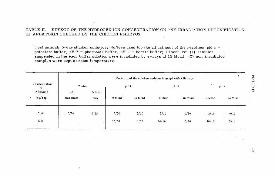
T A B L E II. E F F E C T OF THE HYDROGEN IO N C O N C E N T R A TIO N ON THE IR R A D IA T IO N D E T O X IF IC A T IO N OF A F L A T O X IN CH E CKED B Y THE CH ICKE N EM BRYO S
T es t anim al: 5-day chicken em bryos; B u ffe rs used fo r the adjustment o f the reaction : pH 4 — phthalate bu ffe r, pH 7 — phosphate bu ffer, pH 9 — bora te bu ffer; P roced u re : (1 ) sam ples suspended in the each bu ffer solution w ere irra d ia ted by 7 - ra y s at 15 M rad , (2 ) n on -irrad ia ted sam p les w ere kept at room tem peratu re.
Concentrationof
Aflatoxin
Control
No Saline
M ortality of the chicken embryos in jected with Aflatoxin
pH 4 pH 7 pH 9
PL-199/17
(Mg/egg) treatm ent only 0 Mrad 15 Mrad 0 Mrad 15 Mrad 0 Mrad 15 Mrad
1.0 0/10 0/15 7/10 3/10 2/10 0/10 . 3/10 0/10
5.0 10/10 6/10 10/10 0/10 10/10 2/10
o>CO

64 MIYAKI e t a l .
Aflatoxin was quite resistant to irradiation (Table I). Aflatoxin was, however, decomposed in some inorganic salt solutions and this tendency accelerated by the irradiation treatment.
B io log ica l a s s a y of i r r a d i a t e d A fla tox in (T a b le s I and II)
FIG.5. Fluorescence spectra of Aflatoxin irradiated with gamma rays; Sample: Aflatoxin mixture in MeOH; Excitation wavelength: 365 nm; Fluorospectrophotometer: SHIMAZU GF-16 E.
A C K N O W L E D G E M E N T S
This work has been perform ed under a Research Agreem ent with the International Atom ic Energy Agency entitled Radiosensitivity of, and radiation-induced changes in botulinus toxins and mycotoxins.
R E F E R E N C E S
[1] WEBB, B .D ., THIERS, H .D ., RICHARDSON, L .R ., Appl. Microbiol. 7 (1959) 329-33. t2] ADYE, I . , MATELES, R .I ., Biochim. biophys. acta 86 (1964) 418 .

X-RAY INACTIVATION OF FOOT-AND-MOUTH DISEASE VIRUS AND VESICULAR STOMATITIS VIRUS IN AQUEOUS MEDIA
C .D . JOHNSONTHE J. J. THOMPSON PHYSICAL LABORATORY,
THE UNIVERSITY OF READING,READING, BERKS., UNITED KINGDOM
Abstract
X-RAY INACTIVATION OF FOOT-AND-MOUTH DISEASE VIRUS AND VESICULAR STOMATITIS VIRUS IN AQUEOUS MEDIA. Radiation induced modifications to the biological activities and morphology of viruses are very dependent upon the techniques used in irradiation. Inactivation of foot-and-mouth disease virus and vesicular stomatitis virus by the effects of X-irradiation in dilute aqueous solutions has been studied and the post-irradiation effects and virus sensitization effects are described. Comparisons are made between X-radiation and hydrogen peroxide as virus inactivating agents. Survival curves resulting from these modes of inactivation give information on the reaction kinetics and on the function of the virus protein. Virus morphology, as detected in electron micrographs, is more readily changed by irradiation in solution (indirect effect) than by irradiation of the virus in the frozen state (direct effect).In each case the observed physical changes are related to loss of infectivity and the results are shown to be consistent with the inactivation processes previously inferred.
1. INDIRECT EFFECTS IN D ILUTE AQUEOUS SOLUTIONS
Under certain conditions, exponential survival curves result from the inactivation o f virus by radiation-induced diffusible agents in aqueous solutions. These conditions, in particular that a molecule after inactivation reacts as read ily as it did before inactivation, are often satisfied fo r the inactivation of b iological macromolecules by the short-lived agents in irradiated water and aqueous solutions. Figure 1 shows that the viruses o f foot-and-mouth disease and vesicu lar stomatitis are inactivated exponentially when irradiated in aqueous solutions of low non-viral protein concentration.
The reaction yield in irradiated solutions, in this case the number of virus particle inactivating events per unit dose, is often found to be independent of solute concentration, provided that this is sufficiently great for recombination of the radicals to be negligible. This has the effect of causing the inactivation of virus in aqueous solution (measured as the fraction surviving a particular dose) to be ve ry dependent upon the concentration of virus particles and radical scavengers in solution.
A common finding for the indirect effects of radiation on m acromolecules is that, after allowing for the presence of non-viral radical scavengers and for some recombination, there are many reactions between virus and free radicals for every one which causes an inactivation [1].
In the course of irradiation experiments, at various concentrations, it became evident that, in dilute solutions, virus continued to be inactivated after the termination of the irradiation. The results of an experiment
65

66 JOHNSON
Dose ; (R x 105)
F1G.1. Inactivation of foot-and-mouth disease virus (FMDV) and vesicular stomatitis virus (VSV) by X-irradiation in aqueous buffer solutions of low non-viral protein concentration.
in which foot-and-mouth disease virus was diluted 1/300 in phosphate buffer and irradiated for 30 min at 1200 R/min are shown in Fig. 2. Sample were titrated in m ice at the end of the irradiation and at 30 min intervals thereafter. The resulting survival curve shows that the inactivation proceeded at a gradually reducing rate for at least 2\ hours after irra d iation. A s im ilar post-irradiation inactivation effect was reported by A lper in 1954 [2]. She showed that dilute suspensions of bacteriophage were inactivated by p re-irrad iated buffer solutions and also that phage, irradiated but still infective, was more susceptible than non-irradiated m ateria l to this form of inactivation. The post-irradiation inactivation seen in Fig. 2. was subsequently shown to be the combined effect of inactivation by agents form ed in the irradiated solution and radiation sensitization of the virus to the action o f these agents.
Inactivation in pre-irrad iated buffer solution.
Suspensions of foot-and-mouth disease virus and vesicu lar stomatitis virus are readily inactivated by pre-irrad iated buffer solutions provided that the concentration o f v irus and o f non-viral protein or other protective agents is sufficiently low. The same is true of the inactivation of these viruses by hydrogen peroxide.
Pre-irrad ia ted samples of phosphate buffer, phosphate-buffered saline and 0.1M ammonium bicarbonate were capable of inactivating virus to about the same extent. A lso, when trip le-d istilled , aerated water was irradiated and then added to 0.4M phosphate buffer solution

P L -1 9 9 /4 67
T I M E AFTER START OF I R R A D I A T I O N ( h )
FIG.2. Continuing inactivation of foot-and-mouth disease virus, irradiated at a dilution of 1/300 in phosphate buffer, for up to 2¿ h after the end of the irradiation.
so as to produce a final concentration of 0.04M at pH 7.6, this buffer inactivated vir.us in a sim ilar manner. Typical survival curves are shown in Fig. 3. A ll such curves are characterized by a slope which decreased continuously with increasing reaction time when plotted on a sem i- logarithmic scale. Over the reaction times of about 3 h. normally employed in these experiments the viruses were quite stable in non- irradiated buffer solutions. S im ilar inactivation curves to those illustrated in Fig. 3 were observed when hydrogen peroxide was used as the inactivating agent.
The sensitivity of these inactivating processes to changes in virus concentration is demonstrated in Fig. 4. The overa ll inactivation of diluted virus suspensions, reacted for a fixed time with identical samples of p re-irrad iated buffer solution, is related to the virus concentration in the reaction m ixtures. A s im ilar strong dependence upon virus concentration was shown when the inactivating agent was hydrogen peroxide. In the latter case the effect o f varying the hydrogen peroxide concentration is also illustrated.
Protection against the post-irradiation effects
The presence of peptone, glutathione or thiourea decreased the rate of virus inactivation in pre-irrad iated buffer solutions and in hydrogen peroxide. At peptone concentrations in excess of 1 g/ litre no significant
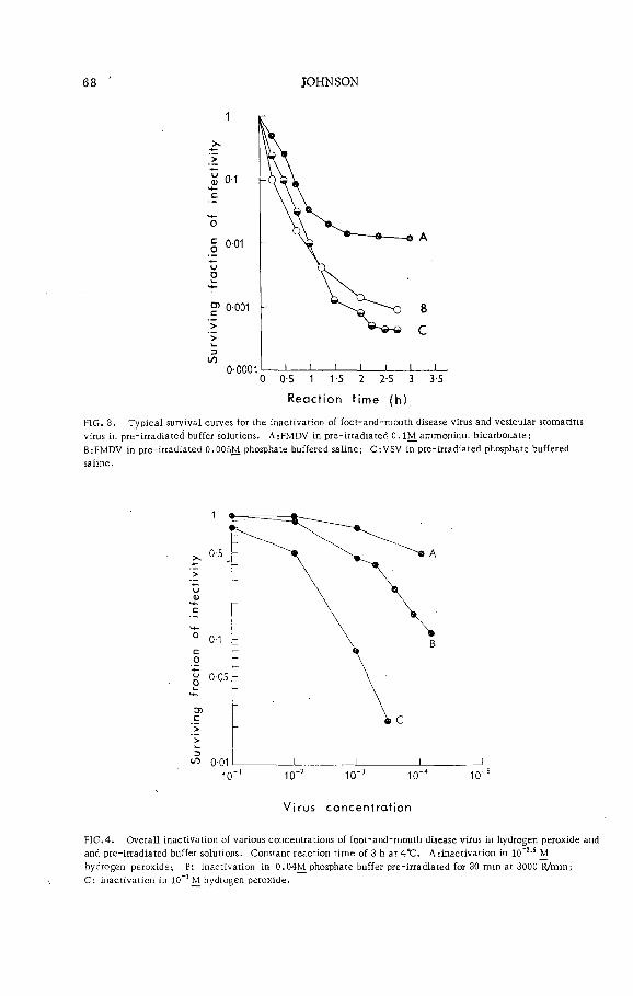
68 JOHNSON
FIG. 3. Typical survival curves for the inactivation of foot-and-mouth disease virus and vesicular stomatitis virus in pre-irradiated buffer solutions. AjFMDV in pre-irradiated 0.1M ammonium bicarbonate;B:FMDV in pre-irradiated 0.005M phosphate buffered saline; C:VSV in pre-irradiated phosphate buffered saline.
Virus concentrat ion
FIG.4. Overall inactivation of various concentrations of foot-and-mouth disease virus in hydrogen peroxide and and pre-irradiated buffer solutions. Constant reaction time of 3 h at 4 “C. A rinactivation in 10~3,5 M hydrogen peroxide-, B: inactivation in 0.04M phosphate buffer pre-irradiated for 30 min at 3000 R/min;C: inactivation in 10"3 M hydrogen peroxide.

P L -1 9 9 /4 69
inactivation was observed. Thus a convenient method of studying reaction rates was to take samples from a reaction mixture at appropriate times and to stop the reaction in these samples by the addition of an equal volume of peptone at a concentration of 100 g/litre. The whole set of samples could then be titrated for in fectivity at the term ination of the reaction.
Rates of inactivation
Apart from the dependence upon concentration of reactants, the rate of inactivation in pre-irrad iated buffer solution was found to increase with the temperature of the reaction mixture and with the quantity of air or oxygen dissolved in the buffer solution at the time of irradiation. A ll data presented here are for reactions at 4°C with air-saturated buffer solutions. No inactivation was observed when buffer solution irradiated in the frozen state was thawed and mixed with virus at 4°C.
Virus sensitization effect
As observed by A lper for bacteriophage, the X-radiation of aqueous suspensions of virus rendered the virus more susceptible to subsequent inactivation by pre-irrad iated buffer solution and to the action of hydrogen peroxide. A lso pre-treatm ent of virus suspensions with hydrogen peroxide rendered them more sensitive to inactivation by pre-irrad iated buffer. These effects were demonstrated in a set of experiments which have been previously reported [3].
Reaction kinetics
It was important, in connection with the reaction kinetics of these inactivation processes, to know if the inactivating agent was in excess throughout the reaction. It is likely that the pronounced curvature of some of the survival curves (e. g. curve A of Fig. 3) was partly due to a decrease in the concentration of the inactivating agent (which is assumed to be formed hydrogen peroxide) as the reaction proceeded. However, fo r other reaction m ixtures, particu larly those employing 10_3M hydrogen peroxide, the hydrogen peroxide concentration remained constant for the period of the reaction.
To test this point two sets of reaction mixtures were prepared, one consisted of foot-and-mouth disease virus (from BHK ce lls ) at dilutions from 10_I in ?-log steps to 103-5 in phosphate buffer containing 10~3M hydrogen peroxide; the second was a s im ilar set of m ixtures of 10'3M hydrogen peroxide with |-log dilutions of 5 wt. /vol.(%) of bovine plasma albumin. A fter 4 h at 4°C the amounts of hydrogen peroxide remaining in the individual reaction mixtures were determined and compared with a control of 10"3M hydrogen peroxide in phosphate buffer. In the presence of foot-and-mouth disease virus there was a significant reduction in the concentration of hydrogen peroxide- in those samples which contained virus at a concentration of 10-2 or more. This is thirty times the concentration normally employed in the experiments described previously. Hydrogen peroxide was also found to be stable in a ll except the highest concentrations of bovine plasma albumin.
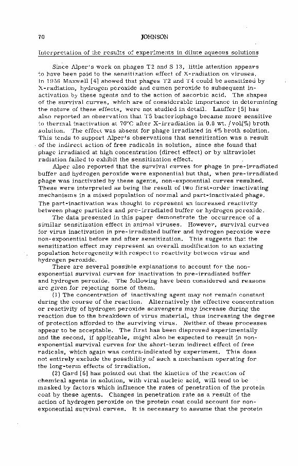
70 JOHNSON
Interpretation of the results of experiments in dilute aqueous solutions
Since A lper 's work on phages T2 and S 13, little attention appears to have been paid to the sensitization effect of X-radiation on viruses.In 1956 Maxwell [4] showed that phages T2 and T4 could be sensitized by X-radiation, hydrogen peroxide and cumen peroxide to subsequent inactivation by these agents and to the action of ascorbic acid. The shapes of the survival curves, which are of considerable importance in determining the nature of these effects, were not studied in detail. Lauffer [5] has also reported an observation that T5 bacteriophage became more sensitive to thermal inactivation at 70°C after X -irrad iation in 0.8 wt. /vol(%) broth solution. The effect was absent for phage irradiated in 4% broth solution. This tends to support A lper 's observations that sensitization was a result of the indirect action of free radicals in solution, since she found that phage irradiated at high concentration (direct effect) or by ultraviolet radiation failed to exhibit the sensitization effect.
A lper also reported that the survival curves for phage in pre-irrad iated buffer and hydrogen peroxide were exponential but that, when pre-irrad iated phage was inactivated by these agents, non-exponential curves resulted. These were interpreted as being the result of two firs t-o rd er inactivating mechanisms in a mixed population of normal and part-inactivated phage.The' part-inactivation was thought to represent an increased reactiv ity between phage particles and pre-irrad iated buffer or hydrogen peroxide.
The data presented in this paper demonstrate the occurrence of a s im ilar sensitization effect in animal viruses. However, survival curves for virus inactivation in pre-irrad iated buffer and hydrogen peroxide were non-exponential before and after sensitization. This suggests that the sensitization effect may represent an overa ll modification to an existing population heterogeneity with respect to reactiv ity between virus and hydrogen peroxide.
There are severa l possible explanations to account for the nonexponential survival curves for inactivation in pre-irrad iated buffer and hydrogen peroxide. The following have been considered and reasons are given for rejecting some of them.
(1) The concentration of inactivating agent may not remain constant during the course of the reaction. A lternatively the effective concentration or reactiv ity of hydrogen peroxide scavengers may increase during the reaction due to the breakdown of virus m aterial, thus increasing the degree of protection afforded to the surviving virus. Neither, of these processes appear to be acceptable. The firs t has been disproved experimentallyand the second, if applicable, might also be expected to result in nonexponential survival curves for the short-term indirect effect of free radicals, which again was contra-indicated by experiment. This does not en tirely exclude the possib ility of such a mechanism operating for the long-term effects of irradiation.
(2) Gard [6] has pointed out that the kinetics of the reaction of chem ical agents in solution, with v ira l nucleic acid, w ill tend to be masked by factors which influence the rates o f penetration of the protein coat by these agents. Changes in penetration rate as a result of the action of hydrogen peroxide on the protein coat could account for nonexponential survival curves. It is necessary to assume that the protein
PL -Í99/4 71
coat becomes less permeable to the hydrogen peroxide during the reaction. Survival curves of the type shown in Fig. 3 would'then result.
This is an attractive hypothesis since virus sensitization may be interpreted as a modification to the protein coat in the reverse manner (i. e. so as to increase the perm eability of the protein coat to hydrogen peroxide). That such an effect could be attributed to the action o f free radicals seems ve ry reasonable since, as w ill be seen later, these agents rapidly break up the virus protein in the in itial stages of virus degradation.
The observations in this work, and previously by Maxwell, that hydrogen peroxide can itse lf cause sensitization does not appear to fit this hypothesis. From the shape of the survival curves it would appear that pre-treatm ent of virus with hydrogen peroxide should reduce and not, as observed, increase the subsequent reactiv ity between the treated virus and hydrogen peroxide. The fact that the treatment which caused sensitization was carried out in re la tive ly concentrated virus suspensions, whereas the subsequent inactivation was observed in dilute suspensions, may have some bearing on this apparent anomaly.
(3) The third inactivation mechanism which may be considered is that proposed by Salk and Gori [7] to account fo r the kinetics of fo r maldehyde inactivation. They have shown that survival curves of the type shown in Fig. 3 may be due to an inherent heterogeneity in the virus population with respect to reactiv ity between virus and the chemical inactivating agent.
If the possibility of a heterogeneous population is admitted, then a further explanation of the shapes of the survival curves is that inactivation is in part a multi-step process. A virus sample of which the m ajority is inactivated by a single-step reaction and the rest by a cumulative multi-step reaction can result in overa ll survival curves of the required shape. Such heterogeneity in the virus population could represent variation between individual virus particles or may m erely indicate the presence o f clumps of infective particles behaving as s in g le in fect ious units.
A more plausible hypothesis is one in which clumps o f virus particles exist, but with only a single infectious particle present, per clump. The non-infective particles would serve to protect the infective one against chemical inactivation.
The virus sensitization effect may be incorporated into the general hypothesis of a heterogeneous population by postulating that: the actionsof free radicals in irradiated virus suspension have the effect of modifying the population distribution so as to increase the overa ll rate of inactivation when the virus is subsequently exposed to hydrogen peroxide or pre-irrad iated buffer.
Damage to virus protein
In the foregoing discussion it has been assumed that virus inactivation is a manifestation of some fundamental damage to the v ira l nucleic acid core. This is consistent with current belie fs that the nucleic acid is the essential infectious component of most viruses and that it retains its in fectivity in the absence of its protein coat. Nevertheless, damage to the surface protein must occur during exposure of virus to the indirect

72 JOHNSON
action of X-radiation. This may be expected to produce observable changes in virus properties, fo r example, in the specific receptor mechanism by which the virus is thought to enter the host cell. It is probable that the virus sensitization effect is a consequence of damage to the virus protein. In the next section it w ill be shown that, in a virus sample which had lost about half of its in itial in fectivity by the action of free radicals in solution (as was the case for virus exhibiting the sensitization effect), the surviving particles must have suffered considerable damage to their protein coats.
2. X -RAD IATIO N DEGRADATION OF FOOT-AND-M OUTHDISEASE VIRUS OBSERVED AS LOSS OF CHARACTERISTICPARTICLES IN ELECTRON MICROGRAPHS
Therm al inactivation of foot-and-mouth disease virus at 56°C has been shown to result in degradation of. the 25-nm infective particle into 7-nm sub-units, and ribonucleic acid which is destroyed by environmental ribonuclease [8]. The net effect of these gross changes in morphology is observed in electron micrographs as a reduction in the concentration of characteristic 25-nm particles. The 7-nm degradation products are too sm all tp be resolved from other detail in the specimen support film s.
In this work quantitative measurements of the concentrations of characteristic particles in irradiated samples of foot-and-mouth disease virus, type O, strain M. 11, have been made to study the radiation damage to the virus particles during and after the loss of infectivity. Because of the need to use high concentrations of virus to observe adequate numbers of particles in the sprayed droplets used to determine the particle concentrations, it was only possible to apply this technique to undiluted material. The effects of pre-irrad iated buffer solution were normally observed at virus concentrations which were too low to perm it examination by electron m icroscopy. The work was therefore lim ited to a comparison between the direct effect of X-radiation in the frozen state and the action of free radicals in aqueous solutions of re lative ly high virus concentration (1010 plaque form ing units per m illilitre ).
Virus was grown on ro lle r cultures of baby hamster kidney ce lls and purified by ultracentrifugation. Suspensions were made in 0.1M ammonium bicarbonate at pH 7.6. C larified bovine plasma albumin was added to a ll samples before spraying and in those samples irradiated in solution it was included before the irradiation. This served to swamp protection effects due to extraneous protein in the virus samples, thus making the irradiations in solution more reproducible. It was also required to improve the contrast of the droplets formed in the spray technique. Despite the scavenging of free radicals by the plasma albumin, at the concentration used (0.1 wt./vol.(% )) the virus inactivation in solution was almost entirely due to the indirect effect.
A fter irradiation, samples were mixed with 88-nm polystyrene latex particles and sprayed to form droplet patterns.
D irect effect
The results of an experiment in which foot-and-mouth disease virus was irradiated in the frozen state are given in Fig. 5. The virus/latex

PL-199/4 73
inz>LO
+
0 2 4 . 6 8
D O S E , (Rx 105)
FIG. 5. Surviving fraction of characteristic particles (points О & в) and of infectivity (points +) for foot- and-mouth disease virus irradiated at -60“C (direct effect).
ratios (for a constant latex concentration) are shown for samples irra d iated at three different doses. Three control samples (30 droplets) and two samples for each irradiation dose (20 droplets) were used.
Figure 5 demonstrates that there was no significant loss in relative number of characteristic virus particles in the irradiated samples. In fectivity lost by virus irradiated under identical conditions is shown in the same figure.
Indirect effect
Several semi-quantitative experiments were perform ed in which it was observed that the indirect effect of X-irradiation in aqueous solutions considerably reduced the numbers of characteristic particles in electron m icrographs. Specimens of irradiated virus samples were prepared for the electron m icroscope by allowing volumes of about 0.005 ml of virus suspension to dry down onto support grids covered by collodian substrates. Estimates o f the numbers of characteristic, particles in the sample were obtained by scanning large areas of the specimen at a magnification of 20 000 X. Although by no means as satisfactory as the spray droplet counting technique, this method could be applied to solutions containing too few virus particles to provide adequate particle counts in droplet residues of about 12-/um diameter, which were the largest that could be conveniently employed in the spraying technique. (This lim itation was due to the fact that it was not convenient to photograph la rger drops and yet maintain sufficient magnification to be able to c learly distinguish the 25-nm virus particles. ) In this manner it was possible to observe losses of characteristic particles over a concentration range of about 10s.

74 ÎOHNSON
These experiments indicated that the loss of particles followed a multi-event type of degradation mechanism. As with the indirect effect of X-radiation on infectivity, little is gained by interpreting the survival curves in term s of the target theory. Hence no distinction is made between a multi-hit or a m ulti-target model. The m ajority of the events corresponded to reactions between virus particles and diffusible agents in solution. The sensitivity of the virus to this form of degradation was naturally very dependant upon the concentration of protective agents in solution.
Two quantitative measurements o f the losses o f characteristic particles in solutions of 1 wt./vol.(% ) bovine plasma albumin in 0.1M ammonium bicarbonate were perform ed using the spray counting technique. Figure 6 shows the results of one of these experiments. The ea rlie r results were confirmed and the curve relating loss of particles to rad iation dose was typical of a multi-event mechanism. Comparison between this curve and that for in fectivity loss shows that degradation of the virus particle occurred quite soon after loss of infectivity.
K-U<
О
>>cuZ)</>
Dose, (R x 105)
FIG. 6. Surviving fraction of characteristic particles (points • ) and infectivity (points O) for foot-and-raouth disease virus irradiated in aqueous solution of 0.1% bovine plasma albumin in 0.1M ammonium bicarbonate (indirect effect).
The results illustrated in F igs 5 and 6 emphasize the d issim ilarities between direct and indirect actions of X-radiation. The differences in re lative sensitivities of the virus to inactivation and degradation in the
PL-199/4 75
two cases are consistent with the be lie f that inactivation, as measured in these experiments, was the result of damage to the v ira l nucleic acid.
In the frozen state the virus would be expected to sustain many inactivating events (ion clusters within the v irus) before sufficient damage had been done to disrupt the entire virus. Whereas, irradiation in aqueous solution presumably causes considerable damage to the virus protein as a result of the frequent surface reactions of the type which do not produce inactivation. This is reflected in the rapid degradation relative to in fectivity loss which results from the less frequent reactions between radiation products in solution and the nucleic acid core.
R E F E R E N C E S
[1] ALLEN, A. O ., Radiation Chemistry of Water and Aqueous Solutions, D. Van Nostrand C o., Toronto (1961).
[2] ALPER, gen. Microbiol. 11 (1954) 313-24.[3] JOHNSON, C .D ., J. gen. Microbiol. 38 (1965) 9-19.[4] MAXWELL, M .D ., Comptes r. 243 (1956) 717-19.[5] LAUFFER, M. A ., Ann. N. Y. Acad. Sci. 83 (1960) 727-40.[6] GARD, S ., Ann. N .Y. Acad. Sci. (1960) 638-48.[7] SALK, J. E ., GORI, J. B., (1960) 609-37.[8] BROWN, F . , Off. int. Epizoot., rep. 167 (1962).


GAMMA RADIATION FOR STERILIZING THE CARCASSES OF FOOT-AND-MOUTH DISEASE VIRUS INFECTED ANIMALS
B. BALDELLI ISTITUTO ZOOPROFILATTICO SPERIMENTALE DELL'UMBRIA E DELLE MARCHE,PERUGIA, ITALY
Abstract
GAMMA RADIATION FOR STERILIZING THE CARCASSES OF FOOT-AND-MOUTH DISEASE VIRUS INFECTED ANIMALS. Experiments on sterilization by means of gamma rays of the carcasses of animals experimentally infected with foot-and-mouth disease virus (FMDV) have been carried out. In the first part the author studied the presence and survival of FMDV in the carcasses and in the organs of infected slaughtered animals. The results obtained are sufficient to underline the problem of the sterilization of carcasses of animals infected by FMDV. The experiments on the inactivation of the FMDV by gamma irradiation in vitro showed the same radiation sensitivity of the three types, O, A and C, of FMDVinaqueous solutions and showed that the fraction of surviving virus is an exponential function of the gamma-ray dose. The results obtained confirm the remarkable resistance of viruses to the effect'of radiation. As far as the dry virus is concerned special tests indicated the necessity of greater doses for inactivating the same virus in the dry as opposed to the liquid state. In the third part the author studied the possibility of utilizing gamma rays for the sterilization of carcasses of infected (or suspected of being infected)FMDV animals using some tissues of infected animals (pigs) (blood, bone marrow, vertebrae, lymph nodes). The results obtained show that the inactivation of FMDV types O, A and С in the carcasses of infected animals can be made by treatment with gamma rays.
INTRODUCTION
This paper presents data on gam m a-ray sterilization of carcasses of animals experimentally infected with foot-and-mouth disease virus (FM DV). However, as the argument is stric tly connected with the problem of the presence and survival of FMDV in the carcasses of slaughtered animals and with that o f the effects of gamma rays on FMDV, problems which have also been experimentally studied by us, this paper has been divided into three parts: (1) the presence and survival of FMDV in the carcasses and organs of infected slaughtered animals;(2) the results of experiments of inactivation of FMDV by gamma radiation in vitro ; and (3) the employment of gamma rays fo r the sterilization of the carcasses of infected animals.
1. PRESENCE AND SURVIVAL OF FMDV IN THE CARCASSES AND ORGANS OF INFECTED ANIM ALS
The danger of the presence and survival of FMDV in the carcasses and organs of slaughtered animals is a very serious problem, stric tly connected with the control of foot-and-mouth disease and it has acquired new importance because of the recent increase in the trade of meat from various countries in A frica , where exotic types of FMDV have been recovered, to the Near East and Europe.
77
T A B L E I. S U R V IV A L O F F M D V IN TISSUES AND ORGANS F R O M C A R C A S S E S O F E X P E R I M E N T A L L YI N F E C T E D C A T T L E K E P T A T D I F F E R E N T CONDITIO NS O F P R E S E R V A T I O N
<1CO
M aterial At slaughter48 h
Chilled
8 d 60 d
Frozen
120 d 210 d
Blood + + + + + +
Kidney + + - + + +
Spleen + + - + + -
Liver + - - + + +
Brain - - - - - -
Lung + + + + + +
Bone marrow + + + + + +
Lymph nodes + + + + + +
Heart - - - -
Stomach + - - - - -
Rumen + + + + + +
Intestine + - - + + +
Tongue + + + + . + +
Muscle + - - - • -
Parotis + + + + + +
Testicle + - - + + + .
Uterus + + + + + +
+ Recovery of FMDV - Negative finding
BA
I.DELLI
T A B L E II . S U R V IV A L O F F M D V IN TISSU ES AND O RG A NS F R O M C A R C A S S E S O F E X P E R I M E N T A L L YI N F E C T E D P IG S K E P T A T D I F F E R E N T C O N D ITIO N S O F P R E S E R V A T I O N
M aterial At slaughter24 h
Chilled
10 d 70 d 90 d
Frozen
120 d 144 d 210 d
Blood + + + + + + + +
Kidney + + - + NE NE + +
Spleen + + - + NE NE + +
Liver + + - + NE NE + +
Brain + + + + NE NE + +
Lung + + + + NE NE + +
Bone marrow + + + - + + + + +
Lymph nodes + + + . + + + + +
Heart - - - - - - - -
Stomach - + + + + + + +
Intestine + + + + NE NE + +
Tongue + + + + NE NE + +
Muscle + + - + - - - -
Bacon + + ' - + + + + +
Parotis + + + + + + + +
+ Recovery of FMDV - Negative finding NE Not exam ined

80 BALDELLI
The literature on this problem provides sufficient data and observations: those o f the British Committee [1-3] , Henderson and Brooksby [4] , Wittman [5, 6] and Cottral et al. [7, 8] are fundamental. Intimately related with the research of these authors is that carried out recently in the Istituto Zooprofilattico Sperimentale dell'Um bria e delle Marche [9-14] which has led to some important conclusions on the presence and survival of FMDV in different tissues and organs of infected animals purposely slaughtered at a time considered to be near the peak of virus production and virem ia and kept in different conditions of re frigeration and deep freeze .
For the recovery of FMDV in the infected tissues and organs we adopted the tissue culture method utilizing ca lf kidney-cell monolayers which provide not only the same guarantee of valid ity as the b iological tests on naturally susceptible animals, but are more easily and econom ically carried out for this kind of study.
The results of our experiments (Tables I and II) can be summarized thus:
(a) FMDV is present in almost all organs and tissues of experimentally infected pigs and cattle slaughtered at the peak of disease;
(b) FMDV persists in many organs and tissues kept at normal re frigeration temperature, except for the muscle tissue in which the virus is often prematurely inactivated because of the influence of the changes o f the pH which take place in rigo r mortis of the animals; in those tissues — such as lymph nodes, bone marrow and blood — which do not develop the degree of acidity that is present in the muscle tissue, the survival of FMDV is longer and its concentration is la rger;
(c ) Under deep-frozen conditions FMDV persists fo r a long time in almost a ll the organs and tissues of infected animals and our experimental data show that FMDV persisted up to seven months after slaughter (seven months was the maximum period tested).
These results are in perfect accord with those of the AA, who have dealt with this argument, so that we have reached the conclusion that, because the usual operations of slaughter and preservation of meat do not guarantee the inactivation of FMDV, the trade of fresh, salted, cooled and frozen meat should be subjected to severe and restric tive measures which must prevent the occurrence and spread of FMD to the free countries and regions. These measures must also prevent the introduction of new types o f virus against which the vaccinated cattle in the infected countries have not been immunized. In this connection we agree with Brooksby [15] who a ffirm s that prohibition should be placed^ on the importation of meat from areas where the exotic types of virus occur.
Before concluding this argument it is useful to re fe r b r ie fly to some other investigations perform ed in 1962 [16] to control the in troduction o f FMDV into Italy through meat imported from abroad. This control has been carried out by v iro log ica l examination on ca lf kidneyce ll monolayers of samples o f bone marrow of vertebrae taken from carcasses o f slaughtered animals coming in at the frontier. S ixty-seven imported cattle carcasses w ere examined and the investigations proved positive fo r FMDV in 6 carcasses (8. 98%): 2 cases were frozen meat from cattle slaughtered 2 months prior to a rr iva l at the Italian frontier o f Pontebba and 4 cases w ere re frigera ted meat of cattle slaughtered

PL -199/3 81
2 days before a rr iva l at the Italian frontier o f Fortezza. These infected meats w ere from two different European countries. The 6 FM DV strains, checked also by b iological tests in susceptible cattle, were identified as FMDV type C.
The research carried out by us and the results obtained are sufficient to underline the problem of the sterilization o f carcasses o f animals infected by FMDV, and in view of the importance of this ■ we tried to obtain the sterilization of meat by means o f gamma radiation.
2. ■ RESULTS OF EXPERIM ENTS ON THE IN ACTIVATIO N OF FMDV BY GAMMA RADIATION in vitro
R ela tive ly few and scanty reports are available on the effects of gamma radiation on FMDV. For this reason, and to get some prelim inary data fo r the work on the employment o f gamma rays fo r the
F M D V irrodiated in aqueous state
FIG .l. Exponential survival curve of irradiated FMDV propagated in vitro.
sterilization of the carcasses of infected animals, some quantitative research on the inactivation by gamma rays of FMDV types O, A and С was undertaken, using a 'Hot Spot Mk IV ' 60Co source.
In all cases the virus materials used in these experiments were obtained from virus propagated in v itro on ca lf kidney-cell monolayers grown in Roux flasks containing 0. 5 lactalburçiin hydrolysate in Earle buffer solution. In the case of FMDV type C, the effects of radiation w ere studied also on the dry virus, obtained after lyophilization of the above-mentioned liquid.
Several samples of the above virus were prepared and irradiated at a constant dose-rate of 0. 25 Mrad fo r various tim es, whilê a sample of each virus was kept as an unexposed control. The infective titre of the various exposed and unexposed virus samples was estimated in v itro by inoculation of series of ten-fold dilutions into groups of 5 tubes of ca lf kidney-cell monolayers and expressed by the method o f Reed and Muench [17] in cytopathogenic doses (DCP5o/ml). A ll irradiated virus samples which by in v itro tests proved inactivated were also controlled by b iological tests in unweaned m ice.
The results o f these studies [18] indicated that a ll three types of FMDV in aqueous solutions showed the same radiation sensitivity, the

82 BALDELLI
inactivation curves being practically identical (F ig. 1), and showed that the fraction o f surviving virus is an exponential function of the gamma- ray dose, within the e rro r of the experiment. A ll three types of FMDV lose their infectiveness fo r ca lf kidney-cell monolayers and fo r unweanèd m ice after irradiation with 3 Mrad, i. e. after a very high gamma dose.The results again confirm the remarkable resistance of viruses in general, and of FMDV in particular, to the effect of radiation, considering that an inverse relation is known to exist between inactivation doses and the size o f the virus [19, 2 0 ] .
As regards the dry virus, the tests indicated that inactivation was effected by treatment with 4 Mrad, i .e . 1 Mrad more was .required above the dose for inactivating the same virus in the liquid state. This is in full agreement with the observations by Lea [21] that dry virus is inactivated by d irect effect of radiation alone, whereas in the case of virus irradiated in liquid state to the d irect effect must be added the indirect effect of free radicals and other radiation products in solution.
3. EM PLO YM ENT OF GAMMA RAYS FOR STERILIZING THECARCASSES OF INFECTED ANIM ALS
As a result o f the above-mentioned experiments, further research has been undertaken to study the possibility of utilizing gamma rays fo r the sterilization of carcasses of infected or suspected infected FMDV animals, in order to prevent fresh, chilled and deep frozen meat constituting a means o f spreading the disease through trade and transport.
For practical reasons, the investigations were carried out on tissues of experimentally infected animals and for this purpose we used samples o f blood, bone marrow from vertebrae and lymph nodes, tissues which, according to our research [ 2 2 ] and that of other authors, harbour the highest contents o f FMDV and where the survival o f the virus is longer.
M aterials and method
Four unexposed, unvaccinated and susceptible pigs, each weighing about 1 2 0 kg, were injected intraderm ally in the posterior right foot with FMDV: the firs t two received 5X 10⁶ DCP50 o f type О virus, the third 2 X 10⁶ DCP50 of type A virus, and the fourth 5 X 10⁶ DCP50
o f type С virus. A fte r 40-45 hours post inoculation, when the animals showed symptoms of FMD (high rise o f body temperature, typical generalized lesions, etc. ), they were sacrificed by total bleeding and the blood was collected, diluted 50% with A lsever liquid, distributed in equal quantities into glass holders and harvested at -30°C. The muscular, mesenteric and mediastinic lymph nodes and all the vertebrae w ere removed aseptically from the carcasses, collected into glass holders and harvested at -30°C, until treatment with gamma rays.Having chosen nine samples of blood, nine samples of lymph nodes and nine samples of vertebrae from each pig, and retaining one sample o f each m aterial as an untreated control, the remaining 8 samples o f each of the m aterials w ere treated with increasing doses o f gamma rays (from 0.25 to 2 Mrad).
PL -199 /3 83
Tests fo r FMDV w ere perform ed with the tissue culture method on ca lf kidney-cell monolayers on the treated and untreated m aterials. The blood samples w ere also tested fo r the in fective titre o f the virus to establish the concentration o f virus in the original untreated m aterial, and in the individual samples exposed to increasing doses of gamma rays. To this end, series of ten-fold dilutions o f blood w ere injected into groups o f 5 tubes o f ca lf kidney-cell monolayers and the infective titre determined by the method of Reed and Muench and expressed in cytopathogenic doses (DCP50 /ml).
As regards the lymph nodes and bone m arrow o f vertebrae in pig No. 1 infected with type О virus, a quantitative research of virus was effected in relation to the weight of the m aterial only on the untreated sample.
Results
The results are summarzied in Table III. From an examination o f the table, the follow ing observations can be made.
•
(a) In pig No. 1, infected with FMDV type O, the strongest resistance o f the virus to gamma rays was observed in the blood and bone marrow, where inactivation o f FMDV required 1.75 Mrad, while to inactivatethe virus in lymph nodes 1. 5 Mrad w ere necessary.
(b) In pig No. 2, also infected with FM DV type O, low er doses of radiation appeared to inactivate the virus in the different tissues examined: 0. 75 Mrad w ere sufficient to destroy the in fectivity o f blood and bone m arrow, and 1.25 Mrad to inactivate the virus in the lymph nodes.
(c ) In pig No. 3, infected with FM DV type A , the in fectivity o f the blood was destroyed with 0. 75 Mrad,and that o f the bone marrow and lymph nodes with 0. 5 and Q. 25 Mrad respectively,
(d) In pig No. 4 , ’ infected with FM DV type C, the strongest resistance to the gamma rays was shown by the virus harboured in the blood, where inactivation required. 1. 75 Mrad,whereas the in fectivity of the lymph nodes and the bone marrow was destroyed by 1. 25 and 1 Mrad respectively.
The results obtained show that the inactivation of FMDV types O,A and С in the carcasses of infected animals can be effected by treatment with gamma rays which succeed in destroying the in fectivity of those tissues (blood, lymph nodes and bone m arrow) where the virus contents are the highest and the persistence of the virus is longer.
The variab ility in the dose o f radiation found necessary to inactivate FMDV harboured in the various experimental samples could be due either to the physico-chem ical and biological factors connected with the tissues harbouring the virus — factors which may give a protective or favourable action with regard to the effects of the radiation — or, more effectively, to the differing virus content o f the different tissues. This last hypothesis is supported by the fact that the highest doses of gamma rays were r e quired to destroy the virus in carcass No. 1, infected with FMDV type O, and in carcass No. 4, infected with FMDV type C, namely in m aterial from those pigs where the highest v ira l blood titres w ere ascertained. Another indication that the virus concentration in the tissues operates to require a changing gam ma-ray dose to obtain the inactivating effect is given by the results of virus titration of the various unexposed and
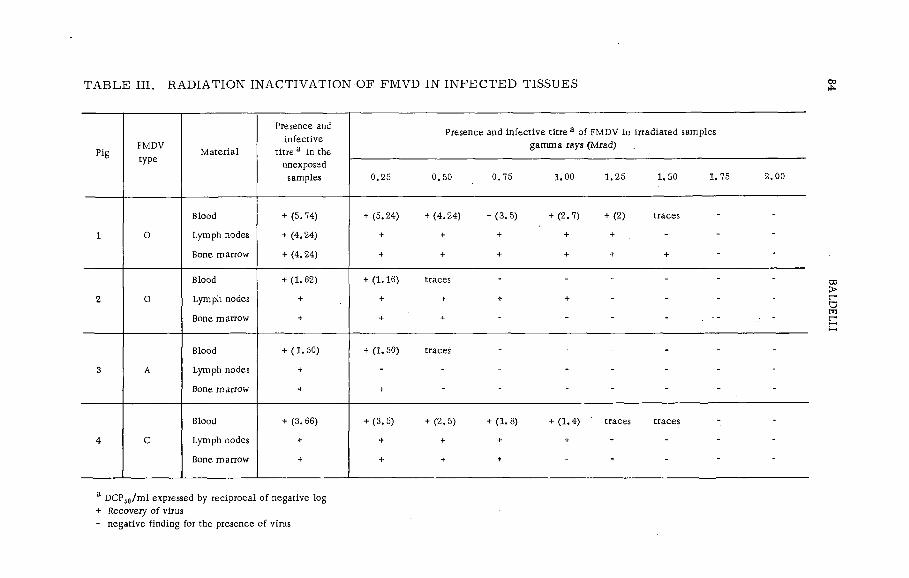
T A B L E III . R A D IA T IO N IN A C T IV A T IO N O F F M V D IN I N F E C T E D TIS S U E S
PigFMDVtype
M aterial
Presence and infective
titre a in the unexposed
samples
Presence and in fective titre a of FMDV in irradiated samples gam m a rays (Mrad)
.0 .25 0 .50 0 .75 1.00 1 .25 1 .50 1. 75 2 .00
1 0
Blood
Lymph nodes
Bone marrow
+ (5.74)
+ (4.24)
+ (4.24)
+ (5.24) + (4 .2 4 ) + (3 .5 ) + (2 .7 ) + (2 ) traces
+ + + + + . -
+ + + + + + ~
2 0
Blood
Lymph nodes
Bone marrow
+ (1.62)
+
+
+ (1.16) traces - -
+ + + + - - - -
+ + - - -
3 A
Blood
Lymph nodes
Bone marrow
+ (1 .50)
+
+
+ (1.50) traces - -
4 С
Blood
Lymph nodes
Bone marrow
+ (3.66)
+
+
+ (3.5) + (2 .5 ) + (1 .8 ) + (1 .4 ) ' traces traces
* + + + - - - -
+ + t -
a DCPs0/m l expressed by reciprocal of negative log + Recovery of virus- negative finding for the presence of virus

PL -199/3 85
exposed samples of blood of a ll the pigs. These results, which agree with those obtained in the experiments carried out with FMDV propagated in v itro , show that also in the case o f FMDV harboured by the blood of infected animals and exposed to gamma radiation, the fraction of surviving virus is an exponential function of the gamma ray dose.
With regard to the dose of gamma rays which can guarantee, within an adequate margin of safety, the sterilization of the carcasses of animals infected by FMDV, the above-mentioned experiments provide a basis for reasonable expectation that after treatment with 2 Mrad all the m aterial of every infected or suspected infected animal is free of infectious virus. This is slightly higher than the maximum dose ascertained in experiments which, though necessarily restricted to few animals, have been carried out purposely in particularly severe conditions on m aterial coming from animals experimentally infected and slaughtered at a time considered to be near the peak of virus, production. These conditions are unlikely to occur in practice with animals naturally infected with FMD, and they very seldom occur with experimentally infected animals (in none o f the many pigs examined in our ea rlie r research did we find such high blood infective titre as in pig No. 1).
CONCLUSIONS
Based on these results, the following conclusions may be drawn:(1) Until technical progress w ill make available some method
capable of s teriliz in g the carcasses of slaughtered animals, the trade of fresh, chilled and frozen meat must be subjected to severe and restric tive measures, as it can be a means of spreading FMDV with dangerous'consequences both fo r the free countries and for those countries which, though already infected, try to prevent the introduction of new types of FMDV able to cross the immunizing barrier which protects the national livestock.
(2) Gamma radiation could play a very important ro le in curing the carcasses o f slaughtered animals infected or suspected of being infected with FMDV as the gamma rays ensure the inactivation of FMDV harboured in those tissues and organs which o ffer the most favourable conditions fo r the survival of FMDV.
(3) Owing to the high resistance o f FMDV to gamma rays, shown both by FMDV propagated in v itro and by FMDV in the tissue o f experimentally infected animals, the radiation treatment of the carcasses must, however, demand high doses of gamma rays (at least 2 Mrad). Therefore, the possible application of the method depends on problems, above all of a biochemical order, which concern the nutritional value and the edib ility o f the meat which has received such a high irradiation. These last problems are not covered by the present investigation.
R E F E R E N C E S
[1] MINISTRY OF AGRICULTURE AND FISHERIES, Second Progress Report of the Foot-and-Mouth Disease Research Committee, Her Majesty's Stationery Office, London (1927) 1-417.
[2 ] MINISTRY OF AGRICULTURE AND FISHERIES, Third Progress Report of the Foot-and-Mouth Disease Research Committee, Her Majesty's Stationery Office, London (1928) 1-141.

86 BALDELLI
[3] MINISTRY OF AGRICULTURE AND FISHERIES, Fourth Progress Report of the Food-and-Mouth Disease Research Committee, Her Majesty's Stationery Office, London (1931) 1-375.
[4] HENDERSON, W .M ., BROOKSBY, J. В. , ]. Hyg. 46 (1948) 394.[5] WITTMAN, G ., Berl. u. Münch. tierarztl. Wschr., 70 (1957) 321-23.[6] WITTMAN, G ., Monatsh. f. Tierheilk. 9 (1957) 215-30.[7] COTTRAL, G .E ., COX. B .F ., BALDWIN, D .E ., Am. J. vet. Res. 21 (1960) 288.[8] COX, B .F ., COTTRAL, G .E ., BALDWIN, D .E ., Am. J. vet. Res. 22 (1961)224.[9] SA VI, P ., BALDELLI. B ., MOROZZI, A ., Atti Soc. Ital. Sci. Vet. 15 (1961) 734.
[10] SA VI, P ., BALDELLI. B .. MOROZZI, A ., Atti Soc. Ital. Sci. Vet. 15 (1961) 740.[11] SA VI, P ., BALDELLI. B .. MOROZZI, A ., Atti Soc. Ital. Sci. Vet. 15 (1961) 743.[1 2 ] SA VI, P ., BALDELLI, B .. MOROZZI, A ., FRESCURA. T ., Atti Soc. Ital. Sci. Vet. 15 (1961)
750.[13] SA VI, P ., BALDELLI. B ., MOROZZI, A .. Bull. Off. Int. Epiz. 57 (1962) 853.[14] SA VI, P ., BALDELLI, B .. Bull. Off. Int. Epiz. 57 (1962) 891.[15] BROOKSBY, J .B ., Bull. Off. Int. Epiz. 57 (1962) 847.[16] SA VI, P .. BALDELLI, B ., FRESCURA. T . , Atti Soc. Ital. Sci. Vet. 16 (1962) 475.[17] REED. L .. MUENCH, H .. Am. J. Hygiene 27 (1938) 493.[18] BALDELLI, B ., BEGLIOMINI. A .. FRESCURA, T . , MASSA, D ., Atti Soc. Ital. Microbiol. 12
(1963) 179.[19] DESROSIER, N .W ., ROSENSTOCK, H .M ., Radiation Technology in Food, Agriculture and Biology,
Avi Publishing Company, Westport (1960).[20] JORDAN, R .T ., KEMPE, L .L ., Proc. Soc. Exper. Biol. Med. 91 (1956) 212.[21] LEA, D .E ., Action of Radiations on Living Cells, 2nd e d . , University Press, Cambridge (1955).

THE EFFECTS OF ADDITIVES ON RADIATION-.RESISTANCE OF Cl. botulinum IN MEAT
A .W . ANDERSON, D. A. CORLETT, Jr.,AND K. L. KRABBENHOFT OREGON STATE UNIVERSITY,CORVALLIS, OREGON,UNITED STATES OF AMERICA
Abstract
THE EFFECTS OF ADDITIVES ON RADIATION-RESISTANCE OF Cl. Botulinum IN MEAT.Beef ground round inoculated with 1 million spores of Cl. botulinum per gram and containing varrious additives was exposed to gamma radiation. Mild heat treatment applied after irradiation reduced the survival of Q . 'botulinum, strain A-5 spores in raw ground beef. Heat applied before or during irradiation appears to have no effect. Sodium chloride, calcium chloride, mustard oil (allyl isothiocyanate), nutmeg, sodium citrate and Versene reduced the dose or irradiation required to inactivate Cl. botulinum spores (strains A-5 and B-115) in raw ground beef. Spores were inactivated in samples (irradiated at 2.0, 2.5 and 3.0 Mrad) which contained sodium nitrate (1000 ppm) plus sodium chloride (2.5%). Similar results were obtained when sodium nitrite (200 ppm) was substituted for sodium nitrate, except that there was evidence of spore survival in 1 of 120 cans irradiated at 2.0 Mrad. Spore destruction was based upon the absence of spores and mouse-lethal toxin in meat sub-cultures made from cans incubated at 35°C for 120 days. Spores were not destroyed when exposed to 2.5 or 3.0 Mrad in the absence of sodium nitrate, sodium nitrite or sodium chloride. Furthermore, the use of these chemicals individually together with radiation was ineffective. The additives alone in the absence of radiation also did not cause spore destruction. Radiation levels of 2.0, 2.5 and 3.0 Mrad, when used with sodium chloride at 1.5 or 2.0°/o and sodium nitrate at 500 ppm or sodium nitrite at 100 ppm, were ineffective.
INTRODUCTION
Cl. botulinum is one of the most radiation resistant food intoxication sporeform ers [1-3]. This fact has created a serious problem in the radiation sterilization of non-acid foods. It is the objective of food scientists to make radiation sterilized foods equal in safety, from the public health standpoint, that achieved for other accepted methods for com m ercia lly processed foods. At the radiation leve l now recommended fo r sterilization, serious defects in odour, flavour, colour, and texture occur which make the product unacceptable.
Recent investigations indicate that the radiation leve l required to destroy Cl. botulinum may be reduced appreciably by the use of common food additives, or to some degree by post-radiation heat treatment.Spices and chemicals of various kinds as w ell as m ild heat treatments are used in many types of specialty meat products. Chemical additives, such as sodium chloride, sodium nitrite, and sodium nitrate, have wide use as preservatives in meat [4]. Their effect on Cl. botulinum was demonstrated by Y esa ir and Cameron [5], who found a 70% reduction in spore counts brought about by the use of 0.1% sodium nitrate, 0.005% sodium nitrite, or 2% sodium chloride in a meat infusion agar. Bulman and Ayres [6] also noted that nitrite inhibited spore germination of a putrefactive anaerobe (P. A. 3679) in inoculated meat emulsions. These
87
88 ANDERSON e t a l.
chemical additives have been more recently suggested for canned meat and other foods [7].
The present study was undertaken to determine the effect of spices, heat and common chemical additives on the dose of gamma radiation required to destroy spores of Cl. botulinum inoculated into canned ground steak. Spores of type 33-A, 5A, and 115B were used, since recent studies [8] showed these to be among the most radiation-resistant.
M ATERIALS AND METHODS
Preparation of spores •
Original cultures of Cl. botulinum type A5, A33, and B115 were obtained from the National Canners Association, Washington, D. C. and the Quartermaster Food and Container Institute for the Armed Forces, Natick, Mass. The large number of spores necessary for these experiments were obtained by introducing a 0.5% inoculum, heat shocked for 10 minutes at 80°C [9], into fresh ly steamed trypticase- thioglycollate broth (trypticase, 5%; Bacto-peptone, 1.5%; glucose, 0.01%; sodium thioglycollate, 0.1%; pH 7.0). The inoculated flasks were then incubated at 37°C for 7-8 days, to allow maximum spore formation.
A fter the growth period spores were harvested by centrifugation, washed twice with M/15 phosphate buffer (pH 7.0), combined, and stored in phosphate buffer solution (titre 1 X 10⁸ spores/ml) at 4°C until needed. Before use, viable spore titres were checked by inoculating trypticase-thioglycollate broth with appropriate heat shocked dilutions of the spore suspension.
Radiation source
A ll cans were irradiated at the Gamma Test Facilities , A rco, Idaho. Dosim etry was checked every eight hours by eerie sulphate bottles or ion chamber dosimeters. In the case of the submersible water bath the dosim eters were located inside the cans at the top, bottom, and centre inside the inner chamber.
Meat packs
The meat was high quality (85% lean and 15% fat and fib re ) fresh ground round obtained from a loca lly operated, government inspected, meat packing plant. 1000-g portions were inoculated with heat-shocked spores to a concentration of approximately 106 spores per gram.100 g of inoculated meat was packed into each baby food can (202 X 204). The cans were sealed using an automatic can sealer, rinsed in 1000 ppm chlorinated detergent solution, packed in wet ice, and shipped to the Gamma Test Facilities where they were held at wet ice temperature until irradiated.

P L -199/10 89
Heat source
The concurrent heat treatment was provided by specially designed equipment which could be lowered into the irradiation field. Hot water, kept at a constant temperature (± 0.5°C) was provided from two 25-gallon reservo irs . The hot-water supply also served as the come-up tank and the water supply re servo ir for the temperature-controlled submersible bath. A pump was used to circulate the water through the submersible bath and back to the reservo ir.
The temperature in the submersible bath was determined experimentally by a thermocouple in the inner chamber. A temperature drop of approximately 5°C was recorded between the hot-water re servo ir and the tank when exposed under water at 20°C in the irradiation field.Control experiments were made prior to the actual irradiation tests under simulated conditions as described below.
The come-up times for the meat packs were determined on an automatic recorder by inserting thermocouples in the centre of cans [10] filled with meat. The cans were im m ersed in water 5°C higher than the desired temperature. It was observed experimentally that 35 minutes was the maximum time required for a can to reach the desired internal temperature irrespective of the final heat leve l.
The cans treated before irradiation were heated before being shipped to the irradiation source. Cans treated after irradiation were heated upon their return from the source. During transporting and/or storage the cans were held at wet-ice temperature.
Spice and chemical additives
Meat was weighed out in 1000-g quantities and the spices and/or chemicals added in the quantities indicated in the tables. These were usually added in amounts perm itted in foods under the existing regulation of the Food and Drug Administration [11]. The meat mixture was held at 4°C for 16 h to allow the additives to diffuse thoroughly throughout the meat.
The test sample was then inoculated with 10® heat-shocked spores per gram , and thoroughly mixed. 100 g of the test samples was packed into each baby food can (202X204), sealed, packed in insulated wet-ice containers, and shipped to the Gamma Test Facility, for irradiation.
Assay for spore survival
The irradiated samples returned to Oregon State University were incubated at 37°C and observed fo r a hard swell indicative of spoilage. Most spoilage, if it were to occur, would take place within a week to 10 days.
Those cans which did not spoil after a time lapse of 30 to 120 days were aseptically opened. F ive grams of meat were removed from the centre of the sample and from various parts of the can for a total of 15 g and placed into a screw-cap test tube. Seven m illim etres of freshly steamed trypticase-th ioglycollate broth was added per tube and incubated for a minimum of 48 h at 37°C.
The incubated sub-cultures were centrifuged at 800 X g for 20 min. The supernatant fluid was decanted o ff and divided into two portions.

90 ANDERSON e t a l.
One portion was transferred to a sterile culture tube (15 X 150 mm), and served as the active preparation; the remaining portion was transferred into another culture tube and boiled for 15 min to inactivate any toxin present, centrifuged, and used as a. control. Both sets of culture tubes were covered with rubber-hooded stoppers and taped shut.
0.2 m l of a sample was then injected into a 15-20 g white mouse intraperitoneally. Two m ice received the unboiled inoculum and one rece ived the boiled inoculum. Toxic ity of the preparation was characterized by the typical symptoms of botulinum toxin poisoning followed by death.The m ice were observed for toxin effects for a period of 3 days. Toxic symptoms were absent in the boiled control.
Wagenaar and Dack [12] and Bonventre and Kempe [13] observed that the amount of radiation needed to inactivate the toxin was considerably more than that needed fo r sterilization . Hence, if the toxin was present, it could be introduced with the original inoculum and could
T A B L E I. V IS IB LE S PO ILA G E AND MOUSE L E T H A L IT Y OF GROUND ROUND IN O C U LA TE D W ITH C l. botulinum (A -5 ),(106 spores/g)
Irradiationlevel(Mrad)
Timeheat
applied
Treatment temperature Г С )
■ 20 60 65 70 75 77.5 80 82.5 85 87.5
0.0 5a 5 5 5 5 5 5 5 5 5
before 5 5 5 5 5 4 5 2 5 2
2.0 during 5 5 _5 5 4 5 4 4 3 3after 5 ■ 5 5 5 3 5 1 5 1 1
before 1 4 1 1 0 0 5 0 3 2
2.5 during 1 5 1 2 0 2 2 1 0 2
after 1 1 0 1 0 0 1 1 0 1
before 0 0 1 1 2 1 5 0 5 13.0 during 0 0 1 1 0 1 0 0 0 0
after 0 0 0 0 1 0 0 0 0 1
Figures underlined indicate no visible spoilage due to can swelling; however Cl. botulinum was recovered and shown to be toxic.
a Five replications per sample (500 cans pet experiment).
P L -199/10 91
remain after radiation processing. Thus, to ascertain the presence of viable spores in the toxin-containing samples, heat-shocked subcultures were made from the 15-g meat samples. These sub-cultures w ere then examined m icroscopica lly for the presence of spores and toxin-producing ability.
RESULTS AND DISCUSSION
Results shown in Table I indicate that heat, if used in the radiation sterilization process, should be applied following, not before or during, the irradiation treatment. Heat applied before or during the irradiation actually increased the radiation resistance of the type A spores, especially in the temperature range 80-87.5°C. This effect might be explained in one or more of the following ways:
(a) Shrinkage and loss of water due to the effect of the heat on the meat substrate could reduce the diffusion of the lethal ions produced by the irradiation
(b) Fats rendered by the heat treatments might afford protection by coating the spore's surface simulating dehydration and thus increase their resistance to the irradiation.
(c) Active sulphur groups produced by heating the protein o f the meat may act as free radical acceptors affording further protection to the spores.
Again re ferr ing to Table I, it appears that heat treatments in a number of instances affected the spores in such a way that germination did not occur during incubation of the cans. However, upon opening the cans and sub-culturing the contents, spore germination and toxin production could be demonstrated. This phenomena was especially pronounced in the heating before and during irradiation and occasionally in the heat treatment following irradiation. Such a practice could create a potentially dangerous condition. Before heat treatments can be recommended for reducing radiation sterilization doses, further confirm atory experiments should be carried out.
Sodium chloride increased the lethal effect of radiation on the spores of Cl. botulinum. Experimental data shown in Table II indicate that the lethal action of sodium chloride was exerted most e ffective ly at a concentration of 3.0% at an irradiation leve l of 3.0 Mrad. Low er leve ls of
\sodium chloride had little or no demonstrable effect. Strain A-5 was less resistant than strain B-115 to the combined effects of salt and irradiation. Calcium chloride, at a concentration of 3.0%,was as effective as an equivalent concentration of sodium chloride in destroying the spores of strain B-115. The chloride ion may be the active agent in enhancing the lethal action of radiation since NaCl and CaC l2 at near equivalent concentrations-were equally effective.
A number of spices were investigated including nutmeg, sage, coriander, cinnamon, allspice, caraway, cardamon, mace, marjoram , ce lery, thyme, garlic , red pepper, mustard and cloves. Of these only musta.rd appeared synergistic with irradiation. Nutmeg, coriander and sage exhibited a slight effect.
Data in Table III show that mustard o il (a lly l isothiocyanate), at a concentration 0.1%, increased the lethal action of irradiation with the

CDЮ
T A B L E II. M O U SE L E T H A L IT Y O F IR R A D IA T E D G RO U ND ROUND (1 0 6 s p o r e s / g )
MradN o. o f
sam plesa
U ninocu latedcontro l
Inoculatedcontrol
salt
(b)
Sodium ch lor id e
No.
(a)
1%
(a) (b)
2%
(a) (b)
3%
(a) Cb)
4P¡0
(a) (b )
0.0 2 0 18 18 18 18 18 18 18 18 18 18
• 2.0 2 0 18 18 18 18 6 18 0 18 0 0
2.5 2 0 18 18 16 18 16 18 0 18 0 0
3.0 2 0 18 18 1 9 1 10 0 2 0 0
a N in e rep lica tions per sam ple, 792 cans per experim ent.
(a ) A -5 ; (b ) B-115 "
ANDERSON et
al,

PL-199/10 93
T A B L E III. M OUSE L E T H A L IT Y OF IN O C U L A T E D G R O UND R O U N D (106 SPO R ES/g)
Irradiation
le v e l
(M rad )
U ninocu lated
control
Inocu lated
control
(a ) (b )
Mustard
o . i%
(a ) (b )
Mustard
0 .1% +
1% NaC l
(b )
Mustard
0.1%
2% N aC l
(b )
Mustard
0.1%
3% N aC l
(b )
.. 0 .0 0 9 a 9 9 9 9 9 9
1 .5 0 9 9 9 9 9 9 9
2 .0 0 9 9 0 0 0 0 0
2 .5 0 9 9 0 0 0 0 0
3 .0 0 9 9 0 0 0 0 0
a1 sam ple, 9 rep lications (360 cans per exper im en t),
(a ) A -5 ; (b ) B-115
T A B L E IV . M O USE L E T H A L IT Y O F G R O U N D R O U N D P L U S SODIUM C IT R A T E OR V E R S E N E W ITH C l. botulinum (T Y P E B115), (106 SPO R ES/g)
L e v e l o f
irradiated
(M rad )
N o .o f
samples
Uninocu lated
control
inocu lated
control
Sodium c itra te
0 .01 0 .1 1 .0
% % %
Versene
(d i- Na)
salt
0.186
Verse n e-C A (fo od grade)
0.05% 0.5%
0.0 2 0 18 18 18 18 18 18 18
2 .5 2 0 18 18 18 9 0 18 18
3 .0 3 0 27 4 0 0 •o 0 1
Num ber o f cans from which spores o f C l . botulinum w ere recovered and on sub-culturing w ere
tox ic for m ic e .
9 rep lications per sam ple (504 cans per exp er im en t).
total inactivation of spores occurring at 2.0 M rad. Again, strain B-115 appeared more resistant than strain A -5 . Sodium chloride and mustard oil in combination was no more effective than mustard alone.
The results with the chelating agents, ethylene-diam ine-tetra-acetic acid (EDTA) and sodium citrate, are shown in Table IV. Concentrations of 0.186% Versene and 0.05% Versene CA, in inoculated samples irradiated at 3.0 Mrad, inactivated spores of Cl. botilinum. However, 0.5% Versene

94 ANDERSON et al.
CA appeared to enhance survival. A concentration of 0.1% sodium citrate was sufficient to inactivate spores at 3.0 M rad. It is possible that both EDTA and sodium citrate may induce by their chelating properties the pregermination state, a condition in which the spores are more sensitive to irradiation.
Sodium citrate also exhibited a desirable effect on the organoleptic qualities of raw irradiated beef. The texture was preserved and there appeared to be a marked reduction of the odours characteristic of irradiated meat.
TABLE V. SPOILAGE OF IRRADIATED CANNED GROUND ROUND STEAK INOCULATED WITH Cl. botulinum SPORES (33-A) PLUS SODIUM NITRATE OR SODIUM NITRITE PLUS SODIUM CHLORIDE
A d d itive
R ad ia tion
dose
(M rad )
T o ta l
cans
Swollen
cans3
Unswollen
cans
contain ing
toxin b
Unswollen
cans and
no
toxin
None 0 25 25 - -
2 .0 25 25 -
2 .5 25 . 23 0 2
3 .0 25 15 0 10
Sodium nitrate
(1000 ppm )
0 25 25 - ' -
plus 2 . Ef/o 2 .0 125 0 0 125sodium ch loride
2 .5 25 0 0 25
3 .0 25 0 0 25
Sodium n itrite
(200 ppm )
0 25 25 - -
plus 2 . ЭУо 2 .0 125 1 5 119sodium ch loride
2 .5 25 0 0 25
3 .0 25 0 0 25
Inoculum consisted o f 106 spores per g o f m ea t,
aCans w ere held at 35 С until hard swells d eve lop ed . A l l cans which showed hard swells w ere
exam ined m ic roscop ica lly for spore d eve lop m en t and for le th a l concentrations o f tox in .
Cans not hav ing hard swells w ith in 120 days w ere sub-cultured and tested for tox in by mouse
assay and exam ined m ic roscop ica lly for outgrowth o f spores.
b

PL-199/10 95
T A B L E V I. S P O IL A G E OF IR R A D IA T E D G R O UND R O U N D STEAK M IX E D W ITH V A R IO U S A D D IT IV E S A N D IN O C U L A TED. W ITH C l. botulinum SPO R ES (3 3 -A )
A d d it ive
Radiation
dose
(M rad )
T o ta l
cans
Swollen
cans3
Unswollen
cans
conta in ing
toxin*3
Unswollen
cans and
no
toxin
None 0 25 25
(1000 ppm)
2 .0 25 5 20 -
2 .5 25 10 15 0
3 .0 25. 1 24 0
Sodium nitrate 0 25 25 ,
(200 ppm ) '
2 .0 25 12 13 0
2 .5 25 13 12 0
3 .0 25 4 21 0
Sodium ch loride 0 10 10 _ _
(2 .5 ? »)
2 .0 25 15 10 0
Inoculum consisted o f 106 spores per g o f m eat,
aCans w ere held at 35 С until hard sweljs d eve lop ed . A l l cans which showed hard swells
were exam ined m ic ro scop ica lly for spore d eve lop m en t and for le th a l concentrations o f
tox in .
^Cans not having hard swells w ithin 120 days w ere sub-cultured and tested for toxin by
mouse assay and exam ined m ic roscop ica lly for outgrowth o f spores.
Prelim inary experiments revealed that the use of gamma irradiation in combination with sodium chloride (1.5 or 2.0%) plus sodium nitrite (100 ppm) or sodium nitrate (500 ppm) was not effective in causing the destruction of Cl. botulinum spores which had been inoculated into canned ground round steak. Hence, the chemical additives in these experiments were used at higher concentrations but still within the maxim al tolerable levels as specified by the Food and Drug Administration [11].
The results obtained when various dosages of gamma irradiation were used in combination with sodium chloride (2.5%) and sodium nitrate (1000 ppm) are shown in Table V. The use of these additives with any
96 ANDERSON et al.
of the radiation dosages was effective with 25 cans, an additional 100 cans were tested to increase the number of samples.
The results obtained when sodium nitrite (200 ppm) was substituted for sodium nitrate in the preceding experiment were sim ilar (Table V). Under these conditions, 1 can of 125 developed a hard swell within the 120-d incubation period, and 5 of 124 samples contained sufficient toxin to cause lethal effects when injected into mice. Hence, on the basis of these results and at the levels tested, sodium nitrite at 200 ppm does not appear to be as effective as sodium nitrate at 1000 ppm when used with gamma irradiation to inactivate C l. botulinum spores.
The data also reveal that the use of either the combined additives or irradiation alone (2.0, 2.5 and 3.0 M rad) was not effective in inactivating the spores or in preventing the development of hard swells. M icroscopic examination of the irradiated samples (no additives) with hard swells revealed rods morphologically sim ilar to Cl. botulinum, which indicated the presence of Cl. botulinum in these samples.
On the basis of the results (Table VI), the use of’the various additives individually is not as effective as a combination of additives. When 2.0 M rad of irradiation were used in combination with these additives, it was once again observed that sodium nitrate at 1000 ppm and sodium chloride alone are less effective than either sodium nitrite or sodium nitrate.
Toxin may be present in cans exhibiting no swelling or visible deterioration, in agreement with Kempe and Graikoski [14] who suggested that C l. botulinum spores may be inactivated by irradiation but still be capable of germination and growth through one or two vegetative cycles. This would explain the presence of demonstrable toxin in meat showing no gross spoilage. However, it has been our experience that where toxin was present there were also viable spores in cans showing no signs of spoilage. Hence, it appears that the p re sence of lethal concentrations of toxin was associated with spore germination and subsequent cell growth rather than with toxin introduced with the original inoculum.
The observation that combinations of either sodium nitrate or sodium nitrite with sodium chloride lower the dose of gamma radiation necessary to destroy Cl. botulinum offers’ the possibility of the use of these additives in radiation-preserved foods. Additional studies directed toward the practicability of using these additives in food sterilization and pasteurization program m es are suggested.
R E F E R E N C E S
[ i l - KEMPE, L. L . , GRAIKOSKI, J. T . , GILLIES, R, A . , Gamma ray sterilization o f canned meat
previously inoculated with anaerobic bacterial spores, Appl. M icrob iol. 2 (1954) 330.
[2 ] MORGAN, В. H . , REED, J. M . , Resistance o f bacterial spores to gamma irradiation, Fd Res. 19
(1954) 357. _
[3 ] NIVEN, C. F . , Jr., M icrob iological aspects o f radiation preservation o f food, A . Rev. M icrob iol. 4 (1965) 149.
[4 ] NIVEN, C .F . , Jr., CJESBRO, W .R ., "Chem icals and antibiotics in m eat preservation", Am erican
Meat Institute Foundation, Science o f Meat and Meat Products, W. H. Freeman & C o .,
San Francisco (1960) 309-14.[5 ] YESAIR, J ., CAMERON, E .J ., Inhibitive e ffec t o f curing agents on anaerobic spores, The
Canner 94 (1942) 89.
PL-199/10 97
[6 ] BULMAN, С ., AYRES, J. С . , Preservative e ffec t o f various concentrations o f curing
salts in comminuted port, Fd T ec h n o l., Champaign j5 (1952) 255.[7 ] SILLIKER, J. H . , GREENBERG, R. A . , SCHACK, W. R ., Effect o f individual curing ingredients
on the shelf stability o f canned comminuted meats, Fd Tech n o l., Champaign 12 (1958) 551.
[8 ] ANELLIS, A . , KOCH, R .B ., Comparative resistance o f strains o f C l. botulinum to gamma rays,
Appl. M icrobiol. 1£ (1963) 326.
[9 ] EVANS, F .R . , CURRAN, H .R ., The accelerating e ffect o f sublethal heat upon the germination
o f aerobic mesophilic spores, J. Bact. 45 (1943) 47.. [1 0 ] ECKLUND, O .F . , Apparatus for the measured o f the rate o f heat penetration in canned foods,
Fd T ech n o l., Champaign 2 (1957) 349.
[11] US DEPTARTMENT OF AGRICULTURE, Regulations Governing the M eat Inspection o f the United
States Department o f Agriculture (1960) 100-101.[12] WAGENAAR, R .D ., DACK, G. M . , Studies on the inactivation o f type A Cl. botulinum
toxin by irradiation with cobalt-60, Fd Res. 25 (1960) 279.
[13] BON VENTRE, P .F . , KEMPE, L .L , » Physiology o f toxin production by C l. botulinum types A and
IV Activation o f the toxin, J. Bact. _79 (1960) 24.
[14] KEMPE, L. L . , GRAIKOSKI, J. T . , Appl. M icrobiol. 10 (1962) 31-36.

COMBINATION TREATMENT OF SPORES OF Cl. botulinum WITH HEAT PLUS RADIATION
N. GRECZ, J. UPADHYAY, T .C . TANG AND C. A. LIN ILLINOIS INSTITUTE OF TECHNOLOGY,CHICAGO, ILL., UNITED STATES OF AMERICA
Abstract
COMBINATION TREATMENT OF SPORES OF Cl. botulinum W ITH HEAT PLUS RADIATION.
Radiation resistance o f spores o f C l. botulinum is strongly affected by the temperature during irradiation.
Very low radiation resistance was consistently observed at 0°C when samples were in the liquid state.
Below 0”C, the resistance o f spores increased because the solidly frozen medium presumably decreased
the diffusion o f free radicals. As temperature increased above 0“C processes o f radiation protection
occurred. When spores were subjected to low leve ls o f radiation (0 .6 -0 .8 Mrad) the heat resistance
o f the surviving spores was very remarkedly decreased. Experiments were designed to study what kind
o f radiation damage, i . e . d irect hit or indirect action, is responsible for the loss o f heat resistance o f
spores. Indirect effects were reduced by freezing the medium and lowering the temperature during
irradiation down to -196“C . Spores o f C l. botulinum 33A in phosphate buffer were irradiated to 0 .6 ,
0 .8 and 1.0 Mrad at irradiation temperatures ranging from +25 to -196°C and subsequently heated at
99*C. Survival curves revealed that a ll spores irradiated at +25 and 0°C were highly sensitive to heat
with D 10 = 5. 5 min (a fter 0 .6 Mrad), D 10 = 3 .0 min (a fter 0 .8 Mrad) and D 10 = 2. 3 min (after 1.0 Mrad).
For nonTirradiated controls D10 was 23 min. Pre-irradiation at -25 through -196"C resulted in a much
smaller loss o f heat resistance with Du) clustering around 17. 4 min (after 0. 6 Mrad), 13. 5 min (after
0 .8 Mrad) and 11.5 min (a fter 1 .0 Mrad). Loss o f heat resistance after pre-irradiation at + 25 and 0"C
was highly influenced by the liquid state o f suspending medium whereas at -25 through -196 'C it depended
prim arily on radiation dose. The mechanism o f heat sensitization o f spores seems to be related primarily
to m igrating active free radicals at +25 and 0”C and to random splitting o f molecular bonds at -25 to
-196"C.
INTRODUCTION
Two basic survival mechanisms of bacterial spores are being investigated in our laboratory: resistance to heat and resistance to radiation. Let us briefly review the information developed to date with respect to the effect of heat and radiation on bacterial spores, especially as related to the possibility of a combined heat plus radiation food sterilization process.
In the early 1950’s, i . e. soon after initiation of the food irradiation programme in the US, it was discovered by such investigators as Morgan and R e e d [[l ], Kempe [2, 3], and Kempe, Graikoski and Bonventre [4] that the order in which radiation and heating followed each other played a definite role in the inactivation sensitivity of spores. Table I. When the spores were heated first and then irradiated, there was no difference between the survival of heated and non-heated spores. However, when spores were irradiated by low levels of radiation, such as 0. 6 to 0. 8 Mrad, the heat resistance of the surviving spores was very rem arkably decreased. Most astonishing, however, was the effect of mild heat and radiation applied simultaneously, the spores became apparently very highly resistant at elevated temperatures, reaching a peak of resistance between 70 and 80°C. As the result of this high
99
100 GRECZ e t al.
TABLE I. ADDITIVITY OF THE LETHAL EFFECTS OF HEAT PLUS IRRADIATION ON BACTERIAL SPORES
Treatm en t o f sporesE ffect on resistance to
Heat Radiation
Pre-heating - No e ffe c t
Pre-irrad iation Sensitized A d d it iv e
Simultaneous radiation
plus heatSensitized H igh ly resistant
F IG . l . Generalized survival curve illustrating the e ffec t o f temperature during irradiation on the radiation
sensitivity o f spores o f C l. botulinum.
resistance of bacterial spores a very characteristic pattern of radiation survival was recognized, as illustrated in a rather conceptual manner in F i g .1.
Effect of temperature on radiation resistance of spores
A recent critical re-examination of available data in our laboratory showed that in aqueous media a very low radiation resistance was consistently observed at 0°C. In the past we have published some survival

PL-199/12 101
%ç>
Ç ООО
*ç>
СОВAL т - 6 0DOSE RATE 0.363 MEGARAD PER HR
S r i 0 7 MEGARAD
O ' a 9 MEGARAD
_L
CLOSTRIDIUM BOTULINUM 33A
BORATE B UFFER, pH6
» Ф О 7 MEGARAO
0 —0 0 .9 MEGARAD
А НЕАГ ONLY, ><6 Min. (time neceisory to d«hvQr 0.7 megarod >
Д HEAT ONLY, 140Min.(lime necessary to deliver 0 9 roegorod )
-1 0 0 - 5 0 -20 0 + 2 0 +40 *-60 + 80
TEM PERATU RE Í *C Ï
FIG. 2. Effect o f irradiation temperature on survival o f spores o f C l. botulinum 33A in borate buffer, pH 6.
*§
* 0.00 $
$§ - 1.00
COBAL T - 6 0DOSE RATE: 0.363 MEGARAD PER HR
_ 0__
' a _ > c r
CLOSTRIDIUM BOTULINUM 33A
BEEF DINNER, pH6
ф Ф 0.7 MEGARAD
О — - о 0 .9 MEGARAOA HEAT ONLY, н б Mm.( time necessory to deliver 0.7megorod I
A HEAT ONLY, i46Min.(timeriece«*ory to deliver 0.9megorod )
_L-150
_ L J _____ I_____ I______1_____ I__ L-100 - 50 -20 0 *20 1-*0 >60 « 8 0 *9 0
TEMPERATURE ("C t
FIG .3. Effect o f irradiation temperature on survival o f spores o f C l. botulinum 33A in b ee f dinner, pH 6.
curves in which the point of lowest resistance appeared to fa ll between -20 and +20°C [5]. This appears to have been due to the arb itrary manner of fitting of curves and to random scatter of experimental points. Additional data showed that the actual point of lowest resistance appeared to be always at 0°C. This type of survival curves have been obtained
102 GRECZ et al.
in tris, borate and phosphate buffer as well as in beef dinner and in pork pea broth. Figure 2 shows some of our data obtained with spores of Cl. botulinum 33A in borate buffer. Here at 0. 9 Mrad a very definite minimum was found at 0°C. The 0 .7 -M rad curve can be drawn so as to give a low between 0°C and -10°C. Figure 3 shows another survival curve in beef dinner, a food paste for m ilitary use in squeeze tubes. Here again at 0. 7 Mrad, a very definite low at 0°C was observed. At 0.9 Mrad the point at +40°C was exceptionally low, but otherwise the points indicated a low survival at around 0°C. The low survival at 0°C in aqueous media has been explained as follows, Fig. 4. At temperatures lower than 0°C the resistance of spores increases because the medium freezes. The solidly frozen state causes a one-step decrease in diffusion rates of radiation induced radicals, this in turn contributes to a greatly increased efficiency of indirect action of radiation effects in the liquid state around 0°C as compared with the solid state. As temperature increases above 0°C processes of radiation protection occur. They may
FIG. 4. Postulated explanations o f the differences in radiation resistance o f spores at different irradiation
tempera tures.
be due to repair, charge transfer, radical annealment or perhaps to an active heat-resistance mechanism providing an effective sink for harmful radiation energy. These processes contribute to increased resistance of spores about 0°C. The net result is a definite maximum of indirect effects of radiation at 0°C. Therefore, it may be postulated as we have done in F ig. 5, that a bacterial spore killed at -196°C would have received less extensive damage to its peripheral components, i .e. that radiation damage would be randomly distributed. At 0°C one would expect extensive indirect activity at the periphery of the spores,- resulting in extensive damage to vital proteins which may be involved in thermal survival of spores. The situation with respect to indirect effects in liquid media between 0°C and +80°C as well as 80 to 95°C is virtually unknown.

PL-199/12 103
This general analysis provided the background for several experiments in our laboratory designed to study the mechanism by which sub-lethal irradiation affects heat resistance of bacterial spores.
» « » •
• * ' • • * • • • • » »
-I96°C o ecF IG .5. Effect o f temperature on direct and indirect action o f radiation on a bacterial spore. This
hypothetical m odel represents a volume o f water containing a spore with its sensitive target (DNA) in the
centre. The dots indicate harmful events (ionizations plus free radicals). A t -196°C essentially a ll dots
are ionizations from direct hits; at 0 t h a lf o f the dots are ionizations, whereas the other half are free radicals. Inside the spore there is a near dry environment so that the density o f harmful events within the
spore is essentially the same at 0 eC as at -196 °C. However, at the spore surface there is much more damage at 0“С than at -196X1 due to contribution o f free radicals to the indirect e ffec t o f radiation.
FIG. 6. Formation o f an ion pair as the result o f a direct hit on a m olecu le. M odel shows chain o f a
6 carbon molecule with 2 covalent electrons in each bond. Hydrogens are attached on the sides o f the
carbon atoms.
Direct and indirect effects of radiation
It is well known that radiation affects biological cells either by direct hit or by indirect action. Direct hit by definition is the purely physical damage, usually thought of as breakage of covalent bonds in molecules which are important to the survival of the cell. The breakage of bonds results either in ions or in free radicals. When a covalent bond is broken in such a way that the electrons are distributed unevenly between the two fragments, two ions are formed, one positive and one negative ion, called an ion pair (F ig. 6). On the other hand, when a covalent bond is broken in such a way that the electrons are distributed evenly between the two fragments, two free radicals are formed (Fig. 7). Free radicals are usually highly reactive and may be very short lived or they may have a relatively long life depending on their chemical species
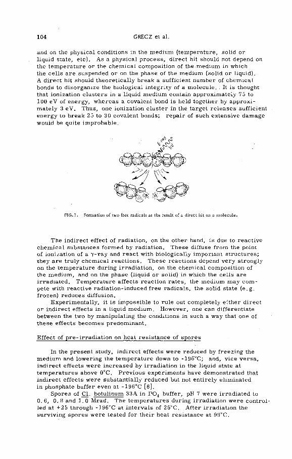
104 GRECZ et al.
and on the physical conditions in the medium (temperature, solid or liquid state, etc). As a physical process, direct hit should not depend on the temperature or the chemical composition of the medium in which the cells are suspended or on the phase of the medium (solid or liquid).A direct hit should theoretically break a sufficient number of chemical bonds to disorganize the biological integrity of a molecule. . It is thought that ionization clusters in a liquid medium contain approximately 75 to 100 eV of energy, whereas a covalent bond is held together by approximately 3 eV. Thus, one ionization cluster in the target releases sufficient energy to break 25 to 30 covalent bonds; repair of such extensive damage would be quite improbable.
F IG .7. Formation o f two free radicals as the result o f a direct hit on a m olecu le.
The indirect effect of radiation, on the other hand, is due to reactive chemical substances formed by radiation. These diffuse from the point of ionization of a y -ray and react with biologically important structures; they are truly chemical reactions. These reactions depend very strongly on the temperature during irradiation, on the chemical composition of the medium, and on the phase (liquid or solid) in which the cells are irradiated. Temperature affects reaction rates, the medium may com pete with reactive radiation-induced free radicals, the solid state (e.g. frozen) reduces diffusion.
Experimentally, it is im possible to rule out completely either direct or indirect effects in a liquid medium. However, one can differentiate between the two by manipulating the conditions in such a way that one of these effects becomes predominant.
Effect of pre-irradiation on heat resistance of spores
In the present study, indirect effects were reduced by freezing the medium and lowering the temperature down to -196°C; and, vice versa, indirect effects were increased by irradiation in the liquid state at temperatures above 0°C. Previous experiments have demonstrated that indirect effects were substantially reduced but not entirely eliminated in phosphate buffer even at -196“C [6].
Spores of Cl. botulinum 33A in P 0 4 buffer, pH 7 were irradiated to 0. 6, 0. 8 and 1. 0 Mrad. The temperatures during irradiation were controlled at +25 through -196“C at intervals of 25°C. After irradiation the surviving spores were tested for their heat resistance at 99“C.
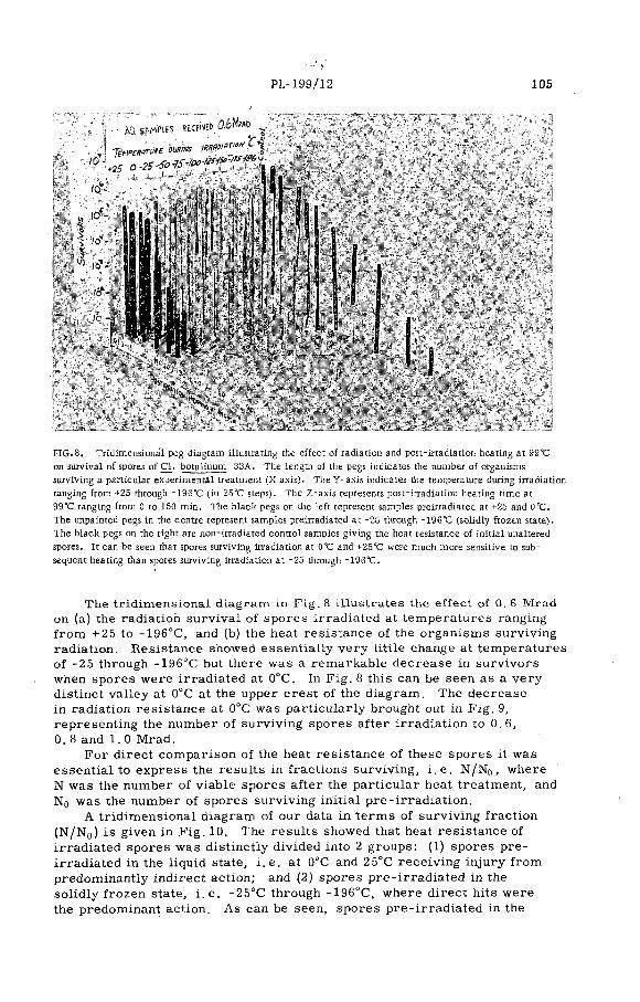
PL-199/12 105
i- 1
t
FIG. 8. Tridimensional peg diagram illustrating the e ffect o f radiation and post-irradiation heating at 99°C
on survival o f spores o f C l. botulinum 33A. The length o f the pegs indicates the number o f organisms
surviving a particular experimental treatment (X axis). The Y -ax is indicates the temperature during irradiation ranging from +25 through -196°C (in 25°C steps). The Z-axis represents post-irradiation heating tim e at
99°C ranging from 0 to 150 m in. The black pegs on the le ft represent samples preirradiated at +25 and 0°C.The unpainted pegs in the centre represent samples preirradiated at -25 through -196*0 (solidly frozen state).
The black pegs on the right are non-irradiated control samples g iv ing the heat resistance o f in itia l unaltered
spores. It can be seen that spores surviving irradiation at O'C and +25°C were much more sensitive to subsequent heating than spores surviving irradiation at -25 through -196°C.
The tridimensional diagram in F ig. 8 illustrates the effect of 0. 6 Mrad on (a) the radiation survival of spores irradiated at temperatures ranging from +25 to -196°C, and (b) the heat resistance of the organism s surviving radiation. Resistance showed essentially very little change at temperatures of -25 through -196°C but there was a remarkable decrease in survivors when spores were irradiated at 0°C. In Fig. 8 this can be seen as a very distinct valley at 0°C at the upper crest of the diagram . The decrease in radiation resistance at 0°C was particularly brought out in Fig. 9, representing the number of surviving spores after irradiation to 0. 6,0. 8 and 1. 0 Mrad.
For direct comparison of the heat resistance of these spores it was essential to express the results in fractions surviving, i .e. N/N0, where N was the number of viable spores after the particular heat treatment, and N 0 was the number of spores surviving initial pre-irradiation.
A tridimensional diagram of our data in term s of surviving fraction (N/N0) is given in F ig. 10. The results showed that heat resistance of irradiated spores was distinctly divided into 2 groups: (1) spores pre- irradiated in the liquid state, i .e. at 0°C and 25°C receiving injury from predominantly indirect action; and (2) spores pre-irrad iated in the solidly frozen state, i . e. -25°C through -196°C, where direct hits were the predominant action. As can be seen, spores pre-irrad iated in the

106 GRECZ et al.
FIG .9.
TEMPERATURE D U R ING IR R A D IA T IO N
Effect o f irradiation temperature on survival o f spores o f C l. botulinum 33A.
to saMfies ксйда
0.2S-Sb-yS-t°° ^ • '
!| 1 11FIG. 10. Norm alized tridimensional peg diagram illustrating the e ffec t o f radiation and post-irradiation heating at 99°C on survival o f spores o f C l. botulinum 33A. Sim ilar to Fig. 8, but results expressed as
fraction o f spores surviving: N/N0 where N 0 - number o f spores remaining viab le after in itia l exposure to
0 .6 Mrad, i . e . number o f spores at zero tim e o f heating; N = number o f spores remaining v iab le after the
period o f heating. Irradiation and heating was in P04 buffer, pH 7.
lisíi^r
P.L-199/12 107
liquid medium were considerably 'more heat sensitive than spores pre- irradiated in the solidly frozen medium.
Figure 11 presents the spread of points for heat survival of spores pre-irrad iated to 0. 6 Mrad at the various temperatures from +25 to -196°C. There was a substantial scattering of points for both the liquid and the solidly frozen pre-irradiations. Spores pre-irradiated
to ° - |
ur2-
1СГ3
ic r -
1СГ*
ALL SAMPLES 0 6 Mrad TEMP DURING IRRADIATION °C
□ ♦ 25© 0
о - 25
e - 50a - 75P - Ю0
- 125o - 150• - 175
A - 196
« CONTROL
0 20 40 60 80 t00 120
P O S TIR R A D IA TIO N H E A TIN G AT 99°C (m in i
FIG. 11. Spread o f points for heat survival o f spores preirradiated to 0 .6 Mrad at various temperatures from
+25 to -196°C (in 25°C steps). N/N0 as in F ig . l0 ; irradiation and heating were in P04 buffer, pH 7.
at 0°C and 25°C showed very little difference in their subsequent heat re sistance. Spores pre-irradiated in the solidly frozen state showed a re latively active intermingling of points; although there was a tendency toward a somewhat higher heat resistance of spores pre-irradiated at the lower temperatures.
The relatively close agreement between heat resistance of spores pre- irradiated in the liquid state versus those pre-irradiated in the solidly frozen state, seemed to justify averaging of the surviving fraction of these two groups as was done in the next graph, F ig. 12. Spores pre- irradiated at 0°C to 25°C were consistently more sensitive to subsequent heat than spores pre-irradiated at -25 to -196°C.
The difference between pre-irradiation in the liquid state and in the solidly frozen state was especially emphasized by plotting the surviving fraction obtained after 5 minutes of heating of pre-irradiated spores,Fig. 13. The number of survivors changed in a very definite stepwise manner between +25 and 0°C versus -25 through -196°C. This stepwise difference was accentuated by two factors: (1) the pre-irradiation dose,i. e. 0.6, 0. 8 or 1. 0 Mrad (especially in the liquid state); and (2) the length of the post-irradiation heating. The second point is illustrated

108 GRECZ et al.
zzсоuiccoCL.Ф
POSTIRRADIATION HEATING AT 99eC (min)
FIG .12. E ffect o f radiation dose and radiation temperature on subsequent heat resistance o f surviving spores
o f C l. botulinum 33A . N/N0 as in F ig .10; irradiation and heating were in P 0 4 buffer, pH 7.
t e m p e r a t u r e d u r i n g IR R A D IA TIO N (<>C)
FIG .13, E ffect o f so lid-liqu id transition during irradiation on heat resistance o f surviving spores (plotted
with respect to three different preirradiation doses). N/N0 = fraction o f spores surviving, where N0 = in itia l
number o f spores forming colonies prior to irradiation or heating; N = number o f spores remaining viab le
after the indicated radiation plus heat treatment. (Note that N/N0 in Figs 13 and 14 has a different meaning
than N/N0 in Figs 10-12, 15 and 16.)
in F ig. 14, comparing results obtained after 5, 10, 20 minutes of heating at 99°C. A very definite increase in the step can be seen with increase in time of heating.
Here we have seen that the change in irradiation conditions from solidly frozen to liquid led to a very pronounced change in subsequent heat resistance of spores.
PL-199/12 109
<шI
z 10'1 -о►—<о<CE
tnОa.
оz
Z>«/)1ЛШ
T E M P E R A T U R E D U R IN G IR R A D IA T IO N ( ° C ) 0 .6 M r a d
FIG .14. E ffect o f so lid-liqu id transition during irradiation on heat resistance o f surviving spores (plotted with
respect to three different times o f heating). N/N0 = fraction o f spores surviving, where N0 = in itia l number o f
spores forming colonies prior to irradiation or heating; N = number o f spores remaining v iab le after the
indicated radiation plus heat treatment.
□
ju_ l ------ 1------ 1------1------ 1------ 1-------1------1-------1— I-----+2 5 0 - 2 5 - 5 0 - 7 5 -1 0 0 -1 2 5 -15 0 -175 -19 6
P O S T IR R A D IA T IO N H E A T I N G A T 9 9 ° C (m in )
F IG .15. Heat sensitivity o f spores o f C l. botulinum 33A preirradiated in the liquid state. N/M0 as in
Fig. 10; irradiation and heating were in P04 buffer, pH 7.
Figure 15 shows that heat sensitivity of spores pre-irradiated in the liquid state increased with increasing doses of pre-irradiation, i. e. in the order 0.6, 0. 8 and 1.0 Mrad, respectively.
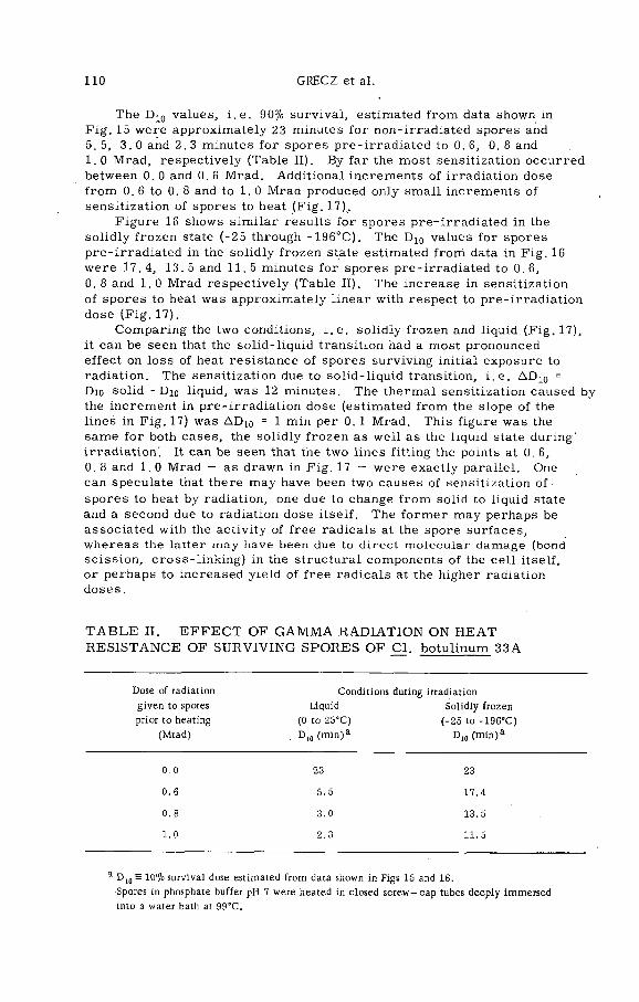
110 GRECZ et al.
The D 10 values, i .e. 90% survival, estimated from data shown in Fig. 15 were approximately 23 minutes for non-irradiated spores and 5.5, 3.0 and 2. 3 minutes for spores pre-irradiated to 0. 6, 0. 8 and1.0 Mrad, respectively (Table II). By far the most sensitization occurred between 0. 0 and 0. 6 Mrad. Additional increments of irradiation dose from 0. 6 to 0. 8 and to 1. 0 Mrad produced only sm all increments of sensitization of spores to heat (Fig. 17).
Figure 16 shows sim ilar results for spores pre-irradiated in the solidly frozen state (-25 through -196°C). The D10 values for spores pre-irrad iated in the solidly frozen state estimated from data in Fig. 16 were 17.4, 13.5 and 11.5 minutes for spores pre-irradiated to 0.6,0. 8 and 1. 0 Mrad respectively (Table II). The increase in sensitization of spores to heat was approximately linear with respect to pre-irradiation dose (F ig .17).
Comparing the two conditions, i .e. solidly frozen and liquid (F ig. 17), it can be seen that the solid-liquid transition had a most pronounced effect on loss of heat resistance of spores surviving initial exposure to radiation. The sensitization due to solid-liquid transition, i .e. AD10 - Dio solid - Dio liquid, was 12 minutes. The thermal sensitization caused by the increment in pre-irradiation dose (estimated from the slope of the lines in Fig. 17) was AD 10 = 1 min per 0. 1 Mrad. This figure was the same for both cases, the solidly frozen as well as the liquid state during' irradiation'. It can be seen that the two lines fitting the points at 0.6,0. 8 and 1. 0 Mrad — as drawn in Fig. 17 — were exactly parallel. One can speculate that there may have been two causes of sensitization of spores to heat by radiation, one due to change from solid to liquid state and a second due to radiation dose itself. The form er may perhaps be associated with the activity of free radicals at the spore surfaces, whereas the latter may have been due to direct molecular damage (bond scission, cross-linking) in the structural components of the cell itself, or perhaps to increased yield of free radicals at the higher radiation doses.
T A B L E II. E F F E C T OF G A M M A R A D IA T IO N ON H E A T R E S IST A N C E OF SU R V IV IN G SPO R ES OF C l. botulinum 33A
Dose o f radiation Conditions during irradiation
g iv en to spores Liquid S olid ly frozen
prior to heating (0 to 25°C ) (-2 5 to -196 °C )
(M rad ) . D 10 (m in )a Dio (m in )a
0 .0 23 23
0 .6 5.5 17.4
0 .8 3 .0 13.5
1.0 2 .3 11.5
a D 10 = 10% survival dose estim ated from data shown in Figs 15 and 16.
Spores in phosphate buffer pH 7 w ere heated in closed s c rew - cap tubes deep ly im m ersed
into a w ater bath at 99°C.

PL-199/12 111
FIG. 16. Heat sensitivity o f spores o f C l. botulinum 33A preirradiated in the solidly frozen state. N/M0 as in Fig. 10; irradiation and heating were in P04 buffer, pH 7.
30 Г
difference due to solid-liquid transition
spores preirradlated in the solidly frozen state (-25 to -196°C)
spores preirradlated in the liquid state
(0 to +25°C)
0.2 0.4 0.6 0.8Dose of Preirradiation
(Mrad)FIG. 17. Heat resistance o f spores (in D10 values) surviving in itia l exposure to gamma radiation in the liquid
and in the solidly frozen medium. D10 = tim e necessary to k ill 90% o f spore population at 99°C.
CONCLUSIONS
These experiments were undertaken in order to study the fact that pre-irradiation of bacterial spores makes them highly susceptible to subsequent heat inactivation. In particular, it was of interest to find which component of radiation, the direct hit or the indirect action, makes the spore become heat sensitive.
The results support the idea that the indirect effects of radiation are the prim ary causes for the loss of heat resistance of spores sur-

112 GRECZ et al.
viving exposure to sublethal doses of radiation. Whenever the indirect effects of radiation could be increased, the heat sensitivity of spores was correspondingly increased. A change in amount of indirect effects would be expected in (1) the solid-liquid transition of the medium because of the drastic change in diffusion rates, (2) in change of temperature in the solidly frozen medium because of decrease in reactivity of radiation
'induced free radicals, and finally (3) change in radiation dose because of its effect on the yield of free radicals. We have shown that a ll three .of these factors affected the heat sensitivity of spores in such a way as would be predicted from the hypothesis that indirect effects of radiation- are the prim ary agents lowering heat sensitivity in pre-irrad iated spores.
These findings appear to give some idea about the location of thermal injury. Let us distinguish between (i) the action of radiation as related to individual molecules within the cell, and (ii) the action of radiation as related to the entire cell. It can be visualized that an increase of indirect effects in the liquid medium as opposed to the solidly frozen medium w ill lead to increased peripheral damage at,.spore surfaces to constituents which are most probably not involved in radiation survival of spores.
In this connection'let us point out that the range of diffusion of indirect free radicals has been estimated to be of the order of 10-40 Â [7 -9 ], whereas the diameter of the spore is of the order of 1-2 мт,i .e. 10 000-20 000 Â. Furtherm ore, structural components of the spore are essentially hydrophobic [10], i . e. water cannot contact intimately the vital molecules in the spore.
With respect to water permeability, the spore can be visualized illustratively as some kind of a spongy m aterial made of a relatively water inert material. W ater can penetrate into the spore [11, 12], but it cannot get into intimate contact with vital proteins and nucleic acids within the spore. Supposedly the proteins and nucleic acids in the spore are physically shielded by a masking cement made up of a C a -D P A complex [13]. Thus it may be visualized that most indirect processes are taking place at internal and external spore surfaces.
Usually DNA — the prim ary target of radiation — is located in deeper regions of the cell. In the spore the DNA seems to be especially guarded by several protective membranes. Therefore, it appears unlikely that DNA would be extensively attacked by external free radicals. (This may be the reason why spores are more resistant to radiation than vegetative c e lls . )
These considerations lead to the conclusion that loss of heat resistance by spores surviving exposure to low levels of radiation is accompanied by surface reactions with free radicals, namely by peripheral reactions with the protein components of the spore, especially involving spore coats and perhaps spore membranes. Since heat resistance is usually thought to be associated with protein dénaturation, it is tempting to conclude that radiation weakens surviving spores through damage to proteins. However, no conclusive evidence is available at present to implicate proteins as the cause of thermal death either in pre-irradiated or in unirradiated spores. Therefore, further studies in our laboratory are directed toward identification of the molecules responsible for radiation death and those responsible for thermal death of spores of Cl. botulinum.

PL-199/12 113
The work described here was supported in part by Public Health Service Grant EF-00517, and P . H . S . C areer Development Award 5-K 3-A I-21, 763. The irradiations were carried out at the US Army Natick Radiation Laboratory under contract NLABS 212.
A C K N O W L E D G E M E N T S
R E F E R E N C E S
[1 ] MORGAN, B .H ., REED, J .M ., Resistance o f bacterial spores to gamma irradiation, Food. Res. 19
(1954) 357. “
[2 ] KEMPE, L. L . , Combined effects o f heat and radiation in food sterilization, Appl. M icrob iol. 3
(1955) 346.
[3 ] KEMPE, L. L . , Complementary effects o f heat and radiation on food microorganisms, Nucleonics 18
(1960) 108. ~[4 ] KEMPE, L .L ., GRAIKOSKI, J .T . , BONVENTRE, P .F ., Combined irradiation-heat processing of
canned foods. I. Cooked ground bee f inoculated with Clostridium botulinum spores, Appl. M icrobiol.
5 (1957) 292.
[5 ] GRECZ, N ., Biophysical aspects o f Clostridia, J. Appl. Bacteriol. 28 (1965) 17.
[6 ] GRECZ, N ., UPADHYAY, J ., TANG , T . , Effect o f temperature on radiation resistance o f spores o f
Clostridium botulinum 33A, Can. J. M icrob iol. (Feb.1967).
[7 ] HUTCHINSON, F . , The distance that a radical formed by ionizing radiation can diffuse in a yeastce ll, Rad. Res. 7 (1957)473.
[8 ] HUTCHINSON, F . , POLLARD, E., "Target theory and radiation effects on b io log ica l molecules", Mechanisms in radiobiology I. (ERRARA, М ., FORSSBERG, A . , Eds) Academ ic Press, In c.,
New York (1961) 71-92.
[9 ] GRECZ, N ., Estimation o f the distance that a radical formed by radiation can diffuse in a spore
o f Clostridium botulinum, Manuscript in preparation (1967).
[1 0 ] GRECZ, N .» SMITH, R ., Moisture affin ity o f spores and vegeta tive cells o f Clostridium botulinum,
Bacteriol. Proc. G 100 (1966) 32.
[1 1 ] BLACK, S .H ., GERHARDT, P ., Perm eability o f bacterial spores. IV . Water content, uptake and
distribution, J. Bacteriol. 83 (1962) 960.
[1 2 ] GERHARDT, P., BLACK, S .H ., "Perm eabilty o f bacterial spores” , in Spores, I I (HALVORSON, Ed.), Burgess Publishing C o . , Minneapolis, Minn. (1961) 218-28.
[1 3 ] TANG , T . , Relation o f d ip ico lin ic acid and calcium to heat resistance o f spores o f Clostridium
botulinum, Thesis, Illinois Institute o f Technology, Chicago (1967).


EFFECTS OF HEATING AND GAMMA RADIATION ON THE INHIBITION OF BACTERIAL SPORES BY CURING AGENTS
T . A . ROBERTS
AGRICULTURAL RESEARCH C O U N C IL ,
M E A T RESEARCH IN S T IT U T E .
CAM BRIDGE, U N IT E D K ING DO M
Abstract
EFFECTS OF HEATING AND G AM M A RADIATION ON THE INHIBITION OF BACTERIAL SPORES BY
CURING AGENTS. Studies on the resistance o f bacterial spores to inhibition by sodium chloride in the recovery medium have shown that both heat and gamma radiation are capable o f in flicting damage to the
spores which manifests itself as a reduction in salt tolerance. Radiation is at least as e ffe c t ive as heat in causing this damage. Implications o f these observations are discussed with respect to cured meat
products.
Canned cured meats, such as ham and tongue, have an excellent public health record ( i .e . are safe as regards pathogenic organisms) and are also stable regarding spoilage organism s. The heat process received by such products is relatively mild and sufficient to destroy at best only autolytic enzymes and vegetative cells of most bacteria. Bacteria l spoilage would therefore be attributable to organism s surviving heating, i . e . spores, o r relatively heat resistant vegetative bacteria ( e . g . Streptococcus faecium). It is known that both aerobic and anaerobic bacterial spores are often present in the final canned cured meat product, yet are unstable to grow.
Attempts are made to minimize the heating of such products, since overheating results in loss of eating, quality and in the case of hams, shrinkage and loss of weight. C learly the amount of heating required to process adequately a large ham is difficult to gauge, there being two opposing requirements, (a) to process lightly to avoid shrinkage and quality and weight loss, and (b) to process sufficiently to render the product safe and stable. If bacterial spores are assumed to persist throughout a ham, to obtain adequate heating at the centre the periphery must be heated to a greater extent, and probably overheated. The use of gamma radiation would overcome this difficulty, since penetration through such a product would be no problem and adequate dosage at the centre would not involve overdosage at the periphery.
The possible use of gamma radiation raises several m icrobiological questions. It would not be possible to use sterilizing doses of radiation as this would cause intolerable organoleptic changes and in any case, if we accept that heat and radiation each set out to achieve a sim ilar goal, sterilizing doses are not necessary. Doses of the order of 0.1-0.4 Mrad are commonly used by us to pasteurize experimentally (radurize) lamb carcasses and beef, and are capable of effecting considerable extension of storage life under chill conditions [ l ] . Such doses inactivate a large proportion of the vegetative bacterial flora, but have little or no effect
115
116 ROBERTS
on bacterial spores [2] . Doses of this order have already been considered for use on cured products and, although initial work indicated that irradiated hams were in ferior from the point of view of flavour [3] , more recent work at very high dose rates is more promising.
Although canned cured meats are both stable and safe, the reasons for this are not clear. Fo r example, whereas the viable spores present in cured products are incapable of growth in that product, growth readily occurs if these spores are sub-cultured into fresh medium. Sim ilarly, ■spores which have not been heated, when injected into the final cured product, readily grow and cause spoilage. Evidently the process of heating has inflicted some degree of damage to the spores which renders them m ore sensitive to subsequent inhibition by curing ingredients.
The possible use of radiation in the curing/canning process made it necessary to determine whether radiation is capable of causing sim ilar damage to the spores, so as to affect sim ilarly their inhibition by curing ingredients, since it is now generally accepted that substitution of heat processing by radiation should involve no lowering of safety m argins from the point of view of public health and safety.
The effect of curing ingredients on bacterial spores, although apparently thoroughly investigated under different conditions, is not well explained. The following factors have been shown to have some effect either on germination of spores, o r their outgrowth, the cumulative effect being the safe, stable canned cured product: spore load; pH of product; concentrations of curing ingredients ( e.g. sodium chloride, potassium nitrate, sodium nitrite, sugar); and the changed sensitivity of heated spores to inhibition by curing ingredients. Although the effect of tem perature on stability is not well documented, it is now clear that it plays a role in the interactions of the above factors. Of these factors, no one alone seems to be capable of producing a product as stable and sáfe as the com m ercially available canned cured product. Surprisingly little work has been performed on combination of factors despite the obvious need for a universally acceptable explanation. Any evaluation of published data w ill come to the inevitable conclusion that sufficient information is not yet available, although some very broad conclusions may be made.As far as radiation is concerned, a combination process of radiation with mild heating seems inevitable, since autolytic enzymes must be inactivated and 'pasteurizing' doses of radiation are incapable of achieving this. Such a process would inactivate about the same portion of the bacterial flora as the present heat treatment.
One lever to implement com m ercial use of radiation would be if some obvious advantage were offered from the viewpoint of either safety or stability.
Our long-term plan is, therefore, to investigate the effect of radiation on the inhibition of bacterial spores by curing ingredients, although to date only a study on sodium chloride has been completed. Unfortunately, such is the lack of information that, to make a valid comparison with heat, it was necessary first to evaluate quantitatively the efficacy of heating under equivalent conditions.
The investigations described below have taken place over a number of years and, inevitably, the methods used have been modified with experience. The approach, however, has remained the same. Spores suspended in water, o r in an aqueous solution of a curing ingredient, are

PL-199/11 117
F IG .l . The e ffec t o f potassium nitrate on recovery o f heated spores o f C l. sporogenes PA 3679/S2.
ppm N aNQ j
FIG. 2. The e ffec t o f sodium nitrite on recovery o f heated spores o f C l. sporogenes PA 3679/Sz.
FIG .3. The effect of sodium nitrite on recovery of spores of B. subtilis heated a t 80 °C for 30 m in.

118 ROBERTS
heated or irradiated, decimal dilutions prepared, and viable counts made in media containing different concentrations of a single curing ingredient. The effect of the individual ingredients, sodium chloride, potassium nitrate and sodium nitrite on heated spores of clostridia and bacilli has recently been described [4] , the results clearly indicating that potassium nitrate at the concentrations normally used plays a very minor role (F ig . 1) and, m oreover, that p rior heating does not greatly affect the
F IG .4. The e ffec t o f sodium nitrite on recovery o f spores o f B. subtilis heated at 95°C for 45 min.
tolerance of spores to it. The effect of sodium nitrite is highly dependent on the pH of the medium, both in the case of Clostridium sporogenes (F ig . 2) and Bacillus subtilis (F igs 3 and 4). In both cases prior heating tends to render spores more sensitive to subsequent inhibition by nitrite. There is, however, a marked difference in the pH at which nitrite becomes effective; Cl. sporogenes was virtually inhibited by 100 ppm sodium nitrite at pH 6.5 (F ig . 2), whereas recovery of B. subtilis was only re duced to about 40% at pH 6. 6 with 1400 ppm sodium nitrite (F ig . 3) and even after prior heating to 95°C for 45 min about 3% recovery resulted (F ig . 4). The concentrations of sodium nitrite remaining after processing are so low that, if its effect in cured products is at the stages of germ ination or outgrowth, it seems necessary to postulate a combined effect with another, as yet undisclosed, factor. The observation that sodium chloride is more inhibitory against severely heated spores (F ig . 5) led us to study this effect in rather more detail, and to attempt.to correlate the sensitizing effect of heating with some measure of the degree of inactivation [5] . Although results were not entirely conclusive, it was evident that mild heating ( e.g. the normal 'heat-shock' of 80°C for 20 min) did not affect the subsequent pattern of inhibition of spores of Cl. sporogenes by sodium chloride. Treatments up to about FQ0.3 resulted in no sensitization (F ig . 6). More severe heating (to FQ approx. 1.3) made spores m ore sensitive to inhibition by salt in the recovery medium. The presence of up to 6% (w t./vo l.) sodium chloride m erely during heating did not affect the pattern or degree of sensitization.
A sim ilar investigation has been made using gamma radiation as the damaging agent [6] . Spores of two clostridia ( Cl. sporogenes and Cl. oedematiens type C) were irradiated in water and in 3% and 6% aqueous

PL-199/11 119
FIG. 5. The e ffe c t o f sodium chloride on recovery o f heated spores o f C l. sporogenes PA 3679/S2.
7 . N a C l
FIG. 6. The e ffec t o f sodium chloride on recovery o f spores o f C l. sporogenes .unheated and heated in
3ft (w t./ vo l.) NaCl.
V .N a C l added lo' R C A
F IG .7. The e ffec t o f sodium chloride on recovery o f spores o f C l. sporogenes unirradiated and irradiated
in aqueous suspension.
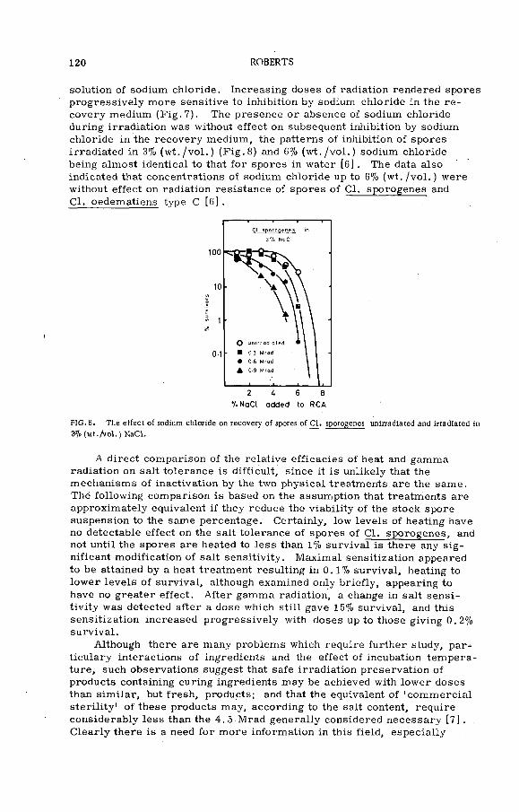
120 ROBERTS
solution of sodium chloride. Increasing doses of radiation rendered spores progressively more sensitive to inhibition by sodium chloride in the re covery medium (F ig . 7). The presence or absence of sodium chloride during irradiation was without effect on subsequent inhibition by sodium chloride in the recovery medium, the patterns of inhibition of spores irradiated in. 3% (w t./vo l.) (F ig . 8) and 6% (w t./vo l.) sodium chloride being almost identical to that for spores in water [6] . The data also indicated that concentrations of sodium chloride up to 6% (w t./vo l.) were without effect on radiation resistance of spores of Cl. sporogenes and Cl. oedematiens type С [6] .
■/.NaCl added to RCA
FIG. 8. The e ffec t o f sodium chloride on recovery o f spores o f C l. sporogenes unirradiated and irradiated in
2fl<! (w t ./ v o l.) NaCl.
A direct comparison of the relative efficacies of heat and gamma radiation on salt tolerance is difficult, since it is unlikely that the mechanisms of inactivation by the two physical treatments are the same. The following comparison is based on the assumption that treatments are approximately equivalent if they reduce the viability of the stock spore suspension to the same percentage. Certainly, low levels of heating have no detectable effect on the salt tolerance of spores of C l. sporogenes, and not until the spores are heated to less than 1% survival is there any sig nificant modification of salt sensitivity. Maxim al sensitization appeared to be attained by a heat treatment resulting in 0.1% survival, heating to low er levels of survival, although examined only briefly, appearing to have no greater effect. A fter gamma radiation, a change in salt sensitivity was detected after a dose which still gave 15% survival, and this sensitization increased progressively with doses up to those giving 0. 2% survival.
Although there are many problems which require further study, par- ticulary interactions of ingredients and the effect of incubation tem perature, such observations suggest that safe irradiation preservation of products containing curing ingredients may be achieved with lower doses than sim ilar, but fresh, products; and that the equivalent o f'com m erc ia l sterility' of these products may, according to the salt content, require considerably less than the 4. 5 Mrad generally considered necessary [7] . . Clearly there is a need for m ore information in this field, especially
PL-199/11 121
with regard to interactions of curing ingredients, but sufficient data is available to indicate it to be promising fo r the commercial implementation of radiation processing.
- A C K N O W L E D G E M E N T S
Figures 1-5 are reproduced with the kind perm ission of Blackwell Scientific Publications Ltd ., Oxford. F igures 6-8 are reproduced with the kind perm ission of Academic P ress Inc. (London) Ltd.
R E F E R E N C E S
[1 ] RHODES, D. N. ( SHEPHERD, H.J. » The treatment o f meats with ionizing radiations X III. Pasteurization o f bee f and lamb, J. Sci. Fd. A grie. 17 (1966) 287.
[2 ] ROBERTS, T . A . , INGRAM, M . , Radiation resistance o f spores o f Clostridium species in aqueous suspension, J. Food. Sci. 30 (1965) 879.
[3 ] HANSEN, P * -L E ., A consumer survey for acceptance evaluation o f cured ham treated by a com bi
nation o f heat and irradiation, Fd. T ec h n o l., Champaign 20 2 (1966) 99.
[4 ] ROBERTS, T . A . , INGRAM, M . , The e ffe c t o f sodium chloride, potassium nitrate and sodium nitrite
on the recovery o f heated bacterial spores, J. Fd. Technol. 1̂ 2 (1966) 147.
[5 ] ROBERTS, T . A . , GILBERT, R. J ., INGRAM, M . , The e ffe c t o f sodium chloride on heat resistanceand recovery o f heated spores o f C l. sporogenes. J. appl. Bact. 2£ (in press).
[6 ] ROBERTS, T . A . , D rrCH ETT, P .J ., INGRAM, М . , The effectofsod ium chlori.de on radiation
resistance and recovery o f irradiated anaerobic spores, J. appl. Bact. 28 (1965) 336.
[7 ] INGRAM, М . , ROBERTS, T . A . , "M icrob io log ica l principles in food irradiation". Food Irradiation,
IAEA, Vienna (1966) 267.

REDUCTION OF RADIATION DOSE REQUIREMENTS OF FOODS BY ADDITIVES
J.. FA R R A S, I . R ISS A N D É. A N D R Á SSY
CENTRAL FOO D RESEARCH IN ST IT U T E ,
B U D A PE ST , H U N G A R Y
Abstract
REDUCTION OF RADIATION DOSE REQUIREMENTS OF FOODS BY ADDITIVES. U tiliz in g a recording photometer, a rapid turbidimetric method was worked out for the determination o f nisin and tylosin activ ity .
Those antibiotics a ffected by irradiation with X-rays in the range o f 0 to 800 krad were also investigated. . In
a solution o f pH 5 .7 containing pea extract, the activ ity o f the antibiotics decreased at a high rate when
irradiated up to 200 krad, however, this rate slowed down with further radiation treatment. Tylosin lactate
proved more radiation resistant than nisin. When treated with dosages between 400 and 800 krad the
activ ity o f nisin decreased by 73 to 87% and that o f tylosin lactate by only 52 to 57%. During one-month
storage at room temperature the residual activ ity o f both the irradiated and untreated antibiotic solutions
remained practically unchanged. The addition o f antibiotics increased the preserving e ffec t o f X-rays.
A 1:1 mixture o f peas and a solution containing 4% sugar and 1.5% salt was heat treated for 15 minutes at
70 'C . Samples taken from the liquid phase o f this mixture were treated with various radiation-antibiotic
combinations. When 100 ppm Nisaplin or 1 ppm tylosin lactate was added the radiation dose requirement
was a quarter o f that needed in the control samples to ensure m icrob iological stability.
INTRODUCTION
In the m icroflora of foods of nearly indifferent or slightly acid character, such as vegetables, spore-form ing bacteria are always present. Since bacterial spores are both heat and radiation resistant, while the heat resistance of other, relatively radiation-resistant, m icro-organism s is generally small, in foods blanched previous to irradiation the number and the resistance of bacterial spores are the principal factors determining radiation dose requirements. The reduction of the radiation dose is desirable not only for economic reasons, but for the sake of reducing the danger <?f undesirable changes caused by high doses. Thus, the possibilities of sensitizing radiation-resistant m icro-organism s or of increasing the m icrobial efficiency of irradiation should be studied. Theoretically, the following main possibilities exist for the enhancement of the preservative effects of ionizing radiations:
(1) The application, combined with irradiation, of physical or chemical factors exerting spore-sensitizing effects
(2) The promotion of the transformation of resistant spores into sensitive cells, before irradiation
(3) The inhibition of the outgrowth or the vegetative reproduction of spores surviving radiation treatment in irradiated foods.
Numerous in v e s t ig a t io n s have been carried out to study substances which enhance the effect of radiation or increase the radiation sensitivity of m icro-organism s. However, the use of the majority of substances found effective does not seem practicable, frequently for reasons of public health [ 1-4] . A number of experiments [5-10] have proved that the sim ultaneous or subsequent application of radiation and heat treatment may have
123
124 FARKAS et al.
a synergie effect. However, there is little experience available as regards the reduction of dose requirement in radiation preservation with the aid of substances stimulating spore germination [11, 12] or inhibiting surviving spores [3, 13].
Last year we reported to the FAO/IAEA Panel on Co-ordination of Research in Radiation Sensitization of M icro-organism s in Food P re servation the results of our experiments aimed at the sensitization of Bacillus cereus spores with germinators [14] . The present paper presents information about the prelim inary study of preservation by the combined application of ionizing radiation and nisin or tylosin.
Nisin and tylosin were found suitable to reduce the heat requirement in the heat treatment of various foods [15-20] . Therefore we investigated the possibility of increasing radiation efficiency by the addition of nisin or tylosin. The radiation resistance of these antibiotics was also studied.
The preparations used in our experiments were: Nisaplin1 of 106 Reading units (RU) activity per gram and a tylosin-lactate preparation2 of unknown purity grade.
Irradiations were carried out with X -ra y apparatus, Stabil 250 (240 kV, 15 mA, without filtration). The dose rate of the apparatus, as measured with a ferrous sulphate dosimeter [21] of the same volume in vessels identical to that of the samples, was between 173 and 256 krad/h, depending on the position of the samples.
M EASUREM ENT OF THE ANTIBIOTIC A C T IV IT Y AND STUDY OF THE EFFE C TS OF THE ANTIBIOTICS ON B AC TER IAL SPORES
During the prelim inary investigations we tried to work out a rapid method of determining with satisfactory accuracy the activity of nisin and tylosin within a few hours and to study simultaneously the mechanism of the effect of the antibiotics on test organism s isolated from peas used in the irradiation experiments.
The test organism s used, as in previous studies [14] , were Bacillus cereus spores heat activated for 30 minutes at 60°C. The resting spores were produced as previously described [22] .
We found the biophotometer of Bonet-Maury and Jouan used for t'/e automatic measurement and recording of the turbidity or optical density of suspensions suitable for our purposes. A pea extract nutrient3, ensuring the rapid germination and growth of the heat-activated Bacillus cereus spores, was filled in the cells, placed in the thermostat of the biophotometer, the antibioticum solution to be investigated was added and this medium was then inoculated to contain 107 spores/m l. During incubation at 30°C the change of turbidity was recorded by the biophotometer. Suspensions without antibiotic served as controls.
During the incubation period the cell-state distribution was controlled under the phase-contrast microscope, too. In the samples taken for microscopic test the cell-state was fixed at the time of sampling by the
1 APLIN and BARRETT Ltd ., Trowbridge, Wilts, England.
1 Eli L illy International Corporation, Indianapolis, Ind., USA.
3 The pea extract was prepared by m ixing peas with water in the proportion 1:1 and steaming for an
hour. The extract was then filtered and heat treated at 0 .5 atm overpressure for 30 minutes. The liquid
had a pH o f 5.7 after sterilization.

PL-199/8 125
addition of sublimate. Figure 1 shows the change of turbidity and ce ll- state distribution in a culture containing no antibiotics.
The germination, or to be more exact the decrease of the refractive indices, was affected by neither nisin nor tylosin. In the first 2 0 to 25 minutes of incubation the turbidity of samples containing antibiotics and of the control samples decreased at the same rate to the same extent. The difference between the samples manifested itself in the vigorous increase 4 of turbidity in the control samples subsequent to reaching the minimum
Proportion of different cell states:
Incubation time
F IG .l . The change in turbidity and cell-s ta te distribution as a function o f time in pea extract inoculated
with Bacillus cereus spores.
Control£5 minutes 150 minutes
250/igjmlNisaplin
Ceil stale walbint a nd i dork spores
gceHs IIM ICC 0/1vegetative cells
■ divc/ing veoetafive
cells
FIG. 2. The cell-s ta te distribution in the control and in samples containing 250 ppm antibiotic after
25 and 150 min incubation.
value, as a consequence of the swelling and outgrowth and vegetative r e production of the germinated, dark spores. In the samples containing antibiotics, after reaching the minimum the increase of turbidity was sm aller or none as compared to that in the control samples. Only in the case of high antibiotic concentrations - a high multiple of that needed to inhibit the transformation of spores into dividing vegetative cells - was observed the presence of more spores not germinating in the antibiotic- containing samples than in the controls (F ig. 2).
When the antibiotic concentration was lowered below the minimum level of complete inhibition, dividing vegetative cells could also be observed in the samples containing antibiotics. In this case the rate of post- germinative development was only slowed down by the presence of antibiotics. Thus our observations support those found by other authors, inasmuch as during transformation of bacterial spores into vegetative cells

126 FARKAS et al.
the phase most sensitive to nisin or tylosin is that of outgrowth or the division of the first vegetative cell [20, 23-26] .
To characterize the inhibiting effect and therewith the activity of antibiotics, the regions below the turbidity curve as plotted from the values obtained during the 150 minutes subsequent to reaching the minimum value were compared (Fig.3) . The regions below the turbidity curves belonging
- incubation time
Relative g row th
Inh ib ition$-,<00
100-G
FIG .3 . The inhibitory e ffec t o f antibiotics as determined on the basis o f turbidity curves.
F IG .4 . The correlation between inhibiting e ffec t and concentration o f antibiotics.
to antibiotic-containing suspensions, Sa , were related to those below the turbidity curves of the controls, SK. By subtracting the G = (Sa/Sk) X 100 value of relative growth from 100, the percentage value, I, of inhibition as caused by the antibiotic was obtained.
There was a close linear correlation between I inhibition and the respective concentrations of the antibiotics ( С м and C t , Fig. 4). The equation of the regression line was for Nisaplin I=-6, 1+6, 4XCn and I = - l , 1+38, I X C t for tylosin lactate. Correlation coefficient values were r N =+ 0.998 (DF=4) and rT =+ 0.948 (DF=5), respectively.

PL- i 99/8 127
The equation of the regression line showed that under the conditions as applied in the activity tests the minimum antibiotic concentrations needed to ensure complete inhibition were: 16.6 /ug Nisaplin per m l (o r 16.6 RU nisin per m l) and 2.7 p g tylosin lactate per ml, respectively.
NISIN AND TYLO SIN L A C T A T E AS A FFE C T E D B Y X -R AYS
Since it is desirable to add the ingredients to the foods before radiation treatment, the use of nisin o r tylosin in radiation preservation of foods is admissible only if the antibiotics are not inactivated by the necessary radiation doses.
Radiation dose (krad)
FIG. 5. The reduction o f Nisaplin and tylosin lactate activ ity as induced by irradiation.
Ley and Hall [27] found that 200 RU nisin per ml dissolved in skim milk, when irradiated at 500 krad level, lost 80% of its activity and was completely inactivated by a dose of 1.5 M rad. Data on the radiation tolerance of tylosin are not known from the literature. We therefore studied the activity of nisin and tylosin-lactate solutions as affected by irradiation in the range up to 800 krad. Nisaplin solution of 100 ц g/ml concentration, and a tylosin-lactate solution of 50 /ug/ml concentration, both containing pea extract at pH = 5.7, were irradiáted. Activity m easurements were carried out the day after irradiation. In between the samples were stored in a refrigerator.
As shown in F ig . 5, the antibiotic activity of both solutions decreased with increasing radiation doses, the tylosin lactate, however, proving more radiation resistant than nisin. (The activity of samples not treated did not change in the 24 hours previous to activity measurement, thus the reduction of activity in irradiated samples occurred during irradiation).
Further investigations are needed to elucidate the mechanism of inactivation as caused by irradiation and to explain the shape of the curves in Fig. 5. The curves illustrating heat inactivation of tylosin, as observed by Tomiyama et al. [28] , were of very sim ilar character. It was shown, further, that tylosin inactivation was promoted by fe rric ions and free radicals originating from auto-oxidized oils. Chelate-form ing agents and antioxidants were, on the other hand, of stabilizing effect. It is supposed that the reduction of activity as caused by irradiation may also be ascribed to oxidative effects. The tailing of the inactivation curves, as shown in

128 FARKAS et al.
TABLE I. RESIDUAL ACTIVITY OF IRRADIATED SAMPLES AFTER STORAGE
A n tib io tic
Radiation
dose
(krad)
Residual a c t iv ity in °¡o o f in itia l
a c t iv ity a fter storage
at З Г at 22°C
24 h 30 d 30 d 60 d
400 27 23 < 3N isap lin
800 13 26
400 48 46 37T y los in lac ta te
800 43 39 “
- not observed.
Fig. 5, is probably due to the graduated exhaustion of dissolved oxygen in the antibiotic solutions not aerated during irradiation.
The residual activity of the irradiated samples, as measured after 30 days storage in a re frigerator or at room temperature, did not change much compared to the first test (Table I). These results support the findings of other authors [29, 30] about the stability of nisin and tylosin in foods stored at temperatures not higher than 37°C.
E F F IC IE N C Y OF RADIATION PASTEUR IZATIO N AS A F F E C T E D BY NISIN AND TYLOSIN
To study the combined effect of antibiotics and ionizing radiation^ a 1:1 mixture of frozen peas after thawing and of an aqueous solution containing 4% sugar and 1.5% salt was prepared. A fter thorough shaking, the mixture was heat treated for 15 minutes at 70°C. According to the cell counts this blanching was survived only by bacterial spores. In these tests both the total cell count and the heat-resistant cell count (spores) were determine'd both before and after heat treatment. A fter cooling, the liquid phase of the mixture was decanted and the liquid divided into several portions. One portion served as control and various amounts of antibiotics were added to the rest. The range of concentrations thus obtained was- then divided into ampullae and 10 paralle l samples were irradiated at each dose level, leaving 10 ampullae of each concentration level for control.
Subsequent to irradiation the samples were stored at 22°C and controlled for spoilage every day (turbidity, film or ring formation, and sedimentation). The number of spoiled samples was plotted against storage time and the time in days needed for spoiling of 10% of the samples was determined by interpolating the spoilage curves. The reciprocals of these values were considered as a characteristic of the spoilage rate.
It was established that the efficiency of radiation treatment substantially increased in the presence of antibiotics. Of sam ples irradiated at the same dose level, those which contained antibiotics showed a much lower spoilage rate than the samples free of antibiotics (F igs 6 and 7).

PL-199/8 129
To achieve m icrobiological stability the radiation dose requirement was four times that needed when 200 ppm Nisaplin or 1 ppm tylosin lactate was added.
Nisaplin ( ppm}'
FIG. 6. The combined effects o f Nisaplin and irradiation.
Tÿlostn L adate ( pprríj
FIG. 7. The combined effects o f tylosin lactate and irradiation.
Thus it was shown that the efficiency of tylosin lactate was two orders of magnitude higher than that of Nisaplin. Another point in favour of the application of tylosin in combination with irradiation is that, according to data found in the pertinent literature, the members of the genus Clostridium are particularly sensitive to tylosin which seems to inhibit the outgrowth and toxin production of all the three types A, В and E of Cl. botulinum strains [29, 31, 32] . It would be interesting to explore the possibility of enhancing the radiation tolerance in antibiotics by the addition of antioxidants. By this means the m icrobiological efficiency of the combination method might also be increased. The investigations into radiation damage in antibiotics and the development of the combination methods are being continued.
130 FARKAS et al.
R E F E R E N C E S
[1 ] BRIDGES, В. A . , "Sensitization by sulphydryl-binding compounds. PL-151/1, FAO/lAEA Panel on* Co-ordination o f Research in Radiation Sensitization o f Micro-organisms in Food Preservation,
Vienna, 5-9 July 1965.
.[2 ] EL-TABEY SHEHATA, A .M . , "M odification o f m icrobicidal e ffect o f ionizing radiations byVitam in K 5 and related compounds", PL-151/4, FAO/IAEA Panel on Co-ordination o f Research in
Radiation Sensitization o f Micro-organisms in Food Preservation, Vienna, 5-9 July 1965.
[3 ] LEE, J .S ., CORLETT. D .A ., Jr., SHIFLETT, M .A . , SINNHUBER, R .O ., Combination processes in
food irradiation: Use o f food additives, Isotopes rad. Technol. 3 1 (1965) 42-48.
[4 ] NAM IK I, М ., "The enhancement o f radiation lethal e ffec t o f micro-organisms by chem ical agents", PL-151/7, FAO/IAEA Panel on Co-ordination o f Research in Radiation Sensitization o f M icro
organisms in Food Preservation, Vienna, 5-9 July 1965.
[5 ] FARKAS, J., Investigations in to the destruction o f bacterial spores by ion izing radiation, Thesis, Budapest (1963).
[6 ] KAN, B .S ., GOLDBLITH, S. A . , PROCTOR, B .E ., Complementary effects o f heat and ionizing
radiation, Food Res. 22 (1957) 509.
[7 ] KEMPE, L .L . , Combined effects o f heat and radiation in food sterilization, Appl. M icrob io l. 3(1955) 346-52.
[8 ] LICCIARDELO, J.J., "Complementary effects o f thermal and ionizing energy” , Exploration in
Future Food-Processing Techniques (GOLDBLITH, S. A ., Ed.). Massachusetts Institute o f T ech
nology, Cambridge (1963).
[9 ] MORGAN, B .H ., REED, J .M ., Resistance o f bacterial spores to gamma irradiation, Food Res. 19
(1954) 357-66.
[10] VINTER, V . , "Some biochem ical aspects o f combined effects o f radiation and heat on bacterial
spores", PL-151/9, FAO/IAEA Panel on Co-ordination o f Research in Radiation Sensitization o f
M icro-organisms in Food Preservation, Vienna, 5-9 July (1965).
[11] ANON, Radiation preservation o f cured meats, M eat Progr. 4 2 (1964) 5-6.
[12] KAN, B .S ., GOLDBLITH, S. A . , PROCTOR, B .E ., Effect o f gamma radiation o f spores that have undergone germination induced by treatment with L-am ino acids. Food Res. 23 (1958) 41-50.
[13] ROBERTS, T .A . , D ITCHETT, P .J ., INGRAM, М .. The e ffec t o f sodium chloride on radiation resistance and recovery o f irradiated anaerobic spores, J. Appl. Bact. 28 (1965) 336-48.
[14] FARKAS, J,, KISS, I . , ANDRASSY, £ ., "Combined effects o f heat and ionizing radiation on bacterial
spores as influenced by some chem ical factors", PL-15Í/5, FAO/IAEA Panel on Co-ordination o f
Research in Radiation Sensitization o f Micro-organisms in Food Preservation, Vienna, 5-9 July 1965.
[15] DENNY, C .B ., REED, J .M ., BOHRER, C .W ., E ffect o f tylosin and heat on spoilage bacteria in canned corn and canned mushrooms, Food Technol. 15̂ (1961) 338-40.
[16] O ’BRIEN, R .T . , TITUS, D .S ., DEVLIN, K .A . , STUMBO, C .R ., LEWIS, J .C ., Antibiotics in food preservation. II . Studies on the influence o f subtilin and nisin on the thermal resistance o f food spoilage bacteria, Food Technol. 1£(1956) 352-55.
[17] SCHALLER, A . , Tylosínlactat, ein H ilfsm ittel für die thermische Konservierung von Lebensmitteln.
Fruchtsaft-Ind. 9 (1964) 341-49.
[18] TRAMER, J., Nisin in food preservation, Chemy Ind. (1966) 446-50.
[19] VAS, К ., KISS, I . , KU TZ, N . , Ûber die Anwendung von Nisin in der Konservenindustrie, Confructa
1 (1961) 155-60.
[20] VAS, К ., KISS, I . , KU TZ, N ., Application o f nisin to reduce heat process requirements in the
sterilization o f canned foods (in Hungarian), Kôzponti Élelm iszeripari Kutatóintézet K ozlem ényei,
Budapest, 1 (1961) i - 5 .
[21] WEISS, J., ALLEN, A .O ., SCHWARZ, H. A ., "Use o f the Fricke ferrous sulfate dosimeter for gamma ray
doses in the range 4 to 40 kr” , Proc. UN int. Conf. PUAE 14 (1956) 179.[22] FARKAS, J ,, KISS, I . , ANDRA'SSY, É., Reduction o f heat and radiation resistance o f Bacillus cereus
spores by initiating germination, Acta m icrob iol. hung. 13 1 (1966) 35-46.
[23] GIBBS, B .M ., HURST, A . , "Lim itations o f nisin as a preservative in non-dairy foods", M icrobial Inhibitors in Food (MOLIN, N . , Ed.), A lm qvist and Wiksell, Stockholm (1964) 151-65.
[24] GOULD, G .W ., "Effect o f food preservatives on the growth o f bacteria from spores", M icrob ial
Inhibitors in Food (MOLIN, N ., Ed.), A lm qvist and Wiksell, Stockholm (1964) 17-24.
[25] GREENBERG, R .A ., S1LLIKER, J .H ., The action o f tylosin on spore-forming bacteria, J.Food Sci. 27
(1962) 64-68.

PL-199/8 131
[26] POOLE, G ., M ALIN , В., Some aspects o f the action o f tylosin on Clostridium species PA 3679,J. Food Sci. 29 (1964) 475-78.
[27] LEY, F .F ., HALL, R .H ., Effect o f gam ma-radiation on nisin, Nature, Lond. 188 (1960) 1184-85.
[28] TO M IYAM A, T . , K ITAHARA, K ., SHIRAISHI, E ., IM A IZU M I, K ., "Stability o f chlortetracycline
and tylosin in tissues", M icrob ial Inhibitors in Food (MOLIN, N . , Ed.)» Almqvist and Wiksell,
Stockholm (1964) 261-68.
[29] MALIN, В., GREENBERG, R .A ., "The e ffec t o f tylosin on spoilage patterns o f inoculated and noninocu- lated cream style corn". M icrob ial Inhibitors in Food (MOLIN, N . , E d .),A lm qvist and Wiksell,Stockholm (1964) 87-95.
[30] TRAMER, J., "Th e inhibitory action o f nisin on Bacillus stearothermophilus", M icrob ial Inhibitors
in Food (MOLIN, N . , Ed.)? A lm qvist and Wiksell, Stockholm (1964) 25-33.[31] GREENBERG, R .A ., SILLIKER, J .H ., "Spoilage patterns in Clostridium botulinum-inculated canned
foods treated with tylosin," M icrob ial Inhibitors in Food (MOLIN, N . , Ed.), Alm qvist and Wiksell, Stockholm (1964) 97-103.
[32] SHENEMAN, J .M ., Prevention o f type E Clostridium botulinum toxin formation in smoked whitefish
chubs with tylosin lactate, J. Food Sci. 30 (1965) 337-43.


THE EFFECT OF IONIZING RADIATION AND ANTIOXIDANT TREATMENT ON THE QUALITY AND STORAGE LIFE OF VACUUM-PACKED TROUT AT 0°C
P. HANSEN
TECHNOLOGICAL LABORATORY,
MINISTRY OF FISHERIES,
COPENHAGEN
AND
В. V. J0RGENSENDEMSTRUPVEJ 5, DRONNINGBORG PR. RANDERS, DENMARK
Abstract
THE EFFECT OF IO NIZING RADIATION AND ANTIO XID AN T TREATMENT ON THE Q U ALITY AND
STORAGE LIFE OF VACUUM -PACKED TROUT A T 0°C. Ice packing provides a simple and econom ical
means o f maintaining the storage temperature o f fresh fish at 0°C. Vacuum packing is valuable in sup
pressing oxidative rancidity during iced storage o f fatty fish. During prolonged storage at 0”C o f vacuum-
packed gutted trout no growth o f sulphite-reducing clostridia is noted, but the total viab le aerobic count
may rise to several millions per gram in the third week of storage. Non-irradiated trout have a maximum
storage l i fe o f 2-2J weeks. Radiation o f vacuum-packed trout increased the bacterial lag phase. A t a
dose o f 100 krad a total viab le count o f 1 m illion per gram was reached at the end o f the fourth week of
storage. A short dip in an aqueous solution o f ascorbic acid w ill reduce harmful effects o f irradiation on
the eating quality o f the trout. Trout dipped in an 0.2% ascorbic acid solution before vacuum packing
could be treated to doses from 50 to 200 krad without im m ediate effects upon the eating quality. The quality, however, decreased steadily independent o f dose during 4 weeks o f iced storage, and first-class
quality was retained for about 3 weeks only , although the fish remained edib le for more than 4 weeks.
The loss o f quality occurred more rapidly when trout were irradiated without prior ascorbic acid treatment.
Ice packing provides a simple and economical means of controlling the temperature of wet fish. Fish which are completely buried in crushed wet ice w ill rapidly reach a temperature of 0°C, which is maintained as long as sufficient ice is present. While m icro-organism s injurious to health cannot grow at this temperature, some species of bacteria norm ally present on wet fish are still active. In normal ice packing the activity of these bacteria w ill limit the storage life of most fish to about two weeks.
During iced storage of fatty fish, such as herring and trout, quality deterioration due to fat oxidation may play an important ro le. Recent investigations have shown that the storage life of gutted trout can be as short as 1 to 1^ weeks and that the limiting factor is oxidation of fat, mainly on the belly wall surface [1, 2] . Two weeks' storage life or more may be obtained, however, by vacuum-packing the gutted trout in plastic bags before ice packing. Both the oxidative changes and the changes due to bacteria are influenced by vacuum packing. It has been shown recently that packing in polyamide bags is m ore effective in the suppression of these quality changes than packing in polyethylene bags [3] . As the form er plastic m aterial is m ore airtight than the latter, it appears that the stor-
133
134 HANSEN and J0RGENSEN
age life of vacuum-packed iced trout depends on the availability of air inside the package. This is in keeping with results of experiments in Great Britain and elsewhere on vacuum packing and ice storage of other species of fish [4] .
The object of current Danish studies on ' combination' processes of vacuum packing and irradiation of gutted trout is to develop processes that w ill allow extended storage at 0°C of the trout in a wholesome and fresh condition. It has been shown elsewhere that fatty fish do not lend themselves readily to irradiation, because of deterioration of fat and pigments [5, 6] . A recent Danish experiment, however, has shown that vacuum-packed gutted trout may be irradiated to doses from 50 to 200 krad without significant oxidative deterioration if the trout are treated with antioxidants before vacuum packing and irradiation [7] . The present studies, therefore, include investigations of the effect of such antioxidant treatment on the quality and storage life of vacuum-packed irradiated trout.
Besides investigations of changes due to bacteria and oxidation, the studies include investigation of some changes of chemical constituents due to the effect of endogenous enzymes, which are probably uninfluenced by the above processes.
M ATERIALS AND METHODS
A ll the present experiments were carried out with rainbow trout raised at commercial trout farm s in Jutland. A ll fish weighed about 150-200 g. The fish were transferred to concrete tanks and kept alive for a few days without food in fresh water. They were then electrocuted and immediately eviscerated and washed free of blood. The fish were then stored in ice for j to 1 hour and again washed. Antioxidant treatment and vacuum packing were carried out immediately afterwards. The fish were again iced and transferred to Ris^ for irradiation, which was carried out about 6 hours later.
Antioxidant treatment, when applied, consisted in a dip of a weak aqueous solution of ascorbic-acid, either the commercial dip for 10-20 seconds in a 0. 2% solution or a controlled dip for 1 minute in a 0. 25% solution.
Two types of vacuum packing were used. One was packing in bags of 0.04-m m thick polyamide foil which were closed in a laboratory machine with controlled evacuation. The other was packing in bags of 0.05 mm thick polyethylene laminated with lacquered cellophane in a com m ercial machine without vacuum gauge. No attempt was made to m easure the oxygen tension inside the packages. It is believed, however, that the form er packing procedure gives a better oxygen rem oval than the latter.
A fter transfer in ice to Risjl the packages were placed in single layers in aluminium trays and irradiated to 100 krads by one passage through thé electron linear accelerator. A fter treatment the packages were again buried in wet ice. ■ ' •
During the following weeks samples were taken about twice a week for examination. Packages showing signs of a ir leakage were discarded. The tail of each fish was used for m icrobial examination. Of the ‘re maining part one fillet was used for organoleptic tests and the other for chemical analyses. A sample always consisted of six fish.

PL-199/6 135
Organoleptic tests
The six fillet pieces per sample were cooked in a casserole by immersion for 15 min in warm water at 90°C containing 2% salt and0.02% of acetic acid. The warm pieces were served under code to a panel of six tasters, each of whom tasted one piece from each experimental group of fish.
M icrobiological examination
After hpmogenization of 10 g of fillet with 90 m l of physiological salt solution in a Waring Blendor, decimal dilutions were made from the suspension and inoculations carried out in plate count agar (D ifco). The total aerobic count was determined after incubation at 25°C for 4 days.
Peroxide values
The above-mentioned 6 fillets per sample were minced together and extracted according to Bligh and D yers' method as modified by Hanson and Olley [8] . The peroxide values were determined on these extracts by L ea 's method [9] .
Hypoxanthin values
The meat of six fillets was minced and blended. The hypoxanthin content was determined by Kalckars' method modified by Jones [10] .
RESULTS 1
The effect of a 10-20 sec dip of gutted trout in a 0. 2% aqueous ascorbic acid solution was tested in an experiment in March 1966. No significant difference in eating quality was observed after 7 or 11 days of ice storage. Both on the 13th and on the 18th day the dipped fish were superior in quality to the non-dipped fish. The quality scores for dipped fish were 5.8 and 5. 3, respectively, and the scores for non-dipped fish 4. 2 and4.0, respectively. On the 18th day the peroxide value of dipped fish was1.0 and that of non-dipped fish 16.7 meq/kg fat.
A m ore thorough investigation was carried out during the following month. The trout were dipped in 0.25% aqueous ascorbic acid solution fo r .l min. As shown in F ig. 1 some difference in eating quality was noted at the end of the third week of storage, the dipped trout again being superior to the non-dipped trout. Figure 2 shows peroxide values determined on the same trout within the 13th to the 26th day of storage. The peroxide values of non-dipped trout are, within most of this périod, more than twice as high as those of dipped trout. Total viable counts were in both cases about 1 million per gram after 3 weèks of storage.
A ll trout investigated in the above experiments weré pácked in a vacuum-packing machine with rapid, non-controlled evacuation.
Figure 3 shows the changes in eating quality'of three lots of trout packed in a laboratory vacuum-packing machine which evacuated the bags slowly to about 75 mm Hg below atmospheric pressure. These-lots were
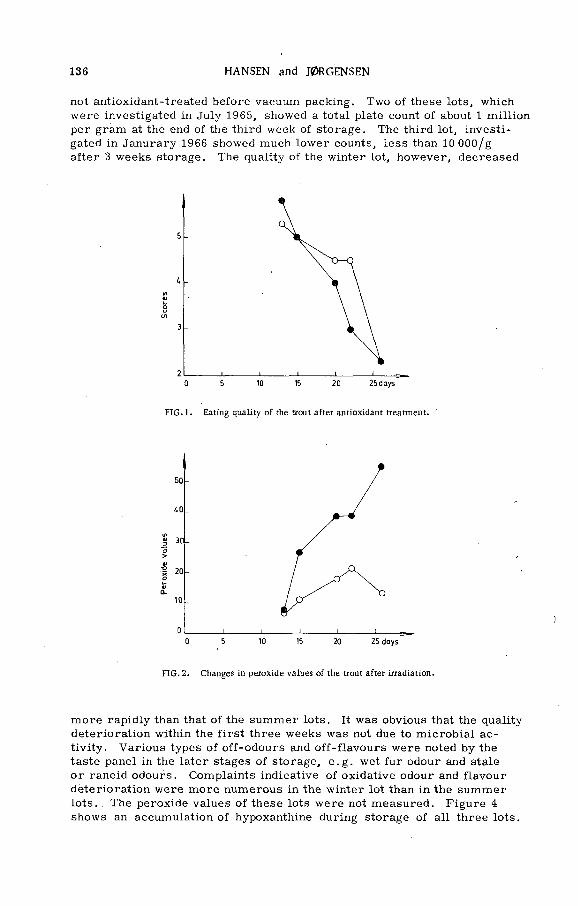
136 HANSEN and J0RGENSEN
not antioxidant-treated before vacuum packing. Two of these lots, which were investigated in July 1965, showed a total plate count of about 1 million per gram at the end of the third week of storage. The third lot, investigated in Janurary 1966 showed much lower counts, less than 10 000/g after 3 weeks storage. The quality of the winter lot, however, decreased
F IG .l . Eating quality o f the trout after antioxidant treatment.
0 5 10 IS 20 25 days
FIG. 2. Changes in peroxide values o f the trout after irradiation.
m ore rapidly than that of the summer lots. It was obvious that the quality deterioration within the first three weeks was not due to m icrobial activity. Various types of off-odours and off-flavours were noted by the taste panel in the later stages of storage, e.g. wet fur odour and stale or rancid odours. Complaints indicative of oxidative odour and flavour deterioration were more numerous in the winter lot than in the summer lots. The peroxide values of these lots were not m easured. F igure 4 shows an accumulation of hypoxanthine during storage of all three lots.
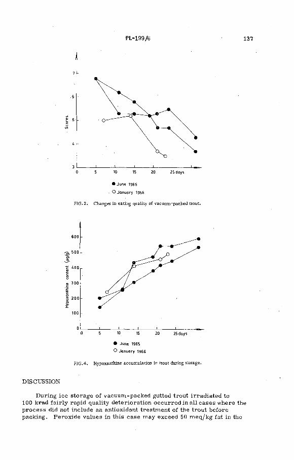
PL-199/6 137
7 -
.6 -
t в- -о
сл
U -
3 I________ I__________ - I ---- ------I__________I_________ I___________■ т-0 5 10 15 20 25 days
• jun e 1965
О January 1956
FIG .3 . Changes in eating quality o f vacuum-packed trout.
600 -
^ , 5 0 0 -O)
3 s
c ¿ 0 0 -4)С О u
« 300 -с '£ с
g 2 0 0 - Q.>»I
too -
0 1__________ i_______I___________ I_______ I_____________ i________ —0 5 10 15 20 25 days
• June 1965
О January 1966
FIG .4. Hypoxanthine accumulation in trout during storage.
DISCUSSION
During ice storage of vacuum-packed gutted trout irradiated to 100 krad fa irly rapid quality deterioration occurred in all cases where the process did not include an antioxidant treatment of the trout before packing. Peroxide values in this case may exceed 50 meq/kg fat in the

138 HANSEN and J0RGFNSKN
fourth week of storage and it appears that oxidative changes play an im portant role in the quality deterioration.
Some preservative effect of a short dip of the fish in a 0. 2 o r 0. 25% ascorbic acid solution before vacuum packing has been demonstrated. The peroxide values of these fish were less than half of non-dipped fish and a definite improvement of storage life was obtained.
The above processes, however, have given lower quality and shorter average life at 0°C than the trout processing described earlie r [7] which combined a dip in a 1% ascorbic acid solution with an efficient subsequent evacuation and vacuum packing in airtight bags. It is concluded therefore that an efficient reduction of the oxygen tension inside the bags plays an important role for the success of irradiation pasteurizing of vacuum- packed gutted trout.
During extended ice storage hypoxanthine is accumulated in the trout m uscle. This is indicative of a loss of inosine monophosphate which is believed to impart pleasant flavour to the fish muscle. Changes of this kind are probably unaffected by the above preservative processes which therefore cannot be expected to stabilize firs t-c la ss quality of this commodity for more than 2-3 weeks although spoilage may be prevented for a much longer period.
R E F E R E N C E S
[1 ] HANSEN, P . , J. Sci. Fd. Agrie . 14 (1963) 781.
[2 ] HANSEN, P . , J. Sci. Fd. A grie. 15 (1964) 344.[3 ] HANSEN, P . , J0RGENSEN, B .V . , J. Sci. Fd. Agrie . 16 (1965) 150.
[4 ] Torry Research Station Annual Report (1962) 10.[5 ] LERKE, P. A . , FARBER, L . , Food Technol. 15 (1961) 145.[6 ] SPINELU, J ., EKLUND, М . . STOLL. N . . M IYAU CH I, D . , Food Technol. 19 (1965) 1016.
[7 ] J0RGENSEN, В. V . , HANSEN, P ., J. Sci. Fd. A grie . 17 (1966) 140.
[8 ] HANSEN, S. W .F ., OLLEY, June, Biochem. J. 89 (1963) 101 P.
[9 ] LEA, С . H . , J. Sci. Fd. Agrie. 3 (1952) 586.
[10] JONES, N .R . et a l . . J. Sci. Fd. Agrie . 15 (1964) 763.
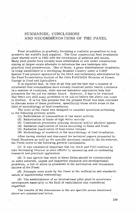
SUMMARIES, CONCLUSIONS AND RECOMMENDATIONS OF THE PANEL
Food irradiation is gradually becoming a realistic proposition to help preserve the w orld 's food supplies. .The first commercial food irradiation plant started work in 1965 with the irradiation of potatoes and onions.Many pilot plants have already been established or are under construction aiming at la rge r-sca le attempts to introduce the new technique into practical food preservation. One of these, a grain disinfestation irradiator, is being established in a developing Member Country under a UNDP Special Fund project sponsored by the IAEA and technically administered by the Food Preservation Section of the Joint FAO/IAEA Division of Atomic Energy in Food and Agriculture.
It is expected that, in view of all this and the fact that a number of irradiated food commodities have already received public health clearance in a number of countries, w ide-spread industrial application bids fair prospects for the not too distant future. However, it has to be realized that there are still many problems to be solved before the above can come to fruition. It is for this purpose that the present Panel has been convened to discuss some of these problems, specifically those which arise in the field of microbiology of food irradiation.
The work of the Panel was designed to consider questions pertaining to the following problem areas:
(1) Radicidation of commodities of low water activity(2) Radurization of foods of high water activity(3) Combination processes utilizing chemical and/or physical agents(4) Radiation inactivation of toxins occurring in foods and feeds(5) Radiation inactivation of food-borne viruses(6) Methodology of research in the microbiology of food irradiation.A fter having studied and discussed the technical papers presented by
Panel M em bers as well as the specific questions raised by the Secretariat, the Panel came to the following general conclusions:
(1) It was considered important that the IAEA and FA O continue to take increasing interest in such efforts by sponsoring and co-ordinating research and practical application
(2) It was agreed that work in these fields should be concentrated on some selected, urgent and important research and developmental problems, a list of which is presented in the summaries and recommendations of the Panel
(3) Attempts were made by the Panel at the unification and standardization of experimental methodology
(4) The establishment of an international pilot plant to accelerate development especially in the field of radicidation was considered important.
The results of the discussions in the six specific, areas mentioned above are summarized below.
139
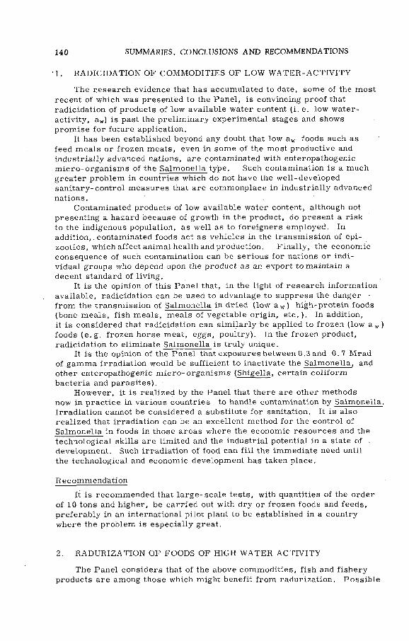
140 SUMMARIES, CONCLUSIONS AND RECOMMENDATIONS
1. RADICIDATION OF COMMODITIES OF LOW WATER- ACT I V I TY
The research evidence that has accumulated to date, some of the most recent of which was presented to the Panel, is convincing proof that radicidation of products of low available water content (i. e. low water- activity, aw) is past the prelim inary experimental stages and shows prom ise for future application.
It has been established beyond any doubt that low aw foods such as feed meals or frozen meats, even in some of the most productive and industrially advanced nations, are contaminated with enteropathogenic m icro-organism s of the Salmonella type. Such contamination is a much greater problem in countries which do not have the well-developed sanitary-control measures that are commonplace in industrially advanced nations.
Contaminated products of low available water content, although not presenting a hazard because of growth in the product, do present a risk to the indigenous population, as well as to foreigners employed. In addition,, contaminated foods act as vehicles in the transmission of epizootics, which affect animal health and production. Finally, the economic consequence of such contamination can be serious for nations or individual groups who depend upon the product as an export to maintain a decent standard of living.
It is the opinion of this Panel that, in the light of research information available, radicidation can be used to advantage to suppress the danger from the transmission of Salmonella in dried (low a w ) high-protein foods (bone meals, fish meals, m eals of vegetable origin, etc.) . In addition, it is considered that radicidation can sim ilarly be applied to frozen (low a w ) foods ( e.g. frozen horse meat, eggs, poultry). In the frozen product, radicidation to eliminate Salmonella is truly unique.
It is the opinion of the Panel that exposures between 0.3 and 0.7 Mrad of gamma irradiation would be sufficient to inactivate the Salmonella, and other enteropathogenic m icro-organism s (Shigella, certain coliform bacteria and parasites).
However, it is realized by the Panel that there are other methods now in practice in various countries to handle contamination by Salmonella. Irradiation cannot be considered a substitute for sanitation. It is also realized that irradiation can be an excellent method for the control of Salmonella in foods in those areas where the economic resources and the technological skills are limited and the industrial potential in a state of . development. Such irradiation of food can fill the immediate need until the technological and economic development has taken place.
Recommendation
It is recommended that la rge -sca le tests, with quantities of the order of 10 tons and higher, be carried out with dry or frozen foods and feeds, preferably in an international pilot plant to be established in a country where the problem is especially great.
2. RADURIZATION OF FOODS OF HIGH W ATER AC T IV ITY
The Panel considers that of the above commodities, fish and fishery products are among those which might benefit from radurization. Possible

SUMMARIES, CONCLUSIONS AND RECOMMENDATIONS 141
public health problems are the potential increased hazards fromCl. botulinum type E and enterobacteriaceae, in the latter case especiallyin shellfish.
Maintenance of storage temperature below 3: C before and after ir rad iation, and preferably during irradiation, would eliminate both problems.
At temperatures above 3°C but below 10°C it is not entirely clear from the evidence available, whether the botulism hazard is increased or not.
Recommendations
(1) L a rge -sca le radurization experiments should .be conducted with both naturally contaminated fish, such as Baltic herring and Great Lakes chub, and inoculated fish, particularly fatty fish which are highly botulinogenic.
(2) Sim ilar experiments should be conducted on pelagic fish such as sardines and anchovies which are a readily available source of protein.Due attention should be paid to special problem s with these fish such as fat oxidation.
(3) In inoculated pack experiments, inoculation should be at two levels: (a ) 10® per gram as proposed by Food and Drug Administration, USA; and (b) 102 per gram , which is c loser to the believed level of natural contamination although still several orders of magnitude in excess of it.
(4) In laboratory and pilot plant tests on radurization of sea foods, proper attention should be paid to the natural contamination level and growth of strictly anaerobic bacteria during post-irradiation storage.This could be done, e . g . , by employing relatively simple procedures such as replica plating.
(5) A systematic laboratory-scale investigation should be carried out to determine which fishery products w ill not support growth and toxin production by Cl. botulinum type E.
(6) In view of the apparent occasional localization of high contamination levels of enterobacteriaceas in shellfish, the most practical approach to control by irradiation would be to irradiate such samples and determine the efficiency of the envisaged process..
(7) Irradiation doses of the order of 0. 1-0. 5 M rad should be considered as giving appreciable extension of refrigerated storage life ( 0-5°C) of vacuum-packed fresh meats with no known health hazard.
In making these recommendations the panel is fully cognizant of the fact that the most satisfactory radurization process for fishery products would include control of storage temperature below 3°C.
3. COM BINATION PROCESSES UT IL IZ IN G CH EM ICAL AND/ORPH YSICAL AGENTS
The Panel is of the opinion that irradiation of products containing curing ingredients holds considerable prom ise with a view to radappertized and radurized products.
Evidence presented to the Panel with respect to sodium chloride, nitrates and nitrites and combinations of these compounds in meat pro

142 SUMMARIES, CONCLUSIONS AND RECOMMENDATIONS
ducts, as well as other quantitative laboratory evidence, clearly indicated that a considerable reduction in the effective radiation dose level is achieved.
The antibiotics nisin and tylosin, which were found suitable to reduce the heat requirements in the thermal preservation of various foods of pH over 4.5, appear to show some promise in lowering the dose requirements of foods, stored ideally below 3°C, to dose levels where radiation-induced sensory changes are not detectable.
It is necessary to reduce the initial viable cell count of raw m aterials and to destroy the tissue enzymes by application of a mild heat treatment.In such combined treatments, the radiation dose and the degree of heating must be sufficient to destroy moulds and other m icro-organism s resistant to antibiotics.
Laboratory investigations have shown that application of heat before irradiation appears to have little benefit with bacterial spores, but may show some synergistic action with some yeasts. (Simultaneous application of heat plus radiation was reported to increase the radiation resistance of bacterial spores, but seems to be effective in eliminating yeasts.) A definite increase in killing of spores is obtained when heat is applied after irradiation of spores. Therefore, in processes concerned with spore destruction, it appears advisable to apply moderate heating of foods after irradiation if such treatment is in accord with food technological considerations.
P ractica l experience with natural food-borne flora indicates that heating before radiation may be just as effective as heating after ir rad iation, but may offer important technological advantages in the preparation of the final product. M icrobiological aspects of this combination process appear to be worthy of more detailed investigations to evaluate any cumulative or synergistic effects of the treatment.
Recommendations
(1) Future work should concentrate on attempts to elucidate, at laboratory level, the synergism of radiation and curing ingredients coupled with the use of combinations of various levels of accepted additives in meat products. It is most desirable that attempts are made to design these two types of experiments in parallel.
(2) It is recommended to continue both basic and applied research simultaneously to .work out the optimal combination, method applying irradiation and nisin or tylosin, respectively, and to determine the most probably effective level of antibiotics and ionizing radiation for any possible application.
(3) It seems to be desirable to study the effect of the sequence of application of combination partners ( i .e. blanching, addition of antibiotics and irradiation).
(4) In the case of vegetative bacterial cells, concurrent radiation and heating seem to be more effective than the two treatments separately.Since this has only been observed with Salmonella, further work should be initiated to establish whether the phenomenon is general, e.g. with Streptococcus and Staphylococcus.
(5) The mechanism of radiation inactivation of antibiotics should be investigated, and both the biological and chemical properties of the decomposition products examined.
SUMMARIES, CONCLUSIONS AND RECOMMENDATIONS 143
(6) It is desirable to. elucidate the post-irradiation stability of antibiotics in different foods at different pH values and at different storage temperatures. The possibilities of producing antibiotic preparations acceptable as food additives and more resistant to radiation should also be explored.
4. RADIATION INACTIVATIO N OF TOXINS OCCURRING IN FOODSAND FEEDS
The Panel recognizes that food-borne diseases due to m icrobial toxins are best controlled by preventing growth of the toxin-producing organisms, but feels that circumstances might arise where elimination of the p re formed toxin by radiation might be considered.
The Panel notes that beside the fact that crude botulinum toxin appears to be rather resistant to ionizing radiation whereas the purified toxins in buffer solutions are sensitive, there is a paucity of information on the radiation resistance of toxins.
Recommendation
The Panel recommends intensification of basic and applied research on (a) radiation resistance»of botulinum toxins and other food- and feed- related toxins, e.g. Staphylococcus toxins, mycotoxins, etc., with special attention to the mechanism of toxin inactivation; (b) the effect of radiation sensitizing and radiation protective agents; and (c) the possibilities of making the toxin more vulnerable to subsequent spontaneous inactivation during storage or during thermal treatment. The convention of a meeting dealing with these problems is considered desirable.
5. RADIATION INACTIVATIO N OF FOO D-BO RNE VIRUSES
In the frozen state, the D value (decimal reduction dose) for the radiation inactivation of foot-and-mouth disease virus is around 0. 6 Mrad. This represents an extreme limit since other animal viruses in general are less radioresistant. At temperatures in excess of 0°C, all viruses may become less radioresistant. Nevertheless, the resistance may be such, even in these cases, that an effective dose for elimination of virus may be so great as to cause undesirable changes in the irradiated m aterial.
Hence, on the account of the extreme radioresistance of the animal viruses of sm all size, large radiation doses may be required for elim ination of viruses in food-stuffs. This is particularly true of the elimination of foot-and-mouth disease virus from frozen meat. It is therefore necessary to decide, in relation to initial infectivity, quantity of meat or other foodstuff involved and acceptable risk, what degree of inactivation is required. The existence of other safety factors, (e. g. thermal and pH inactivation effects) should not be overlooked, and there is some hope that, in certain cases, gam m a-irradiation may represent a useful virus control measure.
Recommendations
(1) It is recommended that a sm all study group should be set up to examine the above problem . Its aims would be: (i) to formulate the

144 SUMMARIES. CONCLUSIONS AND RECOMMENDATIONS
problem in exact terms with'whatever quantitative details are required;(ii) to apply existing experimental data to the problems as formulated in (i) above; and (iii) to decide what further data is required.
(2) In connection with item (iii) above, the following courses for further research should be considered: (a ) further in vivo experiments;(b) the effects on radioresistance of the virus, of the temperature of the irradiated m aterial ( i .e. in the range of -192 to +37°C); (c) the possibility that some reactivation of virus, after inactivation by the direct effect of radiation, may occur; (d) data on the magnitude of radiation doses re quired to fully degrade the virus by direct inactivation may be desired; and (e) the possibility of virus sensitization by physical and chemical agents to the direct and indirect inactivation mechanisms may be investigated.
6. M ETHODOLOGY OF RESEARCH IN THE M ICROBIOLOGY OF FOODIRRADIATION
Great attention has been paid by the Panel to the existence of widely conflicting data on the radioresistance of m icro-organism s as reported by various laboratories, and also to the frequently experienced lack of reproducibility of radiation survival curves as measured by the same researcher. It was generally recognized that part of these problem s could be eliminated by the adoption of suitable experimental designs and standard laboratory techniques, and it was, therefore, thought useful to make recommendations for studies on the radiation resistance of m icro-organism s.
Furtherm ore, in consideration of the fact that radiation research with pathogenic m icro-organism s is associated with dangers of which the personnel of the laboratory may not always be sufficiently aware, the Panel found it necessary to make some recommendations for the safety of work on. radiation resistance of pathogenic m icro-organism s.
Recommendations
(1) A s to the dose range to be applied, based on available evidence the Panel recommends that a range of doses be selected to enable observation of four possible responses: (a ) activation, (b) shoulder, (c ) inactivation, and (d) tail. Fo r example, for anaerobic spores suspended in water or buffer, suitable doses would be 0. 1, 0.2, 0.3, 0.6, 1.1, 1.5, and 2.0 M rad. Under conditions enhancing resistance ( e.g. in foods,or dry systems) correspondingly higher doses should be used.
(2) To facilitate comparison of data from different laboratories, the Panel recommends listing of the following param eters: (a) medium in which the cells have been grown (and if spores, sporulation medium and percentage sporulation); (b) medium in which cells were irradiated, pH, and conditions of anaerobiosis during irradiation (e.g. open vessels, closed vessels, aerated); (c) composition and pH of recovery medium;(d) age of spore suspension and temperature at which stored; (e ) initial concentration of cells, volume of suspension irradiated and number of cells irradiated; (f) temperature during irradiation, or temperature range, specifying whether measured or estimated; (g) radiation dose-

SUMMARIES, CONCLUSIONS AND RECOMMENDATIONS 145
rate and percentage e rro r; (h) conditions of storage of cells before i r radiation and from termination of irradiation to determining viability (time, temperature, and what medium); (i) details of method of recovery; besides composition and pH (cf. с), time and temperature of incubation.
(3) Replication should be clearly stated including: (a) number of samples irradiated at each dose level; (b) number of counts pérformed per irradiated sample; and (c) number of replicate plates or tubes.Ideally, the complete experiment (i. e. growing a fresh batch of cells and repeating irradiation and counting) should be performed 3 times to determine inherent variability between different cell crops of the same strain of organism.
(4) The Panel considers that expert advice should be sought regarding this suggested experimental design and the statistical analysis of results. '
(5) Fo r work on the radiation resistance of pathogenic m icroorganism s, the Panel recommends to have personnel: (a ) experienced in handling pathogens; .(b) immunized if possible; (c) properly dressed (laboratory coats or special clothes); (d) trained in laboratory discipline; disciplinary m easures should include: (1) prohibition of smoking or eating in laboratory; (2) refusal of admission of unauthorized personnel; (3) proper designation of laboratories with warnings for unauthorized personnel; (4) instructions in case of accident; (5) provision of first-a id kits, fire blankets, fire extinguisher and flush shower; (6) address of hospital, of availability of immune sera.
(6) A s regards laboratory design and equipment, the Panel considers that the following aspects should be given careful consideration in work on pathogens: (a) design for handling pathogen if possible, (b) benches and floors easy to clean (impervious to liquid or solvents if possible); (c) centrifuges, pathogen-tight and explosion-proof; (d) special safe for explosive solvents; (e) proper storage for cultures refrigerated or otherwise; (f) disinfectants available for pathogen inactivation (2% NaOH or 500 ppm chlorine solutions, freshly prepared daily); (g) disposal trays and handle trays for pathogenic and/or toxic disposal; (h) plastic bags specially designed for animal disposal, autoclaved before leaving laboratory or disposal by special personnel by means of incinerating; (i) specially designed inoculation boxes or hoods (available com m ercially or to be constructed by the individual).


LIST OF PANEL MEMBERS
A .W . ANDERSON Department of M icrobiology, Oregon State University, C orva llis , Oregon, USA
B. B A L D E L L I
J. FARKAS
Istituto Zooprofilattico Sperimentale dell 'U m b r ia e delle M arche,
V ia S. Costango No. 14,Perug ia , Italy
Department of M icrobiology,Central Food R esearch Institute, Herm an Otto ut 15,Budapest II, Hungary -
N . G R ECZ Biophysics Laboratory , Department of B iology,Illinois Institute of Technology, Chicago 60616,111., USA
G. HOBBS T o rry Research Station, M inistry of Technology,P . O . Box 31, 135 Abbey Road, Aberdeen, Scotland
C . D . JOHNSON
B . V . J0R G ENSEN
The J. J. Thompson Physical Laboratory, University of Reading,Whiteknights Park ,Reading, Be rks . ,England
Dem strupvej 5,Dronningborg pr. Randers,Denm ark
N. N . M ASO K H INA-PO R SH NYAK O VA
A ll-U n ion Scientific R esearch Institute for Canning and Vegetable Drying,
M oscow A -35 , USSR
D . A . A . M OSSEL
(Chairm an)Laborato ry of Bacteriology, Central Institute for Nutrition and
Food Technology (TNO ), Utrechtseweg 48,Ze ist , Netherlands
147

T. A . ROBERTS
G. SAKAGUCHI
O bservers
G. M E IL L A N D
H. IL L I
A . S Z ILV IN Y I
Scientific Secretary
K. VAS
Low Tem perature Research Station, Meat Research Institute, A gricu ltu ra l Research Council, Downing Street, Cam bridge,England
Department of Food Research, National Institute of Health,284 K am iosaki-Chojam aru, Shinagawa-ku, Tokyo, Japan
W HO, .Pa la is de Nations, Geneva, Switzerland
Osterreich ische Studiengesellschaft für Atom energie,
JLenaugasse 10,1090 Vienna, Austria
Institut für Angewandte M ikrobiologie, Hochschule für Bodenkultur, M ichae lerstrasse 25,1180 Vienna, A ustria
Joint F A O /IA E A D ivision of Atomic Energy in Food and Agricu lture
148

IA E A S A L E S A G E N T S
Orders for Agency publications can be pla
agents lis ted below :
A R G E N T I N AComision Nacional de Energía Atóm ica Aven ida del Libertador General San Martin 8250 Buenos A ires • Sue. 29
A U S T R A L I A .Hunter Publications*23 M cK illop Street
d w ith your bookseller or any of our sa les
F I N L A N DAkateeminen Kirjakauppa Keskuskatu 2 H els ink i
F R A N C E
O ffice international de documentation et librairie 48, rue Gay-Lussac F-75, Paris 5 e
Melbourne, C .l
A U S T R I AGeorg Frorame & Co.Spengergasse 39 A*1050t V ienna V
B E L G I U M
O ffice international de librairie 30, avenue Marnix B russels 5
B R A Z I LL ivra ria Kosm os Editora Rua do Rosario , 135-137 R io de Janeiro
A gencia Expoente Oscar M. S ilva Rua X avier de To ledo , 140-1® Andar (C aixa P os ta l No. 5.614)Sao Paulo
B YELO RU SSIAN SO V IE T S O C IA L IS TR E P U B L IC
See under USSR
CAN AD AThe Queen 's Printer Ottawa, Ontario
C H I N A (Ta iw an )
Books and Sc ien tific Supplies Serv ice , L td .,P .O . Box 83
T a ip e i
CZECH O SLO VAK SO C IA L IST R E PU B LICS .N .T .L ,Spolena 51 N ové Mesto Prague 1
D E N M A R KEjnar Munksgaard Ltd.6 Norregade Copenhagen К
G E R M A N Y , Federa l Republic of R. Oldenbourg Rosenheim er Strasse 145 8 Munich 8
H U N G A R YKulturaHungarian Trading Co, for Books and Newspapers P .O .B . 149 Budapest 62
I S R A E LH eilig e r and Co.
3 Nathan Strauss Street Jerusalem
I T A L YA genzia Ed itoria le Intem azionale O rgan izzazion i U n iversa li (A .E .I.O .U .) V ia M eravigli 16 Milan
J A P A NMaruzen Company Ltd .6, T o ri Nichome Nihonbashi (P .O . Box 605)Tokyo Central
M E X I C O
Librería Internacional A v . Sonora 206 M exico 11, D .F .
N E T H E R L A N D S N .V . Martinus N ijh o ff Lange Voorhout 9 The Hague
N E W Z E A L A N DWhitcombe & Tom bs, Ltd .G .P .O . Box 1894 .W ellington, C .l

N O R W A YJohan Grundt Tanum Karl Johans gate 43Oslo
P A K I S T A NKarachi Education Society Haroon Chambers South Nap ier Road (P .O . Box No. 4866)Karachi 2
P O L A N DOsrodek Rozpowszechniana Wydawnictw Naukowych Po lska Akademia Nauk Pa l'a с Kultury i Nauki Warsaw
R O M A N I ACartimexRue A . Briand 14-18 Bucarest
SOUTH A F R IC AVan Schaik*s Bookstore (P ty ) Ltd. L ib ri Building Church Street (P .O . Box 724)Pretoria
S P A I NLibreria BoschRonda de la Universidad 11Barcelona
S W E D E NC .E . F r itzes Kungl. Hovbokhandel Fredsgatan 2 Stockholm 16
S W I T Z E R L A N D Libra irie Payot Rue Grenus 6 1211 Geneva 11
T U R K E Y 'L ibra irie Hachette 469, Is tik lâ l Caddesi Beyoglu , Istanbul
U K R A IN IA N SO V IE T SO C IALISTR E PU B L IC
See under USSR
UNION OF S O V IE T SO C IAL ISTR E PU B LIC S
Mezhdunarodnaya Kniga Smolenskaya-Sennaya 32*34 Moscow G-200
U N ITE D KINGDOM OF G R E A TB R ITA IN AND N O RTH ERN IR E LA N D
Her M ajesty*s Stationery O ffice P .O . Box 569 London, S .E .l
U N ITED S TA TE S O F AM ERICA National Agency for International Pu b lications , Inc.317 East 34th Street New York, N .Y . 10016
V E N E Z U E L ASr. Braulio Gabriel Chacares Gobernador a Candilito 37 Santa Rosalia (Apartado P os ta l 8092)Caracas D .F ,
Y U G O S L A V I AJugoslovenska Knjiga T e ra z ije 27 Belgrade
IA E A publications can a lso be purchased reta il at the United Nations Bookshop at United Nations Headquarters, New York, at the news-stand at the Agency*s Headquarters, V ienna, and at most con ferences, symposia and seminars organized by the
A gency.
In order to fa c ilita te the distribution of its publications, the Agency is prepared to accept payment in UNESCO coupons or in lo ca l currencies.
Orders and inquiries from countries where sa les agents have not yet been appointed
may be sent to :
Distribution and Sales Group, International Atom ic Energy Agency,Kam tner Ring 11, A-1010, Vienna I, Austria

I N T E R N A T I O N A L A T O M I C E N E R G Y A G E N C Y V I E N N A , 1967
PRICE: U S $ 3 .0 0A u s t r i a n Sch il l ings 78 , - (£1.1.2; F .Fr. 14 ,70 ; D M 12,-)