Embed Size (px)
Citation preview
14
EMBRYO TRANSPLANTATION AND PRESERVATION
C. POLGEA. R.C. Institute of Animal Physiology, Animal Research Station,Huntingdon Road, Cambridge, UK
Effective techniques for embryo transplantation in some laboratory and
farm animals are now well established. Methods for collection and transfer
of embryos in the pig were first developed in the early 1960s (Hancock and
Hovell, 1962; Dziuk, Polge and Rowson, 1964; Vincent, Robison and
Ulberg, 1964) and since then they have been applied mainly in research.
Embryo transplantation has proved to be a valuable experimental tool in a
number of studies concerned with early embryonic development, the
survival of embryos in vivo or in vitro, migration and spacing of embryos
within the uterus and factors affecting the maintenance of pregnancy.
Future research is also likely to be concerned increasingly with cellular and
genetic manipulation of eggs and embryos in vitro and the application of
these techniques depends to a large extent on having reliable methods for
the culture of embryos and their subsequent transfer.Practical applications, particularly in farm animals, are also important
and the best example is in cattle where methods developed for research
have now been extended very successfully into the practice of animal
breeding. Applications in pig husbandry have so far been on a relatively
small scale. The high fecundity and reproductive rate in pigs compared
with cattle will never provide the same economic incentive to apply such
methods for the purpose of genetic improvement or to get more offspring
from a few superior animals. On the other hand, strict control of disease,
especially in large intensive units, is a most important aspect of modern pig
husbandry and embryo transplantation should provide the safest method of
introducting new genetic material into closed herds. It is mainly for this
reason, and perhaps also for the possibility of transporting embryos
between countries, that embryo transplantation in pigs is likely to be
applied as a practical measure.
Methods of embryo collectionand transfer
Techniques for collection and transfer of embryos used at different
laboratories are basically very similar to those described by Hancock and
Hovell (1962). The methods used routinely in experiments at the Animal
Research Station are described here.
277
278 Embryo transplantation and preservation
COLLECTION OF EMBRYOS
The tortuous nature of the cervix and uterus in the pig virtually precludesthe collection of embryos from the uterus by non-surgical means. Thesurgical approach, however, is relatively simple and operations on donoranimals can usually be completed in less than 30 minutes. Followingmid-ventral laparotomy under general anaesthesia the reproductive tract isexposed and a region appropriate to the developmental stage of theembryos to be collected (time after ovulation) is then flushed. One-, 2- andearly 4-cell embryos can be collected from the oviducts up to about 40hours after ovulation. A fine glass cannula is inserted into the isthmusthrough a small hole made in the tip of the uterine horn. Flushing fluidpassed down the oviduct from the fimbriated end is collected via thecannula into a glass cup or petri dish. Pig embryos normally enter theuterus much sooner and at an earlier stage of development than in manyother species examined. This means that flushing the oviducts alone shouldbe attempted only when the time of ovulation is known quite precisely.Estimates of ovulation time based on the assumption that the endogenousluteinizing hormone (LH) surge coincides with the onset of oestrus cansometimes be misleading. More precise timing of ovulation can beobtained by injection of human chorionic gonadotrophin (HUG) duringlate pro-oestrus (Dziuk and Baker, 1962; Hunter, 1972). When embryosenter the uterus at the 4-cell stage, it is then virtually impossible to flushthem back through the oviducts due to the valve-like nature of theutero-tubal junction. The most efficient technique for obtaining uterineembryos, or indeed those that may be tubal or uterine, is to flush fluiddown the oviduct and wash all the embryos away from the tip of the horn.The uterus is clamped with bowel forceps at an appropriate length from thetip of the horn and the flushing medium entering from the oviduct ismassaged towards the clamp. A cannula with a larger bore than that usedfor insertion into the isthmus is introduced into the uterine lumen througha small incision made near the tip of the horn and the fluid is 'milked' backout. Up to 5-6 days after oestrus the embryos remain close to the tips ofthe horns and it is only necessary to flush a small section. Later, when theembryos have started to migrate throughout the uterus, the whole of eachindividual horn should be flushed. Several different media have been usedsuccessfully for embryo collection, but the medium now used routinely atthe Animal Research Station is Dulbecco's phosphate buffered salineenriched with lactate, pyruvate and bovine serum albumin (Whittingham,1971). An advantage of a medium buffered with phosphate over someothers that may be buffered with bicarbonate is that pH is quite wellmaintained when the medium is exposed to air.
The methods of embryo recovery described are suitable for animals upto about 12 days after the onset of oestrus. If the uterus is flushed at laterstages when the embryos have become extremely elongated, theembryonic membranes become entangled together. The efficiency of thetechnique is very high and the majority of the embryos within thereproductive tract are generally recovered. For example, in a recentexperiment 205 gilts were used as embryo donors and the operations wereperformed 3-9 days after the onset of oestrus. Eggs or embryos were
C. Polge 279
recovered from all animals except one. In 57% of animals the recovery
rate, estimated by counting corpora lutea and embryos, was 100% and in
the remaining animals only a few embryos were missing. The average
recovery rate for all animals (3234 corpora lutea) was 95%.Repeated operations on donors inevitably tends to build up scar tissue
and adhesions, and in our experience 3-4 operations are about the
maximum that can be performed successfully on one individual.
Superovulation in donor animals can be achieved by administration of
gonadotrophic hormones at an appropriate time in the cycle. A dose of
1000-1500 iu pregnant mare's serum gonadotrophin (PMSG) given early in
the follicular phase (day 15 or 16 of the oestrous cycle) usually produces
25-30 ovulations in mature gilts (Hunter, 1964) although the response is
quite variable. A mixture of 600 iu PMSG + 200 iu HCG given as a single
dose will produce a similar result. Gonadotrophins can also be given after
some treatments used for the synchronization of oestrus (Polge, Day and
Groves, 1968). Heat normally occurs 3.5-4 days after PMSG treatment,
but 500 iu HCG given during the third day after PMSG will synchronize the
time of ovulation in a group of animals. Fertilization in superovulated
animals is generally normal except when the ovulation rate is excessively
high and a number of immature oocytes may be ovulated. Similar
treatments can be used to induce ovulation and obtain embryos from
prepubertal gilts (Dziuk and Gehlbach, 1966; Baker and Coggins, 1968).
EMBRYO TRANSFER
A similar surgical approach to that applied in donors is used for recipient
animals. After exposure of the reproductive tract, embryos can be
transferred either to the oviduct or uterus depending on the stage of
embryonic development. When transferring to the oviduct the embryos are
picked up in about 0.2 ml of fluid in a fine Pasteur pipette which is threaded
down the lumen via the fimbria to a depth of about 5 cm. When embryos
are transferred to the uterus, a small puncture is made in the isthmus
region of the oviduct about 2 cm from the tip of the horn and the tip of the
pipette is slid down into the uterine lumen through the utero-tubal
junction. This method avoids making a puncture wound in the uterus itself
and the consequent possibility of causing endometrial haemorrhage.
In early experiments (Polge, 1966) care was taken to transfer embryos at
very early stages of development (2-cell and early 4-cell) only to the
oviducts and all later stages to the uterus. In some species the site of
transplantation in relation to the stage of embryonic development appears
to be important. In cattle, for example, embryonic survival is reduced if
embryos collected from the oviducts of donors are transplanted to the
uterus of synchronized recipients one or two days earlier than the time they
would normally enter the uterus (Newcomb and Rowson, 1975). In pigs,
however, the length of time that embryos normally remain within the
oviducts is quite short and in recent experiments it has been found that
embryonic survival is not reduced when 2-cell embryos collected from the
oviducts are transplanted to the uterus of recipients at the same stage of the
reproductive cycle. An alternative method of transfer for all embryos can
280 Embryo transplantation and preservation
therefore be used and this is simply to flush them down the oviduct andinto the uterine lumen in a larger volume of fluid. It is generally onlynecessary to transfer the embryos to one side of the uterus since they willlater migrate and become evenly spaced throughout both horns (Dziuk,Polge and Rowson, 1964).
Attempts at non-surgical transfer of embryos via the cervix in the pighave not been very successful (Polge and Day, 1968). It is possible,however, that embryo transfer could be achieved by means of laparoscopy(B.N. Day, personal communication). This approach could be most usefulon farms where suitable facilities for surgery might be lacking.
Synchronization of oestrous cycles
An important factor affecting success in embryo transfer is the degree ofsynchrony between recipient and donor animals. In most species examinedit has been found that pregnancy rate and embryonic survival are reducedif the stage of the reproductive cycle of the recipient is more than aboutone day out of phase with that of the donor.
in many of the early experiments on embryo transfer in the pig,methallibure was used to control the time of onset of oestrus in donor andrecipient gilts. This drug was a most effective synchronizing agent and, inaddition, gonadotrophins could be used following treatment either tostimulate superovulation or induce ovulation at a predetermined time(Polge, Day and Groves, 1968). Since the use of methallibure was withheldin many countries in the early 1970s on the grounds that it was ateratogenetic agent, alternative methods for oestrus synchronization havebeen sought. In pigs, analogues of prostaglandin F2c, do not induceluteolysis effectively when given earlier than day 12 or 13 of the oestrouscycle (Guthrie and Polge, 1976a) and there is no way therefore that thesecompounds alone can be used to synchronize oestrus in randomly cyclinganimals. Various approaches have been attempted in order to creategroups of gilts in which the ovaries of all animals contained corpora luteawhich were old enOugh to respond to exogenous prostaglandin. Themethods included the induction of accessory corpora lutea (Guthrie andPolge, 1976b) and the extension of luteal function by oestrogen. Perhapsthe most successful method was simply to prolong luteal function by meansof pregnancy. In animals injected with cloprostenol (ESTRU-MATE, ICILtd) 12-40 days after mating, abortion was induced and a very highproportion returned to oestrus 4-7 days later. Insemination at the synchro-nized oestrus resulted in normal levels of fertilization and embryonicsurvival (Guthrie and Polge, 1978).
A simpler and more effective method of oestrus synchronization is nowavailable however. In the past, several orally active progestational agentshad been examined in pigs, but there were always major drawbacks to theiruses, including reduced fertility after treatment and the induction offollicular cysts. Encouraging preliminary results with a new orally activeprogestin, allyl trenbolone or altrenogest (REGU-MATE, Roussel Ltd)were first described at a recent Nottingham Easter School (Webel, 1978)and since then trials have been carried out at the Animal Research Station.
C. Polge 281
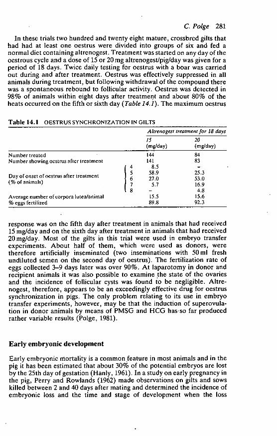
In these trials two hundred and twenty eight mature, crossbred gilts thathad had at least one oestrus were divided into groups of six and fed anormal diet containing altrenogest. Treatment was started on any day of theoestrous cycle and a dose of 15 or 20 mg altrenogest/pig/day was given for aperiod of 18 days. Twice daily testing for oestrus with a boar was carriedout during and after treatment. Oestrus was effectively suppressed in allanimals during treatment, but following withdrawal of the compound therewas a spontaneous rebound to follicular activity. Oestrus was detected in98% of animals within eight days after treatment and about 80% of theheats occurred on the fifth or sixth day (Table 14.1). The maximum oestrus
Table 14.1 OESTRUS SYNCHRONIZATION IN GILTS
Ahrenogest treatment for 18 days
15(mg/day)
20(mg/day)
Number treatedNumber showing oestrus after treatment
Day of onset of oestrus after treatment(% of animals)
Average number of corpora lutea/animal% eggs fertilized
/45678
144141
8.558.927.0
5.7—15.589.8
8483
—25.353.016.94.8
15.692.3
response was on the fifth day after treatment in animals that had received15 mg/day and on the sixth day after treatment in animals that had received20 mg/day. Most of the gilts in this trial were used in embryo transferexperiments. About half of them, which were used as donors, weretherefore artificially inseminated (two inseminations with 50 ml freshundiluted semen on the second day of oestrus). The fertilization rate ofeggs collected 3-9 days later was over 90%. At laparotomy in donor andrecipient animals it was also possible to examine the state of the ovariesand the incidence of follicular cysts was found to be negligible. Altre-nogest, therefore, appears to be an exceedingly effective drug for oestrussynchronization in pigs. The only problem relating to its use in embryotransfer experiments, however, may be that the induction of superovula-tion in donor animals by means of PMSG and HCG has-so far producedrather variable results (Polge, 1981).
Early embryonic development
Early embryonic niortality is a common feature in most animals and in thepig it has been estimated that about 30% of the potential embryos are lostby the 25th day of gestation (Hanly, 1961). In a study on early pregnancy inthe pig, Perry and Rowlands (1962) made observations on gilts and sowskilled between 2 and 40 days after mating and determined the incidence ofembryonic loss and the time and stage of development when the loss
282 Embryo transplantation and preservation
occurred. An important observation was that 22% of the embryos reco-vered from the uterus between the sixth and ninth days after matingappeared to be degenerating.
During the course of experiments on embryo transplantation at theAnimal Research Station an opportunity has been afforded to examine alarge number of embryos collected during the first 10 days after oestrus.These studies are interesting from the point of view of the efficiency offertilization and the extent of early embryonic degeneration in pigs. In theexperiment referred to in an earlier section, the embryos collected from205 donors 3-9 days after the onset of oestrus were examined. Since theaverage recovery rate was 95%, very few embryos were lost throughtechnical reasons and in the majority of cases the whole 'litter' representingall the eggs that had been ovulated was available. A total of 3085 embryoswas examined. Animals had been treated with altrenogest and inseminatedon the second day of oestrus as described above. Out of the 204 animalsfrom which eggs or embryos were recovered, none of the eggs wasfertilized in 11 (5%) pigs. In 82% of the remaining animals all the eggswere fertilized whereas in 18% there was a mixture of fertilized andunfertilized eggs. Eggs were classified as unfertilized if they were single cellor fragmenting and contained no sperm in the zona pellucida. Theproportion of unfertilized eggs in individual animals with partial fertiliza-tion varied quite considerably; in some it was just one or two eggs out ofthe total, but in others the number was much higher. The main cause offertilization failure related to unilateral sperm transport within the uterusand fertilized eggs were recovered from one uterine horn only. Overall,34% of the eggs were unfertilized in the animals in which fertilization wasnot complete. Apart from fertilization failure there was very little evidenceof embryonic loss up to nine days after the onset of oestrus. In all embryosclassified as fertilized, less than 1% showed any obvious signs of degenera-tion. These observations are clearly not in agreement with those of Perryand Rowlands (1962). It should be noted, however, that these authorstreated their data with some caution and stated that it was possible thatsome of the ova collected 6-9 days after mating and classified as degenerat-ing were, in fact, viable. They agree that if this were the case, thenmortality among fertilized eggs up to the ninth day must be very small.
Our data certainly suggest that embryonic mortality up to the ninth dayis very small, but these observations should also be treated cautiously.When embryos are used in transfer experiments it is possible only toexamine them in a relatively superficial manner under a stereomicroscope.It may be that a more detailed examination of fixed and stained embryoscould reveal abnormalities which are not immediately obvious. For exam-ple it was often noted that some of these embryos at the late morula andearly blastocyst stage appeared to have some cells developing outside themain body of the embryos themselves. These were not classified asdegenerate. Also, after the blastocysts had hatched, there were frequentlyconsiderable discrepancies in the size of embryos within a 'litter', althoughall appeared to be viable. Whether such features reflect the ability ofembryos to survive at a later stage is not known. Genetic abnormalitiesresulting from errors arising around the time of fertilization have beensuggested as a cause for early embryonic mortality. However, in a detailed
C. Polge 283
cytogenetic study of pig embryos collected from animals up to 12 days afterovulation, very few chromosomal abnormalities could be detected (Lupse,
1973).Other observations, also recorded in the study of Perry and Rowlands
(1962), suggest that a large amount of embryonic loss, at least during the
first 2-2.5 weeks of pregnancy, is not characteristic of most animals.Embryonic loss in thirteen pigs killed between the 13th and 18th days was '28.4%, but the greater part of this loss occurred in two of the animals. The
average loss in the remaining 11 animals was only 12%. Undoubtedly,however, embryonic losses are higher in the majority of animals by the30th day of pregnancy.
Results achieved in embryo transfer
When embryos have been collected from donors 2-5 days after the onset of
oestrus and transferred to synchronous unmated recipients, the pregnancy
rate achieved has been 60-70% with embryonic survival in pregnantanimals (usually determined about the 30th day of pregnancy) also around60-70% (Dziuk, Polge and Rowson, 1964; Vincent, Robison and Ulberg,
1964). Transfer of embryos at later stages, seven or eight days after theonset of oestrus, has resulted either in a complete failure to establishpregnancy (Webel, Peters and Anderson, 1970) or in much reduced
pregnancy rates (Hunter, Polge and Rowson, 1967). Relatively little isknown about the importance of synchrony between donors and recipients
in the pig, although in one experiment there was evidence that transfers inwhich oestrus in the donor was one or two days earlier or one day laterthan the recipient were as successful as synchronous transfers (Webel,
Peters and Anderson, 1970).Quite a large scale experiment has recently been undertaken at the
Animal Research Station in order to determine more precisely the effectsrelating to degree of synchrony between donors and recipients and stage of
embryonic development at which transfers are made. Although thisexperiment has not yet been completed, enough has been achieved toprovide some interesting results. Embryos have been collected from
donors on days 3, 4, 5, 6, 7, 8 and 9 (onset of oestrus = day 0). Transfershave been made to recipients in which the onset of oestrus was eithersynchronous with that of the donors or one or two days earlier or later.
Five recipients have been allocated to each group and thus the experimentinvolves 175 transfers of which results are now available for 140. The time of
onset of oestrus in groups of animals was controlled by feeding altrenogest
and the methods for collection and transfer of embryos were thosedescribed earlier. The average number of embryos transferred to recipients
was 14 (range 11-18) and embryonic survival in pregnant animals wasdetermined at slaughter on the 30th day of pregnancy.
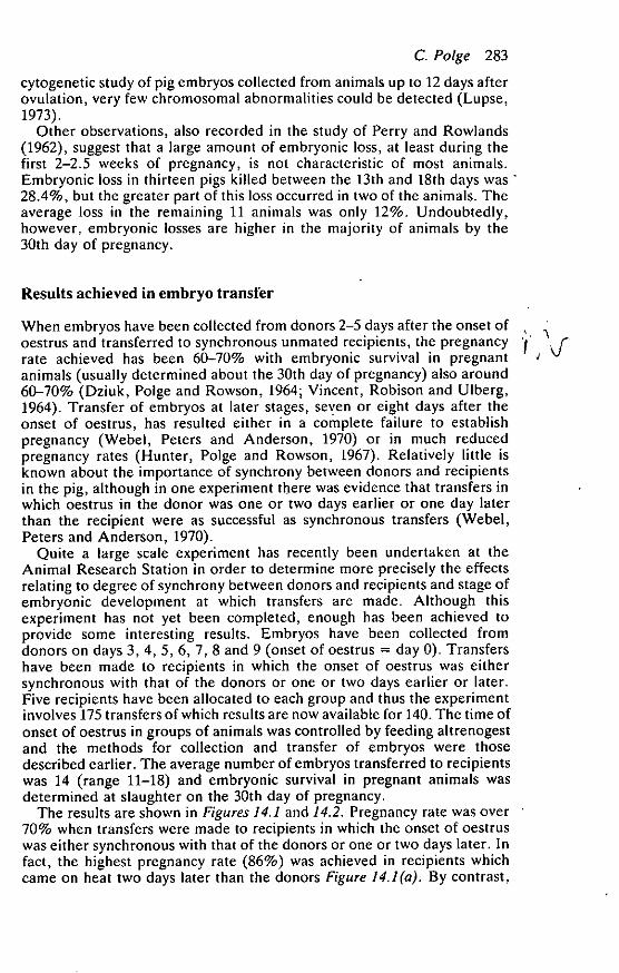
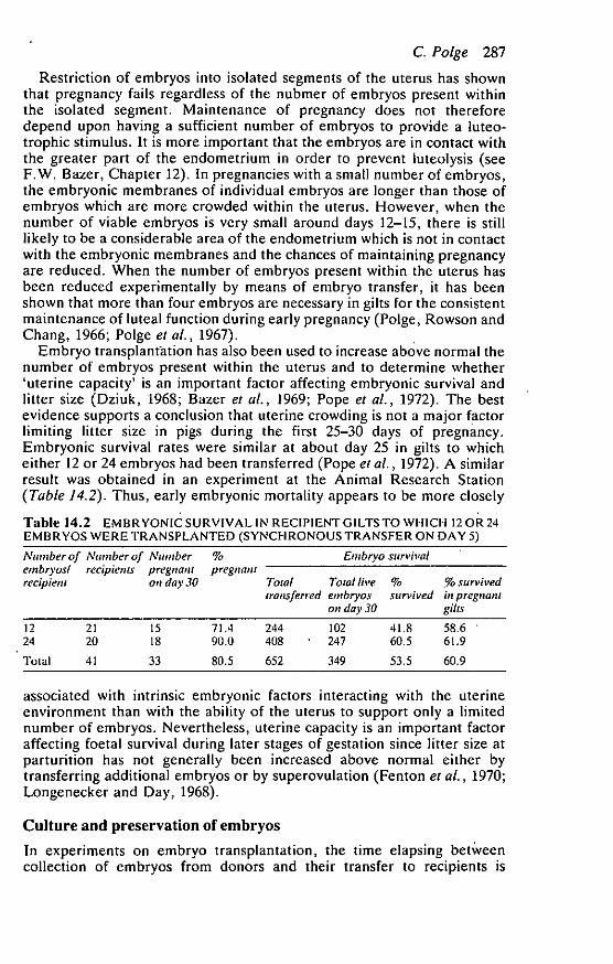
The results are shown in Figures 14.1 and 14.2. Pregnancy rate was over
70% when transfers were made to recipients in which the onset of oestruswas either synchronous with that of the donors or one or two days later. In
fact, the highest pregnancy rate (86%) was achieved in recipients whichcame on heat two days later than the donors Figure 14.1(a). By contrast,

284 Embryo transplantation and preservation
100 lal
8031
36
22
21
2 60
40
ae
20
30
—2 —1 0 1 +2
Oestrus in recipients occurred days after (-1 or before (+) donors
22 •
28
14
^kki
;11:118(
Wh;
• • • •
• • • •• • • •• • • •• • • •
• • • •• • • •• • • •
—1 0 +1+2—2
Oestrus in recipients occurred days after (—) or before (+) donors
100 (b)
80
60
18To
2a
I 40
20
Figure 14.1 Embryo transplantation in gilts: differences in synchrony between recipientsand donors. (a) Pregnancy rate following transfer of embryos collected from donors 3-9 daysafter the onset of oestrus. (b) Embryonic survival rate in pregnant gilts on the 30th day ofpregnancy. Numbers above columns refer to the numbers of recipients in (a) and the numbersof pregnant animals in (b)
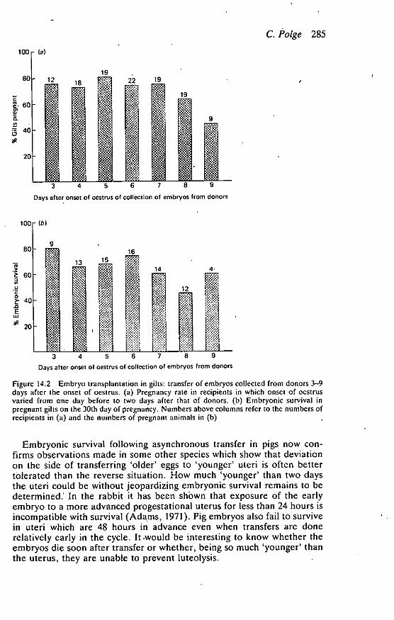
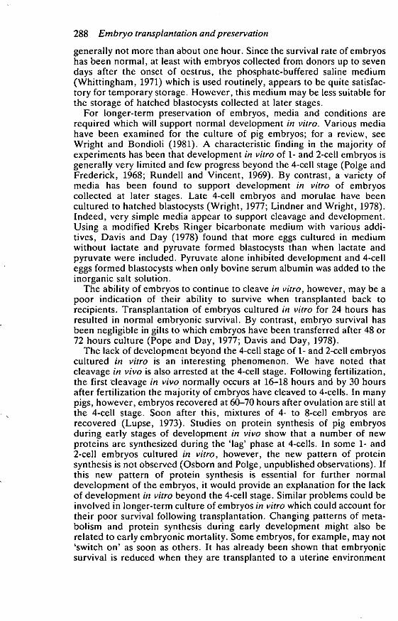
pregnancy rate fell dramatically in recipients which were ahead of thedonors and only one animal out of 22 became pregnant in the groups thatcame on heat two days before the donors. Results were similar whenembryos were collected from donors at any stage of the cycle from days3-9. The average embryonic survival rate in pregnant animals was 65% andwas lowest in the one animal that was two days ahead of the donor (Figure14.1(b). In Figure 14.2, the results have been presented according to theday of the cycle of the donor on which embryos were collected. The figuresinclude results from all recipients except those which were two days aheadof the donors since virtually no pregnancies were achieved in these groups.The main point of interest is that, although the results were somewhatlower in groups receiving embryos collected from donors on days 8 and 9,there was by no means a dramatic fall in pregnancy rate or embryonicstirvival following transfer of these older embryos.
C. Polge 285
100- (a)
80
e'
- 1219
22
19C
8. 60
0.
40
ae
20 II ceeet1.0
1819
3 4 5 6 7 8 9
Days after onset of oestrus of collection of embryos from donors
3 4 5 6 7 a 9Days after onset of oestrus of collection of embryos from donors
Figure 14.2 Embryo transplantation in gilts: transfer of embryos collected from donors 3-9days after the onset of oestrus. (a) Pregnancy rate in recipients in which onset of oestrusvaried from one day before to two days after that of donors. (b) Embryonic survival inpregnant gilts on the 30th day of pregnancy. Numbers above columns refer to the numbers ofrecipients in (a) and the numbers of pregnant animals in (b)
Embryonic survival following asynchronous transfer in pigs now con-firms observations made in some other species which show that deviationon the side of transferring 'older' eggs to 'younger' uteri is often bettertolerated than the reverse situation. How much 'younger' than two daysthe uteri could be without jeopardizing embryonic survival remains to bedetermined.' In the rabbit it has been shOwn that exposure of the earlyembryo to a more advanced progestational uterus for less than 24 hours isincompatible with survival (Adams, 1971). Pig embryos also fail to survivein uteri which are 48 hours in advance even when transfers are donerelatively early in the cycle. It miould be interesting to know whether theembryos die soon after transfer or whether, being so much 'younger' thanthe uterus, they are unable to prevent luteolysis.
286 Embryo transplantation and preservation
Earlier experiments (Webel, Peters and Anderson, 1970) in which noembryos survived when transferred to recipients on days 7 and 8, led to thesuggestion that perhaps embryos need to be present in the uterus of the pigbefore day 7 for pregnancy to be established and maintained. Our recentresults do not confirm this suggestion. A possible explanation of the poorresults achieved following transfer of older embryos in some earlierexperiments could relate to the type of medium used for collection. Wehave noted that when embryos have hatched from the zona pellucida theyare very easily damaged by adverse environmental conditions. Media thatwill support development of early embryos in vitro may be unsuitable forthe storage of embryos at a later stage (Robl and Davis, 1981).
Embryo transplantation in research
Embryo transplantation has been used as an experimental tool in a numberof studies on relationships between the uterus and embryos during earlystages of gestation. In most pregnancies some embryos probably migratefrom one uterine horn to the other before implantation in order toestablish even spacing throughout the uterus. The incidence of transuterinemigration and the mixing of embryos within the uterus has been studiedexperimentally by transferring genetically marked embryos to each uterinehorn (Dziuk, Polge and Rowson, 1964). When embryos from black donorswere transferred to the tip of one uterine horn and embryos from whitedonors were transferred to the tip of the opposite horn, it was found thatnot only did they migrate from the horn of origin, but in most cases theybecame interspersed throughout the uterus.
Migration of embryos from the tips of the horns usually occurs soon afterthe sixth day of gestation. In experiments in which embryos were permittedto enter the uterus from one side only it was found that they started toenter the opposite horn around day 8 or 9 and the uterus was occupiedcompletely by day 15 (Dhindsa, Dziuk and Norton, 1967). The time ofcessation of intrauterine migration was studied by restricting the embryosto anterior sections of the uterine horns by means of ligatures applied soonafter mating. Removal of the ligatures between days 8 and 13 showed thatembryos could still migrate beyond the ligature sites up to day 11 andpregnancy was then maintained. Gilts in which the ligatures were removedon day 13 did not remain pregnant (Polge and Dziuk, 1970).
There is now much evidence to show that the presence of embryosthroughout a large part of the uterus by days 12 or 13 is essential for themaintenance of pregnancy. Non-gravid sections of the uterus have beenestablished experimentally, in some cases by transferring embryos toisolated segments (du Mesnil du Buisson, 1966; Anderson, Rathmacherand Melampy, 1966; Day et al. , 1967; Dhindsa and Dziuk, 1968). It hasbeen shown that a relationship exists between the length of the non-pregnant uterine segment and maintenance of pregnancy. Pregnancy isusually interrupted when over one half of a uterine horn does not containembryos. However, pregnancy continues when a smaller sterile uterinesegment is isolated adjacent to one ovary even though corpora lutea in theipsilateral ovary may regress. Effects of non-pregnant uterine segments aresimilar whether they are located at the tip or base of the uterine horns.
C. Polge 287
Restriction of embryos into isolated segments of the uterus has shownthat pregnancy fails regardless of the nubmer of embryos present withinthe isolated segment. Maintenance of pregnancy does not thereforedepend upon having a sufficient number of embryos to provide a luteo-trophic stimulus. It is more important that the embryos are in contact withthe greater part of the endometrium in order to prevent luteolysis (seeF.W. Bazer, Chapter 12). In pregnancies with a small number of embryos,the embryonic membranes of individual embryos are longer than those ofembryos which are more crowded within the uterus. However, when thenumber of viable embryos is very small around days 12-15, there is stilllikely to be a considerable area of the endometrium which is not in contactwith the embryonic membranes and the chances of maintaining pregnancyare reduced. When the number of embryos present within the uterus hasbeen reduced experimentally by means of embryo transfer, it has beenshown that more than four embryos are necessary in gilts for the consistentmaintenance of luteal function during early pregnancy (Polge, Rowson andChang, 1966; Paige et al., 1967).
Embryo transplantation has also been used to increase above normal thenumber of embryos present within the uterus and to determine whether'uterine capacity' is an important factor affecting embryonic survival andlitter size (Dziuk, 1968; Bazer et al., 1969; Pope et al., 1972). The bestevidence supports a conclusion that uterine crowding is not a major factorlimiting litter size in pigs during the first 25-30 days of pregnancy.Embryonic survival rates were similar at about day 25 in gilts to whicheither 12 or 24 embryos had been transferred (Pope et al., 1972). A similarresult was obtained in an experiment at the Animal Research Station(Table 14.2). Thus, early embryonic mortality appears to be more closely
•Table 14.2 EMBRYONIC SURVIVAL IN RECIPIENT GILTS TO WHICH 12 OR 24EMBRYOS WERE TRANSPLANTED (SYNCHRONOUS TRANSFER ON DAY 5)
Number of Number of Number % Embryo survival
embryos/ recipients pregnant pregnant
recipient on day 30 Total Total live % % survivedtransferred embryos survived in pregnant
on day 30 gills
12 21 15 71.4 244 102 41.8 58.6 •24 20 18 90.0 408 ' 247 60.5 61.9
Total 41 33 80.5 652 349 53.5 60.9
associated with intrinsic embryonic factors interacting with the uterineenvironment than with the ability of the uterus to support only a limitednumber of embryos. Nevertheless, uterine capacity is an important factoraffecting foetal survival during later stages of gestation since litter size atparturition has not generally been increased above normal either bytransferring additional embryos or by superovulation (Fenton et al., 1970;Longenecker and Day, 1968).
Culture and preservation of embryos
In experiments on embryo transplantation, the time elapsing betweencollection of embryos from donors and their transfer to recipients is
288 Embryo transplantation and preservation
generally not more than about one hour. Since the survival rate of embryoshas been normal, at least with embryos collected from donors up to sevendays after the onset of oestrus, the phosphate-buffered saline medium(Whittingham, 1971) which is used routinely, appears to be quite satisfac-tory for temporary storage. However, this medium may be less suitable forthe storage of hatched blastocysts collected at later stages.
For longer-term preservation of embryos, media and conditions arerequired which will support normal development in vitro. Various mediahave been examined for the culture of pig embryos; for a review, seeWright and Bondioli (1981). A characteristic finding in the majority ofexperiments has been that development in vitro of 1- and 2-cell embryos isgenerally very limited and few progress beyond the 4-cell stage (Polge andFrederick, 1968; Rundell and Vincent, 1969). By contrast, a variety ofmedia has been found to support development in vitro of embryoscollected at later stages. Late 4-cell embryos and morulae have beencultured to hatched blastocysts (Wright, 1977; Lindner and Wright, 1978).Indeed, very simple media appear to support cleavage and development.Using a modified Krebs Ringer bicarbonate medium with various addi-tives, Davis and Day (1978) found that more eggs cultured in mediumwithout lactate and pyruvate formed blastocysts than when lactate andpyruvate were included. Pyruvate alone inhibited development and 4-celleggs formed blastocysts when only bovine serum albumin was added to theinorganic salt solution.
The ability of embryos to continue to cleave in vitro, however, may be apoor indication of their ability to survive when transplanted back torecipients. Transplantation of embryos cultured in vitro for 24 hours hasresulted in normal embryonic survival. By contrast, embryo survival hasbeen negligible in gilts to which embryos have been transferred after 48 or72 hours culture (Pope and Day, 1977; Davis and Day, 1978).
The lack of development beyond the 4-cell stage of 1- and 2-cell embryoscultured in vitro is an interesting phenomenon. We have noted thatcleavage in vivo is also arrested at the 4-cell stage. Following fertilization,the first cleavage in vivo normally occurs at 16-18 hours and by 30 hoursafter fertilization the majority of embryos have cleaved to 4-cells. In manypigs, however, embryos recovered at 60-70 hours after ovulation are still atthe 4-cell stage. Soon after this, mixtures of 4- to 8-cell embryos arerecovered (Lupse, 1973). Studies on protein synthesis of pig embryosduring early stages of development in vivo show that a number of newproteins are synthesized during the 'lag' phase at 4-cells. In some 1- and2-cell embryos cultured in vitro, however, the new pattern of proteinsynthesis is not observed (Osborn and Polge, unpublished observations). Ifthis new pattern of protein synthesis is essential for further normaldevelopment of the embryos, it would provide an explanation for the lackof development in vitro beyond the 4-cell stage. Similar problems could beinvolved in longer-term culture of embryos in vitro which could account fortheir poor survival following transplantation. Changing patterns of meta-bolism and protein synthesis during early development might also berelated to early embryonic mortality. Some embryos, for example, may not'switch on' as soon as others. It has already been shown that embryonicsurvival is reduced when they are transplanted to a uterine environment
C. Polge 289
which is more advanced than that from which they have been recovered. Itseems unlikely, however, that there is any very specific uterine factor in thepig which regulates early embryonic development since pig embryossurvive and develop normally when transferred to the oviduct of a rabbit(Polge, Adams and Baker, 1972).
Preservation of embryos by freezing and storage in liquid nitrogen hasnow been achieved in a number of species. So far, however, pig embryoshave not been frozen and thawed successfully. A major problem is theirsensitivity to cooling per se and few have survived after exposure totemperatures below +15 °C (Polge, Wilmut and Rowson, 1974). Duringcooling and rewarming it has been observed that there is a loss ofintracellular lipids, which are very abundant in the pig embryo. It would beinteresting to speculate that it is the lipid composition of the embryonicmembranes which affects their sensitivity to cooling (Polge, 1977).
Despite the inability to freeze pig embryos, some successful long-distance shipments of embryos have been achieved (Baker and Dziuk,1969; James et al., 1980). In the latter experiment, embryos were trans-ported in culture medium from the USA to England and transplanted torecipients within 20-27 hours after collection from donors. Seven of the 12recipients farrowed producing 58 piglets from 227 transferred embryos.
References
ADAMS, C.E. (1971). The fate of fertilized eggs transferred to the uterus oroviduct during advancing pseudopregnancy in the rabbit. J. Reprod.Fert. 26, 99-111
ANDERSON, L.L., RATHMACHER, R.P. and MELAMPY, R.M. (1966). The uterusand unilateral regression of corpora lutea in the pig. Am. J. Physiol. 210,611-614
BAKER, R.D. and COGGINS, E.G. (1968). Control of ovulation rate andfertilization in prepubertal gilts. J. Anim. Sci. 27, 1607-1610
BAKER, R.D. and DZIUK, (1970). Aerial transport of fertilized pig ova.Can. J. Anirn. Sd. 50, 215-216
BAZER, F.W., ROBISON, 0.W., CLAWSON, A.J. and ULBERG, L.C. (1969).Uterine capacity at two stages of gestation in gilts following embryosuperinduction. J. Anim. Sd. 29, 30-34
DAVIS, D.L. and DAY, B.N. (1978). Cleavage and blastocyst formation by pigeggs in vitro. J. Anim. Sci. 46, 1043-1053
DAY, B.N., F'OLGE, C., MOOR, R.M., ROWSON, L.E.A. and BOOTH, D. (1967).Local effect of the uterus on the corpora lutea of early pregnancy inswine. J. Anim. Sci. 26, 499 (Abstract)
DHINDSA, D.S. and DZIUK, P.i. (1968). Influence of varying the proportion ofuterus occupied by embryos on maintenance of pregnancy in the pig. J.Anim. Sd. 27, 668-672
DHINDSA, D.S., DZIUK, P.J. and NORTON, H.W. (1967). Time of transuterinemigration and distribution of embryos in the pig. Anat. Rec. 159,325-330
DU MESNIL DU BUISSON, F. (1966). Contribution al'étude du maintien ducorps jaune de la truie. Thesis. University of Paris
290 Embryo transplantation and preservation
DZIUIC, PI (1968). Effect of number of embryos and uterine space onembryo survival in the pig. J. Anim. Sci. 27, 673-676
DZIUK, P.J. and BAKER, R.D. (1962). Induction and control of ovulation inswine. J. Anim. Sci. 21, 697-699
DZIUK, P.J. and GEHLBACH, G.D. (1966). Induction of ovulation and ferti-lization in the immature gilt. J. Anirn. Sci. 25, 410-413
DZIUK, P.J., POLGE, C. and ROWSON, L.E.A. (1964). Intra - uterine migrationand mixing of embryos in swine following egg transfer. J. Anim. Sci. 23,37-42
FENTON, F.R., BAZER, F.W., ROBISON, 0.W. and ULBERG, L.C. (1970). Effectof quantity of uterus on uterine capacity in gilts. J. Anirn. Sci. 31,104-106
GUTHRIE, H.D. and POLGE, C. (1976a). Luteal function and oestrus in giltstreated with a synthetic analogue of prostaglandin F2e, (ICI 79,939) atvarious times during the oestrous cycle. J. Reprod. Fen. 48, 423-425
GUTHRIE, H.D. and POLGE, C. (1976b). Control of oestrus and fertility ingilts with accessory corpora lutea by prostaglandin analogues ICI 79,939and ICI 80,996. J. Reprod. Fert. 48, 427-430
GUTHRIE, HAD. and POLGE, C. (1978). Treatment of pregnant gilts with aprostaglandin analogue, cloprostenol, to control oestrus and fertility. J.Reprod. Fert. 52, 271-273
HANCOCK, J.L. and HOVELL, G.J.R. (1962). Egg transfer in the sow. J.Reprod. Fert. 4, 195-201
HANLY, S. (1961). Prenatal mortality in farm animals. J. Reprod. Fert. 2,182-194
HUNTER, R.H.F. (1964). Superovulation and fertility in the pig. Anim.Prod. 6, 189-194
HUNTER, R.H.F. (1972). Ovulation in the pig: timing of the response toinjection of human chorionic gonadotrophin. Res. vet. Sci. 13, 356-361
HUNTER, R.H.F., POLGE, C. and ROWSON, L.E.A. (1967). The recovery,transfer and survival of blastocysts in pigs. J. Reprod. Fert. 14, 501- 502
JAMES, J.E., REESER, P.D., DAVIS, D.L., STRAITON, E.C., TALBOT, A.C.•and
POLGE, C. (1980). Culture and long-distance shipment of swine embryos.Theriogenology 14, 463-469
LONGENECKER, D.E. and DAY, B.N. (1968). Fertility level of sows superovu-lated at post weaning estrus. J. Anitn. Sci. 27, 709-711
LINDNER, G.M. and WRIGHT, R.W. (1978). Morphological and quantitativeaspects of the development of swine embryos in vitro. J. Anitn. Sci. 46,711-718
LUPSE, R.M. (1973). Early embryonic development in the pig: A cleavagetiming and cytogenetic study. Thesis. The Graduate School of Arts andSciences, George Washington University, Washington, D.C.
NEWCOMB, R. and ROWSON, L.E.A. (1975). Conception rate after uterinetransfer of cow eggs in relation to synchronization of oestrus and age ofeggs. J. Reprod. Fen. 43, 539-541
PERRY, J.S. and ROWLANDS, I.W. (1962). Early pregnancy in the pig. J.Reprod. Fert. 4, 175-188
POLGE, C. (1966). Egg transplantation in the pig. Wld Rev. Anitn. Prod. 4,79-86
POLGE, C. (1977). The freezing of mammalian embryos: perspectives and
C. Polge 291
possibilities. In The Freezing of Mammalian Embryos (K. Elliott and J.Whelan, Eds.), pp. 3-13. Amsterdam, Elsevier/Exerpta Medica, North-Holland
POLGE, C. (1981). An assessment of techniques for the control of oestrus andovulation in pigs. In Steroids in Animal Reproduction (H. Jasiorowski,Ed.), pp. 73-84. Warsaw Agricultural University: SGGW-AR, Roussel-Uclaf,,Warsaw
POLGE, C. and DAY, B.N. (1968). Pregnancy following non-surgical eggtransfer in pigs. Vet. Rec. 82, 712
POLGE, C. and DZIUK, P.J. (1970). Time of cessation of intrauterine migra-tion of pig embryos. J. Anim. Sci. 31, 565-566
POLGE, C. and FREDERICK, C.L. (1968). Culture and storage of fertilized pigeggs. VIth Int Congr. Anim. Reprod. A.I., Paris, 1, 211 (Abstract)
POLGE, C., ADAMS. C.E. and BAKER, R.D. (1972). Development and survivalof pig embryos in the rabbit oviduct. VIlth Int. Congr. Anim. Reprod.A.L, Munich, 1, 513 - 517
POLGE, C., DAY, B.N. and GROVES, T.W. (1968). Synchronization of ovulation .and artificial insemination in pigs. Vet. Rec. 83, 136-142
POLGE, C., ROWSON, L.E.A. and CHANG, M.C. (1966). The effect of reducingthe number of embryos during early stages of gestation on the mainte-nance of pregnancy in the pig. J. Reprod. Fert. 12, 395-397
POLGE, C., WILMUT, I. and ROWSON, L.E.A. (1974). Low temperature pre-servation of cow, sheep and pig embryos. Cryobiology, 11, 560
POLGE, C., MOOR, R.M., DAY, B.N., BOOTH, W.D. and ROWSON, L.E.A. (1967).
• Embryo numbers and luteal maintenance during early pregnancy inswine. J. Anim. Sci., 26, 1499 (Abstract)
POPE, C.E. and DAY, B.N. (1977). Transfer of preimplantation pig embryosfollowing in vitro culture for 24 or 48 hours. J. Anim. Sci. 44, 1036-1040
POPE, C.E., CHRISTENSEN, R.K., Z1MMERMAN-POPE, V.A. and DAY, B.N.
(1972). Effect of number of embryos on embryonic survival in recipientgilts. J. Anim. Sci. 35, 805 - 808
ROBL, J.M. and DAVIS, D.L. (1981). Effects of serum on swine morulae andblastocysts in vitro. J. Anim. Sci. 52, 1450- 1456
RUNDELL, J.W. and VINCENT, C.K. (1969). In vitro culture of swine ova. J.Anim. Sci. 27, 1196 (Abstract)
VINCENT, C.K., ROBISON, 0.W. and ULBERG, L.C. (1964). A technique forreciprocal embryo transfer in swine. J. Anim. Sci. 23, 1084-1088
WEBEL, S.K. (1978). Ovulation control in the pig. In Control of Ovulation(D.B. Crighton, N.B. Haynes, G.R. Foxcroft and G.E. Lamming,Eds.), pp. 421-434. London, Butterworths
WEBEL, S.K., PETERS, J.B. and ANDERSON, L.L. (1970). Synchronous andasynchronous transfer of embryos in the pig. J. Anim. Sci. 30, 565-568
WHITTINGHAM, D.G. (1971). Survival of mouse embryos after freezing andthawing. Nature, Lond. 233, 125-126
WRIGHT, R.W. (1977). Successful culture in vitro of swine embryos to theblastocyst stage. J. Anim. Sci. 44, 854-858
WRIGHT, R.W. and BONDIOLI, K.R. (1981). Aspects of in vitro fertilizationand embryo culture in domestic animals. J. Anim. Sci. 53, 702-729


