Embed Size (px)
Citation preview
CLOSED-LOOP SYSTEMS FOR NEXT-GENERATION NEUROPROSTHESESEDITED BY : Timothée Levi, Paolo Bonifazi, Paolo Massobrio and
Michela ChiappalonePUBLISHED IN : Frontiers in Neuroscience and Frontiers in Neurology

1 April 2018 | Next Generation Closed-Loop Neuroprostheses
Frontiers Copyright Statement
© Copyright 2007-2018 Frontiers Media SA. All rights reserved.
All content included on this site, such as text, graphics, logos, button
icons, images, video/audio clips, downloads, data compilations and
software, is the property of or is licensed to Frontiers Media SA
(“Frontiers”) or its licensees and/or subcontractors. The copyright in the
text of individual articles is the property of their respective authors, subject to
a license granted to Frontiers.
The compilation of articles constituting this e-book, wherever published,
as well as the compilation of all other content on this site, is the exclusive
property of Frontiers. For the conditions for downloading and
copying of e-books from Frontiers’ website, please see the Terms for
Website Use. If purchasing Frontiers e-books from other websites
or sources, the conditions of the website concerned apply.
Images and graphics not forming part of user-contributed materials may
not be downloaded or copied without permission.
Individual articles may be downloaded and reproduced in accordance
with the principles of the CC-BY licence subject to any copyright or
other notices. They may not be re-sold as an e-book.
As author or other contributor you grant a CC-BY licence to others to
reproduce your articles, including any graphics and third-party materials
supplied by you, in accordance with the Conditions for Website Use and
subject to any copyright notices which you include in connection with your
articles and materials.
All copyright, and all rights therein, are protected by national and
international copyright laws.
The above represents a summary only. For the full conditions see the
Conditions for Authors and the Conditions for Website Use.
ISSN 1664-8714 ISBN 978-2-88945-466-2
DOI 10.3389/978-2-88945-466-2
About Frontiers
Frontiers is more than just an open-access publisher of scholarly articles: it is a pioneering approach to the world of academia, radically improving the way scholarly research is managed. The grand vision of Frontiers is a world where all people have an equal opportunity to seek, share and generate knowledge. Frontiers provides immediate and permanent online open access to all its publications, but this alone is not enough to realize our grand goals.
Frontiers Journal Series
The Frontiers Journal Series is a multi-tier and interdisciplinary set of open-access, online journals, promising a paradigm shift from the current review, selection and dissemination processes in academic publishing. All Frontiers journals are driven by researchers for researchers; therefore, they constitute a service to the scholarly community. At the same time, the Frontiers Journal Series operates on a revolutionary invention, the tiered publishing system, initially addressing specific communities of scholars, and gradually climbing up to broader public understanding, thus serving the interests of the lay society, too.
Dedication to Quality
Each Frontiers article is a landmark of the highest quality, thanks to genuinely collaborative interactions between authors and review editors, who include some of the world’s best academicians. Research must be certified by peers before entering a stream of knowledge that may eventually reach the public - and shape society; therefore, Frontiers only applies the most rigorous and unbiased reviews. Frontiers revolutionizes research publishing by freely delivering the most outstanding research, evaluated with no bias from both the academic and social point of view.By applying the most advanced information technologies, Frontiers is catapulting scholarly publishing into a new generation.
What are Frontiers Research Topics?
Frontiers Research Topics are very popular trademarks of the Frontiers Journals Series: they are collections of at least ten articles, all centered on a particular subject. With their unique mix of varied contributions from Original Research to Review Articles, Frontiers Research Topics unify the most influential researchers, the latest key findings and historical advances in a hot research area! Find out more on how to host your own Frontiers Research Topic or contribute to one as an author by contacting the Frontiers Editorial Office: [email protected]
2 April 2018 | Next Generation Closed-Loop Neuroprostheses
CLOSED-LOOP SYSTEMS FOR NEXT-GENERATION NEUROPROSTHESES
Cloud of the keywords used by the authors contributing to this e-book and logo of the FET European project BrainBow, which depicts a futuristic closed-loop neuroprosthesis for the brain. Copyright: T. Levi, P. Bonifazi, P. Massobrio, M. Chiappalone, S. Chiappalone.
Cover image: Naeblys/Shutterstock.com
Topic Editors: Timothée Levi, University of Bordeaux, FrancePaolo Bonifazi, Biocruces Health Research Institute, SpainPaolo Massobrio, Universita di Genova, ItalyMichela Chiappalone, Istituto Italiano di Tecnologia Genova, Italy
Millions of people worldwide are affected by neurological disorders which disrupt the connec-tions within the brain and between brain and body causing impairments of primary functions and paralysis. Such a number is likely to increase in the next years and current assistive technology is yet limited. A possible response to such disabilities, offered by the neuroscience community, is given by Brain-Machine Interfaces (BMIs) and neuroprostheses.

3 April 2018 | Next Generation Closed-Loop Neuroprostheses
The latter field of research is highly multidisciplinary, since it involves very different and dis-perse scientific communities, making it fundamental to create connections and to join research efforts. Indeed, the design and development of neuroprosthetic devices span/involve different research topics such as: interfacing of neural systems at different levels of architectural complexity (from in vitro neuronal ensembles to human brain), bio-artificial interfaces for stimulation (e.g. micro-stimulation, DBS: Deep Brain Stimulation) and recording (e.g. EMG: Electromyography, EEG: Electroencephalography, LFP: Local Field Potential), innovative signal processing tools for coding and decoding of neural activity, biomimetic artificial Spiking Neural Networks (SNN) and neural network modeling. In order to develop functional communication with the nervous system and to create a new generation of neuroprostheses, the study of closed-loop systems is mandatory. It has been widely recognized that closed-loop neuroprosthetic systems achieve more favorable outcomes for users then equivalent open-loop devices. Improvements in task performance, usability, and embodiment have all been reported in systems utilizing some form of feedback. The bi-directional communication between living neurons and artificial devices is the main final goal of those studies. However, closed-loop systems are still uncommon in the literature, mostly due to requirement of multidisciplinary effort. Therefore, through eBook on closed-loop systems for next-generation neuroprostheses, we encourage an active discussion among neurobiologists, electrophysiologists, bioengineers, computational neuroscientists and neuromorphic engineers.
This eBook aims to facilitate this process by ordering the 25 contributions of this research in which we highlighted in three different parts: (A) Optimization of different blocks composing the closed-loop system, (B) Systems for neuromodulation based on DBS, EMG and SNN and (C) Closed-loop BMIs for rehabilitation.
Citation: Levi, T., Bonifazi, P., Massobrio, P., Chiappalone, M., eds. (2018). Closed-Loop Systems for Next-Generation Neuroprostheses. Lausanne: Frontiers Media. doi: 10.3389/978-2-88945-466-2

4 April 2018 | Next Generation Closed-Loop Neuroprostheses
Table of Contents
07 Editorial: Closed-Loop Systems for Next-Generation NeuroprosthesesTimothée Levi, Paolo Bonifazi, Paolo Massobrio and Michela Chiappalone
Optimization of Different Blocks Composing The Closed-Loop System10 A Sliced Inverse Regression (SIR) Decoding the Forelimb Movement from
Neuronal Spikes in the Rat Motor CortexShih-Hung Yang, You-Yin Chen, Sheng-Huang Lin, Lun-De Liao, Henry Horng-ShingLu, Ching-Fu Wang, Po-Chuan Chen, Yu-Chun Lo, Thanh Dat Phan, Hsiang-Ya Chao, Hui-Ching Lin, Hsin-Yi Lai and Wei-Chen Huang
20 Proof of Concept of an Online EMG-Based Decoding of Hand Postures and Individual Digit Forces for Prosthetic Hand ControlAlycia Gailey, Panagiotis Artemiadis and Marco Santello
35 Prediction of STN-DBS Electrode Implantation Track in Parkinson’s Disease by Using Local Field PotentialsIlknur Telkes, Joohi Jimenez-Shahed, Ashwin Viswanathan, Aviva Abosch and Nuri F. Ince
51 EEG-Based Quantification of Cortical Current Density and Dynamic Causal Connectivity Generalized across Subjects Performing BCI-Monitored Cognitive TasksHristos Courellis, Tim Mullen, Howard Poizner, Gert Cauwenberghs and John R. Iversen
68 An Improved Unscented Kalman Filter Based Decoder for Cortical Brain-Machine InterfacesSimin Li, Jie Li and Zheng Li
87 An FPGA Platform for Real-Time Simulation of Spiking Neuronal NetworksDanilo Pani, Paolo Meloni, Giuseppe Tuveri, Francesca Palumbo, Paolo Massobrio and Luigi Raffo
100 A Bidirectional Brain-Machine Interface Featuring a Neuromorphic Hardware DecoderFabio Boi, Timoleon Moraitis, Vito De Feo, Francesco Diotalevi, Chiara Bartolozzi, Giacomo Indiveri and Alessandro Vato
115 Qualitative-Modeling-Based Silicon Neurons and Their NetworksTakashi Kohno, Munehisa Sekikawa, Jing Li, Takuya Nanami and Kazuyuki Aihara
131 Detection of Movement Related Cortical Potentials from EEG Using Constrained ICA for Brain-Computer Interface ApplicationsFatemeh Karimi, Jonathan Kofman, Natalie Mrachacz-Kersting, Dario Farina and Ning Jiang

5 April 2018 | Next Generation Closed-Loop Neuroprostheses
141 Factors of Influence on the Performance of a Short-Latency Non-Invasive Brain Switch: Evidence in Healthy Individuals and Implication for Motor Function RehabilitationRen Xu, Ning Jiang, Natalie Mrachacz-Kersting, Kim Dremstrup and Dario Farina
Systems for Neuromodulation Based on DBS, EMG and SNN150 Proceedings of the Third Annual Deep Brain Stimulation Think Tank: A Review of
Emerging Issues and TechnologiesP. Justin Rossi, Aysegul Gunduz, Jack Judy, Linda Wilson, Andre Machado, James J. Giordano, W. Jeff Elias, Marvin A. Rossi, Christopher L. Butson, Michael D. Fox, Cameron C. McIntyre, Nader Pouratian, Nicole C. Swann, Coralie de Hemptinne, Robert E. Gross, Howard J. Chizeck, Michele Tagliati, Andres M. Lozano, Wayne Goodman, Jean-Philippe Langevin, Ron L. Alterman, Umer Akbar, Greg A. Gerhardt, Warren M. Grill, Mark Hallett, Todd Herrington, Jeffrey Herron, Craig van Horne, Brian H. Kopell, Anthony E. Lang, Codrin Lungu, Daniel Martinez-Ramirez, Alon Y. Mogilner, Rene Molina, Enrico Opri, Kevin J. Otto, Karim G. Oweiss, Yagna Pathak, Aparna Shukla, Jonathan Shute, Sameer A. Sheth, Ludy C. Shih, G. Karl Steinke, Alexander I. Tröster, Nora Vanegas, Kareem A. Zaghloul, Leopoldo Cendejas-Zaragoza, Leonard Verhagen, Kelly D. Foote and Michael S. Okun
165 Detecting a Cortical Fingerprint of Parkinson’s Disease for Closed-Loop NeuromodulationKevin Kern, Georgios Naros, Christoph Braun, Daniel Weiss and Alireza Gharabaghi
174 A Rodent Model of Dynamic Facial Reanimation Using Functional Electrical StimulationMark A. Attiah, Julius de Vries, Andrew G. Richardson and Timothy H. Lucas
182 Evoked Electromyographically Controlled Electrical StimulationMitsuhiro Hayashibe
189 Generation of Locomotor-Like Activity in the Isolated Rat Spinal Cord Using Intraspinal Electrical Microstimulation Driven by a Digital Neuromorphic CPGSébastien Joucla, Matthieu Ambroise, Timothée Levi, Thierry Lafon, Philippe Chauvet, Sylvain Saïghi, Yannick Bornat, Noëlle Lewis, Sylvie Renaud and Blaise Yvert
198 Bio-Inspired Controller on an FPGA Applied to Closed-Loop Diaphragmatic StimulationAdeline Zbrzeski, Yannick Bornat, Brian Hillen, Ricardo Siu, James Abbas, Ranu Jung and Sylvie Renaud
Closed-Loop Bmis for Rehabilitation212 A Review of Control Strategies in Closed-Loop Neuroprosthetic Systems
James Wright, Vaughan G. Macefield, André van Schaik and Jonathan C. Tapson225 Control of an Ambulatory Exoskeleton with a Brain–Machine Interface for
Spinal Cord Injury Gait RehabilitationEduardo López-Larraz, Fernando Trincado-Alonso, Vijaykumar Rajasekaran, Soraya Pérez-Nombela, Antonio J. del-Ama, Joan Aranda, Javier Minguez, Angel Gil-Agudo and Luis Montesano
240 A Personalized Multi-Channel FES Controller Based on Muscle Synergies to Support Gait Rehabilitation after StrokeSimona Ferrante, Noelia Chia Bejarano, Emilia Ambrosini, Antonio Nardone, Anna M. Turcato, Marco Monticone, Giancarlo Ferrigno and Alessandra Pedrocchi

López-Larraz et al. Control of an Ambulatory Exoskeleton for Paraplegics with a BMI
Kwak, N.-S., Müller, K.-R., and Lee, S.-W. (2015). A lower limb exoskeleton control
system based on steady state visual evoked potentials. J. Neural Eng. 12:056009.
doi: 10.1088/1741-2560/12/5/056009
Lebedev, M. A., and Nicolelis, M. A. L. (2006). Brain-machine interfaces:
past, present and future. Trends Neurosci. 29, 536–546. doi:
10.1016/j.tins.2006.07.004
López-Larraz, E., Antelis, J. M., Montesano, L., Gil-Agudo, A., and Minguez, J.
(2012). “Continuous decoding of motor attempt and motor imagery from EEG
activity in spinal cord injury patients,” in 34th Annual International Conference
of the IEEE Engineering in Medicine and Biology Society (EMBC) (San Diego,
CA), 1798–1801. doi: 10.1109/EMBC.2012.6346299
López-Larraz, E., Montesano, L., Gil-Agudo, Á., and Minguez, J. (2014).
Continuous decoding of movement intention of upper limb self-initiated
analytic movements from pre-movement EEG correlates. J. Neuroeng. Rehabil.
11:153. doi: 10.1186/1743-0003-11-153
López-Larraz, E., Montesano, L., Gil-Agudo, Á., Minguez, J., and Oliviero,
A. (2015a). Evolution of EEG motor rhythms after spinal cord injury: a
longitudinal study. PLoS ONE 10:e0131759. doi: 10.1371/journal.pone.0131759
López-Larraz, E., Trincado-Alonso, F., and Montesano, L. (2015b). “Brain-
machine interfaces for motor rehabilitation: is recalibration important?,” in
14th International Conference on Rehabilitation Robotics (ICORR) (Singapore),
223–228. doi: 10.1109/ICORR.2015.7281203
Maeder, C. L., Sannelli, C., Haufe, S., and Blankertz, B. (2012). Pre-stimulus
sensorimotor rhythms influence brain-computer interface classification
performance. IEEE Trans. Neural Syst. Rehabil. Eng. 20, 653–662. doi:
10.1109/TNSRE.2012.2205707
Marino, R. J., Barros, T., Biering-Sorensen, F., Burns, S. P., Donovan, W. H.,
Graves, D. E., et al. (2003). International standards for neurological
classification of spinal cord injury. J. Spinal Cord Med. 26(Suppl 1),
S50–S56.
Mattia, D., Pichiorri, F., Molinari, M., and Rupp, R. (2013). “Brain computer
interface for hand motor function restoration and rehabilitation,” in Towards
Practical Brain-Computer Interfaces, eds B. Z. Allison, S. Dunne, R. Leeb, J. D.
R. Millán, and A. Nijholt (Berlin; Heidelberg: Springer), 131–153.
Millán, J. D. R., Rupp, R., Müller-Putz, G. R., Murray-Smith, R., Giugliemma,
C., Tangermann, M., et al. (2010). Combining brain-computer interfaces and
assistive technologies: state-of-the-art and challenges. Front. Neurosci. 4:161.
doi: 10.3389/fnins.2010.00161
Mrachacz-Kersting, N., Kristensen, S. R., Niazi, I. K., and Farina, D. (2012).
Precise temporal association between cortical potentials evoked by motor
imagination and afference induces cortical plasticity. J. Physiol. 590, 1669–1682.
doi: 10.1113/jphysiol.2011.222851
Müller-Putz, G. R., Zimmermann, D., Graimann, B., Nestinger, K., Korisek, G.,
and Pfurtscheller, G. (2007). Event-related beta EEG-changes during passive
and attempted foot movements in paraplegic patients. Brain Res. 1137, 84–91.
doi: 10.1016/j.brainres.2006.12.052
Nathan, K., and Contreras-Vidal, J. L. (2016). Negligible motion artifacts in
scalp electroencephalography (EEG) during treadmill walking. Front. Hum.
Neurosci. 9:708. doi: 10.3389/fnhum.2015.00708
Nene, A. V., Hermens, H. J., and Zilvold, G. (1996). Paraplegic locomotion: a
review. Spinal Cord 34, 507–524. doi: 10.1038/sc.1996.94
Niazi, I. K., Jiang, N., Tiberghien, O., Nielsen, J. F., Dremstrup, K., and Farina, D.
(2011). Detection of movement intention from single-trial movement-related
cortical potentials. J. Neural Eng. 8:066009. doi: 10.1088/1741-2560/8/6/066009
Nolan, H., Whelan, R., and Reilly, R. B. (2010). FASTER: fully automated statistical
thresholding for EEG artifact rejection. J. Neurosci. Methods 192, 152–162. doi:
10.1016/j.jneumeth.2010.07.015
Pfurtscheller, G., Leeb, R., Keinrath, C., Friedman, D., Neuper, C., Guger,
C., et al. (2006). Walking from thought. Brain Res. 1071, 145–152. doi:
10.1016/j.brainres.2005.11.083
Pfurtscheller, G., and Lopes da Silva, F. H. (1999). Event-related EEG/MEG
synchronization and desynchronization: basic principles. Clin. Neurophysiol.
110, 1842–1857. doi: 10.1016/S1388-2457(99)00141-8
Pfurtscheller, G., Müller, G. R., Pfurtscheller, J., Gerner, H. J., and Rupp, R. (2003).
‘Thought’ - Control of functional electrical stimulation to restore hand grasp
in a patient with tetraplegia. Neurosci. Lett. 351, 33–36. doi: 10.1016/S0304-
3940(03)00947-9
Pichiorri, F., Cincotti, F., Fallani, F. D. V., Pisotta, I., and Morone, G. (2011).
“Towards a brain computer interface-based rehabilitation : from bench to
bedside,” in Proceedings of the 5th International Brain-Computer Interface
Conference (Graz), 268–271.
Pons, J. L., and Torricelli, D. (eds.). (2014). Emerging Therapies in
Neurorehabilitation, 1st Edn. Berlin; Heidelberg: Springer-Verlag.
Rajasekaran, V., Aranda, J., and Casals, A. (2015). “Adaptive walking assistance
based on human- orthosis interaction,” in IEEE/RSJ International Conference
on Intelligent Robots and Systems (IROS) (Hamburg), 6190–6195. doi:
10.1109/IROS.2015.7354260
Ramos-Murguialday, A., Broetz, D., Rea, M., Läer, L., Yilmaz, O., Brasil, F. L., et al.
(2013). Brain-machine interface in chronic stroke rehabilitation: a controlled
study. Ann. Neurol. 74, 100–108. doi: 10.1002/ana.23879
Ramos-Murguialday, A., Schürholz, M., Caggiano, V., Wildgruber, M., Caria,
A., Hammer, E. M., et al. (2012). Proprioceptive feedback and brain
computer interface (BCI) based neuroprostheses. PLoS ONE 7:e47048. doi:
10.1371/journal.pone.0047048
Rohm, M., Schneiders, M., Müller, C., Kreilinger, A., Kaiser, V., Müller-Putz, G. R.,
et al. (2013). Hybrid brain-computer interfaces and hybrid neuroprostheses
for restoration of upper limb functions in individuals with high-level spinal
cord injury. Artif. Intell. Med. 59, 133–142. doi: 10.1016/j.artmed.2013.
07.004
Rupp, R. (2014). Challenges in clinical applications of brain computer
interfaces in individuals with spinal cord injury. Front. Neuroeng. 7:38. doi:
10.3389/fneng.2014.00038
Sburlea, A. I., Montesano, L., and Minguez, J. (2015). Continuous detection of
the self-initiated walking pre-movement state from EEG correlates without
session-to-session recalibration. J. Neural Eng. 12:036007. doi: 10.1088/1741-
2560/12/3/036007
Scivoletto, G., Tamburella, F., Laurenza, L., Torre, M., and Molinari, M.
(2014). Who is going to walk? A review of the factors influencing
walking recovery after spinal cord injury. Front. Hum. Neurosci. 8:141. doi:
10.3389/fnhum.2014.00141
Shibasaki, H., and Hallett, M. (2006). What is the Bereitschaftspotential? Clin.
Neurophysiol. 117, 2341–2356. doi: 10.1016/j.clinph.2006.04.025
van Hedel, H. J., Wirz, M., and Dietz, V. (2005). Assessing walking ability in
subjects with spinal cord injury: validity and reliability of 3 walking tests. Arch.
Phys. Med. Rehabil. 86, 190–196. doi: 10.1016/j.apmr.2004.02.010
Velu, P. D., and de Sa, V. R. (2013). Single-trial classification of gait and
point movement preparation from human EEG. Front. Neurosci. 7:84. doi:
10.3389/fnins.2013.00084
Venkatakrishnan, A., Francisco, G. E., and Contreras-Vidal, J. L. (2014).
Applications of brain-machine interface systems in stroke recovery and
rehabilitation. Curr. Phys. Med. Rehabil. Rep. 2, 93–105. doi: 10.1007/s40141-
014-0051-4
Wirz, M., Zemon, D. H., Rupp, R., Scheel, A., Colombo, G., Dietz, V., et al.
(2005). Effectiveness of automated locomotor training in patients with chronic
incomplete spinal cord injury: a multicenter trial. Arch. Phys. Med. Rehabil. 86,
672–680. doi: 10.1016/j.apmr.2004.08.004
Wolpaw, J. R., Birbaumer, N., McFarland, D. J., Pfurtscheller, G., and Vaughan,
T. M. (2002). Brain-computer interfaces for communication and control. Clin.
Neurophysiol. 113, 767–791. doi: 10.1016/S1388-2457(02)00057-3
Wyndaele, M., andWyndaele, J. J. (2006). Incidence, prevalence and epidemiology
of spinal cord injury: what learns a worldwide literature survey? Spinal Cord 44,
523–529. doi: 10.1038/sj.sc.3101893
Xu, R., Jiang, N., Lin, C., Mrachacz-kersting, N., Dremstrup, K., and Farina,
D. (2014). Enhanced low-latency detection of motor intention from EEG for
closed-loop brain-computer interface applications. IEEE Trans. Biomed. Eng.
61, 288–296. doi: 10.1109/TBME.2013.2294203
Conflict of Interest Statement: The authors declare that the research was
conducted in the absence of any commercial or financial relationships that could
be construed as a potential conflict of interest.
Copyright © 2016 López-Larraz, Trincado-Alonso, Rajasekaran, Pérez-Nombela,
del-Ama, Aranda, Minguez, Gil-Agudo and Montesano. This is an open-access
article distributed under the terms of the Creative Commons Attribution License (CC
BY). The use, distribution or reproduction in other forums is permitted, provided the
original author(s) or licensor are credited and that the original publication in this
journal is cited, in accordance with accepted academic practice. No use, distribution
or reproduction is permitted which does not comply with these terms.
Frontiers in Neuroscience | www.frontiersin.org August 2016 | Volume 10 | Article 359 | 239

ORIGINAL RESEARCHpublished: 16 September 2016doi: 10.3389/fnins.2016.00425
Frontiers in Neuroscience | www.frontiersin.org September 2016 | Volume 10 | Article 425 |
Edited by:
Paolo Massobrio,
University of Genoa, Italy
Reviewed by:
Erika G. Spaich,
Aalborg University, Denmark
Marco Knaflitz,
Polytechnic University of Turin, Italy
*Correspondence:
Simona Ferrante
†Co-first authors.
Specialty section:
This article was submitted to
Neuroprosthetics,
a section of the journal
Frontiers in Neuroscience
Received: 07 March 2016
Accepted: 30 August 2016
Published: 16 September 2016
Citation:
Ferrante S, Chia Bejarano N,
Ambrosini E, Nardone A, Turcato AM,
Monticone M, Ferrigno G and
Pedrocchi A (2016) A Personalized
Multi-Channel FES Controller Based
on Muscle Synergies to Support Gait
Rehabilitation after Stroke.
Front. Neurosci. 10:425.
doi: 10.3389/fnins.2016.00425
A Personalized Multi-Channel FESController Based on MuscleSynergies to Support GaitRehabilitation after StrokeSimona Ferrante 1*†, Noelia Chia Bejarano 1 †, Emilia Ambrosini 1, 2, Antonio Nardone 3, 4,
Anna M. Turcato 3, 4, Marco Monticone 2, 5, Giancarlo Ferrigno 1 and Alessandra Pedrocchi 1
1Neuroengineering and Medical Robotics Laboratory, Department of Electronics, Information and Bioengineering, Politecnico
di Milano, Milan, Italy, 2 Physical Medicine and Rehabilitation Unit, Scientific Institute of Lissone, Fondazione Salvatore
Maugeri (IRCCS), Lissone, Monza Brianza, Italy, 3 Posture and Movement Laboratory, Division of Physical Medicine and
Rehabilitation, Scientific Institute of Veruno, Fondazione Salvatore Maugeri (IRCCS), Veruno, Novara, Italy, 4Department of
Translational Medicine, University of Eastern Piedmont, Novara, Italy, 5Department of Public Health, Clinical and Molecular
Medicine, University of Cagliari, Cagliari, Italy
It has been largely suggested in neuroscience literature that to generate a vast
variety of movements, the Central Nervous System (CNS) recruits a reduced set
of coordinated patterns of muscle activities, defined as muscle synergies. Recent
neurophysiological studies have recommended the analysis of muscle synergies to
finely assess the patient’s impairment, to design personalized interventions based
on the specific nature of the impairment, and to evaluate the treatment outcomes.
In this scope, the aim of this study was to design a personalized multi-channel
functional electrical stimulation (FES) controller for gait training, integrating three novel
aspects: (1) the FES strategy was based on healthy muscle synergies in order to
mimic the neural solutions adopted by the CNS to generate locomotion; (2) the
FES strategy was personalized according to an initial locomotion assessment of
the patient and was designed to specifically activate the impaired biomechanical
functions; (3) the FES strategy was mapped accurately on the altered gait kinematics
providing a maximal synchronization between patient’s volitional gait and stimulation
patterns. The novel intervention was tested on two chronic stroke patients. They
underwent a 4-week intervention consisting of 30-min sessions of FES-supported
treadmill walking three times per week. The two patients were characterized by a
mild gait disability (walking speed > 0.8m/s) at baseline. However, before treatment
both patients presented only three independent muscle synergies during locomotion,
resembling two different gait abnormalities. After treatment, the number of extracted
synergies became four and they increased their resemblance with the physiological
muscle synergies, which indicated a general improvement in muscle coordination. The
originally merged synergies seemed to regain their distinct role in locomotion control.
The treatment benefits were more evident for one patient, who achieved a clinically
important change in dynamic balance (Mini-Best Test increased from 17 to 22) coupled
with a very positive perceived treatment effect (GRC = 4). The treatment had started
240

Ferrante et al. Synergy-Based Neuroprosthesis for Stroke Rehabilitation
the neuro-motor relearning process also on the second subject, but twelve sessions were
not enough to achieve clinically relevant improvements. This attempt to apply the novel
theories of neuroscience research in stroke rehabilitation has provided promising results,
and deserves to be further investigated in a larger clinical study.
Keywords: functional electrical stimulation, stroke rehabilitation, locomotion, treadmill, muscle synergies
INTRODUCTION
The rehabilitation of neurological patients strongly benefits oftask-oriented, immersive, repetitive exercises when the patientexperiences an enriched, augmented sensorial feedback. Indeed,such interventions stimulate the activity-dependent plasticityof the Central Nervous System (CNS) thus facilitating motorrelearning (Ting et al., 2015). Activity-dependent plasticity isfurther enhanced when Functional Electrical Stimulation (FES)is synchronized with task-oriented volitional exercises (Shefflerand Chae, 2007; Chae et al., 2008; Gandolla et al., 2014, 2016;Kafri and Laufer, 2015), such as cycling (Ferrante et al., 2008;Ambrosini et al., 2011, 2012) and walking (Kesar et al., 2009;Embrey et al., 2010; Sabut et al., 2010, 2011; Daly et al., 2011; Kimet al., 2012). Indeed, the increased afferent feedback provided byFES modulates motor cortex function and excitability to facilitaterecovery (Ridding et al., 2000; Gandolla et al., 2014, 2016).
The first FES-based gait systems were designed for thetreatment of foot drop, combining single-channel stimulationof the peroneal nerve with a pressure sensor to detect theinitial contact of the foot with the ground (Melo et al., 2015).Since then, multi-channel FES strategies have been proposedand tested in stroke patients (Kesar et al., 2009; Ambrosiniet al., 2010; Embrey et al., 2010; Sabut et al., 2010, 2011; Dalyet al., 2011; Kim et al., 2012). However, in all FES-based gaitsystems, only the two main gait phases (i.e., the stance and swingphase) were detected and used to trigger the stimulation of thedifferent muscles involved in the movement. The stimulationwaveforms were mainly trapezoidal (Melo et al., 2015). Thesewaveforms use a ramp up of stimulation amplitude at a constantpulse width to avoid sudden and jerky muscle responses bothin the agonist and antagonist muscles, and a ramp downto avoid a sudden and unpleasant slap of the foot on theground. Biomimetic stimulation controllers, which modulate thestimulation amplitude based on physiological EMG activations,were proposed for a single-channel drop-foot stimulator andwere tested on a single patient, resulting to be more efficient thantrapezoidal profiles (O’Keeffe et al., 2003). Biomimetic multi-channel FES systems have shown promising therapeutic benefitswhen applied in stroke patients during cycling (Ferrante et al.,2008; Ambrosini et al., 2011, 2012). However, to the authors’knowledge, a biomimetic multi-channel FES system has not yetbeen proposed and tested during gait.
To design a biomimetic FES controller, it is essential to mimicthe neural solutions adopted by the CNS to generate movements.It has been largely suggested in neuroscience literature that inorder to generate a vast variety of movements, the CNS recruits areduced set of coordinated patterns of muscle activities, definedas muscle synergies or motor modules (d’Avella et al., 2003;
d’Avella and Bizzi, 2005). Further, a study on spinalized ratshas provided experimental evidence that the CNS simplifies thecomplexity and high dimensionality of neural commands andmechanical outputs by means of a modular organization at theneuromuscular level (Mussa-Ivaldi and Bizzi, 2000).
The concept of muscle synergy has been formalized witha mathematical model based on factorization algorithms thatdecompose the EMG signals into the product of two components.The weighting component reveals the muscle composition ofeach synergy and the relative level of activation of each muscle,whereas the temporal component reflects the activation timingof each synergy throughout the execution of the movement. Eachmuscle synergy contributes to the mechanical output neededto generate task-specific biomechanical functions, also calledbiomechanical correlates (Lacquaniti et al., 2012). Many studieson physiological gait have agreed in the definition of foursynergies as responsible of the main biomechanical correlates onhealthy subjects (Clark et al., 2010; Barroso et al., 2014; Routsonet al., 2014):
Synergy 1 (weight acceptance): activation of the hip andknee extensors during early stance that is associated with weightacceptance;
Synergy 2 (push off): activation of the ankle plantar-flexors inlate stance that is associated with forward propulsion;
Synergy 3 (foot clearance): activation of the rectus femorisand the tibialis anterior during early stance and early swing,which provides foot dorsi-flexion immediately after heel strikeand ground clearance of the foot, respectively;
Synergy 4 (leg deceleration): activation of the hamstringsduring late swing and early stance to decelerate the leg and propelthe body.
An additional synergy can be found when the trunk musclesare also recorded (Ivanenko et al., 2005). Simulation studies haveconfirmed the validity of the biomechanical correlates of themuscle synergies (Neptune et al., 2009; Allen andNeptune, 2012).
Muscle synergies have been shown to be “fixed” becausethey are consistent across different subjects despite variabilityand noise in the neuro-musculo-skeletal system, but also“flexible” so that they can adapt to slight changes in theenvironment or be affected by pathologies and then modulatedwith rehabilitation training (Santello and Lang, 2014). Forinstance, during locomotion post-stroke individuals exhibit areduced number of synergies in their paretic side due to themerging of motor modules that imply a non-functional muscleco-contraction reflecting walking dysfunctions (Bowden et al.,2010; Clark et al., 2010; Ting et al., 2015). It is likely that thisreduction is caused by a lack of independence of the corticospinaldrive to the spinal cord, which ultimately causes a poor musclecontrol.
Frontiers in Neuroscience | www.frontiersin.org September 2016 | Volume 10 | Article 425 | 241

Ferrante et al. Synergy-Based Neuroprosthesis for Stroke Rehabilitation
Muscle-synergy analysis is currently considered a usefulmethodology to assess sensorimotor individual deficits(Safavynia et al., 2011). Further, it could be a potentialground upon which novel therapies aimed at enhancing motorrelearning could be designed (Ting et al., 2015). In this scope, aFES training based on healthy muscle synergies has been recentlyproposed for a balance control task. However, the experimentalapparatus was rather complex, discouraging its translation to theclinical practice (Galeano et al., 2014).
Our study merges the potentialities of FES-based gaittreatments with the strength of muscle-synergy trainingapproach. Indeed, this study was aimed at designing apersonalized, biomimetic, multi-channel stimulation controllerto support gait rehabilitation after stroke, integrating thefollowing novel aspects:
1) the FES strategy is based on the physiological muscle synergiesobtained during overground locomotion.
2) the FES strategy is personalized according to an initiallocomotion assessment of the patient, and is used to properlyactivate impaired biomechanical correlates.
3) the FES strategy is mapped accurately on the altered gaitkinematics taking advantage of a segmentation algorithm ableto discriminate in real time between 6 gait phases (ChiaBejarano et al., 2015b), allowing a maximal synchronizationbetween the subject’s volitional gait and the stimulationpatterns.
Preliminary results obtained from two chronic stroke patientswith the proposed FES gait controller will be presented in orderto show the potentiality of this novel intervention.
MATERIALS AND METHODS
The Stimulation Controller ArchitectureThe FES-controller architecture includes the subject that canwalk overground or on a treadmill, a PC running Linux RTAI,which hosts the whole control system, and a current-controlledstimulator (Rehastim R©, Hasomed GmbH) delivering biphasicpulses to surface electrodes (Pals R©, Axelgaard ManufacturingCo., Ltd.) placed on up to 8 muscles of the paretic leg. Thesubjects wear two inertial sensors (Mtx R©, Xsens Technology),on both shanks, which provide a real-time kinematic measureused to accurately synchronize the stimulation to the gait cycle.The control system comprises a graphical user interface (GUI)implemented in QtTMsoftware and two real-time applications.The GUI allows the therapist to customize the treatment onthe single patient, to start, pause, and stop the treatment, tosave data, and to access the stored data. The first real-timeapplication of the control system is the gait segmentation block(see Figure 1), which receives the signals from the inertial sensorsand estimates the Initial Contact (IC), the End Contact (EC), andthe Mid-Swing (MS) gait events for each leg. This algorithm wasadapted from Chia Bejarano et al. (2015b), in order to be usedrobustly also in a magnetically disturbed environment, and wasvalidated on healthy subjects using the force-sensitive resistorsas a gold standard (Chia Bejarano et al., 2015a). The algorithmdemonstrated an excellent accuracy in detecting the IC and ECevents (F1-score of 0.98 for the IC and 0.96 for the EC), and
allows the detection of the following 6 gait phases: paretic doublesupport, non-paretic initial swing, non-paretic terminal swing,non-paretic double support, paretic initial swing, paretic terminalswing (Figure 1).
The second real-time application is themulti-channel synergy-based stimulation controller that is personalized on each patientfollowing the steps reported in Figure 2 and detailed below.
Healthy Muscle SynergiesThe starting point of the stimulation controller is the definition ofa set of representative healthy muscle synergies. Thirteen healthysubjects (7 men and 6 women; age: 24.8 ± 1.3 years; height:1.73 ± 0.11m, weight: 60.8 ± 11.4 kg) volunteered to participatein this study. They were asked to walk overground and on atreadmill at their self-selected speed. The EMG was measuredon the following muscles of the dominant leg: gluteus maximus(GM), rectus femoris (RF), vastus medialis (VM), hamstringmedialis (HM), hamstring lateralis (HL), gastrocnemius medialis(MG), and tibialis anterior (TA). Kinematics data were acquiredat 50 Hz by means of 2 inertial and magnetic sensors (MTx,Xsens) worn on both shanks (Chia Bejarano et al., 2015b).
The EMG signals were acquired at 1024Hz, band-passfiltered (3rd-order Butterworth filter, cut-off frequencies of40 and 400Hz), rectified, and low-pass filtered (3rd-orderButterworth filter, cut-off frequency of 5Hz) to obtain theEMG envelopes. Afterwards, the envelopes were segmented intosingle strides using the first contacts of the ipsilateral foot withthe pavement (IC events). Then, each stride was normalizedin time by interpolating the signals into 100 points, and inamplitude by dividing the EMG signals of each muscle bythe median maximum value obtained across strides for eachwalking condition (treadmill and overground). After removingthe initial acceleration and the final deceleration phases fromeach acquisition, 20 representative strides for each subject andcondition were extracted as suggested by Oliveira et al. (2014).The non-negative matrix factorization (NMF) algorithm wasapplied separately to the 20 envelopes obtained for each subjectand walking condition, in order to extract their muscle synergies(Lee and Seung, 1999). The quality of the factorization wasmeasured by computing the variability accounted for (VAF) andthe number of muscle synergies was chosen as the smallestnumber that allowed the reconstruction with a total VAF higherthan 90%, or that did not improve the single-muscle VAF morethan 5% when adding a new synergy (Clark et al., 2010). Theindividual muscle synergies of each walking condition wereextracted using the most representative number of synergiesobtained according to the chosen VAF criterion. Then, theweights of each individual muscle synergy were normalized tohave a unitary norm, applying the corresponding transformationto their respective activations profiles, to maintain constant theirproduct. For each walking condition, the average set of musclesynergies across subjects was calculated. To compare the healthymuscle synergies obtained during overground and treadmillwalking, the mean and standard deviation of the followingmetrics were computed: (1) the similarity, i.e., the normalizedscalar product between the weights (W) extracted in the twowalking conditions; (2) the circular cross correlation between theactivation profiles (H) extracted in the two conditions; (3) the
Frontiers in Neuroscience | www.frontiersin.org September 2016 | Volume 10 | Article 425 | 242

Ferrante et al. Synergy-Based Neuroprosthesis for Stroke Rehabilitation
FIGURE 1 | The stimulation controller architecture. In the control system block, real-time signals and non-real-time signals are indicated with solid and dashed
arrows respectively. GUI, graphical user interface; freq, frequency; PW, pulse width; A, amplitude.
FIGURE 2 | The methodology used to define the personalized biomimetic stimulation strategy. NNR, Non-Negative Matrix Reconstruction.
Frontiers in Neuroscience | www.frontiersin.org September 2016 | Volume 10 | Article 425 | 243

Ferrante et al. Synergy-Based Neuroprosthesis for Stroke Rehabilitation
lag in percentage of gait cycle in which the maximal circularcross correlation was obtained. If the two walking conditionswere comparable in terms of muscle synergies, the healthysynergies extracted from the overgroundwalking condition couldbe used both as a reference to evaluate the overground wakingcoordination of patients before and after treatment, and tobuild the biomimetic stimulation strategy to be applied duringtreadmill training.
Pre-treatment Assessment of the Patient’s EMGBefore starting the treatment, the patient was asked to walkoverground and the surface EMG activation signals weremeasured on eight muscles of the paretic leg following the sameprotocol described in the previous paragraph for healthy subjects.Analogously, the signal processing procedure described abovewas used to obtain the EMG envelopes.
NNR of the Patient’s EMG Envelopes with the Healthy
Muscle SynergiesThe mean set of weights (WHEALTHY) and activation profiles(HHEALTHY) obtained during overground walking in healthysubjects were used to perform the two Non-Negative MatrixReconstructions (NNR) of the EMG envelopes obtained on theparetic side during the patient’s pre-treatment assessment. TheNNR was applied by fixing the synergy vectors as WHEALTHY andletting only the synergy activation coefficients H update at everyalgorithm iteration, according to the following multiplicativeupdate rule:
H← H
(
WTHEALTHY ·M
)
(
WTHEALTHY ·WHEALTHY ·H
)
where M is the matrix of the EMG envelopes measured on the8 muscles during 20 gait cycles. Each vector of WHEALTHY wasnormalized to unit norm before applying NNR.
Afterwards, the NNR was applied to the EMG envelopes ofthe specific patient by fixing the synergy activation coefficientsHHEALTHY and deriving the patient’s weights, using the followingupdate rule:
W ← W
(
M ·HTHEALTHY
)
(
W ·HHEALTHY · HTHEALTHY
)
Identification of the Patient’s Impaired Muscle
SynergiesEach of the four reconstructed patient’s muscle synergies werecompared to the mean healthy synergies by computing thefollowing metrics:
• the similarity between the patient’s reconstructed weights andWHEALTHY.• the circular cross correlation computed between the patient’s
reconstructed activation profiles and HHEALTHY.
• the time lag computed as: Tlag = 1 −∣
∣
∣
lag100
∣
∣
∣, where lag
is the percentage of gait cycle (lag value can vary between−50 and 50) in which the maximal correlation between thereconstructed activation profile and HHEALTHY was obtained.
• the activation duration was defined as:
Act = 1−
∣
∣
∣ActHp−ActHhealthy
∣
∣
∣
100 , where ActHp and ActHhealthy
are the durations, in percentage of the gait cycle, of theactivation phases. These were computed on the patient’sreconstructed activation profile and on the mean activationprofile of the healthy group, respectively. The activationduration was defined based on the onset and offset values,which were determined from the activation profile using athreshold fixed at the minimum of each profile plus 20% ofthe cycle peak-to-peak amplitude.
For all metrics a value close to 1 indicates a behavior similar to thehealthy subjects. The metrics were first computed on the group ofhealthy subjects in order to obtain the normality ranges and thespecific thresholds to be used to discriminate the impairedmusclesynergies. For each metric, a cut-off point of the mean−2·SDwaschosen to define a threshold common to all muscle synergies. Apatient’s muscle synergy was defined as impaired when at leastone of the metrics resulted under threshold.
Definition of the Personalized Biomimetic Stimulation
StrategyThe individual muscle activations were reconstructed from therepresentative physiological muscle synergies by multiplying themean muscle weights and the mean activation profiles of thesynergies that resulted to be impaired in the patient as follows:EMG N×100 = Whealthy N×J ∗ HhealthyJ × 100 where N is numberof considered muscles (N = 8) and J the number of impairedsynergies (J ≤ 4).
To avoid excessive fatigue due to FES, the stimulation ofeach muscle was set to zero when the profile was lower thana threshold defined as the value of the baseline plus the 20%of the peak-to-peak amplitude. Finally, when muscles werecharacterized by very similar activation profiles, if possible,they were grouped to be activated by a single stimulationchannel using stimulation electrodes covering both muscles.The stimulation strategy modulated the stimulation pulse widthbetween 0 and a predefined maximum value of 400µs. Thestimulation frequency was common to all muscles and was setto 20 Hz, whereas the stimulation amplitude was identifiedindividually for each muscle, during an initial calibrationprocedure, in order to produce a visible contraction withoutdiscomfort. To identify the stimulation amplitude a pulse widthof 400µs was used.
The control system adapted the predefined biomimeticstimulation strategy to changes in walking speed within session.Indeed, when a subject entered a new gait phase, the average ofits duration over the last five strides was computed. This estimatewas used to stretch or expand the corresponding part of thestimulation profile in order to fully adapt to the subject’s gaittiming.
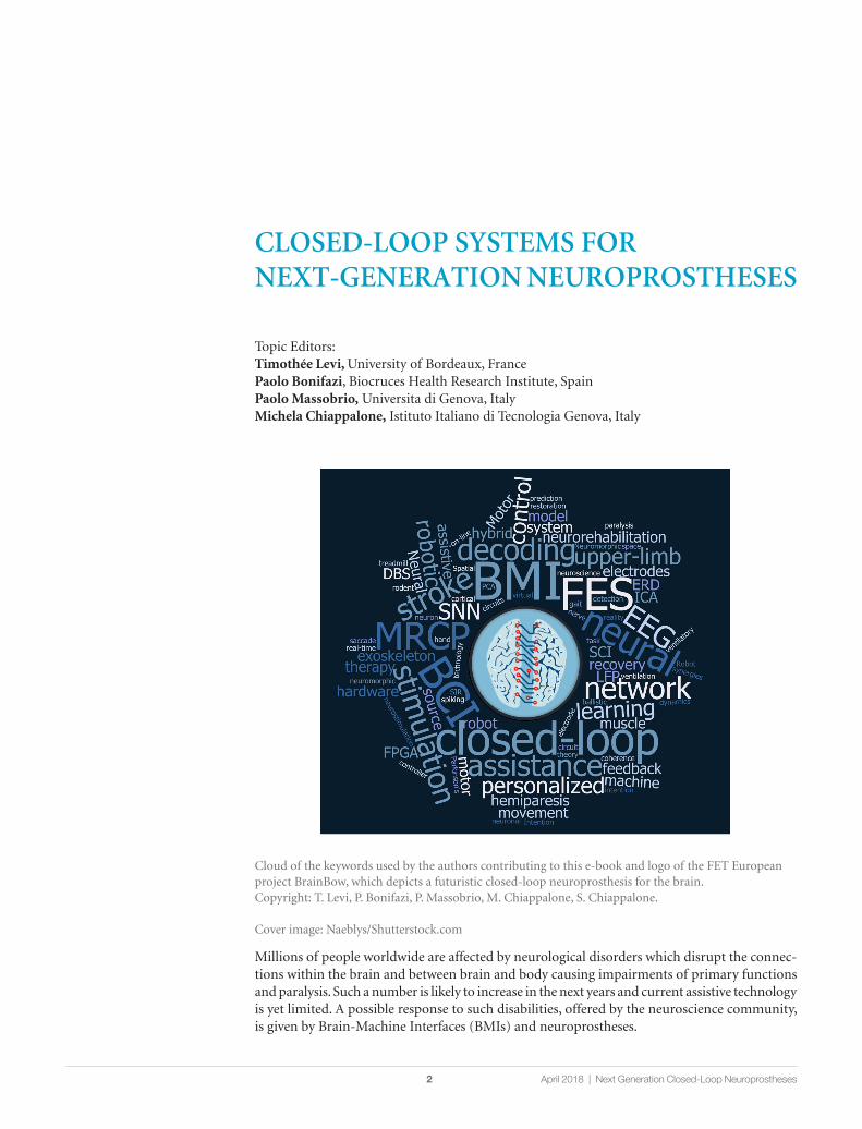
A Preliminary Evaluation of the FESTreatment Effect on Two Chronic StrokePatientsTwo patients with chronic hemiparesis due to ischemic stroke(Table 1) were asked to undergo a 4-week intervention consistingof 30-min sessions of FES-supported treadmill walking three
Frontiers in Neuroscience | www.frontiersin.org September 2016 | Volume 10 | Article 425 | 244
Ferrante et al. Synergy-Based Neuroprosthesis for Stroke Rehabilitation
TABLE 1 | Patient details.
Age (years) Gender Time from stroke Hemiparetic side
S1 67 Man 11 years Left
S2 64 Man 9 months Right
times per week. Each session consisted of 5 min of warmingup without FES, 20 min of gait supported by the multi-channelpersonalized FES controller, and 5 min of cooling down withoutFES. The patient was asked to select his preferred walking velocityduring the warming up phase. Before and after the end of theintervention, two clinical scales were assessed: the motor sub-scale of the Functional Independence Measure (FIM) whichevaluates the patient’s motor disability during daily life activitiesand ranges from 13 to 91 (independent), (Kidd et al., 1995) andthe Mini Best test (MBT) which evaluated the dynamic balanceand ranges from 0 to 28 (normal balance; Franchignoni et al.,2010). To evaluate specific improvements in terms of walkingability, the same test used to identify the patient’s impairedmuscle synergies was repeated at the end of the intervention.Both kinematics and EMG data were collected. The meancadence was computed from the kinematics data. EMG envelopeswere computed and the NMF algorithm was applied to extractthe muscle synergies as previously described for healthy subjects.At the end of the intervention, the patients were also asked to ratethe global perceived effect of the treatment using the global ratingchange (GRC), which is an 11-point scale (−5 = made thingsworse; 0= not changed; 5= completely recovered; Kamper et al.,2009).
The protocol was approved by the Central Ethics Committeeof the Fondazione Salvatore Maugeri (IRCCS) and both patientsprovided their written informed consent before participation.
RESULTS
Functioning of Multi-ChannelSynergy-Based Stimulation ControlerHealthy Muscle SynergiesThe WHEALTHY and HHEALTHY during overground walking arereported in Figure 3. All healthy subjects were characterizedby four muscle synergies corresponding to the four gait sub-functions identified in literature: Weight Acceptance (WA),Push Off (PO), Foot Clearance (FC) and Leg Deceleration(LD). The same modular organization, both in terms of spatialcomposition and temporal recruitment, was obtained duringtreadmill walking. Indeed, comparing the extracted musclesynergies in the two walking conditions and averaging acrosssubjects, a mean (Standard Deviation, SD) similarity of 0.89(0.11), a circular cross correlation of 0.94 (0.06), and a timelag of 2 (1) in percentage of the gait cycle were found. Thisconfirms that the two walking conditions share the same musclecoordination and thus it is possible to define both a treadmill andan overground treatment based on the same set of physiologicalmuscle synergies.
The synergies extracted from the overground walkingcondition were used to perform the two NNR of the EMG
envelopes obtained on the paretic side during the patient’s pre-treatment assessment.
NNR of the Patient’s EMG Envelopes with the Healthy
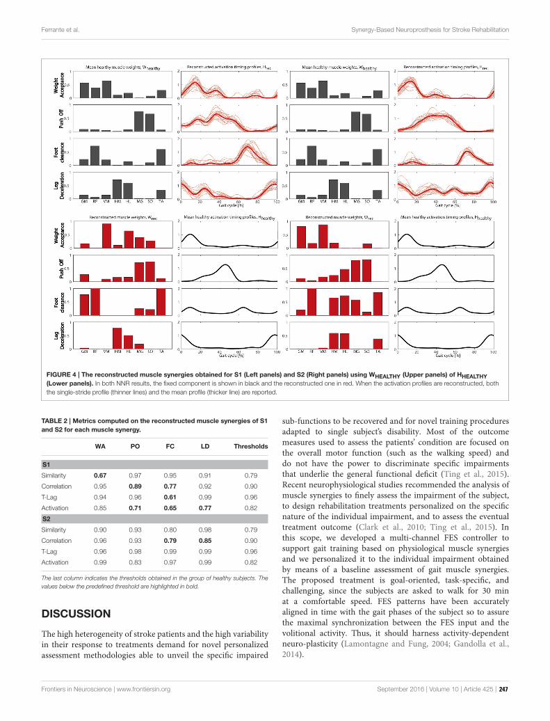
SynergiesFigure 4 shows the NNR results obtained for both patients. Theobtained VAF values were 0.85 and 0.77 for S1, and 0.90 and0.84 for S2 when the NNR was applied fixing WHEALTHY andHHEALTHY, respectively.
Identification of the Patient’s Impaired Muscle
SynergiesTable 2 reports for each metric and each muscle synergy, thethresholds computed on the healthy subjects group (last column)and the values obtained by the two patients during the pre-treatment assessment.
The metrics confirmed what was visually observed by thereconstructed synergies: S1 had an impaired spatial compositionin the WA synergy (similarity was under threshold for WA), awider activation timing of PO and LD synergies, and a delayedrecruitment of the FC synergy. Concerning S2, a low crosscorrelation was found for FC and LD synergies. Thus, all foursynergies were defined as impaired for S1 and only FC and LDwere considered impaired synergies for S2.
Definition of the Personalized Stimulation StrategyThe final FES strategy obtained and used for both patientsis shown in Figure 5. The medial and lateral hamstrings andthe medial gastrocnemius and soleus showed similar activationprofiles and therefore the FES strategy coupled into onestimulation channel both hamstrings, and the calf muscles intoanother. Thus, a total of six muscle groups were stimulated.
S1 had a FES strategy based on all four healthy synergies,whereas for S2 only the FC and LD synergies was used to obtainthe muscle stimulation profiles. When a reduced number ofsynergies was used to create the stimulation strategy, a subsetof the six muscle groups was stimulated. In particular, the calfmuscles, which were not recruited by the FC and LD healthysynergies, were not stimulated for S2.
Figure 5 also shows the different kinematic patterns of the twopatients. Indeed, both patients were characterized by a prolongeddouble support, but S1 extended the paretic double supportphase (gait phase 1) whereas S2 extended the non-paretic doublesupport phase (gait phase 4). In both cases, the FES strategywas able to adapt to these changes in gait pattern, mapping thestimulation profiles accordingly.
For both patients the first (upper panels) and last (lowerpanels) sessions of the intervention are shown to highlight thedifferences in gait speed together with slight differences in thekinematic pattern. In the first session of FES-supported gait,S1 presented an impaired kinematic pattern characterized by adouble support phase of the paretic leg (phase 1) lasting the 48%of the gait cycle and a very short paretic single support (21%of the gait cycle). In the last treatment session, this kinematicpattern changed: the duration of the paretic double support wasthe 25% of the gait cycle and the duration of the paretic singlesupport was 29%. These improvements in the kinematic pattern
Frontiers in Neuroscience | www.frontiersin.org September 2016 | Volume 10 | Article 425 | 245

Ferrante et al. Synergy-Based Neuroprosthesis for Stroke Rehabilitation
FIGURE 3 | The physiological muscle synergies: muscle weights (Left panel) and temporal activation profiles (Right panel) obtained during overground
walking. Mean values and standard deviation are reported in both panels. GM, gluteus maximus; RF, rectus femoris; VM, vastus medialis; HM, hamstring medialis;
HL, hamstring lateralis; MG, gastrocnemius medialis; TA, tibialis anterior.
corresponded to a walking speed that in the last session doubledits value with respect to the first session. Concerning S2, thekinematics pattern in the first session was characterized by areduced paretic swing phase that was augmented by 52% in thelast session, achieving a final duration equal to the 38% of the gaitcycle.
The FES Treatment Effect on the TwoChronic Stroke PatientsBoth patients completed the treatment without difficulties andreported a positive global perceived effect of the treatment (GRCwas +4 (improved a lot) and +2 (improved) for S1 and S2,respectively). The treadmill speed used in the first and last dayof treatment increased from 0.43 to 0.83 m/s, and from 0.38 to0.68m/s for S1 and S2, respectively.
The extracted muscle synergies before and after the treatmentduring the overground walking tests are shown in Figures 6, 7for S1 and S2, respectively. The treatment induced an increase ofthe number of extracted synergies in both patients from 3 to 4indicating a general improvement in muscle coordination. TheVAF was 0.87 before and 0.88 after treatment, and 0.90 beforeand 0.93 after treatment for S1 and S2, respectively. The visualcomparison between the extracted synergies obtained before
intervention for S1 and the healthy synergies (Figure 6) suggeststhat the first extracted synergy (S1-1) resembles the FC synergyexcept for the GM activation, the second extracted synergy (S1-2)mostly recruits the MG and SO muscles as it is in the PO synergywith an anticipatory activation profile, and the third synergy(S1-3) merges the WA and LD synergies. After treatment, fourmuscle synergies were found, generally resembling the spatialcomposition of the healthy muscle synergies in Figure 3. Anearly recruitment of the plantar-flexors is still present in the POsynergy.
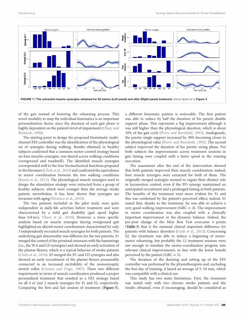
Concerning S2 (Figure 7), before treatment the first synergy(S2-1) can be associated to the WA synergy, the second (S2-2)seems to be the merging of the FC and LD synergies, and thethird (S2-3) seems to be the PO synergy with a slight contributionof the hamstring muscles. After treatment, four synergies wereobtained displaying a behavior more similar to healthy subjects;however, an early activation of the plantar-flexors is still visible inPO synergy.
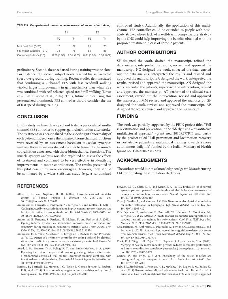
The clinical evaluation of the two patients is reported inTable 3. Both patients had a mild motor disability at baseline. Inboth cases the improvement gained in muscle coordination wasnot yet enough to be transferred into a significant difference ingeneral motor disability.
Frontiers in Neuroscience | www.frontiersin.org September 2016 | Volume 10 | Article 425 | 246
Ferrante et al. Synergy-Based Neuroprosthesis for Stroke Rehabilitation
FIGURE 4 | The reconstructed muscle synergies obtained for S1 (Left panels) and S2 (Right panels) using WHEALTHY (Upper panels) of HHEALTHY
(Lower panels). In both NNR results, the fixed component is shown in black and the reconstructed one in red. When the activation profiles are reconstructed, both
the single-stride profile (thinner lines) and the mean profile (thicker line) are reported.
TABLE 2 | Metrics computed on the reconstructed muscle synergies of S1
and S2 for each muscle synergy.
WA PO FC LD Thresholds
S1
Similarity 0.67 0.97 0.95 0.91 0.79
Correlation 0.95 0.89 0.77 0.92 0.90
T-Lag 0.94 0.96 0.61 0.99 0.96
Activation 0.85 0.71 0.65 0.77 0.82
S2
Similarity 0.90 0.93 0.80 0.98 0.79
Correlation 0.96 0.93 0.79 0.85 0.90
T-Lag 0.96 0.98 0.99 0.99 0.96
Activation 0.99 0.83 0.97 0.99 0.82
The last column indicates the thresholds obtained in the group of healthy subjects. The
values below the predefined threshold are highlighted in bold.
DISCUSSION
The high heterogeneity of stroke patients and the high variabilityin their response to treatments demand for novel personalizedassessment methodologies able to unveil the specific impaired
sub-functions to be recovered and for novel training proceduresadapted to single subject’s disability. Most of the outcomemeasures used to assess the patients’ condition are focused onthe overall motor function (such as the walking speed) anddo not have the power to discriminate specific impairmentsthat underlie the general functional deficit (Ting et al., 2015).Recent neurophysiological studies recommended the analysis ofmuscle synergies to finely assess the impairment of the subject,to design rehabilitation treatments personalized on the specificnature of the individual impairment, and to assess the eventualtreatment outcome (Clark et al., 2010; Ting et al., 2015). Inthis scope, we developed a multi-channel FES controller tosupport gait training based on physiological muscle synergiesand we personalized it to the individual impairment obtainedby means of a baseline assessment of gait muscle synergies.The proposed treatment is goal-oriented, task-specific, andchallenging, since the subjects are asked to walk for 30 minat a comfortable speed. FES patterns have been accuratelyaligned in time with the gait phases of the subject so to assurethe maximal synchronization between the FES input and thevolitional activity. Thus, it should harness activity-dependentneuro-plasticity (Lamontagne and Fung, 2004; Gandolla et al.,2014).
Frontiers in Neuroscience | www.frontiersin.org September 2016 | Volume 10 | Article 425 | 247

Ferrante et al. Synergy-Based Neuroprosthesis for Stroke Rehabilitation
FIGURE 5 | The personalized stimulation strategy obtained for S1 (Left panels) and S2 (Right panels) in the first (Upper panels) and last (Lower panels)
sessions of the intervention. Same labels as in Figure 3.
FIGURE 6 | The extracted muscle synergies obtained for S1 before (Left panel) and after (Right panel) treatment. Same labels as in Figure 3.
In literature, FES controllers have been based on very simplesegmentation algorithms able to discriminate between the stanceand swing phases and the FES strategy has been linearly mappedthrough the gait stride adopting sub-optimal time rules in orderto automatically deactivate stimulation (Daly et al., 2011). Thenovelty of our proposed control system lies in the capabilityto accurately map the subject’s gait timing based on the real-time detection of six gait phases. This mapping is able to
stretch or extend the stimulation profiles according to theactual duration of all six phases. For instance, if a patient’s gaitpattern is characterized by an extended double support phase,the stimulation profile of the muscles supporting this phase areextended accordingly in order to follow the correct muscle timingand coordination involved in this phase (see Figure 5). Thischoice avoids the unwanted activation of antagonist muscles inthe extended kinematic phase that could increase the instability
Frontiers in Neuroscience | www.frontiersin.org September 2016 | Volume 10 | Article 425 | 248
Ferrante et al. Synergy-Based Neuroprosthesis for Stroke Rehabilitation
FIGURE 7 | The extracted muscle synergies obtained for S2 before (Left panel) and after (Right panel) treatment. Same labels as in Figure 3.
of the gait instead of fostering the relearning process. Thisnovel modality to map the individual kinematics is an importantpersonalization factor, since the duration of each gait phase ishighly dependent on the patient’s level of impairment (Olney andRichards, 1996).
The starting point to design the proposed biomimetic multi-channel FES controller was the identification of the physiologicalset of synergies during walking. Results obtained in healthysubjects confirmed that a common motor-control strategy basedon four muscles synergies, was shared across walking conditions(overground and treadmill). The identified muscle synergiescorresponded well to the four biomechanical functions proposedin the literature (Clark et al., 2010) and confirmed the equivalencein motor coordination between the two walking conditions(Kautz et al., 2011). The physiological muscle synergies used todesign the stimulation strategy were extracted from a group ofhealthy subjects, which were younger than the average strokepatient; nevertheless, it has been shown that synergies areinvariant with aging (Monaco et al., 2010).
The two patients included in the pilot study were quiteindependent in daily-life activities before treatment and werecharacterized by a mild gait disability (gait speed higherthan 0.8m/s; Tilson et al., 2010). However, a more specificanalysis based on muscle synergies during overground gaithighlighted an altered motor coordination characterized by only3 independently recruitedmuscle synergies for both patients. Theunderlying gait abnormality was different for the two patients. S1merged the control of the proximal extensors with the hamstrings(i.e., theWA and LD synergies) and showed an early activation ofthe plantar-flexors, which is a typical behavior of stroke patients(Clark et al., 2010). S2 merged the FC and LD synergies and alsoshowed an early recruitment of the plantar-flexors presumablyconnected to an increased excitability of the monosynapticstretch reflex (Crenna and Frigo, 1987). These two differentimpairments in terms of muscle coordination produced a properpersonalized treatment that resulted in a FES strategy basedon all 4 or just 2 muscle synergies for S1 and S2, respectively.Comparing the first and last session of treatment (Figure 5),
a different kinematic pattern is noticeable. The first patientwas able to reduce by half the duration of his paretic doublesupport phase. This represents a big improvement although itwas still higher than the physiological duration, which is about10% of the gait cycle (Perry and Burnfield, 1992). Analogously,the paretic single support increased by 38% becoming closer tothe physiological value (Perry and Burnfield, 1992). The secondsubject improved the duration of his paretic swing phase. Forboth subjects the improvements across treatment sessions ingait timing were coupled with a faster speed in the trainingexecution.
The assessment after the end of the intervention showedthat both patients improved their muscle coordination; indeed,four muscle synergies were extracted for both of them. Theoriginally merged synergies seemed to regain their distinct rolein locomotion control, even if the PO synergy maintained ananticipated recruitment and a prolonged timing in both patients.The benefits of the treatment were more evident for S1 andthis was confirmed by the patient’s perceived effect; indeed, S1stated that, thanks to the treatment, he was able to achieve avery good walking improvement (GRC = 4). The improvementin motor coordination was also coupled with a clinicallyimportant improvement in the dynamic balance. Indeed, thepre-post change of the Mini Best Test overcame 4 points(Table 3) that is the minimal clinical important difference forpatients with balance disorders (Godi et al., 2013). ConcerningS2, the treatment was able to induce a beginning of neuro-motor relearning, but probably the 12 treatment sessions werenot enough to translate the motor-coordination progress intorelevant clinical improvements, in line with the lower benefitperceived by the patient (GRC= 2).
The duration of the donning and setting up of the FEScontroller was performed by the physiotherapists and, excludingthe first day of training, it lasted an average of 5–10 min, whichwas compatible with a clinical use.
This study has two main limitations. First, the treatmentwas tested only with two chronic stroke patients and theresults obtained, even if encouraging, should be considered as
Frontiers in Neuroscience | www.frontiersin.org September 2016 | Volume 10 | Article 425 | 249
Ferrante et al. Synergy-Based Neuroprosthesis for Stroke Rehabilitation
TABLE 3 | Comparison of the outcome measures before and after training.
S1 S2
T0 T1 T0 T1
Mini Best Test (0–28) 17 22 21 23
FIM motor subscale (13–91) 78 78 85 85
Cadence (strides/s) (SD) 0.98 (0.03) 1.01 (0.03) 0.81 (0.03) 0.80 (0.02)
preliminary. Second, the speed used during training was too slow.For instance, the second subject never reached his self-selectedspeed overground during training. Recent studies demonstratedthat combining a 2-channel FES with fast treadmill walkingyielded larger improvements in gait mechanics than when FESwas combined with self-selected speed treadmill walking (Kesaret al., 2011; Awad et al., 2016). Thus, future studies using thispersonalized biomimetic FES controller should consider the useof fast speed during training.
CONCLUSION
In this study we have developed and tested a personalized multi-channel FES controller to support gait rehabilitation after stroke.The treatment was personalized to the specific gait abnormality ofeach patient. Indeed, once the impaired biomechanical functionswere revealed by an assessment based on muscular synergiesanalysis, the exercise was shaped in order to train only the musclecoordination associated with those biomechanical functions. Themuscle-synergy analysis was also exploited to assess the effectsof treatment and confirmed to be very effective in identifyingimprovements in motor coordination. The results presented inthis pilot case study were encouraging; however, they shouldbe confirmed by a wider statistical study (e.g., a randomized
controlled study). Additionally, the application of this multi-channel FES controller could be extended to people with post-acute stroke, whose lack of a well-learnt compensatory strategyby the CNS could help improving the benefits obtained with theproposed treatment in case of chronic patients.
AUTHOR CONTRIBUTIONS
SF designed the work, drafted the manuscript, refined thedata analysis, interpreted the results, revised and approved themanuscript. NC designed the work, collected the data, carriedout the data analysis, interpreted the results and revised andapproved the manuscript. EA designed the work, interpreted theresults, revised and approved the manuscript. AN designed thework, recruited the patients, supervised the intervention, revisedand approved the manuscript. AT performed the clinical scaleassessment, carried out the intervention, revised and approvedthe manuscript. MM revised and approved the manuscript. GFdesigned the work, revised and approved the manuscript. APdesigned the work, revised and approved the manuscript.
FUNDING
The work was partially supported by the PRIN project titled “Fallrisk estimation and prevention in the elderly using a quantitativemultifactorial approach” (grant no.: 2010R277FT) and partlyby the project titled “Fall prevention and locomotion recoveryin post-stroke patients: a multimodal training towards a moreautonomous daily life” funded by the Italian Ministry of Health(grant no.: GR-2010-2312228).
ACKNOWLEDGMENTS
The authors would like to acknowledge AxelgaardManufacturingLtd. for donating the stimulation electrodes.
REFERENCES
Allen, J. L., and Neptune, R. R. (2012). Three-dimensional modular
control of human walking. J. Biomech. 45, 2157–2163. doi:
10.1016/j.jbiomech.2012.05.037
Ambrosini, E., Ferrante, S., Pedrocchi, A., Ferrigno, G., and Molteni, F. (2011).
Cycling induced by electrical stimulation improvesmotor recovery in postacute
hemiparetic patients: a randomized controlled trial. Stroke 42, 1068–1073. doi:
10.1161/STROKEAHA.110.599068
Ambrosini, E., Ferrante, S., Ferrigno, G., Molteni, F., and Pedrocchi, A. (2012).
Cycling induced by electrical stimulation improves muscle activation and
symmetry during pedaling in hemiparetic patients. IEEE Trans. Neural Syst.
Rehabil. Eng. 20, 320–330. doi: 10.1109/TNSRE.2012.2191574
Ambrosini, E., Ferrante, S., Schauer, T., Ferrigno, G., Molteni, F., and Pedrocchi,
A. (2010). Design of a symmetry controller for cycling induced by electrical
stimulation: preliminary results on post-acute stroke patients. Artif. Organs 34,
663–667. doi: 10.1111/j.1525-1594.2009.00941.x
Awad, L. N., Reisman, D. S., Pohlig, R. T., and Binder-Macleod, S. A. (2016).
Reducing the cost of transport and increasing walking distance after stroke:
a randomized controlled trial on fast locomotor training combined with
functional electrical stimulation. Neurorehabil. Neural Repair 30, 661–670. doi:
10.1177/1545968315619696
Barroso, F., Torricelli, D., Moreno, J. C., Taylor, J., Gomez-Soriano, J., Esteban,
E. B., et al. (2014). Shared muscle synergies in human walking and cycling. J.
Neurophysiol. 112, 1984–1998. doi: 10.1152/jn.00220.2014
Bowden, M. G., Clark, D. J., and Kautz, S. A. (2010). Evaluation of abnormal
synergy patterns poststroke: relationship of the fugl-meyer assessment to
hemiparetic locomotion. Neurorehabil. Neural Repair 24, 328–337. doi:
10.1177/1545968309343215
Chae, J., Sheffler, L., and Knutson, J. (2008). Neuromuscular electrical stimulation
for motor restoration in hemiplegia. Top. Stroke Rehabil. 15, 412–426. doi:
10.1310/tsr1505-412
Chia Bejarano, N., Ambrosini, E., Baccinelli, W., Nardone, A., Monticone, M.,
Ferrigno, G., et al. (2015a). A multi-channel biomimetic neuroprosthesis to
support treadmill gait training in stroke patients. Conf. Proc. IEEE Eng. Med.
Biol. Soc. 2015, 7159–7162. doi: 10.1109/EMBC.2015.7320043
Chia Bejarano, N., Ambrosini, E., Pedrocchi, A., Ferrigno, G., Monticone, M., and
Ferrante, S. (2015b). A novel adaptive, real-time algorithm to detect gait events
from wearable sensors. IEEE Trans. Neural Syst. Rehabil. Eng. 23, 413–422. doi:
10.1109/TNSRE.2014.2337914
Clark, D. J., Ting, L. H., Zajac, F. E., Neptune, R. R., and Kautz, S. A. (2010).
Merging of healthy motor modules predicts reduced locomotor performance
andmuscle coordination complexity post-stroke. J. Neurophysiol. 103, 844–857.
doi: 10.1152/jn.00825.2009
Crenna, P., and Frigo, C. (1987). Excitability of the soleus H-reflex arc
during walking and stepping in man. Exp. Brain Res. 66, 49–60. doi:
10.1007/BF00236201
Daly, J. J., Zimbelman, J., Roenigk, K. L., McCabe, J. P., Rogers, J. M., Butler, K.,
et al. (2011). Recovery of coordinated gait: randomized controlled stroke trial of
Functional Electrical Stimulation (FES) versus No, FES, with weight-supported
Frontiers in Neuroscience | www.frontiersin.org September 2016 | Volume 10 | Article 425 | 250