Embed Size (px)
Citation preview

2.1 Weak Interactions in Aqueous Systems 47
2.2 Ionization of Water, Weak Acids, and Weak Bases 58
2.3 Buffering against pH Changes in Biological Systems 63
2.4 Water as a Reactant 69
2.5 The Fitness of the Aqueous Environment for Living Organisms 69
Water is the most abundant substance in living systems, making up 70% or more of the weight of most organisms. The first living organisms on
Earth doubtless arose in an aqueous environment, and the course of evolution has been shaped by the proper-ties of the aqueous medium in which life began. This chapter begins with descriptions of the physi-cal and chemical properties of water, to which all aspects of cell structure and function are adapted. The attractive forces between water molecules and the slight tendency of water to ionize are of crucial impor-tance to the structure and function of biomolecules. We review the topic of ionization in terms of equilibrium constants, pH, and titration curves, and consider how aqueous solutions of weak acids or bases and their salts act as buffers against pH changes in biological systems. The water molecule and its ionization products, H1 and OH2, profoundly influence the structure, self-assembly, and properties of all cellular components, including proteins, nucleic acids, and lipids. The noncovalent interactions responsible for the strength and specificity of “recognition” among biomolecules are decisively influenced by water’s properties as a solvent, including its ability to form hydrogen bonds with itself and with solutes.
2.1 Weak Interactions in Aqueous SystemsHydrogen bonds between water molecules provide the cohesive forces that make water a liquid at room tem-perature and a crystalline solid (ice) with a highly ordered arrangement of molecules at cold tempera-
tures. Polar biomolecules dissolve readily in water because they can replace water-water interactions with more energetically favorable water-solute interactions. In contrast, nonpolar biomolecules are poorly soluble in water because they interfere with water-water interac-tions but are unable to form water-solute interactions. In aqueous solutions, nonpolar molecules tend to clus-ter together. Hydrogen bonds and ionic, hydrophobic (Greek, “water-fearing”), and van der Waals interac-tions are individually weak, but collectively they have a very significant influence on the three-dimensional structures of proteins, nucleic acids, polysaccharides, and membrane lipids.
Hydrogen Bonding Gives Water Its Unusual PropertiesWater has a higher melting point, boiling point, and heat of vaporization than most other common solvents (Table 2–1). These unusual properties are a conse-quence of attractions between adjacent water molecules that give liquid water great internal cohesion. A look at the electron structure of the H2O molecule reveals the cause of these intermolecular attractions. Each hydrogen atom of a water molecule shares an electron pair with the central oxygen atom. The geometry of the molecule is dictated by the shapes of the outer electron orbitals of the oxygen atom, which are similar to the sp3 bonding orbitals of carbon (see Fig. 1–15). These orbitals describe a rough tetrahe-dron, with a hydrogen atom at each of two corners and unshared electron pairs at the other two corners (Fig. 2–1a). The H—O—H bond angle is 104.58, slightly less than the 109.58 of a perfect tetrahedron because of crowding by the nonbonding orbitals of the oxygen atom. The oxygen nucleus attracts electrons more strongly than does the hydrogen nucleus (a proton); that is, oxygen is more electronegative. This means that the shared electrons are more often in the vicinity of the oxygen atom than of the hydrogen. The result of this unequal electron sharing is two electric dipoles in the water molecule, one along each of the H—O bonds;
47
Water
2
c02Water.indd Page 47 30/08/12 7:46 AM user-F408 /Users/user-F408/Desktop

Water48
each hydrogen atom bears a partial positive charge (�1), and the oxygen atom bears a partial negative charge equal in magnitude to the sum of the two partial positives (2�2). As a result, there is an electrostatic attraction between the oxygen atom of one water molecule and the hydrogen of another (Fig. 2–1b), called a hydrogen bond. Throughout this book, we represent hydrogen bonds with three parallel blue lines, as in Figure 2–1b. Hydrogen bonds are relatively weak. Those in liquid water have a bond dissociation energy (the energy required to break a bond) of about 23 kJ/mol, compared with 470 kJ/mol for the covalent O—H bond in water or 348 kJ/mol for a covalent C—C bond. The hydrogen
bond is about 10% covalent, due to overlaps in the bonding orbitals, and about 90% electrostatic. At room temperature, the thermal energy of an aqueous solution (the kinetic energy of motion of the individual atoms and molecules) is of the same order of magnitude as that required to break hydrogen bonds. When water is heated, the increase in temperature reflects the faster motion of individual water molecules. At any given time, most of the molecules in liquid water are hydrogen bonded, but the lifetime of each hydrogen bond is just 1 to 20 picoseconds (1 ps 5 10212 s); when one hydrogen bond breaks, another hydrogen bond forms, with the same partner or a new one, within 0.1 ps. The apt phrase “flickering clusters” has been applied to the short-lived groups of water molecules interlinked by hydrogen bonds in liquid water. The sum of all the hydrogen bonds between H2O molecules confers great internal cohesion on liquid water. Extended networks of hydrogen-bonded water molecules also form bridges between solutes (proteins and nucleic acids, for exam-ple) that allow the larger molecules to interact with each other over distances of several nanometers with-out physically touching. The nearly tetrahedral arrangement of the orbitals about the oxygen atom (Fig. 2–1a) allows each water molecule to form hydrogen bonds with as many as four neighboring water molecules. In liquid water at room temperature and atmospheric pressure, however, water molecules are disorganized and in continuous motion, so that each molecule forms hydrogen bonds with an average of only 3.4 other molecules. In ice, on the other hand, each water molecule is fixed in space and forms hydrogen bonds with a full complement of four other water molecules to yield a regular lattice structure (Fig. 2–2). Hydrogen bonds account for the relatively high melting point of water, because much thermal energy is required to break a sufficient proportion of hydrogen bonds to destabilize the crystal lattice of ice
Melting point (8C) Boiling point (8C) Heat of vaporization (J/g)*Water 0 100 2,260
Methanol (CH3OH) 298 65 1,100
Ethanol (CH3CH2OH) 2117 78 854
Propanol (CH3CH2CH2OH) 2127 97 687
Butanol (CH3(CH2)2CH2OH) 290 117 590
Acetone (CH3COCH3) 295 56 523
Hexane (CH3(CH2)4CH3) 298 69 423
Benzene (C6H6) 6 80 394
Butane (CH3(CH2)2CH3) 2135 20.5 381
Chloroform (CHCl3) 263 61 247
*The heat energy required to convert 1.0 g of a liquid at its boiling point and at atmospheric pressure into its gaseous state at the same temperature. It is a
direct measure of the energy required to overcome attractive forces between molecules in the liquid phase.
TABLE 2–1 Melting Point, Boiling Point, and Heat of Vaporization of Some Common Solvents
FIGURE 2–1 Structure of the water molecule. (a) The dipolar nature of
the H2O molecule is shown in a ball-and-stick model; the dashed lines
represent the nonbonding orbitals. There is a nearly tetrahedral arrange-
ment of the outer-shell electron pairs around the oxygen atom; the two
hydrogen atoms have localized partial positive charges (�1) and the
oxygen atom has a partial negative charge (�2). (b) Two H2O molecules
joined by a hydrogen bond (designated here, and throughout this book,
by three blue lines) between the oxygen atom of the upper molecule
and a hydrogen atom of the lower one. Hydrogen bonds are longer and
weaker than covalent O—H bonds.
c02Water.indd Page 48 27/08/12 1:48 PM user-F408 /Users/user-F408/Desktop
104.5�
Hydrogen bond
0.177 nm
Covalent bond
0.0965 nm
H
��
��
��
��
H
O
(a) (b)
��
��
��
��
�� ��

2.1 Weak Interactions in Aqueous Systems 49
(Table 2–1). When ice melts or water evaporates, heat is taken up by the system:
H2O(solid)¡ H2O(liquid) ¢H 5 15.9 kJ/mol
H2O(liquid)¡ H2O(gas) ¢H 5 144.0 kJ/mol
During melting or evaporation, the entropy of the aqueous system increases as the highly ordered arrays of water molecules in ice relax into the less orderly hydrogen-bonded arrays in liquid water or into the wholly disordered gaseous state. At room temperature, both the melting of ice and the evaporation of water occur spontaneously; the tendency of the water mole-cules to associate through hydrogen bonds is out-weighed by the energetic push toward randomness. Recall that the free-energy change (DG) must have a negative value for a process to occur spontaneously: DG 5 DH 2 T DS, where DG represents the driving force, DH the enthalpy change from making and break-ing bonds, and DS the change in randomness. Because DH is positive for melting and evaporation, it is clearly the increase in entropy (DS) that makes DG negative and drives these changes.
Water Forms Hydrogen Bonds with Polar SolutesHydrogen bonds are not unique to water. They readily form between an electronegative atom (the hydrogen acceptor, usually oxygen or nitrogen) and a hydrogen atom covalently bonded to another electronegative atom (the hydrogen donor) in the same or another molecule (Fig. 2–3). Hydrogen atoms covalently bonded to car-
bon atoms do not participate in hydrogen bonding, because carbon is only slightly more electronegative than hydrogen and thus the C—H bond is only very weakly polar. The distinction explains why butanol (CH3(CH2)2CH2OH) has a relatively high boiling point of 117 8C, whereas butane (CH3(CH2)2CH3) has a boiling point of only 20.5 8C. Butanol has a polar hydroxyl group and thus can form intermolecular hydrogen bonds. Uncharged but polar biomolecules such as sugars dissolve readily in water because of the stabilizing effect of hydrogen bonds between the hydroxyl groups or car-bonyl oxygen of the sugar and the polar water molecules. Alcohols, aldehydes, ketones, and compounds contain-ing N—H bonds all form hydrogen bonds with water molecules (Fig. 2–4) and tend to be soluble in water.
FIGURE 2–3 Common hydrogen bonds in biological systems. The
hydrogen acceptor is usually oxygen or nitrogen; the hydrogen donor is
another electronegative atom.
Hydrogen
donor
Hydrogen
acceptor
H
O
D
OO
J
H
N
O
OO
DJ
H
N
O
DD
H
O
O
OO
H
N
O
DD
H
O
OJO
ECA
JO
ECA
GDN
GDN
GD
FIGURE 2–4 Some biologically important hydrogen bonds.
Between the
hydroxyl group
of an alcohol
and water
Between the
carbonyl group
of a ketone
and water
Between peptide
groups in
polypeptides
Between
complementary
bases of DNA
HNAH
ECA
NO
HA
AN
HR EC
H
NC
ECH3
HC
K HN
HENN ENH
NER
OCH
CKO
NBC
CAC
l
A
Thymine
Adenine
i
H
O
HAO
GR
H HHE
R1
H
R2
AO
HE
JO
ECA
OR
E
AH
B
NC
C
CA
HA H
NH
H
R
JO
GH
G DCO
FIGURE 2–2 Hydrogen bonding in ice. In ice, each water molecule forms
four hydrogen bonds, the maximum possible for a water molecule, creat-
ing a regular crystal lattice. By contrast, in liquid water at room tempera-
ture and atmospheric pressure, each water molecule hydrogen-bonds
with an average of 3.4 other water molecules. This crystal lattice structure
makes ice less dense than liquid water, and thus ice floats on liquid water.
c02Water.indd Page 49 27/08/12 1:48 PM user-F408 /Users/user-F408/Desktop

Water50
Hydrogen bonds are strongest when the bonded mol-ecules are oriented to maximize electrostatic interaction, which occurs when the hydrogen atom and the two atoms that share it are in a straight line—that is, when the acceptor atom is in line with the covalent bond between the donor atom and H (Fig. 2–5). This arrange-ment puts the positive charge of the hydrogen ion directly between the two partial negative charges. Hydrogen bonds are thus highly directional and capable of holding two hydrogen-bonded molecules or groups in a specific geometric arrangement. As we shall see later, this prop-erty of hydrogen bonds confers very precise three-dimensional structures on protein and nucleic acid mol-ecules, which have many intramolecular hydrogen bonds.
Water Interacts Electrostatically with Charged SolutesWater is a polar solvent. It readily dissolves most bio-molecules, which are generally charged or polar com-pounds (Table 2–2); compounds that dissolve easily in water are hydrophilic (Greek, “water-loving”). In contrast, nonpolar solvents such as chloroform and
benzene are poor solvents for polar biomolecules but easily dissolve those that are hydrophobic—nonpolar molecules such as lipids and waxes. Water dissolves salts such as NaCl by hydrating and stabilizing the Na1 and Cl2 ions, weakening the electro-static interactions between them and thus counteract-ing their tendency to associate in a crystalline lattice (Fig. 2–6). Water also readily dissolves charged bio-molecules, including compounds with functional groups such as ionized carboxylic acids (—COO2), protonated amines (2NH1
3 ), and phosphate esters or anhydrides. Water replaces the solute-solute hydrogen bonds link-ing these biomolecules to each other with solute-water hydrogen bonds, thus screening the electrostatic inter-actions between solute molecules. Water is effective in screening the electrostatic interactions between dissolved ions because it has a high dielectric constant, a physical property that reflects the number of dipoles in a solvent. The strength, or force (F), of ionic interactions in a solution depends on the magnitude of the charges (Q), the distance between the charged groups (r), and the dielectric constant (�, which is dimensionless) of the solvent in which the interactions occur:
F 5Q1Q2
er 2
For water at 25 8C, � is 78.5, and for the very nonpolar solvent benzene, � is 4.6. Thus, ionic interactions between dissolved ions are much stronger in less polar environments. The dependence on r2 is such that ionic attractions or repulsions operate only over short distances—in the range of 10 to 40 nm (depend-ing on the electrolyte concentration) when the sol-vent is water.
FIGURE 2–5 Directionality of the hydrogen bond. The attraction between
the partial electric charges (see Fig. 2–1) is greatest when the three atoms
involved in the bond (in this case O, H, and O) lie in a straight line. When
the hydrogen-bonded moieties are structurally constrained (when they
are parts of a single protein molecule, for example), this ideal geometry
may not be possible and the resulting hydrogen bond is weaker.
Strong
hydrogen bondWeaker
hydrogen bond
PKO
HAOAR
PKO
HAOAR
G
DOG
DO
TABLE 2–2 Some Examples of Polar, Nonpolar, and Amphipathic Biomolecules (Shown as Ionic Forms at pH 7)
H
HO
CH2OHO
OH
OH
OH
CH2�NH3 COO�
CH2�OOC COO�
H
H
H
H
�NH3
CH
CH
OH
OH
CH3 COO�CH
CH2OHHOCH2
CH CHCH3(CH2)7 (CH2)6 CH2 C
CH CH(CH2)7 (CH2)7 CH2CH3
CH2 CH
GNH3
GN(CH3)3
O
O
COOJ
CH3(CH2)15CH2
CH2CH2 CH2O
O
OJ
C
CH3(CH2)15CH2 CHO
O
CH2O
P
C
O
O
Polar groups Nonpolar groups
Polar Nonpolar
Glucose Typical wax
Amphipathic
Glycine Phenylalanine
Aspartate
Phosphatidylcholine
Lactate
Glycerol
c02Water.indd Page 50 27/08/12 1:48 PM user-F408 /Users/user-F408/Desktop

2.1 Weak Interactions in Aqueous Systems 51
Entropy Increases as Crystalline Substances DissolveAs a salt such as NaCl dissolves, the Na1 and Cl2 ions leaving the crystal lattice acquire far greater freedom of motion (Fig. 2–6). The resulting increase in entropy (randomness) of the system is largely responsible for the ease of dissolving salts such as NaCl in water. In thermodynamic terms, formation of the solution occurs with a favorable free-energy change: DG 5 DH 2 T DS, where DH has a small positive value and T DS a large positive value; thus DG is negative.
Nonpolar Gases Are Poorly Soluble in WaterThe molecules of the biologically important gases CO2, O2, and N2 are nonpolar. In O2 and N2, electrons are shared equally by both atoms. In CO2, each CPO bond is polar, but the two dipoles are oppositely directed and cancel each other (Table 2–3). The movement of mole-cules from the disordered gas phase into aqueous solu-tion constrains their motion and the motion of water molecules and therefore represents a decrease in entropy. The nonpolar nature of these gases and the decrease in entropy when they enter solution combine to make
them very poorly soluble in water (Table 2–3). Some organisms have water-soluble “carrier proteins” (hemo-globin and myoglobin, for example) that facilitate the transport of O2. Carbon dioxide forms carbonic acid (H2CO3) in aqueous solution and is transported as the HCO2
3 (bicarbonate) ion, either free—bicarbonate is very soluble in water (~100 g/L at 25 8C)—or bound to hemoglobin. Three other gases, NH3, NO, and H2S, also have biological roles in some organisms; these gases are polar, dissolve readily in water, and ionize in aqueous solution.
Nonpolar Compounds Force Energetically Unfavorable Changes in the Structure of WaterWhen water is mixed with benzene or hexane, two phases form; neither liquid is soluble in the other. Non-polar compounds such as benzene and hexane are hydrophobic—they are unable to undergo energetically favorable interactions with water molecules, and they interfere with the hydrogen bonding among water molecules. All molecules or ions in aqueous solution interfere with the hydrogen bonding of some water
TABLE 2–3 Solubilities of Some Gases in Water Solubility Gas Structure* Polarity in water (g/L)†
Nitrogen NqN Nonpolar 0.018 (40 8C)
Oxygen OPO Nonpolar 0.035 (50 8C)
Carbon dioxide OPCPO
�� �� Nonpolar 0.97 (45 8C)
Ammonia HG
NAH
DH
��
Polar 900 (10 8C)
Hydrogen sulfide HG
SD
H
��
Polar 1,860 (40 8C)
*The arrows represent electric dipoles; there is a partial negative charge (d2) at the head of the arrow, a partial positive charge (d1 ; not
shown here) at the tail.†Note that polar molecules dissolve far better even at low temperatures than do nonpolar molecules at relatively high temperatures.
FIGURE 2–6 Water as solvent. Water dissolves many
crystalline salts by hydrating their component ions. The
NaCl crystal lattice is disrupted as water molecules clus-
ter about the Cl2 and Na1 ions. The ionic charges are
partially neutralized, and the electrostatic attractions
necessary for lattice formation are weakened.
c02Water.indd Page 51 27/08/12 1:49 PM user-F408 /Users/user-F408/Desktop
�
Hydrated
Na+ ion
Hydrated
Cl– ionH2O
Na+
Cl–
�
��
�
�
�
�
�
�
�
�
Note the nonrandom
orientation of the
water molecules
�
�� �
Cl–
�
Na+
�
Water52
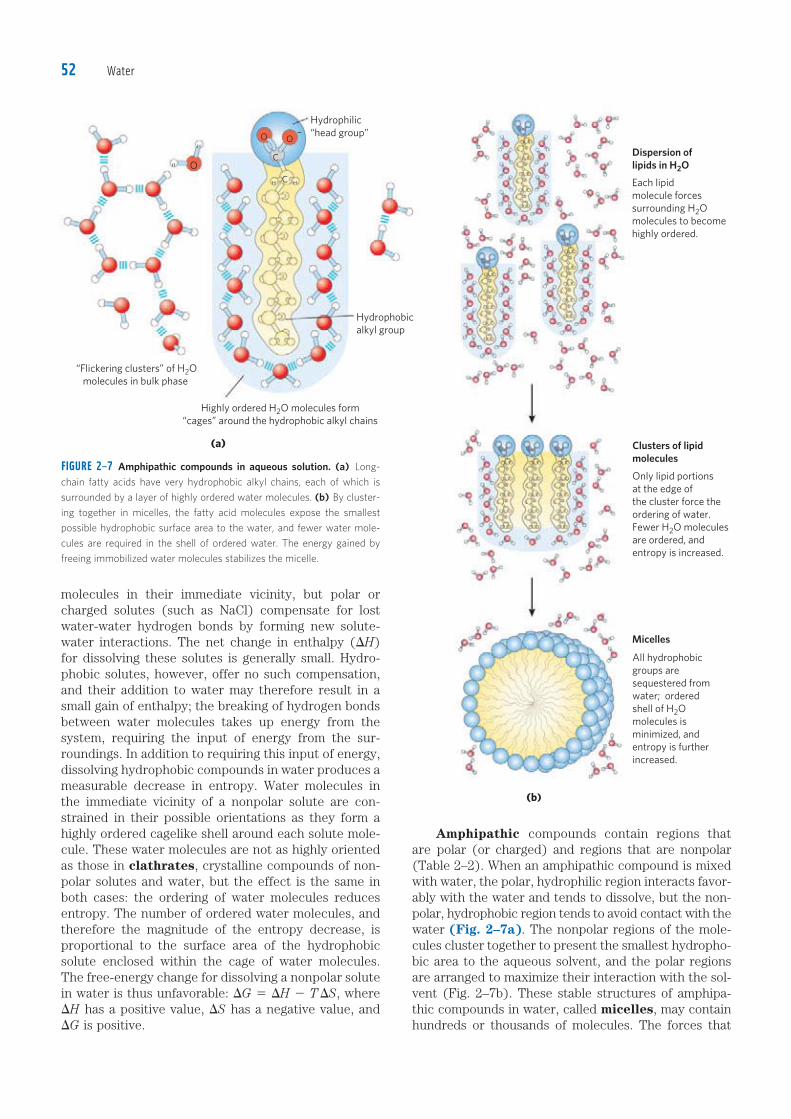
Amphipathic compounds contain regions that are polar (or charged) and regions that are nonpolar (Table 2–2). When an amphipathic compound is mixed with water, the polar, hydrophilic region interacts favor-ably with the water and tends to dissolve, but the non-polar, hydrophobic region tends to avoid contact with the water (Fig. 2–7a). The nonpolar regions of the mole-cules cluster together to present the smallest hydropho-bic area to the aqueous solvent, and the polar regions are arranged to maximize their interaction with the sol-vent (Fig. 2–7b). These stable structures of amphipa-thic compounds in water, called micelles, may contain hundreds or thousands of molecules. The forces that
FIGURE 2–7 Amphipathic compounds in aqueous solution. (a) Long-
chain fatty acids have very hydrophobic alkyl chains, each of which is
surrounded by a layer of highly ordered water molecules. (b) By cluster-
ing together in micelles, the fatty acid molecules expose the smallest
possible hydrophobic surface area to the water, and fewer water mole-
cules are required in the shell of ordered water. The energy gained by
freeing immobilized water molecules stabilizes the micelle.
molecules in their immediate vicinity, but polar or charged solutes (such as NaCl) compensate for lost water-water hydrogen bonds by forming new solute-water interactions. The net change in enthalpy (DH) for dissolving these solutes is generally small. Hydro-phobic solutes, however, offer no such compensation, and their addition to water may therefore result in a small gain of enthalpy; the breaking of hydrogen bonds between water molecules takes up energy from the system, requiring the input of energy from the sur-roundings. In addition to requiring this input of energy, dissolving hydrophobic compounds in water produces a measurable decrease in entropy. Water molecules in the immediate vicinity of a nonpolar solute are con-strained in their possible orientations as they form a highly ordered cagelike shell around each solute mole-cule. These water molecules are not as highly oriented as those in clathrates, crystalline compounds of non-polar solutes and water, but the effect is the same in both cases: the ordering of water molecules reduces entropy. The number of ordered water molecules, and therefore the magnitude of the entropy decrease, is proportional to the surface area of the hydrophobic solute enclosed within the cage of water molecules. The free-energy change for dissolving a nonpolar solute in water is thus unfavorable: DG 5 DH 2 T DS, where DH has a positive value, DS has a negative value, and DG is positive.
c02Water.indd Page 52 27/08/12 1:49 PM user-F408 /Users/user-F408/Desktop
Dispersion oflipids in H2O
Clusters of lipid molecules
Micelles
(b)
(a)
“Flickering clusters” of H2O
molecules in bulk phase
Highly ordered H2O molecules form
“cages” around the hydrophobic alkyl chains
Hydrophilic
“head group”O O
C
CH H
H
H O
Each lipid
molecule forces
surrounding H2O
molecules to become
highly ordered.
Only lipid portions
at the edge of
the cluster force the
ordering of water.
Fewer H2O molecules
are ordered, and
entropy is increased.
All hydrophobic
groups are
sequestered from
water; ordered
shell of H2O
molecules is
minimized, and
entropy is further
increased.
–
Hydrophobic
alkyl group

2.1 Weak Interactions in Aqueous Systems 53
hold the nonpolar regions of the molecules together are called hydrophobic interactions. The strength of hydrophobic interactions is not due to any intrinsic attraction between nonpolar moieties. Rather, it results from the system’s achieving the greatest thermody-namic stability by minimizing the number of ordered water molecules required to surround hydrophobic por-tions of the solute molecules. Many biomolecules are amphipathic; proteins, pig-ments, certain vitamins, and the sterols and phospholipids of membranes all have both polar and nonpolar surface regions. Structures composed of these molecules are sta-bilized by hydrophobic interactions among the nonpolar regions. Hydrophobic interactions among lipids, and between lipids and proteins, are the most important determinants of structure in biological membranes. Hydrophobic interactions between nonpolar amino acids also stabilize the three-dimensional structures of proteins. Hydrogen bonding between water and polar solutes also causes an ordering of water molecules, but the energetic effect is less significant than with nonpolar solutes. Disruption of ordered water molecules is part of the driving force for binding of a polar substrate (reac-tant) to the complementary polar surface of an enzyme: entropy increases as the enzyme displaces ordered water from the substrate, and as the substrate displaces ordered water from the enzyme surface (Fig. 2–8).
van der Waals Interactions Are Weak Interatomic AttractionsWhen two uncharged atoms are brought very close together, their surrounding electron clouds influence each other. Random variations in the positions of the elec-trons around one nucleus may create a transient electric dipole, which induces a transient, opposite electric dipole
in the nearby atom. The two dipoles weakly attract each other, bringing the two nuclei closer. These weak attrac-tions are called van der Waals interactions (also known as London forces). As the two nuclei draw closer together, their electron clouds begin to repel each other. At the point where the net attraction is maximal, the nuclei are said to be in van der Waals contact. Each atom has a characteristic van der Waals radius, a measure of how close that atom will allow another to approach (Table 2–4). In the “space-filling” molecular models shown throughout this book, the atoms are depicted in sizes proportional to their van der Waals radii.
TABLE 2–4
van der Waals Radii and Covalent (Single-Bond) Radii of Some Elements
van der Waals Covalent radius for Element radius (nm) single bond (nm) H 0.11 0.030
O 0.15 0.066
N 0.15 0.070
C 0.17 0.077
S 0.18 0.104
P 0.19 0.110
I 0.21 0.133
Sources: For van der Waals radii, Chauvin, R. (1992) Explicit periodic trend of van der Waals
radii. J. Phys. Chem. 96, 9194–9197. For covalent radii, Pauling, L. (1960) Nature of the
Chemical Bond, 3rd edn, Cornell University Press, Ithaca, NY.
Note: van der Waals radii describe the space-filling dimensions of atoms. When two atoms
are joined covalently, the atomic radii at the point of bonding are less than the van der
Waals radii, because the joined atoms are pulled together by the shared electron pair. The
distance between nuclei in a van der Waals interaction or a covalent bond is about equal to
the sum of the van der Waals or covalent radii, respectively, for the two atoms. Thus the
length of a carbon–carbon single bond is about 0.077 nm 1 0.077 nm 5 0.154 nm.
FIGURE 2–8 Release of ordered water favors formation of an enzyme-substrate complex. While separate, both enzyme and substrate force
neighboring water molecules into an ordered shell. Binding of substrate
to enzyme releases some of the ordered water, and the resulting
increase in entropy provides a thermodynamic push toward formation of
the enzyme-substrate complex (see p. 198).
c02Water.indd Page 53 27/08/12 1:49 PM user-F408 /Users/user-F408/Desktop
Substrate
Enzyme
Disordered water displaced by
enzyme-substrate interaction
Ordered water interacting
with substrate and enzyme
Enzyme-substrate
interaction stabilized
by hydrogen-bonding,
ionic, and hydrophobic
interactions

Water54
Weak Interactions Are Crucial to Macromolecular Structure and Function
I believe that as the methods of structural chemis-try are further applied to physiological problems, it will be found that the significance of the hydrogen bond for physiology is greater than that of any other single structural feature.
—Linus Pauling,The Nature of the Chemical Bond, 1939
The noncovalent interactions we have described—hydrogen bonds and ionic, hydrophobic, and van der Waals interactions (Table 2–5)—are much weaker than covalent bonds. An input of about 350 kJ of energy is required to break a mole of (6 3 1023) C—C single bonds, and about 410 kJ to break a mole of C—H bonds, but as little as 4 kJ is sufficient to disrupt a mole of typical van der Waals interactions. Hydrophobic inter-actions are also much weaker than covalent bonds, although they are substantially strengthened by a highly polar solvent (a concentrated salt solution, for exam-ple). Ionic interactions and hydrogen bonds are variable in strength, depending on the polarity of the solvent and the alignment of the hydrogen-bonded atoms, but they are always significantly weaker than covalent bonds. In aqueous solvent at 25 8C, the available thermal energy can be of the same order of magnitude as the strength of these weak interactions, and the interaction between solute and solvent (water) molecules is nearly as favor-
able as solute-solute interactions. Consequently, hydro-gen bonds and ionic, hydrophobic, and van der Waals interactions are continually forming and breaking. Although these four types of interactions are indi-vidually weak relative to covalent bonds, the cumulative effect of many such interactions can be very significant. For example, the noncovalent binding of an enzyme to its substrate may involve several hydrogen bonds and one or more ionic interactions, as well as hydrophobic and van der Waals interactions. The formation of each of these weak bonds contributes to a net decrease in the free energy of the system. We can calculate the stability of a noncovalent interaction, such as the hydrogen bonding of a small molecule to its macromolecular part-ner, from the binding energy, the reduction in the energy of the system when binding occurs. Stability, as measured by the equilibrium constant (see below) of the binding reaction, varies exponentially with binding energy. In order to dissociate two biomolecules (such as an enzyme and its bound substrate) that are associated noncovalently through multiple weak interactions, all these interactions must be disrupted at the same time. Because the interactions fluctuate randomly, such simultaneous disruptions are very unlikely. Therefore, 5 or 20 weak interactions bestow much greater molecular stability than would be expected intuitively from a sim-ple summation of small binding energies. Macromolecules such as proteins, DNA, and RNA contain so many sites of potential hydrogen bonding or ionic, van der Waals, or hydrophobic interactions that the cumulative effect of the many small binding forces can be enormous. For macromolecules, the most stable (that is, the native) structure is usually that in which weak interactions are maximized. The folding of a single polypeptide or polynucleotide chain into its three-dimensional shape is determined by this principle. The binding of an antigen to a specific antibody depends on the cumulative effects of many weak interactions. As noted earlier, the energy released when an enzyme binds noncovalently to its substrate is the main source of the enzyme’s catalytic power. The binding of a hormone or a neurotransmitter to its cellular receptor protein is the result of multiple weak interactions. One consequence of the large size of enzymes and receptors (relative to their substrates or ligands) is that their extensive surfaces provide many opportunities for weak interactions. At the molecular level, the complementarity between interact-ing biomolecules reflects the complementarity and weak interactions between polar, charged, and hydrophobic groups on the surfaces of the molecules. When the structure of a protein such as hemoglobin (Fig. 2–9) is determined by x-ray crystallography (see Box 4–5), water molecules are often found to be bound so tightly that they are part of the crystal structure; the same is true for water in crystals of RNA or DNA. These bound water molecules, which can also be detected in aqueous solutions by nuclear magnetic resonance, have distinctly different properties from
GCD P
O
GCD P
O
HOOO
GD
HON
B�NH3
O
�OOO C
�NH3 H3N� O
O
A
O
CH3 CH3
CH2
CH2
A
A
GCH
D
Water
Hydrogen bonds Between neutral groups
Between peptide bonds
Ionic interactions Attraction
Repulsion
Hydrophobicinteractions
van der Waals Any two atoms in interactions close proximity
TABLE 2–5 Four Types of Noncovalent (“Weak”) Interactions among Biomolecules in Aqueous Solvent
c02Water.indd Page 54 27/08/12 1:49 PM user-F408 /Users/user-F408/Desktop

2.1 Weak Interactions in Aqueous Systems 55
those of the “bulk” water of the solvent. They are, for example, not osmotically active (see below). For many proteins, tightly bound water molecules are essential to their function. In a key reaction in photosynthesis, for example, protons flow across a biological membrane as light drives the flow of electrons through a series of electron-carrying proteins (see Fig. 19–62). One of these proteins, cytochrome f, has a chain of five bound water molecules (Fig. 2–10) that may provide a path for protons to move through the membrane by a process known as “proton hopping” (described below). Another such light-driven proton pump, bacteriorhodopsin, almost certainly uses a chain of precisely oriented bound water molecules in the transmembrane move-ment of protons (see Fig. 19–69b). Tightly bound water molecules can also form an essential part of the binding site of a protein for its ligand. In a bacterial arabinose-binding protein, for example, five water molecules form hydrogen bonds that provide critical cross-links between the sugar (arabinose) and the amino acid residues in the sugar-binding site (Fig. 2–11).
Solutes Affect the Colligative Properties of Aqueous SolutionsSolutes of all kinds alter certain physical properties of the solvent, water: its vapor pressure, boiling point, melting point (freezing point), and osmotic pressure. These are called colligative properties (colligative meaning “tied together”), because the effect of solutes on all four properties has the same basis: the concentra-tion of water is lower in solutions than in pure water. The effect of solute concentration on the colligative properties of water is independent of the chemical properties of the solute; it depends only on the number of solute particles (molecules or ions) in a given amount of water. For example, a compound such as NaCl, which dissociates in solution, has an effect on osmotic pressure
FIGURE 2–9 Water binding in hemoglobin. (PDB ID 1A3N) The crystal
structure of hemoglobin, shown (a) with bound water molecules (red
spheres) and (b) without the water molecules. The water molecules are
so firmly bound to the protein that they affect the x-ray diffraction pat-
tern as though they were fixed parts of the crystal. The two � subunits
of hemoglobin are shown in gray, the two � subunits in blue. Each sub-
unit has a bound heme group (red stick structure), visible only in the �
subunits in this view. The structure and function of hemoglobin are dis-
cussed in detail in Chapter 5.
(a) (b)
FIGURE 2–10 Water chain in cytochrome f. Water is bound in a proton
channel of the membrane protein cytochrome f, which is part of the
energy-trapping machinery of photosynthesis in chloroplasts (see Fig.
19–61). Five water molecules are hydrogen-bonded to each other and to
functional groups of the protein: the peptide backbone atoms of valine,
proline, arginine, and alanine residues, and the side chains of three
asparagine and two glutamine residues. The protein has a bound heme
(see Fig. 5–1), its iron ion facilitating electron flow during photosynthesis.
Electron flow is coupled to the movement of protons across the mem-
brane, which probably involves “proton hopping” (see Fig. 2–14) through
this chain of bound water molecules.
FIGURE 2–11 Hydrogen-bonded water as part of a protein's sugar-binding site. In the L-arabinose-binding protein of the bacterium E. coli,
five water molecules are essential components of the hydrogen-bonded
network of interactions between the sugar arabinose (center) and at
least 13 amino acid residues in the sugar-binding site. Viewed in three
dimensions, these interacting groups constitute two layers of binding
moieties; amino acid residues in the first layer are screened in red, those
in the second layer in green. Some of the hydrogen bonds are drawn lon-
ger than others for clarity; they are not actually longer than the others.
c02Water.indd Page 55 27/08/12 1:49 PM user-F408 /Users/user-F408/Desktop
Asn232
Arg156
Asn168
Asn153
Hemepropionate
NH2
Gln158
Gln59Val60
WaterH
H
H
H
H
H
H
H
N
H
O
O
–O
O
O
O
O
O
O
O
N
N
N
N
HN
HN
Fe
HH
HO CCH
H
N
Ala27
Pro231
OO
Asn232
H
H
H
H
H
H
OH
HO
OH
OH
HO
O
N
N
N
NHAsp235
O
Asn205
Thr208H
HO N
H
HN
H
HN
Gln11
Lys10
Asp90
O
NH2
�
�
�
�
�
�
Glu14
OO
Asp89
Met108
Met204
Arg151
Thr147
O
O
O
O
OO
O
HN

Water56
that is twice that of an equal number of moles of a non-dissociating solute such as glucose. Water molecules tend to move from a region of higher water concentration to one of lower water con-centration, in accordance with the tendency in nature for a system to become disordered. When two different aqueous solutions are separated by a semipermeable membrane (one that allows the passage of water but not solute molecules), water molecules diffusing from the region of higher water concentration to the region of lower water concentration produce osmotic pressure (Fig. 2–12). Osmotic pressure, ß, measured as the force necessary to resist water movement (Fig. 2–12c), is approximated by the van’t Hoff equation:
ß 5 icRT
in which R is the gas constant and T is the absolute temperature. The symbol i is the van’t Hoff factor, which is a measure of the extent to which the solute dissociates into two or more ionic species. The term ic is the osmolarity of the solution, the product of the van’t Hoff factor i and the solute’s molar concentration c. In dilute NaCl solutions, the solute completely disso-ciates into Na1 and Cl2, doubling the number of solute particles, and thus i 5 2. For all nonionizing solutes, i 5 1. For solutions of several (n) solutes, ß is the sum of the contributions of each species:
ß 5 RT(i1c1 1 i2c2 1 p 1 incn)
Osmosis, water movement across a semipermeable membrane driven by differences in osmotic pressure, is an important factor in the life of most cells. Plasma membranes are more permeable to water than to most other small molecules, ions, and macromolecules because protein channels (aquaporins; see Fig. 11–45) in the membrane selectively permit the passage of water. Solutions of osmolarity equal to that of a cell’s cytosol are said to be isotonic relative to that cell. Sur-rounded by an isotonic solution, a cell neither gains nor loses water (Fig. 2–13). In a hypertonic solution, one with higher osmolarity than that of the cytosol, the cell shrinks as water moves out. In a hypotonic solution, one with a lower osmolarity than the cytosol, the cell swells as water enters. In their natural environments, cells generally contain higher concentrations of biomol-ecules and ions than their surroundings, so osmotic pressure tends to drive water into cells. If not somehow counterbalanced, this inward movement of water would distend the plasma membrane and eventually cause bursting of the cell (osmotic lysis). Several mechanisms have evolved to prevent this catastrophe. In bacteria and plants, the plasma mem-brane is surrounded by a nonexpandable cell wall of sufficient rigidity and strength to resist osmotic pressure
FIGURE 2–12 Osmosis and the measurement of osmotic pressure. (a) The initial state. The tube contains an aqueous solution, the beaker
contains pure water, and the semipermeable membrane allows the pas-
sage of water but not solute. Water flows from the beaker into the tube
to equalize its concentration across the membrane. (b) The final state.
Water has moved into the solution of the nonpermeant compound,
diluting it and raising the column of solution within the tube. At equilib-
rium, the force of gravity operating on the solution in the tube exactly
balances the tendency of water to move into the tube, where its concen-
tration is lower. (c) Osmotic pressure (ß) is measured as the force that
must be applied to return the solution in the tube to the level of the
water in the beaker. This force is proportional to the height, h, of the col-
umn in (b).
FIGURE 2–13 Effect of extracellular osmolarity on water movement across a plasma membrane. When a cell in osmotic balance with its
surrounding medium—that is, a cell in (a) an isotonic medium—is trans-
ferred into (b) a hypertonic solution or (c) a hypotonic solution, water
moves across the plasma membrane in the direction that tends to
equalize osmolarity outside and inside the cell.
c02Water.indd Page 56 27/08/12 1:49 PM user-F408 /Users/user-F408/Desktop
h
Piston
Nonpermeant
solute dissolved
in water
Pure
water
Force (�)
resists osmosis
Semipermeable
membrane
(a) (b) (c)
(b) Cell in hypertonicsolution; water moves out and cell shrinks.
(c) Cell in hypotonicsolution; water moves in, creating outward pressure; cell swells, may eventually burst.
(a) Cell in isotonicsolution; no net water movement.
Extracellularsolutes
Intracellularsolutes

2.1 Weak Interactions in Aqueous Systems 57
and prevent osmotic lysis. Certain freshwater protists that live in a highly hypotonic medium have an organelle (contractile vacuole) that pumps water out of the cell. In multicellular animals, blood plasma and interstitial fluid (the extracellular fluid of tissues) are maintained at an osmolarity close to that of the cytosol. The high concen-tration of albumin and other proteins in blood plasma contributes to its osmolarity. Cells also actively pump out Na1 and other ions into the interstitial fluid to stay in osmotic balance with their surroundings. Because the effect of solutes on osmolarity depends on the number of dissolved particles, not their mass, macromolecules (proteins, nucleic acids, polysaccha-rides) have far less effect on the osmolarity of a solution than would an equal mass of their monomeric compo-nents. For example, a gram of a polysaccharide com-posed of 1,000 glucose units has the same effect on osmolarity as a milligram of glucose. Storing fuel as polysaccharides (starch or glycogen) rather than as glucose or other simple sugars avoids an enormous increase in osmotic pressure in the storage cell. Plants use osmotic pressure to achieve mechanical rigidity. The very high solute concentration in the plant cell vacuole draws water into the cell (Fig. 2–13), but the nonexpandable cell wall prevents swelling; instead, the pressure exerted against the cell wall (turgor pressure) increases, stiffening the cell, the tissue, and the plant body. When the lettuce in your salad wilts, it is because loss of water has reduced turgor pressure. Osmosis also has consequences for laboratory protocols. Mitochon-dria, chloroplasts, and lysosomes, for example, are enclosed by semipermeable membranes. In isolating these organelles from broken cells, biochemists must perform the fractionations in isotonic solutions (see Fig. 1–8) to prevent excessive entry of water into the organ-elles and the swelling and bursting that would follow. Buffers used in cellular fractionations commonly contain sufficient concentrations of sucrose or some other inert solute to protect the organelles from osmotic lysis.
are the numbers of particles each solute yields in solu-tion (i 5 2 for KCl and NaCl). The osmotic strength of the lysosomal contents is
ßlysosome 5 RT(iKClcKCl 1 iNaClcNaCl)
5 RT [(2)(0.1 mol/L) 1 (2)(0.03 mol/L)4
5 RT(0.26 mol/L)
The osmotic strength of a sucrose solution is given by
ßsucrose 5 RT(isucrosecsucrose)
In this case, isucrose 5 1, because sucrose does not ion-ize. Thus,
ßsucrose 5 RT(csucrose)
The osmotic strength of the lysosomal contents equals that of the sucrose solution when
ßsucrose 5 ßlysosome
RT(csucrose) 5 RT(0.26 mol/L)
csucrose 5 0.26 mol/L
So the required concentration of sucrose (FW 342) is (0.26 mol/L)(342 g/mol) 5 88.92 g/L. Because the sol-ute concentrations are only accurate to one significant figure, csucrose 5 0.09 kg/L.
WORKED EXAMPLE 2–1 Osmotic Strength of an Organelle I
Suppose the major solutes in intact lysosomes are KCl (,0.1 M) and NaCl (,0.03 M). When isolating lyso-somes, what concentration of sucrose is required in the extracting solution at room temperature (25 8C) to prevent swelling and lysis?
Solution: We want to find a concentration of sucrose that gives an osmotic strength equal to that produced by the KCl and NaCl in the lysosomes. The equation for calcu-lating osmotic strength (the van’t Hoff equation) is
ß 5 RT(i1c1 1 i2c2 1 i3c3 1 p 1 incn)
where R is the gas constant 8.315 J/mol ? K, T is the absolute temperature (Kelvin), c1, c2, and c3 are the molar concentrations of each solute, and i1, i2, and i3
WORKED EXAMPLE 2–2 Osmotic Strength of an Organelle II
Suppose we decided to use a solution of a polysac-charide, say glycogen (p. 255), to balance the osmotic strength of the lysosomes (described in Worked Exam-ple 2–1). Assuming a linear polymer of 100 glucose units, calculate the amount of this polymer needed to achieve the same osmotic strength as the sucrose solution in Worked Example 2–1. The Mr of the glucose polymer is ,18,000, and, like sucrose, it does not ionize in solution.
Solution: As derived in Worked Example 2–1,
ßsucrose 5 RT(0.26 mol/L)
Similarly,
ßglycogen 5 RT(iglycogencglycogen) 5 RT(cglycogen)
For a glycogen solution with the same osmotic strength as the sucrose solution,
ßglycogen 5 ßsucrose
RT(cglycogen) 5 RT(0.26 mol/L)
cglycogen 5 0.26 mol/L 5 (0.26 mol/L)(18,000 g/mol)
5 4.68 kg/L
Or, when significant figures are taken into account, cglycogen 5 5 kg/L, an absurdly high concentration. As we’ll see later (p. 256), cells of liver and muscle store carbohydrate not as low molecular weight sugars such as glucose or sucrose but as the high molecular weight polymer glycogen. This allows the cell to contain a large mass of glycogen with a minimal effect on the osmolarity of the cytosol.
c02Water.indd Page 57 27/08/12 1:49 PM user-F408 /Users/user-F408/Desktop

Water58
SUMMARY 2.1 Weak Interactions in Aqueous Systems� The very different electronegativities of H and O
make water a highly polar molecule, capable of forming hydrogen bonds with itself and with solutes. Hydrogen bonds are fleeting, primarily electrostatic, and weaker than covalent bonds. Water is a good solvent for polar (hydrophilic) solutes, with which it forms hydrogen bonds, and for charged solutes, with which it interacts electrostatically.
� Nonpolar (hydrophobic) compounds dissolve poorly in water; they cannot hydrogen-bond with the solvent, and their presence forces an energetically unfavorable ordering of water molecules at their hydrophobic surfaces. To minimize the surface exposed to water, nonpolar compounds such as lipids form aggregates (micelles) in which the hydrophobic moieties are sequestered in the interior, associating through hydrophobic interactions, and only the more polar moieties interact with water.
� Weak, noncovalent interactions, in large numbers, decisively influence the folding of macromolecules such as proteins and nucleic acids. The most stable macromolecular conformations are those in which hydrogen bonding is maximized within the molecule and between the molecule and the solvent, and in which hydrophobic moieties cluster in the interior of the molecule away from the aqueous solvent.
� The physical properties of aqueous solutions are strongly influenced by the concentrations of solutes. When two aqueous compartments are separated by a semipermeable membrane (such as the plasma membrane separating a cell from its surroundings), water moves across that membrane to equalize the osmolarity in the two compartments. This tendency for water to move across a semipermeable membrane produces the osmotic pressure.
2.2 Ionization of Water, Weak Acids, and Weak BasesAlthough many of the solvent properties of water can be explained in terms of the uncharged H2O molecule, the small degree of ionization of water to hydrogen ions (H1) and hydroxide ions (OH2) must also be taken into account. Like all reversible reactions, the ionization of water can be described by an equilibrium constant. When weak acids are dissolved in water, they contribute H1 by ionizing; weak bases consume H1 by becoming protonated. These processes are also governed by equi-librium constants. The total hydrogen ion concentration from all sources is experimentally measurable and is expressed as the pH of the solution. To predict the state of ionization of solutes in water, we must take into account the relevant equilibrium constants for each ionization reaction. We therefore turn now to a brief
discussion of the ionization of water and of weak acids and bases dissolved in water.
Pure Water Is Slightly IonizedWater molecules have a slight tendency to undergo reversible ionization to yield a hydrogen ion (a proton) and a hydroxide ion, giving the equilibrium
H2OΔ H1 1 OH2 (2–1)
Although we commonly show the dissociation product of water as H1, free protons do not exist in solution; hydrogen ions formed in water are immediately hydrat-ed to form hydronium ions (H3O
1). Hydrogen bonding between water molecules makes the hydration of dis-sociating protons virtually instantaneous:
OH
H � OH�OH
H O�H H
H
÷
The ionization of water can be measured by its elec-trical conductivity; pure water carries electrical current as H3O
1 migrates toward the cathode and OH2 toward the anode. The movement of hydronium and hydroxide ions in the electric field is extremely fast compared with that of other ions such as Na1, K1, and Cl2. This high ionic mobility results from the kind of “proton hopping” shown in Figure 2–14. No individual proton moves very
O+
O
O
O
O
O
O
H HProton hop
Hydronium ion gives up a proton
Water accepts proton and
becomes a hydronium ion
H
H
H
H
H
H
H
H
H
H
H
H
H
H
O H
FIGURE 2–14 Proton hopping. Short “hops” of protons between a series
of hydrogen-bonded water molecules result in an extremely rapid net
movement of a proton over a long distance. As a hydronium ion (upper
left) gives up a proton, a water molecule some distance away (bottom)
acquires one, becoming a hydronium ion. Proton hopping is much faster
than true diffusion and explains the remarkably high ionic mobility of H1
ions compared with other monovalent cations such as Na1 and K1.
c02Water.indd Page 58 27/08/12 1:49 PM user-F408 /Users/user-F408/Desktop

2.2 Ionization of Water, Weak Acids, and Weak Bases 59
far through the bulk solution, but a series of proton hops between hydrogen-bonded water molecules causes the net movement of a proton over a long distance in a remarkably short time. (OH2 also moves rapidly by pro-ton hopping, but in the opposite direction.) As a result of the high ionic mobility of H1, acid-base reactions in aqueous solutions are exceptionally fast. As noted above, proton hopping very likely also plays a role in biological proton-transfer reactions (Fig. 2–10; see also Fig. 19–69b). Because reversible ionization is crucial to the role of water in cellular function, we must have a means of expressing the extent of ionization of water in quantita-tive terms. A brief review of some properties of revers-ible chemical reactions shows how this can be done. The position of equilibrium of any chemical reaction is given by its equilibrium constant, Keq (sometimes expressed simply as K). For the generalized reaction
A 1 BΔ C 1 D (2–2)
the equilibrium constant Keq can be defined in terms of the concentrations of reactants (A and B) and products (C and D) at equilibrium:
Keq 5[C]eq[D]eq
[A]eq[B]eq
Strictly speaking, the concentration terms should be the activities, or effective concentrations in nonideal solutions, of each species. Except in very accurate work, however, the equilibrium constant may be approximated by measuring the concentrations at equilibrium. For reasons beyond the scope of this dis-cussion, equilibrium constants are dimensionless. Nonetheless, we have generally retained the concentra-tion units (M) in the equilibrium expressions used in this book to remind you that molarity is the unit of concentration used in calculating Keq. The equilibrium constant is fixed and characteristic for any given chemical reaction at a specified tempera-ture. It defines the composition of the final equilibrium mixture, regardless of the starting amounts of reactants and products. Conversely, we can calculate the equilib-rium constant for a given reaction at a given tempera-ture if the equilibrium concentrations of all its reactants and products are known. As we showed in Chapter 1 (p. 26), the standard free-energy change (DG8) is directly related to ln Keq.
The Ionization of Water Is Expressed by an Equilibrium ConstantThe degree of ionization of water at equilibrium (Eqn 2–1) is small; at 25 8C only about two of every 109 molecules in pure water are ionized at any instant. The equilibrium constant for the reversible ionization of water is
Keq 5[H1][OH2]
[H2O] (2–3)
In pure water at 25 8C, the concentration of water is 55.5 M—grams of H2O in 1 L divided by its gram molecu-lar weight: (1,000 g/L)/(18.015 g/mol)—and is essen-tially constant in relation to the very low concentrations of H1 and OH2, namely 1 3 1027 M. Accordingly, we can substitute 55.5 M in the equilibrium constant expression (Eqn 2–3) to yield
Keq 5[H1][OH2]
[55.5 M]
On rearranging, this becomes
(55.5 M)(Keq) 5 [H1][OH2] 5 Kw (2–4)
where Kw designates the product (55.5 M)(Keq), the ion product of water at 25 8C. The value for Keq, determined by electrical-conductivity measurements of pure water, is 1.8 3 10216 M at 25 8C. Substituting this value for Keq in Equation 2–4 gives the value of the ion product of water:
Kw 5 [H1][OH2] 5 (55.5 M)(1.8 3 10216 M)
5 1.0 3 10214 M2
Thus the product [H1][OH2] in aqueous solutions at 25 8C always equals 1 3 10214 M2. When there are exactly equal concentrations of H1 and OH2, as in pure water, the solution is said to be at neutral pH. At this pH, the concentration of H1 and OH2 can be calculated from the ion product of water as follows:
Kw 5 [H1][OH2] 5 [H1]2 5 [OH2]2
Solving for [H1] gives
[H1] 5 2Kw 5 21 3 10214 M2
[H1] 5 [OH2] 5 1027 M
As the ion product of water is constant, whenever [H1] is greater than 1 3 1027 M, [OH2] must be less than 1 3 1027 M, and vice versa. When [H1] is very high, as in a solution of hydrochloric acid, [OH2] must be very low. From the ion product of water we can calculate [H1] if we know [OH2], and vice versa.
WORKED EXAMPLE 2–3 Calculation of [H1]What is the concentration of H1 in a solution of 0.1 M NaOH?
Solution: We begin with the equation for the ion product of water:
Kw 5 [H1][OH2]
With [OH2] 5 0.1 M, solving for [H1] gives
[H1 4 5Kw
[OH2]5
1 3 10214 M2
0.1 M5
10214 M2
1021 M 5 10213 M
c02Water.indd Page 59 27/08/12 1:49 PM user-F408 /Users/user-F408/Desktop
Water60
tration of hydrogen ions is 1.0 3 1027 M, the pH can be
calculated as follows:
pH 5 log1
1.0 3 10275 7.0
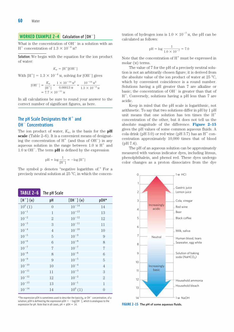
Note that the concentration of H1 must be expressed in molar (M) terms. The value of 7 for the pH of a precisely neutral solu-tion is not an arbitrarily chosen figure; it is derived from the absolute value of the ion product of water at 25 8C, which by convenient coincidence is a round number. Solutions having a pH greater than 7 are alkaline or basic; the concentration of OH2 is greater than that of H1. Conversely, solutions having a pH less than 7 are acidic. Keep in mind that the pH scale is logarithmic, not arithmetic. To say that two solutions differ in pH by 1 pH unit means that one solution has ten times the H1 concentration of the other, but it does not tell us the absolute magnitude of the difference. Figure 2–15 gives the pH values of some common aqueous fluids. A cola drink (pH 3.0) or red wine (pH 3.7) has an H1 con-centration approximately 10,000 times that of blood (pH 7.4). The pH of an aqueous solution can be approximately measured with various indicator dyes, including litmus, phenolphthalein, and phenol red. These dyes undergo color changes as a proton dissociates from the dye
TABLE 2–6 The pH Scale[H1] (M) pH [OH2] (M) pOH*100 (1) 0 10214 14
1021 1 10213 13
1022 2 10212 12
1023 3 10211 11
1024 4 10210 10
1025 5 1029 9
1026 6 1028 8
1027 7 1027 7
1028 8 1026 6
1029 9 1025 5
10210 10 1024 4
10211 11 1023 3
10212 12 1022 2
10213 13 1021 1
10214 14 100 (1) 0
*The expression pOH is sometimes used to describe the basicity, or OH2 concentration, of a
solution; pOH is defined by the expression pOH 5 2log[OH2], which is analogous to the
expression for pH. Note that in all cases, pH 1 pOH 5 14.
WORKED EXAMPLE 2–4 Calculation of [OH2]What is the concentration of OH2 in a solution with an H1 concentration of 1.3 3 1024 M?
Solution: We begin with the equation for the ion product of water:
Kw 5 [H1][OH2]
With [H1] 5 1.3 3 1024 M, solving for [OH2] gives
[OH2 4 5Kw
[H1]5
1 3 10214 M2
0.00013 M5
10214 M2
1.3 3 1024 M 5 7.7 3 10211 M
In all calculations be sure to round your answer to the correct number of significant figures, as here.
The pH Scale Designates the H1 and OH2 ConcentrationsThe ion product of water, Kw, is the basis for the pH scale (Table 2–6). It is a convenient means of designat-ing the concentration of H1 (and thus of OH2) in any aqueous solution in the range between 1.0 M H1 and 1.0 M OH2. The term pH is defined by the expression
pH 5 log1
[H1]5 2log [H1]
The symbol p denotes “negative logarithm of.” For a precisely neutral solution at 25 8C, in which the concen-
FIGURE 2–15 The pH of some aqueous fluids.
c02Water.indd Page 60 27/08/12 1:49 PM user-F408 /Users/user-F408/Desktop
13
12
11
10
9
8
7
6
5
4
3
2
1
0
Household bleach
Household ammonia
Solution of baking
soda (NaHCO3)
Seawater, egg white
Human blood, tears
Milk, saliva
Black coffee
Beer
Red wine
Cola, vinegar
Lemon juice
Gastric juice
1 m HCl
14 1 m NaOH
Neutral
Increasingly
basic
Increasingly
acidic

2.2 Ionization of Water, Weak Acids, and Weak Bases 61
molecule. Accurate determinations of pH in the chemi-cal or clinical laboratory are made with a glass electrode that is selectively sensitive to H1 concentration but insensitive to Na1, K1, and other cations. In a pH meter, the signal from the glass electrode placed in a test solu-tion is amplified and compared with the signal generat-ed by a solution of accurately known pH. Measurement of pH is one of the most important
and frequently used procedures in biochemistry. The pH affects the structure and activity of biological macromolecules; for example, the catalytic activity of enzymes is strongly dependent on pH (see Fig. 2–22). Measurements of the pH of blood and urine are com-monly used in medical diagnoses. The pH of the blood plasma of people with severe, uncontrolled diabetes, for example, is often below the normal value of 7.4; this condition is called acidosis (described in more detail below). In certain other diseases the pH of the blood is higher than normal, a condition known as alkalosis. Extreme acidosis or alkalosis can be life-threatening. ■
Weak Acids and Bases Have Characteristic Acid Dissociation ConstantsHydrochloric, sulfuric, and nitric acids, commonly called strong acids, are completely ionized in dilute aqueous solutions; the strong bases NaOH and KOH are also
completely ionized. Of more interest to biochemists is the behavior of weak acids and bases—those not com-pletely ionized when dissolved in water. These are ubiquitous in biological systems and play important roles in metabolism and its regulation. The behavior of aqueous solutions of weak acids and bases is best understood if we first define some terms. Acids may be defined as proton donors and bases as proton acceptors. When a proton donor such as acetic acid (CH3COOH) loses a proton, it becomes the corre-sponding proton acceptor, in this case the acetate anion (CH3COO2). A proton donor and its corresponding pro-ton acceptor make up a conjugate acid-base pair (Fig. 2–16), related by the reversible reaction
CH3COOHΔ CH3COO2 1 H1
Each acid has a characteristic tendency to lose its proton in an aqueous solution. The stronger the acid, the greater its tendency to lose its proton. The tendency of any acid (HA) to lose a proton and form its conjugate base (A2) is defined by the equilibrium constant (Keq) for the reversible reaction
HAΔ H1 1 A2
for which
Keq 5[H1][A2]
[HA]5 Ka
FIGURE 2–16 Conjugate acid-base pairs consist of a proton donor and a proton acceptor. Some compounds, such as acetic acid and ammonium
ion, are monoprotic; they can give up only one proton. Others are diprotic
(carbonic acid and glycine) or triprotic (phosphoric acid). The dissociation
reactions for each pair are shown where they occur along a pH gradient.
The equilibrium or dissociation constant (Ka) and its negative logarithm,
the pKa, are shown for each reaction. *For an explanation of apparent dis-
crepancies in pKa values for carbonic acid (H2CO3), see p. 67.
Monoprotic acidsAcetic acid
(Ka � 1.74 � 10�5 M)
Diprotic acidsCarbonic acid
(Ka � 1.70 � 10�4 M);
Bicarbonate
(Ka � 6.31 � 10�11 M)
Triprotic acidsPhosphoric acid
(Ka � 7.25 � 10�3 M);
Dihydrogen phosphate
(Ka � 1.38 � 10�7 M);
Monohydrogen phosphate
(Ka � 3.98 � 10�13 M)
Glycine, carboxyl
(Ka � 4.57 � 10�3 M);
Glycine, amino
(Ka � 2.51 � 10�10 M)
Ammonium ion
(Ka � 5.62 � 10�10 M)
pKa � 4.76
pKa � 3.77* pKa � 10.2
pKa � 9.25
pKa � 2.14 pKa � 6.86 pKa � 12.4
pKa � 2.34 pKa � 9.60
21 3 4 5 6 7 8 9 10 11 12 13
pH
CH3COH
O
CH3C �
O�
H�
O
H2CO3 HCO3� � H� HCO3
� CO32� � H�
NH4� NH3 � H�
H3PO4 H2PO4� � H�
H2PO4� HPO4
2� � H� HPO42� PO4
3� � H�
CH2COH
O
CH2C �
O�
H�
ONH3� NH3
�
CH2CO�
O
CH2C �
O�
H�
ONH3� NH2
c02Water.indd Page 61 27/08/12 1:49 PM user-F408 /Users/user-F408/Desktop

Water62
Equilibrium constants for ionization reactions are usu-ally called ionization constants or acid dissociation constants, often designated Ka. The dissociation con-stants of some acids are given in Figure 2–16. Stronger acids, such as phosphoric and carbonic acids, have larger ionization constants; weaker acids, such as mono-hydrogen phosphate (HPO22
4 ), have smaller ionization constants. Also included in Figure 2–16 are values of pKa, which is analogous to pH and is defined by the equation
pKa 5 log1Ka
5 2log Ka
The stronger the tendency to dissociate a proton, the stronger is the acid and the lower its pKa. As we shall now see, the pKa of any weak acid can be determined quite easily.
Titration Curves Reveal the pKa of Weak AcidsTitration is used to determine the amount of an acid in a given solution. A measured volume of the acid is titrated with a solution of a strong base, usually sodium hydroxide (NaOH), of known concentration. The NaOH is added in small increments until the acid is consumed (neutralized), as determined with an indicator dye or a pH meter. The concentration of the acid in the original solution can be calculated from the volume and concen-tration of NaOH added. The amounts of acid and base in titrations are often expressed in terms of equivalents, where one equivalent is the amount of a substance that will react with, or supply, one mole of hydrogen ions in an acid-base reaction. A plot of pH against the amount of NaOH added (a titration curve), reveals the pKa of the weak acid. Consider the titration of a 0.1 M solution of acetic acid with 0.1 M NaOH at 25 8C (Fig. 2–17). Two reversible equilibria are involved in the process (here, for simplic-ity, acetic acid is denoted HAc):
H2OΔ H1 1 OH2 (2–5)
HAcΔ H1 1 Ac2 (2–6)
The equilibria must simultaneously conform to their characteristic equilibrium constants, which are, respec-tively,
Kw 5 [H1][OH2 4 5 1 3 10214 M2 (2–7)
Ka 5[H1][Ac2]
[HAc]5 1.74 3 1025 M (2–8)
At the beginning of the titration, before any NaOH is added, the acetic acid is already slightly ionized, to an extent that can be calculated from its ionization con-stant (Eqn 2–8). As NaOH is gradually introduced, the added OH2 combines with the free H1 in the solution to form H2O, to an extent that satisfies the equilibrium relationship in Equation 2–7. As free H1 is removed, HAc dissociates
further to satisfy its own equilibrium constant (Eqn 2–8). The net result as the titration proceeds is that more and more HAc ionizes, forming Ac2, as the NaOH is added. At the midpoint of the titration, at which exactly 0.5 equivalent of NaOH has been added per equivalent of the acid, one-half of the original acetic acid has under-gone dissociation, so that the concentration of the proton donor, [HAc], now equals that of the proton acceptor, [Ac2]. At this midpoint a very important relationship holds: the pH of the equimolar solution of acetic acid and acetate is exactly equal to the pKa of acetic acid (pKa 5 4.76; Figs 2–16, 2–17). The basis for this rela-tionship, which holds for all weak acids, will soon become clear. As the titration is continued by adding further increments of NaOH, the remaining nondissociated ace-tic acid is gradually converted into acetate. The end point of the titration occurs at about pH 7.0: all the ace-tic acid has lost its protons to OH2, to form H2O and acetate. Throughout the titration the two equilibria (Eqns 2–5, 2–6) coexist, each always conforming to its equilibrium constant. Figure 2–18 compares the titration curves of three weak acids with very different ionization con-stants: acetic acid (pKa 5 4.76); dihydrogen phosphate,
FIGURE 2–17 The titration curve of acetic acid. After addition of each
increment of NaOH to the acetic acid solution, the pH of the mixture is
measured. This value is plotted against the amount of NaOH added,
expressed as a fraction of the total NaOH required to convert all the
acetic acid (CH3COOH) to its deprotonated form, acetate (CH3COO2).
The points so obtained yield the titration curve. Shown in the boxes are
the predominant ionic forms at the points designated. At the midpoint of
the titration, the concentrations of the proton donor and proton acceptor
are equal, and the pH is numerically equal to the pKa. The shaded zone
is the useful region of buffering power, generally between 10% and 90%
titration of the weak acid.
1.0
CH3COO�
CH3COOH
pH � pKa � 4.76
pHBuffering
region
OH� added (equivalents)
0 100%50
Percent titrated
9
8
7
3
2
1
00 0.5 0.6 0.7 0.8 0.90.40.30.20.1
[CH3COOH] � [CH3COO�]
pH 5.76
pH 3.76
6
5
4
c02Water.indd Page 62 27/08/12 1:49 PM user-F408 /Users/user-F408/Desktop

2.3 Buffering against pH Changes in Biological Systems 63
H2PO24 (pKa 5 6.86); and ammonium ion, NH1
4 (pKa 5 9.25). Although the titration curves of these acids have the same shape, they are displaced along the pH axis because the three acids have different strengths. Acetic acid, with the highest Ka (lowest pKa) of the three, is the strongest of the three weak acids (loses its proton most readily); it is already half dissociated at pH 4.76. Dihy-drogen phosphate loses a proton less readily, being half dissociated at pH 6.86. Ammonium ion is the weakest acid of the three and does not become half dissociated until pH 9.25. The titration curve of a weak acid shows graphically that a weak acid and its anion—a conjugate acid-base pair—can act as a buffer, as we describe in the next section.
SUMMARY 2.2 Ionization of Water, Weak Acids, and Weak Bases� Pure water ionizes slightly, forming equal numbers
of hydrogen ions (hydronium ions, H3O1) and
hydroxide ions. The extent of ionization is described
by an equilibrium constant, Keq 5[H1][OH2]
[H2O],
from which the ion product of water, Kw, is derived. At 25 8C, Kw 5 [H1][OH2] 5 (55.5 M)(Keq) 5
10214 M2.
� The pH of an aqueous solution reflects, on a logarithmic scale, the concentration of hydrogen ions:
pH 5 log1
[H1]5 2log [H1].
� The greater the acidity of a solution, the lower its pH. Weak acids partially ionize to release a hydrogen ion, thus lowering the pH of the aqueous solution. Weak bases accept a hydrogen ion, increasing the pH. The extent of these processes is characteristic of each particular weak acid or base and is expressed as an acid dissociation constant:
Keq 5[H1][A2]
[HA]5 Ka.
� The pKa expresses, on a logarithmic scale, the relative strength of a weak acid or base:
pKa 5 log1Ka
5 2log Ka.
� The stronger the acid, the smaller its pKa; the stronger the base, the larger its pKa. The pKa can be determined experimentally; it is the pH at the midpoint of the titration curve for the acid or base.
2.3 Buffering against pH Changes in Biological SystemsAlmost every biological process is pH-dependent; a small change in pH produces a large change in the rate of the process. This is true not only for the many reac-tions in which the H1 ion is a direct participant, but also for those reactions in which there is no apparent role for H1 ions. The enzymes that catalyze cellular reactions, and many of the molecules on which they act, contain ionizable groups with characteristic pKa values. The protonated amino and carboxyl groups of amino acids and the phosphate groups of nucleotides, for example, function as weak acids; their ionic state is determined by the pH of the surrounding medium. (When an ioniz-able group is sequestered in the middle of a protein, away from the aqueous solvent, its pKa, or apparent pKa, can be significantly different from its pKa in water.) As we noted above, ionic interactions are among the forces that stabilize a protein molecule and allow an enzyme to recognize and bind its substrate. Cells and organisms maintain a specific and con-stant cytosolic pH, usually near pH 7, keeping biomole-cules in their optimal ionic state. In multicellular organ-isms, the pH of extracellular fluids is also tightly regulated. Constancy of pH is achieved primarily by biological buffers: mixtures of weak acids and their con-jugate bases.
FIGURE 2–18 Comparison of the titration curves of three weak acids. Shown here are the titration curves for CH3COOH, H2PO2
4 , and NH14 .
The predominant ionic forms at designated points in the titration are
given in boxes. The regions of buffering capacity are indicated at the
right. Conjugate acid-base pairs are effective buffers between approxi-
mately 10% and 90% neutralization of the proton-donor species.
1.0
NH3
Midpoint
of
titration
Buffering
regions:pKa � 9.25
NH3
[NH�4]�[NH3]
CH3COO�pKa � 6.86
pKa � 4.76
[CH3COOH] � [CH3COO�]
CH3COOH
pH
10.25
5.76
3.76
14
13
12
11
10
9
8
7
6
5
4
3
2
1
00 0.50.40.30.20.1 0.6 0.7 0.8 0.9
[H2PO4�] � [HPO2
4�]
Phosphate
Acetate
NH4�
H2PO4�
8.257.86
5.86
HPO42�
OH� added (equivalents)
0 100%50
Percent titrated
c02Water.indd Page 63 27/08/12 1:49 PM user-F408 /Users/user-F408/Desktop

Water64
Buffers Are Mixtures of Weak Acids and Their Conjugate BasesBuffers are aqueous systems that tend to resist changes in pH when small amounts of acid (H1) or base (OH2) are added. A buffer system consists of a weak acid (the proton donor) and its conjugate base (the proton accep-tor). As an example, a mixture of equal concentrations of acetic acid and acetate ion, found at the midpoint of the titration curve in Figure 2–17, is a buffer system. Notice that the titration curve of acetic acid has a rela-tively flat zone extending about 1 pH unit on either side of its midpoint pH of 4.76. In this zone, a given amount of H1 or OH2 added to the system has much less effect on pH than the same amount added outside the zone. This relatively flat zone is the buffering region of the acetic acid–acetate buffer pair. At the midpoint of the buffering region, where the concentration of the proton donor (acetic acid) exactly equals that of the proton acceptor (acetate), the buffering power of the system is maximal; that is, its pH changes least on addition of H1 or OH2. The pH at this point in the titration curve of acetic acid is equal to its pKa. The pH of the acetate buf-fer system does change slightly when a small amount of H1 or OH2 is added, but this change is very small com-pared with the pH change that would result if the same amount of H1 or OH2 were added to pure water or to a solution of the salt of a strong acid and strong base, such as NaCl, which has no buffering power. Buffering results from two reversible reaction equi-libria occurring in a solution of nearly equal concentra-tions of a proton donor and its conjugate proton accep-tor. Figure 2–19 explains how a buffer system works. Whenever H1 or OH2 is added to a buffer, the result is a small change in the ratio of the relative concentrations of the weak acid and its anion and thus a small change in pH. The decrease in concentration of one component of the system is balanced exactly by an increase in the other. The sum of the buffer components does not change, only their ratio. Each conjugate acid-base pair has a characteristic pH zone in which it is an effective buffer (Fig. 2–18). The H2PO2
4 /HPO224 pair has a pKa of 6.86 and thus can
serve as an effective buffer system between approxi-mately pH 5.9 and pH 7.9; the NH1
4 /NH3 pair, with a pKa of 9.25, can act as a buffer between approximately pH 8.3 and pH 10.3.
The Henderson-Hasselbalch Equation Relates pH, pKa, and Buffer ConcentrationThe titration curves of acetic acid, H2PO2
4 , and NH14
(Fig. 2–18) have nearly identical shapes, suggesting that these curves reflect a fundamental law or relationship. This is indeed the case. The shape of the titration curve of any weak acid is described by the Henderson-Hassel-balch equation, which is important for understanding buffer action and acid-base balance in the blood and tis-
sues of vertebrates. This equation is simply a useful way of restating the expression for the ionization constant of an acid. For the ionization of a weak acid HA, the Hen-derson-Hasselbalch equation can be derived as follows:
Ka 5[H1][A2]
[HA]
First solve for [H1]:
[H1] 5 Ka
[HA]
[A2]
Then take the negative logarithm of both sides:
2log [H1] 5 2log Ka 2 log[HA]
[A2]
Substitute pH for 2log [H1] and pKa for 2log Ka:
pH 5 pKa 2 log[HA]
[A2]
Now invert 2log [HA]/[A2], which involves changing its sign, to obtain the Henderson-Hasselbalch equation:
pH 5 pKa 1 log [A2]
[HA] (2–9)
This equation fits the titration curve of all weak acids and enables us to deduce some important quantitative relationships. For example, it shows why the pKa of a
FIGURE 2–19 The acetic acid–acetate pair as a buffer system. The sys-
tem is capable of absorbing either H1 or OH2 through the reversibility of
the dissociation of acetic acid. The proton donor, acetic acid (HAc), con-
tains a reserve of bound H1, which can be released to neutralize an
addition of OH2 to the system, forming H2O. This happens because the
product [H1][OH2] transiently exceeds Kw (1 3 10214 M2). The equilib-
rium quickly adjusts to restore the product to 1 3 10214 M2 (at 25 8C),
thus transiently reducing the concentration of H1. But now the quotient
[H1][Ac2]/[HAc] is less than Ka, so HAc dissociates further to restore
equilibrium. Similarly, the conjugate base, Ac2, can react with H1 ions
added to the system; again, the two ionization reactions simultaneously
come to equilibrium. Thus a conjugate acid-base pair, such as acetic
acid and acetate ion, tends to resist a change in pH when small amounts of
acid or base are added. Buffering action is simply the consequence of two
reversible reactions taking place simultaneously and reaching their points
of equilibrium as governed by their equilibrium constants, Kw and Ka.
Kw � [H�][OH�]
Acetic acid
(CH3COOH)HAc Ac�
H�
OH� H2O
Acetate
(CH3COO�)
[H�][Ac�][HAc]
Ka �
c02Water.indd Page 64 27/08/12 1:49 PM user-F408 /Users/user-F408/Desktop

2.3 Buffering against pH Changes in Biological Systems 65
weak acid is equal to the pH of the solution at the mid-point of its titration. At that point, [HA] 5 [A2], and
pH 5 pKa 1 log 1 5 pKa 1 0 5 pKa
The Henderson-Hasselbalch equation also allows us to (1) calculate pKa, given pH and the molar ratio of pro-ton donor and acceptor; (2) calculate pH, given pKa and the molar ratio of proton donor and acceptor; and (3) calculate the molar ratio of proton donor and accep-tor, given pH and pKa.
Weak Acids or Bases Buffer Cells and Tissues against pH ChangesThe intracellular and extracellular fluids of multicellular organisms have a characteristic and nearly constant pH. The organism’s first line of defense against changes in internal pH is provided by buffer systems. The cytoplasm of most cells contains high concentrations of proteins, and these proteins contain many amino acids with functional groups that are weak acids or weak bases. For example, the side chain of histidine (Fig. 2–20) has a pKa of 6.0 and thus can exist in either the protonated or unproton-ated form near neutral pH. Proteins containing histidine residues therefore buffer effectively near neutral pH.
FIGURE 2–20 Ionization of histidine. The amino acid histidine, a compo-
nent of proteins, is a weak acid. The pKa of the protonated nitrogen of
the side chain is 6.0.
A
ACH2
A
ACH2
pH 5 pH 7
Protein Protein
3::4C
NH
CHHC HC
NH
�
CNH
CH � H�
N
WORKED EXAMPLE 2–5 Ionization of HistidineCalculate the fraction of histidine that has its imidazole side chain protonated at pH 7.3. The pKa values for histidine are pK1 5 1.8, pK2 (imidazole) 5 6.0, and pK3 5 9.2 (see Fig. 3–12b).
Solution: The three ionizable groups in histidine have suffi-ciently different pKa values that the first acid (—COOH) is completely ionized before the second (protonated imidaz-ole) begins to dissociate a proton, and the second ionizes completely before the third (—NH1
3) begins to dissociate its proton. (With the Henderson-Hasselbalch equation, we can easily show that a weak acid goes from 1% ionized at 2 pH units below its pKa to 99% ionized at 2 pH units above its pKa; see also Fig. 3–12b.) At pH 7.3, the carboxyl group of histidine is entirely deprotonated (—COO2) and the a-amino group is fully protonated (—NH1
3). We can there-fore assume that at pH 7.3, the only group that is partially
dissociated is the imidazole group, which can be proton-ated (we’ll abbreviate as HisH1) or not (His). We use the Henderson-Hasselbalch equation:
pH 5 pKa 1 log[A2]
[HA]
Substituting pK2 5 6.0 and pH 5 7.3:
7.3 5 6.0 1 log[His]
[HisH1]
1.3 5 log[His]
[HisH1]
antilog 1.3 5[His]
[HisH1]5 2.0 3 101
This gives us the ratio of [His] to [HisH1] (20 to 1 in this case). We want to convert this ratio to the fraction of total histidine that is in the unprotonated form His at pH 7.3. That fraction is 20/21 (20 parts His per 1 part HisH1, in a total of 21 parts histidine in either form), or about 95.2%; the remainder (100% minus 95.2%) is protonated—about 5%.
Nucleotides such as ATP, as well as many metabo-lites of low molecular weight, contain ionizable groups that can contribute buffering power to the cytoplasm. Some highly specialized organelles and extracellular compartments have high concentrations of compounds that contribute buffering capacity: organic acids buffer the vacuoles of plant cells; ammonia buffers urine. Two especially important biological buffers are the phosphate and bicarbonate systems. The phosphate buffer system, which acts in the cytoplasm of all cells, consists of H2PO2
4 as proton donor and HPO224 as proton acceptor:
H2PO24 Δ H1 1 HPO22
4
The phosphate buffer system is maximally effective at a pH close to its pKa of 6.86 (Figs 2–16, 2–18) and thus tends to resist pH changes in the range between about 5.9 and 7.9. It is therefore an effective buffer in biological fluids; in mammals, for example, extracellular fluids and most cyto-plasmic compartments have a pH in the range of 6.9 to 7.4.
WORKED EXAMPLE 2–6 Phosphate Buffers(a) What is the pH of a mixture of 0.042 M NaH2PO4 and 0.058 M Na2HPO4?
Solution: We use the Henderson-Hasselbalch equation, which we’ll express here as
pH 5 pKa 1 log[conjugate base]
[acid]
In this case, the acid (the species that gives up a proton) is H2PO2
4 , and the conjugate base (the species that gains a proton) is HPO22
4 . Substituting the given concentra-tions of acid and conjugate base and the pKa (6.86),
pH 5 6.86 1 log 0.0580.042
5 6.86 1 0.14 5 7.0
c02Water.indd Page 65 27/08/12 1:49 PM user-F408 /Users/user-F408/Desktop

Water66
in turn depends on the concentration of dissolved CO2, which in turn depends on the concentration of CO2 in the gas phase, or the partial pressure of CO2, denoted pCO2. Thus the pH of a bicarbonate buffer exposed to a gas phase is ultimately determined by the concentration of HCO2
3 in the aqueous phase and by pCO2 in the gas phase. The bicarbonate buffer system is an effective
physiological buffer near pH 7.4, because the H2CO3 of blood plasma is in equilibrium with a large reserve capacity of CO2(g) in the air space of the lungs. As noted above, this buffer system involves three reversible equilibria, in this case between gaseous CO2
in the lungs and bicarbonate (HCO23 ) in the blood
plasma (Fig. 2–21). Blood can pick up H1, such as from the lactic acid produced in muscle tissue during vigorous exercise. Alternatively, it can lose H1, such as by protonation of the NH3 produced during protein catabolism. When H1
is added to blood as it passes through the tissues, reac-tion 1 in Figure 2–21 proceeds toward a new equilibrium, in which [H2CO3] is increased. This in turn increases [CO2(d)] in the blood (reaction 2) and thus increases the partial pressure of CO2(g) in the air space of the lungs (reaction 3); the extra CO2 is exhaled. Conversely, when H1 is lost from the blood, the opposite events occur: more H2CO3 dissociates into H1 and HCO2
3 and thus more CO2(g) from the lungs dissolves in blood plasma. The rate of respiration—that is, the rate of inhal-ing and exhaling—can quickly adjust these equilibria to keep the blood pH nearly constant. The rate of respi-ration is controlled by the brain stem, where detection of an increased blood pCO2 or decreased blood pH trig-gers deeper and more frequent breathing. At the pH of blood plasma (7.4) very little H2CO3 is present in comparison with HCO2
3 , and the addition of a small amount of base (NH3 or OH2) would titrate this H2CO3, exhausting the buffering capacity. The important
FIGURE 2–21 The bicarbonate buffer system. CO2 in the air space of
the lungs is in equilibrium with the bicarbonate buffer in the blood
plasma passing through the lung capillaries. Because the concentration
of dissolved CO2 can be adjusted rapidly through changes in the rate of
breathing, the bicarbonate buffer system of the blood is in near-equilibrium
with a large potential reservoir of CO2.
Aqueous phase
(blood in capillaries)
H2CO
HCOH
3
3
H2OH2O
reaction 2
CO2(g)
reaction 3
Gas phase
(lung air space)
reaction 1
CO2(d)
We can roughly check this answer. When more con-jugate base than acid is present, the acid is more than 50% titrated and thus the pH is above the pKa (6.86), where the acid is exactly 50% titrated.
(b) If 1.0 mL of 10.0 M NaOH is added to a liter of the buffer prepared in (a), how much will the pH change?
Solution: A liter of the buffer contains 0.042 mol of NaH2PO4. Adding 1.0 mL of 10.0 M NaOH (0.010 mol) would titrate an equivalent amount (0.010 mol) of NaH2PO4 to Na2HPO4, resulting in 0.032 mol of NaH2PO4 and 0.068 mol of Na2HPO4. The new pH is
pH 5 pKa 1 log[HPO22
4 ]
[H2PO24 ]
5 6.86 1 log0.0680.032
5 6.86 1 0.33 5 7.2
(c) If 1.0 mL of 10.0 M NaOH is added to a liter of pure water at pH 7.0, what is the final pH? Compare this with the answer in (b).
Solution: The NaOH dissociates completely into Na1 and OH2, giving [OH2] 5 0.010 mol/L 5 1.0 3 1022 M. The pOH is the negative logarithm of [OH2], so pOH 5 2.0. Given that in all solutions, pH 1 pOH 5 14, the pH of the solution is 12. So, an amount of NaOH that increases the pH of water from 7 to 12 increases the pH of a buffered solu-tion, as in (b), from 7.0 to just 7.2. Such is the power of buffering!
Blood plasma is buffered in part by the bicarbonate system, consisting of carbonic acid (H2CO3) as proton donor and bicarbonate (HCO2
3 ) as proton acceptor (K1 is the first of several equilibrium constants in the bicarbonate buffering system):
H2CO3Δ H1 1 HCO23
K1 5[H1][HCO2
3 ]
[H2CO3]
This buffer system is more complex than other conjugate acid-base pairs because one of its components, carbonic acid (H2CO3), is formed from dissolved (d) carbon diox-ide and water, in a reversible reaction:
CO2(d) 1 H2OΔ H2CO3
K2 5[H2CO3]
[CO2(d)][H2O]
Carbon dioxide is a gas under normal conditions, and CO2 dissolved in an aqueous solution is in equilibrium with CO2 in the gas (g) phase:
CO2(g)Δ CO2(d)
K3 5[CO2(d)]
[CO2(g)]
The pH of a bicarbonate buffer system depends on the concentration of H2CO3 and HCO2
3 , the proton donor and acceptor components. The concentration of H2CO3
c02Water.indd Page 66 27/08/12 1:49 PM user-F408 /Users/user-F408/Desktop

2.3 Buffering against pH Changes in Biological Systems 67
role for H2CO3 (pKa 5 3.57 at 37 8C) in buffering blood plasma (,pH 7.4) seems inconsistent with our earlier statement that a buffer is most effective in the range of 1 pH unit above and below its pKa. The explanation for this paradox is the large reservoir of CO2(d) in blood. Its rapid equilibration with H2CO3 results in the formation of additional H2CO3:
CO2(d) 1 H2OΔ H2CO3
It is useful in clinical medicine to have a simple expres-sion for blood pH in terms of dissolved CO2, which is commonly monitored along with other blood gases. We can define a constant, Kh, which is the equilibrium con-stant for the hydration of CO2 to form H2CO3:
Kh 5[H2CO3]
[CO2(d)]
Then, to take the CO2(d) reservoir into account, we can express [H2CO3] as Kh[CO2(d)], and substitute this expression for [H2CO3] in the equation for the acid dis-sociation of H2CO3:
Ka 5[H1][HCO2
3 ]
[H2CO3]5
[H1][HCO23 ]
Kh[CO2 (d)]
Now, the overall equilibrium for dissociation of H2CO3 can be expressed in these terms:
KhKa 5 Kcombined 5[H1][HCO2
3 ]
[CO2 (d)]
We can calculate the value of the new constant, Kcombined, and the corresponding apparent pK, or pKcombined, from the experimentally determined values of Kh (3.0 3 1023
M) and Ka (2.7 3 1024 M) at 37 8C:
Kcombined 5 (3.0 3 1023 M)(2.7 3 1024 M)
5 8.1 3 1027 M2
pKcombined 5 6.1
In clinical medicine, it is common to refer to CO2(d) as the conjugate acid and to use the apparent, or com-bined, pKa of 6.1 to simplify calculation of pH from [CO2(d)]. In this convention,
pH 5 6.1 1 log[HCO2
3 ]
(0.23 3 pCO2)
where pCO2 is expressed in kilopascals (kPa; typically, pCO2 is 4.6 to 6.7 kPa) and 0.23 is the corresponding solubility coefficient for CO2 in water; thus the term 0.23 3 pCO2 < 1.2 kPa. Plasma [HCO2
3 ] is normally about 24 mM. ■
Untreated Diabetes Produces Life-Threatening Acidosis Human blood plasma normally has a pH between
7.35 and 7.45, and many of the enzymes that function in the blood have evolved to have maximal activ-ity in that pH range. Enzymes typically show maximal
catalytic activity at a characteristic pH, called the pH optimum (Fig. 2–22). On either side of this optimum pH, catalytic activity often declines sharply. Thus, a small change in pH can make a large difference in the rate of some crucial enzyme-catalyzed reactions. Bio-logical control of the pH of cells and body fluids is there-fore of central importance in all aspects of metabolism and cellular activities, and changes in blood pH have marked physiological consequences (described with gusto in Box 2–1!). In individuals with untreated diabetes mellitus, the lack of insulin, or insensitivity to insulin (depending on the type of diabetes), disrupts the uptake of glucose from blood into the tissues and forces the tissues to use stored fatty acids as their primary fuel. For reasons we will describe in detail later (see Fig. 24–30), this depen-dence on fatty acids results in the accumulation of high concentrations of two carboxylic acids, b-hydroxybutyric acid and acetoacetic acid (blood plasma level of 90 mg/100 mL, compared with ,3 mg/100 mL in control (healthy) individuals; urinary excretion of 5 g/24 hr, compared with ,125 mg/24 hr in controls). Dissociation of these acids lowers the pH of blood plasma to less than 7.35, causing acidosis. Severe acidosis leads to headache, drowsiness, nausea, vomiting, and diarrhea, followed by stupor, coma, and convulsions, presumably because at the lower pH, some enzyme(s) do not function optimally. When a patient is found to have high blood glucose, low plasma pH, and high levels of b-hydroxybutyric acid and acetoacetic acid in blood and urine, diabetes mellitus is the likely diagnosis. Other conditions can also produce acidosis. Fasting and starvation force the use of stored fatty acids as fuel,
FIGURE 2–22 The pH optima of some enzymes. Pepsin is a digestive
enzyme secreted into gastric juice, which has a pH of ,1.5, allowing
pepsin to act optimally. Trypsin, a digestive enzyme that acts in the
small intestine, has a pH optimum that matches the neutral pH in the
lumen of the small intestine. Alkaline phosphatase of bone tissue is a
hydrolytic enzyme thought to aid in bone mineralization.
100
7
pH
Pe
rce
nt
ma
xim
um
ac
tiv
ity
Alkaline
phosphatase
50
01 2 3 4 5 6 8 9 10
Pepsin
Trypsin
c02Water.indd Page 67 27/08/12 1:49 PM user-F408 /Users/user-F408/Desktop

Water68
with the same consequences as for diabetes. Very heavy exertion, such as a sprint by runners or cyclists, leads to temporary accumulation of lactic acid in the blood. Kid-ney failure results in a diminished capacity to regulate bicarbonate levels. Lung diseases (such as emphysema, pneumonia, and asthma) reduce the capacity to dispose of the CO2 produced by fuel oxidation in the tissues, with the resulting accumulation of H2CO3. Acidosis is treated by dealing with the underlying condition—administering insulin to people with diabetes, and ste-roids or antibiotics to people with lung disease. Severe acidosis can be reversed by administering bicarbonate solution intravenously. ■
This is an account by J.B.S. Haldane of physiological experiments on controlling blood pH, from his book Possible Worlds (Harper and Brothers, 1928). “I wanted to find out what happened to a man when one made him more acid or more alkaline . . . One might, of course, have tried experiments on a rabbit first, and some work had been done along these lines; but it is difficult to be sure how a rabbit feels at any time. Indeed, some rabbits make no serious attempt to cooperate with one. “. . . A human colleague and I therefore began experiments on one another . . . My colleague Dr. H.W. Davies and I made ourselves alkaline by over-breathing and by eating anything up to three ounces of bicar-bonate of soda. We made ourselves acid by sitting in an airtight room with between six and seven per cent of carbon dioxide in the air. This makes one breathe as if one had just completed a boat-race, and also gives one a rather violent headache . . . Two hours was as long as any one wanted to stay in the carbon diox-ide, even if the gas chamber at our disposal had not retained an ineradicable odour of ‘yellow cross gas' from some wartime experiments, which made one weep gently every time one entered it. The most obvi-ous thing to try was drinking hydrochloric acid. If one takes it strong it dissolves one’s teeth and burns one’s throat, whereas I wanted to let it diffuse gently all through my body. The strongest I ever cared to drink was about one part of the commercial strong acid in a hundred of water, but a pint of that was enough for me, as it irritated my throat and stomach, while my calculations showed that I needed a gallon and a half to get the effect I wanted . . . I argued that if one ate ammonium chloride, it would partly break up in the body, liberating hydrochloric acid. This proved to be correct . . . the liver turns ammonia into a harmless substance called urea before it reaches the heart and brain on absorption from the gut. The hydrochloric
acid is left behind and combines with sodium bicar-bonate, which exists in all the tissues, producing sodium chloride and carbon dioxide. I have had this gas produced in me in this way at the rate of six quarts an hour (though not for an hour on end at that rate) . . . “I was quite satisfied to have reproduced in myself the type of shortness of breath which occurs in the terminal stages of kidney disease and diabetes. This had long been known to be due to acid poisoning, but in each case the acid poisoning is complicated by other chemical abnormalities, and it had been rather uncertain which of the symptoms were due to the acid as such. “The scene now shifts to Heidelberg, where Freudenberg and György were studying tetany in babies . . . it occurred to them that it would be well worth trying the effect of making the body unusually acid. For tetany had occasionally been observed in patients who had been treated for other complaints by very large doses of sodium bicarbonate, or had lost large amounts of hydrochloric acid by constant vomit-ing; and if alkalinity of the tissues will produce tetany, acidity may be expected to cure it. Unfortunately, one could hardly try to cure a dying baby by shutting it up in a room full of carbonic acid, and still less would one give it hydrochloric acid to drink; so nothing had come of their idea, and they were using lime salts, which are not very easily absorbed, and which upset the diges-tion, but certainly benefit many cases of tetany. “However, the moment they read my paper on the effects of ammonium chloride, they began giving it to babies, and were delighted to find that the tetany cleared up in a few hours. Since then it has been used with effect both in England and America, both on children and adults. It does not remove the cause, but it brings the patient into a condition from which he has a very fair chance of recovering.”
BOX 2–1 MEDICINE On Being One's Own Rabbit (Don't Try This at Home!)
WORKED EXAMPLE 2–7 Treatment of Acidosis with Bicarbonate
Why does intravenous administration of a bicarbonate solution raise the plasma pH?
Solution: The ratio of [HCO23 ] to [CO2(d)] determines the
pH of the bicarbonate buffer, according to the equation
pH 5 6.1 1 log[HCO2
3 ]
(0.23 3 pCO2)
If [HCO23 ] is increased with no change in pCO2, the pH
will rise.
c02Water.indd Page 68 27/08/12 1:49 PM user-F408 /Users/user-F408/Desktop

2.5 The Fitness of the Aqueous Environment for Living Organisms 69
SUMMARY 2.3 Buffering against pH Changes in Biological Systems� A mixture of a weak acid (or base) and its salt
resists changes in pH caused by the addition of H1 or OH2. The mixture thus functions as a buffer.
� The pH of a solution of a weak acid (or base) and its salt is given by the Henderson-Hasselbalch
equation: pH 5 pKa 1 log[A2]
[HA].
� In cells and tissues, phosphate and bicarbonate buffer systems maintain intracellular and extracellular fluids at their optimum (physiological) pH, which is usually close to pH 7. Enzymes generally work optimally at this pH.
� Medical conditions that lower the pH of blood, causing acidosis, or raise it, causing alkalosis, can be life threatening.
2.4 Water as a ReactantWater is not just the solvent in which the chemical reac-tions of living cells occur; it is very often a direct par-ticipant in those reactions. The formation of ATP from ADP and inorganic phosphate is an example of a con-densation reaction in which the elements of water are eliminated (Fig. 2–23). The reverse of this reaction—cleavage accompanied by the addition of the elements of water—is a hydrolysis reaction. Hydrolysis reac-tions are also responsible for the enzymatic depolymer-ization of proteins, carbohydrates, and nucleic acids. Hydrolysis reactions, catalyzed by enzymes called hydrolases, are almost invariably exergonic; by pro-ducing two molecules from one, they lead to an increase in the randomness of the system. The formation of cel-lular polymers from their subunits by simple reversal of hydrolysis (that is, by condensation reactions) would be endergonic and therefore does not occur. As we shall see, cells circumvent this thermodynamic obstacle by coupling endergonic condensation reactions to exer-gonic processes, such as breakage of the anhydride bond in ATP.
You are (we hope!) consuming oxygen as you read. Water and carbon dioxide are the end products of the oxidation of fuels such as glucose. The overall reaction can be summarized as
C6H12O6Glucose
1 6O2¡ 6CO2 1 6H2O
The “metabolic water” formed by oxidation of foods and stored fats is actually enough to allow some animals in very dry habitats (gerbils, kangaroo rats, camels) to survive for extended periods without drinking water. The CO2 produced by glucose oxidation is converted in erythrocytes to the more soluble HCO2
3 , in a reaction catalyzed by the enzyme carbonic anhydrase:
CO2 1 H2OΔ HCO23 1 H1
In this reaction, water not only is a substrate but also functions in proton transfer by forming a network of hydrogen-bonded water molecules through which proton hopping occurs (Fig. 2–14). Green plants and algae use the energy of sunlight to split water in the process of photosynthesis:
2H2O 1 2A 88nlight O2 1 2AH2
In this reaction, A is an electron-accepting species, which varies with the type of photosynthetic organism, and water serves as the electron donor in an oxidation-reduction sequence (see Fig. 19–59) that is fundamen-tal to all life.
SUMMARY 2.4 Water as a Reactant� Water is both the solvent in which metabolic
reactions occur and a reactant in many biochemical processes, including hydrolysis, condensation, and oxidation-reduction reactions.
2.5 The Fitness of the Aqueous Environment for Living OrganismsOrganisms have effectively adapted to their aqueous environment and have evolved means of exploiting the unusual properties of water. The high specific heat of water (the heat energy required to raise the temperature of 1 g of water by 1 8C) is useful to cells and organisms because it allows water to act as a “heat buffer,” keeping the temperature of an organism relatively constant as the temperature of the surroundings fluctuates and as heat is generated as a byproduct of metabolism. Furthermore, some vertebrates exploit the high heat of vaporization of water (Table 2–1) by using (thus losing) excess body heat to evaporate sweat. The high degree of internal cohesion of liquid water, due to hydrogen bonding, is exploited by plants as a means of transporting dissolved nutrients from the roots to the leaves during the process of transpiration. Even the density of ice, lower than that of liquid water, has important biological consequences in the life cycles of aquatic organisms. Ponds freeze from
FIGURE 2–23 Participation of water in biological reactions. ATP is a
phosphoanhydride formed by a condensation reaction (loss of the ele-
ments of water) between ADP and phosphate. R represents adenosine
monophosphate (AMP). This condensation reaction requires energy.
The hydrolysis of (addition of the elements of water to) ATP to form
ADP and phosphate releases an equivalent amount of energy. These
condensation and hydrolysis reactions of ATP are just one example of
the role of water as a reactant in biological processes.
ROOOPBO
AO�
OOOPBO
AO�
OO� � H2O
(ATP)
R OOPBO
AO�
O H � HOOPBO
AO�O
(ADP)
O O
O�
Phosphoanhydride
c02Water.indd Page 69 27/08/12 1:49 PM user-F408 /Users/user-F408/Desktop

Water70
the top down, and the layer of ice at the top insulates the water below from frigid air, preventing the pond (and the organisms in it) from freezing solid. Most fundamental to all living organisms is the fact that many physical and biological properties of cell macromolecules, particularly the proteins and nucleic acids, derive from their interac-tions with water molecules of the surrounding medium. The influence of water on the course of biological evolu-tion has been profound and determinative. If life forms have evolved elsewhere in the universe, they are unlikely to resemble those of Earth unless liquid water is plentiful in their planet of origin.
Kandori, H. (2000) Role of internal water molecules in bacteriorhodopsin. Biochim. Biophys. Acta 1460, 177–191.
Intermediate-level review of the role of an internal chain of water molecules in proton movement through this protein.
Kornblatt, J. & Kornblatt, J. (1997) The role of water in recognition and catalysis by enzymes. Biochemist 19 (3), 14–17.
A short, useful summary of the ways in which bound water influences the structure and activity of proteins.
Lemieux, R.U. (1996) How water provides the impetus for molecular recognition in aqueous solution. Acc. Chem. Res. 29, 373–380.
A study of the role of water in the binding of a sugar to its binding protein.
Luecke, H. (2000) Atomic resolution structures of bacteriorhodopsin photocycle intermediates: the role of discrete water molecules in the function of this light-driven ion pump. Biochim. Biophys. Acta 1460, 133–156.
Advanced review of a proton pump that employs an internal chain of water molecules.
Nicolls, P. (2000) Introduction: the biology of the water molecule. Cell. Mol. Life Sci. 57, 987–992.
A short review of the properties of water, introducing several excellent advanced reviews published in the same issue (see especially Pocker, 2000, and Rand et al., 2000, below).
Symons, M.C. (2000) Spectroscopy of aqueous solutions: protein and DNA interactions with water. Cell. Mol. Life Sci. 57, 999–1007.
Wiggins, P.M. (1990) Role of water in some biological processes. Microbiol. Rev. 54, 432–449.
A review of water in biology, including discussion of the physical structure of liquid water, its interaction with biomolecules, and the state of water in living cells.
OsmosisCayley, D.S., Guttman, H.J., & Record, M.T., Jr. (2000) Biophysical characterization of changes in amounts and activity of Escherichia coli cell and compartment water and turgor pressure in response to osmotic stress. Biophys. J. 78, 1748–1764.
An advanced physical investigation of the cytoplasmic water fraction of the bacterium Escherichia coli grown in media of different osmolarities. (See also Record et al., 1998, below.)
Rand, R.P., Parsegian, V.A., & Rau, D.C. (2000) Intracellular osmotic action. Cell. Mol. Life Sci. 57, 1018–1032.
Review of the roles of water in enzyme catalysis as revealed by studies in water-poor solutes.
Record, M.T., Jr., Courtenay, E.S., Cayley, D.S., & Guttman, H.J. (1998) Responses of E. coli to osmotic stress: large changes in amounts of cytoplasmic solutes and water. Trends Biochem. Sci. 23, 143–148.
Intermediate-level review of the ways in which a bacterial cell counters changes in the osmolarity of its surroundings. (See also Cayley et al., 2000, above.)
Zonia, L. & Munnik, T. (2007) Life under pressure: hydrostatic pressure in cell growth and function. Trends Plant Sci. 12, 90–97.
Weak Interactions in Aqueous SystemsBaldwin, R.L. (2007) Energetics of protein folding. J. Mol. Biol. 371, 283–301.
Advanced discussion of the thermodynamic factors, including weak interactions, that determine the course of protein folding.
Ball, P. (2008) Water as an active constituent in cell biology. Chem. Rev. 108, 74–108.
An advanced discussion of the role of water in biological structure and function.
Blokzijl, W. & Engberts, J.B.F.N. (1993) Hydrophobic effects. Opinions and facts. Angew. Chem. Int. Ed. Engl. 32, 1545–1579.
Advanced, monumental, and critical review.
Chaplin, M. (2006) Do we underestimate the importance of water in cell biology? Nat. Rev. Mol. Cell Biol. 7, 861–866.
hydrogen bond 48bond energy 48hydrophilic 50hydrophobic 50amphipathic 52micelle 52hydrophobic
interactions 53van der Waals
interactions 53osmolarity 56osmosis 56isotonic 56hypertonic 56hypotonic 56equilibrium constant
(Keq) 59
ion product of water (Kw) 59
pH 60acidosis 61alkalosis 61conjugate acid-base
pair 61acid dissociation constant
(Ka) 62pKa 62titration curve 62buffer 64buffering region 64Henderson-Hasselbalch
equation 64condensation 69hydrolysis 69
Key TermsTerms in bold are defined in the glossary.
Further Reading
GeneralBall, P. (2001) Life’s Matrix: A Biography of Water, University of California Press, Berkeley, CA.
A highly accessible and entertaining description of water, from the Big Bang to its many roles in the chemistry of life.
Denny, M.W. (1993) Air and Water: The Biology and Physics of Life’s Media, Princeton University Press, Princeton, NJ.
A wonderful investigation of the biological relevance of the properties of water.
Eisenberg, D. & Kauzmann, W. (1969) The Structure and Properties of Water, Oxford University Press, New York.
An advanced, classic treatment of the physical chemistry of water and hydrophobic interactions.
Franks, F. & Mathias, S.F. (eds). (1982) Biophysics of Water, John Wiley & Sons, Inc., New York.
A large collection of papers on the structure of pure water and of the cytoplasm.
Gerstein, M. & Levitt, M. (1998) Simulating water and the molecules of life. Sci. Am. 279 (November), 100–105.
A well-illustrated description of the use of computer simulation to study the biologically important association of water with proteins and nucleic acids.
c02Water.indd Page 70 27/08/12 1:49 PM user-F408 /Users/user-F408/Desktop

Problems 71
Fersht, A.R. (1987) The hydrogen bond in molecular recognition. Trends Biochem. Sci. 12, 301–304.
A clear, brief, quantitative discussion of the contribution of hydrogen bonding to molecular recognition and enzyme catalysis.
Jeffrey, G.A. (1997) An Introduction to Hydrogen Bonding, Oxford University Press, New York.
A detailed, advanced discussion of the structure and properties of hydrogen bonds, including those in water and biomolecules.
Kauzmann, W. (1959) Some factors in the interpretation of protein denaturation. Adv. Protein Chem. 14, 1–63.
Remains the classic statement of the importance of hydrophobic interactions in the stability of proteins.
Ladbury, J. (1996) Just add water! The effect of water on the specificity of protein-ligand binding sites and its potential application to drug design. Chem. Biol. 3, 973–980.
Levy, Y. & Onuchic, J.N. (2006) Water mediation in protein folding and molecular recognition. Annu. Rev. Biophys. Biomol. Struct. 35, 389–415.
An advanced discussion of the role of water in protein structure.
Martin, T.W. & Derewenda, Z.S. (1999) The name is bond—H bond. Nat. Struct. Biol. 6, 403–406.
Brief review of the evidence that hydrogen bonds have some covalent character.
Pace, C.N. (2009) Energetics of protein hydrogen bonds. Nat. Struct. Mol. Biol. 16, 681–682.
Brief account of the historical contributions to understanding the strength of hydrogen bonds in proteins.
Pocker, Y. (2000) Water in enzyme reactions: biophysical aspects of hydration-dehydration processes. Cell. Mol. Life Sci. 57, 1008–1017.
Review of the role of water in enzyme catalysis, with carbonic anhydrase as the featured example.
Schwabe, J.W.R. (1997) The role of water in protein-DNA interactions. Curr. Opin. Struct. Biol. 7, 126–134.
An examination of the important role of water in both the specificity and the affinity of protein-DNA interactions.
Stillinger, F.H. (1980) Water revisited. Science 209, 451–457.A short review of the physical structure of water, including the
importance of hydrogen bonding and the nature of hydrophobic interactions.
Tanford, C. (1978) The hydrophobic effect and the organization of living matter. Science 200, 1012–1018.
A classic review of the chemical and energetic bases for hydrophobic interactions between biomolecules in aqueous solutions.
Weak Acids, Weak Bases, and Buffers: Problems for PracticeSegel, I.H. (1976) Biochemical Calculations, 2nd edn, John Wiley & Sons, Inc., New York.
Problems
1. Solubility of Ethanol in Water Explain why ethanol (CH3CH2OH) is more soluble in water than is ethane (CH3CH3).
2. Calculation of pH from Hydrogen Ion Concentra-tion What is the pH of a solution that has an H1 concentration of (a) 1.75 3 1025 mol/L; (b) 6.50 3 10210 mol/L; (c) 1.0 31024 mol/L; (d) 1.50 3 1025 mol/L?
3. Calculation of Hydrogen Ion Concentration from pH What is the H1 concentration of a solution with pH of (a) 3.82; (b) 6.52; (c) 11.11?
4. Acidity of Gastric HCl In a hospital laboratory, a 10.0 mL sample of gastric juice, obtained several hours
after a meal, was titrated with 0.1 M NaOH to neutrality; 7.2 mL of NaOH was required. The patient’s stomach contained no ingested food or drink; thus assume that no buffers were present. What was the pH of the gastric juice?
5. Calculation of the pH of a Strong Acid or Base (a) Write out the acid dissociation reaction for hydrochloric acid. (b) Calculate the pH of a solution of 5.0 3 1024 M HCl. (c) Write out the acid dissociation reaction for sodium hydroxide. (d) Calculate the pH of a solution of 7.0 3 1025 M NaOH.
6. Calculation of pH from Concentration of Strong Acid Calculate the pH of a solution prepared by diluting 3.0 mL of 2.5 M HCl to a final volume of 100 mL with H2O.
7. Measurement of Acetylcholine Levels by pH Changes The concentration of acetylcholine (a neurotransmitter) in a sample can be determined from the pH changes that accom-pany its hydrolysis. When the sample is incubated with the enzyme acetylcholinesterase, acetylcholine is converted to choline and acetic acid, which dissociates to yield acetate and a hydrogen ion:
�N
Acetylcholine
H2OCH3 CH2C O
O
CH2 CH3
CH3
CH3
O�
Choline Acetate
�N H�C
O
CH3HO � �
CH3
CH3
CH3CH2 CH2
In a typical analysis, 15 mL of an aqueous solution containing an unknown amount of acetylcholine had a pH of 7.65. When incubated with acetylcholinesterase, the pH of the solution decreased to 6.87. Assuming there was no buffer in the assay mixture, determine the number of moles of acetylcholine in the 15 mL sample.
8. Physical Meaning of pKa Which of the following aque-ous solutions has the lowest pH: 0.1 M HCl; 0.1 M acetic acid (pKa 5 4.86); 0.1 M formic acid (pKa 5 3.75)?
9. Meanings of Ka and pKa (a) Does a strong acid have a greater or lesser tendency to lose its proton than a weak acid? (b) Does the strong acid have a higher or lower Ka than the weak acid? (c) Does the strong acid have a higher or lower pKa than the weak acid?
10. Simulated Vinegar One way to make vinegar (not the preferred way) is to prepare a solution of acetic acid, the sole acid component of vinegar, at the proper pH (see Fig. 2–15)
c02Water.indd Page 71 27/08/12 1:49 PM user-F408 /Users/user-F408/Desktop

Water72
and add appropriate flavoring agents. Acetic acid (Mr 60) is a liquid at 25 8C, with a density of 1.049 g/mL. Calculate the volume that must be added to distilled water to make 1 L of simulated vinegar (see Fig. 2–16).
11. Identifying the Conjugate Base Which is the conjugate base in each of the pairs below?
(a) RCOOH, RCOO2 (c) H2PO24 , H3PO4
(b) RNH2, RNH13 (d) H2CO3, HCO2
3
12. Calculation of the pH of a Mixture of a Weak Acid and Its Conjugate Base Calculate the pH of a dilute solution that contains a molar ratio of potassium acetate to acetic acid (pKa 5 4.76) of (a) 2:1; (b) 1:3; (c) 5:1; (d) 1:1; (e) 1:10.
13. Effect of pH on Solubility The strongly polar, hydrogen-bonding properties of water make it an excellent solvent for ionic (charged) species. By contrast, nonionized, nonpolar organic molecules, such as benzene, are relatively insoluble in water. In principle, the aqueous solubility of any organic acid or base can be increased by converting the molecules to charged species. For example, the solubility of benzoic acid in water is low. The addition of sodium bicarbonate to a mixture of water and benzoic acid raises the pH and deprotonates the benzoic acid to form benzoate ion, which is quite soluble in water.
CBO
OOH COO�
Benzoic acid Benzoate ionpKa ≈ 5
BO
Are the following compounds more soluble in an aqueous solu-tion of 0.1 M NaOH or 0.1 M HCl? (The dissociable protons are shown in red.)
NI
AH
Pyridine ionpKa ≈ 5
(b)
(c)
(a)
� -NaphtholpKa ≈ 10
CB
H
GN
DH
OCAH
AC
JO
GOOCH3
OCH2
N-Acetyltyrosine methyl esterpKa ≈ 10
CH3
DOH
O
O
14. Treatment of Poison Ivy Rash The compo-nents of poison ivy and poison oak that produce the
characteristic itchy rash are catechols substituted with long-chain alkyl groups.
OH
(CH2)nOCH3
pKa ≈ 8
OH
If you were exposed to poison ivy, which of the treatments below would you apply to the affected area? Justify your choice.
(a) Wash the area with cold water.(b) Wash the area with dilute vinegar or lemon juice.(c) Wash the area with soap and water.(d) Wash the area with soap, water, and baking soda
(sodium bicarbonate).
15. pH and Drug Absorption Aspirin is a weak acid with a pKa of 3.5 (the ionizable H is shown in red):
CBO
GO
CBO
GOH
CH3
D
It is absorbed into the blood through the cells lining the stom-ach and the small intestine. Absorption requires passage through the plasma membrane, the rate of which is deter-mined by the polarity of the molecule: charged and highly polar molecules pass slowly, whereas neutral hydrophobic ones pass rapidly. The pH of the stomach contents is about 1.5, and the pH of the contents of the small intestine is about 6. Is more aspirin absorbed into the bloodstream from the stomach or from the small intestine? Clearly justify your choice.
16. Calculation of pH from Molar Concentrations What is the pH of a solution containing 0.12 mol/L of NH4Cl and 0.03 mol/L of NaOH (pKa of NH1
4 /NH3 is 9.25)?
17. Calculation of pH after Titration of Weak Acid A compound has a pKa of 7.4. To 100 mL of a 1.0 M solution of this compound at pH 8.0 is added 30 mL of 1.0 M hydrochloric acid. What is the pH of the resulting solution?
18. Properties of a Buffer The amino acid glycine is often used as the main ingredient of a buffer in biochemical experi-ments. The amino group of glycine, which has a pKa of 9.6, can exist either in the protonated form (—NH1
3 ) or as the free base (—NH2), because of the reversible equilibrium
RONH13 Δ RONH2 1 H1
(a) In what pH range can glycine be used as an effective buffer due to its amino group?
(b) In a 0.1 M solution of glycine at pH 9.0, what fraction of glycine has its amino group in the —NH1
3 form?(c) How much 5 M KOH must be added to 1.0 L of 0.1 M
glycine at pH 9.0 to bring its pH to exactly 10.0?(d) When 99% of the glycine is in its —NH1
3 form, what is the numerical relation between the pH of the solution and the pKa of the amino group?
c02Water.indd Page 72 27/08/12 1:49 PM user-F408 /Users/user-F408/Desktop

Problems 73
19. Calculation of the pKa of an Ionizable Group by Titration The pKa values of a compound with two ionizable groups are pK1 5 4.10 and pK2 between 7 and 10. A biochemist has 10 mL of a 1.0 M solution of this compound at a pH of 8.00. She adds 10.0 mL of 1.00 M HCl, which changes the pH to 3.20. What is pK2?
20. Calculation of the pH of a Solution of a Polyprotic Acid Histidine has ionizable groups with pKa values of 1.8, 6.0, and 9.2, as shown below (His 5 imidazole group). A bio-chemist makes up 100 mL of a 0.100 M solution of histidine at a pH of 5.40. She then adds 40 mL of 0.10 M HCl. What is the pH of the resulting solution?
21. Calculation of the Original pH from the Final pH after Titration A biochemist has 100 mL of a 0.10 M solution of a weak acid with a pKa of 6.3. She adds 6.0 mL of 1.0 M HCl, which changes the pH to 5.7. What was the pH of the original solution?
22. Preparation of a Phosphate Buffer What molar ratio of HPO22
4 to H2PO24 in solution would produce a pH of 7.0?
Phosphoric acid (H3PO4), a triprotic acid, has three pKa val-ues: 2.14, 6.86, and 12.4. Hint: Only one of the pKa values is relevant here.
23. Preparation of Standard Buffer for Calibration of a pH Meter The glass electrode used in commercial pH meters gives an electrical response proportional to the concentration of hydrogen ion. To convert these responses to a pH reading, the electrode must be calibrated against standard solutions of known H1 concentration. Determine the weight in grams of sodium dihydrogen phosphate (NaH2PO4 ? H2O; FW 138) and disodium hydrogen phosphate (Na2HPO4; FW 142) needed to prepare 1 L of a standard buffer at pH 7.00 with a total phos-phate concentration of 0.100 M (see Fig. 2–16). See problem 22 for the pKa values of phosphoric acid.
24. Calculation of Molar Ratios of Conjugate Base to Weak Acid from pH For a weak acid with a pKa of 6.0, calcu-late the ratio of conjugate base to acid at a pH of 5.0.
25. Preparation of Buffer of Known pH and Strength Given 0.10 M solutions of acetic acid (pKa 5 4.76) and sodium acetate, describe how you would go about preparing 1.0 L of 0.10 M acetate buffer of pH 4.00.
26. Choice of Weak Acid for a Buffer Which of these compounds would be the best buffer at pH 5.0: formic acid
(pKa 5 3.8), acetic acid (pKa 5 4.76), or ethylamine (pKa 5 9.0)? Briefly justify your answer.
27. Working with Buffers A buffer contains 0.010 mol of lactic acid (pKa 5 3.86) and 0.050 mol of sodium lactate per liter. (a) Calculate the pH of the buffer. (b) Calculate the change in pH when 5 mL of 0.5 M HCl is added to 1 L of the buffer. (c) What pH change would you expect if you added the same quantity of HCl to 1 L of pure water?
28. Use of Molar Concentrations to Calculate pH What is the pH of a solution that contains 0.20 M sodium acetate and 0.60 M acetic acid (pKa 5 4.76)?
29. Preparation of an Acetate Buffer Calculate the concentrations of acetic acid (pKa 5 4.76) and sodium acetate necessary to prepare a 0.2 M buffer solution at pH 5.0.
30. pH of Insect Defensive Secretion You have been observing an insect that defends itself from enemies by secreting a caustic liquid. Analysis of the liquid shows it to have a total concentration of formate plus formic acid (Ka 5 1.8 3 1024) of 1.45 M; the concentration of formate ion is 0.015 M. What is the pH of the secretion?
31. Calculation of pKa An unknown compound, X, is thought to have a carboxyl group with a pKa of 2.0 and another ionizable group with a pKa between 5 and 8. When 75 mL of 0.1 M NaOH is added to 100 mL of a 0.1 M solution of X at pH 2.0, the pH increases to 6.72. Calculate the pKa of the second ionizable group of X.
32. Ionic Forms of Alanine Alanine is a diprotic acid that can undergo two dissociation reactions (see Table 3–1 for pKa values). (a) Given the structure of the partially protonated form (or zwitterion; see Fig. 3–9) below, draw the chemical structures of the other two forms of alanine that predominate in aqueous solution: the fully protonated form and the fully deprotonated form.
Alanine
H3N C H
COO�
CH3
�
Of the three possible forms of alanine, which would be present at the highest concentration in solutions of the following pH: (b) 1.0; (c) 6.2; (d) 8.02; (e) 11.9. Explain your answers in terms of pH relative to the two pKa values.
33. Control of Blood pH by Respiratory Rate(a) The partial pressure of CO2 in the lungs can be varied
rapidly by the rate and depth of breathing. For example, a com-mon remedy to alleviate hiccups is to increase the concentra-tion of CO2 in the lungs. This can be achieved by holding one’s breath, by very slow and shallow breathing (hypoventilation), or by breathing in and out of a paper bag. Under such condi-tions, pCO2 in the air space of the lungs rises above normal.
H3N CH
COOH
CH2
C
HN
CHCH N
H
�
1.8pK1
6.0pK2
9.2pK3
H3N��
CH
COO�
CH2
C
HN
CHCH N
H
�
H3N�
CH
COO�
CH2
C
HN
CHCH N
H2N CH
COO�
CH2
C
HN
CHCH N
COOH COO�
HisH� HisNH3 NH2
�Ionizable
group
c02Water.indd Page 73 27/08/12 1:49 PM user-F408 /Users/user-F408/Desktop

Water74
Qualitatively explain the effect of these procedures on the blood pH.
(b) A common practice of competitive short-distance run-ners is to breathe rapidly and deeply (hyperventilate) for about half a minute to remove CO2 from their lungs just before the race begins. Blood pH may rise to 7.60. Explain why the blood pH increases.
(c) During a short-distance run, the muscles produce a large amount of lactic acid (CH3CH(OH)COOH; Ka 5 1.38 3 1024 M) from their glucose stores. In view of this fact, why might hyperventilation before a dash be useful?
34. Calculation of Blood pH from CO2 and Bicarbonate Levels Calculate the pH of a blood plasma sample with a total CO2 concentration of 26.9 mM and bicarbonate concentration of 25.6 mM. Recall from page 67 that the relevant pKa of car-bonic acid is 6.1.
35. Effect of Holding One’s Breath on Blood pH The pH of the extracellular fluid is buffered by the bicarbonate/carbonic acid system. Holding your breath can increase the concentration of CO2(g) in the blood. What effect might this have on the pH of the extracellular fluid? Explain by show-ing the relevant equilibrium equation(s) for this buffer system.
Data Analysis Problem
36. “Switchable” Surfactants Hydrophobic molecules do not dissolve well in water. Given that water is a very com-monly used solvent, this makes certain processes very diffi-cult: washing oily food residue off dishes, cleaning up spilled oil, keeping the oil and water phases of salad dressings well mixed, and carrying out chemical reactions that involve both hydrophobic and hydrophilic components.
Surfactants are a class of amphipathic compounds that includes soaps, detergents, and emulsifiers. With the use of surfactants, hydrophobic compounds can be suspended in aqueous solution by forming micelles (see Fig. 2–7). A micelle has a hydrophobic core consisting of the hydrophobic com-pound and the hydrophobic “tails” of the surfactant; the hydrophilic “heads” of the surfactant cover the surface of the micelle. A suspension of micelles is called an emulsion. The more hydrophilic the head group of the surfactant, the more powerful it is—that is, the greater its capacity to emulsify hydrophobic material.
When you use soap to remove grease from dirty dishes, the soap forms an emulsion with the grease that is easily removed by water through interaction with the hydrophilic head of the soap molecules. Likewise, a detergent can be used to emulsify spilled oil for removal by water. And emulsifiers in commercial salad dressings keep the oil suspended evenly throughout the water-based mixture.
There are some situations in which it would be very useful to have a “switchable” surfactant: a molecule that could be reversibly converted between a surfactant and a nonsurfactant.
(a) Imagine such a “switchable” surfactant existed. How would you use it to clean up and then recover the oil from an oil spill?
Liu et al. describe a prototypical switchable surfactant in their 2006 article “Switchable Surfactants.” The switching is based on the following reaction:
Amidine form Amidinium form
H2OCO2 �� HCO3�
�N CH3
CH3
NHR
EN
CH3A
�N CH3
CH3
N
H
HR EE
CH3A
C C
(b) Given that the pKa of a typical amidinium ion is 12.4, in which direction (left or right) would you expect the equilib-rium of the above reaction to lie? (See Fig. 2–16 for relevant pKa values.) Justify your answer. Hint: Remember the reaction H2O 1 CO2Δ H2CO3.
Liu and colleagues produced a switchable surfactant for which R 5 C16H33. They do not name the molecule in their article; for brevity, we’ll call it s-surf.
(c) The amidinium form of s-surf is a powerful surfactant; the amidine form is not. Explain this observation.
Liu and colleagues found that they could switch between the two forms of s-surf by changing the gas that they bubbled through a solution of the surfactant. They demonstrated this switch by measuring the electrical conductivity of the s-surf solution; aqueous solutions of ionic compounds have higher conductivity than solutions of nonionic compounds. They started with a solution of the amidine form of s-surf in water. Their results are shown below; dotted lines indicate the switch from one gas to another.
A
B
Time (min)
Gas bubbled in:
Ele
ctr
ica
l
co
nd
uc
tiv
ity
00
100 200
CO2 Ar CO2 Ar
(d) In which form is the majority of s-surf at point A? At point B?
(e) Why does the electrical conductivity rise from time 0 to point A?
(f) Why does the electrical conductivity fall from point A to point B?
(g) Explain how you would use s-surf to clean up and recover the oil from an oil spill.
ReferenceLiu, Y., Jessop, P.G., Cunningham, M., Eckert, C.A., & Liotta, C.L. (2006) Switchable surfactants. Science 313, 958–960.
c02Water.indd Page 74 27/08/12 1:49 PM user-F408 /Users/user-F408/Desktop