Embed Size (px)
Citation preview

Inflammation
1

Definition
• Inflammation – represents a typicalpathological process, an answer tocellular injury of different etiology,oriented toward diminishing activity andelimination of pathogenic factors fromthe body, delimitation of injuries,liquidation of injured structures andtheir replacement with viable structures.
2

• The clinical definition of inflammatory process in different organs is formed from Latin or Greek roots of organs name adding the suffix -it or –itis (ex: inflammation of the gum -gingivitis, of the pulp–pulpitis, of the tongue -glossitis)
3

General biological characteristics of inflammation
•Inflammation is a pathologic process ;
•Inflammation is a typical pathologic process ;
•Inflammation is the body answer to every injury withpredominant local manifestations, but also with generalreactions;
• Inflammation represents a complex of vascular-tissularreactions and can develop only at the level of tissues and organs.

The role of inflammation
• Without inflammation infections would go unchecked, wounds
would never heal, and injured tissues might
remain permanent festering sores.
5

ETIOLOGY OF INFLAMMATION = FLOGOGENIC FACTORS
Infections (bacterial, viral, fungal, parasitic) and microbial
toxins.
Tissue necrosis
Foreign bodies
Immune-mediated inflammation

INFLAMMATION
A C U T E C H R O N I C
• Rapid in onset (typically minutes);
• Short duration, lasting for hours or a few days;
• It is characteristic: exudation of fluid and plasma proteins (edema) and emigration of leukocytes, predominantly neutrophils.
• Longer duration;
• Is associated with the presence of lymphocytes and macrophages infiltration;
• It is characteristic: proliferation of blood vessels, fibrosis, and tissue destruction.
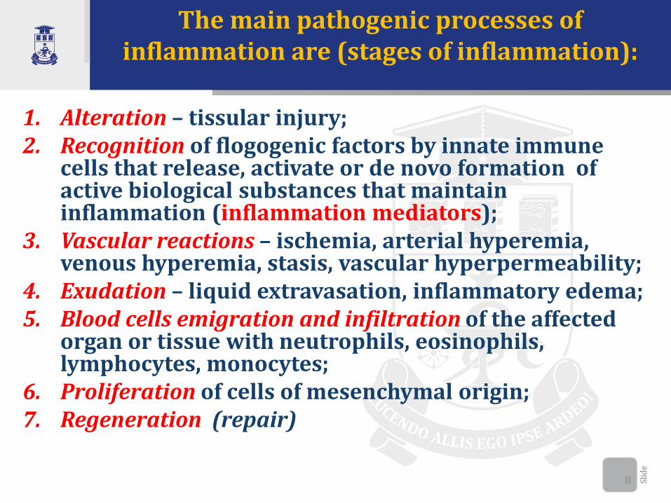
The main pathogenic processes of inflammation are (stages of inflammation):
1. Alteration – tissular injury;2. Recognition of flogogenic factors by innate immune
cells that release, activate or de novo formation of active biological substances that maintain inflammation (inflammation mediators);
3. Vascular reactions – ischemia, arterial hyperemia, venous hyperemia, stasis, vascular hyperpermeability;
4. Exudation – liquid extravasation, inflammatory edema;5. Blood cells emigration and infiltration of the affected
organ or tissue with neutrophils, eosinophils, lymphocytes, monocytes;
6. Proliferation of cells of mesenchymal origin;7. Regeneration (repair)
8

9

Inflammation of the pulp
• Schematic illustration of a tooth with a healthy pulp (left panel) and an inflamed pulp (right panel) subjacent to a caries lesion

I. ALTERATION IN INFLAMATORY PROCESS
P R I M A R Y S E C O N D A R Y
• Initial alteration caused by theinitial harmful factor (flogogenfactors).
• Structural and functionaldisorders provoked by theharmful factor directly in theplace where it acts.
• Primary alteration representsthe trigger mechanism andinitiates the onset ofinflammation.
Alteration as a consequence of action of pathogenic factors.
Persistent modification of cells and acellular elements structures at the level of tissues and organs
accompanied by functional disorders

II. Recognition of pathogenic factors by innate immune cells (DAMP)
PATHOGEN ASSOCIATED MOLECULAR PATTERN
(PAMP)(lipopolysaccharides, peptidoglycan,
endotoxin)
DAMAGE ASSOCIATED –MOLECULAR PATTERN
(DAMP)Products of cell injury, cell
necrosis
PATTERN RECOGNITION RECEPTORS(PAMP) (DAMP)
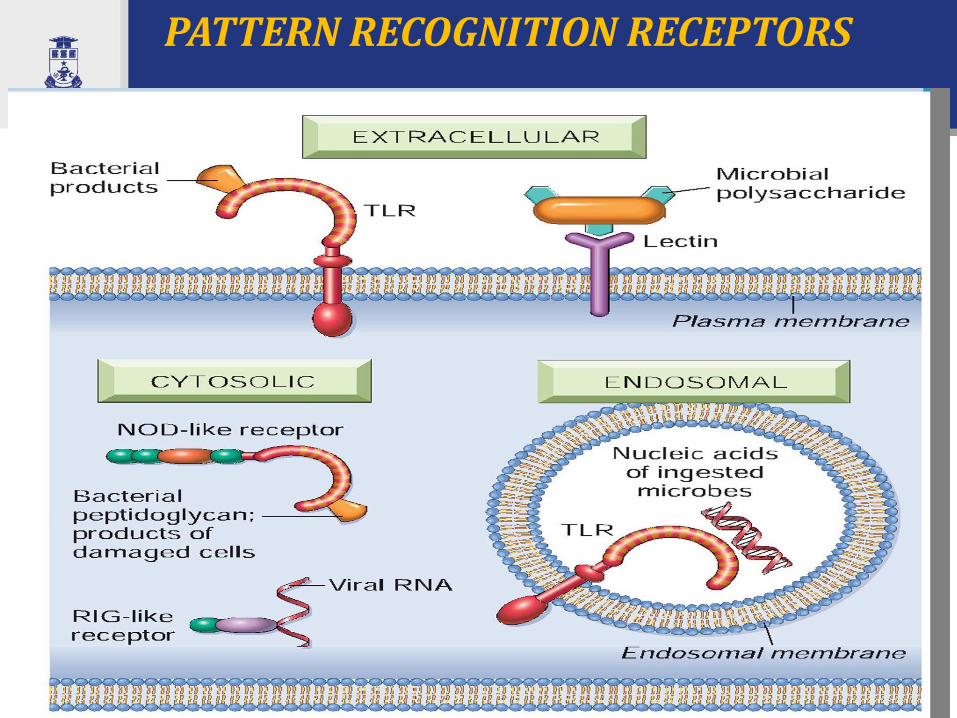
PATTERN RECOGNITION RECEPTORS

INFLAMMATORY MEDIATORS
Inflammatory mediatorsare soluble factors that areproduced by various cells orderived from plasma proteins andare generated or activated inresponse to the inflammatorystimulus.
They initiate and regulateinflammatory reactions and theirbiological goal is: protection thebody by diminish pathogenicactivity, delimitation and isolationof the focus of alteration andrestoration of the injuredstructures
14

INFLAMMATORY MEDIATORS

1. Presynthetizied cellular inflammatory
mediators
16
Histamine;Beta-glucosaminidase;
Triptase;CFE (chemotactic factor for eosinophils);CFN (chemotactic factor for neutrophils);
Heparine.
• LYSOSOMAL ENZYMES (glycolytic enzymes, photolytic enzymes, lipolytic enzymes)
• BACTERICIDE PRODUCTS- Oxygen dependent (H2O2, O2-, OH-,OCl-);- Oxygen-independent (cationic proteins, which
damage cellular membrane ofmicroorganisms, lysozim (muraminidasis) -which break down the muraminic acid frommucoproteins of microbial wall, lactoferinthat bind iron ions necessary for vital activityof the microbe.
Mast cell

17
Bactericide oxygen-dependent products(H2O2, OH-, OCl-, O2-) and specific mediators:
- cationic proteins and main basic protein with a direct anti-parasitic action;
- peroxidase – that break down oxygen peroxide till H2O2 andatomar oxygen, and in presence of halogens forms OCl-;
- histaminase – eoxidative deamination of histamine,- arylsulphatase - inactivates leukotrienes;- phospholipase D – inactivate the thrombocyte activator
factor;- perforins
• LYMPHOKINS- Mitogen factor, stimulates proliferation of non-
sensitized lymphocyte;- Factor of vascular wall hyperpermeability;- Lymphocytotoxin - have direct cytotoxic action;- Chemoattractant factor, which contributes to
lymphocyte migration from vascular bed intoinflammatory focus;
• - Inhibitory factor of macrophage migration

Platelets
18
• Serotonin (5-hydroxytryptamine) is
realized from platelets, when they
interact with collagen, thrombin,
adenosine diphosphate, and antigen-
antibody complexes. Thus, the
platelets release reaction, which is a
key component of coagulation, also
results in increased vascular
permeability. This is one of several
links between clotting and
inflammation.
• Thromboxane A2 (vasoconstriction
and platelet aggregation)

De novo synthesized inflammatory mediators
19

• Cytokines are proteins produced by many cell types (principally activated lymphocytes and macrophages, but also endothelial, epithelial, and connective tissue cells) that modulate the functions of other cell types.
20
CYTOKINES - INFLAMMATORY MEDIATORS

Cytokine Principal Sources
Principal Actions in
Inflammation
IN ACUTE INFLAMMATION
TNF Macrophages, mast cells, T
lymphocytes
Stimulates expression of endothelial
adhesion molecules and secretion of
other cytokines; systemic effects
IL-1 Macrophages, endothelial cells, some
epithelial cells
Similar to TNF; greater role in fever
IL-6 Macrophages, other cells Systemic effects (acute-phase
response)
Chemokines Macrophages, endothelial cells, T
lymphocytes, mast cells, other cell
types
Recruitment of leukocytes to sites of
inflammation; migration of cells to
normal tissues
IN CHRONIC INFLAMMATION
IL-12 Dendritic cells, macrophages Increased production of IFN-γ
IFN-γ T lymphocytes, NK cells Activation of macrophages
(increased ability to kill microbes
and tumor cells)
IL-17 T lymphocytes Recruitment of neutrophils and
monocytes
21

Systemic action of TNF and IL-1
22

CHEMOKINESChemokines (Greek -kinos,
movement) are a family of small cytokines, (8 to 10 kD) proteins that act primarily as chemoattractants for specific types of leukocytes.
They are secreted by activated macrophages, endothelial cells, and other cell types.
Chemokines have two main functions: they stimulate leukocyte recruitment in inflammation and control the normal migration of cells through various tissues. Examples: IL-8, Monocytechemoattractant protein (MCP-1), eotaxin, macrophage inflammatory protein-1α (MIP-1α), Lymphotactin , Fractalkine

2. PLASMA DERIVED MEDIATORS
• Plasma-derived mediators (e.g., Hageman factor, complement proteins, kinins) are produced mainly in the liver and present in the circulation as inactive precursors that must be activated, usually by a series of proteolytic cleavages, to acquire their biologic properties.
24
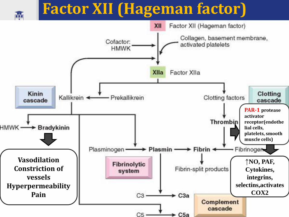
Factor XII (Hageman factor)
25
PAR-1 protease activator receptor(endothelial cells, platelets, smooth muscle cells)
↑NO, PAF,
Cytokines,
integrins,
selectins,activates
COX2
VasodilationConstriction of
vesselsHyperpermeability
Pain

Complement System
• The complement system consists of more than 20proteins, some of which are numbered C1 throughC9.
• This system functions in both innate and adaptiveimmunity for defense against microbial pathogens.
• It cause vascular permeability, chemotaxis, andopsonization.
26

Complement system
27

28
Vascular reactions

III. Vascular reactions in inflammatory process

INFLAMMATORY ARTERIAL HYPEREMIAC3a, C5a, PG, histamine,
bradykinin,
prostoglandin PGE2
• Increased blood flow assures optimal trophic conditions, and accumulation of leukocytes in vessels of inflammatory tissue,
which later will lead to release of inflammatory mediators,
phagocytosis, cellular infiltration, proliferation and regeneration
• Hemorrhage from dilatedvessels.• Spread from inflammatoryfocus of biologic active and toxicsubstances, with general effects.• Dissemination of pathogenagent and development ofsecondary inflammatory foci.

INFLAMMATORY VENOUS HYPEREMIA, STASIS, THROMBOSIS
Mechanisms of development
• Endothelial factors - endothelial cells become more spherical suchnarrowing vascular diameter; decreased negative charge ofendothelium which lead to adhesion of blood cells;
• Plasmatic factors – hemoconcentration, increased blood viscosity andhematocrit index, increased hemocirculatory resistance;
• Rheological factors –thrombocyte and erythrocyte aggregation, bloodcoagulation and thrombosis (active Hageman factor);
• Extravascular factors - tissue edema as result of extravasation due toblood and lymph vessels compression, which provoke hemostasis andlymphostasis.
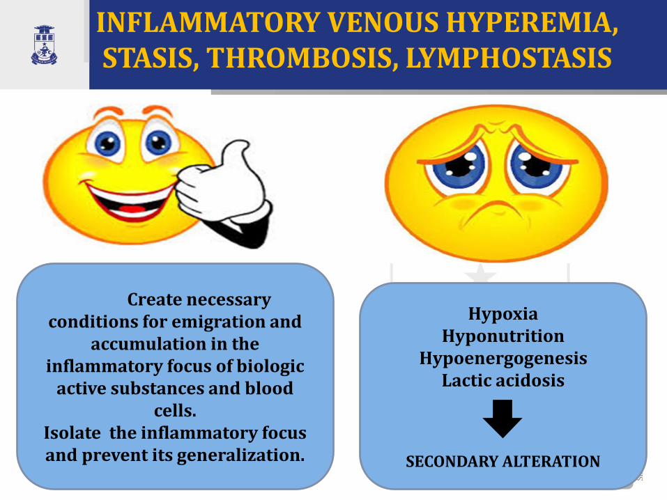
INFLAMMATORY VENOUS HYPEREMIA, STASIS, THROMBOSIS, LYMPHOSTASIS
Create necessary conditions for emigration and
accumulation in the inflammatory focus of biologic
active substances and blood cells.
Isolate the inflammatory focus and prevent its generalization.
HypoxiaHyponutrition
HypoenergogenesisLactic acidosis
SECONDARY ALTERATION

Exudation in inflammatory focus and edema Exudation
(inflammatory edema) represents
the extravasation of intravascular liquid
in the interstitial space or serous
cavities of the body.
• Contains more than 2% proteins, these having high molecular weight (globulin, fibrinogen)
• Contains cells (erythrocytes, thrombocyte, leucocytes);
• In case of infectious inflammation, exudates
is septic – contains pathogenic agents and
its vital products (toxins, enzymes, antigens).

TYPES OF EXUDATE
Serous exudates –contains up to 3% low
molecular weight proteins (predominantly
albumins), few neutrophils, these
determine its physical properties – low viscosity
(watery), fluid (flow easily), almost
transparent.
Fibrinous exudate – contains high molecular weight
proteins (globulins) and fibrinogen, the last being transformed into fibrin,
which causes the clotting of exudate, which has a gel
consistence, and attach to the tissues, blocking the
drainage

TYPES OF EXUDATE
Hemorrhagic exudate –occurs as result of increased vessel
permeability and contains erythrocytes
Purulent exudate –is an inflammatory exudate rich
in leukocytes(mostly degenerated
neutrophils), the debris of dead cells and, in many
cases, microbes.

V. Leukocytes emigration in inflammatory focus
36
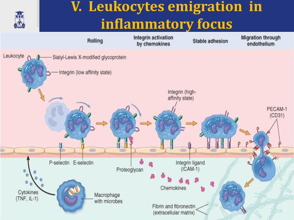
V. Leukocytes emigration in inflammatory focus
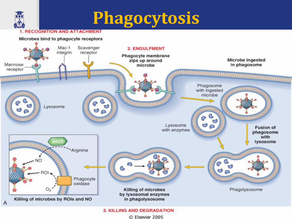
Phagocytosis
38

VI. Proliferation of cells in the inflammatory focus
39

VI. Proliferation of cells in the inflammatory focus
Proliferation represents themultiplication and
accumulation in theinflammatory focus of cells
of mesenchymal origin:
• hematopoietic stem-cells –monocites (macrophages),
T- and B-lymphocytes, plasmocytes
• local fibroblasts, epithelialcambial cells.
• Fibroblasts produceproduce ECM proteins and
collagen fibrils. 40
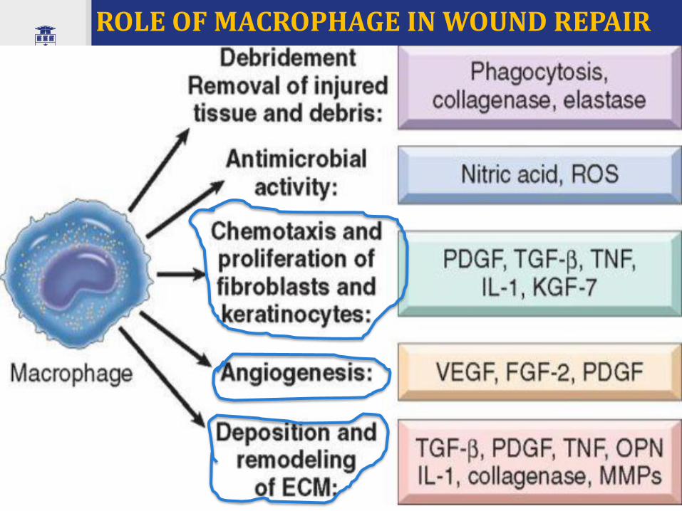
ROLE OF MACROPHAGE IN WOUND REPAIR

VII. Regeneration
• Regeneration represents the process of recovery of injured structure in the inflammatory focus, and it is directly proportional to the volume of destruction and to the regenerative capacity of affected organ. In function of these conditions the regeneration can be complete or incomplete
42

SYSTEMIC EFFECTS IN INFLAMMATORY REACTION - = ACUTE PHASE RESPONSE

Termination of the acute inflammatory response
Pro –inflammatory mediators
• Leukotriens(LTB4, LTC4, LTD4)
• TNF – α and IL-1
from macrophages and other cells
• pro-inflammatory lipid mediators
• TNF in macrophages
Anti-inflammatory mediators
• Lipoxins (LXA4, LXB4)
• transforming growth factor-β (TGF-β) and IL-10, from macrophages and other cells
• anti-inflammatory lipid mediators -resolvins and protectins
• Neural impulses (cholinergic discharge)
44

5 cardinal signs of inflammation
45

46