Embed Size (px)
Citation preview
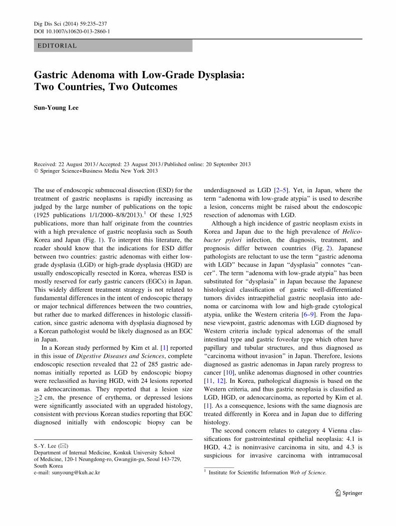
EDITORIAL
Gastric Adenoma with Low-Grade Dysplasia:Two Countries, Two Outcomes
Sun-Young Lee
Received: 22 August 2013 / Accepted: 23 August 2013 / Published online: 20 September 2013
� Springer Science+Business Media New York 2013
The use of endoscopic submucosal dissection (ESD) for the
treatment of gastric neoplasms is rapidly increasing as
judged by the large number of publications on the topic
(1925 publications 1/1/2000–8/8/2013).1 Of these 1,925
publications, more than half originate from the countries
with a high prevalence of gastric neoplasia such as South
Korea and Japan (Fig. 1). To interpret this literature, the
reader should know that the indications for ESD differ
between two countries: gastric adenomas with either low-
grade dysplasia (LGD) or high-grade dysplasia (HGD) are
usually endoscopically resected in Korea, whereas ESD is
mostly reserved for early gastric cancers (EGCs) in Japan.
This widely different treatment strategy is not related to
fundamental differences in the intent of endoscopic therapy
or major technical differences between the two countries,
but rather due to marked differences in histologic classifi-
cation, since gastric adenoma with dysplasia diagnosed by
a Korean pathologist would be likely diagnosed as an EGC
in Japan.
In a Korean study performed by Kim et al. [1] reported
in this issue of Digestive Diseases and Sciences, complete
endoscopic resection revealed that 22 of 285 gastric ade-
nomas initially reported as LGD by endoscopic biopsy
were reclassified as having HGD, with 24 lesions reported
as adenocarcinomas. They reported that a lesion size
C2 cm, the presence of erythema, or depressed lesions
were significantly associated with an upgraded histology,
consistent with previous Korean studies reporting that EGC
diagnosed initially with endoscopic biopsy can be
underdiagnosed as LGD [2–5]. Yet, in Japan, where the
term ‘‘adenoma with low-grade atypia’’ is used to describe
a lesion, concerns might be raised about the endoscopic
resection of adenomas with LGD.
Although a high incidence of gastric neoplasm exists in
Korea and Japan due to the high prevalence of Helico-
bacter pylori infection, the diagnosis, treatment, and
prognosis differ between countries (Fig. 2). Japanese
pathologists are reluctant to use the term ‘‘gastric adenoma
with LGD’’ because in Japan ‘‘dysplasia’’ connotes ‘‘can-
cer’’. The term ‘‘adenoma with low-grade atypia’’ has been
substituted for ‘‘dysplasia’’ in Japan because the Japanese
histological classification of gastric well-differentiated
tumors divides intraepithelial gastric neoplasia into ade-
noma or carcinoma with low and high-grade cytological
atypia, unlike the Western criteria [6–9]. From the Japa-
nese viewpoint, gastric adenomas with LGD diagnosed by
Western criteria include typical adenomas of the small
intestinal type and gastric foveolar type which often have
papillary and tubular structures, and thus diagnosed as
‘‘carcinoma without invasion’’ in Japan. Therefore, lesions
diagnosed as gastric adenomas in Japan rarely progress to
cancer [10], unlike adenomas diagnosed in other countries
[11, 12]. In Korea, pathological diagnosis is based on the
Western criteria, and thus gastric neoplasia is classified as
LGD, HGD, or adenocarcinoma, as reported by Kim et al.
[1]. As a consequence, lesions with the same diagnosis are
treated differently in Korea and in Japan due to differing
histology.
The second concern relates to category 4 Vienna clas-
sifications for gastrointestinal epithelial neoplasia: 4.1 is
HGD, 4.2 is noninvasive carcinoma in situ, and 4.3 is
suspicious for invasive carcinoma with intramucosal
S.-Y. Lee (&)
Department of Internal Medicine, Konkuk University School
of Medicine, 120-1 Neungdong-ro, Gwangjin-gu, Seoul 143-729,
South Korea
e-mail: [email protected] 1 Institute for Scientific Information Web of Science.
123
Dig Dis Sci (2014) 59:235–237
DOI 10.1007/s10620-013-2860-1

carcinoma [8]. Dysplasia is a precancerous lesion associ-
ated with a carcinoma risk increasing in parallel with the
histological grade [9]. Most Western pathologists use the
term ‘‘dysplasia’’ to describe a neoplastic premalignant
abnormality; evidence of invasion into the lamina propria
is needed to diagnose cancer. Nonetheless, while most
Western pathologists require a structurally invasive focus
for a cancer diagnosis, in the Japanese classification, there
is no formal adenocarcinoma in situ classification [8]. The
fundamental concepts for the diagnosis of cancer are
completely different between WHO classification and
Japanese classification. Japanese pathologists diagnose
cancer based on severe cytologic dysplasia, irrespective of
the presence of invasion [6, 13]. Therefore, a considerable
number of non-invasive intramucosal (intraepithelial)
gastric neoplasms, so-called HGD outside Japan, are
resected by ESD after being diagnosed as EGC in Japan.
Although the Vienna classification was designed to reduce
diagnostic discrepancies with Japan, it has been difficult to
adopt due to differing criteria used to diagnose gastric
epithelial neoplastic lesions [6, 8]. Thus, the prevalence of
EGC is inflated in Japan since lesions that most Western
pathologists identify as HGD are often considered intra-
mucosal adenocarcinomas in Japan (Fig. 3).
In addition to the inconsistency between the countries
regarding the diagnosis of EGC, the grading system for EGC
is not the same in Japan as elsewhere [6–9, 13]. There are
discrepancies with respect to the depth of invasion and the
EGC cell types. For example, when cancer cells are limited
to the first submucosal (SM1) layer with microvascular
invasion (i.e., lymphatic, venous, or perineural invasion) in
the third submucosal (SM3) layer, the depth of invasion is
identified as the ‘‘SM1 layer with microvascular invasion’’
outside Japan but as the ‘‘SM3 layer’’ within Japan. Based on
the concept that one cannot completely distinguish between
microvascular invasion and true direct invasion (even if there
is a distance between the tumor mass and distinct lymphatic/
vascular invasion), the depth of cancer invasion is defined as
the deepest level of the gastric wall in Japan. Nonetheless,
when an EGC is 80 % moderately-differentiated adenocar-
cinoma and 20 % poorly-differentiated adenocarcinoma,
the main diagnosis outside Japan would be the dominant
cell type of ‘‘moderately-differentiated adenocarcinoma’’,
whereas some Japanese pathologists define the diagnosis
‘‘poorly-differentiated adenocarcinoma’’ based on the most
advanced cell type. While neither diagnostic criteria can be
considered right or wrong, Japanese diagnoses for gastric
neoplasms are usually of higher grade than for comparable
lesions diagnosed in other countries.
Gastric adenoma with low-grade dysplasia
WHO pathological diagnostic criteria
Japanese pathological diagnostic criteria
Low-grade intraepithelial neoplasia (dysplasia)
Endoscopic resection*
Non-invasive benignneoplasia (some atypia)
Follow up orendoscopic resection
*Some reveal high-grade dysplasia or cancer after resection.
Fig. 2 Different diagnosis and management for gastric adenoma with
low-grade dysplasia. According to the Japanese classification of
gastric carcinoma, low-grade dysplasia is closer to group 3 (benign
neoplasia) and some of group 2 (atypical epithelium which cannot be
diagnosed as neoplastic atypia) lesions. Therefore, follow up is
preferred to endoscopic resection in Japan
Gastric adenoma with high-grade dysplasia
Endoscopic resection*
Intramucosal cancer if there is a nuclear or glandular
atypical change (even it is limited to the lamina propria)
*Some reveal cancer after resection.
Endoscopic resection
WHO pathological diagnostic criteria
Japanese pathological diagnostic criteria
High-grade intraepithelial neoplasia (dysplasia)
Fig. 3 Different diagnosis and management for gastric adenoma with
high-grade dysplasia. Japanese pathologists are reluctant to use the
term ‘‘high-grade dysplasia’’ for the gastric lesions, and therefore, it is
often diagnosed as intramucosal carcinoma in Japan
Japan(n=846)
Korea(n=302)
USA (n=130)
Germany (n=101)
China (n=111)
Others(n=435)
Fig. 1 Publications on endoscopic submucosal dissection according
to the countries. Since 2000, there were 1,925 publications in SCI
expanding SSCI, and A&HCI journals. Most of the papers were
published from countries where there is a high incidence of gastric
neoplasm due to the high prevalence of Helicobacter pylori infection
236 Dig Dis Sci (2014) 59:235–237
123
The final concern is the need for a unified therapeutic
plan for gastric dysplasia. In countries such as Japan where
the term ‘‘low and high-grade atypia’’ are used instead of
‘‘LGD and HGD’’ for a gastric adenoma, ESD is mostly
indicated for EGC, including intramucosal carcinomas,
which would be considered severe dysplasia or HGD out-
side of Japan. On the contrary, in countries where gastric
adenomas are classified as either LGD or HGD, as in the
study by Kim et al. [1], ESD is indicated not only for
gastric adenomas with HGD but also for those with LGD.
In conclusion, the diagnosis of gastric adenoma with
LGD in Korea carries a substantial risk of cancer develop-
ment, as reported by Kim et al. [1]. Efforts should be made
to develop unified international guidelines for the manage-
ment of gastric adenoma with LGD by overcoming the
differences in the diagnostic criteria that are currently used
in different countries and lead to a different natural course.
Acknowledgments This work was supported by the National
Research Foundation of Korea funded by the Korean Government
(NRF 2012K2A2A4010622). The author thanks to Dr. Yasuo Ohkura
at the Department of Pathology, Kyorin University School of Medi-
cine, Tokyo, Japan and Dr. Tetsuo Nemoto at the Department of
Surgical Pathology, School of Medicine, Toho University, Tokyo,
Japan for their valuable comments in revising Fig. 1 and descriptions
on the Japanese criteria.
Conflict of interest None.
References
1. Kim MK, Jang JY, Kim JW, et al. Is lesion size an independent
indication for endoscopic resection of biopsy-proven low-grade
gastric dysplasia? Dig Dis Sci. (Epub ahead of print). doi: 10.
1007/s10620-013-2805-8.
2. Choi CW, Kang DH, Kim HW, et al. Endoscopic submucosal
dissection as a treatment for gastric adenomatous polyps: pre-
dictive factors for early gastric cancer. Scand J Gastroenterol.
2012;47:1218–1225.
3. Cho SJ, Choi IJ, Kim CG, et al. Risk of high-grade dysplasia or
carcinoma in gastric biopsy-proven low-grade dysplasia: an
analysis using the Vienna classification. Endoscopy. 2011;43:
465–471.
4. Kim YJ, Park JC, Kim JH, et al. Histologic diagnosis based on
forceps biopsy is not adequate for determining endoscopic
treatment of gastric adenomatous lesions. Endoscopy. 2010;42:
620–626.
5. Sung HY, Cheung DY, Cho SH, et al. Polyps in the gastroin-
testinal tract: discrepancy between endoscopic forceps biopsies
and resected specimens. Eur J Gastroenterol Hepatol. 2009;21:
190–195.
6. Schlemper RJ, Riddell RH, Kato Y, et al. The Vienna classifi-
cation of gastrointestinal epithelial neoplasia. Gut. 2000;47:
251–255.
7. Tamura W, Fukami N. Early gastric cancer and dysplasia. Gas-
trointest Endosc Clin N Am. 2013;23:77–94.
8. Stolte M. The new Vienna classification of epithelial neoplasia of
the gastrointestinal tract: advantages and disadvantages. Virch-
ows Arch. 2003;442:99–106.
9. Lauwers GY, Srivastava A. Gastric preneoplastic lesions and
epithelial dysplasia. Gastroenterol Clin North Am. 2007;36:
813–829.
10. Yamada H, Ikegami M, Shimoda T, et al. Long-term follow-up
study of gastric adenoma/dysplasia. Endoscopy. 2004;36:390–396.
11. Rugge M, Cassaro M, Di Mario F, et al. The long term outcome
of gastric noninvasive neoplasia. Gut. 2003;52:1111–1116.
12. Park SY, Jeon SW, Jung MK, et al. Long-term follow-up study of
gastric intraepithelial neoplasias: progression from low-grade
dysplasia to invasive carcinoma. Eur J Gastroenterol Hepatol.
2008;20:966–970.
13. Japanese Gastric Cancer Association. Japanese classification of
gastric carcinoma: 3rd English edition. Gastric Cancer. 2011;14:
101–112.
Dig Dis Sci (2014) 59:235–237 237
123