Embed Size (px)
Citation preview
NMR IN BIOMEDICINENMR Biomed. 2006; 19: 368–378
DOI:10.1002/nbm.1050
Published online in Wiley InterScience (www.interscience.wiley.com).Potential and feasibility of parallel MRI at high field
Florian Wiesinger,1 Pierre-Francois Van de Moortele,2 Gregor Adriany,2 Nicola De Zanche,1
Kamil Ugurbil2 and Klaas P. Pruessmann1*
1Institute for Biomedical Engineering, University and ETH Zurich, Zurich, Switzerland2Center for MR research (CMRR), University of Minnesota, Minneapolis, Minnesota, USA
Received 23 November 2004; Accepted 14 March 2006
*CorrespondenceEngineering, UniZurich, SwitzerlaE-mail: PruessmaContract/grantE12061/4178.1INContract/grant sgrant number: T
Abbreviation usogeneity; BOLDEPI, echo planarview; GRAPPA,sition; RF, radioSAR, specific absencoding; SMASSNR, signal-to-n
Copyright # 200
ABSTRACT: This survey focuses on the fusion of two major lines of recent progress in MRI methodology: parallel imaging
with receiver coil arrays and the transition to high and ultra-high field strength for human applications. As discussed in this
paper, combining the two developments has vast potential due to multiple specific synergies. First, parallel acquisition and
high field are highly complementary in terms of their individual advantages and downsides. As a consequence, the joint
approach generally offers enhanced flexibility in the design of scanning strategies. Second, increasing resonance frequency
changes the electrodynamics of the MR signal in such a way that parallel imaging becomes more effective in large objects.
The underlying conceptual and theoretical considerations are reviewed in detail. In further sections, technical challenges and
practical aspects are discussed. The feasibility of parallel MRI at ultra-high field is illustrated by current results of parallel
human MRI at 7 T. Copyright # 2006 John Wiley & Sons, Ltd.
KEYWORDS: parallel imaging; high field; 7 T; SNR; SMASH; SENSE; GRAPPA
INTRODUCTION
This survey addresses the combination of parallelimaging (PI) with another important line of recentdevelopment in MRI methodology: the transition to highand ultra-high field strength for human applications.Large-scale research into ultra-high-field in vivo MR isdriven by the promises of increased intrinsic signal-to-noise ratio (SNR) and enhanced susceptibility contrast, astypically used for functional brain MRI. These benefitscome at the expense of serious fundamental issues such asincreased tissue heating, B1 inhomogeneity and limitedradiofrequency (RF) penetration (1–3), prompting somehesitation in the early days of ultra-high-field MR.However, within approximately the past decade boththeoretical (4–10) and experimental studies (11–13) haveestablished the feasibility of in vivoMR in humans at fieldstrengths as high as 7 Tand beyond. Concurrently, parallelMRI technology has entered the scene. Enhancing the
to: K. P. Pruessmann, Institute for Biomedicalversity and ETH Zurich, Gloriastrasse 35, [email protected]: EUREKA/KTI; contract/grant number:CA-MRI.ponsor: SEP Life Sciences, ETH Zurich; contract/H7/02-2.
ed: B0, main magnetic field strength; DB0, B0 inhom-contrast, blood oxygenation level dependent contrast;imaging; FLASH, fast low-angle shot; FOV, field-of-generalized autocalibrating partially parallel acqui-frequency; SOS image, root-sum-of-squares image;orption rate; PI, parallel imaging; SENSE, sensitivityH, simultaneous acquisition of spatial harmonics;oise ratio.
6 John Wiley & Sons, Ltd.
efficiency of spatial encoding in a fundamental fashion,parallel acquisition with coil arrays is widely recognizedas holding great potential for a broad range ofapplications. With several implementations available,such as SMASH (14), SENSE (15) and GRAPPA (16), PIis increasingly used in clinical practice (17,18).
The importance of these two developments isunderscored by their impact on the MRI industry. Todate, most major MRI vendors have expanded their rangeof human whole-body MRI systems toward 3 T and even7 T. Most of the currently available commercial systemsoffer embedded PI capability, with the number ofindependent receiver channels now ranging up to 32.For recent reviews of high-field and parallel in vivo MRsee, e.g., Refs (17–21).
High-field and parallel MRI are conceptually inde-pendent developments. However, it has recently beenrecognized that their combination holds particularpromise and entails vast and partly surprising synergieson two levels. First, it turns out that the two approachesare highly complementary in terms of their individualadvantages and downsides, hence adding significantflexibility in the design of scanning strategies. Second, ithas been found that high field conditions favor parallelimaging in an even more fundamental way, which isrooted in the physics of MR signal propagation anddetection. As it turns out, the transition to ultra-high fieldchanges these physics in such a way that spatial encodingby parallel acquisition becomes more effective.
This article aims to review these considerations in detail.We will first discuss the individual strengths andweaknesses of the two approaches and how they combine.
NMR Biomed. 2006; 19: 368–378
Table 1. Characteristics of high field and parallel ima-ging. ‘xxx’ indicates that there is no immediate effect.The two concepts are highly complementary and formvarious synergies when combined, as indicated by the‘R’ symbols. In this context, parallel imaging can beregarded as a converter, which puts the SNR benefits
PARALLEL MRI AT HIGH FIELD 369
Special emphasis is then laid upon the crucial role ofelectrodynamics and their impact on the inherent potentialand limitations of PI as a function of B0. Finally, we willdiscuss high-field PI from a practical perspective, high-lighting technical issues and initial results.
of high field towork in favor of alternative advantages
High FieldParallelImaging Combination
SNR Increased Decreased þScan efficiency
time, resolution,coverage
��� Increased ���
DB0 artifactsblurring, ghosting,distortions
Increased Decreased þ
Motion artifacts ��� Decreased ���RF energy absorption Increased Decreased þAcoustic gradient noise Increased Decreased þIntrinsic PPI performance Increased ��� þ
BASIC SYNERGIES
Compared with any known gradient encoding scheme,spatial encoding by means of distinct coil sensitivitiesstands out by the fact that it does not interfere with thenuclear spin magnetization that it encodes. This importantproperty underlies the versatility of the mechanism,which permits parallelizing virtually any MR imagingsequence. The enhanced encoding efficiency available inPI translates into numerous advantages. Most notablythese are: higher scan efficiency (reduced acquisitiontime, higher resolution and coverage), reduction ofartifacts due to B0 inhomogeneity (22–25) and motion(26–28), reduction of the specific absorption rate (SAR)(29), and mitigation of acoustic gradient noise (30). Inthe majority of applications these benefits come at theexpense of reduced SNR efficiency, which forms themajor downside of PI. As a consequence, PI is most usefulin situations where some spare SNR yield of aconventional technique can be traded for one or multipleof the advantages listed above.
High field (>1.5 T) and ultra-high field (>7T),1
conversely, provide two main benefits: higher baselineSNR and enhanced susceptibility contrast (e.g. T2
�,BOLD contrast) (19,20). However, increasing B0 alsogives rise to a range of specific problems, such asincreased B0 inhomogeneity, reduced T2 and T2
�
relaxation times, increased RF energy absorption in thetissue (SAR) and higher acoustic noise levels. The B0
homogeneity in biological samples generally degrades asB0 increases because the interfering effects of magnetictissue susceptibility scale with B0. For fast imagingtechniques, such as EPI (31), spiral scanning (32), andbalanced steady-state methods (33), B0 inhomogeneityresults in enhanced blurring, distortions, ghosting, or darkband artifacts. Therefore effective higher-order shimming(34–36) is vital at 3 T and above. Scanning at ultra-highfield typically requires further compromises for addres-sing field inhomogeneity and faster signal decay, such ask-space segmentation (37) and restricting the FOV (38–40). RF energy deposition in the tissue increasesapproximately as the square of the resonance frequencyand is a serious safety issue at field strengths of 3 T andbeyond (4,6,41). Strict SAR limitations impose signifi-cant restrictions on the design of RF-intensive MRsequences at high field. Finally, Lorentz forces on the
1Ultra-high field, since the associated 1H frequencies are in the ultra-high frequency range (300MHz to 1GHz).
Copyright # 2006 John Wiley & Sons, Ltd.
gradient coils also scale with B0, causing enhancedacoustic noise levels at high B0.
Table 1 summarizes these strengths and limitations.Given the listed characteristics, the two approachesevidently exhibit a high level of complementarity. Forexample, high field strength affords higher intrinsic SNRbut suffers from B0 inhomogeneity and SAR limitations.In turn, PI is an effective means of tackling B0
inhomogeneity and reducing the SAR at the expense ofthe SNR. By combining the two options one caneffectively address the shortcomings of both conceptsand frequently secure a significant net benefit comparedto non-parallel imaging at lower field. In this combi-nation, the parallelization plays the crucial role of aconverter, which puts the SNR benefit of high field towork in favor of alternative advantages.
These basic arguments illustrate the more straightfor-ward synergies between PI and high field. Additionally, asindicated in the last row of Table 1, there is another, moresophisticated kind of synergy, involving the inherentlimits of PI performance and the physics of MR signaldetection. This aspect is discussed in the following.
ELECTRODYNAMICS AND INTRINSICPOTENTIAL
In the late 1980s, Roemer et al. revolutionized signaldetection in MRI by introducing phased-array coils(42,43). In combination with appropriate reconstructionschemes, the array approach permits increasing the SNRyield by covering the imaged region with multiple coilshaving individual localized reception characteristics. InPI the parallel nature of array reception is additionallyutilized for supplementary spatial encoding (14–16,44–48). Both the SNR benefit and the encoding power of coilarrays depend crucially on the individual coils’ reception
NMR Biomed. 2006; 19: 368–378
370 F. WIESINGER ET AL.
characteristics. The latter are governed by the laws of RFelectrodynamics, i.e. Maxwell’s equations, which dependon the signal frequency and hence on B0. To study PIperformance as a function of B0 it is therefore essential toanalyze the role of electrodynamics in some detail. Thissection aims to summarize the relevant theoreticalconsiderations and the key conclusions that have resultedthus far.
Parallel imaging in terms of RFelectrodynamics
With respect to image reconstruction and SNR consider-ations, the relevant characteristics of a receiver coil arrayare the individual, spatially varying MR signal sensi-tivities of the array elements and the noise statistics of theentire array. Based on the reciprocal nature of theunderlying electrodynamics (49,50), these properties canbe expressed in terms of the electric (E) and magnetic (H)fields generated by the array when driving each individualcoil with unit input current at the appropriate Larmorfrequency. The coil sensitivities correspond to thetransverse components of the magnetic fields:
scðrÞ ¼ mðrÞ½HðcÞx ðrÞ � iHðcÞ
y ðrÞ� (1)
where c counts the array elements, r denotes position inthree-dimensional space, m denotes the magneticpermeability of the scanned object, and B0 is assumedto be aligned with the z-axis. Owing to the negative sign incombining the transverse field components, expression(1) is frequently referred to as B�
1 of the respective coilelement. The noise statistics are conveniently describedby the noise covariance matrix C, whose entries arerelated to the electric fields:
Cc;c0 ¼Z
sample
sðrÞEðcÞðrÞ � Eðc0ÞðrÞd3r; (2)
where s denotes the electric conductivity of the scannedobject and the bar indicates complex conjugation.In the common case of Cartesian k-space sampling, PI
reconstruction can be performed by image domainunfolding (15 and references therein). This is doneessentially by inverting a matrix of sensitivity values foreach set of pixels that alias in single-coil reconstruction.Let the index r count the pixels in any such set and rrdenote their positions. Then the sensitivity matrix for theset is given by
Sc;r ¼ scðrrÞ (3)
Similar to the mathematical description of phased-array imaging with full k-space density (42), the SNR ofeach of the reconstructed pixels values can now be
Copyright # 2006 John Wiley & Sons, Ltd.
expressed as (15):
SNRPIr / B2
0ffiffiffiffiffiffiffiffiffiffiffiffiffiffiffiffiffiffiffiffiffiffiffiffiffiffiffiffiffiffiffiffiffiffiffiffiffiffiffiR SHC�1S
� ��1� �
r;r
r ; (4)
where the reduction factor R indicates the factor by whichk-space was undersampled and the superscript H denotesthe complex conjugate transpose.
Equations (1)–(4) illustrate the close relationshipbetween the SNR yield in PI and the electrodynamicsof the receiver coil array. In practice the SNR yield in PI isoften stated relative to the SNR that would be achievedwith full-density k-space sampling (15):
SNRPIr ¼
SNRfullrffiffiffi
Rp
gr; (5)
where g is the so-called geometry factor. This equationemphasizes that the characteristic SNR loss in PI may beviewed as due to two independent mechanisms. Thesquare root of R reflects reduced overall data acquisitionand hence reduced intrinsic signal averaging. Conversely,the g factor describes noise amplification related to theconditioning of the unfolding operation. Hence it reflectsthe suitability of the coil configuration for the specific PItask, which is characterized by the size, shape, anddielectric properties of the object, the imaged slice orvolume, and the undersampling scheme. In a broad sense,the g factor may as well be regarded as a measure ofdistinctness among the set of coil sensitivities. Highlydistinct coil sensitivities yield low, i.e. favorable gfactors, which however can never drop below the optimalvalue of 1.
It is important to note that eqns (4) and (5) are based onthe assumption that all considered scans are performedwith the same pulse sequence and differ only in thenumber of repetitions. This leads to conservative SNRestimates because the application of PI frequently permitsoptimizing the acquisition strategy in terms of its baselineSNR yield. Higher baseline SNR can result, for instance,from adjusting the sequence timing, the length of echotrains, or the rate of contrast agent administration. Mostnotably, in some circumstances the SNR benefits of PI-driven sequence optimization actually outweigh theinherent SNR losses (25,51).
Based on the aforementioned considerations, PIperformance has recently been studied by means of boththeoretical models and experiments (52–58). In suchstudies the SNR yield of a specific imaging setup istypically quantified using formulas similar to eqn (4),requiring the determination of coil sensitivities andnoise statistics of the array under investigation. Inactual experiments, sc(r) and C can be determinedrelatively easily by calibration measurements (15,42,52).For the theoretical assessment of PI performance viaeqns (1) and (2) the RF coil’s reciprocal fields must becalculated by solving Maxwell’s equations (59).
NMR Biomed. 2006; 19: 368–378
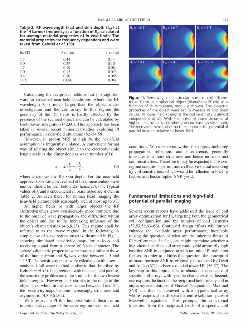
Table 2. RF wavelength (lRF) and skin depth (dRF) atthe 1H Larmor frequency as a function of B0, calculatedfor average material properties of in vivo brain. Thematerial properties are frequency dependent andweretaken from Gabriel et al. (98)
B0 (T) lRF (m) d RF (m)
1.5 0.44 0.143.0 0.27 0.104.7 0.19 0.0847.0 0.13 0.0739.4 0.10 0.06511.5 0.086 0.061
Figure 1. Sensitivity of a circular surface coil (diame-ter¼10 cm) in a spherical object (diameter¼ 20 cm) as afunction of B0 (simulated, modulus shown). The dielectricproperties of the object were set to average in vivo brainvalues. At lower field strengths the coil sensitivity is almostindependent of B0. With the onset of wave behavior athigher field the coil sensitivities grow increasingly structured.This increase in sensitivity structure enhances the potential ofparallel imaging relative to lower field
PARALLEL MRI AT HIGH FIELD 371
Calculating the reciprocal fields is fairly straightfor-ward in so-called near-field conditions, where the RFwavelength l is much larger than the object underinvestigation and the coil array. In this regime thegeometry of the RF fields is hardly affected by thepresence of the scanned object and can be calculated byBiot–Savart integration (42,60). This approach has beentaken in several recent numerical studies exploring PIperformance in near-field situations (52–54,58).
However, in proton MRI at high B0 the near-fieldassumption is frequently violated. A convenient formalway of relating the object size L to the electrodynamiclength scale is the dimensionless wave number (61):
k ¼ 2pL
lþ i
L
d; (6)
where d denotes the RF skin depth. For the near-fieldapproach to be valid the real part of the dimensionless wavenumber should be well below 2p, hence L/l< 1. Typicalvalues of l and d encountered in brain tissue are shown inTable 2. As seen there, for human head imaging thenear-field picture holds reasonably well at most up to 3T.
At higher fields or with larger objects the RFelectrodynamics grow considerably more complex dueto the onset of wave propagation and diffraction withinthe object and due to the increasing influence of theobject’s characteristics (4,6,8,13). This regime shall bereferred to as the ‘wave regime’ in the following. Asimple case of wave regime onset is illustrated in Fig. 1,showing simulated sensitivity maps for a loop coilreceiving signal from a sphere of 20 cm diameter. Thesphere’s dielectric properties were chosen similar to thoseof the human brain and B0 was varied between 1.5 and11.5 T. The sensitivity maps were calculated with a semi-analytical full-wave method similar to that described byKeltner et al. (4). In agreement with the near-field picture,the sensitivity profiles are quite similar for the two lowestfield strengths. However, as l shrinks to the range of theobject size, which in this case occurs between 4 and 5 T,the sensitivity maps become increasingly structured andasymmetric (4,8,9,61,62).
With respect to PI this last observation illustrates animportant advantage of the wave regime over near-field
Copyright # 2006 John Wiley & Sons, Ltd.
conditions. Wave behavior within the object, includingpropagation, reflection, and interference, generallytranslates into more structured and hence more distinctcoil sensitivities. Therefore it may be expected that wave-regime conditions permit more effective spatial encodingby coil sensitivities, which would be reflected in lower gfactors and hence higher SNR yield.
Fundamental limitations and high-fieldpotential of parallel imaging
Several recent reports have addressed the issue of coilarray optimization for PI, targeting both the geometricalcoil configuration and the number of coil elements(52,53,58,63–66). Continued design efforts will furtherenhance the available array performance, inevitablyraising the question of what are the inherent limits ofPI performance. In fact, one might speculate whether ahypothetical perfect coil array could yield arbitrarily highbaseline SNR in conjunction with unlimited PI reductionfactors. In order to address this question, the concept ofultimate intrinsic SNR as originally introduced by Ocaliand Atalar (67) has been extended toward PI (56,57). Thekey step in this approach is to abandon the concept ofspecific coil arrays with specific characteristics. Instead,one exploits the fact that the reciprocal fields of any coil inany array are solutions of Maxwell’s equations. MaximalSNR can thus be achieved with a hypothetical arraywhose reciprocal fields span the entire solution space ofMaxwell’s equations. This prompts the conceptualtransition from the reciprocal fields of a specific coil
NMR Biomed. 2006; 19: 368–378
372 F. WIESINGER ET AL.
array to a complete basis of Maxwell solutions:
fEcðrÞ;HcðrÞg ! facðrÞ; bcðrÞg (7)
Each element of this basis can be regarded as onebuilding block of a virtual ‘complete’ coil array.Evaluating eqns (1)–(5) with the full basis {ac(r),bc(r)} yields maps of the ultimate SNR for conventionalFourier imaging, and of the ultimate g factor for anyspecific PI experiment.To date, ultimate PI performance has been studied in
two independent investigations (56,57), assuming differ-ent specifications for the object geometry and materialproperties and using different Maxwell bases. Ohligeret al. (56) chose the same specifications as previouslyused in Ocali and Atalar (67), i.e. an elliptical cylinderwith dimensions and material properties similar to thehuman torso in conjunction with a plane wave basis. Inref. (57) spherical objects were studied using a multipolebasis. The key findings of these studies are similar andshall be summarized here along with selected results fromref. (57). For brevity, the discussion is limited to PIperformance in the center of the sphere when imaging thecentral transverse plane with k-space undersampling inone dimension.Figure 2 shows the ultimate SNR at the sphere’s center
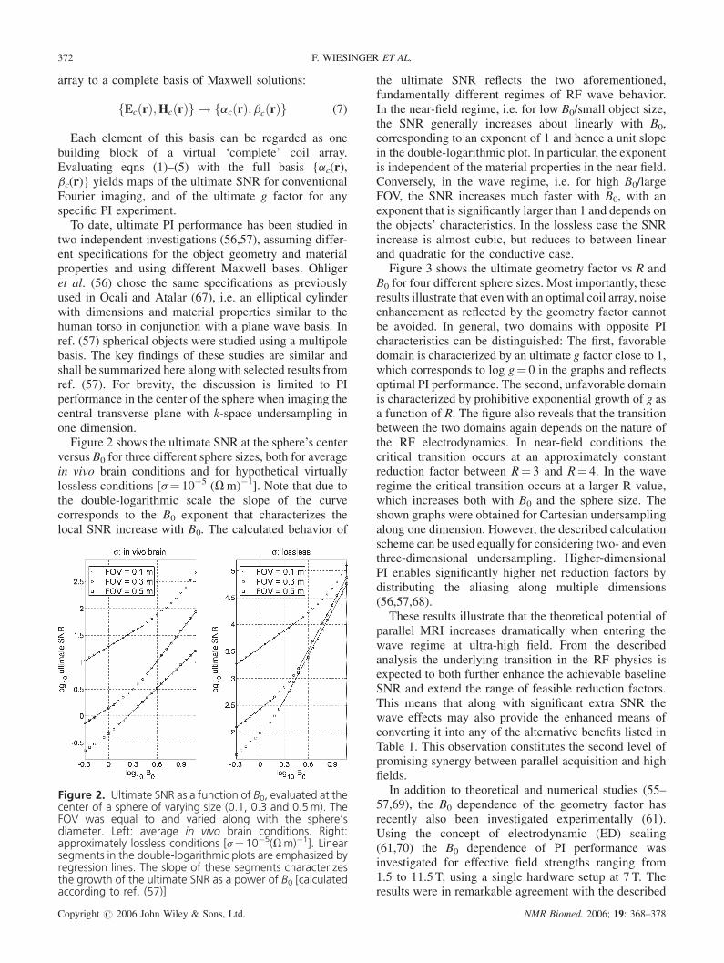
versus B0 for three different sphere sizes, both for averagein vivo brain conditions and for hypothetical virtuallylossless conditions [s¼ 10�5 (Vm)�1]. Note that due tothe double-logarithmic scale the slope of the curvecorresponds to the B0 exponent that characterizes thelocal SNR increase with B0. The calculated behavior of
igure 2. Ultimate SNR as a function of B0, evaluated at theenter of a sphere of varying size (0.1, 0.3 and 0.5m). TheOV was equal to and varied along with the sphere’siameter. Left: average in vivo brain conditions. Right:pproximately lossless conditions [s¼10�5(Vm)�1]. Linearegments in the double-logarithmic plots are emphasized byegression lines. The slope of these segments characterizeshe growth of the ultimate SNR as a power of B0 [calculatedccording to ref. (57)]
FcFdasrta
Copyright # 2006 John Wiley & Sons, Ltd.
the ultimate SNR reflects the two aforementioned,fundamentally different regimes of RF wave behavior.In the near-field regime, i.e. for low B0/small object size,the SNR generally increases about linearly with B0,corresponding to an exponent of 1 and hence a unit slopein the double-logarithmic plot. In particular, the exponentis independent of the material properties in the near field.Conversely, in the wave regime, i.e. for high B0/largeFOV, the SNR increases much faster with B0, with anexponent that is significantly larger than 1 and depends onthe objects’ characteristics. In the lossless case the SNRincrease is almost cubic, but reduces to between linearand quadratic for the conductive case.
Figure 3 shows the ultimate geometry factor vs R andB0 for four different sphere sizes. Most importantly, theseresults illustrate that even with an optimal coil array, noiseenhancement as reflected by the geometry factor cannotbe avoided. In general, two domains with opposite PIcharacteristics can be distinguished: The first, favorabledomain is characterized by an ultimate g factor close to 1,which corresponds to log g¼ 0 in the graphs and reflectsoptimal PI performance. The second, unfavorable domainis characterized by prohibitive exponential growth of g asa function of R. The figure also reveals that the transitionbetween the two domains again depends on the nature ofthe RF electrodynamics. In near-field conditions thecritical transition occurs at an approximately constantreduction factor between R¼ 3 and R¼ 4. In the waveregime the critical transition occurs at a larger R value,which increases both with B0 and the sphere size. Theshown graphs were obtained for Cartesian undersamplingalong one dimension. However, the described calculationscheme can be used equally for considering two- and eventhree-dimensional undersampling. Higher-dimensionalPI enables significantly higher net reduction factors bydistributing the aliasing along multiple dimensions(56,57,68).
These results illustrate that the theoretical potential ofparallel MRI increases dramatically when entering thewave regime at ultra-high field. From the describedanalysis the underlying transition in the RF physics isexpected to both further enhance the achievable baselineSNR and extend the range of feasible reduction factors.This means that along with significant extra SNR thewave effects may also provide the enhanced means ofconverting it into any of the alternative benefits listed inTable 1. This observation constitutes the second level ofpromising synergy between parallel acquisition and highfields.
In addition to theoretical and numerical studies (55–57,69), the B0 dependence of the geometry factor hasrecently also been investigated experimentally (61).Using the concept of electrodynamic (ED) scaling(61,70) the B0 dependence of PI performance wasinvestigated for effective field strengths ranging from1.5 to 11.5 T, using a single hardware setup at 7 T. Theresults were in remarkable agreement with the described
NMR Biomed. 2006; 19: 368–378
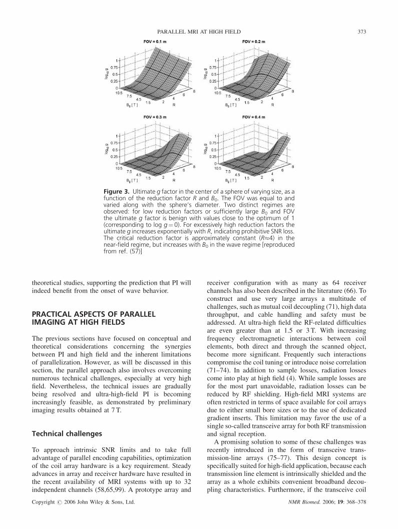
Figure 3. Ultimate g factor in the center of a sphere of varying size, as afunction of the reduction factor R and B0. The FOV was equal to andvaried along with the sphere’s diameter. Two distinct regimes areobserved: for low reduction factors or sufficiently large B0 and FOVthe ultimate g factor is benign with values close to the optimum of 1(corresponding to log g¼ 0). For excessively high reduction factors theultimate g increases exponentially with R, indicating prohibitive SNR loss.The critical reduction factor is approximately constant (R�4) in thenear-field regime, but increases with B0 in the wave regime [reproducedfrom ref. (57)]
PARALLEL MRI AT HIGH FIELD 373
theoretical studies, supporting the prediction that PI willindeed benefit from the onset of wave behavior.
PRACTICAL ASPECTS OF PARALLELIMAGING AT HIGH FIELDS
The previous sections have focused on conceptual andtheoretical considerations concerning the synergiesbetween PI and high field and the inherent limitationsof parallelization. However, as will be discussed in thissection, the parallel approach also involves overcomingnumerous technical challenges, especially at very highfield. Nevertheless, the technical issues are graduallybeing resolved and ultra-high-field PI is becomingincreasingly feasible, as demonstrated by preliminaryimaging results obtained at 7 T.
Technical challenges
To approach intrinsic SNR limits and to take fulladvantage of parallel encoding capabilities, optimizationof the coil array hardware is a key requirement. Steadyadvances in array and receiver hardware have resulted inthe recent availability of MRI systems with up to 32independent channels (58,65,99). A prototype array and
Copyright # 2006 John Wiley & Sons, Ltd.
receiver configuration with as many as 64 receiverchannels has also been described in the literature (66). Toconstruct and use very large arrays a multitude ofchallenges, such as mutual coil decoupling (71), high datathroughput, and cable handling and safety must beaddressed. At ultra-high field the RF-related difficultiesare even greater than at 1.5 or 3 T. With increasingfrequency electromagnetic interactions between coilelements, both direct and through the scanned object,become more significant. Frequently such interactionscompromise the coil tuning or introduce noise correlation(71–74). In addition to sample losses, radiation lossescome into play at high field (4). While sample losses arefor the most part unavoidable, radiation losses can bereduced by RF shielding. High-field MRI systems areoften restricted in terms of space available for coil arraysdue to either small bore sizes or to the use of dedicatedgradient inserts. This limitation may favor the use of asingle so-called transceive array for both RF transmissionand signal reception.
A promising solution to some of these challenges wasrecently introduced in the form of transceive trans-mission-line arrays (75–77). This design concept isspecifically suited for high-field application, because eachtransmission line element is intrinsically shielded and thearray as a whole exhibits convenient broadband decou-pling characteristics. Furthermore, if the transceive coil
NMR Biomed. 2006; 19: 368–378
374 F. WIESINGER ET AL.
array setup permits independent phase and amplitudecontrol, it also enables RF shimming (78), i.e. theoptimization of transmit field uniformity. Even morepromising are the prospects of operating each transmitchannel with an individual RF waveform. This approachpermits accelerating multidimensional selective RFexcitation (transmit SENSE, (79,80) and is a promisingmeans of addressing B1 inhomogeneity and SARissues (81).Partly associated with the coil design issue is the
problem of sensitivity calibration. Accurate sensitivitymapping is a prerequisite for robust image-domainunfolding, specifically for high reduction factors.Standard sensitivity mapping relies on eliminating objectinformation based on additional data acquired with ahomogeneously sensitive reference coil, such as a bodycoil. However, such coil is typically not available at ultra-high field. The common way of replacing the body coilimage is by a root-sum-of-squares (SOS) image from theentire array. However, this approach is limited because itremoves only the object’s amplitude modulations, whileleaving the object’s phase modulation unaffected. Thelatter can be quite substantial at high field due to B0
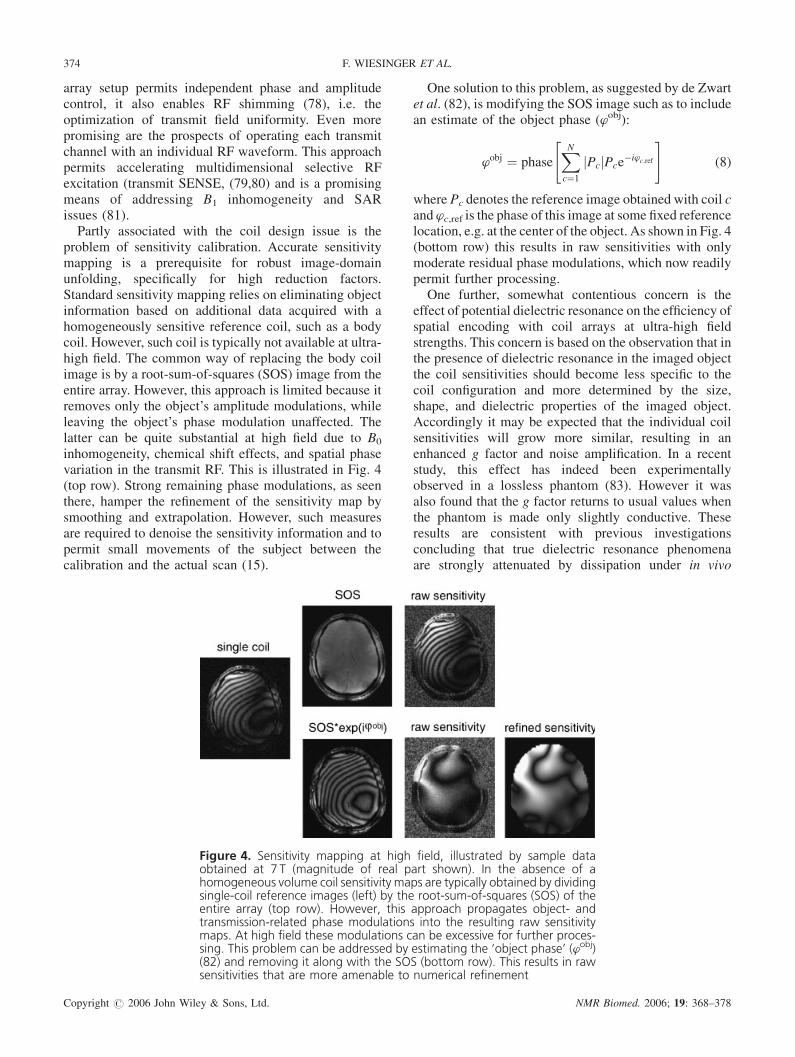
inhomogeneity, chemical shift effects, and spatial phasevariation in the transmit RF. This is illustrated in Fig. 4(top row). Strong remaining phase modulations, as seenthere, hamper the refinement of the sensitivity map bysmoothing and extrapolation. However, such measuresare required to denoise the sensitivity information and topermit small movements of the subject between thecalibration and the actual scan (15).
Figure 4. Sensitivity mapping at high field, illustrated by sample dataobtained at 7 T (magnitude of real part shown). In the absence of ahomogeneous volume coil sensitivity maps are typically obtained by dividingsingle-coil reference images (left) by the root-sum-of-squares (SOS) of theentire array (top row). However, this approach propagates object- andtransmission-related phase modulations into the resulting raw sensitivitymaps. At high field these modulations can be excessive for further proces-sing. This problem can be addressed by estimating the ‘object phase’ (’obj)(82) and removing it along with the SOS (bottom row). This results in rawsensitivities that are more amenable to numerical refinement
Copyright # 2006 John Wiley & Sons, Ltd.
One solution to this problem, as suggested by de Zwartet al. (82), is modifying the SOS image such as to includean estimate of the object phase (wobj):
’obj ¼ phaseXNc¼1
Pcj jPce�i’c;ref
" #(8)
where Pc denotes the reference image obtained with coil cand wc,ref is the phase of this image at some fixed referencelocation, e.g. at the center of the object. As shown in Fig. 4(bottom row) this results in raw sensitivities with onlymoderate residual phase modulations, which now readilypermit further processing.
One further, somewhat contentious concern is theeffect of potential dielectric resonance on the efficiency ofspatial encoding with coil arrays at ultra-high fieldstrengths. This concern is based on the observation that inthe presence of dielectric resonance in the imaged objectthe coil sensitivities should become less specific to thecoil configuration and more determined by the size,shape, and dielectric properties of the imaged object.Accordingly it may be expected that the individual coilsensitivities will grow more similar, resulting in anenhanced g factor and noise amplification. In a recentstudy, this effect has indeed been experimentallyobserved in a lossless phantom (83). However it wasalso found that the g factor returns to usual values whenthe phantom is made only slightly conductive. Theseresults are consistent with previous investigationsconcluding that true dielectric resonance phenomenaare strongly attenuated by dissipation under in vivo
NMR Biomed. 2006; 19: 368–378
PARALLEL MRI AT HIGH FIELD 375
conditions and hence are of little practical relevance (7–10). In this context it is important to note that the notion ofdielectric resonance has sometimes been also used todescribe another phenomenon. The constructive anddestructive interference of array coil characteristics isalso most prominent in wave-regime conditions and canlook similar to dielectric resonance. However, it isfundamentally different from the actual resonance effect(10) and raises no concerns with respect to PIperformance because it does not change the individualcoil characteristics.
Gradually, computer hardware resources may alsobecome a limiting factor. Highly parallel acquisition withlarge coil arrays not only yields higher encodingefficiency but also larger amounts of data to be managedand processed. With current reconstruction algorithmsthe computational load is particularly high for PI withnon-Cartesian k-space trajectories (84–87) . Even higherare the computational demands of advanced algorithmsfor dealing with increased B0 inhomogeneity at highfield (88–90).
Initial experimental results
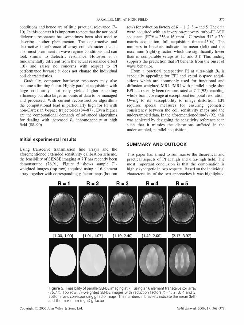
Using transceive transmission line arrays and theaforementioned extended sensitivity calibration scheme,the feasibility of SENSE imaging at 7 T has recently beendemonstrated (76,91). Figure 5 shows sample T1-weighted images (top row) acquired using a 16-elementarray together with corresponding g-factor maps (bottom
Figure 5. Feasibility of parallel SENSE imaging at 7 T using a 16 element transceive coil array(76,77). Top row: T1-weighted SENSE images with reduction factors R¼ 1, 2, 3, 4 and 5.Bottom row: corresponding g factor maps. The numbers in brackets indicate the mean (left)and the maximum (right) g factor
Copyright # 2006 John Wiley & Sons, Ltd.
row) for reduction factors of R¼ 1, 2, 3, 4 and 5. The datawere acquired with an inversion-recovery turbo-FLASHsequence (FOV¼ 256� 160mm2, Cartesian 512� 320matrix acquisition, full acquisition time¼ 450 s). Thenumbers in brackets indicate the mean (left) and themaximum (right) g-factor, which are significantly lowerthan in comparable setups at 1.5 and 3 T. This findingsupports the prediction that PI benefits from the onset ofwave behavior.
From a practical perspective PI at ultra-high B0 isespecially appealing for EPI and spiral k-space acqui-sitions which are commonly used for functional anddiffusion-weighted MRI. fMRI with parallel single-shotEPI has recently been demonstrated at 7 T (92), enablingwhole-brain coverage at exceptional temporal resolution.Owing to its susceptibility to image distortion, EPIrequires special measures for ensuring geometricconsistency between the coil sensitivity maps and theundersampled data. In the aforementioned study (92), thiswas achieved by designing the sensitivity reference scansuch that it mimics the distortions suffered in theundersampled, parallel acquisition.
SUMMARY AND OUTLOOK
This paper has aimed to summarize the theoretical andpractical aspects of PI at high and ultra-high field. Themost important conclusion is that the combination ishighly synergetic in two respects. Based on the individualcharacteristics of the two approaches it was highlighted
NMR Biomed. 2006; 19: 368–378
376 F. WIESINGER ET AL.
that PI and high field are largely complementary in termsof their basic strengths and weaknesses (cf. Table 1). Inaddition, increasing field strength changes the electro-dynamics of the resonance signal in favor of PIperformance (cf. Figs. 1–3). Higher resonance frequencyresults in reduced RF wavelength, giving rise to enhancedRF propagation and interference effects. These translateinto more distinct coil sensitivities and hence into moreeffective sensitivity encoding. Changing RF behavior athigh field affects PI also in the sense that it favorsdifferent coil design concepts. Addressing variousspecific requirements of the joint approach, the transceivetransmission-line architecture is a particularly promisingcurrent array concept.In order to take full advantage of the described
synergies, a number of issues must still be addressed. Oneimportant challenge is consistent sensitivity calibration,especially for distortion-sensitive readout strategies.Enhanced B0 correction (88–90) and auto-calibrated PIschemes (93–96) are promising approaches to thisproblem. Advanced calibration must also address thefact that greater distinctness of the coil sensitivities goesalong with more spread-out spatial frequency content.Hence the coil calibration must rely on more referenceinformation and take greater care in smoothing andextrapolating sensitivity maps.For better exploiting the physically limited SNR
reserve, coil array optimization will continue to be ofmajor significance. Enhancing the number of independentcoil elements will be one main objective in this process.The reasonable number of coils will ultimately be limitedby the associated increase of coil conductor andpreamplifier noise contributions. However, since thenoise becomes increasingly sample dominated withincreasing frequency, high field generally favors largerarrays.Besides parallel acquisition, parallel transmission with
a coil array is a promising concept in light of thechallenges of ultra-high fields. Transmit SENSE has beenshown to enhance multidimensional selective RFexcitation and also to enable SAR reduction (79–81).However, from a technical perspective this approach israther demanding. Besides a transmit coil array it requiresseparate waveform generators and RF amplifiers for eachcoil element. Furthermore, similar to the receiveanalogue, accurate transmit SENSE requires reliableknowledge of the transmit characteristics. It is importantto note that the transmit characteristics are generally notidentical to the receive sensitivities, especially at highfield (50,97). Hence specialized schemes for transmitcalibration are called for.Ultra-high-field MRI in humans has been pioneered for
several years. At the time of writing this paper aconsiderable number of further 7 T whole-body systemsare being installed or projected in laboratories all over theworld, reflecting the community’s high expectations fromthis technology. The years to come will show how and to
Copyright # 2006 John Wiley & Sons, Ltd.
which extent the considerable challenges of routine ultra-high-fieldMR can be addressed. It also remains to be seenfor which human applications 7 T and higher will becomethe field range of choice. Based on the considerationscompiled in this survey it seems fair to predict thatparallel reception and transmission will play a vital role inthis continuing development.
REFERENCES
1. Bottomley PA, Andrew ER. RF magnetic field penetration, phaseshift and power dissipation in biological tissue: implications forNMR imaging. Phys. Med. Biol. 1978; 23 (4): 630–643.
2. Roschmann P. Radiofrequency penetration and absorption in thehuman-body—limitations to high-field whole-body nuclear-mag-netic-resonance imaging. Med. Phys. 1987; 14 (6): 922–931.
3. Bomsdorf H, Helzel T, Kunz D, Roschmann P, Tschendel O,Wieland J. Spectroscopy and imaging with a 4 tesla whole-bodyMR system. NMR Biomed. 1988; 1 (3): 151–158.
4. Keltner JR, Carlson JW, Roos MS, Wong ST, Wong TL, BudingerTF. Electromagnetic fields of surface coil in vivo NMR at highfrequencies. Magn. Reson. Med. 1991; 22 (2): 467–480.
5. Simunic D, Wach P, Renhart W, Stollberger R. Spatial distributionof high-frequency electromagnetic energy in human head duringMRI: numerical results and measurements. IEEE Trans. Biomed.Engng 1996; 43 (1): 88–94.
6. Collins CM, Li S, Smith MB. SAR and B1 field distributions in aheterogeneous human head model within a birdcage coil. Specificenergy absorption rate.Magn. Reson. Med. 1998; 40 (6): 847–856.
7. Kangarlu A, Baertlein BA, Lee R, Ibrahim T, Yang L, AbduljalilAM, Robitaille PM. Dielectric resonance phenomena in ultra highfield MRI. J. Comput. Assist. Tomogr. 1999; 23 (6): 821–831.
8. Hoult DI. Sensitivity and power deposition in a high-field imagingexperiment. J. Magn. Reson. Imag. 2000; 12 (1): 46–67.
9. Yang QX, Wang J, Zhang X, Collins CM, Smith MB, Liu H, ZhuXH, Vaughan JT, Ugurbil K, ChenW. Analysis of wave behavior inlossy dielectric samples at high field.Magn. Reson. Med. 2002; 47(5): 982–989.
10. Collins CM, Liu W, Schreiber W, Yang QX, Smith MB. Centralbrightening due to constructive interference with, without, anddespite dielectric resonance. J. Magn. Reson. Imag. 2005; 21 (2):192–196.
11. Ugurbil K, Garwood M, Ellermann J, Hendrich K, Hinke R, Hu X,Kim SG, Menon R, Merkle H, Ogawa S, Salmi R. Imaging at highmagnetic fields: initial experiences at 4 T.Magn. Reson. Q. 1993; 9(4): 259–277.
12. Robitaille PM, Abduljalil AM, Kangarlu A, Zhang X, Yu Y,Burgess R, Bair S, Noa P, Yang L, Zhu H, Palmer B, Jiang Z,Chakeres DM, Spigos D. Human magnetic resonance imaging at8 T. NMR Biomed. 1998; 11 (6): 263–265.
13. Vaughan JT, Garwood M, Collins CM, Liu W, DelaBarre L,Adriany G, Andersen P, Merkle H, Goebel R, Smith MB, UgurbilK. 7T vs. 4T: RF power, homogeneity, and signal-to-noise com-parison in head images. Magn. Reson. Med. 2001; 46 (1): 24–30.
14. Sodickson DK, Manning WJ. Simultaneous acquisition of spatialharmonics (SMASH): fast imaging with radiofrequency coilarrays. Magn. Reson. Med. 1997; 38 (4): 591–603.
15. Pruessmann KP, Weiger M, Scheidegger MB, Boesiger P. SENSE:sensitivity encoding for fast MRI.Magn. Reson. Med. 1999; 42 (5):952–962.
16. Griswold MA, Jakob PM, Heidemann RM, Nittka M, Jellus V,Wang J, Kiefer B, Haase A. Generalized autocalibrating partiallyparallel acquisitions (GRAPPA).Magn. Reson. Med. 2002; 47 (6):1202–1210.
17. van den Brink JS, Watanabe Y, Kuhl CK, Chung T, Muthupillai R,Van Cauteren M, Yamada K, Dymarkowski S, Bogaert J, Maki JH,Matos C, Casselman JW, Hoogeveen RM. Implications of SENSEMR in routine clinical practice. Eur. J. Radiol. 2003; 46 (1): 3–27.
18. Heidemann RM, Ozsarlak O, Parizel PM, Michiels J, Kiefer B,Jellus V,MullerM, Breuer F, BlaimerM, GriswoldMA, Jakob PM.
NMR Biomed. 2006; 19: 368–378
PARALLEL MRI AT HIGH FIELD 377
A brief review of parallel magnetic resonance imaging. Eur.Radiol. 2003; 13 (10): 2323–2337.
19. Ugurbil K, Adriany G, Andersen P, Chen W, Garwood M, GruetterR, Henry PG, Kim SG, Lieu H, Tkac I, Vaughan T, Van DeMoortele PF, Yacoub E, Zhu XH. Ultrahigh field magnetic reson-ance imaging and spectroscopy.Magn. Reson. Imag. 2003; 21 (10):1263–1281.
20. Norris DG. High field human imaging. J. Magn. Reson. Imag.2003; 18 (5): 519–529.
21. Pruessmann KP. Parallel imaging at high field strength: synergiesand joint potential. Top. Magn. Reson. Imag. 2004; 15 (4): 237–244.
22. Griswold MA, Jakob PM, Chen Q, Goldfarb JW, Manning WJ,Edelman RR, Sodickson DK. Resolution enhancement insingle-shot imaging using simultaneous acquisition of spatialharmonics (SMASH). Magn. Reson. Med. 1999; 41 (6): 1236–1245.
23. Bammer R, Keeling SL, Augustin M, Pruessmann KP, Wolf R,Stollberger R, Hartung HP, Fazekas F. Improved diffusion-weighted single-shot echo-planar imaging (EPI) in stroke usingsensitivity encoding (SENSE). Magn. Reson. Med. 2001; 46 (3):548–554.
24. Bammer R, Auer M, Keeling SL, Augustin M, Stables LA,Prokesch RW, Stollberger R, Moseley ME, Fazekas F. Diffusiontensor imaging using single-shot SENSE-EPI. Magn. Reson. Med.2002; 48 (1): 128–136.
25. Jaermann T, Crelier G, Pruessmann KP, Golay X, Netsch T, vanMuiswinkel AM, Mori S, van Zijl PC, Valavanis A, Kollias S,Boesiger P. SENSE-DTI at 3 T. Magn. Reson. Med. 2004; 51 (2):230–236.
26. Bydder M, Larkman DJ, Hajnal JV. Detection and elimination ofmotion artifacts by regeneration of k-space. Magn. Reson. Med.2002; 47 (4): 677–686.
27. Bydder M, Atkinson D, Larkman DJ, Hill DL, Hajnal JV. SMASHnavigators. Magn. Reson. Med. 2003; 49 (3): 493–500.
28. Atkinson D, Larkman DJ, Batchelor PG, Hill DL, Hajnal JV. Coil-based artifact reduction. Magn. Reson. Med. 2004; 52 (4): 825–830.
29. Hennig J, Weigel M, Thiel T. Optimizing SAR-reduction for high-field TSE with asymmetric hyperechoes with partial Fourierparallel imaging. In Proc. 12th Annual Meeting of ISMRM, Kyoto2004; p.539.
30. de Zwart JA, van Gelderen P, Kellman P, Duyn JH. Reduction ofgradient acoustic noise in MRI using SENSE-EPI. Neuroimage2002; 16 (4): 1151–1155.
31. Mansfield P. Multiplanar image formation using NMR spin echoes.J. Phys. C 1977; 10: L55–L58.
32. Ahn BC, Kim JH, Cho ZH. High-speed spiral-scan echo planarNMR imaging. IEEE Trans. Med. Imag. 1986; MI-5: 2–7.
33. Oppelt A, Graumann R, Barfuss H, Fischer H, Hartl W, Shajor A.FISP—a new fast MRI sequence. Electromedica 1986; 54: 15–18.
34. Gruetter R, Tkac I. Field mapping without reference scan usingasymmetric echo-planar techniques. Magn. Reson. Med. 2000; 43(2): 319–323.
35. Gruetter R. Automatic, localized in vivo adjustment of all first- andsecond-order shim coils. Magn. Reson. Med. 1993; 29 (6): 804–811.
36. Schar M, Kozerke S, Fischer SE, Boesiger P. Cardiac SSFPimaging at 3 Tesla. Magn. Reson. Med. 2004; 51 (4): 799–806.
37. McKinnon GC. Ultrafast interleaved gradient-echo-planar ima-ging on a standard scanner.Magn. Reson. Med. 1993; 30 (5): 609–616.
38. Pfeuffer J, Adriany G, Shmuel A, Yacoub E, Van De Moortele PF,Hu X, Ugurbil K. Perfusion-based high-resolution functionalimaging in the human brain at 7 Tesla. Magn. Reson. Med.2002; 47 (5): 903–911.
39. Pfeuffer J, van de Moortele PF, Yacoub E, Shmuel A, Adriany G,Andersen P, Merkle H, Garwood M, Ugurbil K, Hu X. Zoomedfunctional imaging in the human brain at 7 Tesla with simultaneoushigh spatial and high temporal resolution. Neuroimage 2002; 17(1): 272–286.
40. Yacoub E, Duong TQ, Van De Moortele PF, Lindquist M, AdrianyG, Kim SG, Ugurbil K, Hu X. Spin-echo fMRI in humans usinghigh spatial resolutions and high magnetic fields. Magn. Reson.Med. 2003; 49 (4): 655–664.
Copyright # 2006 John Wiley & Sons, Ltd.
41. Collins CM, Liu W, Wang J, Gruetter R, Vaughan JT, Ugurbil K,Smith MB. Temperature and SAR calculations for a human headwithin volume and surface coils at 64 and 300MHz. J. Magn.Reson. Imag. 2004; 19 (5): 650–656.
42. Roemer PB, Edelstein WA, Hayes CE, Souza SP, Mueller OM.The NMR phased array. Magn. Reson. Med. 1990; 16 (2): 192–225.
43. Hayes CE, Roemer PB. Noise correlations in data simultaneouslyacquired from multiple surface coil arrays. Magn. Reson. Med.1990; 16 (2): 181–191.
44. Carlson JW. An algorithm for NMR imaging reconstruction basedon multiple RF receiver coils. J. Magn. Reson. 1987; 74 (2): 376–380.
45. Hutchinson M, Raff U. Fast MRI data acquisition using multipledetectors. Magn. Reson. Med. 1988; 6 (1): 87–91.
46. Kelton JR, Magin RL, Wright SM. An algorithm for rapid imageacquisition using multiple receiver coils. In Proc. 8th AnnualMeeting of SMRM1989; p. 1172.
47. Ra JB, Rim CY. Fast imaging using subencoding data sets frommultiple detectors. Magn. Reson. Med. 1993; 30 (1): 142–145.
48. Kyriakos WE, Panych LP, Kacher DF, Westin CF, Bao SM,Mulkern RV, Jolesz FA. Sensitivity profiles from an array of coilsfor encoding and reconstruction in parallel (SPACE RIP). Magn.Reson. Med. 2000; 44 (2): 301–308.
49. Hoult DI, Lauterbur PC. The sensitivity of the zeugmatographicexperiment involving human samples. J. Magn. Reson. 1979; 34:425–433.
50. Hoult DI. The principle of reciprocity in signal strengthcalculations – a mathematical guide. Conc. Magn. Reson. 2000;12 (4): 173–187.
51. Weiger M, Boesiger P, Hilfiker PR, Weishaupt D, Pruessmann KP.Sensitivity encoding as a means of enhancing the SNR efficiency insteady-state MRI.Magn. Reson. Med. 2005; 53 (1): 177– 185.
52. Weiger M, Pruessmann KP, Leussler C, Roschmann P, Boesiger P.Specific coil design for SENSE: a six-element cardiac array.Magn.Reson. Med. 2001; 45 (3): 495–504.
53. de Zwart JA, Ledden PJ, Kellman P, van Gelderen P, Duyn JH.Design of a SENSE-Optimized High-Sensitivity MRI receive coilfor brain imaging. Magn. Reson. Med. 2002; 47 (6): 1218–1227.
54. Bankson JA, Wright SM. Simulation-based investigation of par-tially parallel imaging with a linear array at high accelerations.Magn. Reson. Med. 2002; 47 (4): 777–786.
55. Ledden P, Duyn JH. Ultra-high frequency array performance:predicted effects of dielectric resonance. In Proc. 10th AnnualMeeting of ISMRM, Honolulu, HI, 2002; 324.
56. Ohliger MA, Grant AK, Sodickson DK. Ultimate intrinsicsignal-to-noise ratio for parallel MRI: electromagnetic field con-siderations.Magn. Reson. Med. 2003; 50 (5): 1018– 1030.
57. Wiesinger F, Boesiger P, Pruessmann KP. Electrodynamics andultimate SNR in parallel MR imaging. Magn. Reson. Med. 2004;52 (2): 376–390.
58. Zhu Y, Hardy CJ, Sodickson DK, Giaquinto RO, Dumoulin CL,Kenwood G, Niendorf T, Lejay H, McKenzie CA, Ohliger MA,Rofsky NM. Highly parallel volumetric imaging with a 32-elementRF coil array. Magn. Reson. Med. 2004; 52 (4): 869– 877.
59. Jackson JD. Classical Electrodynamics. New York: John Wileyand Sons. 1999.
60. Wang J, Reykowski A, Dickas J. Calculation of the signal-to-noiseratio for simple surface coils and arrays of coils. IEEE Trans.Biomed. Engng 1995; 42 (9): 908–917.
61. Wiesinger F, Van de Moortele PF, Adriany G, De Zanche N,Ugurbil K, Pruessmann KP. Parallel imaging performance as afunction of field strength-An experimental investigation usingelectrodynamic scaling. Magn. Reson. Med. 2004; 52 (5): 953–964.
62. Glover GH, Hayes CE, Pelc NJ, Edelstein WA, Mueller OM, HartHR, Hardy CJ, Odonnell M, Barber WD. Comparison of linear andcircular-polarization for magnetic-resonance imaging. J. Magn.Reson. 1985; 64 (2): 255–270.
63. Ohliger MA, Greenman R, McKenzie CA, Sodickson DK. Con-centric coil arrays for spatial encoding in parallel MRI. In Proc. 9thAnnual Meeting of ISMRM, Glasgow 2001; 21.
64. de Zwart JA, Ledden PJ, van Gelderen P, Bodurka J, Chu R, DuynJH. Signal-to-noise ratio and parallel imaging performance of a
NMR Biomed. 2006; 19: 368–378
378 F. WIESINGER ET AL.
16-channel receive-only brain coil array at 3.0 Tesla.Magn. Reson.Med. 2004; 51 (1): 22–26.
65. Hardy CJ, Darrow RD, Saranathan M, Giaquinto RO, Zhu Y,Dumoulin CL, Bottomley PA. Large field-of-view real-time MRIwith a 32-channel system. Magn. Reson. Med. 2004; 52 (4): 878–884.
66. McDougall MP, Wright SM. 64-channel array coil for single echoacquisition magnetic resonance imaging. Magn. Reson. Med.2005; 54 (2): 386–392.
67. Ocali O, Atalar E. Ultimate intrinsic signal-to-noise ratio in MRI.Magn. Reson. Med. 1998; 39 (3): 462–473.
68. Weiger M, Pruessmann KP, Boesiger P. 2D SENSE for faster 3DMRI. Magnetic Resonance Materials in Physics, Biology andMedicine 2002; 14 (1): 10–19.
69. Duyn JH, de Zwart JA, van Gelderen P, Ledden P. SENSEperformance in human brain at 1.5 and 3.0 Tesla. In Proc. 10thAnnual Meeting of ISMRM, Honolulu, HI, 2002; 192.
70. Yang QX, Wang J, Collins CM, Smith MB, Zhang X, Ugurbil K,Chen W. Phantom design method for high-field MRI humansystems. Magn. Reson. Med. 2004; 52 (5): 1016–1020.
71. Ohliger MA, Ledden P, McKenzie CA, Sodickson DK. Effects ofinductive coupling on parallel MR image reconstructions. Magn.Reson. Med. 2004; 52 (3): 628–639.
72. Duensing GR, Brooker HR, Fitzsimmons JR. Maximizing signal-to-noise ratio in the presence of coil coupling. J. Magn. Reson. B1996; 111 (3): 230–235.
73. Duensing GR, Peterson DM, Wolverton LW, Fitzsimmons JR.Transceive Phased Array Designed for Imaging at 3.0 T. In Proceed-ings of the 6th Annual Meeting of International Society for MagneticResonance in Medicine, Sydney, 1998; 441.
74. King SB, Duensing GR, Varosi S, Peterson DM,Molyneaux DA. Afour channel transceive phased array head coil for 3 T. In Proceed-ings of the 9th Annual Meeting of International Society forMagnetic Resonance in Medicine, Glasgow, 2001; 12.
75. Lee RF, Westgate CR, Weiss RG, Newman DC, Bottomley PA.Planar strip array (PSA) for MRI.Magn. Reson. Med. 2001; 45 (4):673–683.
76. Adriany G, Van deMoortele PF, Wiesinger F, Moeller S, Strupp JP,Andersen P, Snyder C, Zhang X, Chen W, Pruessmann KP,Boesiger P, Vaughan T, Ugurbil K. Transmit and receive trans-mission line arrays for 7 Tesla parallel imaging. Magn. Reson.Med. 2005; 53 (2): 434–445.
77. Vaughan JT. RF coil for imaging system. US patent no. 6,633,161(2003).
78. Boskamp EB, Lee RF. Whole body LPSA transceive array withoptimized transmit homogeneity. In Proc. 10th Annual Meeting ofISMRM, Honolulu, HI, 2002; 903.
79. Katscher U, Bornert P, Leussler C, van den Brink JS. TransmitSENSE. Magn. Reson. Med. 2003; 49 (1): 144–150.
80. Zhu Y. Parallel excitation with an array of transmit coils. Magn.Reson. Med. 2004; 51 (4): 775–784.
81. Zhu Y. RF Power reduction with parallel excitation. In Proc. 12thAnnual Meeting of ISMRM, Kyoto 2004; 331.
82. de Zwart JA, van Gelderen P, Kellman P, Duyn JH. Application ofsensitivity-encoded echo-planar imaging for blood oxygen level-dependent functional brain imaging. Magn. Reson. Med. 2002; 48(6): 1011–1020.
83. Wiesinger F, Van De Moortele PF, Adriany G, De Zanche N,Snyder CJ, Vaughan JT, Ugurbil K, Pruessmann K. An
Copyright # 2006 John Wiley & Sons, Ltd.
investigation into the role of dielectric resonance in parallelimaging. In Proc. 12th Annual Meeting of ISMRM, Kyoto 2004;2393.
84. Pruessmann KP, Weiger M, Bornert P, Boesiger P. Advances insensitivity encoding with arbitrary k-space trajectories. Magn.Reson. Med. 2001; 46 (4): 638–651.
85. Kannengießer SAR, Brenner AR, Noll TG. Acceleratedimage reconstruction for sensitivity encoded imaging with arbi-trary k-space trajectories. In Proceedings of the 8th Annual Meet-ing of International Society for Magnetic Resonance in Medicine,Denver, 2000; 155.
86. Yeh EN, McKenzie CA, Ohliger MA, Sodickson DK. Parallelmagnetic resonance imaging with adaptive radius in k-space(PARS): constrained image reconstruction using k-space localityin radiofrequency coil encoded data. Magn. Reson. Med. 2005; 53(6): 1383–1392.
87. Eggers H, Boernert P, Boesiger P. Comparison of gridding- andconvolution-based iterative reconstruction algorithms for sensi-tivity-encoded non-cartesian acquisition. In Proc. 10th AnnualMeeting of ISMRM, Honolulu, HI, 2002; 743.
88. Jezzard P, Balaban RS. Correction for geometric distortion in echoplanar images from B0 field variations. Magn. Reson. Med. 1995;34 (1): 65–73.
89. Barmet C, Tsao J, Pruessmann KP. Efficient iterative reconstruc-tion for MRI in strongly inhomogeneous B0. In Proc. 12th AnnualMeeting of ISMRM, Kyoto, 2004; 347.
90. Eggers H, Boesiger P. Iterative d B0 and T2� correction for radial
multi-gradient-echo imaging. In Proc. 12th Annual Meeting ofISMRM, Kyoto, 2004; 348.
91. Wiesinger F, Van deMoortele P, Adriany G, Boesiger P, Ugurbil K,Pruessmann KP. Sensitivity encoding at 7 Tesla using transceivemicrostrip coil arrays. In Proc. 20th Meeting of ESMRMB, Rot-terdam, 2003; 39.
92. Van De Moortele PF, Adriany G, Moeller S, Strupp J, Andersen P,Snyder CJ, Vaughan JT, Ugurbil K. Whole brain fMRI in human atultra-high field with parallel SENSE imaging. In Proc. 12th AnnualMeeting of ISMRM, Kyoto, 2004; 1027.
93. Jakob PM, Griswold MA, Edelman RR, Sodickson DK. AUTO-SMASH: a self-calibrating technique for SMASH imaging. Sim-ultaneous acquisition of spatial harmonics. Magnetic ResonanceMaterials in Physics Biology and Medicine 1998; 7 (1): 42–54.
94. Heidemann RM, Griswold MA, Haase A, Jakob PM. VD-AUTO-SMASH imaging. Magn. Reson. Med. 2001; 45 (6): 1066–1074.
95. McKenzie CA, Yeh EN, Ohliger MA, Price MD, Sodickson DK.Self-calibrating parallel imaging with automatic coil sensitivityextraction. Magn. Reson. Med. 2002; 47 (3): 529–538.
96. Bammer R, Vigen KK, Pruessmann KP, Markl M, Moseley ME.Self-Calibrating Radial Generalized SENSE. In Proc. 12th AnnualMeeting of ISMRM, Kyoto, 2004; 2414.
97. Wang J, Yang QX, Zhang X, Collins CM, Smith MB, Zhu XH,Adriany G, Ugurbil K, Chen W. Polarization of the RF field in ahuman head at high field: a study with a quadrature surface coil at7.0 T. Magn. Reson. Med. 2002; 48 (2): 362–369.
98. Gabriel S, Lau RW, Gabriel C. The dielectric propertiesof biological tissues.3. Parametric models for the dielectric spec-trum of tissues. Phys. Med. Biol. 1996; 41 (11): 2271–2293.
99. Buehrer M, Baltes C, Kozerke S, Boesiger P. Whole-heart coronaryMRA using a 32 channel coil array in combination with 2D-SENSE.Proc. 13th Meeting of ISMRM, Miami, FL, 2005; 1606.
NMR Biomed. 2006; 19: 368–378