Embed Size (px)
Citation preview
REVIEW
Micro-algal biosensors
Roberta Brayner & Alain Couté & Jacques Livage &
Catherine Perrette & Clémence Sicard
Received: 3 February 2011 /Revised: 4 May 2011 /Accepted: 13 May 2011 /Published online: 29 May 2011# Springer-Verlag 2011
Abstract Fighting against water pollution requires theability to detect pollutants for example herbicides or heavymetals. Micro-algae that live in marine and fresh water offera versatile solution for the construction of novel biosensors.These photosynthetic microorganisms are very sensitive tochanges in their environment, enabling the detection oftraces of pollutants. Three groups of micro-algae aredescribed in this paper: chlorophyta, cyanobacteria, anddiatoms.
Keywords Biological samples . Biosensors . Fluorescence/luminescence .Metals/heavy metals . Pesticides/endocrinedisruptors . Quality assurance/control
Introduction
Water pollution arising from industry and agriculture isbecoming a major problem in developing countries, while
in under-developed countries nearly 500 millions people donot have access to safe drinking water. Usual contaminantsinclude both organic (detergents, insecticides, volatilecompounds …) and inorganic (heavy metals, chemicalwastes …) compounds. The objective of all water treatmentprocesses is to remove existing pollutants or at least toreduce their concentration so that water becomes fit for itsdesired end-use. It is therefore very important nowadaysto be able to control the nature and amount ofcontaminants in our environment. Two general analyticalapproaches are currently used for this purpose. They arebased either on conventional physicochemical analyses oron biological assays. Although standard analytical tech-niques enable highly accurate detection and quantifica-tion of specific pollutants, exhaustive analyses arecomplex and costly. Moreover, they fail to provide dataon the bioavailability of pollutants, their effects on livingsystems and their synergistic or antagonistic behavior inmixtures. As a partial response to these needs, biosensorshave been developed and used in environmentallyoriented bioassays.
A biosensor contains two parts: a bioreceptor, thebiological sensing element, and a transducer that detectsthe biochemical signal and transforms it into an electrical oroptical signal. These microelectronic devices enable rapid,accurate, and low-level detection of a variety of substancesin body fluids, water, or air. The need for early-warningprocedures to detect contaminants has prompted thedevelopment of cell-based sensors. The rapid and accurateevaluation of water toxicity is nowadays an important issuefor environmental water safety. However, it is difficult tomeasure the toxicity of individual chemicals contained inwater, because a wide variety of chemicals co-exist inenvironmental water, and a mixture may have even morecomplex toxicity. Bioassays seem to be one of the most
R. Brayner :C. SicardUniversité Paris Diderot (Paris 7), CNRS UMR 7086, Interfaces,Traitements, Organisation et Dynamiquedes Systèmes (ITODYS),15 rue Jean de Baïf,75205 Paris cedex 13, France
A. Couté :C. PerretteMuséum National d’Histoire Naturelle, Département RDDM,FRE 3206, USM 505, 57 rue Cuvier,75005 Paris, France
J. Livage (*) :C. SicardCollège de France,11 place Marcelin Berthelot,75005 Paris, Francee-mail: [email protected]
Anal Bioanal Chem (2011) 401:581–597DOI 10.1007/s00216-011-5107-z
useful methods of measuring toxicity in environmental andindustrial wastewaters. Many bioassays based on algae [1–4],bacteria [5], plant tissues [6], and animal cells [7] have beendeveloped in recent years. In particular, micro-algae havebeen widely used for toxicity assays because of their highsensitivity and reproducibility. The use of micro-algae inthe design of biosensors is a very recent topic inbiotechnology. Several algal biosensors have been devel-oped during the past decade in order to detect herbicides,volatile organic compounds (VOC), heavy metals andeven chemical warfare agents [8]. In micro-algal bio-sensors the metabolic activity of the living organism ismeasured. Toxic substances in the surroundings of thecells have a large effect on their metabolic activity and thiseffect can be transformed into electrical or optical signals.One of the main target analytes of micro-algal biosensorsis pesticides, a very broad term which includes herbicides,insecticides, and fungicides. Herbicides that target theenzyme acetolactate synthase (ALS) or acetohydroxy acidsynthase (AHAS) are among the most widely used in theworld. However, most scientific papers published duringthe past decade deal with PSII inhibitors. Therefore ourreview will focus on the analysis of photosynthesisquenching by these herbicides.
One of the main advantages of micro-algal biosensors isthat frequent measurements can be made without extensivepreparation of the sample. However, they usually have onlylow selectivity with regard to separate analyses. Only aglobal signal corresponding to a range of toxic substancesis obtained. However, this global response is often moreuseful for assessment of water quality than the measure-ment of individual concentrations [9].
The micro-algal biosensors discussed in this review aresummarized in Table 1.
Immobilization of micro-algae
One of the limiting steps in the development of whole-cellmicro-algal biosensors is immobilization of the biomaterialin a matrix that prevents leaching without reducing thestability and activity of the cells. Most of the immobiliza-tion techniques rely on the use of organic supports forexample poly(vinyl alcohol) (PVA) or polysulfone (PSU),which some algal strains may find toxic. More biocompat-ible supports include biopolymers such as calcium alginate.However they suffer from a lack of stability over time,precluding their use in long-term devices. Thus mostappropriate entrapment matrices seem to be porous silicamaterials that are non-toxic to living cells and resistmicrobial attack. An in-depth review paper by Moreno-Garrido describes in detail the most recent advancesrealized in the immobilization of micro-algae. [1].
Gel entrapment is the most widely used technique for algalimmobilization. It can be performed with synthetic polymers(acrylamide, photo-crosslinkable resins, polyurethanes),proteins (gelatin, collagen or egg white), or naturalpolysaccharides (agars, carrageenans or alginates).
To improve the stability of biological functions, variousimmobilization techniques have been proposed:
& microencapsulation within a permeable membrane [10];& adsorption on to cellulose derivatives [11];& gel entrapment [12, 13];& reticulation in glutaraldehyde [14]; and& co-reticulation in an albumin–glutaraldehyde matrix
[15–17].
Among the gels, poly(vinyl alcohol) (PVA) is frequentlyused as matrix for immobilization of a variety of enzymes andcells [18]. A variant of this polymer uses styrylpyridinum
Table 1 The micro-algal biosensors discussed in this review
Strain Classification Inorganic/organic Detection limit Ref.
Chlorella vulgaris in alginate gel Amperometric Organic 2–3000 μmol dm−3 [40]
Chlorella vulgaris Amperometric VOCs 1 μmol dm−3 [41]
Dictyosphaerium chlorelloides Optical Organic 0.5 μmol L−1 [42]
Chlorella vulgaris in silica micro-columns Sequential elution and determination Inorganic 0.5–4 μg L−1 [43]
Chlorella vulgaris between two platinumelectrodes
Conductometric Inorganic 10 ppb [44]
Chlorella vulgaris immobilized in BSA Optical Inorganic 1 ppb [45, 46]
Dictyosphaerium chlorelloides in sol-gelsilica matrix
Optical Inorganic 0.6 mg L−1 [47]
Synechococcus PCC 7942 immobilizedin PVA-SbQ
Optical Inorganic/organic 0.2 and 0.06 mmol L−1 [53]
Synechococcus PCC7942 Optical Inorganic <5 mg L−1 [60]
Anabaena torulosa immobilized on anoxygen electrode
Amperometric Inorganic 0.4 mg L−1 [61]
Thalassiosira rotula frustule optical Organic 10 ppm [63, 64]
582 R. Brayner et al.
groups attached to the poly(vinyl alcohol) (PVA-SbQ) tocross-link the polymer chains under mild conditions withoutdamaging the biological material to be entrapped [19].Synechococcus sp. PCC 7942 entrapped in PVA-SbQenabled efficient detection of herbicides [19]. In thesephotoelectrochemical cells, cyanobacteria were used tocapture light energy and convert it into reduced species.The PSII acceptor DCBQ could be used as an electroactivemediator to transport electrons from the reduced species inthe photosynthetic membrane to the working platinumelectrode. A method to detect pollutants, for example diuron,that inhibit the photosynthetic electron flow in entrappedcyanobacteria was developed. The percentage inhibitioncould be measured after just 5 min of contact with thepollutant and 1 min of illumination, performance better thanwith bioluminescent cyanobacteria.
Micro-algal biosensors have to be regenerated after use,but most immobilization techniques irreversibly bind cellspreventing the production of reusable biosensors. Severalgroups are trying to improve this process, but results are notyet very good and studies are still in progress. A recentpublication suggests that a convenient method could be to usemicro-algae coated with biocompatible magnetic nanopar-ticles (NdFeB). Living cells are attracted to the surface of ascreen-printed electrode by a magnetic field. They can then beeasily removed when the magnetic field is switched off [20].
Photosynthetically active whole cells within silica gel matrix
Silica is a biocompatible material very attractive forencapsulation of enzymes and whole cells. If a material is
biocompatible it is neither cyto-toxic or geno-toxic, andneither are its eventual degradation products. Silica gels havenumerous advantages as material host, for example mechan-ical and chemical stability and photo-transparency, which issometimes required for photosynthetically active cells.
Photobiochemical hybrid materials have been obtainedby immobilization of Synechococcus sp. PCC 6301 andPCC 7002 cyanobacteria within silica gels [21–24]. Evenafter 12 weeks, entrapped cells continue to autofluoresce, asproved by epifluorescence microscopy images (Fig. 1).This is indicative of the preservation of their photosyntheticapparatus and, therefore, their ability to photosynthesize.Both chlorophyll a and phycocyanin can be detected viacharacteristic reflectance bands. Chlorophyll a was detect-able after 12 weeks for Synechococcus PCC 6301 yet thephycocyanin component had disappeared. For SynechococcusPCC 7002 this trend was reversed with the water-solublephycocyanin remaining longer. These hybrid materials couldbe used as biosensors to detect traces of metals andherbicides by monitoring the photosynthetic activity of thecyanobacteria.
Sol–gel process for encapsulation of vegetal cells [25]
Silica matrixes are relatively inexpensive to synthesize andhave interesting properties including optical transparency,biocompatibility and chemical inertness. Moreover, shapematerials such as microspheres, fibers, or thin films can beeasily processed from the precursor solution. Algal silicafor instance could be deposited in front of the tip of anoptical fiber to construct an optical biosensor. The optical
Fig. 1 Epifluorescence microscopy images of Synechococcus encapsulated within silica gel after (ii) one week, (iii) four weeks, (iv) 12 weeks, forstrains a PCC 6301 and b PCC 7002. Scale bar represents 10 μm. (Reprinted from Ref.[22], Copyright (2010), with permission from Elsevier)
Micro-algal biosensors 583
fiber would be used to send the excitation radiation to thealgal cells and convey the fluorescence radiation up to afluorimeter. Similarly, sol–gel films can be deposited on toan electrode for amperometric measurements.
The objective of this review is to highlight micro-algalbiosensors based on three families of eukaryotic (greenalgae, diatoms) and prokaryotic (cyanobacteria) micro-algae. They are all photosynthetic, using CO2 and waterto make sugar, with oxygen as a by-product. Theircharacteristic color depends on the nature of their photo-synthetic pigments. Their metabolic activity is verysensitive to changes in the surrounding medium, enablingthe bio-detection of water pollution.
Chlorophyta or green algae
Green algae are eukaryotic with their genetic materialconfined inside a nucleus surrounded by a membrane.Some green algae, called macro-algae are quite large,
visible to the naked eye, whereas others, called micro-algae, are much smaller and not visible without amicroscope. Only this second group will be discussed here.
The Chlorophyta thallus is organized into one of fourpatterns:
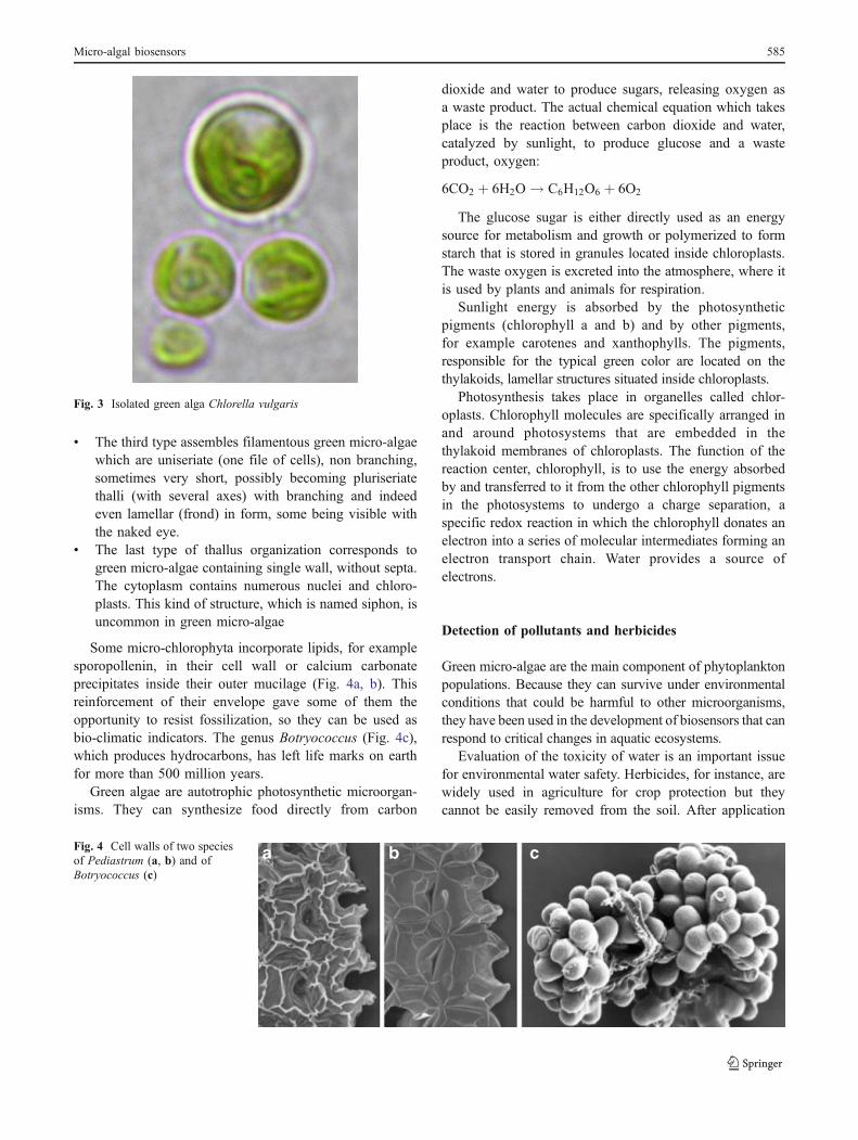
& The first, and the most important here, consists ofisolated microorganisms constituted of a single cellseparated from the outer medium by a membrane oftenprotected by a cellulosic wall (Fig. 2). The wallthickness, morphology, and ornamentation are verydiverse. A mucilaginous substance (mucus) of variablethickness produced by the cell separates it from theoutside medium. This group includes Chlorella vulgaris,the green micro-alga most used for biosensor applica-tions (Fig. 3). This micro-alga has solitary and sphericalcells, with a distinct wall, one or two chloroplasts, andsometimes one pyrenoid.
& In the second group, green micro-algae form colonies madeof several cells that may have different morphologies.
Fig. 2 Isolated green algae: a Chlamydomonas, b Cylindrocystis, c Micrasterias, d Closterium, e Euastrum, f Xanthidium
584 R. Brayner et al.
& The third type assembles filamentous green micro-algaewhich are uniseriate (one file of cells), non branching,sometimes very short, possibly becoming pluriseriatethalli (with several axes) with branching and indeedeven lamellar (frond) in form, some being visible withthe naked eye.
& The last type of thallus organization corresponds togreen micro-algae containing single wall, without septa.The cytoplasm contains numerous nuclei and chloro-plasts. This kind of structure, which is named siphon, isuncommon in green micro-algae
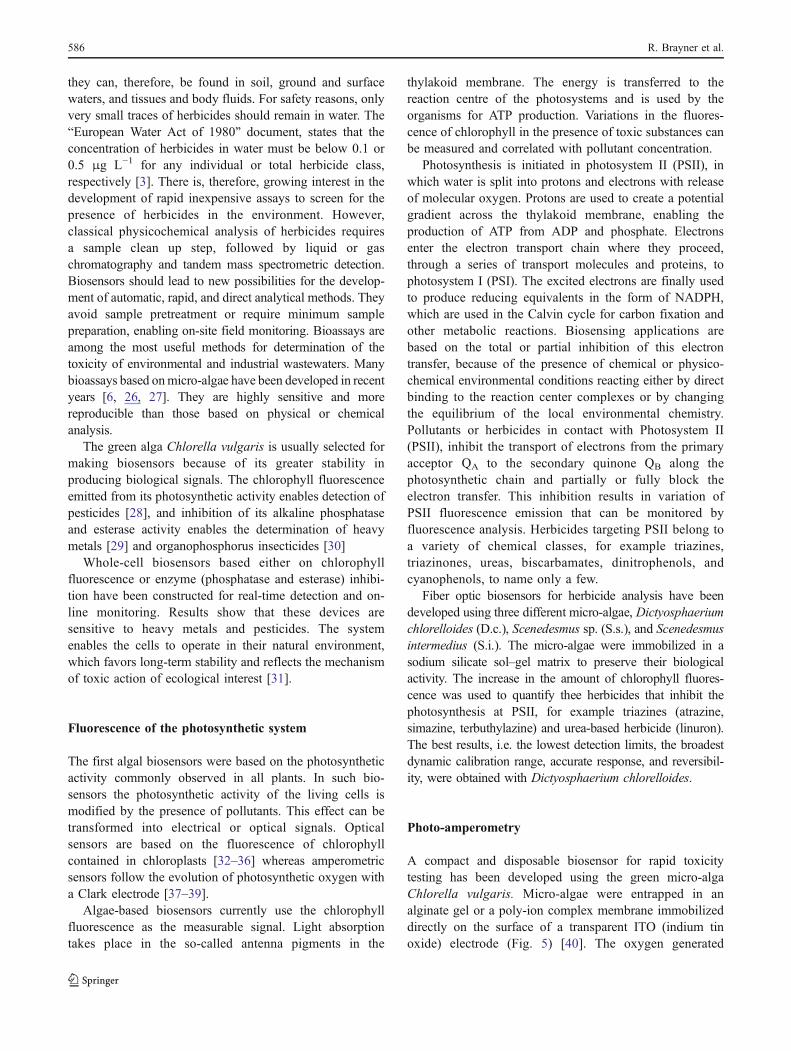
Some micro-chlorophyta incorporate lipids, for examplesporopollenin, in their cell wall or calcium carbonateprecipitates inside their outer mucilage (Fig. 4a, b). Thisreinforcement of their envelope gave some of them theopportunity to resist fossilization, so they can be used asbio-climatic indicators. The genus Botryococcus (Fig. 4c),which produces hydrocarbons, has left life marks on earthfor more than 500 million years.
Green algae are autotrophic photosynthetic microorgan-isms. They can synthesize food directly from carbon
dioxide and water to produce sugars, releasing oxygen asa waste product. The actual chemical equation which takesplace is the reaction between carbon dioxide and water,catalyzed by sunlight, to produce glucose and a wasteproduct, oxygen:
6CO2 þ 6H2O ! C6H12O6 þ 6O2
The glucose sugar is either directly used as an energysource for metabolism and growth or polymerized to formstarch that is stored in granules located inside chloroplasts.The waste oxygen is excreted into the atmosphere, where itis used by plants and animals for respiration.
Sunlight energy is absorbed by the photosyntheticpigments (chlorophyll a and b) and by other pigments,for example carotenes and xanthophylls. The pigments,responsible for the typical green color are located on thethylakoids, lamellar structures situated inside chloroplasts.
Photosynthesis takes place in organelles called chlor-oplasts. Chlorophyll molecules are specifically arranged inand around photosystems that are embedded in thethylakoid membranes of chloroplasts. The function of thereaction center, chlorophyll, is to use the energy absorbedby and transferred to it from the other chlorophyll pigmentsin the photosystems to undergo a charge separation, aspecific redox reaction in which the chlorophyll donates anelectron into a series of molecular intermediates forming anelectron transport chain. Water provides a source ofelectrons.
Detection of pollutants and herbicides
Green micro-algae are the main component of phytoplanktonpopulations. Because they can survive under environmentalconditions that could be harmful to other microorganisms,they have been used in the development of biosensors that canrespond to critical changes in aquatic ecosystems.
Evaluation of the toxicity of water is an important issuefor environmental water safety. Herbicides, for instance, arewidely used in agriculture for crop protection but theycannot be easily removed from the soil. After application
Fig. 3 Isolated green alga Chlorella vulgaris
Fig. 4 Cell walls of two speciesof Pediastrum (a, b) and ofBotryococcus (c)
Micro-algal biosensors 585
they can, therefore, be found in soil, ground and surfacewaters, and tissues and body fluids. For safety reasons, onlyvery small traces of herbicides should remain in water. The“European Water Act of 1980” document, states that theconcentration of herbicides in water must be below 0.1 or0.5 μg L−1 for any individual or total herbicide class,respectively [3]. There is, therefore, growing interest in thedevelopment of rapid inexpensive assays to screen for thepresence of herbicides in the environment. However,classical physicochemical analysis of herbicides requiresa sample clean up step, followed by liquid or gaschromatography and tandem mass spectrometric detection.Biosensors should lead to new possibilities for the develop-ment of automatic, rapid, and direct analytical methods. Theyavoid sample pretreatment or require minimum samplepreparation, enabling on-site field monitoring. Bioassays areamong the most useful methods for determination of thetoxicity of environmental and industrial wastewaters. Manybioassays based onmicro-algae have been developed in recentyears [6, 26, 27]. They are highly sensitive and morereproducible than those based on physical or chemicalanalysis.
The green alga Chlorella vulgaris is usually selected formaking biosensors because of its greater stability inproducing biological signals. The chlorophyll fluorescenceemitted from its photosynthetic activity enables detection ofpesticides [28], and inhibition of its alkaline phosphataseand esterase activity enables the determination of heavymetals [29] and organophosphorus insecticides [30]
Whole-cell biosensors based either on chlorophyllfluorescence or enzyme (phosphatase and esterase) inhibi-tion have been constructed for real-time detection and on-line monitoring. Results show that these devices aresensitive to heavy metals and pesticides. The systemenables the cells to operate in their natural environment,which favors long-term stability and reflects the mechanismof toxic action of ecological interest [31].
Fluorescence of the photosynthetic system
The first algal biosensors were based on the photosyntheticactivity commonly observed in all plants. In such bio-sensors the photosynthetic activity of the living cells ismodified by the presence of pollutants. This effect can betransformed into electrical or optical signals. Opticalsensors are based on the fluorescence of chlorophyllcontained in chloroplasts [32–36] whereas amperometricsensors follow the evolution of photosynthetic oxygen witha Clark electrode [37–39].
Algae-based biosensors currently use the chlorophyllfluorescence as the measurable signal. Light absorptiontakes place in the so-called antenna pigments in the
thylakoid membrane. The energy is transferred to thereaction centre of the photosystems and is used by theorganisms for ATP production. Variations in the fluores-cence of chlorophyll in the presence of toxic substances canbe measured and correlated with pollutant concentration.
Photosynthesis is initiated in photosystem II (PSII), inwhich water is split into protons and electrons with releaseof molecular oxygen. Protons are used to create a potentialgradient across the thylakoid membrane, enabling theproduction of ATP from ADP and phosphate. Electronsenter the electron transport chain where they proceed,through a series of transport molecules and proteins, tophotosystem I (PSI). The excited electrons are finally usedto produce reducing equivalents in the form of NADPH,which are used in the Calvin cycle for carbon fixation andother metabolic reactions. Biosensing applications arebased on the total or partial inhibition of this electrontransfer, because of the presence of chemical or physico-chemical environmental conditions reacting either by directbinding to the reaction center complexes or by changingthe equilibrium of the local environmental chemistry.Pollutants or herbicides in contact with Photosystem II(PSII), inhibit the transport of electrons from the primaryacceptor QA to the secondary quinone QB along thephotosynthetic chain and partially or fully block theelectron transfer. This inhibition results in variation ofPSII fluorescence emission that can be monitored byfluorescence analysis. Herbicides targeting PSII belong toa variety of chemical classes, for example triazines,triazinones, ureas, biscarbamates, dinitrophenols, andcyanophenols, to name only a few.
Fiber optic biosensors for herbicide analysis have beendeveloped using three different micro-algae, Dictyosphaeriumchlorelloides (D.c.), Scenedesmus sp. (S.s.), and Scenedesmusintermedius (S.i.). The micro-algae were immobilized in asodium silicate sol–gel matrix to preserve their biologicalactivity. The increase in the amount of chlorophyll fluores-cence was used to quantify thee herbicides that inhibit thephotosynthesis at PSII, for example triazines (atrazine,simazine, terbuthylazine) and urea-based herbicide (linuron).The best results, i.e. the lowest detection limits, the broadestdynamic calibration range, accurate response, and reversibil-ity, were obtained with Dictyosphaerium chlorelloides.
Photo-amperometry
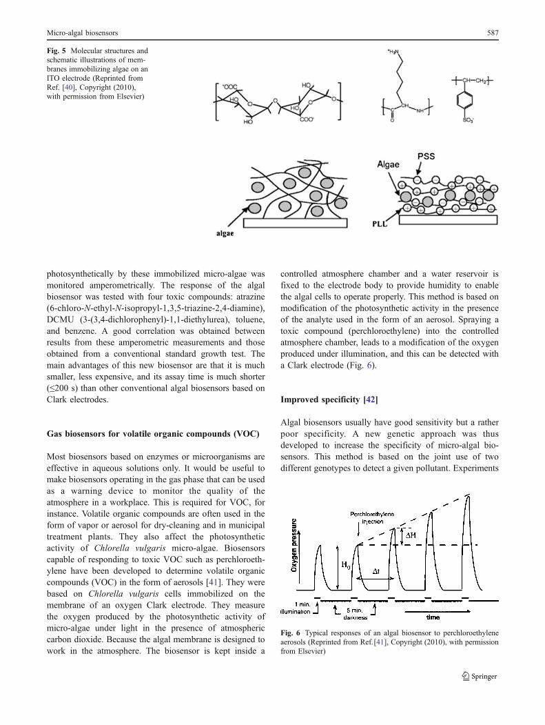
A compact and disposable biosensor for rapid toxicitytesting has been developed using the green micro-algaChlorella vulgaris. Micro-algae were entrapped in analginate gel or a poly-ion complex membrane immobilizeddirectly on the surface of a transparent ITO (indium tinoxide) electrode (Fig. 5) [40]. The oxygen generated
586 R. Brayner et al.
photosynthetically by these immobilized micro-algae wasmonitored amperometrically. The response of the algalbiosensor was tested with four toxic compounds: atrazine(6-chloro-N-ethyl-N-isopropyl-1,3,5-triazine-2,4-diamine),DCMU (3-(3,4-dichlorophenyl)-1,1-diethylurea), toluene,and benzene. A good correlation was obtained betweenresults from these amperometric measurements and thoseobtained from a conventional standard growth test. Themain advantages of this new biosensor are that it is muchsmaller, less expensive, and its assay time is much shorter(≤200 s) than other conventional algal biosensors based onClark electrodes.
Gas biosensors for volatile organic compounds (VOC)
Most biosensors based on enzymes or microorganisms areeffective in aqueous solutions only. It would be useful tomake biosensors operating in the gas phase that can be usedas a warning device to monitor the quality of theatmosphere in a workplace. This is required for VOC, forinstance. Volatile organic compounds are often used in theform of vapor or aerosol for dry-cleaning and in municipaltreatment plants. They also affect the photosyntheticactivity of Chlorella vulgaris micro-algae. Biosensorscapable of responding to toxic VOC such as perchloroeth-ylene have been developed to determine volatile organiccompounds (VOC) in the form of aerosols [41]. They werebased on Chlorella vulgaris cells immobilized on themembrane of an oxygen Clark electrode. They measurethe oxygen produced by the photosynthetic activity ofmicro-algae under light in the presence of atmosphericcarbon dioxide. Because the algal membrane is designed towork in the atmosphere. The biosensor is kept inside a
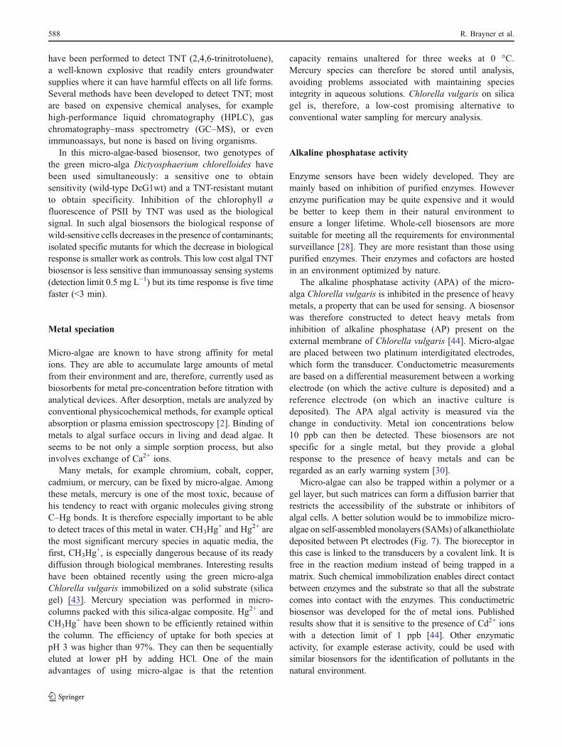
controlled atmosphere chamber and a water reservoir isfixed to the electrode body to provide humidity to enablethe algal cells to operate properly. This method is based onmodification of the photosynthetic activity in the presenceof the analyte used in the form of an aerosol. Spraying atoxic compound (perchloroethylene) into the controlledatmosphere chamber, leads to a modification of the oxygenproduced under illumination, and this can be detected witha Clark electrode (Fig. 6).
Improved specificity [42]
Algal biosensors usually have good sensitivity but a ratherpoor specificity. A new genetic approach was thusdeveloped to increase the specificity of micro-algal bio-sensors. This method is based on the joint use of twodifferent genotypes to detect a given pollutant. Experiments
Fig. 5 Molecular structures andschematic illustrations of mem-branes immobilizing algae on anITO electrode (Reprinted fromRef. [40], Copyright (2010),with permission from Elsevier)
Fig. 6 Typical responses of an algal biosensor to perchloroethyleneaerosols (Reprinted from Ref.[41], Copyright (2010), with permissionfrom Elsevier)
Micro-algal biosensors 587
have been performed to detect TNT (2,4,6-trinitrotoluene),a well-known explosive that readily enters groundwatersupplies where it can have harmful effects on all life forms.Several methods have been developed to detect TNT; mostare based on expensive chemical analyses, for examplehigh-performance liquid chromatography (HPLC), gaschromatography–mass spectrometry (GC–MS), or evenimmunoassays, but none is based on living organisms.
In this micro-algae-based biosensor, two genotypes ofthe green micro-alga Dictyosphaerium chlorelloides havebeen used simultaneously: a sensitive one to obtainsensitivity (wild-type DcG1wt) and a TNT-resistant mutantto obtain specificity. Inhibition of the chlorophyll afluorescence of PSII by TNT was used as the biologicalsignal. In such algal biosensors the biological response ofwild-sensitive cells decreases in the presence of contaminants;isolated specific mutants for which the decrease in biologicalresponse is smaller work as controls. This low cost algal TNTbiosensor is less sensitive than immunoassay sensing systems(detection limit 0.5 mg L−1) but its time response is five timefaster (<3 min).
Metal speciation
Micro-algae are known to have strong affinity for metalions. They are able to accumulate large amounts of metalfrom their environment and are, therefore, currently used asbiosorbents for metal pre-concentration before titration withanalytical devices. After desorption, metals are analyzed byconventional physicochemical methods, for example opticalabsorption or plasma emission spectroscopy [2]. Binding ofmetals to algal surface occurs in living and dead algae. Itseems to be not only a simple sorption process, but alsoinvolves exchange of Ca2+ ions.
Many metals, for example chromium, cobalt, copper,cadmium, or mercury, can be fixed by micro-algae. Amongthese metals, mercury is one of the most toxic, because ofhis tendency to react with organic molecules giving strongC–Hg bonds. It is therefore especially important to be ableto detect traces of this metal in water. CH3Hg
+ and Hg2+ arethe most significant mercury species in aquatic media, thefirst, CH3Hg
+, is especially dangerous because of its readydiffusion through biological membranes. Interesting resultshave been obtained recently using the green micro-algaChlorella vulgaris immobilized on a solid substrate (silicagel) [43]. Mercury speciation was performed in micro-columns packed with this silica-algae composite. Hg2+ andCH3Hg
+ have been shown to be efficiently retained withinthe column. The efficiency of uptake for both species atpH 3 was higher than 97%. They can then be sequentiallyeluted at lower pH by adding HCl. One of the mainadvantages of using micro-algae is that the retention
capacity remains unaltered for three weeks at 0 °C.Mercury species can therefore be stored until analysis,avoiding problems associated with maintaining speciesintegrity in aqueous solutions. Chlorella vulgaris on silicagel is, therefore, a low-cost promising alternative toconventional water sampling for mercury analysis.
Alkaline phosphatase activity
Enzyme sensors have been widely developed. They aremainly based on inhibition of purified enzymes. Howeverenzyme purification may be quite expensive and it wouldbe better to keep them in their natural environment toensure a longer lifetime. Whole-cell biosensors are moresuitable for meeting all the requirements for environmentalsurveillance [28]. They are more resistant than those usingpurified enzymes. Their enzymes and cofactors are hostedin an environment optimized by nature.
The alkaline phosphatase activity (APA) of the micro-alga Chlorella vulgaris is inhibited in the presence of heavymetals, a property that can be used for sensing. A biosensorwas therefore constructed to detect heavy metals frominhibition of alkaline phosphatase (AP) present on theexternal membrane of Chlorella vulgaris [44]. Micro-algaeare placed between two platinum interdigitated electrodes,which form the transducer. Conductometric measurementsare based on a differential measurement between a workingelectrode (on which the active culture is deposited) and areference electrode (on which an inactive culture isdeposited). The APA algal activity is measured via thechange in conductivity. Metal ion concentrations below10 ppb can then be detected. These biosensors are notspecific for a single metal, but they provide a globalresponse to the presence of heavy metals and can beregarded as an early warning system [30].
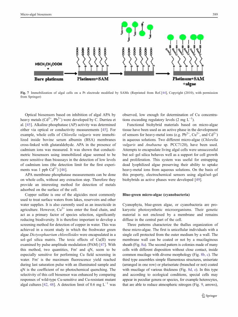
Micro-algae can also be trapped within a polymer or agel layer, but such matrices can form a diffusion barrier thatrestricts the accessibility of the substrate or inhibitors ofalgal cells. A better solution would be to immobilize micro-algae on self-assembledmonolayers (SAMs) of alkanethiolatedeposited between Pt electrodes (Fig. 7). The bioreceptor inthis case is linked to the transducers by a covalent link. It isfree in the reaction medium instead of being trapped in amatrix. Such chemical immobilization enables direct contactbetween enzymes and the substrate so that all the substratecomes into contact with the enzymes. This conductimetricbiosensor was developed for the of metal ions. Publishedresults show that it is sensitive to the presence of Cd2+ ionswith a detection limit of 1 ppb [44]. Other enzymaticactivity, for example esterase activity, could be used withsimilar biosensors for the identification of pollutants in thenatural environment.
588 R. Brayner et al.
Optical biosensors based on inhibition of algal APA byheavy metals (Cd2+, Pb2+) were developed by C. Durrieu etal. [45]. Alkaline phosphatase (AP) activity was determinedeither via optical or conductivity measurements [45]. Forexample, whole cells of Chlorella vulgaris were immobi-lized inside bovine serum albumin (BSA) membranescross-linked with glutaraldehyde. APA in the presence ofcadmium ions was measured. It was shown that conducti-metric biosensors using immobilized algae seemed to bemore sensitive than bioassays in the detection of low levelsof cadmium ions (the detection limit for the first experi-ments was 1 ppb Cd2+) [46].
APA membrane phosphatase measurements can be doneon whole cells, without any extraction step. Therefore theyprovide an interesting method for detection of metalsadsorbed on the surface of the cell.
Copper sulfate is one of the algicides most commonlyused to treat surface waters from lakes, reservoirs and otherwater supplies. It is also currently used as an insecticide inagriculture. However, Cu2+ ions enter the food chain, andact as a primary factor of species selection, significantlyreducing biodiversity. It is therefore important to develop ascreening method for detection of copper in water. This wasachieved in a recent study in which the freshwater greenalgae Dictyosphaerium chlorelloides were encapsulated in asol–gel silica matrix. The toxic effects of Cu(II) wereexamined by pulse amplitude modulation (PAM) [47]. Withthis method, two quantities, Fm′ and qN, seem to beespecially sensitive for performing Cu field screening inwater. Fm′ is the maximum fluorescence yield reachedduring last saturation pulse with an illuminated sample andqN is the coefficient of no photochemical quenching. Theselectivity of this cell biosensor was enhanced by comparingresponses of wild-type Cu-sensitive and Cu-resistant mutantalgal cultures [42, 48]. A detection limit of 0.6 mg L−1 was
observed, low enough for determination of Cu concentra-tions exceeding regulatory levels (2 mg L−1).
Functional biohybrid materials based on micro-algaetissue have been used as an active phase in the developmentof sensors for heavy-metal ions (e.g. Pb2+, Cu2+, and Cd2+)in aqueous solutions. Two different micro-algae (Chlorellavulgaris and Anabaena sp. PCC7120), have been used.Attempts to encapsulate living algal cells were unsuccessfulbut sol–gel silica behaves well as a support for cell growthand proliferation. This system was useful for entrappingdead lyophilized algae preserving their ability to uptakeheavy-metal ions from aqueous solutions. On the basis ofthis property, electrochemical sensors using algal/sol–gelbiohybrids as active phases were developed [49].
Blue-green micro-algae (cyanobacteria)
Cyanophyta, blue-green algae, or cyanobacteria are pro-karyotic photosynthetic microorganisms. Their geneticmaterial is not enclosed by a membrane and remainsdiffuse in the central part of the cell.
Three patterns characterize the thallus organization ofthese micro-algae. The first is unicellular individuals with asingle cell protected from the outer medium by a wall. Themembrane wall can be coated or not by a mucilaginoussheath (Fig. 8a). The second pattern is colonies made of manycells with different disposition without close contact, insidecommon mucilage with diverse morphology (Fig. 8b, c). Thethird type assembles simple filamentous structures, uniseriate(arranged in one row) or pluriseriate (branched or not) coatedwith mucilage of various thickness (Fig. 8d, e). In this typeand according to ecological conditions, special cells mayappear in peculiar genera or species, for example heterocytes,that are able to reduce atmospheric nitrogen (Fig. 9, arrows),
Fig. 7 Immobilization of algal cells on a Pt electrode modified by SAMs (Reprinted from Ref.[44], Copyright (2010), with permissionfrom Springer)
Micro-algal biosensors 589
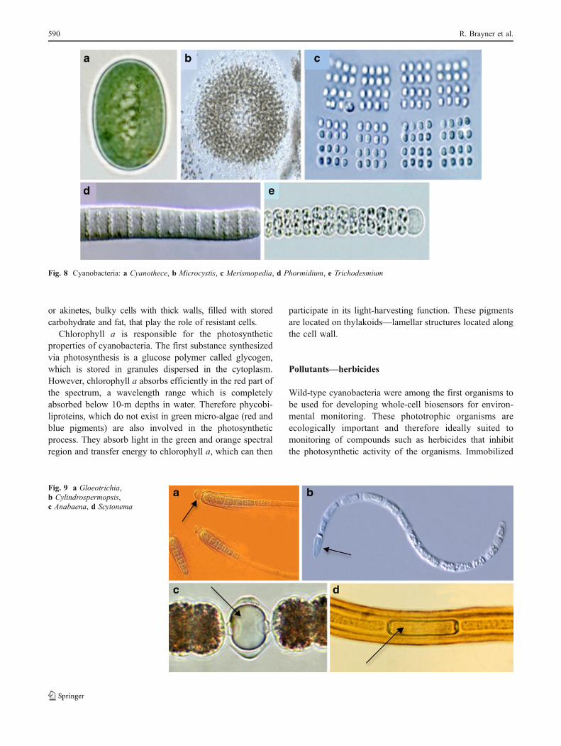
or akinetes, bulky cells with thick walls, filled with storedcarbohydrate and fat, that play the role of resistant cells.
Chlorophyll a is responsible for the photosyntheticproperties of cyanobacteria. The first substance synthesizedvia photosynthesis is a glucose polymer called glycogen,which is stored in granules dispersed in the cytoplasm.However, chlorophyll a absorbs efficiently in the red part ofthe spectrum, a wavelength range which is completelyabsorbed below 10-m depths in water. Therefore phycobi-liproteins, which do not exist in green micro-algae (red andblue pigments) are also involved in the photosyntheticprocess. They absorb light in the green and orange spectralregion and transfer energy to chlorophyll a, which can then
participate in its light-harvesting function. These pigmentsare located on thylakoids—lamellar structures located alongthe cell wall.
Pollutants—herbicides
Wild-type cyanobacteria were among the first organisms tobe used for developing whole-cell biosensors for environ-mental monitoring. These phototrophic organisms areecologically important and therefore ideally suited tomonitoring of compounds such as herbicides that inhibitthe photosynthetic activity of the organisms. Immobilized
Fig. 8 Cyanobacteria: a Cyanothece, b Microcystis, c Merismopedia, d Phormidium, e Trichodesmium
Fig. 9 a Gloeotrichia,b Cylindrospermopsis,c Anabaena, d Scytonema
590 R. Brayner et al.
cyanobacteria (Synechocystis and Synechococcus) can alsobe used as biosensors for detection of water toxicitybecause of their versatile metabolism, for example photo-synthetic activity (thylakoids membranes in vegetativecells), respiration, fermentation, and nitrogen fixation(heterocyt cells). Amperometric biosensors based oncyanobacteria have also been used for detection ofphytotoxic pollutants [50, 51]. However, they are not verystable and robust.
To overcome these disadvantages, microbial cells can begenetically modified by introduction of a “reporter gene” toconnect the initial biological interaction of the testedchemical or physical event to an easily recordable outputsignal (e.g. light). The most commonly used reporterproteins are β-galactosidase, green-fluorescent protein(GFP) and luciferase.
Shao et al. described a bioluminescent SynechocystisPCC 6803-derived reporter strain used to monitor thecorrelation between cyanobacterial activity and the pres-ence of herbicides [52]. To make this reporter strain theyintroduced a construct made of the constitutive tacpromoter fused to luc (the gene encoding firefly luciferase)and luxAB (encoding the bacterial luciferase) into thecyanobacterial cells by means of an integrative vector. Inthis case Synechocystis PCC 6803 reporter strain is enabledfor assessment of the bioavailability and effects of variousherbicides (diuron, atrazine, propazine, and simazine) onthe cyanobacterial cell. The sensitivity of the system for theanalytes (determined as EC50 values) reached the lowmg L−1 level. The optimum pH for assay of biolumines-cence was found to be 6.5. This may be for one of tworeasons: either this pH supports the optimum physiology ofthe cyanobacterial biosensor or it is the pH at whichcyanobacteria are most permeable to the luciferin substrate.Bioluminescence measurements are quite fast (10 s) but aminimum exposure time of 30 min is required to reachthese values. In this case, the tested herbicides specificallyreduce the energy state of the cells by blocking the electronflow in PSII, resulting in inhibition of photosynthesis.With regard to environmental relevance, cyanobacterialbiosensors are sensitive to herbicides at the parts-per-million level, which is appropriate for detecting residuesin groundwater or soil. In addition, these biosensorsprovide information on the bioavailability of the herbicidein environmental samples. Although phenoxy acid herbi-cides are less toxic than other herbicides, they are highlysoluble, enabling easy determination of EC20 and EC50.Compared with the green alga Selenastrum capricornutum,the cyanobacterial biosensor is more sensitive to glyphosatein terms of reaction time and sensitivity.
Whole-cell luminescent cyanobacterial biosensors seemto be more simple, rapid, accurate, and economical thanother methods, for example photosystem-based whole-cell
and tissue biosensors, in detection of the toxicity ofherbicides. They could also be used to indicate the type ofherbicide and possibly its potential mode of action. Thedisadvantage of this method is the need for knowledge ofgenetic tools. To circumvent this limitation, the photosyn-thetic activity of cyanobacteria in the presence of herbicideswas followed by photoelectrochemical [53] and ampero-metric measurements [54]. Herbicides interact with theprocess of photosynthesis and are generally inhibitors ofPSII-dependent electron flow. However, the major incon-venience of this method is the very short time in which freebiological material is available.
Detection of heavy metals
Some organisms carrying luminescent reporter genes fusedto metal ion-inducible promoters may be used as biosensorsfor detection of bioavailable heavy metal ions in environ-mental samples. Mercury ion biosensors have been con-structed by fusing the lux genes of Vibrio fischeri bacteriato the mercury resistance operon of Tn21 [55, 56].Biosensors have also been designed for specific responsesto copper, nickel, zinc, chromate, and thallium ions byfusion of the lux genes to a number of metal ion-responsivepromoters from Alcaligenes eutrophus [57]. Use of the geneexpression driven by the smt operator/promoter of thecyanobacterium Synechococcus sp. for detection of metalions in aquatic environments has also been proposed [58].The smt locus of Synechococcus sp. consists of theprokaryotic metallothionein gene, smtA (absent in greenalgae and diatoms), and a divergently transcribed geneencoding a repressor of smtA transcription, designatedsmtB. The transcription of smtA is induced in the presenceof trace metal cations (cadmium, zinc, copper, mercury,cobalt, and nickel) [59, 60]. To determine the sensitivity ofSynechococcus PCC 7942 (pJLE23) light emission to tracesof heavy metal cations, cultures were incubated in thepresence of different concentrations and luminescence wasmonitored over 5 h [60]. Synechococcus PCC 7942(pJLE23) was able to detect cations such as Zn2+ inaqueous solutions at levels well below the World HealthOrganization recommended maximum for drinking water(5 mg L−1, 80 μmol L−1).
To improve the reproducibility of toxicological tests inthe presence of heavy metals, cyanobacteria can beimmobilized in different matrices. The cyanobacteriumAnabaena torulosa was immobilized on an oxygen elec-trode by use of a poly(2-hydroxylethyl methacrylate) matrix[61]. The behavior of this microorganism in the presence oflead was monitored by measuring changes of photosyn-thetic oxygen release. In this case, the EC50 of thecyanobacteria for lead was 0.4 mg L−1.
Micro-algal biosensors 591
Diatoms
Diatoms, Diatomophyceae or Bacillariophyceae are themost prominent micro-algae in oceans and fresh waters.They are eukaryotic microorganisms, with the geneticmaterial confined to the cell nucleus. These photosyntheticalgae play a critical role in carbon cycling, fixing carbondioxide (CO2) and releasing oxygen. Almost 25% of allorganic carbon fixation on the planet (transformation ofcarbon dioxide and water into sugars, using light energy) isperformed by diatoms. They synthesize chlorophylls aand c, xanthophylls (amongst them diatoxanthin), andcarotenoids (which are responsible for their typicalyellow color). The pigmentary sites are located withinchloroplasts (photosynthetic apparatus) thylakoids. The firstsubstance produced during the photosynthetic process is apolymer of glucose called chrysolaminarin (named alsoleucosin and chrysose), together with lipids. It appears likelarge refringent grey colored granules.
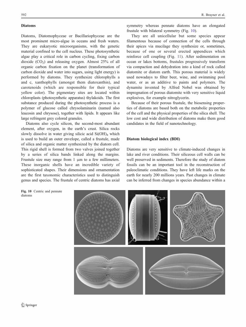
Diatoms also cycle silicon, the second-most abundantelement, after oxygen, in the earth’s crust. Silica rocksslowly dissolve in water giving silicic acid Si(OH)4 whichis used to build an outer envelope, called a frustule, madeof silica and organic matter synthesized by the diatom cell.This rigid shell is formed from two valves joined togetherby a series of silica bands linked along the margins.Frustule size may range from 1 μm to a few millimeters.These inorganic shells have an incredible variety ofsophisticated shapes. Their dimensions and ornamentationare the first taxonomic characteristics used to distinguishgenus and species. The frustule of centric diatoms has axial
symmetry whereas pennate diatoms have an elongatedfrustule with bilateral symmetry (Fig. 10).
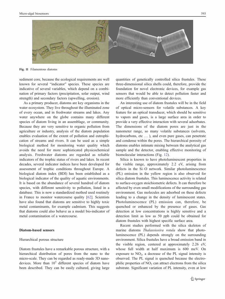
They are all unicellular but some species appearfilamentous because of connection of the cells throughtheir apices via mucilage they synthesize or, sometimes,because of one or several erected appendices whichreinforce cell coupling (Fig. 11). After sedimentation onocean or lakes bottoms, frustules progressively transformvia compaction and dehydration into a kind of rock calleddiatomite or diatom earth. This porous material is widelyused nowadays to filter beer, wine, and swimming poolwater, or as an additive to paints and polymers. Thedynamite invented by Alfred Nobel was obtained byimpregnation of porous diatomite with very sensitive liquidexplosives, for example nitroglycerin.
Because of their porous frustule, the biosensing proper-ties of diatoms are based both on the metabolic propertiesof the cell and the physical properties of the silica shell. Thelow cost and wide distribution of diatoms make them goodcandidates in the field of nanotechnology.
Diatom biological index (BDI)
Diatoms are very sensitive to climate-induced changes inlake and river conditions. Their siliceous cell walls can bewell preserved in sediments. Therefore the study of diatomfossils can be an important tool in the reconstruction ofpaleoclimatic conditions. They have left life marks on theearth for nearly 200 millions years. Past changes in climatecan be inferred from changes in species abundance within a
Fig. 10 Centric and pennatediatoms
592 R. Brayner et al.
sediment core, because the ecological requirements are wellknown for several “indicator” species. These species areindicative of several variables, which depend on a combi-nation of primary factors (precipitation, solar output, windstrength) and secondary factors (upwelling, erosion).
As a primary producer, diatoms are key organisms in thewater ecosystem. They live throughout the illuminated zoneof every ocean, and in freshwater streams and lakes. Anywater anywhere on the globe contains many differentspecies of diatom living in an assemblage, or community.Because they are very sensitive to organic pollution fromagriculture or industry, analysis of the diatom populationenables evaluation of the extent of pollution and eutrophi-cation of streams and rivers. It can be used as a simplebiological method for monitoring water quality whichavoids the need for more sophisticated physicochemicalanalysis. Freshwater diatoms are regarded as reliableindicators of the trophic status of rivers and lakes. In recentdecades, several indicator indices have been developed forassessment of trophic conditions throughout Europe. Abiological diatom index (BDI) has been established as abiological indicator of the quality of aquatic environments.It is based on the abundance of several hundred of diatomspecies, with different sensitivity to pollution, listed in adatabase. This is now a standardized method used routinelyin France to monitor watercourse quality [62]. Scientistshave also found that diatoms are sensitive to highly toxicmetal contaminants, for example cadmium. This suggeststhat diatoms could also behave as a model bio-indicator ofmetal contamination of a watercourse.
Diatom-based sensors
Hierarchical porous structure
Diatom frustules have a remarkable porous structure, with ahierarchical distribution of pores from the nano to themicro-scale. They can be regarded as ready-made 3D nano-devices. More than 105 different species of diatom havebeen described. They can be easily cultured, giving large
quantities of genetically controlled silica frustules. Thesethree-dimensional silica shells could, therefore, provide thefoundation for novel electronic devices, for example gassensors that would be able to detect pollution faster andmore efficiently than conventional devices.
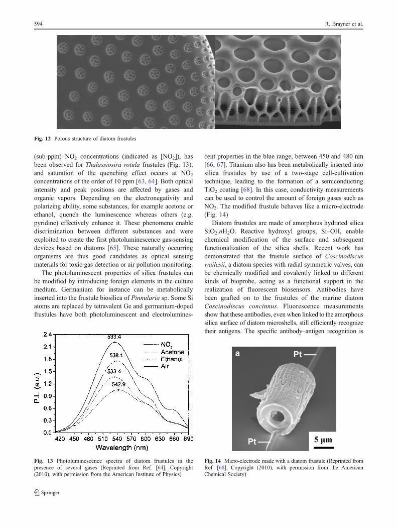
An interesting use of diatom frustules will be in the fieldof optical micro-sensors for volatile substances. A keyfeature for an optical transducer, which should be sensitiveto vapors and gases, is a large surface area in order toprovide a very effective interaction with several adsorbates.The dimensions of the diatom pores are just in thenanometer range, so many volatile substances (solvents,hydrocarbons, etc … ), and even pure gases, can penetrateand condense within the pores. The hierarchical porosity ofdiatoms enables intimate mixing between the analytical gassample and the detector, enabling effective monitoring ofbiomolecular interactions (Fig. 12).
Silica is known to have photoluminescent properties inthe visible range, approximately 2.2 eV, arising fromdefects in the Si–O network. Similar photoluminescence(PL) emission in the yellow region is also observed forsilica diatom frustules. This luminescence activity is relatedto surface-oxygen stoichiometric defects. It can therefore beaffected by even small modifications of the surrounding gasenvironment. Gas molecules are adsorbed on these defectsleading to a change in the density of luminescent states.Photoluminescence (PL) emission can, therefore, bequenched or enhanced by the presence of gases. Gasdetection at low concentrations is highly sensitive and adetection limit as low as 50 ppb could be obtained fordiatom frustules with highest specific surface area.
Recent studies performed with the silica skeleton ofmarine diatoms Thalassiosira rotula show that photo-luminescence (PL) depends strongly on the surroundingenvironment. Silica frustules have a broad emission band inthe visible region, centered at approximately 2.26 eV,whose full width at half maximum is 600 meV. Onexposure to NO2, a decrease of the PL signal intensity isobserved. The PL signal is quenched because the electro-philic properties of NO2 can attract electrons from the silicasubstrate. Significant variation of PL intensity, even at low
Fig. 11 Filamentous diatoms
Micro-algal biosensors 593
(sub-ppm) NO2 concentrations (indicated as [NO2]), hasbeen observed for Thalassiosira rotula frustules (Fig. 13),and saturation of the quenching effect occurs at NO2
concentrations of the order of 10 ppm [63, 64]. Both opticalintensity and peak positions are affected by gases andorganic vapors. Depending on the electronegativity andpolarizing ability, some substances, for example acetone orethanol, quench the luminescence whereas others (e.g.pyridine) effectively enhance it. These phenomena enablediscrimination between different substances and wereexploited to create the first photoluminescence gas-sensingdevices based on diatoms [65]. These naturally occurringorganisms are thus good candidates as optical sensingmaterials for toxic gas detection or air pollution monitoring.
The photoluminescent properties of silica frustules canbe modified by introducing foreign elements in the culturemedium. Germanium for instance can be metabolicallyinserted into the frustule biosilica of Pinnularia sp. Some Siatoms are replaced by tetravalent Ge and germanium-dopedfrustules have both photoluminescent and electrolumines-
cent properties in the blue range, between 450 and 480 nm[66, 67]. Titanium also has been metabolically inserted intosilica frustules by use of a two-stage cell-cultivationtechnique, leading to the formation of a semiconductingTiO2 coating [68]. In this case, conductivity measurementscan be used to control the amount of foreign gases such asNO2. The modified frustule behaves like a micro-electrode(Fig. 14)
Diatom frustules are made of amorphous hydrated silicaSiO2.nH2O. Reactive hydroxyl groups, Si–OH, enablechemical modification of the surface and subsequentfunctionalization of the silica shells. Recent work hasdemonstrated that the frustule surface of Coscinodiscuswailesii, a diatom species with radial symmetric valves, canbe chemically modified and covalently linked to differentkinds of bioprobe, acting as a functional support in therealization of fluorescent biosensors. Antibodies havebeen grafted on to the frustules of the marine diatomCoscinodiscus concinnus. Fluorescence measurementsshow that these antibodies, even when linked to the amorphoussilica surface of diatom microshells, still efficiently recognizetheir antigens. The specific antibody–antigen recognition is
Fig. 13 Photoluminescence spectra of diatom frustules in thepresence of several gases (Reprinted from Ref. [64], Copyright(2010), with permission from the American Institute of Physics)
Fig. 12 Porous structure of diatom frustules
Fig. 14 Micro-electrode made with a diatom frustule (Reprinted fromRef. [68], Copyright (2010), with permission from the AmericanChemical Society)
594 R. Brayner et al.
revealed by changes in the photoluminescence emission ofdiatoms frustules [69–71].
Diatom frustules could also be used as templates for thefabrication of nanostructured materials. Silica shells can bechemically converted into other oxide materials withoutlosing their 3D nanostructure. In such a process, currentlycalled BaSIC (bioclastic and shape-preserving inorganicconversion), silica has been converted into a new compo-sition via a shape-preserving gas–silica displacementreaction. The silica shell, for instance, can be transformedinto MgO on heating in magnesium vapor at 900 °C for 4 h[72]. Many other nanostructured oxide materials (TiO2,ZrO2, BaTiO3) have thus been synthesized [73, 74]. Silicacan even be reduced to porous silicon, leading to newpossibilities in micro-electronics. Such a synergistic com-bination of biological nanostructures with synthetic chem-ical functionalization could lead to a large number of 3Dmicro/nanostructures with chemistry and properties that canbe designed for sensing applications.
Diatom frustules as photonic crystals
Some diatoms behave like “living opals”—they haveiridescent properties arising from their peculiar porousstructure. Pores are not randomly distributed among thesilica shell; they sometimes form a regular periodic network
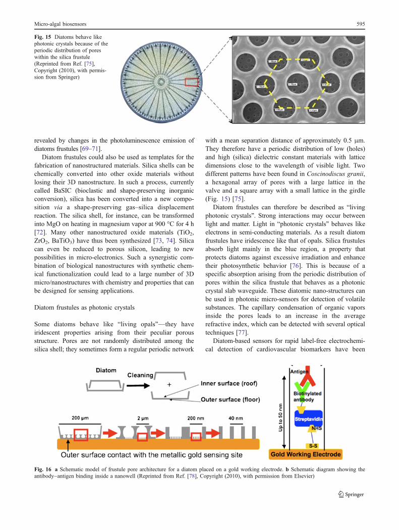
with a mean separation distance of approximately 0.5 μm.They therefore have a periodic distribution of low (holes)and high (silica) dielectric constant materials with latticedimensions close to the wavelength of visible light. Twodifferent patterns have been found in Coscinodiscus granii,a hexagonal array of pores with a large lattice in thevalve and a square array with a small lattice in the girdle(Fig. 15) [75].
Diatom frustules can therefore be described as “livingphotonic crystals”. Strong interactions may occur betweenlight and matter. Light in “photonic crystals” behaves likeelectrons in semi-conducting materials. As a result diatomfrustules have iridescence like that of opals. Silica frustulesabsorb light mainly in the blue region, a property thatprotects diatoms against excessive irradiation and enhancetheir photosynthetic behavior [76]. This is because of aspecific absorption arising from the periodic distribution ofpores within the silica frustule that behaves as a photoniccrystal slab waveguide. These diatomic nano-structures canbe used in photonic micro-sensors for detection of volatilesubstances. The capillary condensation of organic vaporsinside the pores leads to an increase in the averagerefractive index, which can be detected with several opticaltechniques [77].
Diatom-based sensors for rapid label-free electrochemi-cal detection of cardiovascular biomarkers have been
Fig. 16 a Schematic model of frustule pore architecture for a diatom placed on a gold working electrode. b Schematic diagram showing theantibody–antigen binding inside a nanowell (Reprinted from Ref. [78], Copyright (2010), with permission from Elsevier)
Fig. 15 Diatoms behave likephotonic crystals because of theperiodic distribution of poreswithin the silica frustule(Reprinted from Ref. [75],Copyright (2010), with permis-sion from Springer)
Micro-algal biosensors 595
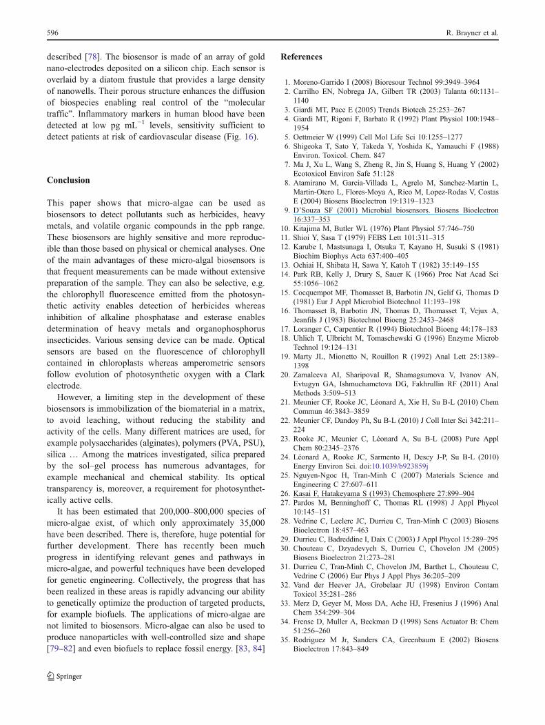
described [78]. The biosensor is made of an array of goldnano-electrodes deposited on a silicon chip. Each sensor isoverlaid by a diatom frustule that provides a large densityof nanowells. Their porous structure enhances the diffusionof biospecies enabling real control of the “moleculartraffic”. Inflammatory markers in human blood have beendetected at low pg mL−1 levels, sensitivity sufficient todetect patients at risk of cardiovascular disease (Fig. 16).
Conclusion
This paper shows that micro-algae can be used asbiosensors to detect pollutants such as herbicides, heavymetals, and volatile organic compounds in the ppb range.These biosensors are highly sensitive and more reproduc-ible than those based on physical or chemical analyses. Oneof the main advantages of these micro-algal biosensors isthat frequent measurements can be made without extensivepreparation of the sample. They can also be selective, e.g.the chlorophyll fluorescence emitted from the photosyn-thetic activity enables detection of herbicides whereasinhibition of alkaline phosphatase and esterase enablesdetermination of heavy metals and organophosphorusinsecticides. Various sensing device can be made. Opticalsensors are based on the fluorescence of chlorophyllcontained in chloroplasts whereas amperometric sensorsfollow evolution of photosynthetic oxygen with a Clarkelectrode.
However, a limiting step in the development of thesebiosensors is immobilization of the biomaterial in a matrix,to avoid leaching, without reducing the stability andactivity of the cells. Many different matrices are used, forexample polysaccharides (alginates), polymers (PVA, PSU),silica … Among the matrices investigated, silica preparedby the sol–gel process has numerous advantages, forexample mechanical and chemical stability. Its opticaltransparency is, moreover, a requirement for photosynthet-ically active cells.
It has been estimated that 200,000–800,000 species ofmicro-algae exist, of which only approximately 35,000have been described. There is, therefore, huge potential forfurther development. There has recently been muchprogress in identifying relevant genes and pathways inmicro-algae, and powerful techniques have been developedfor genetic engineering. Collectively, the progress that hasbeen realized in these areas is rapidly advancing our abilityto genetically optimize the production of targeted products,for example biofuels. The applications of micro-algae arenot limited to biosensors. Micro-algae can also be used toproduce nanoparticles with well-controlled size and shape[79–82] and even biofuels to replace fossil energy. [83, 84]
References
1. Moreno-Garrido I (2008) Bioresour Technol 99:3949–39642. Carrilho EN, Nobrega JA, Gilbert TR (2003) Talanta 60:1131–
11403. Giardi MT, Pace E (2005) Trends Biotech 25:253–2674. Giardi MT, Rigoni F, Barbato R (1992) Plant Physiol 100:1948–
19545. Oettmeier W (1999) Cell Mol Life Sci 10:1255–12776. Shigeoka T, Sato Y, Takeda Y, Yoshida K, Yamauchi F (1988)
Environ. Toxicol. Chem. 8477. Ma J, Xu L, Wang S, Zheng R, Jin S, Huang S, Huang Y (2002)
Ecotoxicol Environ Safe 51:1288. Atamirano M, Garcia-Villada L, Agrelo M, Sanchez-Martin L,
Martin-Otero L, Flores-Moya A, Rico M, Lopez-Rodas V, CostasE (2004) Biosens Bioelectron 19:1319–1323
9. D’Souza SF (2001) Microbial biosensors. Biosens Bioelectron16:337–353
10. Kitajima M, Butler WL (1976) Plant Physiol 57:746–75011. Shioi Y, Sasa T (1979) FEBS Lett 101:311–31512. Karube I, Mastsunaga I, Otsuka T, Kayano H, Susuki S (1981)
Biochim Biophys Acta 637:400–40513. Ochiai H, Shibata H, Sawa Y, Katoh T (1982) 35:149–15514. Park RB, Kelly J, Drury S, Sauer K (1966) Proc Nat Acad Sci
55:1056–106215. Cocquempot MF, Thomasset B, Barbotin JN, Gelif G, Thomas D
(1981) Eur J Appl Microbiol Biotechnol 11:193–19816. Thomasset B, Barbotin JN, Thomas D, Thomasset T, Vejux A,
Jeanfils J (1983) Biotechnol Bioeng 25:2453–246817. Loranger C, Carpentier R (1994) Biotechnol Bioeng 44:178–18318. Uhlich T, Ulbricht M, Tomaschewski G (1996) Enzyme Microb
Technol 19:124–13119. Marty JL, Mionetto N, Rouillon R (1992) Anal Lett 25:1389–
139820. Zamaleeva AI, SharipovaI R, Shamagsumova V, Ivanov AN,
Evtugyn GA, Ishmuchametova DG, Fakhrullin RF (2011) AnalMethods 3:509–513
21. Meunier CF, Rooke JC, Léonard A, Xie H, Su B-L (2010) ChemCommun 46:3843–3859
22. Meunier CF, Dandoy Ph, Su B-L (2010) J Coll Inter Sci 342:211–224
23. Rooke JC, Meunier C, Léonard A, Su B-L (2008) Pure ApplChem 80:2345–2376
24. Léonard A, Rooke JC, Sarmento H, Descy J-P, Su B-L (2010)Energy Environ Sci. doi:10.1039/b923859j
25. Nguyen-Ngoc H, Tran-Minh C (2007) Materials Science andEngineering C 27:607–611
26. Kasai F, Hatakeyama S (1993) Chemosphere 27:899–90427. Pardos M, Benninghoff C, Thomas RL (1998) J Appl Phycol
10:145–15128. Vedrine C, Leclerc JC, Durrieu C, Tran-Minh C (2003) Biosens
Bioelectron 18:457–46329. Durrieu C, Badreddine I, Daix C (2003) J Appl Phycol 15:289–29530. Chouteau C, Dzyadevych S, Durrieu C, Chovelon JM (2005)
Biosens Bioelectron 21:273–28131. Durrieu C, Tran-Minh C, Chovelon JM, Barthet L, Chouteau C,
Vedrine C (2006) Eur Phys J Appl Phys 36:205–20932. Vand der Heever JA, Grobelaar JU (1998) Environ Contam
Toxicol 35:281–28633. Merz D, Geyer M, Moss DA, Ache HJ, Fresenius J (1996) Anal
Chem 354:299–30434. Frense D, Muller A, Beckman D (1998) Sens Actuator B: Chem
51:256–26035. Rodriguez M Jr, Sanders CA, Greenbaum E (2002) Biosens
Bioelectron 17:843–849
596 R. Brayner et al.
36. Naessens M, Leclerc JC, Tran-Minh C (2000) Ecotoxicol EnvironSafe 46:181–185
37. Pandard P, Rawson DM (1993) Environ Toxicol Water Qual8:323–328
38. Heever J, Grobbelaar J (1997) Water SA 23:233–23839. Duvinsky Z, Falkowski PG, Post AF, Van Hes UM (1987) J Plank
Res 9:607–61240. Shitanda I, Takada K, Sakai Y, Tatsuma T (2005) Anal Chim Acta
530:191–19741. Naessens M, Tren-Minh C (1999) Sensors and Actuators B
59:100–10242. Altamirano M, Garcia-Villada L, Agrelo M, Sanchez-Martin L,
Martin-Otero L, Flores-Moya A, Rico M, Lopez-Rodas V, CostasE (2004) Biosens Bioelectron 19:1319–1323
43. Tajes-Martinez P, Beceiro-Gonzalez E, Muniategui-Lorenzo S,Prada-Rodriguez D (2006) Talanta 68:1489–1496
44. Guedri H, Durrieu C (2008) Microchim Acta 163:179–18445. Durrieu C, Tran-Minh C (2003) Talanta 59:535–54446. Chouteau C, Dzyadevych S, Chovelon J-M, Durrieu C (2004)
Biosens Bioelectron 19:1089–109647. Peña-Vazquez E, Pérez-Conde C, Costas E, Moreno-Bondi MC
(2010) Ecotoxicology 19:1059–106548. Peña-Vazquez E, Maneiro E, Pérez-Conde C, Moreno-Bondi MC,
Costas E (2009) Biosens Bioelectron 24:3538–354349. Darder M, Aranda P, Burgos-Asperilla L, Llobera A, Cadarso VJ,
Fernández-Sánchez C, Ruiz-Hitzky E (2010) J Mater Chem20:9362–9369
50. Rawson DM (1989) Biosensors 4:299–31151. Croisetiere L, Rouillon R, Carpentier R (2001) Appl Microbiol
Biotechnol 56:261–26452. Shao CY, Howe CJ, Porter AJR, Glover LA (2002) Appl Environ
Microbiol 68:5026–503353. Rouillon R, Tocabens M, Carpentier R (1999) Enzyme Microbial
Technol 25:230–23554. Maly J, Masojidek J, Masci A, Ilie M, Cianci E, Foglietti V,
Vastarella W, Pilloton R (2005) Biosens Bioelectron 21:923–93255. Geiselhart L, Osgood M, Holmes DS (1991) Ann NY Acad Sci
646:53–6056. Selifinova O, Burlage R, Barkay T (1993) Appl Environ
Microbiol 59:3083–309057. Collard JM, Corbisier P, Diels L, Dong Q, Jeanthon C, Mergeay
M, Taghavi S, Vander LD, Wilmotte A, Wuertz S (1994) FEMSMicrobiol Rev 14:405–414
58. Turner J, Robinson NJ (1995) J Ind Microbiol 14:119–12559. Huckle JW, Morby AP, Turner JS, Robinson NJ (1993) Mol
Microbiol 7:177–18760. Erbe JL, Adams AC, Taylor KB, Hall LM (1996) J Ind Microbiol
17:80–8361. Chay TC, Surif S, Heng LY (2009) Asian J Biological Sci 2:14–2062. Coste M, Boutry S, Tison-Rosebery J, Delmas F (2009)
Ecological Indicator 9:621–650
63. Lettieri S, Setaro A, De Stefano L, De Stefano M, Maddalena P(2008) Adv Funct Mater 18:1257–1264
64. De Stefano L, Rendina I, De Stefano M, Bismuto A, Maddalena P(2005) Appl Phys Lett 87:233902
65. Steraro A, Lettieri S, Maddalena P, De Stefano L (2007) ApplPhys Lett 91:051921
66. Jeffryes C, Solanki R, Rangineni Y, Wang W, Chang C, Rorrer GL(2008) Adv Mater 20:2633–2637
67. Qin T, Gutu T, Jiao J, Chang C, Rorrer GL (2008) ACS Nano2:1296–1304
68. Jeffryes C, Gutu T, Jiao J, Rorrer G (2008) ACS Nano 2:2103–2112
69. De Stefano L, Rotiroti L, De Stefano M, Lamberti A, LettieriS, Setaro A, Maddalena P (2009) Biosens Bioelectron24:1580–1584
70. De Stefano L, Lamberti A, Rotiroti L, De Stefano M (2008) ActaBiomater 4:126–130
71. Gale DK, Gutu T, Jiao J, Chang C, Rorrer GL (2009) Adv FunctMater 19:926–933
72. Sandhage KH, Dickerson MB, Huseman PM, Caranna MA,Clifton JD, Bull TA, Heibel TJ, Overton WR, SchoenwaelderMEA (2002) Adv Mater 14:429–433
73. Sandhage KH, Allan SM, Dickerson MB, Gaddis CS, Shian S,Weatherspoon MR, Ye C, Ahmad G, Haluska MS, Snyder RL(2005) Int J Appl Ceram Technol 2:317–326
74. Bao Z, Weatherspoon MR, Shian S, Cai Ye, Graham PD,Allan SM, Ahmad G, Dickerson MB, Church BC, Kang Z,Abernathy HW, Summers CJ, Liu M, Sandhage KH (2007)Nature 446:172–175
75. Fuhrmann T, Landwehr S, El Rharbi-Kucki M, Sumper M (2004)Appl Phys B 78:257–260
76. Yamanaka S, Yano R, Usami H, Hayashida N, Ohguchi M,Takeda H, Yoshino K (2008) J Appl Phys 103:074701
77. De Stefano L, Maddalena P, Moretti L, Rea I, Rendina I, DeTommasi E, Mocella V, De Stefano M (2009) Superlattices andMicrostructures 46:84–89
78. Lin KC, Kunduru V, Bothara M, Rege K, Prasad S, RamakrishnaBL (2010) Biosens Bioelectron 25:2336–2342
79. Sicard C, Brayner R, Margueritat J, Hemadi M, Couté A,Yéprémian C, Djediat C, Aubard J, Fiévet F, Livage J, CoradinT (2010) J Mater Chem 20:9342–9347
80. Dahoumane SA, Djedat C, Yéprémian C, Couté A, Fiévet F,Brayner R (2010) Thin Solid Films 518:5432–5436
81. Brayner R, Herbst F, Yéprémian C, Djediat C, Coradin T, LivageJ, Fiévet F, Couté A (2009) Langmuir 25:10062–10067
82. Brayner R, Barberousse H, Hemadi M, Djediat C, Yéprémian C,Coradin T, Livage J, Fiévet F, Couté A (2007) J NanosciNanotech 7:2696–2707
83. Beer LL, Boyd ES, Peters JW, Posewitz MC (2009) Curr OpinionBiotechnol 20:264–271
84. Kruse O, Hankamer B (2010) Curr Opinion Biotechnol 21:238–243
Micro-algal biosensors 597