Embed Size (px)
Citation preview
R
P
MD
a
ARRAA
KPGTB
C
0d
Sensors and Actuators B 143 (2009) 430–443
Contents lists available at ScienceDirect
Sensors and Actuators B: Chemical
journa l homepage: www.e lsev ier .com/ locate /snb
eview
olypyrrole based amperometric glucose biosensors
inni Singh ∗, Pavan Kumar Kathuroju, Nagaraju Jampanaepartment of Instrumentation, Indian Institute of Science, Bangalore-560012, Karnataka, India
r t i c l e i n f o
rticle history:eceived 15 June 2009eceived in revised form 21 August 2009ccepted 2 September 2009vailable online 9 September 2009
eywords:olypyrrolelucoseransduceriosensor
a b s t r a c t
Biosensors have gained immense acceptance in the field of medical diagnostics, besides environmental,food safety and biodefence applications due to its attributes of real-time and rapid response. This syn-ergistic combination of biotechnology and microelectronics comprises a biological recognition elementcoupled with a compatible transducer device. Diabetes is a disease of major concern since the ratio ofworld population suffering from it is increasing at an alarming rate and therefore the need for develop-ment of accurate and stable glucose biosensors is evident. There are many commercial glucose biosensorsavailable yet some limitations need attention. This review presents a detailed account of the polypyr-role based amperometric glucose biosensors. The polymer polypyrrole is used extensively as a matrixfor immobilization of glucose oxidase enzyme owing to its favourable features such as stability underambient conditions, conductivity that allows it to be used as an electron relay, ability to be polymerizedunder neutral and aqueous mild conditions, and more. The simple one-step electrodeposition on theelectrode surface allows easy entrapment of the enzyme. The review is structured into three categories(a) the first-stage biosensors: which report the studies from the inception of use of polypyrrole in glu-cose biosensors during which time the role of the polymer and the use of mediators was established. Thisperiod saw extensive work by two separate groups of Schuhmann and Koopal who contributed a greatdeal in understanding the electron transfer pathways in polypyrrole based glucose biosensors, (b) thesecond-stage biosensors: which highlight the shift of polypyrrole from a conventional matrix to com-
posite matrices with extensive use of mediators focused at improving the selectivity of response, and(c) third-stage biosensors: the remarkable properties of nanoparticles and carbon nanotubes and theiroutstanding ability to mediate electron transfers have seen their indispensable use in conjugation withpolypyrrole for development of glucose biosensors with improved sensitivity and stability characteristicswhich is accounted in the review, which thus traces the evolution of polypyrrole from a conventional matrix, to composites and thence to the form of nanotube arrays, with the objective of addressing thevital issue of diabetes management through the development of stable and reliable glucose biosensors.© 2009 Elsevier B.V. All rights reserved.
ontents
1. Introduction . . . . . . . . . . . . . . . . . . . . . . . . . . . . . . . . . . . . . . . . . . . . . . . . . . . . . . . . . . . . . . . . . . . . . . . . . . . . . . . . . . . . . . . . . . . . . . . . . . . . . . . . . . . . . . . . . . . . . . . . . . . . . . . . . . . . . . . . . . 4311.1. The glucose biosensor: background and sensing principle . . . . . . . . . . . . . . . . . . . . . . . . . . . . . . . . . . . . . . . . . . . . . . . . . . . . . . . . . . . . . . . . . . . . . . . . . . . . . . . . . . 4311.2. Mechanism of polymerization of pyrrole monomer and immobilization of the enzyme . . . . . . . . . . . . . . . . . . . . . . . . . . . . . . . . . . . . . . . . . . . . . . . . . . . 431
2. Polypyrrole as a matrix in glucose biosensors . . . . . . . . . . . . . . . . . . . . . . . . . . . . . . . . . . . . . . . . . . . . . . . . . . . . . . . . . . . . . . . . . . . . . . . . . . . . . . . . . . . . . . . . . . . . . . . . . . . . . . . 4322.1. First-stage biosensors: conventional polypyrrole in glucose biosensors . . . . . . . . . . . . . . . . . . . . . . . . . . . . . . . . . . . . . . . . . . . . . . . . . . . . . . . . . . . . . . . . . . . . 432
2.1.1. The pathway of electron transfer . . . . . . . . . . . . . . . . . . . . . . . . . . . . . . . . . . . . . . . . . . . . . . . . . . . . . . . . . . . . . . . . . . . . . . . . . . . . . . . . . . . . . . . . . . . . . . . . . . . 4342.1.2. First-stage biosensors: a step ahead. . . . . . . . . . . . . . . . . . . . . . . . . . . . . . . . . . . . . . . . . . . . . . . . . . . . . . . . . . . . . . . . . . . . . . . . . . . . . . . . . . . . . . . . . . . . . . . . 437
2.2. Second-stage biosensors: polypyrrole composites in glucose biosensors . . . . . . . . . . . . . . . . . . . . . . . . . . . . . . . . . . . . . . . . . . . . . . . . . . . . . . . . . . . . . . . . . . . 4382.3. Third-stage biosensors: polypyrrole based nanoparticles and carbon nanotubes in glucose biosensors . . . . . . . . . . . . . . . . . . . . . . . . . . . . . . . . . . . 439
3. Conclusions . . . . . . . . . . . . . . . . . . . . . . . . . . . . . . . . . . . . . . . . . . . . . . . . . . . . . . . . . . . . . . . . .Acknowledgements . . . . . . . . . . . . . . . . . . . . . . . . . . . . . . . . . . . . . . . . . . . . . . . . . . . . . . . .References . . . . . . . . . . . . . . . . . . . . . . . . . . . . . . . . . . . . . . . . . . . . . . . . . . . . . . . . . . . . . . . . . .Biographies . . . . . . . . . . . . . . . . . . . . . . . . . . . . . . . . . . . . . . . . . . . . . . . . . . . . . . . . . . . . . . . . .
∗ Corresponding author. Tel.: +91 80 22932273; fax: +91 80 23600135.E-mail addresses: [email protected] (M. Singh), [email protected] (P.K. K
925-4005/$ – see front matter © 2009 Elsevier B.V. All rights reserved.oi:10.1016/j.snb.2009.09.005
. . . . . . . . . . . . . . . . . . . . . . . . . . . . . . . . . . . . . . . . . . . . . . . . . . . . . . . . . . . . . . . . . . . . . . . . . 441. . . . . . . . . . . . . . . . . . . . . . . . . . . . . . . . . . . . . . . . . . . . . . . . . . . . . . . . . . . . . . . . . . . . . . . . . . 442. . . . . . . . . . . . . . . . . . . . . . . . . . . . . . . . . . . . . . . . . . . . . . . . . . . . . . . . . . . . . . . . . . . . . . . . . 442. . . . . . . . . . . . . . . . . . . . . . . . . . . . . . . . . . . . . . . . . . . . . . . . . . . . . . . . . . . . . . . . . . . . . . . . . 443
athuroju), [email protected] (N. Jampana).
Actua
1
bc[asiormaatctiapcttad
aedaewadaimaOoTe(eepcamst
1
cuiff3pgaim
M. Singh et al. / Sensors and
. Introduction
A biosensor is an analytical device that consists of a confinediological component in conjunction with a transducer device thatonverts a biochemical signal into an amplified electrical signal1]. Enzymes, whole cells, tissues, receptors, antibodies and nucleiccids comprise the biological component. Enzymes being highlypecific find extensive applications in biosensors. It is, however,ncumbent for the biological element involved in the developmentf a biosensor to be immobilized, to allow reuse with consistency ofesponse, a prerequisite for any device aimed at subsequent com-ercial scale up. Various immobilization procedures are used such
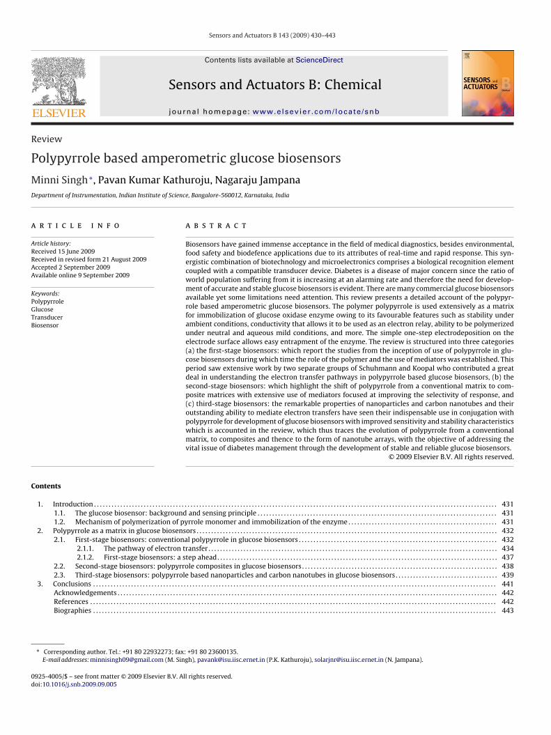
s entrapment and encapsulation, covalent binding, cross-linkingnd adsorption [2]. In general, the method is based on the nature ofhe biocomponent, the type of transducer used, the physicochemi-al properties of the analyte and the operating conditions in whichhe biosensor is to function, and overriding all these considerationss the necessity for the biological component to exhibit maximumctivity in its immobilized microenvironment. The biological com-onent specifically recognizes the analyte, and the biochemicalhange that results by the interaction of the two is indicated byhe transducer, the key component of the biosensor. This necessi-ates the coupling of a transducer compatible with the biochemicalssay under study. Several types of transducers are used in theevelopment of biosensors as shown in Fig. 1.
Electrochemical are the most widely used class of transducers-mperometric transducers detect the current drain through anlectrode at an applied potential; potentiometric transducersetect the change in potential across the membrane of an electrodes it comes in contact with specific ions; conductometric transduc-rs detect the change in conductivity as a result of the reaction,hereas impedimetric transducers track the change in impedance
s a result of redox biochemical response. Thermal transducersetect the change in temperature whereas piezoelectric and surfacecoustic wave transducers are mass based and detect the changen frequency of oscillation after adsorption or desorption of analyte
olecules on the surface of piezoelectric detector materials suchs quartz, lithium niobate, lead–zinc niobate–lead titanate, etc.ptical transducers measure electromagnetic radiations absorbedr emitted by reactants or products of the biochemical system.he field effect transistors (FETs) are a combination of solid-statelectronics with ion selective electrodes. Enzyme sensitive FETsENFETs) are developed by applying thin layers of immobilizednzymes over FETs [3]. The major attributes of biosensors whichxplain their exponential increase in the market are specificity,ortability (in most cases), fast response, ability to function in opti-ally opaque solutions, real-time analysis without pre treatment,nd ease of use [4] leading to their wide range of applications inedical diagnostics [5–7], environmental monitoring [8,9], food
afety [10,11], on- and off-line industrial processes [12] and detec-ion of warfare agents [13,14].
.1. The glucose biosensor: background and sensing principle
Diabetes is a chronic disease with no cure. It occurs when pan-reas do not produce enough insulin, or the body cannot effectivelyse the insulin it produces. This leads to hyperglycemia, character-
zed by increased blood glucose conc. The disease is a major concernor world population as in the year 2000, 171 million people suf-ered from diabetes, a figure that is expected to reach an alarming66 million by 2030 [15]. Blood glucose monitoring is an integral
art of diabetes management and in this regard self-monitoring oflucose has been considered by many as one of the most importantdvances in management of the disease ever since the discovery ofnsulin [16]. The most commercially successful devices for glucoseonitoring are the blood glucose biosensors, the initial concept of
tors B 143 (2009) 430–443 431
which was proposed by Clark and Lyons [17] which led to the firstcommercial glucose analyzer launched by Yellow Spring Instru-ment Co., Ohio. There are now over 40 blood glucose meters inthe market, yet many challenges need attention [6,18].
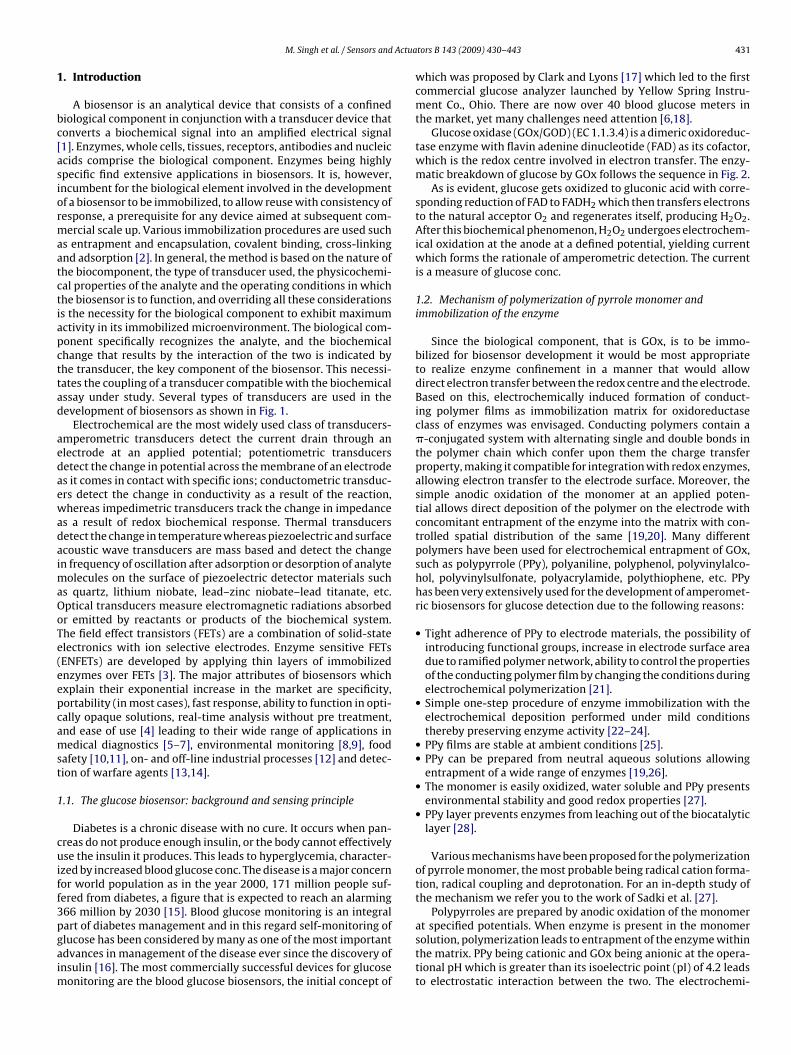
Glucose oxidase (GOx/GOD) (EC 1.1.3.4) is a dimeric oxidoreduc-tase enzyme with flavin adenine dinucleotide (FAD) as its cofactor,which is the redox centre involved in electron transfer. The enzy-matic breakdown of glucose by GOx follows the sequence in Fig. 2.
As is evident, glucose gets oxidized to gluconic acid with corre-sponding reduction of FAD to FADH2 which then transfers electronsto the natural acceptor O2 and regenerates itself, producing H2O2.After this biochemical phenomenon, H2O2 undergoes electrochem-ical oxidation at the anode at a defined potential, yielding currentwhich forms the rationale of amperometric detection. The currentis a measure of glucose conc.
1.2. Mechanism of polymerization of pyrrole monomer andimmobilization of the enzyme
Since the biological component, that is GOx, is to be immo-bilized for biosensor development it would be most appropriateto realize enzyme confinement in a manner that would allowdirect electron transfer between the redox centre and the electrode.Based on this, electrochemically induced formation of conduct-ing polymer films as immobilization matrix for oxidoreductaseclass of enzymes was envisaged. Conducting polymers contain a�-conjugated system with alternating single and double bonds inthe polymer chain which confer upon them the charge transferproperty, making it compatible for integration with redox enzymes,allowing electron transfer to the electrode surface. Moreover, thesimple anodic oxidation of the monomer at an applied poten-tial allows direct deposition of the polymer on the electrode withconcomitant entrapment of the enzyme into the matrix with con-trolled spatial distribution of the same [19,20]. Many differentpolymers have been used for electrochemical entrapment of GOx,such as polypyrrole (PPy), polyaniline, polyphenol, polyvinylalco-hol, polyvinylsulfonate, polyacrylamide, polythiophene, etc. PPyhas been very extensively used for the development of amperomet-ric biosensors for glucose detection due to the following reasons:
• Tight adherence of PPy to electrode materials, the possibility ofintroducing functional groups, increase in electrode surface areadue to ramified polymer network, ability to control the propertiesof the conducting polymer film by changing the conditions duringelectrochemical polymerization [21].
• Simple one-step procedure of enzyme immobilization with theelectrochemical deposition performed under mild conditionsthereby preserving enzyme activity [22–24].
• PPy films are stable at ambient conditions [25].• PPy can be prepared from neutral aqueous solutions allowing
entrapment of a wide range of enzymes [19,26].• The monomer is easily oxidized, water soluble and PPy presents
environmental stability and good redox properties [27].• PPy layer prevents enzymes from leaching out of the biocatalytic
layer [28].
Various mechanisms have been proposed for the polymerizationof pyrrole monomer, the most probable being radical cation forma-tion, radical coupling and deprotonation. For an in-depth study ofthe mechanism we refer you to the work of Sadki et al. [27].
Polypyrroles are prepared by anodic oxidation of the monomer
at specified potentials. When enzyme is present in the monomersolution, polymerization leads to entrapment of the enzyme withinthe matrix. PPy being cationic and GOx being anionic at the opera-tional pH which is greater than its isoelectric point (pI) of 4.2 leadsto electrostatic interaction between the two. The electrochemi-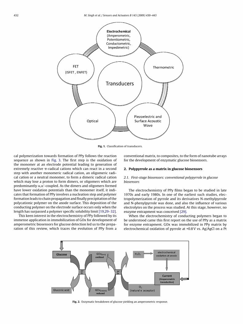
432 M. Singh et al. / Sensors and Actuators B 143 (2009) 430–443
ation
cstescwphcfpcl
iar
enzyme entrapment was conceived [29].
Fig. 1. Classific
al polymerization towards formation of PPy follows the reactionequence as shown in Fig. 3. The first step is the oxidation ofhe monomer at an electrode potential leading to generation ofxtremely reactive �-radical cations which can react in a secondtep with another monomeric radical cation, an oligomeric radi-al cation or a neutral monomer, to form a dimeric radical cationhich may lose a proton to form dimers, or oligomers which areredominantly �,�′-coupled. As the dimers and oligomers formedave lower oxidation potentials than the monomer itself, it indi-ates that formation of PPy involves a nucleation step and polymerormation leads to chain propagation and finally precipitation of theolycationic polymer on the anode surface. This deposition of theonducting polymer on the electrode surface occurs only when theength has surpassed a polymer specific solubility limit [19,29–32].
This keen interest in the electrochemistry of PPy followed by itsmmense application in immobilization of GOx for development ofmperometric biosensors for glucose detection led us to the prepa-ation of this review, which traces the evolution of PPy from a
Fig. 2. Enzymatic breakdown of glucose y
of transducers.
conventional matrix, to composites, to the form of nanotube arraysfor the development of enzymatic glucose biosensors.
2. Polypyrrole as a matrix in glucose biosensors
2.1. First-stage biosensors: conventional polypyrrole in glucosebiosensors
The electrochemistry of PPy films began to be studied in late1970s and early 1980s. In one of the earliest such studies, elec-tropolymerization of pyrrole and its derivatives N-methylpyrroleand N-phenylpyrrole was done, and also the influence of variouselectrolytes on the process was studied. At this stage, however, no
When the electrochemistry of conducting polymers began tobe understood came this first report on the use of PPy as a matrixfor enzyme entrapment. GOx was immobilized in PPy matrix byelectrochemical oxidation of pyrrole at +0.8 V vs. Ag/AgCl on a Pt
ielding an amperometric response.
M. Singh et al. / Sensors and Actua
FMe
wpinoiccsve(+taspoiPiiitwaacp
loading, pH, temperature, stability and kinetic parameters Km and
ig. 3. Sequence of electrochemical polymerization to form PPy. (Reprinted fromikrochim Acta Schuhmann W, Conducting polymer based amperometric enzyme
lectrodes, 1–29, © 1995 (121), with permission from Springer.)
orking electrode. GOx being negatively charged at an operationalH of 7.0, and the polymer with a net positive charge [33], ionic
ncorporation of GOx into the matrix was the proposed mecha-ism of immobilization. The sensor had a linear detection rangef 0–100 mM glucose. The electrochemical oxidation of enzymat-cally produced H2O2 at +0.7 V was used as a measure of glucoseonc. though it was known that at this potential various interferingompounds contribute to anodic current when real-time samplesuch as biological fluids are used. To avoid such scrupulous obser-ations due to these compounds, response characteristics of thelectrodes in the presence of mediators phenazine methosulphatePMS) and benzoquinone were observed at oxidation potentials of0.35 and +0.4 V, respectively. The key advantageous features ofhe sensor were the ease of preparation, reproducibility, stabilitynd disposability [31]. The feasibility of GOx/PPy/Pt electrodes wastudied and the characterization and optimization of operationalarameters was done. The sensor exhibited linear detection rangef 0.2–1.0 mM glucose. The limitation, however, was the instabil-ty of the electrode due to leaching out of the enzyme from thePy matrix owing to the porous nature of the polymer. Anothermportant finding was the degradation of the conducting polymern the presence of H2O2 [34]. GOx was entrapped by electrochem-cal polymerization of a pyrrole derivative, N-methyl pyrrole andhe electrode response towards glucose, in terms of current density,as correlated with theoretical expressions per se film thickness
nd enzyme loading. An agreement was observed for thick filmsnd theoretical studies, which took into account factors such asurrent (i), bulk substrate conc. (s∞), diffusion coefficient (Ds) andartition coefficient (K) of glucose in the film, film thickness (l),
tors B 143 (2009) 430–443 433
rate const (Kcat) of the biochemical reaction, conc. of the enzyme(e∑) and the Michaelis–Menten constant (Km), in comparison to
thinner films, in which the observed responses were lower thanpredicted. Thickness, in turn, was a function of charge. Interestingly,the dependence of response on rotation speed (in case of rotat-ing disc working electrodes) with respect to enzyme loading wasdemonstrated, which was dependent on the expression 1/Xk whereXk is the kinetic length given by (DsKm/Kcate∑)1/2. It was reported
that when 1/Xk > 1 the electrode response is independent of enzymeconc. and when 1/Xk � 1, the response varied with enzyme load-ing and became inversely dependent on the rotation speed of theelectrode, explained by the differential dispersion of H2O2 at differ-ent speeds. The study also explained a rather intricate observationof immense practical significance, of anomalously large electroderesponse towards glucose being due to enzyme adsorption onto thebare electrode if not subjected to electrochemical polymerizationas soon as the electrode is immersed into the monomer-enzymesolution. The sensor had a linear detection range of 0–0.22 M glu-cose and a stability of 5 days [35].
With increasing understanding of the electron transfer pro-cess in PPy based glucose biosensors, in this first report, anattempt was made to develop Py-mediator conjugates with fer-rocene derivatives with an objective of devising a more efficientmeans of electron transfer between the enzyme and the electrode.Two conjugates for catalytic mediation were examined-Py/FAPP[(ferrocenyl)amidopropyl] pyrrole and Py/FAPAPP [(ferrocenyl)amidopentyl amidopropyl] pyrrole. Due to higher catalytic effi-ciency observed for Py/FAPP conjugate possibly due to differentialmobility or orientation of the ferrocenyl ligands, the former was theconjugate of choice. An operating potential of +0.3 V vs. Ag/AgCl wasused to study the electron exchange between reduced GOx and theferricinium ion. A marked limitation of this conjugate based elec-trode was its low stability of as low as 2 days [33]. GOx has also beenimmobilized on PPy-lecithin bilayer lipid membranes by cross-linking and ionic adsorption. The sensor was able to detect glucosein the range 0–50 mM. Ultrathin film (<10 nm), stability and electri-cal conductivity of the polymer membranes were claimed to be itsdesirable features [36]. The electrochemistry of PPy–GOx electrodewas detailed and the electrochemical behavior of the electrodewith respect to charge and potential were reported. The enzymewas entrapped in the PPy matrix potentiostatically at +0.65 V vs.SCE. It was found that 7% of the total charge input was consumedfor electrochemical oxidation of pyrrole leading to polymerizationand entrapment of GOx. To study the state of electroactivity andthe response of the PPy–GOx electrodes for glucose estimation, avariation in the cyclic voltammograms was induced with respect toapplied potential. Due to this variation the electroactivity of the PPyfilm was lost, though the electrode continued to furnish a substan-tial response towards glucose. To identify the potential at which theelectrode loses electroactivity but continues to respond to glucose,a series of cyclic voltammetric experiments were performed alter-nating with glucose determination and this potential threshold wasidentified to be +0.55 V. Therefore the study suggested the use of+0.5 V for clinical applications which would eliminate co-oxidationof interfering compounds, in comparison to +0.7 V and concludedthat response of PPy–GOx electrode to glucose is observed onlyafter loss of PPy electroactivity, which is the result of over-oxidationof PPy by the H2O2 produced [37]. The response characteristics ofthe PPy/GOx electrodes were further probed and the response withrespect to operational parameters such as film thickness, enzyme
Imax was reported. The thickness of the film was measured in rela-tion to the charge deposited, in accordance with Holdcroft and Funt[38] and a charge of 0.75 mC/cm2 was chosen which was equiva-lent to a film thickness of 0.17 �m. With increase in thickness a
4 Actua
roorGiHGs1s[Prroeolsmvcdp
2
wsegotce
imntgetpntnbotmrfTch
att–tide
34 M. Singh et al. / Sensors and
educed electrode response (current) was observed owing to lossf H2O2 into the bulk. Besides film thickness, the turnover numberf GOx was shown to play a significant role in the electrochemicalesponse of the sensor. An optimum response was observed in theOx conc. range of 25–100 U/ml beyond which the response dimin-
shed which was attributed to the reduced internal diffusion of2O2 to the underlying Pt electrode. At a conc. higher than 100 U/mlOx, H2O2 generated at the PPy/solution interface diffused into theolution instead. The sensor furnished a linear detection range of–7.5 mM glucose with a response time of 5 min, an operationaltability of 80 days, and a remarkable storage stability of 500 days39]. In an extended study it was observed that incorporation oft microparticles into the PPy/GOx preparations enhanced the cur-ent response by 40%. This was due to increased surface area as aesult of dispersion of Pt particles which act as catalyst for H2O2xidation. Since the response was observed only after loss of PPylectroactivity, it further confirmed the role of Pt. The response timef the sensor reduced to 10–15 s [40]. Belanger’s reports [37,39] onoss of electroactivity of PPy clearly laid the foundation for sub-equent studies and detailed investigations into electron transferechanisms in amperometric biosensors. In another set of obser-
ations by another group in the same year, electron transfer waslaimed to be between GOx and PPy. The authors, though contra-icted Belanger’s observations, failed to provide any substantialroof of their findings [41].
.1.1. The pathway of electron transferBy now, the biochemistry of enzymatic breakdown of glucose
ith respect to electrochemical measurements had been under-tood and the use of PPy as an entrapment matrix was also wellstablished. As will be seen henceforth, the focus of the studyained impetus in terms of pathway of electron transfer in amper-metric glucose biosensors. Significant work in this orientation bywo groups – Schuhmann (Germany) and Koopal (Netherlands)ontributed to the understanding of the phenomenon to a greatxtent.
The electrocatalytic properties of functionalized PPy were stud-ed. Photometric analysis of covalently immobilized GOx on the
odified matrix revealed that activity was independent of thick-ess of the film since the enzyme is immobilized on the surface ofhe matrix through amide bonds. For electrochemical analysis oflucose, however, which is dependent upon H2O2 oxidation at thelectrode, for which H2O2 needs to penetrate through the matrix tohe surface of the metal electrode, thickness of the PPy matrix is aarameter of paramount relevance. Activities with respect to thick-ess in the range 0.3–14.2 �m were observed and 0.5 �m furnishedhe maximum response (a charge of 0.45 mC/cm2 gives a film thick-ess of 1.0 �m—[38]). This size exclusion effect of the matrix wille explained elsewhere. Since most work is focused on the studyf electron transfer between the redox centre of the enzyme andhe electrode surface, it was viable to modify suitably the poly-
er matrix to attain a consistent physiologically relevant range ofesponse. Therefore, covalent attachment of the enzyme on the sur-ace of the matrix by chemical modifications of the same was done.o further enhance the response characteristics, the use of artifi-ial electron acceptors have been proposed, which are discussedenceforth [47].
In one of the first reports of covalent binding of GOx to function-lized PPy, the amperometric response of the immobilized enzymeowards glucose was studied. The procedure involved –NH2 func-ionalization of PPy at �-position, carbodiimide activation of
COOH groups on GOx and thereafter covalent immobilizationhrough amide bond formation. A substantial loss of enzyme activ-ty was observed in comparison to native enzyme, as expectedue to covalent bond formation, leading to denaturation of thenzyme. The sensor had a linear detection range of 0-5 mM glucose,tors B 143 (2009) 430–443
a response time of 25 s and a stability of 3 days with approximately70% activity retention on the third day. Incorporation of artificialelectron acceptors was studied but their use in comparison to thenatural electron acceptor O2 was negated due to leaching of thesame [vide infra] which later led to covalently bonded mediatorsat N- and �-positions of PPy, however, its success in dehyroge-nase based sensors was demonstrated [48]. The adsorption of aredox mediator to facilitate electron transfer between the enzymeactive centre and the metal surface was, as earlier mentioned, aninappropriate proposition. It was seen that the adsorbed redoxmediator 1,1′-dimethylferrocene leached out as was evident bya quasi-exponential decrease of electrode response towards glu-cose, which regenerated upon reloading the electrode with themediator. The operational stability of only a few hours of the elec-trode as against 3 days for an unmodified electrode confirms thatnot the loss of enzyme but that of the mediator is responsible forthe diminished response [49]. A possible remedy to the loss ofthe mediator is covalent binding of the same in the matrix whichhas proven successful as will also be noticed in future work. Theredox properties of PPy for development of amperometric biosen-sors showed that for oxidase class of enzymes the electrocatalyticactivity is due to the sieve-effect of PPy, which allows smaller sizedmolecules such as H2O2 to penetrate to the surface of the electrode,allowing electron transfer. It was also proposed that this selectivefeature of porosity could reduce electrochemical co-oxidation ofinterfering compounds such as ascorbate which would have sig-nificant relevance to clinical applications of such biosensors [50].In an extended work it was further proven that covalently immo-bilized GOx activity is independent of the thickness of the film.However, the time required to attain stationary background cur-rent increased with the thickness of the polymer film since H2O2needs to diffuse through the thickness of the film to undergo oxi-dation at the electrode surface. A comparison of stationary andflow injection analysis (FIA) system led to the conclusion that FIAwas a preferred choice for analysis. The outcome of the studywas applied for the analysis of sucrose in the presence of glucose,besides glucose determination under stationary conditions [51].Schuhmann further demonstrated the size exclusion property ofPPy matrix reiterating the characteristics of covalently immobi-lized GOx on the surface of functionalized PPy as an experimentallysound approach to glucose analysis. Fig. 4 demarks the functionalaspect of entrapped (herein referred to as Type 1) and covalentlyimmobilized (herein referred to as Type 2) GOx and explains clearlythe simple working principle of the two types of electrodes.
It is clear from Fig. 4(a) that in Type 1 electrodes glucosemolecules traverse through the pores to reach the active site of theenzyme, which, due to spatial rearrangements during entrapmentmight not be fully accessible. On the other hand, in Type 2 elec-trodes, as is seen in Fig. 4(b) glucose has easy access to the activesite of the enzyme which is surface bound, producing H2O2, which,owing to its smaller size travels the distance of the film thicknessto the electrode surface to undergo oxidation. Also, the large sizedenzyme molecules on the surface limit the entry of interferentascorbic acid into the matrix substantially. Therefore, an obviousadvantage of Type 2 over Type 1 electrodes is the reliable deter-mination of glucose in the presence of co-oxidizable compounds,especially ascorbic and uric acid, which have comparable molecularweight to that of glucose. A further investigation of the Type 2 elec-trode by comparing its response with standard spectrophotometricassay showed the reliability of the biosensor for glucose determi-nation in complex samples. The size exclusion property of PPy films
were further probed and an attempt to understand the mecha-nism of GOx entrapment, whether adsorption or entrapment, wasmade. In this study, PPy/GOx electrodes were prepared by poten-tiostatic and galvanostatic methods. Till now, the thickness of thefilm, which is a function of charge applied, decided the response
M. Singh et al. / Sensors and Actuators B 143 (2009) 430–443 435
F the sA enzym
tootaff0tecseetwhdbatcanai
ctHmuceecsmtab
The involvement of the PPy backbone in electron transfer is,however, limited to the exclusion of O2, since H2O2 which is formedenzymatically in the presence of O2 is known to destroy the con-ducting properties of the polymer film [vide supra]. By modifying
ig. 4. (a) GOx entrapped in PPy matrix and (b) GOx covalently immobilized onmperometric substrate determination in flow injection systems with polypyrrole
o glucose. Greater the thickness, lesser was the observed responsewing to porosity, however, it was noticed that though the abovebservation stands true but for films prepared galvanostatically,he above inverse proportionality principle did not obey. Instead,n optimum thickness corresponding to a charge of 50 mC/cm2
urnished most favourable size exclusion characteristics. There-ore, for subsequent analysis, PPy electrodes were prepared at.2 �A (galvanostatic). To study the effect of location of enzyme onhe electrode, response characteristics of PPy/GOx electrodes werevaluated in terms of kinetic parameters, Km (Michaelis–Mentenonstant, higher the Km lower the affinity of the enzyme for theubstrate and vice-versa) and Imax (maximum current, a measure ofnzyme activity) and various kinds of films were prepared. Wherelectrodeposition was not done, PPy/GOx composite layer struc-ures were prepared by dip adsorption. The Km values observedere in the range 7–10 mM glucose, which, as expected, wereigher than 6.2 mM for soluble GOx, owing to enzyme loss due toesorption or denaturation. Among the layered films prepared, theest response was observed for electropolymerized Pt/(PPy–GOx)s against adsorbed GOx on PPy films, in which case it was observedhat the H2O2 produced is lost to the bulk solution. Although a con-lusion of electropolymerization being the preferred choice overdsorption for immobilization was made, however, the mecha-ism of immobilization-entrapment or adsorption, could not bescertained though in most reported works the terminology useds ‘entrapment’ [52].
An established prerequisite for amperometric biosensors is thelose proximity between the active site of the enzyme and the elec-rode surface to allow direct electron transfer between the two.owever, the spatial organization of the biological recognition ele-ents does not always allow such proximity and therefore, the
se of redox relays is envisaged. In this study the authors clearlyategorized the means to electrical communication between thenzyme and the electrode, highlighting the advantages and oth-rwise, of covalent binding and entrapment, and demonstratedo-polymerization of pyrrole modified GOx with osmium-complex
ubstituted pyrrole derivative and N-methyl pyrrole as an efficientechanism of direct electron transfer [53]. In a rather illustra-ive and nearly conclusive work on electron transfer pathway inmperometric biosensors, the mediator ferrocene was covalentlyound through amide linked spacers to the outer surface of perio-
urface of PPy matrix. (Reprinted from Sensors and Actuators B, Schuhmann W,e electrodes, 41–49, © 1991 (4), with permission from Elsevier.)
date oxidized/ carbodiimide activated GOx, as shown in Fig. 5 andentrapped this modified enzyme within the PPy matrix and there-after hypothesized two different pathways of electron transfer: (a)electron transfer between the active site of reduced GOx to the fer-rocene redox relay due to swinging of the mediator close to theactive site of the enzyme after which the electron could be trans-ferred via direct electron hopping to other ferrocene moieties andfinally to the electrode surface, or through the conducting poly-mer which acts as a ‘molecular-wire’ between the enzyme and theelectrode surface (b) direct electron transfer between the enzymeactive site and the PPy backbone without the intervention of fer-rocene, or a combination of the two pathways.
Fig. 5. Ferrocene bound through long flexible spacer arms to the surface of theenzyme. (Reprinted from Biosensors and Bioelectronics, Schuhmann W, Elec-tron transfer pathways in amperometric biosensors. Ferrocene modified enzymesentrapped in conducting polymer layers, 181–193, © 1995 (10), with permissionfrom Elsevier.)
436 M. Singh et al. / Sensors and Actuators B 143 (2009) 430–443
F modifie nd Sc©
tm(rwdfwccictiammm(l
naei[
gwfdtwipP[
tocHTart
Pt
owing to the corrugated surface of PPy. Furthermore, the dimen-sions of PPy corrugations matched the molecular dimensions ofthe enzyme, ensuring that redox active sites of the enzyme areindeed in closest proximal range of the conducting polymer result-ing in direct electronic interaction [59]. The track-etch filtration
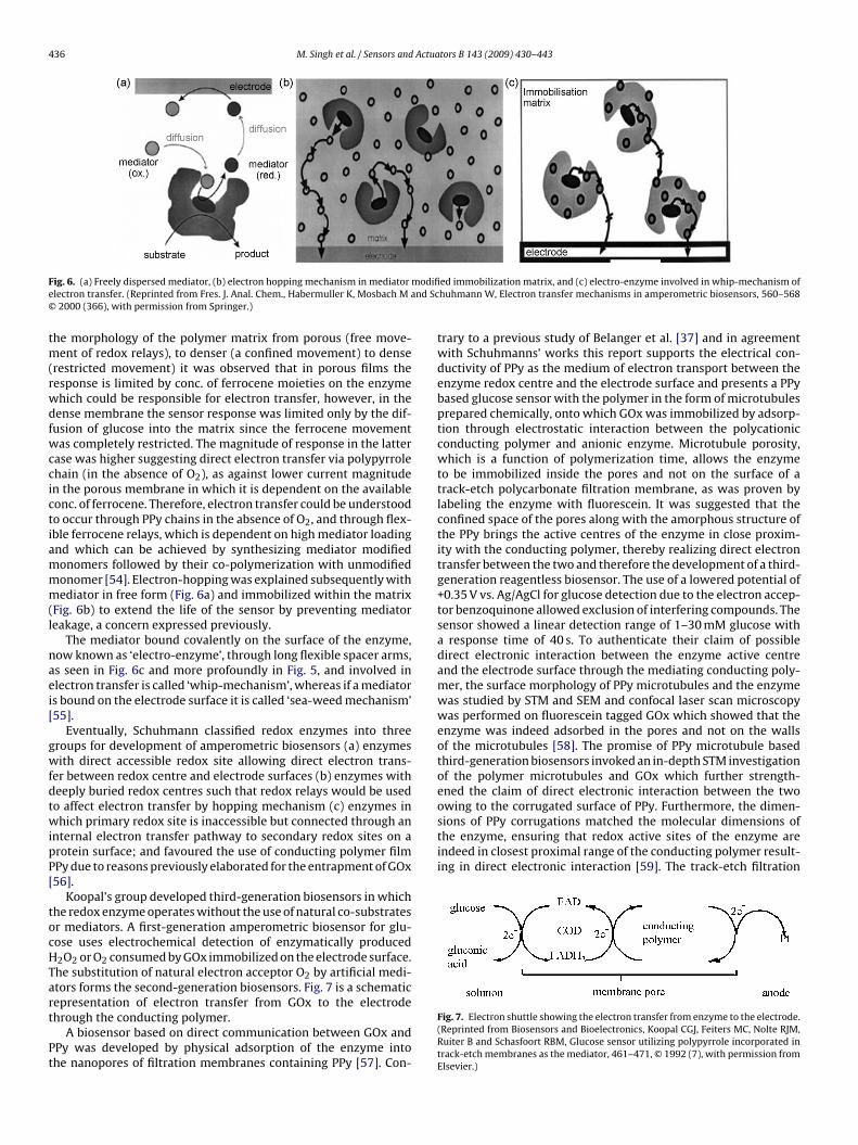
ig. 6. (a) Freely dispersed mediator, (b) electron hopping mechanism in mediatorlectron transfer. (Reprinted from Fres. J. Anal. Chem., Habermuller K, Mosbach M a2000 (366), with permission from Springer.)
he morphology of the polymer matrix from porous (free move-ent of redox relays), to denser (a confined movement) to dense
restricted movement) it was observed that in porous films theesponse is limited by conc. of ferrocene moieties on the enzymehich could be responsible for electron transfer, however, in theense membrane the sensor response was limited only by the dif-usion of glucose into the matrix since the ferrocene movementas completely restricted. The magnitude of response in the latter
ase was higher suggesting direct electron transfer via polypyrrolehain (in the absence of O2), as against lower current magnituden the porous membrane in which it is dependent on the availableonc. of ferrocene. Therefore, electron transfer could be understoodo occur through PPy chains in the absence of O2, and through flex-ble ferrocene relays, which is dependent on high mediator loadingnd which can be achieved by synthesizing mediator modifiedonomers followed by their co-polymerization with unmodifiedonomer [54]. Electron-hopping was explained subsequently withediator in free form (Fig. 6a) and immobilized within the matrix
Fig. 6b) to extend the life of the sensor by preventing mediatoreakage, a concern expressed previously.
The mediator bound covalently on the surface of the enzyme,ow known as ‘electro-enzyme’, through long flexible spacer arms,s seen in Fig. 6c and more profoundly in Fig. 5, and involved inlectron transfer is called ‘whip-mechanism’, whereas if a mediators bound on the electrode surface it is called ‘sea-weed mechanism’55].
Eventually, Schuhmann classified redox enzymes into threeroups for development of amperometric biosensors (a) enzymesith direct accessible redox site allowing direct electron trans-
er between redox centre and electrode surfaces (b) enzymes witheeply buried redox centres such that redox relays would be usedo affect electron transfer by hopping mechanism (c) enzymes inhich primary redox site is inaccessible but connected through an
nternal electron transfer pathway to secondary redox sites on arotein surface; and favoured the use of conducting polymer filmPy due to reasons previously elaborated for the entrapment of GOx56].
Koopal’s group developed third-generation biosensors in whichhe redox enzyme operates without the use of natural co-substratesr mediators. A first-generation amperometric biosensor for glu-ose uses electrochemical detection of enzymatically produced2O2 or O2 consumed by GOx immobilized on the electrode surface.he substitution of natural electron acceptor O2 by artificial medi-tors forms the second-generation biosensors. Fig. 7 is a schematic
epresentation of electron transfer from GOx to the electrodehrough the conducting polymer.A biosensor based on direct communication between GOx andPy was developed by physical adsorption of the enzyme intohe nanopores of filtration membranes containing PPy [57]. Con-
ed immobilization matrix, and (c) electro-enzyme involved in whip-mechanism ofhuhmann W, Electron transfer mechanisms in amperometric biosensors, 560–568
trary to a previous study of Belanger et al. [37] and in agreementwith Schuhmanns’ works this report supports the electrical con-ductivity of PPy as the medium of electron transport between theenzyme redox centre and the electrode surface and presents a PPybased glucose sensor with the polymer in the form of microtubulesprepared chemically, onto which GOx was immobilized by adsorp-tion through electrostatic interaction between the polycationicconducting polymer and anionic enzyme. Microtubule porosity,which is a function of polymerization time, allows the enzymeto be immobilized inside the pores and not on the surface of atrack-etch polycarbonate filtration membrane, as was proven bylabeling the enzyme with fluorescein. It was suggested that theconfined space of the pores along with the amorphous structure ofthe PPy brings the active centres of the enzyme in close proxim-ity with the conducting polymer, thereby realizing direct electrontransfer between the two and therefore the development of a third-generation reagentless biosensor. The use of a lowered potential of+0.35 V vs. Ag/AgCl for glucose detection due to the electron accep-tor benzoquinone allowed exclusion of interfering compounds. Thesensor showed a linear detection range of 1–30 mM glucose witha response time of 40 s. To authenticate their claim of possibledirect electronic interaction between the enzyme active centreand the electrode surface through the mediating conducting poly-mer, the surface morphology of PPy microtubules and the enzymewas studied by STM and SEM and confocal laser scan microscopywas performed on fluorescein tagged GOx which showed that theenzyme was indeed adsorbed in the pores and not on the wallsof the microtubules [58]. The promise of PPy microtubule basedthird-generation biosensors invoked an in-depth STM investigationof the polymer microtubules and GOx which further strength-ened the claim of direct electronic interaction between the two
Fig. 7. Electron shuttle showing the electron transfer from enzyme to the electrode.(Reprinted from Biosensors and Bioelectronics, Koopal CGJ, Feiters MC, Nolte RJM,Ruiter B and Schasfoort RBM, Glucose sensor utilizing polypyrrole incorporated intrack-etch membranes as the mediator, 461–471, © 1992 (7), with permission fromElsevier.)
Actuators B 143 (2009) 430–443 437
mF
r‘lataotlihmbblbrgAtrnc
2
toemfobiwiw−baipotoatwg−cgptIwimp(dGw
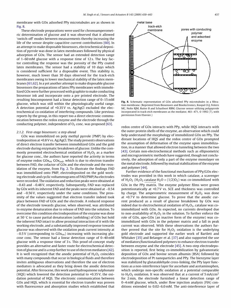
Fig. 8. Schematic representation of GOx adsorbed PPy microtubules in a filtra-tion membrane. (Reprinted from Biosensors and Bioelectronics, Koopal CGJ, Feiters
M. Singh et al. / Sensors and
embrane with GOx adsorbed PPy microtubules are as shown inig. 8.
These electrode preparations were used for chronoamperomet-ic determination of glucose and it was observed that it allowedswitch-off’ modes between measurements thereby increasing theife of the sensor despite capacitive current contributions [60]. Inn attempt to make disposable biosensors, electrochemical deposi-ion of pyrrole was done in latex membranes followed by physicaldsorption of GOx. The sensor had an extended detection rangef 1–60 mM glucose with a response time of 12 s. The key fac-or controlling the response was the porosity of the PPy coatedatex membranes. The sensor had a stability of 10 days whichs considered sufficient for a disposable event. This stability is,owever, much lower than 30 days observed for the track-etchembranes owing to lower mechanical stability of the latex mem-
ranes [61,62]. In a yet another attempt to make disposable glucoseiosensors the preparations of latex PPy membranes with immobi-
ized GOx were further processed with graphite to make conductingiosensor ink and incorporate onto a pre printed electrode. Theesulting biocomponent had a linear detection range of 0–15 mMlucose, which was still within the physiologically useful range.
detection potential of +0.35 V vs. Ag/AgCl excluded the elec-rochemical co-oxidation of interfering compounds. Like previouseports by the group, in this report too a direct electronic commu-ication between the redox enzyme and the electrode through theonducting polymer, independent of O2 conc. was proposed [63].
.1.2. First-stage biosensors: a step aheadGOx was immobilized on poly methyl pyrrole (PMP) by elec-
rodeposition at +0.8 V vs. Ag/AgCl. The study presents observationsf direct electron transfer between immobilized GOx and the goldlectrode during enzymatic breakdown of glucose. Unlike the com-only presented electrochemical oxidation of H2O2 as a marker
or glucose conc., the authors have reported the activity in termsf enzyme redox GOxox GOxred, which is due to electron transferetween FAD, the cofactor of GOx and the electrode and the reox-
dation of the enzyme, from Fig. 2. To illustrate the findings FADas immobilized onto PMP, electrodeposited on the gold work-
ng electrode and cyclic voltammograms of FAD/PMP/Au electrodesere recorded. The oxidation and reduction peaks were obtained at0.43 and −0.48 V, respectively. Subsequently, FAD was replacedy GOx with its inherent FAD and the peaks were obtained at −0.54nd −0.56 V, respectively under the same conditions. The prox-mity of the values suggested that electron transfer indeed tooklace between FAD of GOx and the electrode. A reduced responsef the electrode towards glucose, when observed, was attributedo enzyme denaturation due to release of FAD into the solution. Tovercome this condition electrodeposition of the enzyme was donet 50 ◦C to cause partial denaturation (unfolding) of GOx but holdhe inherent FAD intact to allow proximal interaction of it with theorking electrode, and a substantially improved response towards
lucose was observed with the oxidation peak current intensity at0.5 V (corresponding to GOxox) increasing with increasing glu-
ose conc. The sensor had a linear detection range of 0–40 mMlucose with a response time of 3 s. This proof-of-concept studyrovides an alternative and faster route for electrochemical detec-ion of glucose and is a step further to use of electron mediators [42].t is well recognized that the anodic potential of H2O2 coincides
ith many compounds that occur in biological fluids and thereforenvites ambiguous observations and therefore the use of electron
ediators is advocated as they bring down the anodic detection
otential. After ferrocene, this work used hydroquinone sulphonateHQS) which lowered the detection potential to +0.35 V, the oxi-ation potential of HQS. The electronic communication betweenOx and HQS, which is essential for electron transfer was provenith fluorescence and absorption studies which established thatMC, Nolte RJM, Ruiter B and Schasfoort RBM, Glucose sensor utilizing polypyrroleincorporated in track-etch membranes as the mediator, 461–471, © 1992 (7), withpermission from Elsevier.)
redox centre of GOx interacts with PPy, while HQS interacts withthe outer protein shells of the enzyme, an observation which couldhelp understand the morphology of immobilized GOx on PPy. Thedistant locations of HQS and the redox centre of GOx promptedthe assumption of deformation of the enzyme upon immobiliza-tion, in a manner that allowed electron tunneling between the two[43]. Certain non-electrochemical methods such as ellipsometricand microgravimetric methods have suggested, though not conclu-sively, the adsorption of only a part of the enzyme monolayer onthe metal electrode, followed by mutual stabilization of the enzymeand polymer [44].
Further evidence of the functional mechanism of PPy/GOx elec-trodes was provided in this work in which catalase, a scavengerof H2O2 (H2O2 catalase−−−−−→ H2O + (1/2)O2) was co-immobilized with
GOx in the PPy matrix. The enzyme polymer films were grownpotentiostatically at +0.7 V vs. SCE and thickness was controlledby charge. The amperometric measurement of H2O2 was doneat +0.7 V to determine glucose. To prove that the anodic cur-rent produced as a result of glucose breakdown by GOx wasindeed due to electrochemical oxidation of H2O2, catalase was co-immobilized with GOx. As expected, no currents developed dueto non-availability of H2O2 in the solution. To further enforce therole of GOx, apo-GOx (an inactive form of the enzyme) was co-immobilized with GOx in the polymer matrix and a fall in peakcurrent was observed. With these observations the authors fur-ther proved that the site for H2O2 oxidation is the underlyinggold electrode and supported the earlier work of Bartlett andWhitaker [35] and Belanger et al. [37] and also supported the useof mediators/functionalized polymers to enhance electron transferbetween enzyme and the electrode [45]. A two-step electrodepo-sition is reported, first being co-immobilization by galvanostaticdeposition of GOx and bovine serum albumin (BSA) followed byelectrodeposition of Pt nanoparticles and PPy. The bienzyme layerwas stabilized by glutaraldehyde cross-linking, the PPy layer func-tions as a non-interference layer for ascorbate and acetaminophen,which undergo non-specific oxidation at a potential comparable
to H2O2 oxidation. It was observed that at a current of 5 mA/cm2for 2 min, the electrode furnished a linear detection range of0–4 mM glucose, which, under flow injection analysis (FIA) con-ditions extended to 0.02–6.0 mM. The anti-interference layer of
4 Actua
Paamobsoa0erewlomantf
2b
ma
arttOtlmavpaasi[iIpprarstttwllFesioOt
38 M. Singh et al. / Sensors and
Py masked the interfering compounds. The electrodes had a stor-ge stability of 70–80 days [22]. This mentioned role of PPy, ofnti-interference layer, is in agreement with the work of Schu-ann [vide infra] who have explained the same as a ‘sieve-effect’
f PPy. Multilayered structures with adsorbed GOx sandwichedetween two poly(amphiphilic)pyrrole layers were prepared byequential polymerization. This resulted in improved sensitivitywing to high diffusion limitation with respect to the substratend an extended linearity from 2.5 mM to 5.0 mM glucose despite.35–0.40% activity retention upon immobilization [46]. Further,nzymatic layers were prepared with amphiphilic substituted pyr-ole dodecyltriethylammoniumtetrafluoroborate monomer. The Ptlectrodes were coated with a dispersion of monomer-GOx andater was removed by evacuation leading to adsorption of the
ayer on the metal surface. This was followed by electrodepositionf the dry monomer enzyme film leading to irreversible entrap-ent of the enzyme. The resulting enzyme electrodes displayed
nionic exchange properties owing to the presence of alkylammo-ium groups. This novel procedure of enzyme entrapment claimedo save enzyme and monomer since a high loading is demanded tournish substantial current peaks [25].
.2. Second-stage biosensors: polypyrrole composites in glucoseiosensors
The use of conventional PPy was replaced by composites of theatrix with conducting salts and non-conducting polymers in an
ttempt to get improved characteristics of response.Bare and sputtered Pt electrodes on a 16 channel multielectrode
rray were prepared for estimation of glucose. The experimentsevealed that FAD was responsible for glucose oxidation and thathe two processes took place – the direct electron transfer betweenhe enzyme and the electrode surface and electron transfer to2, the acceptor, therefore leading to a combination of first and
hird-generation biosensor [64]. The operating conditions control-ing the analytical performance of a PPy/GOx biosensor in FIA
ode were studied in detail which included the effect of dopingnion, phosphates, charge and flow rate. In agreement with pre-ious reports, the thickness of the polymer film was considered aarameter of immense importance and a 20 nm thick film furnishedn optimum response in terms of substrate product diffusions controlled by porosity and molecular exclusion, and polymerolution interface considerations. The sensor exhibited a stabil-ty of 50 h of measurement at a frequency of 10 measurements/h65]. The FIA system allows use of decreased sample volumes,ncreased amperometric response and increased reproducibility.n FIA mode an organic conducting salt (OCS) tetrathiafulvalene--tetracyanoquinodimethane (TTF-TCNQ) has been used in thereparation of PPy/GOx biosensor owing to its ability to reoxidizeeduced sites in FAD thereby allowing the biosensor to oper-te at lower potentials, obviating the need of additional artificialedox mediators. TTF-TCNQ is an efficient mediator in air-saturatedolutions since O2 does not compete with it as an electron accep-or. In this work OCS was mixed with silicone oil and pressedightly onto the paste electrode followed by addition of GOx andhen glutaraldehyde for cross-linking. Onto this preparation PPyas electropolymerized with the purpose of molecular exclusion
eading to OCS/GOx/PPy electrode preparations. The sensor had ainear detection range of 1–10 mM glucose with 500 injections inIA. A low operating potential of +0.15 mV along with the sieveffect of PPy offered enhanced selectivity. To further improve the
electivity characteristics a bilayer was prepared by polymeriz-ng o-phenylenediamine (o-PDA), a non-conducting polymer alsoffering exclusion properties, onto the OCS/GOx/PPy, leading toCS/GOx/PPy/o-PPDA preparations. It is worthy of mention thathe mediating property of the OCS is preserved within the mono-
tors B 143 (2009) 430–443
and bilayer. The monolayer, however, offered higher sensitivity incomparison to the bilayer due to diffusive characteristics [66]. Witha mild variant to the above work, a layer of Prussian Blue (PB)was coated onto the Pt working electrode followed by GOx–PPyelectropolymerization to form Pt/PB/GOx–PPy electrodes. PB isan electrocatalyst that allows both anodic oxidation and cathodicreduction of H2O2 and is able to diminish the overpotential of theanodic oxidation of H2O2 allowing its measurement at a lowerpotential of +0.6 V leading to exclusion of interferents, and also at acathodic potential of −0.2 V. The work, however, negated the exclu-sive property of PPy and supported the same for PPDA. The sensor,however, failed to furnish any linear dependence for cathodic oranodic currents on glucose concentrations therefore questioningthe recommendation of PB based glucose biosensors for furtherstudy [67].
A PPy/GOx glucose biosensor was prepared galvanostaticallyat 342 �A/cm2 and the effect of pH during fabrication on theresponse of the biosensor towards glucose was reported. Forstrongly acidic conditions a poor sensitivity towards glucose detec-tion was observed owing to positive charge on GOx molecules andsubstrate and product diffusion barrier due to thick film formation.Under alkaline conditions a non-conductive thin film is formed dueto preferential attachment of nucleophilic OH− over GOx leadingto minimal enzyme immobilization. In medium acidic to neutralconditions, GOx was effectively entrapped within the PPy matrixfurnishing a sensitivity of 7 nA/mM glucose making neutral pH thechoice for fabrication of the biosensor which had a linear detectionrange of 0–10 mM glucose with a response time of 15 s. A limita-tion though was the short-term stability owing to oxidation of thematrix by H2O2 and leaching of the enzyme from the matrix [68].Further immobilization of GOx in PPy/polyacrylamide microparti-cles was done and an amperometric response towards glucose inthe presence of PPy suggested that electron transfer indeed tookplace via the conducting polymer [69]. Though polypyrrole hasbeen accepted and proven to be a suitable matrix for GOx immobi-lization for multiple reasons previously mentioned, yet the concernabout lower signal outputs with PPy/GOx preparations needs atten-tion. It is observed that the current magnitude falls dramaticallyduring the process of electrochemical deposition when GOx isincorporated in the monomer solution which once again supportsthe previous observations of loss of conductivity of the polymer. Inthis study, GOx was entrapped in multi lamellar vesicles (MLV) andthereafter electrochemically deposited onto the electrode. MLVsare lipid layers that protect the enzyme during electrochemi-cal entrapment, thereby enhancing the long-term stability of thebiosensors. PPy when used with MLV showed better retentionof conductivity and supported higher current magnitudes. Sub-sequent study of the kinetic parameters of GOx–MLV–PPy filmsshowed an increased Km which was attributed to increased filmthickness. A significant feature was the enhanced linear range ofupto ca. 200 mM glucose and an improved stability of upto 20days [70,71]. Next, microfabrication of a glucose biosensor wasdone by preparing carbon post microarrays by electropolymerizingGOx and pyrrole on SU-8 photoresist carbon posts using dodecyl-benzenesulfonate as a dopant. The sensor had a linear detectionrange of 0.5–20 mM glucose with a response time of 20 s [72]. Inthe most recent development, a composite was prepared by cova-lently attaching PPy to the –COOH moieties of alginate and thenceattaching GOx to the composite. The blend showed a high sensitiv-ity of 16 �A mM−1 cm−2 and a narrow linear range of 0–3.5 mMglucose with a response time of 4 s. Futher on, a new matrix
was developed by the co-electropolymerization of pyrrole-biotinwith pyrrole-alginate. It involved the conjugation of biotinylated-GOx to biotinylated-pyrrole through avidin bridges, followed byco-polymerization with alginate-pyrrole. The conjugate had a lin-ear detection range of 0–3.5 mM glucose with a response time of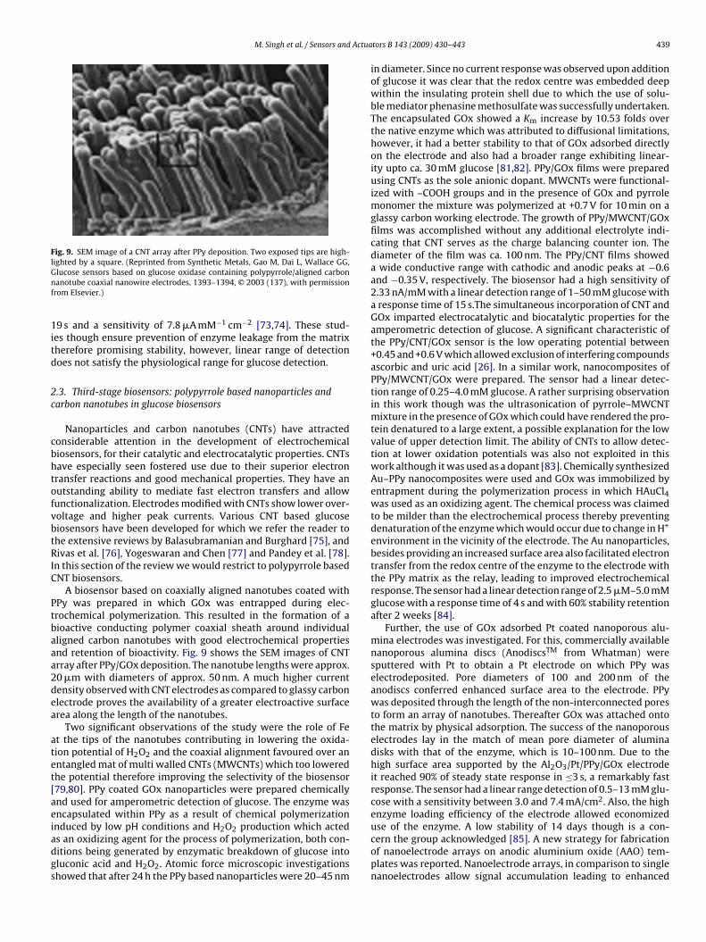
M. Singh et al. / Sensors and Actua
Fig. 9. SEM image of a CNT array after PPy deposition. Two exposed tips are high-lighted by a square. (Reprinted from Synthetic Metals, Gao M, Dai L, Wallace GG,Gnf
1itd
2c
cbhtofvbtRIC
Ptbaaa2dea
atet[aeiadgs
use of the enzyme. A low stability of 14 days though is a con-
lucose sensors based on glucose oxidase containing polypyrrole/aligned carbonanotube coaxial nanowire electrodes, 1393–1394, © 2003 (137), with permission
rom Elsevier.)
9 s and a sensitivity of 7.8 �A mM−1 cm−2 [73,74]. These stud-es though ensure prevention of enzyme leakage from the matrixherefore promising stability, however, linear range of detectionoes not satisfy the physiological range for glucose detection.
.3. Third-stage biosensors: polypyrrole based nanoparticles andarbon nanotubes in glucose biosensors
Nanoparticles and carbon nanotubes (CNTs) have attractedonsiderable attention in the development of electrochemicaliosensors, for their catalytic and electrocatalytic properties. CNTsave especially seen fostered use due to their superior electronransfer reactions and good mechanical properties. They have anutstanding ability to mediate fast electron transfers and allowunctionalization. Electrodes modified with CNTs show lower over-oltage and higher peak currents. Various CNT based glucoseiosensors have been developed for which we refer the reader tohe extensive reviews by Balasubramanian and Burghard [75], andivas et al. [76], Yogeswaran and Chen [77] and Pandey et al. [78].
n this section of the review we would restrict to polypyrrole basedNT biosensors.
A biosensor based on coaxially aligned nanotubes coated withPy was prepared in which GOx was entrapped during elec-rochemical polymerization. This resulted in the formation of aioactive conducting polymer coaxial sheath around individualligned carbon nanotubes with good electrochemical propertiesnd retention of bioactivity. Fig. 9 shows the SEM images of CNTrray after PPy/GOx deposition. The nanotube lengths were approx.0 �m with diameters of approx. 50 nm. A much higher currentensity observed with CNT electrodes as compared to glassy carbonlectrode proves the availability of a greater electroactive surfacerea along the length of the nanotubes.
Two significant observations of the study were the role of Fet the tips of the nanotubes contributing in lowering the oxida-ion potential of H2O2 and the coaxial alignment favoured over anntangled mat of multi walled CNTs (MWCNTs) which too loweredhe potential therefore improving the selectivity of the biosensor79,80]. PPy coated GOx nanoparticles were prepared chemicallynd used for amperometric detection of glucose. The enzyme wasncapsulated within PPy as a result of chemical polymerizationnduced by low pH conditions and H O production which acted
2 2s an oxidizing agent for the process of polymerization, both con-itions being generated by enzymatic breakdown of glucose intoluconic acid and H2O2. Atomic force microscopic investigationshowed that after 24 h the PPy based nanoparticles were 20–45 nmtors B 143 (2009) 430–443 439
in diameter. Since no current response was observed upon additionof glucose it was clear that the redox centre was embedded deepwithin the insulating protein shell due to which the use of solu-ble mediator phenasine methosulfate was successfully undertaken.The encapsulated GOx showed a Km increase by 10.53 folds overthe native enzyme which was attributed to diffusional limitations,however, it had a better stability to that of GOx adsorbed directlyon the electrode and also had a broader range exhibiting linear-ity upto ca. 30 mM glucose [81,82]. PPy/GOx films were preparedusing CNTs as the sole anionic dopant. MWCNTs were functional-ized with –COOH groups and in the presence of GOx and pyrrolemonomer the mixture was polymerized at +0.7 V for 10 min on aglassy carbon working electrode. The growth of PPy/MWCNT/GOxfilms was accomplished without any additional electrolyte indi-cating that CNT serves as the charge balancing counter ion. Thediameter of the film was ca. 100 nm. The PPy/CNT films showeda wide conductive range with cathodic and anodic peaks at −0.6and −0.35 V, respectively. The biosensor had a high sensitivity of2.33 nA/mM with a linear detection range of 1–50 mM glucose witha response time of 15 s.The simultaneous incorporation of CNT andGOx imparted electrocatalytic and biocatalytic properties for theamperometric detection of glucose. A significant characteristic ofthe PPy/CNT/GOx sensor is the low operating potential between+0.45 and +0.6 V which allowed exclusion of interfering compoundsascorbic and uric acid [26]. In a similar work, nanocomposites ofPPy/MWCNT/GOx were prepared. The sensor had a linear detec-tion range of 0.25–4.0 mM glucose. A rather surprising observationin this work though was the ultrasonication of pyrrole–MWCNTmixture in the presence of GOx which could have rendered the pro-tein denatured to a large extent, a possible explanation for the lowvalue of upper detection limit. The ability of CNTs to allow detec-tion at lower oxidation potentials was also not exploited in thiswork although it was used as a dopant [83]. Chemically synthesizedAu–PPy nanocomposites were used and GOx was immobilized byentrapment during the polymerization process in which HAuCl4was used as an oxidizing agent. The chemical process was claimedto be milder than the electrochemical process thereby preventingdenaturation of the enzyme which would occur due to change in H+
environment in the vicinity of the electrode. The Au nanoparticles,besides providing an increased surface area also facilitated electrontransfer from the redox centre of the enzyme to the electrode withthe PPy matrix as the relay, leading to improved electrochemicalresponse. The sensor had a linear detection range of 2.5 �M–5.0 mMglucose with a response time of 4 s and with 60% stability retentionafter 2 weeks [84].
Further, the use of GOx adsorbed Pt coated nanoporous alu-mina electrodes was investigated. For this, commercially availablenanoporous alumina discs (AnodiscsTM from Whatman) weresputtered with Pt to obtain a Pt electrode on which PPy waselectrodeposited. Pore diameters of 100 and 200 nm of theanodiscs conferred enhanced surface area to the electrode. PPywas deposited through the length of the non-interconnected poresto form an array of nanotubes. Thereafter GOx was attached ontothe matrix by physical adsorption. The success of the nanoporouselectrodes lay in the match of mean pore diameter of aluminadisks with that of the enzyme, which is 10–100 nm. Due to thehigh surface area supported by the Al2O3/Pt/PPy/GOx electrodeit reached 90% of steady state response in ≤3 s, a remarkably fastresponse. The sensor had a linear range detection of 0.5–13 mM glu-cose with a sensitivity between 3.0 and 7.4 mA/cm2. Also, the highenzyme loading efficiency of the electrode allowed economized
cern the group acknowledged [85]. A new strategy for fabricationof nanoelectrode arrays on anodic aluminium oxide (AAO) tem-plates was reported. Nanoelectrode arrays, in comparison to singlenanoelectrodes allow signal accumulation leading to enhanced
440 M. Singh et al. / Sensors and Actuators B 143 (2009) 430–443
F preparE nanof
raitaspcwPsit
crrTttwcbtiPweatwobboHd
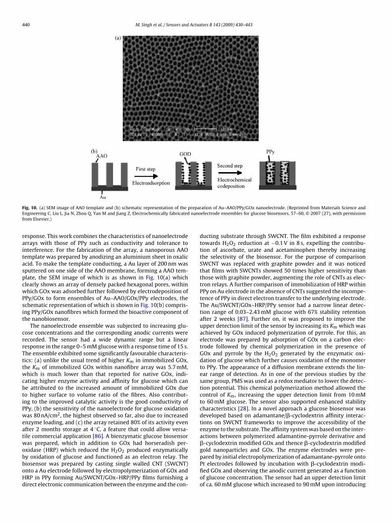
ig. 10. (a) SEM image of AAO template and (b) schematic representation of thengineering C, Liu L, Jia N, Zhou Q, Yan M and Jiang Z, Electrochemically fabricatedrom Elsevier.)
esponse. This work combines the characteristics of nanoelectroderrays with those of PPy such as conductivity and tolerance tonterference. For the fabrication of the array, a nanoporous AAOemplate was prepared by anodizing an aluminium sheet in oxaliccid. To make the template conducting, a Au layer of 200 nm wasputtered on one side of the AAO membrane, forming a AAO tem-late, the SEM image of which is as shown in Fig. 10(a) whichlearly shows an array of densely packed hexagonal pores, withinhich GOx was adsorbed further followed by electrodeposition of
Py/GOx to form ensembles of Au–AAO/GOx/PPy electrodes, thechematic representation of which is shown in Fig. 10(b) compris-ng PPy/GOx nanofibres which formed the bioactive component ofhe nanobiosensor.
The nanoelectrode ensemble was subjected to increasing glu-ose concentrations and the corresponding anodic currents wereecorded. The sensor had a wide dynamic range but a linearesponse in the range 0–5 mM glucose with a response time of 15 s.he ensemble exhibited some significantly favourable characteris-ics: (a) unlike the usual trend of higher Km in immobilized GOx,he Km of immobilized GOx within nanofibre array was 5.7 mM,hich is much lower than that reported for native GOx, indi-
ating higher enzyme activity and affinity for glucose which cane attributed to the increased amount of immobilized GOx dueo higher surface to volume ratio of the fibres. Also contribut-ng to the improved catalytic activity is the good conductivity ofPy, (b) the sensitivity of the nanoelectrode for glucose oxidationas 80 nA/cm2, the highest observed so far, also due to increased
nzyme loading, and (c) the array retained 80% of its activity evenfter 2 months storage at 4 ◦C, a feature that could allow versa-ile commercial application [86]. A bienzymatic glucose biosensoras prepared, which in addition to GOx had horseradish per-
xidase (HRP) which reduced the H2O2 produced enzymatically
y oxidation of glucose and functioned as an electron relay. Theiosensor was prepared by casting single walled CNT (SWCNT)nto a Au electrode followed by electropolymerization of GOx andRP in PPy forming Au/SWCNT/GOx–HRP/PPy films furnishing airect electronic communication between the enzyme and the con-ation of Au–AAO/PPy/GOx nanoelectrode. (Reprinted from Materials Science andelectrode ensembles for glucose biosensors, 57–60, © 2007 (27), with permission
ducting substrate through SWCNT. The film exhibited a responsetowards H2O2 reduction at −0.1 V in 8 s, expelling the contribu-tion of ascorbate, urate and acetaminophen thereby increasingthe selectivity of the biosensor. For the purpose of comparisonSWCNT was replaced with graphite powder and it was noticedthat films with SWCNTs showed 50 times higher sensitivity thanthose with graphite powder, augmenting the role of CNTs as elec-tron relays. A further comparison of immobilization of HRP withinPPy on Au electrode in the absence of CNTs suggested the incompe-tence of PPy in direct electron transfer to the underlying electrode.The Au/SWCNT/GOx–HRP/PPy sensor had a narrow linear detec-tion range of 0.03–2.43 mM glucose with 67% stability retentionafter 2 weeks [87]. Further on, it was proposed to improve theupper detection limit of the sensor by increasing its Km which wasachieved by GOx induced polymerization of pyrrole. For this, anelectrode was prepared by adsorption of GOx on a carbon elec-trode followed by chemical polymerization in the presence ofGOx and pyrrole by the H2O2 generated by the enzymatic oxi-dation of glucose which further causes oxidation of the monomerto PPy. The appearance of a diffusion membrane extends the lin-ear range of detection. As in one of the previous studies by thesame group, PMS was used as a redox mediator to lower the detec-tion potential. This chemical polymerization method allowed thecontrol of Km, increasing the upper detection limit from 10 mMto 60 mM glucose. The sensor also supported enhanced stabilitycharacteristics [28]. In a novel approach a glucose biosensor wasdeveloped based on adamantane/�-cyclodextrin affinity interac-tions on SWCNT frameworks to improve the accessibility of theenzyme to the substrate. The affinity system was based on the inter-actions between polymerized adamantine-pyrrole derivative and�-cyclodextrin modified GOx and thence �-cyclodextrin modifiedgold nanoparticles and GOx. The enzyme electrodes were pre-
pared by initial electropolymerization of adamantane-pyrrole ontoPt electrodes followed by incubation with �-cyclodextrin modi-fied GOx and observing the anodic current generated as a functionof glucose concentration. The sensor had an upper detection limitof ca. 60 mM glucose which increased to 90 mM upon introducing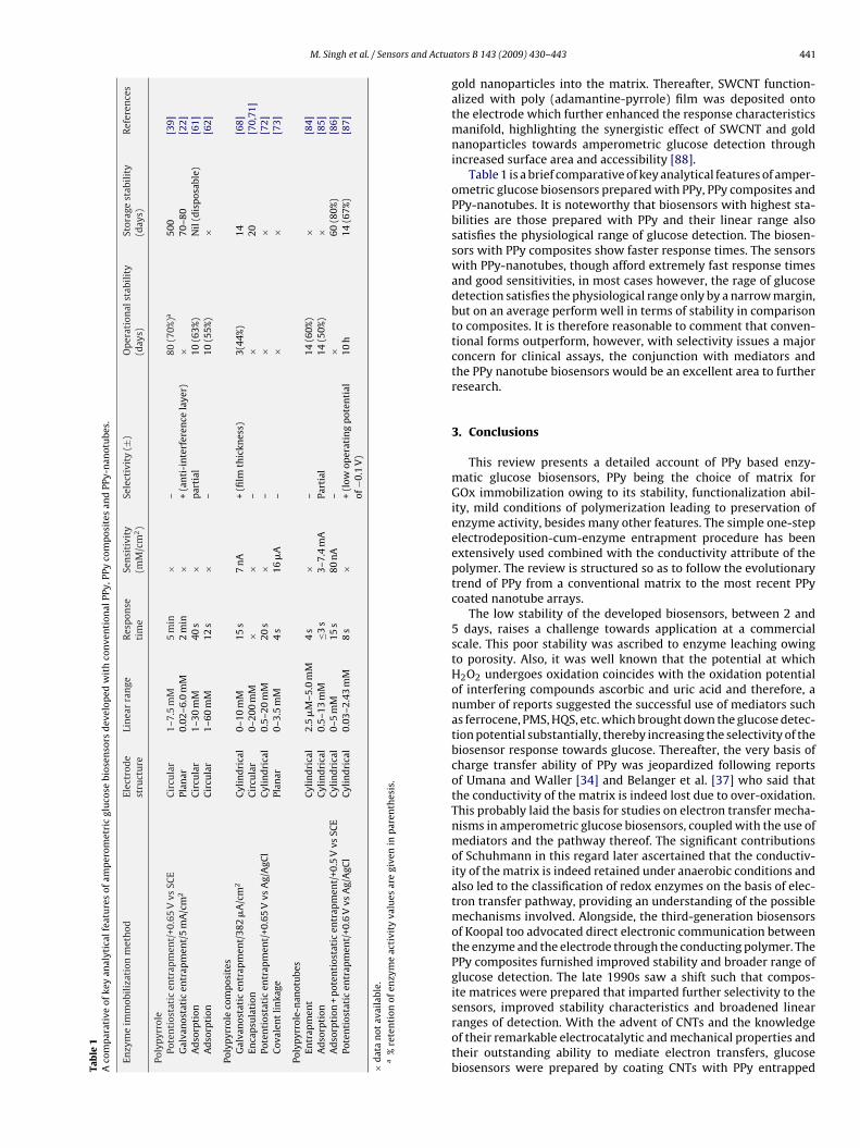
M. Singh et al. / Sensors and Actua
Tab
le1
Aco
mp
arat
ive
ofke
yan
alyt
ical
feat
ure
sof
amp
erom
etri
cgl
uco
sebi
osen
sors
dev
elop
edw
ith
con
ven
tion
alPP
y,PP
yco
mp
osit
esan
dPP
y-n
anot
ube
s.
Enzy
me
imm
obil
izat
ion
met
hod
Elec
trod
est
ruct
ure
Lin
ear
ran
geR
esp
onse
tim
eSe
nsi
tivi
ty(m
M/c
m2)
Sele
ctiv
ity
(±)
Op
erat
ion
alst
abil
ity
(day
s)St
orag
est
abil
ity
(day
s)R
efer
ence
s
Poly
pyr
role
Pote
nti
osta
tic
entr
apm
ent/
+0.6
5V
vsSC
EC
ircu
lar
1–7.
5m
M5
min
×–
80(7
0%)a
500
[39]
Gal
van
osta
tic
entr
apm
ent/
5m
A/c
m2
Plan
ar0.
02–6
.0m
M2
min
×+
(an
ti-i
nte
rfer
ence
laye
r)×
70–8
0[2
2]A
dso
rpti
onC
ircu
lar
1–30
mM
40s
×p
arti
al10
(63%
)N
il(d
isp
osab
le)
[61]
Ad
sorp
tion
Cir
cula
r1–
60m
M12
s×
–10
(55%
)×
[62]
Poly
pyr
role
com
pos
ites
Gal
van
osta
tic
entr
apm
ent/
382
�A
/cm
2C
ylin
dri
cal
0–10
mM
15s
7n
A+
(film
thic
knes
s)3(
44%
)14
[68]
Enca
psu
lati
onC
ircu
lar
0–20
0m
M×
×–
×20
[70,
71]
Pote
nti
osta
tic
entr
apm
ent/
+0.6
5V
vsA
g/A
gCl
Cyl
ind
rica
l0.
5–20
mM
20s
×–
××
[72]
Cov
alen
tli
nka
gePl
anar
0–3.
5m
M4
s16
�A
–×
×[7
3]
Poly
pyr
role
-nan
otu
bes
Entr
apm
ent
Cyl
ind
rica
l2.
5�
M–5
.0m
M4
s×
–14
(60%
)×
[84]
Ad
sorp
tion
Cyl
ind
rica
l0.
5–13
mM
≤3s
3–7.
4m
APa
rtia
l14
(50%
)×
[85]
Ad
sorp
tion
+p
oten
tios
tati
cen
trap
men
t/+0
.5V
vsSC
EC
ylin
dri
cal
0–5
mM
15s
80n
A–
×60
(80%
)[8
6]Po
ten
tios
tati
cen
trap
men
t/+0
.6V
vsA
g/A
gCl
Cyl
ind
rica
l0.
03–2
.43
mM
8s
×+
(low
oper
atin
gp
oten
tial
of−0
.1V
)10
h14
(67%
)[8
7]
×d
ata
not
avai
labl
e.a
%re
ten
tion
ofen
zym
eac
tivi
tyva
lues
are
give
nin
par
enth
esis
.
tors B 143 (2009) 430–443 441
gold nanoparticles into the matrix. Thereafter, SWCNT function-alized with poly (adamantine-pyrrole) film was deposited ontothe electrode which further enhanced the response characteristicsmanifold, highlighting the synergistic effect of SWCNT and goldnanoparticles towards amperometric glucose detection throughincreased surface area and accessibility [88].
Table 1 is a brief comparative of key analytical features of amper-ometric glucose biosensors prepared with PPy, PPy composites andPPy-nanotubes. It is noteworthy that biosensors with highest sta-bilities are those prepared with PPy and their linear range alsosatisfies the physiological range of glucose detection. The biosen-sors with PPy composites show faster response times. The sensorswith PPy-nanotubes, though afford extremely fast response timesand good sensitivities, in most cases however, the rage of glucosedetection satisfies the physiological range only by a narrow margin,but on an average perform well in terms of stability in comparisonto composites. It is therefore reasonable to comment that conven-tional forms outperform, however, with selectivity issues a majorconcern for clinical assays, the conjunction with mediators andthe PPy nanotube biosensors would be an excellent area to furtherresearch.
3. Conclusions
This review presents a detailed account of PPy based enzy-matic glucose biosensors, PPy being the choice of matrix forGOx immobilization owing to its stability, functionalization abil-ity, mild conditions of polymerization leading to preservation ofenzyme activity, besides many other features. The simple one-stepelectrodeposition-cum-enzyme entrapment procedure has beenextensively used combined with the conductivity attribute of thepolymer. The review is structured so as to follow the evolutionarytrend of PPy from a conventional matrix to the most recent PPycoated nanotube arrays.
The low stability of the developed biosensors, between 2 and5 days, raises a challenge towards application at a commercialscale. This poor stability was ascribed to enzyme leaching owingto porosity. Also, it was well known that the potential at whichH2O2 undergoes oxidation coincides with the oxidation potentialof interfering compounds ascorbic and uric acid and therefore, anumber of reports suggested the successful use of mediators suchas ferrocene, PMS, HQS, etc. which brought down the glucose detec-tion potential substantially, thereby increasing the selectivity of thebiosensor response towards glucose. Thereafter, the very basis ofcharge transfer ability of PPy was jeopardized following reportsof Umana and Waller [34] and Belanger et al. [37] who said thatthe conductivity of the matrix is indeed lost due to over-oxidation.This probably laid the basis for studies on electron transfer mecha-nisms in amperometric glucose biosensors, coupled with the use ofmediators and the pathway thereof. The significant contributionsof Schuhmann in this regard later ascertained that the conductiv-ity of the matrix is indeed retained under anaerobic conditions andalso led to the classification of redox enzymes on the basis of elec-tron transfer pathway, providing an understanding of the possiblemechanisms involved. Alongside, the third-generation biosensorsof Koopal too advocated direct electronic communication betweenthe enzyme and the electrode through the conducting polymer. ThePPy composites furnished improved stability and broader range ofglucose detection. The late 1990s saw a shift such that compos-ite matrices were prepared that imparted further selectivity to the
sensors, improved stability characteristics and broadened linearranges of detection. With the advent of CNTs and the knowledgeof their remarkable electrocatalytic and mechanical properties andtheir outstanding ability to mediate electron transfers, glucosebiosensors were prepared by coating CNTs with PPy entrapped
4 Actua
Gtaewe
aoo
A
GaE
R
[
[
[
[
[
[[
[
[
[
[
[
[
[
[
[
[
[
[
[
[
[
[
[
[
[
[
[
[
[
[
[
[
[
[
[
[
[
[
[
[
[
[
[
[
[
[
[
42 M. Singh et al. / Sensors and
Ox, enhancing the function of PPy. The highly porous nanostruc-ured frameworks of CNTs led to much higher current densitiesnd improved sensitivities, also at lower oxidation potentials withxtremely fast response times. A further enhancement of responseas observed with nanoelectrode arrays ascribed to accumulative
ffects.The extensive work done towards the development of stable
nd reliable glucose biosensors assures addressing the vital issuef diabetes management and contributing immensely to a sectionf the world population suffering from the disease.
cknowledgements
The authors are grateful to the Department of Biotechnology,ovt. of India for funding the work related to development ofmperometric glucose biosensors, the authors and publishers oflsevier and Springer for granting permission to reproduce figures.
eferences
[1] M. Gronow, Biosensors, Trends Biochem. Sci. 9 (1984) 336–340.[2] J.H.T. Luong, A. Mulchandani, G.G. Guilbault, Developments and applications of
biosensors, Trends Biotechnol. 6 (1988) 310–316.[3] M. Singh, N. Verma, A.K. Garg, N. Redhu, Urea biosensors, Sens. Actuators B 134
(2008) 345–351.[4] S.F. D’Souza, Microbial biosensors, Biosens. Bioelectron. 16 (2001) 337–353.[5] J.C. Pickup, F. Hussain, N.D. Evans, N. Sachedina, In vivo glucose monitoring: the
clinical reality and the promise, Biosens. Bioelectron. 20 (2005) 1897–1902.[6] J.D. Newman, A.P.F. Turner, Home blood glucose monitors: a commercial per-
spective, Biosens. Bioelectron. 20 (2005) 2435–2453.[7] H.E. Koschwanez, W.M. Reichert, In vitro, in vivo and post explantation test-
ing of glucose detecting biosensors: Current methods and recommendations,Biomaterials 28 (2007) 3687–3703.
[8] N. Verma, M. Singh, A Bacillus sphaericus based biosensor for monitoring nickelions in industrial effluents and foodstuffs, J. Automat. Meth. Manage. Chem.(2006) 1–4.
[9] N. Verma, M. Singh, Development of a yeast biosensor for monitoring mercuricions in industrial effluents, Int. J. Environ. Study 62 (1) (2005) 3–13.
10] N. Verma, M. Singh, A disposable microbial based biosensor for quality controlin milk, Biosens. Bioelectron. 18 (2003) 1219–1224.
11] M. Piliarik, L. Parova, J. Homela, High throughput SPR sensor for food safety,Biosens. Bioelectron. 24 (2009) 1399–1404.
12] M. Valach, J. Katrlik, E. Sturdik, P. Gemainer, Ethanol Gluconobacter biosensordesigned for flow injection analysis: application in ethanol fermentation off-line monitoring, Sens. Actuators B 138 (2009) 581–586.
13] W. Shen, R.S. Lakshmanan, L.C. Mathison, V.A. Petrenko, B.A. Chin, Phage coatedmagnetoelastic micro-biosensors for real time detection of Bacillus anthracisspores, Sens. Actuators B 137 (2009) 501–506.
14] A.T. Nirmal, U. Mittal, M. Singh, M. Khaneja, G.K. Kannan, J.C. Kapoor, V. Dubey,P.K. Gutch, G. Lal, K.D. Vyas, D.C. Gupta, Development of handheld SAW vaporsensors for explosives and CW agents, Sens. Actuators B 135 (2) (2009) 399–410.
15] http://www.who.int/topics/diabetes mellitus/en/2008.16] B.H. Ginsberg, An overview of minimally invasive technologies, Clin. Chem. 38
(9) (1992) 1596–1600.17] L.C. Clark Jr., C. Lyons, Electrode systems for continuous monitoring in cardio-
vascular surgery, Ann. NY Acad. Sci. 102 (1962) 29–45.18] J. Wang, Glucose biosensors: 40 years of advances and challenges, Electroanal-
ysis 13 (12) (2001) 983–988.19] W. Schuhmann, Conducting polymer based amperometric enzyme electrodes,
Mikrochim. Acta 121 (1995) 1–29.20] M. Gerard, A. Chaubey, B.D. Malhotra, Application of conducting polymers to
biosensors, Biosens. Bioelectron. 17 (2002) 345–359.21] W. Schuhmann, R. Lammert, M. Hammerle, H.L. Schmidt, Electrocatalytic prop-
erties of polypyrrole in amperometric electrodes, Biosens. Bioelectron. 6 (1991)689–697.
22] D.J. Strike, N.F. Rooij, M.K. Hep, Electrodeposition of glucose oxidase for thefabrication of miniature sensors, Sens. Actuators B 13–14 (1993) 61–64.
23] P.N. Bartlett, P.R. Birkin, The application of conducting polymers in biosensors,Syn. Met. 61 (1993) 15–21.
24] S. Cosnier, Biomolecule immobilization on electrode surfaces by entrapmentor attachment to electrochemically polymerized films. A review, Biosens. Bio-electron. 14 (1999) 443–456.
25] L.C. Guerente, S. Cosnier, C. Innocent, P. Mailley, J.C. Moutet, R.M. Morelis, B.
Leca, P.R. Coulet, Controlled electrochemical preparation of enzymatic layersfor the design of amperometric biosensors, Electroanalysis 5 (1993) 647–652.26] J. Wang, M. Musameh, Carbon nanotubes doped polypyrrole glucose biosensor,Anal. Chim. Acta 539 (2005) 209–213.
27] S. Sadki, P. Schottland, N. Brodie, G. Sabouraud, The mechanisms of pyrroleelectropolymerization, Chem. Soc. Rev. 29 (2000) 283–293.
[
[
tors B 143 (2009) 430–443
28] A. Ramanavicius, A. Kausaite, A. Ramanaviciene, Self encapsulation of oxidasesas a basic approach to tune the upper detection limit of amperometric biosen-sors, Analyst 133 (2008) 1083–1089.
29] A.F. Diaz, J.I. Castillo, J.A. Logan, W.Y. Lee, Electrochemistry of conducting poly-mer films, J. Electroanal. Chem. 129 (1981) 115–132.
30] A. Asavapiriyanont, G.K. Chandler, G.A. Gunawardena, D. Pletcher, The elec-trodeposition of polypyrrole films from aqueous solutions, J. Electroanal. Chem.177 (1984) 229–244.
31] N.C. Foulds, C.R. Lowe, enzyme entrapment in electrically conducting polymers,J. Chem. Soc., Faraday Trans. 82 (1) (1986) 1259–1264.
32] W. Schuhmann, C. Kranz, H. Wohlschlager, J. Strohmeier, Pulse technique forthe electrochemical deposition of polymer films on electrode surfaces, Biosens.Bioelectron. 12 (1997) 1157–1167.
33] N.C. Foulds, C.R. Lowe, Immobilization of glucose oxidase in ferrocene modifiedpyrrole polymers, Anal. Chem. 60 (1988) 2473–2478.
34] M. Umana, J. Waller, Protein modified electrodes. The glucose oxi-dase/polypyrrole system, Anal. Chem. 58 (1986) 2979–2983.
35] P.N. Bartlett, R.G. Whitaker, Electrochemical immobilization of enzymes PartII. Glucose oxidase immobilized in poly-N-methylpyrrole, J. Electroanal. Chem.224 (1987) 37–48.
36] J. Kotowski, T. Janas, H.T. Tien, Immobilization of glucose oxidase on apolypyrrole-lecithin bilayer lipid membrane, Bioelectron. Bioenerg. 19 (1988)277–282.
37] D. Belanger, J. Nadreau, G. Fortier, Electrochemistry of the polypyrrole glucoseoxidase electrode, J. Electroanal. Chem. 274 (1989) 143–155.
38] S. Holdcroft, B.L. Funt, Preparation and electrocatalytic properties of conduct-ing films of polypyrrole containing platinum microparticulates, J. Electroanal.Chem. 240 (1988) 89–103.
39] G. Fortier, E. Brassard, D. Belanger, Optimization of a polypyrrole glucose oxi-dase biosensor, Biosens. Bioelectron. 5 (1990) 473–490.
40] D. Belanger, E. Brassard, G. Fortier, Enhancement of response by incorporationof platinum microparticles into a polypyrrole glucose oxidase electrode, Anal.Chim. Acta 228 (1990) 311–355.
41] S. Yabuki, H. Shinohara, M. Aizawa, Electro-conductive enzyme membrane, J.Chem. Soc. Chem. Commun. (1989) 945–946.
42] P.T. Poet, S. Miyamoto, T. Murakami, J. Kimura, Direct electron transfer withglucose oxidase immobilized in an electropolymerized poly-N-methylpyrrolefilm on a gold microelectrode, Anal. Chim. Acta 235 (1990) 255–263.
43] Y. Kajiya, H. Sugai, C. Iwakura, H. Yoneyama, Glucose sensitivity of polypyrrolefilms containing immobilized glucose oxidase and hydroquinonesulfonate ions,Anal. Chem. 63 (1991) 49–54.
44] J. Rishpon, S. Gottesfield, Investigation of polypyrrole/glucose oxidase elec-trodes by ellipsometric, microgravimetric and electrochemical measurements,Biosens. Bioelectron. 6 (1991) 143–149.
45] J.M. Cooper, D. Bloor, Evidence for the functional mechanism of a polypyrroleglucose oxidase electrode, Electroanalysis 5 (1993) 883–886.
46] L.C. Guerente, A. Deronzier, P. Mailley, J.C. Moutet, Electrochemical immobiliza-tion of glucose oxidase in poly(amphiphilic pyrrole) films and its applicationto the preparation of an amperometric glucose biosensor, Anal. Chim. Acta 289(1994) 143–153.
47] W. Schuhmann, Functionalized polypyrrole. A new material for the construc-tion of biosensors, Syn. Met. 41–43 (1991) 429–432.
48] W. Schuhmann, R. Lammert, B. Uhe, H.L. Schmidt, Polypyrrole, a new possibilityfor covalent bindin of oxidoreductases to electrode surfaces as a base for stablebiosensors, Sens. Actuators B 1 (1990) 537–541.
49] W. Schuhmann, H. Wohlschlager, R. Lammert, H.L. Schmidt, Leaching ofdimethylferrocene, a redox mediator in amperometric enzyme electrodes,Sens. Actuators B 1 (1990) 571–575.
50] W. Schuhmann, R. Lammert, M. Hammerle, H.L. Scmidt, Electrocatalytic prop-erties of polypyrrole in amperometric electrodes, Biosens. Bioelectron. 6 (1991)689–697.
51] W. Schuhmann, R.K. Eberle, Evaluation of polypyrrole/glucose oxidaseelectrodes in flow injection systems for sucrose determination, Biosens. Bio-electron. 6 (1991) 263–273.
52] M. Hammerle, W. Schuhmann, H.L. Schmidt, Amperometric polypyrroleenzyme electrodes: effect of permeability and enzyme location, Sens. ActuatorsB 6 (1992) 106–112.
53] W. Schuhmann, C. Kranz, J. Huber, H. Wohlschlager, Conducting polymer basedamperometric enzyme electrodes. Towards the development of miniaturizedreagentless biosensors, Syn. Met. 61 (1993) 31–35.
54] W. Schuhmann, Electron transfer pathways in amperometric biosensors. Fer-rocene modified enzymes entrapped in conducting polymer layers, Biosens.Bioelectron. 10 (1995) 181–193.
55] K. Habermuller, M. Mosbach, W. Schuhmann, Electron transfer mechanisms inamperometric biosensors, Fres. J. Anal. Chem. 366 (2000) 560–568.
56] W. Schuhmann, Amperometric enzyme biosensors based on optimized elec-tron transfer pathways and non manual immobilization procedures, Rev. Mol.Biotechnol. 82 (2002) 425–441.
57] C.G.J. Koopal, M.C. Feiters, R.J.M. Nolte, B. Ruiter, R.B.M. Schasfoort, Glucosesensor utilizing polypyrrole incorporated in trach etch membranes as the medi-
ator, Biosens. Bioelectron. 7 (1992) 461–471.58] C.G.J. Koopal, M.C. Feiters, R.J.M. Nolte, B. Ruiter, R.B.M. Schasfoort, R. Czajka,H.V. Kempen, Polypyrrole microtubules and their use in the construction of athird generation biosensor, Syn. Met. 51 (1992) 397–405.
59] R. Czajka, C.G.J. Koopal, M.C. Feoters, J.W. Gerritsen, R.J.M. Nolte, H.V. Kem-pen, Scanning tunneling microscopy study of polypyrrole films and of glucose

Actua
[
[
[
[
[
[
[
[
[
[
[
[
[
[
[
[
[
[
[
[
[
[
[
[
[
[
[
[
[
Nagaraju Jampana obtained his M.Sc. (Tech.) degree from Andhra University,
M. Singh et al. / Sensors and
oxidase as used in a third generation biosensor, Bioelectrochem. Bioenerg. 29(1992) 47–57.
60] C.G.J. Koopal, B. Eijsma, R.J.M. Nolte, Chronoamperometric detection of glucoseby a third generation biosensor constructed from conducting microtubules ofpolypyrrole, Syn. Met. 55–57 (1993) 3689–3695.
61] C.G.J. Koopal, M.C. Feiters, R.J.M. Nolte, B. Ruiter, R.B.M. Schasfoort, Third gen-eration amperometric biosensor for glucose. Polypyrrole deposited within amatrix of uniform latex particles as mediator, Bioelectrochem. Bioenerg. 29(1992) 159–175.
62] C.G.J. Koopal, R.J.M. Nolte, Kinetic study of the performance of third generationbiosensors, Bioelectrochem. Bioenerg. 33 (1994) 45–53.
63] C.G.J. Koopal, A.A.C.M. Bos, R.J.M. Nolte, Third generation glucose biosensorincorporated in a conducting printing ink, Sens. Actuators B 18–19 (1994)166–170.
64] P.J.H.J. Os, A. Bult, C.G.J. Koopal, W.P. Bennekom, Glucose detection at bare andsputtered platinum electrodes coated with polypyrrole and glucose oxidase,Anal. Chim. Acta 335 (1996) 209–216.
65] J.C. Vidal, E. Garcia, J.R. Castillo, Electropolymerization of pyrrole and immo-bilization of glucose oxidase in a flow system: influence of the operatingconditions on analytical performance, Biosens. Bioelectron. 13 (1998) 371–382.
66] J.C. Vidal, S. Mendez, J.R. Castillo, Electropolymerization of pyrrole andphenylenediamine over an organic conducting salt based amperometric sensorof increased selectivity for glucose determination, Anal. Chim. Acta 385 (1999)203–211.
67] R. Garjonyte, A. Malinauskas, Glucose biosensor based on glucoseoxidase immobilized in electropolymerized polypyrrole and poly (o-phenylenediamine) films on a Prussian blue modified electrode, Sens.Actuators B 63 (2000) 122–128.
68] Y.M. Uang, T.C. Chou, Fabrication of glucose oxidase/polypyrrole biosensor bygalvanostatic method in various pH aqueous solutions, Biosens. Bioelectron. 19(2003) 141–147.
69] J.R. Retama, D. Mecerreyes, B.L. Ruiz, E.L. Cabarcos, Synthesis and characteriza-tion of semiconducting polypyrrole/polyacrylamide microparticles with GOxfor biosensor applications, Coll. Surf. A: Physiochem. Eng. Aspects 270–271(2005) 239–244.
70] D. Olea, P. Moreau, C. Faure, Polypyrrole glucose oxidase biosensor. Effect ofenzyme encapsulation in multilamellar vesicles on film growth and morphol-ogy, J. Electroanal. Chem. 605 (2007) 125–135.
71] D. Olea, O. Viratelle, C. Faure, Polypyrrole glucose oxidase biosensor. Effectof enzyme encapsulation in multilamellar vesicles on analytical properties,Biosens. Bioelectron. 23 (2008) 788–794.
72] H. Xu, K. Malladi, C. Wang, L. Kulinsky, M. Song, M. Madou, Carbon post microar-rays for glucose sensors, Biosens. Bioelectron. 23 (2008) 1637–1644.
73] K.A. Rabeah, R.S. Marks, Impedance study of the hybrid molecule alginate-pyrrole: demonstration as host matrix for the construction of a highlysensitive amperometric glucose biosensor, Sens. Actuators 136 (2009) 516–522.
74] I.I. Nita, K.A. Rabeah, A.M. Tencaliec, S. Cosnier, R.S. Marks, Amperometricbiosensor based on the electropolymerization of a conductive biotinylated-pyrrole and alginate-pyrrole, Syn. Met. 159 (2009) 1117–1122.
75] K. Balasubramanian, M. Burghard, Biosensors based on carbon nanotubes, Anal.Bioanal. Chem. 385 (2006) 452–468.
76] G. Rivas, M.D. Rubianes, M.C. Rodriguez, N.F. Ferreyra, G.L. Luque, M.L. Pedano,S.A. Miscoria, C. Parrado, Carbon nanotubes for electrochemical biosensing,Talanta 74 (2007) 291–307.
77] U. Yogeswaran, S.M. Chen, Recent trends in the application of carbonnanotubes-polymer composite modified electrodes for biosensors. A review,Anal. Lett. 41-2 (2008) 210–243.
tors B 143 (2009) 430–443 443
78] P. Pandey, M. Datta, B.D. Malhotra, Prospects of nanomaterials in biosensors,Anal. Lett. 41 (2008) 159–209.
79] M. Gao, L. Dai, G.G. Wallace, Glucose sensors based on glucose oxidase con-taining polypyrrole/aligned carbon nanotube coaxial nanowire electrodes, Syn.Met. 137 (2003) 1393–1394.
80] M. Gao, L. Dai, G.G. Wallace, Biosensors based on aligned carbon nan-otubes coated with inherently conducting polymers, Electroanalysis 15 (2003)1089–1094.
81] A. Ramanavicius, A. Kausaite, A. Ramanaviciene, Polypyrrole coated glucoseoxidase nanoparticles for biosensor design, Sens. Actuators B 111–112 (2005)532–539.
82] A. Ramanavicius, A. Kausaite, A. Ramanaviciene, J. Acaite, A. Malinauskas, Redoxenzyme glucose oxidase initiated synthesis of polypyrrole, Syn. Met. 156 (2006)409–413.
83] Y.C. Tsai, S.C. Li, S.W. Liao, Electrodeposition of polypyrrole multiwalled carbonnanotube glucose oxidase nanobiocomposite film for the detection of glucose,Biosens. Bioelectron. 22 (2006) 495–500.
84] J. Njagi, S. Andreescu, Stable enzyme biosensors based on chemically synthe-sized Au polypyrrole nanocomposites, Biosens. Bioelectron. 23 (2007) 168–175.
85] E.M.I.M. Ekanayake, D.M.G. Preethichandra, K. Kaneto, Polypyrrole nanotubearray sensor for enhanced adsorption of glucose oxidase in glucose biosensors,Biosens. Bioelectron. 23 (2007) 107–113.
86] L. Liu, N. Jia, Q. Zhou, M. Yan, Z. Jiang, Electrochemically fabricated nanoelec-trode ensembles for glucose biosensors, Mater. Sci. Eng. C 27 (2007) 57–60.
87] L. Zhu, R. Yang, J. Zhai, C. Tian, Bienzymatic glucose biosensor based on co-immobilization of peroxidase and glucose oxidase on a carbon nanotubeselectrode, Biosens. Bioelectron. 23 (2007) 528–535.
88] M. Holzinger, L. Bouffier, R. Villalonga, S. Cosnier, Adamantane/�-cyclodextrinaffinity biosensors based on single walled carbon nanotubes, Biosens. Bioelec-tron. 24 (2009) 1128–1134.
Biographies
Minni Singh obtained her Ph.D. degree in Biotechnology from Punjabi University,Patiala in 2003, where she worked as faculty till 2008 before joining as a post doctoralfellow at the Department of Instrumentation, Indian Institute of Science. She hasworked on biosensors for 9 years and holds an Indian patent on copper biosensor,as co-inventor. She also works on urea biosensors and has filed a patent for thesame. Her research interests include electrochemical biosensors, both enzyme andmicrobial based with application towards environment, clinical and food safety.
Pavan Kumar Kathuroju obtained his B.Tech. degree in Electronics and Instrumen-tation Engineering from Jawahar Lal Nehru Technological University, Hyderabad,India and Master of Science (Engineering) degree from Indian Institute of Science,Bangalore, India. He is currently pursuing Ph.D. at the Indian Institute of Science.His current research interests include Electrochemical Sensors, Organic Electron-ics, Analog electronic design for biomedical and biosensing applications, design anddevelopment of non-invasive sensing systems for analyte monitoring in body fluidsof human subjects.
Vishakhapatnam, India. He received his M.Phil. and Ph.D. degrees from NagarjunaUniversity, India. At present he is working as associate professor at Indian Insti-tute of Science Bangalore, India. His research interests include solar thermal andphotovoltaics, electrical and thermal contact resistance studies, instrumentationfor energy conservation, impedance spectroscopy.


