Embed Size (px)
Citation preview

RESEARCH ARTICLE
Hypo- and Hypermorphic FOXC1Mutations inDominant Glaucoma: Transactivation andPhenotypic VariabilityCristina Medina-Trillo1,2, Francisco Sánchez-Sánchez1,2, José-Daniel Aroca-Aguilar1,2,Jesús-José Ferre-Fernández1, Laura Morales3, Carmen-Dora Méndez-Hernández3,Fiona Blanco-Kelly4, Carmen Ayuso4, Julián García-Feijoo3, Julio Escribano1,2*
1 Área de Genética, Facultad de Medicina, Universidad de Castilla-La Mancha, Albacete, Spain; Instituto deInvestigación en Discapacidades Neurológicas (IDINE), Universidad de Castilla-La Mancha, Albacete,Spain, 2 Cooperative Research Network on Age-Related Ocular Pathology, Visual and Life Quality, Institutode Salud Carlos III, Madrid, Spain, 3 Servicio de Oftalmología, Hospital Clínico San Carlos, Madrid, Spain;Instituto de Investigación Sanitaria del Hospital Clínico San Carlos, Madrid, Spain, 4 Servicio de Genética,Instituto de Investigación Sanitaria-Fundación Jiménez Díaz (IIS-FJD), Madrid, Spain; Centro deInvestigación Biomédica en Red de Enfermedades Raras (CIBERER), Madrid, Spain
AbstractDominant glaucoma, a heterogeneous, infrequent and irreversible optic neuropathy, is
often associated with elevated intraocular pressure and early-onset. The role of FOXC1 in
this type of glaucoma was investigated in twelve Spanish probands via nucleotide variation
screening of its proximal promoter and unique exon. Functional evaluations of the identified
variants included analyses of the transcriptional activity, protein stability, DNA binding ability
and subcellular localization. Four different mutations that were identified in four probands
(33.3%) were associated with remarkable phenotypic variability and were functionally clas-
sified as either hypermorphic (p.Y47X, p.Q106X and p.G447_G448insDG) or hypomorphic
(p.I126S) alleles. To the best of our knowledge, three of the variants are novel (p.Y47X, p.
I126S and p.G447_G448insDG) and, in addition, hypermorphic FOXC1mutations are re-
ported herein for the first time. The presence of an intact N-terminal activation domain in the
truncated proteins p.Y47X and p.Q106X may underlie their associated transactivation hy-
peractivity by a gain-of-function mechanism involving dysregulated protein-protein interac-
tions. Similarly, altered molecular interactions may also lead to increased p.G447_G448insDG
activity. In contrast, the partial loss-of-function associated with p.I126S was due to im-
paired protein stability, DNA binding, protein phosphorylation and subcellular distribu-
tion. These results support that moderate and variable FOXC1 transactivation changes
are associated with moderate goniodysgenesis, dominant glaucoma and remarkable
phenotypic variability.
PLOS ONE | DOI:10.1371/journal.pone.0119272 March 18, 2015 1 / 22
OPEN ACCESS
Citation: Medina-Trillo C, Sánchez-Sánchez F,Aroca-Aguilar J-D, Ferre-Fernández J-J, Morales L,Méndez-Hernández C-D, et al. (2015) Hypo- andHypermorphic FOXC1 Mutations in DominantGlaucoma: Transactivation and Phenotypic Variability.PLoS ONE 10(3): e0119272. doi:10.1371/journal.pone.0119272
Academic Editor: Tao Cai, NIDCR/NIH, UNITEDSTATES
Received: September 25, 2014
Accepted: January 12, 2015
Published: March 18, 2015
Copyright: © 2015 Medina-Trillo et al. This is anopen access article distributed under the terms of theCreative Commons Attribution License, which permitsunrestricted use, distribution, and reproduction in anymedium, provided the original author and source arecredited.
Data Availability Statement: All relevant data arewithin the paper and its Supporting Information files.
Funding: This study has been supported byresearch grants from the Regional Ministry of Health(GCS-2006_C/12), the Regional Ministry of Scienceand Technology of the Board of the Communities of“Castilla-La Mancha” (PAI-05-002 and PCI08-0036),and the “Instituto de Salud Carlos III” (RD07/0062/0014, RD12/0034/0003 and PI11/00662). Thefunders had no role in study design, data collection
IntroductionGlaucoma is a highly heterogeneous, irreversible and progressive blinding disease produced bythe death of the retinal ganglion cells, which results in the degeneration of the optic nerve.Raised intraocular pressure (IOP) is the primary risk factor for developing glaucoma. Mostglaucoma cases behave as a late-onset complex disease and a minority of cases show an early-onset and follow simple Mendelian inheritance. Dominant transmission is typically observedin juvenile open-angle glaucoma (JOAG; MIM#137750), as well as in some cases of both adult-onset primary open-angle glaucoma (POAG; MIIM# 137760) and primary congenital glauco-ma (PCG; MIM# 231300) [1, 2]. PCG is caused by developmental abnormalities in the anteriorsegment of the eye, which is required for aqueous humour drainage, and manifests clinically inthe neonatal or infantile period, generally before the age of 3. Moreover, late congenital glauco-ma cases (LCG, i.e., glaucoma diagnosed over 3 years of age with abnormal gonioscopy of theanterior segment and dominant inheritance) have also been described [3]. So far two primarygenes, CYP1B1 [4] (MIM# 601771) and LTBP2 [5] (MIM# 602091), have been identified in re-cessive PCG. Loss-of-function CYP1B1mutations are the predominant known genetic cause ofthis type of glaucoma in different populations [6–8].MYOCILIN (MYOC, MIM# 601652) [9,10] and FORKHEAD BOX C1 (FOXC1MIM# 601090) alterations have also been found in asmall number of PCG patients [3, 10]. JOAG presents with an early age of onset, usually be-tween 10 and 35 years. Although at least five loci have been identified for this type of dominantglaucoma, only one disease-causing gene (MYOC) has been identified thus far [11].MYOCmutations are present in approximately 10% of all JOAG cases [12]. Finally, nine dominantadult-onset POAG loci have been reported, but only three genes related to this type of glauco-ma have been identified, OPTN (MIM# 602432) [13], ASB10 (MIM# 615054) [14] andWDR36(MIM# 609669) [15].
FOXC1 lies in the 6p25 forkhead cluster (FOXC1/FOXF2/FOXQ1) and mutations in thisgene cause a spectrum of autosomal dominant anterior eye segment defects, including Axen-feld-Rieger syndrome type 3 (ARS; MIM#602482), causing an increased risk for glaucoma andvarying degrees of iris or extra-ocular abnormalities [16]. This gene encodes a member of theFOX class of transcription factors, which are involved, among other processes, in the regulationof craniofacial, cardiovascular and ocular development [17]. A characteristic and conserved110-amino acid DNA-binding domain, known as the forkhead domain (FHD), is present inthe protein. FOXC1 is expressed in mesoderm and neural-crest-derived cells, including thecells in the anterior segment of the eye, and the periocular mesenchyme and mesenchymal cellsthat have migrated into the eye [18, 19]. The transcriptional activity of this phosphoprotein isregulated by N- and C-terminal activation domains [20]. To date, more than fifty different mis-sense, nonsense, and frameshift FOXC1mutations have been identified, with the majority af-fecting the forkhead domain [21]. These mutations reduce the FOXC1 transactivation ability[22], and none have been described to increase FOXC1 activity. However, increased activity islikely to be associated with duplications in the gene, which has been observed in various typesof anterior segment disorders and glaucoma [23]. It has been shown that patients diagnosedwith Axenfeld-Rieger malformation who carry FOXC1 duplications have a more severe prog-nosis for glaucoma development than patients with FOXC1mutations [24].
To our knowledge, this is the first study to identify hypermorphic FOXC1mutations, and toshow that moderate and variable residual FOXC1 activity levels are involved in dominant glau-coma, and may contribute to the phenotypic variability that is present in this disease.
FOXC1Mutations in Dominant Glaucoma
PLOSONE | DOI:10.1371/journal.pone.0119272 March 18, 2015 2 / 22
and analysis, decision to publish, or preparation ofthe manuscript.
Competing Interests: The authors have declaredthat no competing interests exist.

Materials and Methods
Ethics statementThe study and informed consent procedures were approved by the Ethics Committee forHuman Research of the Hospital Clínico San Carlos, Madrid (Spain), and followed the tenetsof the Declaration of Helsinki. Informed written consent was obtained from all of the partici-pants and was recorded by staff involved in the study.
SubjectsTwelve unrelated probands affected by dominant glaucoma were included in this study. Allsubjects were clinically evaluated by glaucoma specialists. The ophthalmic examination includ-ed slit lamp biomicroscopy, gonioscopy, biometry, intraocular pressure (IOP) measurementand ophthalmoscopy. The PCG clinical diagnosis included at least two of the following clinicalfeatures; increased corneal diameter (>12 mm), along with elevated IOP (>21 mmHg or>16mmHg under general anaesthesia with sevoflurane) and/or Haab’s striae, corneal edema andoptic disc changes, usually before the age of 3. The preoperative PCG diagnosis was based onearly clinical signs and symptoms (photophobia, blepharospasm, tearing, corneal edema andcorneal enlargement). Patients over 3 years of age who were diagnosed with abnormal gonio-scopy of the anterior segment (high iris insertion and absence of an angle recess), were consid-ered to be LCG cases. Cases diagnosed between 10 and 35 years of age or over the age of 40,were classified as JOAG and POAG, respectively. All of the following criteria were required foreither the JOAG or POAG diagnosis: open anterior chamber angle, IOP> 21 mmHg, charac-teristic optic disc changes (e.g., vertical cup-to-disc ratio> 0.4, thin or notched neuroretinalrim or disc haemorrhage), and characteristic visual field changes. The visual field alterationswere classified as previously described [25]. Secondary glaucoma probands were excluded fromthe study. In children, the intraocular pressure was measured using the Perkins applanation to-nometer (Clement Clarke MK2, Harlow, UK), generally under sevoflurane anaesthesia; and assoon as they were sufficiently anesthetized to check the intraocular pressure (during the first 10min after induction and measured again immediately before the children wake up when theybreathe air). Older and more cooperative children were assessed using a slit lamp for tonome-try with a Goldmann applanation tonometer.
Mutation screeningGenomic DNA was extracted from the peripheral blood, using the QIAamp DNA Blood MiniKit (Qiagen). The DNA sequence variation analyses were carried out using automatic Sangersequencing. The promoter (nucleotides −1 to −875), the translated region and the 5'- and 3'-untranslated regions (UTRs) of FOXC1 were amplified via PCR using the primers described inS1 Fig. and S1 Table. The mutations were confirmed in two independent PCR amplificationsby DNA sequencing.
Site directed mutagenesis and cloning of the identified FOXC1mutationsThree FOXC1mutations (p.Y47X, p.Q106X and p.I126S) and the control FOXC1mutation p.I126M were obtained via site-directed mutagenesis using the QuickChange Site-directedMutagenesis Kit (Stratagene), with the primers and PCR conditions indicated in the S1 Materi-als and Methods and S2 Table. The p.G447_G448insDG variant was directly amplified fromthe genomic DNA of the carriers using primers P1 and P2 (S1 Materials and Methods), andwere cloned into the EcoRI/BamHI restriction sites of the pcDNA3.1(-) vector. All of the re-combinant FOXC1 versions were transiently expressed in human embryonic kidney 293T
FOXC1Mutations in Dominant Glaucoma
PLOSONE | DOI:10.1371/journal.pone.0119272 March 18, 2015 3 / 22

(HEK-293T) cells purchased from the American Type Culture Collection (ATCC Rockville,MD) as previously described [26, 27] and indicated in the S1 Materials and Methods. Transientplasmid transfections were carried out using 50–500 ng of plasmid DNA using the SuperfectTransfection Reagent (Qiagen), according to the manufacturer’s instructions.
Transcriptional activity assaysThe FOXC1 transactivation assays were performed using the Luciferase Assay System (Pro-mega) according to the manufacturer’s instructions. A 600-bp fragment of the human CXCR4gene promoter, which contains one FOX-binding element (FBE) [28], was cloned into theNheI/NcoI restriction sites of the pGL3-basic vector (Promega) via directional PCR using nor-mal human genomic DNA as a template and the following primers: 5’TCTGGCTAGCGCGCGGGGAATGGCGTTGG3’ and 5’CTCCATGGTAACCGCTGGTTCTCCAG3’ (the NheIand NcoI sequences are indicated with bold letters, respectively). The monkey kidney derivedCOS7 cell line has been widely used in functional analyses of FOXC1mutations associated withhuman ocular phenotypes [29, 30]. In this study, we employed the human embryonic kidney293T cell line (HEK-293T). HEK-293T cells in 24-well tissue culture plates (2.5x10⁵ cells/well)were transfected with 500 ng of the FOXC1 expression vector, along with 50 ng of the recombi-nant pGL3-basic-CXCR4 luciferase reporter, and 50 ng of the pMirTarget vector (Origene),which expresses red fluorescent protein (RFP) as a transfection control. The total amount oftransfected DNA was equalized to the empty vector. The transactivation assays were performed48 h after the transfection. The cells were harvested and assayed for firefly luciferase activity aspreviously indicated. FOXC1, RFP and LDH were detected by Western blot as the expression,transfection and loading controls, respectively. The RFP and LDH bands were quantitated viadensitometry (n�3) and significant differences were analyzed using the t-test.
Protein stability and half-life calculationThe protein stability was studied via western blot of the transfected cells incubated with cyclo-heximide (300 μg/ml) at different times, as previously reported [31]. The FOXC1 protein levelswere determined by densitometry and the relative amounts at the different times after cyclo-heximide treatment were expressed as a percentage of the levels at time 0 h. At least three inde-pendent assays for each variant were performed. The transfection efficiency was assessed byco-transfecting the cDNAs encoding the different mutants (500 ng) with the pMirTarget vector(50 ng), which encodes RFP. The fluorescent protein was detected via western blotting. Theloaded samples were normalized for total protein content using Bradford reagent (Pierce).FOXC1 decay follows a first-order kinetics. The slope of the decay line was calculated usingstandard linear regression, and the protein half-life was determined as previously described[32].
Nuclear protein extraction and Electrophoretic Mobility–Shift Assay(EMSA)The nuclear extracts of the HEK-293T cells expressing recombinant FOXC1 proteins were pre-pared as previously reported [22] and as briefly described in the S1 Materials and Methods. AnEMSA was performed using the LightShift EMSA Optimization and Control Kit (Thermo Sci-entific) according to the manufacturer's protocol and asdescribed in the S1 Materials andMethods.
FOXC1Mutations in Dominant Glaucoma
PLOSONE | DOI:10.1371/journal.pone.0119272 March 18, 2015 4 / 22
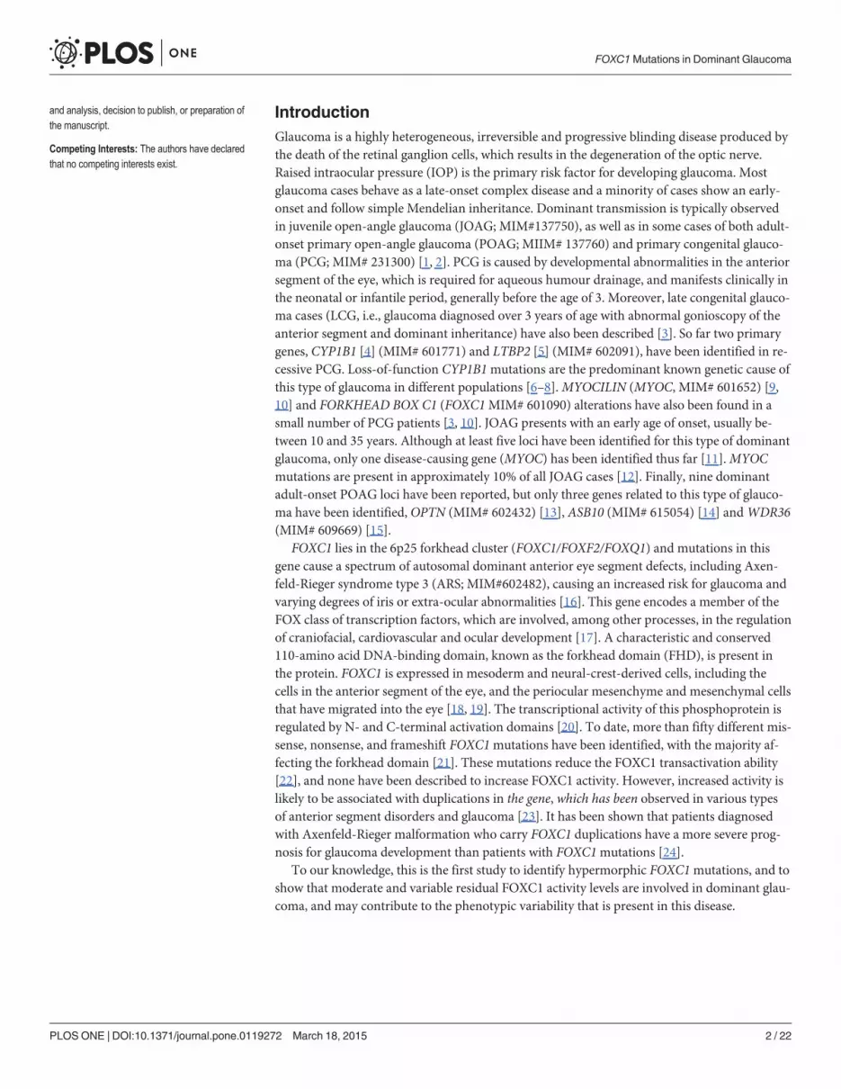
FOXC1 treatment with calf intestinal alkaline phosphataseThe phosphorylation status of the FOXC1 protein expressed in the HEK-293T cells was ana-lyzed by calf intestinal alkaline phosphatase (CIP) treatment of the nuclear extracts. The nucle-ar extracts were incubated with either 5 units of CIP, 11 μm sodium vanadate (NaVO3), orboth for 2 h at 37°C. The recombinant proteins were detected via western immunoblot usingan anti-myc antibody. The percentage of phosphorylated protein was estimated via densitome-try of western blot bands as follows: [upper band volume (phosphorylated FOXC1) x 100]/[upper band volume (phosphorylated FOXC1) + lower band volume (dephosphorylatedFOXC1)]. LDH was detected via western blot as the loading control, and was quantitated viadensitometry (n�3), and significant differences were analyzed using the t-test.
Western and dot blotting and antibodiesFor the western blot analysis, the nuclear extracts were fractionated using sodium dodecyl sul-fate-polyacrylamide gel electrophoresis (SDS-PAGE) using the Mini-PROTEAN III Gel Elec-trophoresis System (BioRad). A tricine-SDS-PAGE was performed as previously described[33]. The protein content of the samples were normalized using the Bradford assay. The gelswere subsequently transferred onto Hybond ECL nitrocellulose membranes (Amersham) forthe immunodetection, And a dot blot analysis of the non-sense mutants was carried out byspotting the normalized nuclear extracts onto Hybond ECL nitrocellulose membranes in a blottransfer apparatus (Bio-Dot, Bio-Rad). A commercial mouse monoclonal anti-myc (SantaCruz Biotechnology) antibody was used as the primary antibody, diluted at 1:1000. Horse-rad-ish peroxidase-conjugated antibodies against mouse IgG (Pierce) were diluted to 1:1000–1:4,000. The chemiluminiscence detection was performed using Supersignal Dura WesternBlot reagents (Pierce), and luminescent imaging was used for the chemiluminescence detection(LAS3000-mini; Fujifilm, Tokyo, Japan). The ensitometry for the protein band quantitation(FOXC1, LDH and RFP) was performed using the Quantity One 4.1 analysis software package(BioRad) in at least two independent experiments performed in triplicate. Ponceau S (Panreac)staining of the blots prior to the antibody incubation was performed to ensure the integrity ofthe samples and that equal amounts of the sample were analyzed [34]. As an additional sampleloading control, lactate dehydrogenase (LDH) was detected in the cell extracts and culturemedia using a goat anti-LDH antibody [AB1222, Chemicon, diluted to 1:5000) and an anti-goat IgG horse-radish peroxidase-conjugated antibody (sc-2033, Santa Cruz Biotechnology, di-luted to 1:2000). The RFP (transfection control) was detected using a rabbit anti-RFP antibody(#AB233, Evrogen), diluted to 1:5000, and an anti-rabbit IgG horse-radish peroxidase-conju-gated antibody (#1858415, Pierce) diluted to 1:1000.
ImmunocytochemistryThe HEK-293T cells were seeded on coverslips placed into 24-well plates, and were transientlytransfected with DNA constructs encoding different FOXC1 variants. All of the transfectionswere performed using the SuperFect Transfection Reagent (Qiagen). After the transfection, thecells were washed once with DPBS and cultured for 24 h. The cells were then fixed with 4%paraformaldehyde for 10 min at room temperature, followed by incubation with phosphate-buffered solution containing 0.2% triton X-100, 10% FBS, and 5% bovine serum albumin for30 min at room temperature. The recombinant proteins were detected using an anti-myc anti-body (Santa Cruz Biotechnology) (at a 1:500 dilution) at 4°C overnight followed by a Cy3-con-jugated anti-mouse IgG (Jackson ImmunoResearch Labs,1:1000 dilution) for 2 h at roomtemperature. Finally, the coverslips were mounted on glass slides using polyvinyl alcohol
FOXC1Mutations in Dominant Glaucoma
PLOSONE | DOI:10.1371/journal.pone.0119272 March 18, 2015 5 / 22

mounting medium with DABCO (Fluka) containing DAPI (40,60-diamidino-2-phenylindole;F6057 SIGMA) and were viewed under a laser scanning confocal microscope (Zeiss LSM 710).
Bioinformatic analysesThe In silico analyses of the sequences of the different FOXC1 variants were carried out using theprograms described in the S1Materials andMethods. Mutations were named according to RefSeq:NM_001453.2 and using the directions fromMutalyzer (https://humgenprojects.lumc.nl/trac/mutalyzer). The transcription start site and 5’-UTR sequence were defined as previously de-scribed [35]. The first nucleotide of the translation initiation site was numbered as nucleotide +1.The novel variants have been submitted to dbSNP (http://www.ncbi.nlm.nih.gov/SNP/).
Statistical analysesThe statistical comparisons between the groups were performed using either the t-test or theone-way analysis of variance (ANOVA). A Bonferroni correction was applied to adjust thetests for multiple comparisons. The data were statistically processed using the SigmaStat 2.0software (SPSS Science).
Results
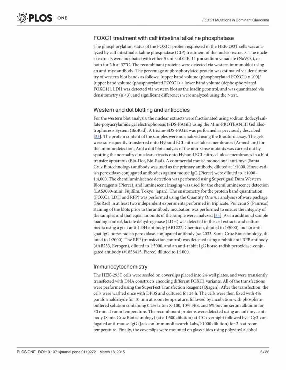
The identification of rare FOXC1mutationsTo analyze the role of FOXC1mutations in dominant glaucoma, a total of twelve unrelatedprobands with no mutations in the glaucoma genesMYOC and CYP1B1 were studied. Four ofthem were affected with PCG, one with LCG, three with JOAG and four with POAG. Four dif-ferent rare heterozygous sequence variations were identified in four probands (33.3%; twoPCGs, one LCG and one POAG; Fig. 1 and Table 1). Three of these variants (p.Y47X, p.I126Sand p.G447_G448insDG) were not found in the Emsembl variation database, the HumanGene Mutation Database, GeneCards, OMIM or in pubMed, and were therefore considered tobe novel mutations. These mutations were also absent in 200 control chromosomes (data notshown). The mutations p.Y47X and p.I126S segregated with the disease in families PCG68 andPCG73, respectively (Fig. 1A and B). These two families showed remarkable phenotypic vari-ability, particularly, but not exclusively, for the disease onset. Proband PCG68, who was classi-fied as LCG, showed a thin iris stroma and posterior embrytoxon in both eyes and no evidentiris hypoplasia, corectopia or systemic alterations (Fig. 2A and B). Her mother, diagnosed withJOAG at the age of 30, also exhibited posterior embryotoxon with no iris attachments or areasof iris atrophy secondary to surgery (Fig. 2C). Generalized stromal thinning, atrophy, corecto-pia (with the pupil pulled in the direction of the most prominent angle findings), or iris holeformation occurring in the quadrant opposite the site of corectopia, were not observed in thesetwo patients; therefore, according to the 9th Consensus Report of the World Glaucoma Associ-ation, the presence of mild iris stroma anomalies in the proband were considered to be compat-ible with congenital glaucoma [36]. Proband PCG73 was diagnosed with congenital glaucomaand her mother was diagnosed with juvenile glaucoma. Both patients presented thinned irisstromal areas, posterior embryotoxon with no iris attachments, surgical iridectomies in botheyes and normal pupils (Fig. 2D-G). Generalized iris stromal thinning, atrophy, corectopia oriris hole formation were also not detected in these patients. Interestingly, another PCG73 fami-ly member, subject II:4, also carried the same FOXC1 genotype and presented corectopia, gen-eralized iris stromal thinning, which could be secondary to surgery, posterior embryotoxonand surgical iridectomy in his left eye (Fig. 2H). He did not show dental, umbilical (Fig. 2I andJ, respectively) or any other systemic malformations, and was diagnosed with Axenfeld-Rieger
FOXC1Mutations in Dominant Glaucoma
PLOSONE | DOI:10.1371/journal.pone.0119272 March 18, 2015 6 / 22

Fig 1. FOXC1mutation segregation in families with autosomal dominant glaucoma. (A, B, C and D) Pedigrees of the families. The numbers in theinferior left side of the symbols indicate age at diagnosis. The oblique lines represent dead subjects. Black, dark grey and light grey symbols indicateglaucoma, Axenfeld-Rieger anomaly and ocular hypertension, respectively. The arrows in the pedigrees show the index cases. +: wild-type allele. The insetsin each panel correspond to the electropherograms of the FOXC1mutations identified in each family. To facilitate the comparison, the wild-type sequence is
FOXC1Mutations in Dominant Glaucoma
PLOSONE | DOI:10.1371/journal.pone.0119272 March 18, 2015 7 / 22
anomaly (ARA). His right eye was eviscerated at the time of the study. In family 559–10, theproband was the only subject available for genetic analysis, and ocular photographs were notavailable. She was diagnosed with PCG and carried the nonsense mutation p.Q106X, (Fig. 1Cand Table 1). Finally, in the adult-onset glaucoma family (556–10), the proband was diagnosedat the age of 66, and her three siblings manifested glaucoma in their fifties (Fig. 1D andTable 1). Only the index case and her sister consented to the genetic analyses and were foundto carry the heterozygous FOXC1 variant p.G447_G448insDG (Fig. 1D, subjects II:2 and II:3).The proband’s iris and iridocorneal angle were normal (Fig. 2K-M) and a careful clinical exam-ination directed to detected Axenfeld-Rieger alterations did not reveal any other relevant ocu-lar or systemic anomalies. All of the described mutations are associated with bilateralglaucoma, variable iris morphology and a disease onset ranging from 1 to 66 years in the indexcases (Table 1). The previously described 3’-UTR variant c.�734A>T (rs35717904) was alsoidentified in probands PCG73 and 556–10 (data not shown). PITX2mutations were ruled outin these patients.
To evaluate the pathogenicity of the identified variants a number of approaches were em-ployed, including bioinformatic analysis, evolutionary conservation of the mutant amino acidsor nucleotides and an in vitro assessment of transcriptional activity, protein stability, DNAbinding ability, phosphorylation status and subcellular localization.
Bioinformatic analysis and evolutionary conservation of the variantsThe analyses of the variants with the programs indicated in the S1 Materials and Methodsshowed that three coding mutations mapped to activating domain 1 (p.Y47X), alpha helix-2(p.Q106X) and alpha-helix 3 (p.I126S) of the forkhead DNA binding domain (Fig. 3A). The
shown above the corresponding mutant sequence. All the mutations were detected in the heterozygous state. Arrows in the electropherograms indicate thelocation of mutations. Duplicated nucleotides in panel D inset are indicated by boxes.
doi:10.1371/journal.pone.0119272.g001
Table 1. FOXC1 gene variations identified in dominant glaucoma cases and associated clinical features.
Proband orfamily member
1Nucleotidechange
Amino acidchange
Phenotype/laterality
Age at diagnosis(years)/sex
IOP (mmHg) atdiagnosis (OD/OS)
C/D (OD/OI)
2Treatment
PCG68 (III:1) 3c.141C>G 3p.Y47X LCG/B 7/F (24/20) NA 1
PCG68 (II:4) c.141C>G p.Y47X JOAG/B 30/F 30/25 0.8/0.8 5+Surgery
559–10 (II:1) c.316C>T p.Q106X PCG/B 3/F (25/25.9) NA Surgery
PCG73 (III:1) 3c.377T>G 3p.I126S PCG/B 1/F (14/15) (0.6–0.7/0.8–0.9)
1+Surgery
PCG73 (II:2) c.377T>G p.I126S JOAG/NA 12/F NA NA NA
PCG73 (II:4) c.377T>G p.I126S ARA/B NA/M NA NA NA
556–10 (II:2) 3c.1337_1342dup 3p.G447_G448insDG
AOG/B 66/F (21/20) NA 1
556–10 (II:3) c.1337_1342dup p.G447_G448insDG
AOG/B >50/F NA NA NA
1The variants were present in the heterozygous state;2Number of drugs and/or surgery;3Novel variants;
AOG/ARA/JOAG/LCG/PCG: Adult-onset glaucoma/Axenfeld-Rieger anomaly/juvenile-onset glaucoma/late-onset primary congenital glaucoma/primary
congenital glaucoma; B: bilateral; C/D: cup/disk ratio; NA: Not available; OD/OS: right eye/left eye. Mutations were named according to RefSeq:
NM_001453.2 and using directions from Mutalyzer (https://humgenprojects.lumc.nl/trac/mutalyzer).
doi:10.1371/journal.pone.0119272.t001
FOXC1Mutations in Dominant Glaucoma
PLOSONE | DOI:10.1371/journal.pone.0119272 March 18, 2015 8 / 22

remaining coding mutation (p.G447_G448insDG) was situated in a Gly-rich sequence that ispart of the inferred intrinsically disordered region 4 (IDR4, Fig. 3A). An in silico analysis car-ried out using the SIFT, PolyPhen-2 and Panther programs to evaluate the functional effect ofthe amino acid substitution p.I126S predicted that it was damaging (SIFT score = 0.01;
Fig 2. The phenotypes associated with the FOXC1mutations identified in this study. (A) Right and (B) left eye of subject PCG68 III:1. (C) Right eye ofsubject PCG68 II:4. (D) Right and (E) left eye of proband PCG73 (subject III:1). (F) Right and (G) left eye of subject III:1. (H) Axenfeld-Rieger anomaly in theleft eye of subject PCG73 II:4. (I) Normal teeth and (J) umbilicus of this patient. (K) Right and (L) left eye, and (M) gonioscopy photography of subject 566–10II:1. Asterisk: area of iris atrophy secondary to surgery. Blue arrows: areas of thin iris. Blue arrowhead: corectopia. Yellow arrows: posterior embrytoxon.White arrowhead: Ahmed valve tube in anterior chamber. White arrows: surgical iridectomies.
doi:10.1371/journal.pone.0119272.g002
FOXC1Mutations in Dominant Glaucoma
PLOSONE | DOI:10.1371/journal.pone.0119272 March 18, 2015 9 / 22

PolyPhen-2 score = 0.99; Panther Pdeleterious = 0.99). The evolutionary conservation analysisrevealed that the amino acid residue I126 is conserved from fish to mammals and that the DGinsertion affects a poly-Gly tract that is shared only by humans and chimpanzees (Fig. 3B).
Transcriptional activityThe transcriptional activity of the recombinant FOXC1 variants was assessed via a transientco-transfection with the luciferase reporter gene coupled to the CXCR4 promoter region,which contains one FBE (Fig. 4A). The detection of the truncated proteins p.Y47X and p.Q106X via standard SDS-PAGE and western blotting was prevented due to their small molecu-lar size. To overcome this difficulty, the electrophoresis was carried out in the presence of tri-cine. Under these conditions, only p.Q106X (but not p.Y47X) could be identified (S2 Fig.). Wealso observed that p.Q106X and the wild-type protein were transferred into the nitrocellulosemembrane with different yields due to their different molecular weights, making the quantifi-cation challenging. Because of these limitations, the truncated proteins were finally analyzedvia dot-blot. Western or dot-blot analyses of RFP and LDH were used as controls for the trans-fection efficiency and sample loading, respectively, and revealed no significant differences.Therefore, the observed FOXC1 band intensity variations can be attributed to alterations in theprotein stability, which will be shown later. Three mutations (p.Y47X, p.Q106X and p.G447_G448insDG) showed significantly increased transcriptional activity, ranging from ap-proximately 160% to almost 200% of the wild-type protein, and were classified as hyper-morphic variants (Fig. 4B and C). In contrast, the activity of the fourth variant (p.I126S) wasonly 20% of the normal protein and was similar to that of the control mutation p.I126M(Fig. 4C), showing that it is a hypomorphic allele. p.I126M has been identified in patients withARS and glaucoma [16] and it has been proposed to be a positive control mutant for activitystudies in the forkhead family of transcription factors [22].
Fig 3. Localization of the FOXC1mutations identified in this study. (A) Localization of the predicted andpreviously identified structural domains and motifs of the polypeptide chain. The predicted Pro-, Ser- and Gly-rich regions reported in the Prosite database are indicated over the scheme. Four IDRs predicted with theDisEMBL [50] and Globplot 2 [51] programs, and alpha-helices 2 (H2) and 3 (H3) of the forkhead domain, areindicated below the protein. AD: activation domain. CR: coding region. IDR: intrinsically disordered region.NLS: nuclear localization signal. Arrows note mutated positions. (B)Multiple alignments of amino acid ornucleotide sequences affected by mutations. The alignments were carried out with ClustalW [52]. The yellowbackground indicates the positions where the amino acids are identical.
doi:10.1371/journal.pone.0119272.g003
FOXC1Mutations in Dominant Glaucoma
PLOSONE | DOI:10.1371/journal.pone.0119272 March 18, 2015 10 / 22

Fig 4. Altered transcriptional activity of the FOXC1 variants identified in dominant glaucoma patients.(A) Scheme of the cDNA construct containing a FOXC1-binding element present in the CXCR4 promoterfused to the luciferasecoding region. The cDNA was cloned into the PGL3 basic vector. This construct wasused as a reporter of transcriptional activity in co-transfection assays with the different FOXC1 variants andthe empty pMirTarget vector, which encodes RFP (transfection efficiency control). The numbers below thescheme correspond to nucleotide positions. cDNA constructs encoding the indicated FOXC1 variants (B)were transiently co-expressed with the reporter cDNA in HEK-293T cells. The transcriptional activity,expressed as a percentage of the luciferase activity of the wild-type protein, was measured as indicated in theMaterials and Methods. The protein levels of the different FOXC1 versions present in HEK-293T cells 48 hafter transfection were determined by western immunoblot using a monoclonal anti-myc antibody (SantaCruz), except for the two non-sense mutants (p.Y47X and p.Q106X) which were difficult to detect andquantify by this technique and had to be assesed by dot-blot. Each lane contained 15 μg of total proteinobtained from the cell lysates. Transfection efficiency was assessed via western immunoblot using an anti-RFP antibody (Evrogen). The sample loading control, endogenous LDH, was also detected via immunoblotusing an anti-LDH antibody (Chemicon). Error bars correspond to the SD of three independent experimentscarried out in triplicate. insDG: p.G447_G448insDG. NT: non-transfected cells (negative control). Asterisksindicate statistical significance as compared to the control: p<0.05 (*); p<0.01 (**); p<0.001 (***).Significance was calculated by one-way ANOVA followed by Tukey multiple-comparison test.
doi:10.1371/journal.pone.0119272.g004
FOXC1Mutations in Dominant Glaucoma
PLOSONE | DOI:10.1371/journal.pone.0119272 March 18, 2015 11 / 22

Protein stabilityThe protein stability of the FOXC1 mutants was assessed in transiently transfected cells treatedwith the protein synthesis inhibitor cycloheximide. The FOXC1 levels were evaluated using ei-ther dot blot or western blot at different times after the inhibition of the protein synthesis. Anysignificant transfection or loading sample variations were ruled out via a western blot analysisof RFP and LDH, respectively (Fig. 5A). p.I126S showed remarkably reduced stability (Fig. 5Aand B), with an estimated half-life value that was approximately 3-fold lower than that of nor-mal FOXC1 (4.35 h vs. 12.95 h, respectively, Fig. 5C), and almost identical to the half-life of thecontrol mutation p.I126M (4.20 h, Fig. 5C). The dot-blot stability analyses of the two dominantnonsense mutations (p.Y47X and p.Q106X) showed the shortest half-lives among all of themutants (< 2.4 h).
p.I126S phosphorylationA careful western immunoblot analysis of FOXC1 revealed a doublet with a low molecularweight band that was more intense in p.I126S than in the wild-type protein (Fig. 6A, lanes 5and 1, respectively). To assess whether this difference is due to phosphorylation, p.I126S was
Fig 5. The p.Y47X, p.Q106X and p.I126S glaucoma-associated FOXC1mutations decrease protein stability. (A) Time course stability analysis ofFOXC1mutant polypeptides was carried out by transient expression of cDNA constructs encoding the different mutations in HEK-293T cells. The transfectedcells were treated with cycloheximide, a protein synthesis inhibitor, and the recombinant proteins were detected by dot or Western immunoblot using an anti-myc monoclonal antibody (Santa Cruz Biotechnology) at the indicated time points. The transfection efficiency was assessed by co-transfection with the non-recombinant pMirTarget vector, which encodes RFP. RFP was detected by dot or Western blot using an anti-RFP antibody (Evrogen). The sample loadingcontrol, endogenous LDH, was also detected via immunoblot using an anti-LDH antibody (Chemicon). (B) The amounts of FOXC1 at the indicated timepoints were determined by densitometry of the corresponding signals obtained via western blot. The relative amounts of FOXC1 are expressed as apercentage of levels at time 0 h. (C) The rate of decay and half-lives of the recombinant FOXC1 variants at the indicated time-points were determined fromlinear regression analysis as described in the Materials and Methods. Error bars correspond to the SD of three independent experiments carried out intriplicate. insDG: p.G447_G448insDG. NT: non-transfected cells (negative control). Asterisks indicate statistical significance compared to the control: p<0.01(**); p<0.001 (***). Two-way ANOVA followed by Tukey multiple-comparison test.
doi:10.1371/journal.pone.0119272.g005
FOXC1Mutations in Dominant Glaucoma
PLOSONE | DOI:10.1371/journal.pone.0119272 March 18, 2015 12 / 22

treated with CIP and analyzed via western blot. As controls, wild-type FOXC1 and the p.I126M mutant were treated in parallel. The LDH detection via western blot showed no signifi-cant sample loading variations (Fig. 6A). The untreated wild-type FOXC1 presented an upperband, estimated to represent 80% of the total protein (Fig. 6A, lane 1 and Fig. 6B). The propor-tion of this band in the untreated p.I126S (Fig. 6A, lane 5) represented less than 60% of the mu-tant protein (Fig. 6B), whereas in the control mutant was similar to the wild-type (Fig. 6A, lane9 and Fig. 6B). The CIP treatment of all of the variants increased the lower band proportion(Fig. 6A, lanes 2, 6 and 10), and this effect was counteracted with the CIP inhibitor NaVO3
Fig 6. The p.I126Smutation decreases FOXC1 phosphorylation. (A) The nuclear extracts of HEK-293Tcells transiently expressing the indicated FOXC1mutants were treated with different combinations of CIP andsodium vanadate. After the treatments the recombinant proteins were detected via western immunoblot usingan anti-myc antibody. The arrowhead indicates the position of the phosphorylated FOXC1. The sampleloading control, endogenous LDH, was detected via immunoblot using an anti-LDH antibody (Chemicon). (B)The percentage of phosphorylated FOXC1 in untreated samples (CIP-, NaVO3-) was estimated bydensitometry of the upper band as indicated in Materials and Methods. Error bars correspond to the SD ofthree independent experiments carried out in triplicate. NT: non-transfected cells (negative control). Theasterisk indicates statistical significance compared to the control: p<0.05 (*); One-way ANOVA.
doi:10.1371/journal.pone.0119272.g006
FOXC1Mutations in Dominant Glaucoma
PLOSONE | DOI:10.1371/journal.pone.0119272 March 18, 2015 13 / 22

(Fig. 6A, lanes 3–4, 7–8 and 11–12), indicating that the upper band corresponds to phosphory-lated FOXC1. In addition, the predicted phosphorylation potential of the mutant Ser residueindicated that it was unphosphorylated (data not shown). These results suggest that p.I126Scan alter the FOXC1 conformation, decreasing the phosphorylation of the whole protein.
EMSA assayThe DNA binding ability of the different mutations was evaluated using EMSA analysis. Com-petition experiments in which similar amounts of pre-bound protein-DNA complexes werechallenged with increasing ratios of competitor DNA containing the FOXC1 binding site(ranging from 0.2 to 10 pmol, i.e., 1- to 50-fold excess), showed that both the intensity and theproportion of the protein-DNA complexes formed by the p.I126S and p.I126M (control) muta-tions, were significantly lower than those of the wild-type protein (Fig. 7A and B). Converesely,and although the intensity of the complexes corresponding to p.G447_G448insDG was higherthan those of the normal protein (Fig. 7A), the DNA binding ability did not differ significantly
Fig 7. The p.I126Smutation decreases FOXC1 DNA-binding. (A) The effect of FOXC1mutations on DNA-binding specificity was assessed via EMSA. The oligonucleotides corresponding to the FOXC1-bindingsequence labeled with biotin at the 5'-end were incubated with 10 μg of nuclear extracts from HEK-293T cellstransfected with the indicated FOXC1 variants. The extracts were separated on a 10% nondenaturingpolyacrylamide gel electrophoresis and were transferred to a positively charged nylon membrane (Hybond-N+, Amersham). FOXC1-oligonucleotide complexes were visualized by incubation with streptavidin-HRPconjugate and chemiluminescent detection (Chemiluminescent EMSA kit, Thermo Scientific). Unlabeledcompetitor oligonucleotides were pre-incubated at increasing concentrations from 0.2 to 10 pmol (1 to 50-foldexcess) with the labeled probe. The arrowhead indicates the position of the full-length FOXC1-DNAcomplexes. (B) The percentage of band volume corresponding to protein-oligonucleotide complexes wasestimated by densitometry as indicated in Materials and Methods. Error bars correspond to the SD of threeindependent experiments carried out in triplicate. insDG: p.G447_G448insDG. NT: non-transfected cells(negative control). Asterisks indicate statistical significance as compared to the control: p<0.001 (***). Two-way ANOVA followed by Tukey multiple-comparison test.
doi:10.1371/journal.pone.0119272.g007
FOXC1Mutations in Dominant Glaucoma
PLOSONE | DOI:10.1371/journal.pone.0119272 March 18, 2015 14 / 22

from the wild-type (Fig. 7B). As expected, the two nonsense mutants failed to bind to the DNAbecause they lacked a complete forkhead domain (data not shown).
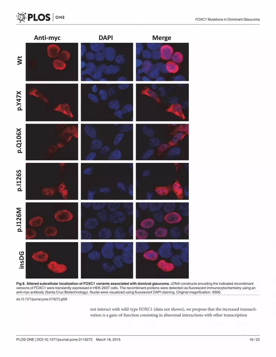
Subcellular localizationFluorescence immunocytochemistry of the control recombinant wild-type FOXC1 localizedthe protein exclusively to the nucleus (Fig. 8, Wt). The two non-sense mutations were detectedin both the nucleus and the cytoplasm (Fig. 8, p.Y47X and p.Q106X). Mutation p.I126S wasalso present in these two cell compartments. In contrast, the p.I126M control mutation was ob-served exclusively in the nucleus. p.G447_G448insDG also showed normal nuclear localization(Fig. 8).
DiscussionGlaucoma is a genetically and clinically heterogeneous disease with a poorly understood genet-ic basis. It has been proposed that dominant glaucoma may originate from goniodysgenesiswith marked variation in the expressivity of dysgenesis, leading to the apparition of symptomsearly or late in life [1]. Typical congenital glaucoma, which is inherited as a non-dominanttrait, is also considered to be part of the anterior segment dysgenesis (ASD) group of disordersbecause it originates via the maldevelopment of structures in the anterior segment of the eye.On the converse, FOXC1 is a gene that regulates the development of the anterior segment ofthe eye, and it is known to be involved in several autosomal dominant eye defects associatedwith an increased risk for glaucoma. Its heterozygous loss-of-function in mice is associatedwith considerable variability in anterior segment abnormalities, similar to those present in pa-tients with ARA and glaucoma [37]. Based on these facts, we explored the role of FOXC1 indominant glaucoma. To this end, we carried out a comprehensive screening of the coding andregulatory regions (UTRs and the promoter) of the gene in twelve affected Spanish familieswith no known alterations in other glaucoma genes.
Pathogenicity of the FOXC1mutationsWe identified three hypermorphic (p.Y47X, p.Q106X and p.G447_G448insDG) and one hypo-morphic (p.I126S) heterozygous FOXC1 variants in four probands, representing a significantproportion (33.3%) of the studied families. Only two of these mutations have previously beenreported. p.Q106X was identified in a patient with an enucleated eye due to severe glaucomaand abnormalities of the anterior segment in the other eye [21]. Interestingly, p.Q106X-relatedmutations have been found to be associated with high phenotypic variability. For instance, p.Q120X was detected in patients with ARS and Peters’ anomaly in the same family [38], and p.Q123X has been identified in ARA patients and even in a normal subject [39].
It is accepted that FOXC1mutations are pathogenic and function by reducing the levels ofprotein, inhibiting DNA binding and/or reducing the levels of FOXC1 transactivation [22, 40].To the best of our knowledge, this is the first report of hypermorphic FOXC1 variants associat-ed with dominant glaucoma, which agrees with previous studies showing a high glaucoma riskassociated with increased gene dosage due to FOXC1 gene duplication [23, 41, 42]. The unex-pected hyperactivity of the two truncated proteins is likely due to the expression of the isolatedactivation domain 1, which appears to act as a potent and autonomous transcription up-regula-tor despite the reduced stability of the mutant polypeptides. According to these results it hasbeen described that expression of N-terminal FOXC1 residues 1–30 and 1–65 present transac-tivation levels that are equivalent to or higher than the full-length FOXC1, and it has been sug-gested that the N-terminal regions are sufficient to activate transcription [20]. Because thetruncated proteins also failed to bind DNA, and pull-down experiments indicated that they did
FOXC1Mutations in Dominant Glaucoma
PLOSONE | DOI:10.1371/journal.pone.0119272 March 18, 2015 15 / 22
not interact with wild-type FOXC1 (data not shown), we propose that the increased transacti-vation is a gain-of-function consisting in abnormal interactions with other transcription
Fig 8. Altered subcellular localization of FOXC1 variants associated with dominat glaucoma. cDNA constructs encoding the indicated recombinantversions of FOXC1 were transiently expressed in HEK-293T cells. The recombinant proteins were detected via fluorescent immunocytochemistry using ananti-myc antibody (Santa Cruz Biotechnology). Nuclei were visualized using fluorescent DAPI staining. Original magnification: X600.
doi:10.1371/journal.pone.0119272.g008
FOXC1Mutations in Dominant Glaucoma
PLOSONE | DOI:10.1371/journal.pone.0119272 March 18, 2015 16 / 22
factors. Our findings complements the haploinsufficiency mechanism, which has frequentlybeen invoked to underlie the pathogenicity of FOXC1 non-sense alleles. The mislocalization ofthe truncated proteins (present in both the nucleus and the cytoplasm) is likely due to theirsmall molecular size (< 50 kDa), which allows free diffusion through the nuclear pores. The re-maining hypermorphic mutation, p.G447_G448insDG, did not differ from the wild-type pro-tein in terms of the protein stability, DNA binding or subcellular localization. Nevertheless, itslocalization in a Gly-rich region, which is predicted to be unstructured (IDR4), may explainthe observed hyperactivity. IDRs are known to lack a fixed tertiary structure and are present indifferent regulatory proteins, which include the transcription factors CREB [43] and p53 [44].They participate in molecular interactions with other proteins or nucleic acids, and althoughthey lack a defined structure when alone in solution, they undergo disorder-to-order transi-tions after binding to specific targets [45]. These data suggest that the increased transcriptionactivity of p.G447_G448insDG may be also due to a gain-of-function that would result in al-tered binding to transcription factors, suggesting a possible role of the mutant amino acids inprotein-protein interactions. p.I126S was hypomorphic and its decreased protein stability andDNA binding affinity were similar to that of the p.I126M control mutation. The reduction inDNA binding affinity may be due to structural alterations in the forkhead domain alpha-helix3, which is predicted to play a role in major DNA groove recognition. Contrary to our results,p.I126M has been described to bind the FBE at near-wild-type FOXC1 levels [22]. This discrep-ancy may reflect different levels of excess unlabelled oligonucleotides used as competitors forFOXC1 DNA binding, which ranged from 1- to 50-fold in the present study and from 0.5- to 1.5-fold in a previous report. In contrast to p.I126M, p.I126S reduced FOXC1 phosphorylation andinduced intracellular mislocalization of this protein. Because the amino acid residues involved inthe p.I126Mmutation are not phosphorylated, we postulate that a mutation-induced conforma-tional change may interfere the interaction between kinases and FOXC1, leading to decreasedFOXC1 phosphorylation. In accordance with our results, it has been described that I126 is criticalfor correct FOXC1 nuclear localization and transactivation, so that substitution of I126 for differ-ent amino acids leads to differentially disrupted nuclear localization and DNA binding [46]. Al-though the differences in protein phosphorylation and subcellular localization between these twomutations do not seem to correlate with the severity of the associated phenotypes, we should bearin mind that unidentified genetic modifiers may also contribute to the final phenotypic outcome.
Phenotypic variability and residual FOXC1 activityAlthough dominant glaucoma was the most outstanding clinical feature in all of the mutationcarriers, a variability in the age of onset was also observed. Moreover, one of the families(PCG73) also showed variable iris morphology, which ranged from thin stromal areas and pos-terior embrytoxon with no iris attachments (subjects II:2 and III:1), to corectopia associatedwith Axenfeld-Rieger anomaly (subject II:4). Mild iris alterations can be present in non-syn-dromic congenital glaucoma cases [47]. Previous reports have also identified a remarkable phe-notypic variability associated with FOXC1mutations [39, 48]. This clinical heterogeneitysuggests an important role for modifier factors (genetic, environmental and/or stochastic) onthe phenotypic outcomes. The FOXC1 associated phenotypic variability may also depend onthe residual transcriptional activity associated with the individual genotypes. Based on thisidea, we observed that none of the glaucoma-associated FOXC1mutations expressed in the cellcultures showed a complete loss-of-function. On the contrary, the glaucoma associated tran-scriptional activity ranged from roughly 20% to 198% of the wild-type levels. Using an additivemodel, we estimated the FOXC1 transactivation ability associated with each glaucoma geno-type as the mean activity of the two alleles and inferred two glaucoma associated activity
FOXC1Mutations in Dominant Glaucoma
PLOSONE | DOI:10.1371/journal.pone.0119272 March 18, 2015 17 / 22
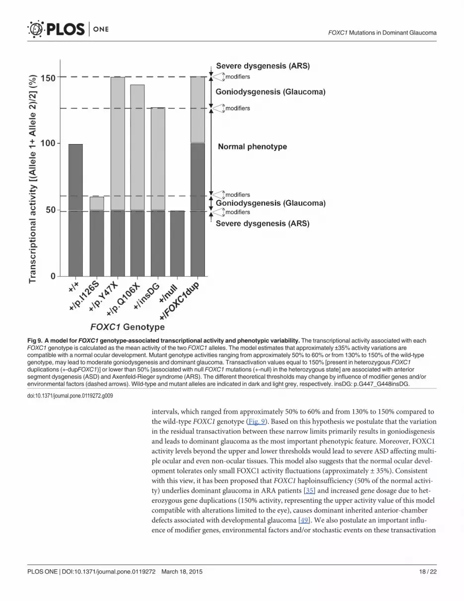
intervals, which ranged from approximately 50% to 60% and from 130% to 150% compared tothe wild-type FOXC1 genotype (Fig. 9). Based on this hypothesis we postulate that the variationin the residual transactivation between these narrow limits primarily results in goniodisgenesisand leads to dominant glaucoma as the most important phenotypic feature. Moreover, FOXC1activity levels beyond the upper and lower thresholds would lead to severe ASD affecting multi-ple ocular and even non-ocular tissues. This model also suggests that the normal ocular devel-opment tolerates only small FOXC1 activity fluctuations (approximately ± 35%). Consistentwith this view, it has been proposed that FOXC1 haploinsufficiency (50% of the normal activi-ty) underlies dominant glaucoma in ARA patients [35] and increased gene dosage due to het-erozygous gene duplications (150% activity, representing the upper activity value of this modelcompatible with alterations limited to the eye), causes dominant inherited anterior-chamberdefects associated with developmental glaucoma [49]. We also postulate an important influ-ence of modifier genes, environmental factors and/or stochastic events on these transactivation
Fig 9. A model for FOXC1 genotype-associated transcriptional activity and phenotypic variability. The transcriptional activity associated with eachFOXC1 genotype is calculated as the mean activity of the two FOXC1 alleles. The model estimates that approximately ±35% activity variations arecompatible with a normal ocular development. Mutant genotype activities ranging from approximately 50% to 60% or from 130% to 150% of the wild-typegenotype, may lead to moderate goniodysgenesis and dominant glaucoma. Transactivation values equal to 150% [present in heterozygous FOXC1duplications (+-dupFOXC1)] or lower than 50% [associated with null FOXC1mutations (+-null) in the heterozygous state] are associated with anteriorsegment dysgenesis (ASD) and Axenfeld-Rieger syndrome (ARS). The different theoretical thresholds may change by influence of modifier genes and/orenvironmental factors (dashed arrows). Wild-type and mutant alleles are indicated in dark and light grey, respectively. insDG: p.G447_G448insDG.
doi:10.1371/journal.pone.0119272.g009
FOXC1Mutations in Dominant Glaucoma
PLOSONE | DOI:10.1371/journal.pone.0119272 March 18, 2015 18 / 22

thresholds. This hypothesis agrees with the proposed existence of critical activity thresholds forFOXC1 and the related protein PITX2 [42].
ConclusionsIn summary, this study shows that FOXC1 sequence variations with moderate activity defectsare associated with dominant glaucoma and remarkable phenotypic variability. In addition,this is the first study to identify hypermorphic variants of this gene and will contribute to a bet-ter understanding of the genetic basis of dominant glaucoma, as well as the function of FOXC1.
Supporting InformationS1 Fig. Localization of primers used in this study to analyze FOXC1. The arrows show theposition of the different PCR primers. Amplicons are indicated by horizontal brackets and thenumbers between parentheses correspond to amplicon length. CR: coding region.(TIF)
S2 Fig. Tricine-SDS-PAGE electrophoresis of the truncated FOXC1mutations. To improvedetection of the small molecular size mutants p.Y47X and p.Q106X, the nuclear extracts ofHEK-293T cells transiently expressing these recombinant proteins were analyzed via Tricine-SDS-PAGE. The proteins were detected via western immunoblot using an anti-myc antibody.NT: non-transfected.(TIF)
S1 Materials and Methods.(DOCX)
S1 Table. The primer sequences and PCR conditions used for FOXC1 sequencing.(DOCX)
S2 Table. The primer sequences and PCR conditions used for FOXC1 site-directed muta-genesis.(DOCX)
AcknowledgmentsWe are indebted to the patients and their families for cooperation in this study. We are gratefulto Mrs. María-José Cabañero for excellent technical assistance and to Mrs. Maite García-Antón, for outstanding support in patient recruitment and sample collection. We greatly ap-preciate the contribution of Dr. Mayte Ariño (Servicio de Oftalmología, Fundación JiménezDíaz, Madrid, SPAIN) and Dr. Gerardo García-García (Servicio de Oftalmología, Hospital elBierzo, Ponferrada, León, SPAIN) for ophthalmic reevaluation and photographs of patients566–10 II:1 and PCG73 II:4, respectively.
Author ContributionsConceived and designed the experiments: JE JGF CMT. Performed the experiments: CMT FSSJDAA JJFF. Analyzed the data: CMT FSS JDAA JJFF LM CDMH FBK CA JGF JE. Contributedreagents/materials/analysis tools: LM CDMH FBK CA JGF JE. Wrote the paper: CMT JDAAJJFF CDMH CA JGF JE.
FOXC1Mutations in Dominant Glaucoma
PLOSONE | DOI:10.1371/journal.pone.0119272 March 18, 2015 19 / 22
References1. Jerndal T. Congenital glaucoma due to dominant goniodysgenesis. A new concept of the heredity of
glaucoma. Am J HumGenet. 1983; 35(4): 645–51. PMID: 6881141
2. Simha N, Verin P, Gauthier L. Congenital glaucoma of dominant autosomal transmission apropos of afamily. Management. Bull Soc Ophtalmol Fr. 1989; 89(10): 1149–51. PMID: 2620399
3. Chakrabarti S, Kaur K, Rao KN, Mandal AK, Kaur I, Parikh RS, et al. The transcription factor geneFOXC1 exhibits a limited role in primary congenital glaucoma. Invest Ophthalmol Vis Sci. 2009; 50(1):75–83. doi: 10.1167/iovs.08-2253 PMID: 18708620
4. Stoilov I, Akarsu AN, Sarfarazi M. Identification of three different truncating mutations in cytochromeP4501B1 (CYP1B1) as the principal cause of primary congenital glaucoma (Buphthalmos) in familieslinked to the GLC3A locus on chromosome 2p21. HumMol Genet. 1997; 6(4): 641–7. PMID: 9097971
5. Ali M, McKibbin M, Booth A, Parry DA, Jain P, Riazuddin SA, et al. Null mutations in LTBP2 cause pri-mary congenital glaucoma. Am J HumGenet. 2009; 84(5): 664–71. doi: 10.1016/j.ajhg.2009.03.017PMID: 19361779
6. Bagiyeva S, Marfany G, Gonzalez-Angulo O, Gonzalez-Duarte R. Mutational screening of CYP1B1 inTurkish PCG families and functional analyses of newly detected mutations. Mol Vis. 2007; 13: 1458–68. PMID: 17893647
7. Lopez-Garrido MP, Medina-Trillo C, Morales-Fernandez L, Garcia-Feijoo J, Martinez-de-la-Casa JM,Garcia-Anton M, et al. NullCYP1B1 genotypes in primary congenital and nondominant juvenile glauco-ma. Ophthalmology. 2013; 120(4): 716–23. doi: 10.1016/j.ophtha.2012.09.016 PMID: 23218183
8. Bejjani BA, Lewis RA, Tomey KF, Anderson KL, Dueker DK, Jabak M, et al. Mutations inCYP1B1, thegene for cytochrome P4501B1, are the predominant cause of primary congenital glaucoma in SaudiArabia. Am J HumGenet. 1998; 62(2): 325–33. PMID: 9463332
9. Chakrabarti S, Kaur K, Komatireddy S, Acharya M, Devi KR, Mukhopadhyay A, et al. Gln48His is theprevalent myocilin mutation in primary open angle and primary congenital glaucoma phenotypes inIndia. Mol Vis. 2005; 11: 111–3. PMID: 15723004
10. Kaur K, Reddy AB, Mukhopadhyay A, Mandal AK, Hasnain SE, Ray K, et al. Myocilin gene implicatedin primary congenital glaucoma. Clin Genet. 2005; 67(4): 335–40. PMID: 15733270
11. Stone EM, Fingert JH, AlwardWL, Nguyen TD, Polansky JR, Sunden SL, et al. Identification of a genethat causes primary open angle glaucoma. Science. 1997; 275(5300): 668–70. PMID: 9005853
12. Wiggs JL, Allingham RR, Vollrath D, Jones KH, De La PM, Kern J, et al. Prevalence of mutations inTIGR/Myocilin in patients with adult and juvenile primary open-angle glaucoma. Am J HumGenet.1998; 63(5): 1549–52. PMID: 9792882
13. Rezaie T, Child A, Hitchings R, Brice G, Miller L, Coca-Prados M, et al. Adult-onset primary open-angleglaucoma caused by mutations in optineurin. Science. 2002; 295(5557): 1077–9. PMID: 11834836
14. Pasutto F, Keller KE, Weisschuh N, Sticht H, Samples JR, Yang YF, et al. Variants in ASB10 are asso-ciated with open-angle glaucoma. HumMol Genet. 2012; 21(6): 1336–49. doi: 10.1093/hmg/ddr572PMID: 22156576
15. Monemi S, Spaeth G, DaSilva A, Popinchalk S, Ilitchev E, Liebmann J, et al. Identification of a noveladult-onset primary open-angle glaucoma (POAG) gene on 5q22.1. HumMol Genet. 2005; 14(6): 725–33. PMID: 15677485
16. Nishimura DY, Swiderski RE, Alward WL, Searby CC, Patil SR, Bennet SR, et al. The forkhead tran-scription factor gene FKHL7 is responsible for glaucoma phenotypes which map to 6p25. Nat Genet.1998; 19(2): 140–7. PMID: 9620769
17. Lines MA, Kozlowski K, Walter MA. Molecular genetics of Axenfeld-Rieger malformations. HumMolGenet. 2002; 11(10): 1177–84. PMID: 12015277
18. Kume T, Deng KY, Winfrey V, Gould DB, Walter MA, Hogan BL. The forkhead/winged helix gene Mf1 isdisrupted in the pleiotropic mouse mutation congenital hydrocephalus. Cell. 1998; 93(6): 985–96.PMID: 9635428
19. Kidson SH, Kume T, Deng K, Winfrey V, Hogan BL. The forkhead/winged-helix gene, Mf1, is necessaryfor the normal development of the cornea and formation of the anterior chamber in the mouse eye. DevBiol. 1999; 211(2): 306–22. PMID: 10395790
20. Berry FB, Saleem RA, Walter MA. FOXC1 transcriptional regulation is mediated by N- and C-terminalactivation domains and contains a phosphorylated transcriptional inhibitory domain. J Biol Chem. 2002;277(12): 10292–7. PMID: 11782474
21. D'Haene B, Meire F, Claerhout I, Kroes HY, Plomp A, Arens YH, et al. Expanding the spectrum ofFOXC1 and PITX2mutations and copy number changes in patients with anterior segment malforma-tions. Invest Ophthalmol Vis Sci. 2011; 52(1): 324–33. doi: 10.1167/iovs.10-5309 PMID: 20881294
FOXC1Mutations in Dominant Glaucoma
PLOSONE | DOI:10.1371/journal.pone.0119272 March 18, 2015 20 / 22
22. Saleem RA, Banerjee-Basu S, Berry FB, Baxevanis AD, Walter MA. Analyses of the effects that dis-ease-causing missense mutations have on the structure and function of the winged-helix proteinFOXC1. Am J HumGenet. 2001; 68(3): 627–41. PMID: 11179011
23. Lehmann OJ, Ebenezer ND, Jordan T, Fox M, Ocaka L, Payne A, et al. Chromosomal duplication in-volving the forkhead transcription factor gene FOXC1 causes iris hypoplasia and glaucoma. Am J HumGenet. 2000; 67(5): 1129–35. PMID: 11007653
24. Strungaru MH, Dinu I, Walter MA. Genotype-phenotype correlations in Axenfeld-Rieger malformationand glaucoma patients with FOXC1 and PITX2mutations. Invest Ophthalmol Vis Sci. 2007; 48(1): 228–37. PMID: 17197537
25. Lopez-Garrido MP, Sanchez-Sanchez F, Lopez-Martinez F, Aroca-Aguilar JD, Blanco-Marchite C,Coca-Prados M, et al. Heterozygous CYP1B1 gene mutations in Spanish patients with primary open-angle glaucoma. Mol Vis. 2006; 12: 748–55. PMID: 16862072
26. Aroca-Aguilar JD, Sanchez-Sanchez F, Ghosh S, Coca-Prados M, Escribano J. Myocilin mutationscausing glaucoma inhibit the intracellular endoproteolytic cleavage of myocilin between amino acidsArg226 and Ile227. J Biol Chem. 2005; 280(22): 21043–51. PMID: 15795224
27. Sanchez-Sanchez F, Martinez-Redondo F, Aroca-Aguilar JD, Coca-Prados M, Escribano J. Character-ization of the intracellular proteolytic cleavage of myocilin and identification of calpain II as a myocilin-processing protease. J Biol Chem. 2007; 282(38): 27810–24. PMID: 17650508
28. Hayashi H, Kume T. Forkhead transcription factors regulate expression of the chemokine receptorCXCR4 in endothelial cells and CXCL12-induced cell migration. Biochem Biophys Res Commun.2008; 367(3): 584–9. doi: 10.1016/j.bbrc.2007.12.183 PMID: 18187037
29. Saleem RA, Banerjee-Basu S, Berry FB, Baxevanis AD, Walter MA. Structural and functional analysesof disease-causing missense mutations in the forkhead domain of FOXC1. HumMol Genet. 2003; 12(22): 2993–3005. PMID: 14506133
30. Saleem RA, Murphy TC, Liebmann JM, Walter MA. Identification and analysis of a novel mutation in theFOXC1 forkhead domain. Invest Ophthalmol Vis Sci. 2003; 44(11): 4608–12. PMID: 14578375
31. Campos-Mollo E, Lopez-Garrido MP, Blanco-Marchite C, Garcia-Feijoo J, Peralta J, Belmonte-Marti-nez J, et al. CYP1B1mutations in Spanish patients with primary congenital glaucoma: phenotypic andfunctional variability. Mol Vis. 2009; 15: 417–31. PMID: 19234632
32. Berry FB, Lines MA, Oas JM, Footz T, Underhill DA, Gage PJ, et al. Functional interactions betweenFOXC1 and PITX2 underlie the sensitivity to FOXC1 gene dose in Axenfeld-Rieger syndrome and ante-rior segment dysgenesis. HumMol Genet. 2006; 15(6): 905–19. PMID: 16449236
33. Schagger H, von JagowG. Tricine-sodium dodecyl sulfate-polyacrylamide gel electrophoresis for theseparation of proteins in the range from 1 to 100 kDa. Anal Biochem. 1987; 166(2): 368–79. PMID:2449095
34. Romero-Calvo I, Ocon B, Martinez-Moya P, Suarez MD, Zarzuelo A, Martinez-Augustin O, et al. Re-versible Ponceau staining as a loading control alternative to actin in Western blots. Anal Biochem.2010; 401(2): 318–20. doi: 10.1016/j.ab.2010.02.036 PMID: 20206115
35. Mears AJ, Jordan T, Mirzayans F, Dubois S, Kume T, Parlee M, et al. Mutations of the forkhead/winged-helix gene, FKHL7, in patients with Axenfeld-Rieger anomaly. Am J HumGenet. 1998; 63(5):1316–28. PMID: 9792859
36. Papadopoulos M, Brandt JD, Sugiyama K, Khaw PT, Chua J, Law S, et al. Establising the diagnosisand determining glaucoma progression. In: Weinreb RN, Grajewski AL, Papadopoulos M, Grigg J,Freedman S, editors. Childhood Glaucoma. Ámsterdam, The Netherlands: Kluger Publications; 2013.p. 15–41. doi: 10.1186/1687-9856-2013-15 PMID: 24025597
37. Smith RS, Zabaleta A, Kume T, Savinova OV, Kidson SH, Martin JE, et al. Haploinsufficiency of thetranscription factors FOXC1 and FOXC2 results in aberrant ocular development. HumMol Genet.2000; 9(7): 1021–32. PMID: 10767326
38. Weisschuh N, Wolf C, Wissinger B, Gramer E. A novel mutation in the FOXC1 gene in a family withAxenfeld-Rieger syndrome and Peters' anomaly. Clin Genet. 2008; 74(5): 476–80. doi: 10.1111/j.1399-0004.2008.01025.x PMID: 18498376
39. Komatireddy S, Chakrabarti S, Mandal AK, Reddy AB, Sampath S, Panicker SG, et al. Mutation spec-trum of FOXC1 and clinical genetic heterogeneity of Axenfeld-Rieger anomaly in India. Mol Vis. 2003;9: 43–8. PMID: 12592227
40. Murphy TC, Saleem RA, Footz T, Ritch R, McGillivray B, Walter MA. The wing 2 region of the FOXC1forkhead domain is necessary for normal DNA-binding and transactivation functions. Invest OphthalmolVis Sci. 2004; 45(8): 2531–8. PMID: 15277473
FOXC1Mutations in Dominant Glaucoma
PLOSONE | DOI:10.1371/journal.pone.0119272 March 18, 2015 21 / 22
41. Lehmann OJ, Ebenezer ND, Ekong R, Ocaka L, Mungall AJ, Fraser S, et al. Ocular developmental ab-normalities and glaucoma associated with interstitial 6p25 duplications and deletions. Invest Ophthal-mol Vis Sci. 2002; 43(6): 1843–9. PMID: 12036988
42. Walter MA. PITs and FOXes in ocular genetics: the Cogan lecture. Invest Ophthalmol Vis Sci. 2003; 44(4): 1402–5. PMID: 12657570
43. Sugase K, Dyson HJ, Wright PE. Mechanism of coupled folding and binding of an intrinsically disor-dered protein. Nature. 2007; 447(7147): 1021–5. PMID: 17522630
44. Tidow H, Melero R, Mylonas E, Freund SM, Grossmann JG, Carazo JM, et al. Quaternary structures oftumor suppressor p53 and a specific p53 DNA complex. Proc Natl Acad Sci USA. 2007; 104(30):12324–9. PMID: 17620598
45. Uversky VN, Gillespie JR, Fink AL. Why are "natively unfolded" proteins unstructured under physiologicconditions? Proteins. 2000; 41(3): 415–27. PMID: 11025552
46. Saleem RA, Banerjee-Basu S, Murphy TC, Baxevanis A, Walter MA. Essential structural and functionaldeterminants within the forkhead domain of FOXC1. Nucleic Acids Res. 2004; 32(14): 4182–93. PMID:15299087
47. Perry LP, Jakobiec FA, Zakka FR, Walton DS. Newborn primary congenital glaucoma: histopathologicfeatures of the anterior chamber filtration angle. J AAPOS. 2012; 16(6): 565–8. doi: 10.1016/j.jaapos.2012.06.012 PMID: 23158552
48. Perveen R, Lloyd IC, Clayton-Smith J, Churchill A, van Heyningen V, Hanson I, et al. Phenotypic vari-ability and asymmetry of Rieger syndrome associated with PITX2mutations. Invest Ophthalmol Vis Sci.2000; 41(9): 2456–60. PMID: 10937553
49. Nishimura DY, Searby CC, Alward WL, Walton D, Craig JE, Mackey DA, et al. A spectrum of FOXC1mutations suggests gene dosage as a mechanism for developmental defects of the anterior chamberof the eye. Am J HumGenet. 2001; 68(2): 364–72. PMID: 11170889
50. Linding R, Jensen LJ, Diella F, Bork P, Gibson TJ, Russell RB. Protein disorder prediction: implicationsfor structural proteomics. Structure. 2003; 11(11): 1453–9. PMID: 14604535
51. Linding R, Russell RB, Neduva V, Gibson TJ. GlobPlot: Exploring protein sequences for globularity anddisorder. Nucleic Acids Res. 2003; 31(13): 3701–8. PMID: 12824398
52. Thompson JD, Higgins DG, Gibson TJ. CLUSTALW: improving the sensitivity of progressive multiplesequence alignment through sequence weighting, position-specific gap penalties and weight matrixchoice. Nucleic Acids Res. 1994; 22(22): 4673–80. PMID: 7984417
FOXC1Mutations in Dominant Glaucoma
PLOSONE | DOI:10.1371/journal.pone.0119272 March 18, 2015 22 / 22