Embed Size (px)
Citation preview
SHORT REPORT
Oligomerization of p53 is necessary to inhibit its transcriptionaltransactivation property at high protein concentration
Arnold Kristjuhan, Viljar Jaks, Ilvi Rimm, Tiia Tooming and Toivo Maimets
Institute of Molecular and Cell Biology, Estonian Biocentre, Tartu University, Riia 23, Tartu, EE2400 Estonia
We have previously shown that transactivation by tumorsuppressor protein p53 can be inhibited in vivo atelevated protein concentrations. In this study wecharacterize the structural requirements of this function.We show that oligomerization domain of p53 is involvedin loss of transactivation at high protein concentrations:mutants not able to oligomerize are neither able tosuppress transactivation, although these transactivatingproperties can be untouched.
Keywords: p53; oligomerization; transactivation
Tumor suppressor protein p53 is involved in regulationof transcription activating the promoters located nearto its DNA binding sites. Number of such genes areidenti®ed, a part of them is clearly involved inregulation of cell proliferation (Barak et al., 1993;Buckbinder et al., 1995; El-Deiry et al., 1993; Kastan etal., 1992; Miyashita and Reed, 1995; Okamoto andBeach, 1994; Owen-Shaub et al., 1995). p53 alsorepresses many viral and cellular promoters (Deb etal., 1992, 1994; Desaintes et al., 1995; Jackson et al.,1993; Subler et al., 1992, 1994). Exact mechanism oftransrepression is obscure.For transactivation, p53 must interact with the basal
transcription machinery and bind sequence-speci®callyto DNA. The corresponding DNA sequence has beencharacterized having consensus 5'-RRRC(A/T)(A/T)GYYYN0±13 RRRC(A/T)(A/T)GYYY-3' (El-Deiryet al., 1992; Funk, et al., 1992).Several functional domains on p53 amino acid chain
have been characterized. The DNA binding domain ofp53 is mapped between amino acids 102 and 290(Bargonetti et al., 1993; Pavletich et al., 1993; Wang etal., 1993). The N-terminal domain of p53 (amino acids1 to 42) is needed for transactivation and it interactswith basal transcription factors. p53 protein is able toform tetramers in vivo, containing the oligomerizationdomain mapped between amino acids 323 and 355(Clore et al., 1994; Wang et al., 1994). Monomeric p53retains transactivating ability (Crook et al., 1994;Pellegata et al., 1995; Sang et al., 1994; Tarunina andJenkins, 1993; Wang et al., 1993), but is incompetentfor transrepression function (Crook et al., 1994;Pellegata et al., 1995; Sang et al., 1994).
We have previously shown that at elevatedexpression conditions the transactivating property ofwt p53 is strongly inhibited. In model systems, lowerconcentrations of p53 lead to activation of a promotercontaining the consensus DNA binding site and higherconcentrations to loss of this activity (Kristjuhan andMaimets, 1995). The same phenomenon was alsoobserved in cell lines where increase of endogenousp53 was induced with U.V. radiation (Lu et al., 1996).In present study we show that oligomerization isnecessary for `self-inhibition' of transactivation byp53.To study the transactivation properties of p53 we
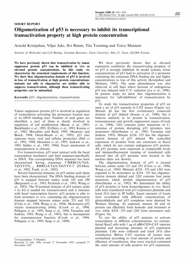
used a set of p53 mutants in CAT assays (Figure 1a).Mutant DI has the ®rst evolutionary conserveddomain of p53 deleted (Soussi et al., 1990), but itbehaves similarly to wt protein in transactivation,transrepression and growth suppression assays (Crooket al., 1994). 1262 contains point mutations in C-terminus of protein disrupting p53 tetramers intomonomers (StuÈ rzbecher et al., 1992; Tarunina andJenkins, 1993). Mutant D324 ± 355 has the oligomer-ization domain of p53 deleted. We controlledexpression of proteins from our constructs in Saos2cells, which do not contain endogenous p53 protein.All p53 proteins were expressed at comparable levelsand immuno¯uorescent staining of transfected cellsshowed that all p53 proteins were located in thenucleus (data not shown).The oligomerization domain of p53 is located
between amino acids 323 and 355 (Clore et al., 1994;Wang et al., 1994). Mutants D324 ± 355 and 1262 wereexpected to be monomers as D324 ± 355 has oligomer-ization domain deleted and 1262 contains four pointmutations which abolish oligomerization of p53(StuÈ rzbecher et al., 1992). We determined the abilityof p53 proteins to form homooligomers in vivo. Saos2cells were transfected with p53 expression plasmids andlysed 24 h later in IP bu�er. Equal amounts of lysatewere crosslinked with di�erent concentrations ofglutaraldehyde and p53 complexes were detected byWestern blotting. As expected, mutant DI and wtprotein can e�ciently form homodi- and tetramers invivo, while D324 ± 355 and 1262 form monomers only(Figure 1b).To test the ability of p53 mutants to activate
transcription at di�erent concentrations, we cotrans-fected Saos2 cells with 1 mg of pBS-CON-CAT reporterplasmid and increasing amounts of p53 expressionplasmids. Cells were collected and lysed 24 h aftertransfection. Before CAT reaction all lysates werenormalized according to total amount of protein ande�ciency of transfection, thus every reaction containedthe same amount of cells positive for p53 expression.
Correspondence: T MaimetsReceived 22 July 1996; revised 27 November 1997; accepted 27November 1997
Oncogene (1998) 16, 2413 ± 2418 1998 Stockton Press All rights reserved 0950 ± 9232/98 $12.00
http://www.stockton-press.co.uk/onc
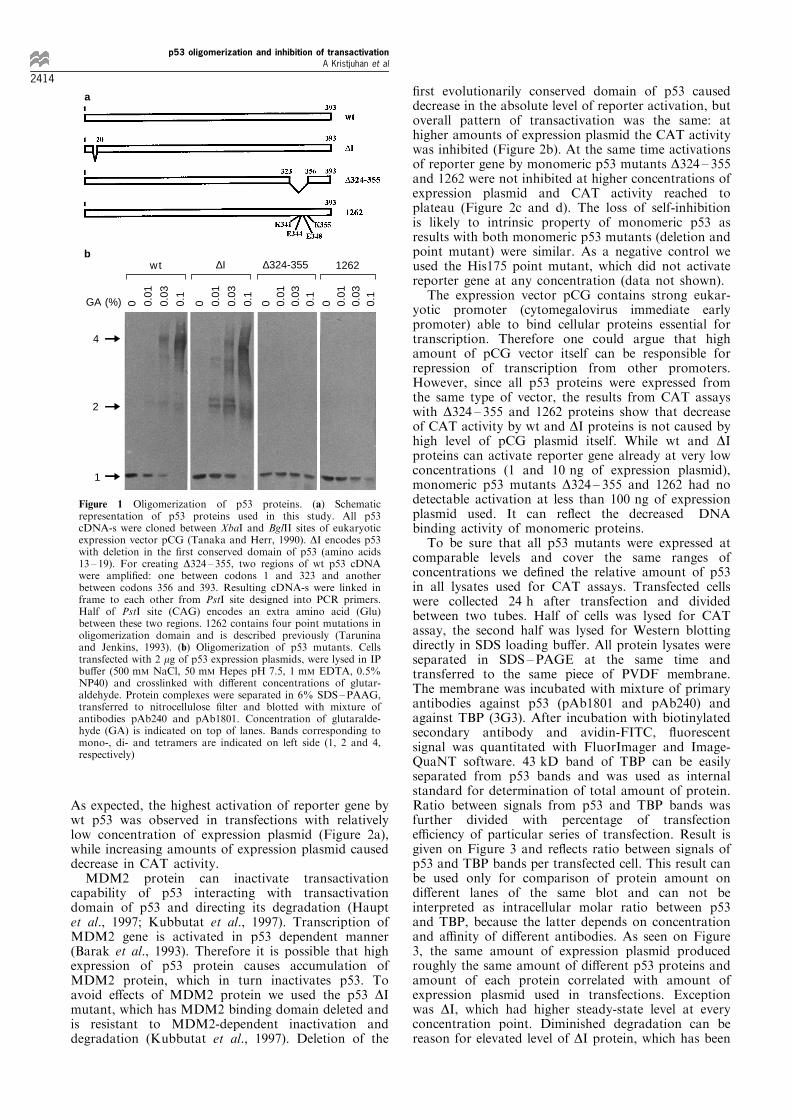
As expected, the highest activation of reporter gene bywt p53 was observed in transfections with relativelylow concentration of expression plasmid (Figure 2a),while increasing amounts of expression plasmid causeddecrease in CAT activity.MDM2 protein can inactivate transactivation
capability of p53 interacting with transactivationdomain of p53 and directing its degradation (Hauptet al., 1997; Kubbutat et al., 1997). Transcription ofMDM2 gene is activated in p53 dependent manner(Barak et al., 1993). Therefore it is possible that highexpression of p53 protein causes accumulation ofMDM2 protein, which in turn inactivates p53. Toavoid e�ects of MDM2 protein we used the p53 DImutant, which has MDM2 binding domain deleted andis resistant to MDM2-dependent inactivation anddegradation (Kubbutat et al., 1997). Deletion of the
®rst evolutionarily conserved domain of p53 causeddecrease in the absolute level of reporter activation, butoverall pattern of transactivation was the same: athigher amounts of expression plasmid the CAT activitywas inhibited (Figure 2b). At the same time activationsof reporter gene by monomeric p53 mutants D324 ± 355and 1262 were not inhibited at higher concentrations ofexpression plasmid and CAT activity reached toplateau (Figure 2c and d). The loss of self-inhibitionis likely to intrinsic property of monomeric p53 asresults with both monomeric p53 mutants (deletion andpoint mutant) were similar. As a negative control weused the His175 point mutant, which did not activatereporter gene at any concentration (data not shown).The expression vector pCG contains strong eukar-
yotic promoter (cytomegalovirus immediate earlypromoter) able to bind cellular proteins essential fortranscription. Therefore one could argue that highamount of pCG vector itself can be responsible forrepression of transcription from other promoters.However, since all p53 proteins were expressed fromthe same type of vector, the results from CAT assayswith D324 ± 355 and 1262 proteins show that decreaseof CAT activity by wt and DI proteins is not caused byhigh level of pCG plasmid itself. While wt and DIproteins can activate reporter gene already at very lowconcentrations (1 and 10 ng of expression plasmid),monomeric p53 mutants D324 ± 355 and 1262 had nodetectable activation at less than 100 ng of expressionplasmid used. It can re¯ect the decreased DNAbinding activity of monomeric proteins.To be sure that all p53 mutants were expressed at
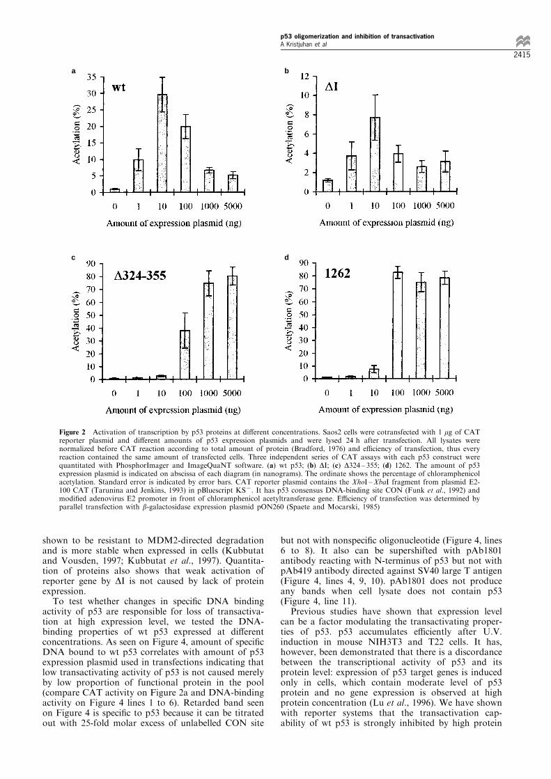
comparable levels and cover the same ranges ofconcentrations we de®ned the relative amount of p53in all lysates used for CAT assays. Transfected cellswere collected 24 h after transfection and dividedbetween two tubes. Half of cells was lysed for CATassay, the second half was lysed for Western blottingdirectly in SDS loading bu�er. All protein lysates wereseparated in SDS ±PAGE at the same time andtransferred to the same piece of PVDF membrane.The membrane was incubated with mixture of primaryantibodies against p53 (pAb1801 and pAb240) andagainst TBP (3G3). After incubation with biotinylatedsecondary antibody and avidin-FITC, ¯uorescentsignal was quantitated with FluorImager and Image-QuaNT software. 43 kD band of TBP can be easilyseparated from p53 bands and was used as internalstandard for determination of total amount of protein.Ratio between signals from p53 and TBP bands wasfurther divided with percentage of transfectione�ciency of particular series of transfection. Result isgiven on Figure 3 and re¯ects ratio between signals ofp53 and TBP bands per transfected cell. This result canbe used only for comparison of protein amount ondi�erent lanes of the same blot and can not beinterpreted as intracellular molar ratio between p53and TBP, because the latter depends on concentrationand a�nity of di�erent antibodies. As seen on Figure3, the same amount of expression plasmid producedroughly the same amount of di�erent p53 proteins andamount of each protein correlated with amount ofexpression plasmid used in transfections. Exceptionwas DI, which had higher steady-state level at everyconcentration point. Diminished degradation can bereason for elevated level of DI protein, which has been
4
2
1
GA (%) 0 0.01
0.03
0.1
0 0.01
0.03
0.1
0 0.01
0.
03
0.1
0 0.01
0.
03
0.1
wt ∆I ∆324-355 1262
a
b
Figure 1 Oligomerization of p53 proteins. (a) Schematicrepresentation of p53 proteins used in this study. All p53cDNA-s were cloned between XbaI and BglII sites of eukaryoticexpression vector pCG (Tanaka and Herr, 1990). DI encodes p53with deletion in the ®rst conserved domain of p53 (amino acids13 ± 19). For creating D324 ± 355, two regions of wt p53 cDNAwere ampli®ed: one between codons 1 and 323 and anotherbetween codons 356 and 393. Resulting cDNA-s were linked inframe to each other from PstI site designed into PCR primers.Half of PstI site (CAG) encodes an extra amino acid (Glu)between these two regions. 1262 contains four point mutations inoligomerization domain and is described previously (Taruninaand Jenkins, 1993). (b) Oligomerization of p53 mutants. Cellstransfected with 2 mg of p53 expression plasmids, were lysed in IPbu�er (500 mM NaCl, 50 mM Hepes pH 7.5, 1 mM EDTA, 0.5%NP40) and crosslinked with di�erent concentrations of glutar-aldehyde. Protein complexes were separated in 6% SDS±PAAG,transferred to nitrocellulose ®lter and blotted with mixture ofantibodies pAb240 and pAb1801. Concentration of glutaralde-hyde (GA) is indicated on top of lanes. Bands corresponding tomono-, di- and tetramers are indicated on left side (1, 2 and 4,respectively)
p53 oligomerization and inhibition of transactivationA Kristjuhan et al
2414
shown to be resistant to MDM2-directed degradationand is more stable when expressed in cells (Kubbutatand Vousden, 1997; Kubbutat et al., 1997). Quantita-tion of proteins also shows that weak activation ofreporter gene by DI is not caused by lack of proteinexpression.To test whether changes in speci®c DNA binding
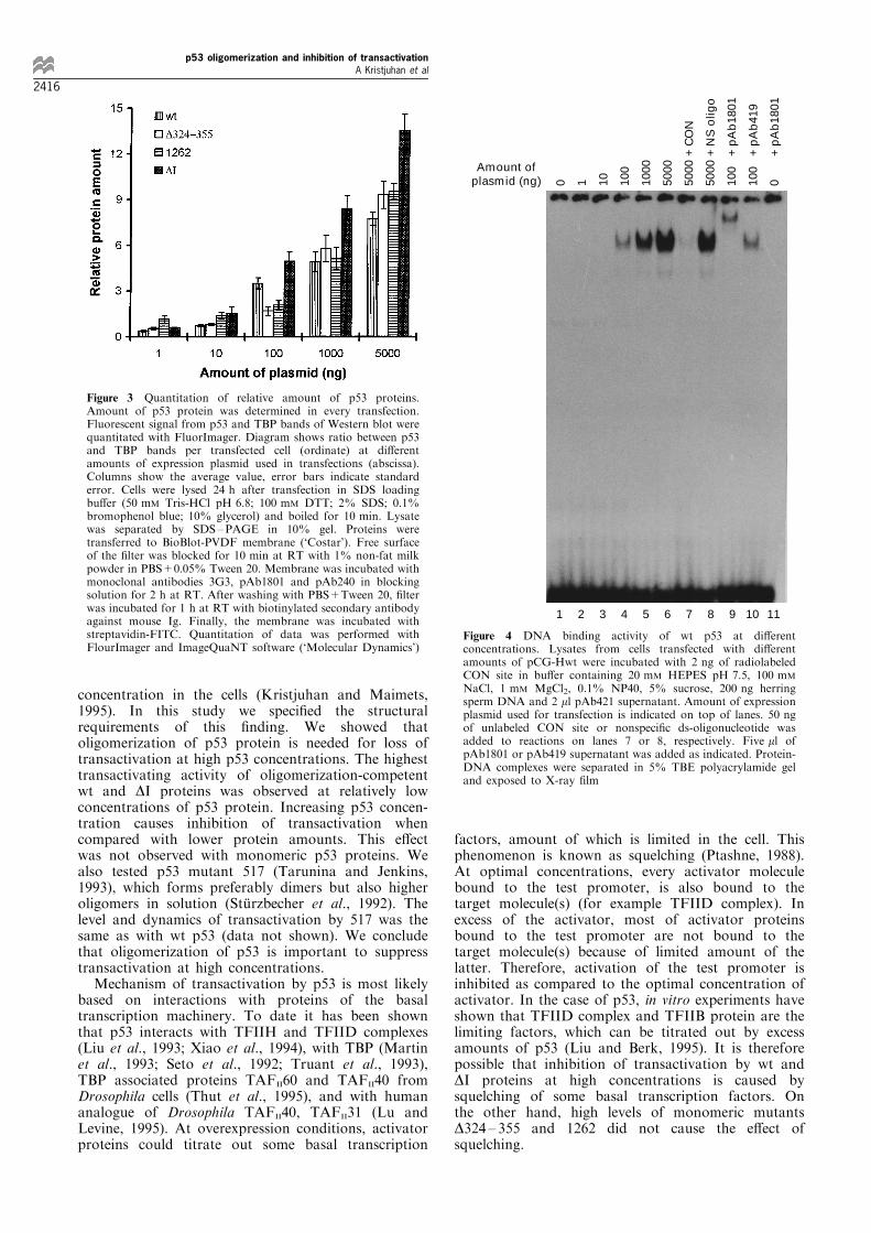
activity of p53 are responsible for loss of transactiva-tion at high expression level, we tested the DNA-binding properties of wt p53 expressed at di�erentconcentrations. As seen on Figure 4, amount of speci®cDNA bound to wt p53 correlates with amount of p53expression plasmid used in transfections indicating thatlow transactivating activity of p53 is not caused merelyby low proportion of functional protein in the pool(compare CAT activity on Figure 2a and DNA-bindingactivity on Figure 4 lines 1 to 6). Retarded band seenon Figure 4 is speci®c to p53 because it can be titratedout with 25-fold molar excess of unlabelled CON site
but not with nonspeci®c oligonucleotide (Figure 4, lines6 to 8). It also can be supershifted with pAb1801antibody reacting with N-terminus of p53 but not withpAb419 antibody directed against SV40 large T antigen(Figure 4, lines 4, 9, 10). pAb1801 does not produceany bands when cell lysate does not contain p53(Figure 4, line 11).Previous studies have shown that expression level
can be a factor modulating the transactivating proper-ties of p53. p53 accumulates e�ciently after U.V.induction in mouse NIH3T3 and T22 cells. It has,however, been demonstrated that there is a discordancebetween the transcriptional activity of p53 and itsprotein level: expression of p53 target genes is inducedonly in cells, which contain moderate level of p53protein and no gene expression is observed at highprotein concentration (Lu et al., 1996). We have shownwith reporter systems that the transactivation cap-ability of wt p53 is strongly inhibited by high protein
a b
c d
Figure 2 Activation of transcription by p53 proteins at di�erent concentrations. Saos2 cells were cotransfected with 1 mg of CATreporter plasmid and di�erent amounts of p53 expression plasmids and were lysed 24 h after transfection. All lysates werenormalized before CAT reaction according to total amount of protein (Bradford, 1976) and e�ciency of transfection, thus everyreaction contained the same amount of transfected cells. Three independent series of CAT assays with each p53 construct werequantitated with PhosphorImager and ImageQuaNT software. (a) wt p53; (b) DI; (c) D324 ± 355; (d) 1262. The amount of p53expression plasmid is indicated on abscissa of each diagram (in nanograms). The ordinate shows the percentage of chloramphenicolacetylation. Standard error is indicated by error bars. CAT reporter plasmid contains the XhoI ±XbaI fragment from plasmid E2-100 CAT (Tarunina and Jenkins, 1993) in pBluescript KS7. It has p53 consensus DNA-binding site CON (Funk et al., 1992) andmodi®ed adenovirus E2 promoter in front of chloramphenicol acetyltransferase gene. E�ciency of transfection was determined byparallel transfection with b-galactosidase expression plasmid pON260 (Spaete and Mocarski, 1985)
p53 oligomerization and inhibition of transactivationA Kristjuhan et al
2415
concentration in the cells (Kristjuhan and Maimets,1995). In this study we speci®ed the structuralrequirements of this ®nding. We showed thatoligomerization of p53 protein is needed for loss oftransactivation at high p53 concentrations. The highesttransactivating activity of oligomerization-competentwt and DI proteins was observed at relatively lowconcentrations of p53 protein. Increasing p53 concen-tration causes inhibition of transactivation whencompared with lower protein amounts. This e�ectwas not observed with monomeric p53 proteins. Wealso tested p53 mutant 517 (Tarunina and Jenkins,1993), which forms preferably dimers but also higheroligomers in solution (StuÈ rzbecher et al., 1992). Thelevel and dynamics of transactivation by 517 was thesame as with wt p53 (data not shown). We concludethat oligomerization of p53 is important to suppresstransactivation at high concentrations.Mechanism of transactivation by p53 is most likely
based on interactions with proteins of the basaltranscription machinery. To date it has been shownthat p53 interacts with TFIIH and TFIID complexes(Liu et al., 1993; Xiao et al., 1994), with TBP (Martinet al., 1993; Seto et al., 1992; Truant et al., 1993),TBP associated proteins TAFII60 and TAFII40 fromDrosophila cells (Thut et al., 1995), and with humananalogue of Drosophila TAFII40, TAFII31 (Lu andLevine, 1995). At overexpression conditions, activatorproteins could titrate out some basal transcription
factors, amount of which is limited in the cell. Thisphenomenon is known as squelching (Ptashne, 1988).At optimal concentrations, every activator moleculebound to the test promoter, is also bound to thetarget molecule(s) (for example TFIID complex). Inexcess of the activator, most of activator proteinsbound to the test promoter are not bound to thetarget molecule(s) because of limited amount of thelatter. Therefore, activation of the test promoter isinhibited as compared to the optimal concentration ofactivator. In the case of p53, in vitro experiments haveshown that TFIID complex and TFIIB protein are thelimiting factors, which can be titrated out by excessamounts of p53 (Liu and Berk, 1995). It is thereforepossible that inhibition of transactivation by wt andDI proteins at high concentrations is caused bysquelching of some basal transcription factors. Onthe other hand, high levels of monomeric mutantsD324 ± 355 and 1262 did not cause the e�ect ofsquelching.
Figure 3 Quantitation of relative amount of p53 proteins.Amount of p53 protein was determined in every transfection.Fluorescent signal from p53 and TBP bands of Western blot werequantitated with FluorImager. Diagram shows ratio between p53and TBP bands per transfected cell (ordinate) at di�erentamounts of expression plasmid used in transfections (abscissa).Columns show the average value, error bars indicate standarderror. Cells were lysed 24 h after transfection in SDS loadingbu�er (50 mM Tris-HCl pH 6.8; 100 mM DTT; 2% SDS; 0.1%bromophenol blue; 10% glycerol) and boiled for 10 min. Lysatewas separated by SDS±PAGE in 10% gel. Proteins weretransferred to BioBlot-PVDF membrane (`Costar'). Free surfaceof the ®lter was blocked for 10 min at RT with 1% non-fat milkpowder in PBS+0.05% Tween 20. Membrane was incubated withmonoclonal antibodies 3G3, pAb1801 and pAb240 in blockingsolution for 2 h at RT. After washing with PBS+Tween 20, ®lterwas incubated for 1 h at RT with biotinylated secondary antibodyagainst mouse Ig. Finally, the membrane was incubated withstreptavidin-FITC. Quantitation of data was performed withFlourImager and ImageQuaNT software (`Molecular Dynamics')
0 1 10
100
1000
5000
5000
5000
100
100
0
1 2 3 4 5 6 7 8 9 10 11
Amount of plasmid (ng)
+ C
ON
+ N
S o
ligo
+ p
Ab
1801
+ p
Ab
419
+ p
Ab
1801
Figure 4 DNA binding activity of wt p53 at di�erentconcentrations. Lysates from cells transfected with di�erentamounts of pCG-Hwt were incubated with 2 ng of radiolabeledCON site in bu�er containing 20 mM HEPES pH 7.5, 100 mM
NaCl, 1 mM MgCl2, 0.1% NP40, 5% sucrose, 200 ng herringsperm DNA and 2 ml pAb421 supernatant. Amount of expressionplasmid used for transfection is indicated on top of lanes. 50 ngof unlabeled CON site or nonspeci®c ds-oligonucleotide wasadded to reactions on lanes 7 or 8, respectively. Five ml ofpAb1801 or pAb419 supernatant was added as indicated. Protein-DNA complexes were separated in 5% TBE polyacrylamide geland exposed to X-ray ®lm
p53 oligomerization and inhibition of transactivationA Kristjuhan et al
2416
One possible explanation here is that monomericp53 can not interact with transcription machinery at alland therefore it can not squelch basal transcriptionfactors. Analogously, oligomeric state of p53 is neededfor interaction with MDM2 (Marston et al., 1995).Although speci®c complex between DNA and mono-meric p53 is poorly detectable in cell lysate, puri®edp53 monomers are capable to bind cooperatively toconsensus DNA (Balagurumoorthy et al., 1995; Wanget al., 1995). p53 binding DNA consensus sequencecontains two tandem decameric elements, each contain-ing two pentameric inverted repeats. Therefore, theDNA itself can be a factor, which brings four p53monomers together and p53 could gain the ability tointeract with transcription machinery only afterbinding to the DNA consensus sequence. Increasingconcentration of monomeric p53 in the cell couldincrease probability that more test promoters arebound to multiple p53 monomers, which in turnassures higher transcription e�ciency from thepromoter.
In this paper we have shown that the ability of p53to activate transcription may be regulated by its stateof oligomerization. In that sense, p53 is similar toanother transcription factor, Drosophila protein KruÈ p-pel (Kr). Monomeric Kr can act as a transcriptionalactivator, whereas Kr oligomers formed at highconcentrations cause repression. Interactions withdi�erent parts of transcriptional machinery areresponsible for these e�ects. (Sauer et al., 1995).
AcknowledgementsWe thank Dr K Vousden for p53 cDNA mutant DI, Dr JJenkins for plasmid E2-100 and mutants of p53 and Dr PChambon for anti-TBP antibody 3G3. The most usefuldiscussions with Arvi JoÄ ers are highly appreciated. Thiswork was partly supported by grants CT94002 andCIPACT930257 from European Commission and grantsfrom Estonian Science Foundation (no. 2315 and 2316).
References
Balagurumoorthy P, Sakamoto H, Lewis MS, Zambrano N,Clore GM, Gronenborn AM, Appella E and HarringtonRE. (1995). Proc. Natl. Acad. Sci. USA, 92, 8591 ± 8595.
Barak Y, Juven T, Ha�ner R and Oren M. (1993). EMBO J.,12, 461 ± 468.
Bargonetti J, Manfredi JJ, Chen X, Marshak DR and PrivesC. (1993). Genes Dev., 7, 2565 ± 2574.
Bradford MM. (1976). Analytical Biochemistry, 72, 248 ±254.
Buckbinder L, Talbott R, Velasco-Miguel S, Takenaka I,Faha B, Seizinger BR and Kley N. (1995). Nature, 377,646 ± 649.
Clore GM, Omichinski JG, Sakaguchi K, Zambrano N.Sakamoto H, Appella E and Gronenborn AM. (1994).Science, 265, 386 ± 391.
Crook T, Marston NJ, Sara EA and Vousden KH. (1994).Cell, 79, 817 ± 827.
Deb S, Jackson CT, Subler MA and Martin DW. (1992). J.Virol., 66, 6164 ± 6170.
Deb SP, MunÄ oz RM, Brown DR, Subler MA and Deb S.(1994). Oncogene, 9, 1341 ± 1349.
Desaintes C, Hallez S, Detremmerie O and Burny A. (1995).Oncogene, 10, 2155 ± 2161.
El-Deiry W, Kern SE, Pietenpol JA, Kinzler KW andVogelstein B. (1992). Nature Genetics, 1, 45 ± 49.
El-Deiry WS, Tokino T, Velculescu VE, Levy DB, ParsonsR, Trent JM, Lin D, Mercer WE, Kinzler KW andVogelstein B. (1993). Cell, 75, 817 ± 825.
Funk WD, Pak DT, Karas RH, Wright WE and Shay JW.(1992). Mol. Cell. Biol., 12, 2866 ± 2871.
Haupt Y, Maya R, Kazaz A and Oren M. (1997). Nature,387, 296 ± 299.
Jackson P, Bos E and Braithwaite AW. (1993). Oncogene, 8,589 ± 597.
Kastan MB, Zhan Q, El-Deiry WS, Carrier F, Jacks T,Walsh WV, Plunkett BS, Vogelstein B and Fornace AJ Jr.(1992). Cell, 71, 587 ± 597.
Kristjuhan A and Maimets T. (1995). Eur. J. Biochem., 234,827 ± 831.
Kubbutat MH and Vousden KH. (1997).Mol. Cell. Biol., 17,460 ± 468.
Kubbutat MH, Jones SN and Vousden KH. (1997). Nature,387, 299 ± 303.
Liu X and Berk AJ. (1995). Mol. Cell. Biol., 15, 6474 ± 6478.
Liu X, Miller CW, Koe�er PH and Berk AJ. (1993). Mol.Cell. Biol., 13, 3291 ± 3300.
Lu H and Levine AJ. (1995). Proc. Natl. Acad. Sci. USA, 92,5154 ± 5158.
Lu X, Burbidge SA, Gri�n S and Smith HM. (1996).Oncogene, 13, 413 ± 418.
Marston NJ, Jenkins JR and Vousden KH. (1995).Oncogene, 10, 1709 ± 1715.
Martin DW, MunÄ oz RM, Subler MA and Deb S. (1993). J.Biol. Chem., 268, 13062 ± 13067.
Miyashita T and Reed JC. (1995). Cell, 80, 293 ± 299.Okamoto K and Beach D. (1994). EMBO J., 13, 4816 ± 4822.Owen-Shaub LB, Zhang W, Cusack JC, Angelo LS, SanteeSM, Fujiwara T, Roth JA, Deisseroth AB, Zhang WW,Kruzel E and Radinsky R. (1995). Mol. Cell. Biol., 15,3032 ± 3040.
Pavletich NP, Chambers KA and Pabo CO. (1993). GenesDev., 7, 2556 ± 2564.
Pellegata NS, Cajot JF and Stanbridge EJ. (1995). Oncogene,11, 337 ± 349.
Ptashne M. (1988). Nature, 335, 683 ± 689.Sang BC, Chen JY, Minna J and Barbosa MS. (1994).
Oncogene, 9, 853 ± 859.Sauer F, Fondell JD, Ohkuma Y, Roeder RG and Jackle H.(1995). Nature, 375, 162 ± 164.
Seto E, Usheva A, Zambetti GP, Momand J, Horikoshi N,Weinmann R, Levine AJ and Shenk T. (1992). Proc. Natl.Acad. Sci. USA, 89, 12028 ± 12032.
Soussi T, Caron de Fromentel C and May P. (1990).Oncogene, 5, 945 ± 952.
Spaete RR and Mocarski ES. (1985). J. Virol., 56, 135 ± 143.StuÈ rzbecher H-W, Brain R, Addison C, Rudge K, Remm M,Grimaldi M, Keenan E and Jenkins JR. (1992). Oncogene,7, 1513 ± 1523.
Subler MA, Martin DW and Deb S. (1992). J. Virol., 66,4757 ± 4762.
Subler MA, Martin DW and Deb S. (1994). Oncogene, 9,1351 ± 1359.
Tanaka M and Herr W. (1990). Cell, 60, 375 ± 386.Tarunina M and Jenkins JR. (1993). Oncogene, 8, 3165 ±3173.
Thut CJ, Chen JL, Klemm R and Tjian R. (1995). Science,267, 100 ± 104.
p53 oligomerization and inhibition of transactivationA Kristjuhan et al
2417
Truant R, Xiao H, Ingles CJ and Greenblatt J. (1993). J.Biol. Chem., 268, 2284 ± 2287.
Wang P, Reed M, Wang, Y, Mayr G, Stenger JE, AndersonME, Schwedes JF and Tegtmeyer P. (1994). Mol. Cell.Biol., 14, 5182 ± 5191.
Wang Y, Reed M, Wang P, Stenger JE, Mayr G, AndersonME, Schwedes JF and Tegtmeyer P. (1993). Genes Dev., 7,2575 ± 2586.
Wang Y, Schwedes JF, Parks D, Mann K and Tegtmeyer P.(1995). Mol. Cell. Biol., 15, 2157 ± 2165.
Xiao H, Pearson A, Coulombe B, Truant R, Zhang S, RegierJL, Triezenberg SJ, Reinberg D, Flores O, Ingles CJ andGreenblatt J. (1994). Mol. Cell. Biol., 14, 7013 ± 7024.
p53 oligomerization and inhibition of transactivationA Kristjuhan et al
2418


