Embed Size (px)
Citation preview
doi:10.1016/j.jmb.2005.12.083 J. Mol. Biol. (2006) 357, 1039–1049
Kinetic Computational Alanine Scanning:Application to p53 Oligomerization
Lillian T. Chong1,2, William C. Swope2*, Jed W. Pitera2
and Vijay S. Pande1*
1Departments of ChemistryStructural Biology, StanfordUniversity, Stanford, CA94305-5447, USA
2IBMAlmaden Research Center650 Harry Road, San JoseCA 95120, USA
0022-2836/$ - see front matter q 2005 E
Abbreviations used: p53tet, p53 odomain; MD, molecular dynamics;squared deviation.
E-mail addresses of the [email protected]; pande@
We have developed a novel computational alanine scanning approach thatinvolves analysis of ensemble unfolding kinetics at high temperature toidentify residues that are critical for the stability of a given protein. Thisapproach has been applied to dimerization of the oligomerization domain(residues 326–355) of tumor suppressor p53. As validated by experimentalresults, our approach has reasonable success in identifying deleteriousmutations, including mutations that have been linked to cancer. We discussa method for determining the effect of mutations on the location of thedimerization transition state.
q 2005 Elsevier Ltd. All rights reserved.
Keywords: tumor suppressor p53; oligomerization; alanine scanning;ensemble unfolding kinetics; molecular dynamics simulation
*Corresponding authorIntroduction
Tumor suppressor p53 is a “gatekeeper” of thegenome, functioning at the center of a network ofbiological pathways that guard the cell frompotential cancer. As a multidomain transcriptionfactor, p53 is comprised of an N-terminal acti-vation domain, a central DNA-binding domain, anoligomerization domain, and a C-terminal regu-latory domain. The oligomerization domainenables p53 to adopt its biologically activetetrameric form. More than half of human cancersresult from mutations in the p53 gene.1 Althoughmost of these mutations are in the DNA-bindingdomain, several studies have identified mutationswithin the oligomerization domain (p53tet) thatare linked to increased incidence of cancer.2–5
More cancer-associated mutations may yet beidentified in p53tet, due to the fact that this regionof the p53 gene was not sequenced in studies thatsearched for mutations in the p53 DNA-bindingdomain.3
Structures of p53tet have been determined byboth NMR spectroscopy (Protein Data Bank 1PES6
and 1SAK7) and X-ray crystallography (ProteinData Bank 1C268 and 1AIE9). This a/b domain,
lsevier Ltd. All rights reserve
ligomerizationRMSD, root-mean-
ding authors:stanford.edu
which is one of the smallest known proteinoligomerization domains, assembles as a dimer ofdimers.10 Each dimer consists of an antiparallelb-sheet and a pair of antiparallel helices. The twodimers associate with one another to form theactive tetramer via an extensive hydrophobicsurface that is formed by the antiparallel helicesof each dimer. Based on kinetics experiments andF-value analysis, tetramerization of p53tet isthought to involve induced-fit associations ofmonomers to form the dimer intermediatesfollowed by “lock-and-key” associations of pre-organized dimers to form the tetramers.10 A recentstudy of p53 biogenesis in vitro has shown that p53dimerizes cotranslationally and then forms thetetramer post-translationally.11 Given the kineticadvantage of dimerization before the p53 chainsleave the polysome, mutations that affect thekinetics could play important biological roles.Consistent with the importance of kinetics in p53function, molecular dynamics (MD) simulations ofp53tet dimerization evolving from the rate-limitingtransition state ensemble have revealed a nuclea-tion-condensation mechanism in which L330, I332,and F338 from each monomer form a foldingnucleus.12
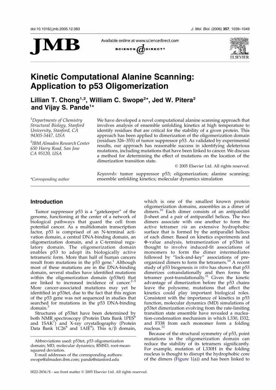
Because of the structural symmetry of p53, pointmutations in the oligomerization domain canreduce the stability of its tetramers significantly.For example, mutation of L330H in the foldingnucleus is thought to disrupt the hydrophobic coreof the dimers (Figure 1(a)) and has been linked to
d.
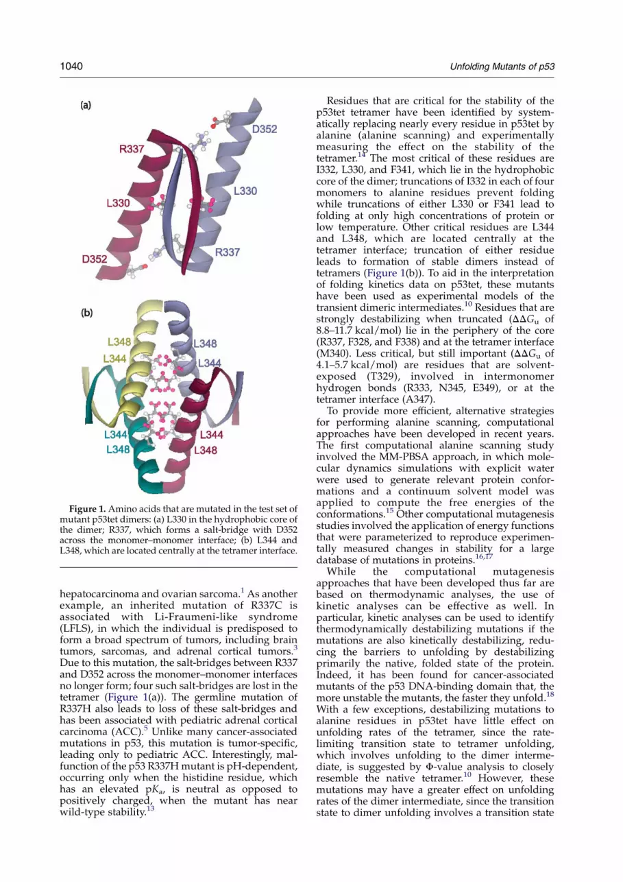
Figure 1. Amino acids that are mutated in the test set ofmutant p53tet dimers: (a) L330 in the hydrophobic core ofthe dimer; R337, which forms a salt-bridge with D352across the monomer–monomer interface; (b) L344 andL348, which are located centrally at the tetramer interface.
1040 Unfolding Mutants of p53
hepatocarcinoma and ovarian sarcoma.1 As anotherexample, an inherited mutation of R337C isassociated with Li-Fraumeni-like syndrome(LFLS), in which the individual is predisposed toform a broad spectrum of tumors, including braintumors, sarcomas, and adrenal cortical tumors.3
Due to this mutation, the salt-bridges between R337and D352 across the monomer–monomer interfacesno longer form; four such salt-bridges are lost in thetetramer (Figure 1(a)). The germline mutation ofR337H also leads to loss of these salt-bridges andhas been associated with pediatric adrenal corticalcarcinoma (ACC).5 Unlike many cancer-associatedmutations in p53, this mutation is tumor-specific,leading only to pediatric ACC. Interestingly, mal-function of the p53 R337H mutant is pH-dependent,occurring only when the histidine residue, whichhas an elevated pKa, is neutral as opposed topositively charged, when the mutant has nearwild-type stability.13
Residues that are critical for the stability of thep53tet tetramer have been identified by system-atically replacing nearly every residue in p53tet byalanine (alanine scanning) and experimentallymeasuring the effect on the stability of thetetramer.14 The most critical of these residues areI332, L330, and F341, which lie in the hydrophobiccore of the dimer; truncations of I332 in each of fourmonomers to alanine residues prevent foldingwhile truncations of either L330 or F341 lead tofolding at only high concentrations of protein orlow temperature. Other critical residues are L344and L348, which are located centrally at thetetramer interface; truncation of either residueleads to formation of stable dimers instead oftetramers (Figure 1(b)). To aid in the interpretationof folding kinetics data on p53tet, these mutantshave been used as experimental models of thetransient dimeric intermediates.10 Residues that arestrongly destabilizing when truncated (DDGu of8.8–11.7 kcal/mol) lie in the periphery of the core(R337, F328, and F338) and at the tetramer interface(M340). Less critical, but still important (DDGu of4.1–5.7 kcal/mol) are residues that are solvent-exposed (T329), involved in intermonomerhydrogen bonds (R333, N345, E349), or at thetetramer interface (A347).
To provide more efficient, alternative strategiesfor performing alanine scanning, computationalapproaches have been developed in recent years.The first computational alanine scanning studyinvolved the MM-PBSA approach, in which mole-cular dynamics simulations with explicit waterwere used to generate relevant protein confor-mations and a continuum solvent model wasapplied to compute the free energies of theconformations.15 Other computational mutagenesisstudies involved the application of energy functionsthat were parameterized to reproduce experimen-tally measured changes in stability for a largedatabase of mutations in proteins.16,17
While the computational mutagenesisapproaches that have been developed thus far arebased on thermodynamic analyses, the use ofkinetic analyses can be effective as well. Inparticular, kinetic analyses can be used to identifythermodynamically destabilizing mutations if themutations are also kinetically destabilizing, redu-cing the barriers to unfolding by destabilizingprimarily the native, folded state of the protein.Indeed, it has been found for cancer-associatedmutants of the p53 DNA-binding domain that, themore unstable the mutants, the faster they unfold.18
With a few exceptions, destabilizing mutations toalanine residues in p53tet have little effect onunfolding rates of the tetramer, since the rate-limiting transition state to tetramer unfolding,which involves unfolding to the dimer interme-diate, is suggested by F-value analysis to closelyresemble the native tetramer.10 However, thesemutations may have a greater effect on unfoldingrates of the dimer intermediate, since the transitionstate to dimer unfolding involves a transition state
Unfolding Mutants of p53 1041
with near-zero F-values10 that have been charac-terized by simulations as having some unstructuredregions and native-like regions that exist as residualstructure in the unfolded state.12
Here, we have probed the contributions ofindividual residues to the thermodynamic stabilityof the tetramer using a novel, kinetic computationalalanine scanning approach that involves high-temperature MD simulations of dimer unfolding.Distributed computing is used to provide necessarystatistical precision by running 100 independentunfolding simulations per mutant dimer. We havetested the robustness of this approach by applying itto a test set that consists of the wild-type dimer andsix mutant dimers that are either cancer-associatedor silent: L330H, R337C, R337H, R337HIP, L344A,and L348A where H and HIP indicate neutral andpositively charged histidine, respectively. Ourapproach has reasonable success in identifying themain cancer-associated mutations in the test set andyields alanine scanning results that are qualitativelyconsistent with data from experiments.14
Figure 2. Ensemble fraction unfolded fu(t) as a functionof time for each p53tet dimer in the test set evolving froma single structure that was equilibrated at either (a) 300 Kor (b) 470 K. Structures are based on the initial F0 dimermodel. Uncertainty estimates represent one standarddeviation, as described in Methods.
Results
Test set
A robust protocol for evaluating unfolding rateswas developed using the test set described above(see Methods for details). For each p53tet dimer inthe test set, an ensemble of unfolding pathways wasgenerated by performing 100 independent simu-lations at 470 K for 10 ns. As experimental modelsof the p53tet dimer,10 the L344A and L348A mutantdimers are likely to have near-wild-type stabilityand thus, unfolding rates similar to that of the wild-type dimer. On the other hand, the L330H, R337C,and R337H mutant dimers would be expected tohave faster unfolding rates, since the mutationsdisrupt the monomer–monomer interface signifi-cantly, leading to a predisposition for cancer.1,4,13
Finally, since the R337HIP mutant tetramer hasnear-wild-type stability,13 the R337HIP mutantdimer is likely to have an unfolding rate that issimilar to that of wild-type.
Unfolding rates were found to be sensitive to thestarting conformations used for the high-tempera-ture simulations. For example, unfolding simu-lations for a given dimer that start from a single,equilibrated structure yield unfolding rates thatdiffer depending on the equilibration temperatureused (300 K and 470 K in Figure 2(a) and (b),respectively). In particular, both the R337H andR337HIP mutant dimers have much faster unfol-ding rates relative to the other mutants when anequilibration temperature of 470 K is used insteadof 300 K.
To reduce the sensitivity of unfolding rates to theinitial structure, 100 independent equilibrationswere performed for each dimer, each of whichwas then used for a single unfolding simulation.Results with this new protocol were much less
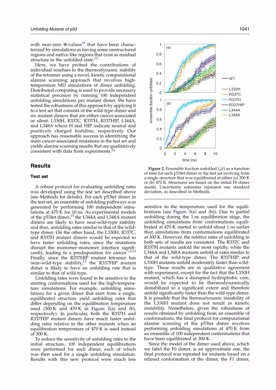
sensitive to the temperature used for the equili-brations (see Figure 3(a) and (b)). Due to partialunfolding during the 1 ns equilibration stage, theunfolding simulations from conformations equili-brated at 470 K started to unfold about 1 ns earlierthan simulations from conformations equilibratedat 300 K. However, the relative rates of unfolding inboth sets of results are consistent. The R337C andR337H mutants unfold the most rapidly, while theL344A and L348A mutants unfold at rates similar tothat of the wild-type dimer. The R337HIP andL330H mutants unfold moderately faster than wild-type. These results are in qualitative agreementwith experiment, except for the fact that the L330Hmutant, which has a disrupted hydrophobic core,would be expected to be thermodynamicallydestabilized to a significant extent and thereforeunfold significantly faster than the wild-type dimer.It is possible that the thermodynamic instability ofthe L330H mutant does not result in kineticinstability. Nonetheless, given the robustness ofresults obtained by unfolding from an ensemble ofconformations, the final protocol for computationalalanine scanning of the p53tet dimer involvesperforming unfolding simulations at 470 K froman ensemble of 100 independent conformations thathave been equilibrated at 300 K.
Since the model of the dimer used above, whichwe label the F0 dimer, is an approximate one, thefinal protocol was repeated for mutants based on arelaxed conformation of the dimer, the F1 dimer,
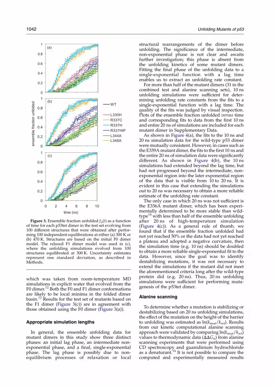
Figure 3. Ensemble fraction unfolded fu(t) as a functionof time for each p53tet dimer in the test set evolving from100 different structures that were obtained after perfor-ming 100 independent equilibrations at either (a) 300 K or(b) 470 K. Structures are based on the initial F0 dimermodel. The relaxed F1 dimer model was used in (c),where the unfolding simulations evolved from 100structures equilibrated at 300 K. Uncertainty estimatesrepresent one standard deviation, as described inMethods.
1042 Unfolding Mutants of p53
which was taken from room-temperature MDsimulations in explicit water that evolved from theF0 dimer.12 Both the F0 and F1 dimer conformationsare likely to be local minima in the folded dimerbasin.12 Results for the test set of mutants based onthe F1 dimer (Figure 3(c)) are in agreement withthose obtained using the F0 dimer (Figure 3(a)).
Appropriate simulation lengths
In general, the ensemble unfolding data formutant dimers in this study show three distinctphases: an initial lag phase, an intermediate non-exponential phase, and a final, single-exponentialphase. The lag phase is possibly due to non-equilibrium processes of relaxation or local
structural rearrangements of the dimer beforeunfolding. The significance of the intermediate,non-exponential phase is not clear and awaitsfurther investigation; this phase is absent fromthe unfolding kinetics of some mutant dimers.Fitting the final phase of the unfolding data to asingle-exponential function with a lag timeenables us to extract an unfolding rate constant.
For more than half of the mutant dimers (31 in thecombined test and alanine scanning sets), 10 nsunfolding simulations were sufficient for deter-mining unfolding rate constants from the fits to asingle-exponential function with a lag time. Thequality of the fits was judged by visual inspection.Plots of the ensemble fraction unfolded versus timeand corresponding fits to data from the first 10 nsand entire 20 ns of simulations are included for eachmutant dimer in Supplementary Data.
As shown in Figure 4(a), the fits to the 10 ns and20 ns simulation data for the wild-type p53 dimerwere mutually consistent. However, in cases such asthe E339A mutant dimer, the fits to the first 10 ns andthe entire 20 ns of simulation data were significantlydifferent. As shown in Figure 4(b), the 10 nssimulations had extended beyond the lag time, buthad not progressed beyond the intermediate, non-exponential region into the later exponential regionof the data that is visible from 10 to 20 ns. It isevident in this case that extending the simulationsout to 20 ns was necessary to obtain a more reliableestimate of the unfolding rate constant.
The only case in which 20 ns was not sufficient isthe E336A mutant dimer, which has been experi-mentally determined to be more stable than wild-type14 with less than half of the ensemble unfoldingafter 20 ns of high-temperature simulation(Figure 4(c)). As a general rule of thumb, wefound that if the ensemble fraction unfolded hadnot yet reached 50% or the data had not yet reacheda plateau and adopted a negative curvature, thenthe simulation time (e.g. 10 ns) should be doubledto obtain a more reliable single-exponential fit to thedata. However, since the goal was to identifydestabilizing mutations, it was not necessary toextend the simulations if the mutant did not meetthe aforementioned criteria long after the wild-typeprotein did (e.g. 20 ns). Thus, 20 ns unfoldingsimulations were sufficient for performing muta-genesis of the p53tet dimer.
Alanine scanning
To determine whether a mutation is stabilizing ordestabilizing based on 20 ns unfolding simulations,the effect of the mutation on the height of the barrierto unfolding was estimated as ln(kmut/kwt). Resultsfrom our kinetic computational alanine scanningapproach were validated by comparing ln(kmut/kwt)values to thermodynamic data (DDGu) from alaninescanning experiments that were performed usingCD spectroscopy and guanidinium hydrochlorideas a denaturant.14 It is not possible to compare thecomputed and experimentally measured results
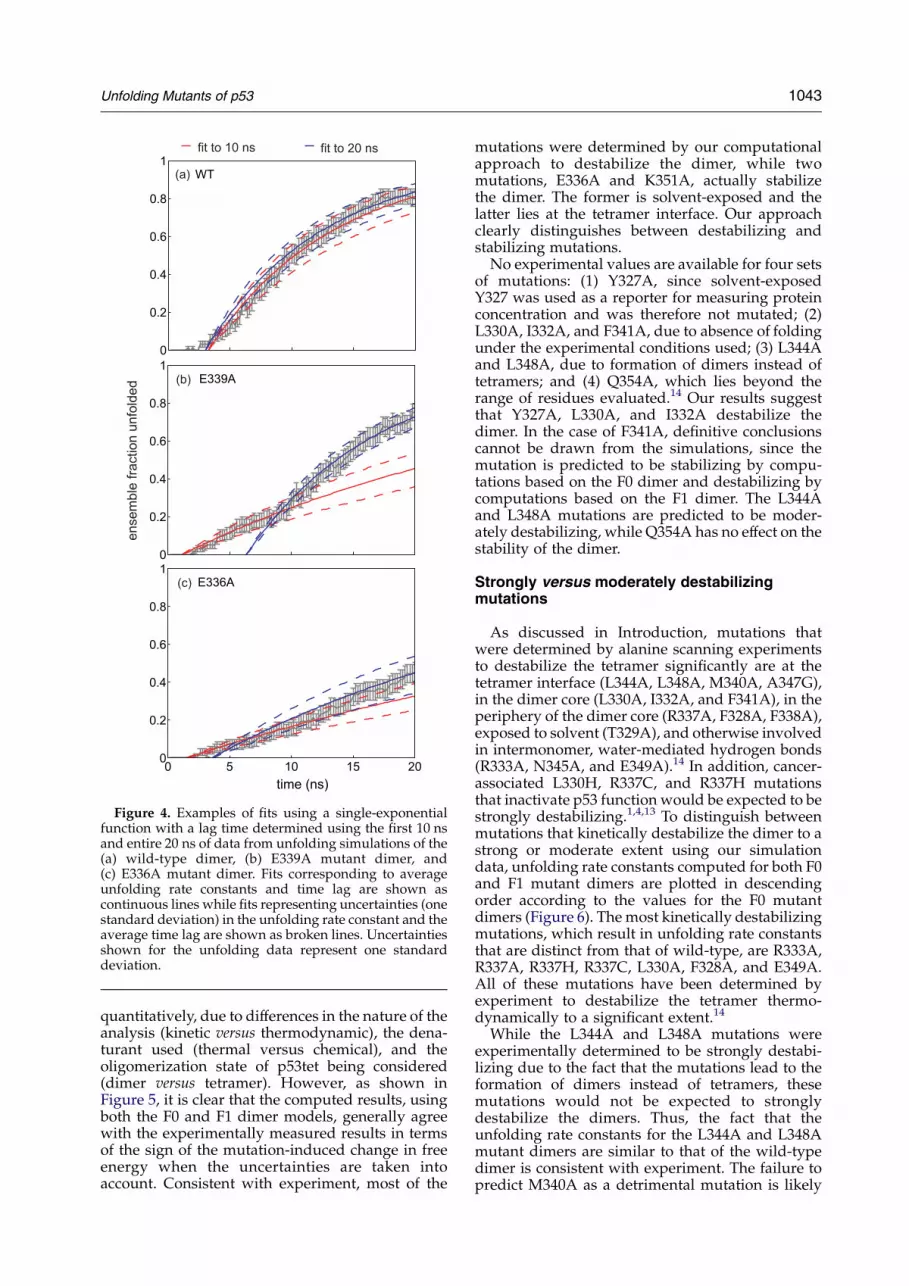
Figure 4. Examples of fits using a single-exponentialfunction with a lag time determined using the first 10 nsand entire 20 ns of data from unfolding simulations of the(a) wild-type dimer, (b) E339A mutant dimer, and(c) E336A mutant dimer. Fits corresponding to averageunfolding rate constants and time lag are shown ascontinuous lines while fits representing uncertainties (onestandard deviation) in the unfolding rate constant and theaverage time lag are shown as broken lines. Uncertaintiesshown for the unfolding data represent one standarddeviation.
Unfolding Mutants of p53 1043
quantitatively, due to differences in the nature of theanalysis (kinetic versus thermodynamic), the dena-turant used (thermal versus chemical), and theoligomerization state of p53tet being considered(dimer versus tetramer). However, as shown inFigure 5, it is clear that the computed results, usingboth the F0 and F1 dimer models, generally agreewith the experimentally measured results in termsof the sign of the mutation-induced change in freeenergy when the uncertainties are taken intoaccount. Consistent with experiment, most of the
mutations were determined by our computationalapproach to destabilize the dimer, while twomutations, E336A and K351A, actually stabilizethe dimer. The former is solvent-exposed and thelatter lies at the tetramer interface. Our approachclearly distinguishes between destabilizing andstabilizing mutations.
No experimental values are available for four setsof mutations: (1) Y327A, since solvent-exposedY327 was used as a reporter for measuring proteinconcentration and was therefore not mutated; (2)L330A, I332A, and F341A, due to absence of foldingunder the experimental conditions used; (3) L344Aand L348A, due to formation of dimers instead oftetramers; and (4) Q354A, which lies beyond therange of residues evaluated.14 Our results suggestthat Y327A, L330A, and I332A destabilize thedimer. In the case of F341A, definitive conclusionscannot be drawn from the simulations, since themutation is predicted to be stabilizing by compu-tations based on the F0 dimer and destabilizing bycomputations based on the F1 dimer. The L344Aand L348A mutations are predicted to be moder-ately destabilizing, while Q354A has no effect on thestability of the dimer.
Strongly versus moderately destabilizingmutations
As discussed in Introduction, mutations thatwere determined by alanine scanning experimentsto destabilize the tetramer significantly are at thetetramer interface (L344A, L348A, M340A, A347G),in the dimer core (L330A, I332A, and F341A), in theperiphery of the dimer core (R337A, F328A, F338A),exposed to solvent (T329A), and otherwise involvedin intermonomer, water-mediated hydrogen bonds(R333A, N345A, and E349A).14 In addition, cancer-associated L330H, R337C, and R337H mutationsthat inactivate p53 function would be expected to bestrongly destabilizing.1,4,13 To distinguish betweenmutations that kinetically destabilize the dimer to astrong or moderate extent using our simulationdata, unfolding rate constants computed for both F0and F1 mutant dimers are plotted in descendingorder according to the values for the F0 mutantdimers (Figure 6). The most kinetically destabilizingmutations, which result in unfolding rate constantsthat are distinct from that of wild-type, are R333A,R337A, R337H, R337C, L330A, F328A, and E349A.All of these mutations have been determined byexperiment to destabilize the tetramer thermo-dynamically to a significant extent.14
While the L344A and L348A mutations wereexperimentally determined to be strongly destabi-lizing due to the fact that the mutations lead to theformation of dimers instead of tetramers, thesemutations would not be expected to stronglydestabilize the dimers. Thus, the fact that theunfolding rate constants for the L344A and L348Amutant dimers are similar to that of the wild-typedimer is consistent with experiment. The failure topredict M340A as a detrimental mutation is likely
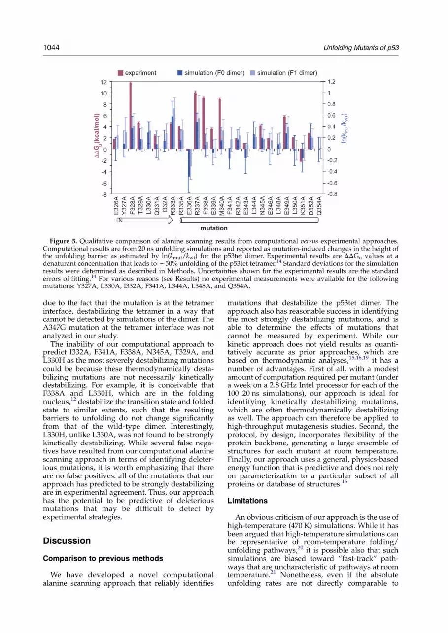
Figure 5. Qualitative comparison of alanine scanning results from computational versus experimental approaches.Computational results are from 20 ns unfolding simulations and reported as mutation-induced changes in the height ofthe unfolding barrier as estimated by ln(kmut/kwt) for the p53tet dimer. Experimental results are DDGu values at adenaturant concentration that leads to w50% unfolding of the p53tet tetramer.14 Standard deviations for the simulationresults were determined as described in Methods. Uncertainties shown for the experimental results are the standarderrors of fitting.14 For various reasons (see Results) no experimental measurements were available for the followingmutations: Y327A, L330A, I332A, F341A, L344A, L348A, and Q354A.
1044 Unfolding Mutants of p53
due to the fact that the mutation is at the tetramerinterface, destabilizing the tetramer in a way thatcannot be detected by simulations of the dimer. TheA347G mutation at the tetramer interface was notanalyzed in our study.
The inability of our computational approach topredict I332A, F341A, F338A, N345A, T329A, andL330H as the most severely destabilizing mutationscould be because these thermodynamically desta-bilizing mutations are not necessarily kineticallydestabilizing. For example, it is conceivable thatF338A and L330H, which are in the foldingnucleus,12 destabilize the transition state and foldedstate to similar extents, such that the resultingbarriers to unfolding do not change significantlyfrom that of the wild-type dimer. Interestingly,L330H, unlike L330A, was not found to be stronglykinetically destabilizing. While several false nega-tives have resulted from our computational alaninescanning approach in terms of identifying deleter-ious mutations, it is worth emphasizing that thereare no false positives: all of the mutations that ourapproach has predicted to be strongly destabilizingare in experimental agreement. Thus, our approachhas the potential to be predictive of deleteriousmutations that may be difficult to detect byexperimental strategies.
Discussion
Comparison to previous methods
We have developed a novel computationalalanine scanning approach that reliably identifies
mutations that destabilize the p53tet dimer. Theapproach also has reasonable success in identifyingthe most strongly destabilizing mutations, and isable to determine the effects of mutations thatcannot be measured by experiment. While ourkinetic approach does not yield results as quanti-tatively accurate as prior approaches, which arebased on thermodynamic analyses,15,16,19 it has anumber of advantages. First of all, with a modestamount of computation required per mutant (undera week on a 2.8 GHz Intel processor for each of the100 20 ns simulations), our approach is ideal foridentifying kinetically destabilizing mutations,which are often thermodynamically destabilizingas well. The approach can therefore be applied tohigh-throughput mutagenesis studies. Second, theprotocol, by design, incorporates flexibility of theprotein backbone, generating a large ensemble ofstructures for each mutant at room temperature.Finally, our approach uses a general, physics-basedenergy function that is predictive and does not relyon parameterization to a particular subset of allproteins or database of structures.16
Limitations
An obvious criticism of our approach is the use ofhigh-temperature (470 K) simulations. While it hasbeen argued that high-temperature simulations canbe representative of room-temperature folding/unfolding pathways,20 it is possible also that suchsimulations are biased toward “fast-track” path-ways that are uncharacteristic of pathways at roomtemperature.21 Nonetheless, even if the absoluteunfolding rates are not directly comparable to
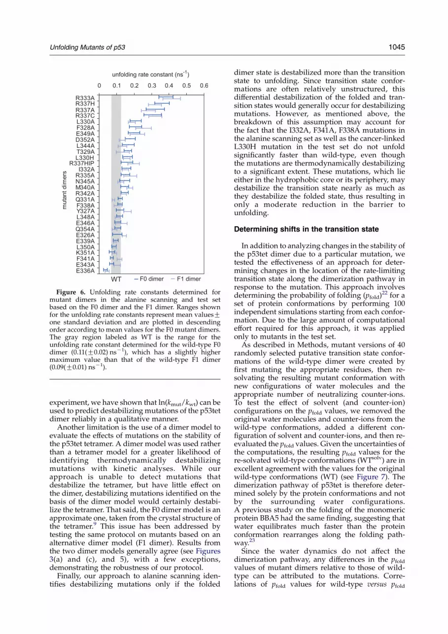
Figure 6. Unfolding rate constants determined formutant dimers in the alanine scanning and test setbased on the F0 dimer and the F1 dimer. Ranges shownfor the unfolding rate constants represent mean valuesGone standard deviation and are plotted in descendingorder according to mean values for the F0 mutant dimers.The gray region labeled as WT is the range for theunfolding rate constant determined for the wild-type F0dimer (0.11(G0.02) nsK1), which has a slightly highermaximum value than that of the wild-type F1 dimer(0.09(G0.01) nsK1).
Unfolding Mutants of p53 1045
experiment, we have shown that ln(kmut/kwt) can beused to predict destabilizing mutations of the p53tetdimer reliably in a qualitative manner.
Another limitation is the use of a dimer model toevaluate the effects of mutations on the stability ofthe p53tet tetramer. A dimer model was used ratherthan a tetramer model for a greater likelihood ofidentifying thermodynamically destabilizingmutations with kinetic analyses. While ourapproach is unable to detect mutations thatdestabilize the tetramer, but have little effect onthe dimer, destabilizing mutations identified on thebasis of the dimer model would certainly destabi-lize the tetramer. That said, the F0 dimer model is anapproximate one, taken from the crystal structure ofthe tetramer.9 This issue has been addressed bytesting the same protocol on mutants based on analternative dimer model (F1 dimer). Results fromthe two dimer models generally agree (see Figures3(a) and (c), and 5), with a few exceptions,demonstrating the robustness of our protocol.
Finally, our approach to alanine scanning iden-tifies destabilizing mutations only if the folded
dimer state is destabilized more than the transitionstate to unfolding. Since transition state confor-mations are often relatively unstructured, thisdifferential destabilization of the folded and tran-sition states would generally occur for destabilizingmutations. However, as mentioned above, thebreakdown of this assumption may account forthe fact that the I332A, F341A, F338A mutations inthe alanine scanning set as well as the cancer-linkedL330H mutation in the test set do not unfoldsignificantly faster than wild-type, even thoughthe mutations are thermodynamically destabilizingto a significant extent. These mutations, which lieeither in the hydrophobic core or its periphery, maydestabilize the transition state nearly as much asthey destabilize the folded state, thus resulting inonly a moderate reduction in the barrier tounfolding.
Determining shifts in the transition state
In addition to analyzing changes in the stability ofthe p53tet dimer due to a particular mutation, wetested the effectiveness of an approach for deter-mining changes in the location of the rate-limitingtransition state along the dimerization pathway inresponse to the mutation. This approach involvesdetermining the probability of folding (pfold)22 for aset of protein conformations by performing 100independent simulations starting from each confor-mation. Due to the large amount of computationaleffort required for this approach, it was appliedonly to mutants in the test set.
As described in Methods, mutant versions of 40randomly selected putative transition state confor-mations of the wild-type dimer were created byfirst mutating the appropriate residues, then re-solvating the resulting mutant conformation withnew configurations of water molecules and theappropriate number of neutralizing counter-ions.To test the effect of solvent (and counter-ion)configurations on the pfold values, we removed theoriginal water molecules and counter-ions from thewild-type conformations, added a different con-figuration of solvent and counter-ions, and then re-evaluated the pfold values. Given the uncertainties ofthe computations, the resulting pfold values for there-solvated wild-type conformations (WTsolv) are inexcellent agreement with the values for the originalwild-type conformations (WT) (see Figure 7). Thedimerization pathway of p53tet is therefore deter-mined solely by the protein conformations and notby the surrounding water configurations.A previous study on the folding of the monomericprotein BBA5 had the same finding, suggesting thatwater equilibrates much faster than the proteinconformation rearranges along the folding path-way.23
Since the water dynamics do not affect thedimerization pathway, any differences in the pfold
values of mutant dimers relative to those of wild-type can be attributed to the mutations. Corre-lations of pfold values for wild-type versus pfold
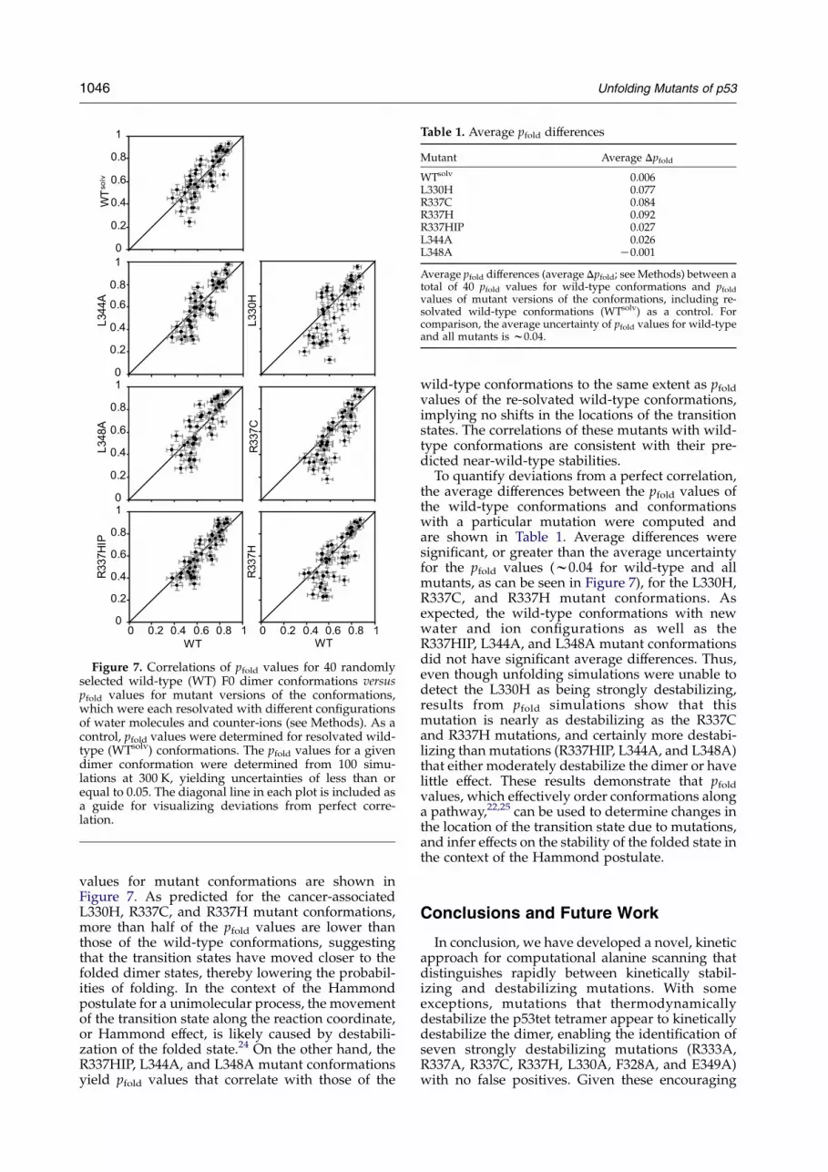
Figure 7. Correlations of pfold values for 40 randomlyselected wild-type (WT) F0 dimer conformations versuspfold values for mutant versions of the conformations,which were each resolvated with different configurationsof water molecules and counter-ions (see Methods). As acontrol, pfold values were determined for resolvated wild-type (WTsolv) conformations. The pfold values for a givendimer conformation were determined from 100 simu-lations at 300 K, yielding uncertainties of less than orequal to 0.05. The diagonal line in each plot is included asa guide for visualizing deviations from perfect corre-lation.
Table 1. Average pfold differences
Mutant Average Dpfold
WTsolv 0.006L330H 0.077R337C 0.084R337H 0.092R337HIP 0.027L344A 0.026L348A K0.001
Average pfold differences (average Dpfold; see Methods) between atotal of 40 pfold values for wild-type conformations and pfold
values of mutant versions of the conformations, including re-solvated wild-type conformations (WTsolv) as a control. Forcomparison, the average uncertainty of pfold values for wild-typeand all mutants is w0.04.
1046 Unfolding Mutants of p53
values for mutant conformations are shown inFigure 7. As predicted for the cancer-associatedL330H, R337C, and R337H mutant conformations,more than half of the pfold values are lower thanthose of the wild-type conformations, suggestingthat the transition states have moved closer to thefolded dimer states, thereby lowering the probabil-ities of folding. In the context of the Hammondpostulate for a unimolecular process, the movementof the transition state along the reaction coordinate,or Hammond effect, is likely caused by destabili-zation of the folded state.24 On the other hand, theR337HIP, L344A, and L348A mutant conformationsyield pfold values that correlate with those of the
wild-type conformations to the same extent as pfold
values of the re-solvated wild-type conformations,implying no shifts in the locations of the transitionstates. The correlations of these mutants with wild-type conformations are consistent with their pre-dicted near-wild-type stabilities.
To quantify deviations from a perfect correlation,the average differences between the pfold values ofthe wild-type conformations and conformationswith a particular mutation were computed andare shown in Table 1. Average differences weresignificant, or greater than the average uncertaintyfor the pfold values (w0.04 for wild-type and allmutants, as can be seen in Figure 7), for the L330H,R337C, and R337H mutant conformations. Asexpected, the wild-type conformations with newwater and ion configurations as well as theR337HIP, L344A, and L348A mutant conformationsdid not have significant average differences. Thus,even though unfolding simulations were unable todetect the L330H as being strongly destabilizing,results from pfold simulations show that thismutation is nearly as destabilizing as the R337Cand R337H mutations, and certainly more destabi-lizing than mutations (R337HIP, L344A, and L348A)that either moderately destabilize the dimer or havelittle effect. These results demonstrate that pfold
values, which effectively order conformations alonga pathway,22,25 can be used to determine changes inthe location of the transition state due to mutations,and infer effects on the stability of the folded state inthe context of the Hammond postulate.
Conclusions and Future Work
In conclusion, we have developed a novel, kineticapproach for computational alanine scanning thatdistinguishes rapidly between kinetically stabil-izing and destabilizing mutations. With someexceptions, mutations that thermodynamicallydestabilize the p53tet tetramer appear to kineticallydestabilize the dimer, enabling the identification ofseven strongly destabilizing mutations (R333A,R337A, R337C, R337H, L330A, F328A, and E349A)with no false positives. Given these encouraging

Unfolding Mutants of p53 1047
results, we are in the process of performing moreextensive mutagenesis of the p53 oligomerizationdomain, analyzing the effects of all possible single-nucleotide polymorphisms at each residue positionin order to predict cancer-associated mutations.Finally, we have shown that pfold simulations can beused to determine mutation-induced shifts in therate-limiting transition state for dimerization thatare consistent with experiment. Both the compu-tational alanine scanning and pfold simulationapproaches can be applied to any system, providedthat the folded and unfolded states are clearlydefined.
† http://www.bioinf.org.uk/software/profit/
Methods
Simulation details
Model building and simulations were performed usingthe GROMACS MD software package26 modified for theFolding@Home distributed computing infrastructure.27
Coordinates of heavy atoms were taken from the crystalstructure of p53tet (Protein Data Bank 1AIE)9 in its activetetrameric form to create a starting model of the wild-typedimer (F0 dimer). An alternative, relaxed model (F1dimer) of the transient dimer intermediate was obtainedfrom room-temperature MD simulations in explicit waterthat evolved from the F0 dimer.12 Heavy atoms forresidue mutations, affecting both monomers, werepositioned using the SCAP side-chain prediction programin the Jackal 1.5 protein structure modeling softwarepackage.28 Acetyl and N-methyl capping groups wereadded to the N terminus and to the C terminus,respectively, of each monomer (residues 326–355).Hydrogen atoms were added using ionization statespresent in neutral solution. Neutral histidine residueswere protonated at the 3-nitrogen atom, which is thepredominant tautomer for free histidine in solution.29 Thedimer was solvated in a cubic box of TIP3P water,30 withan initial box length of 50 A, then charge-neutralized byadding counter-ions.
MD simulations were performed using the modifiedAMBER ff94 force-field developed by Garcia andSonbanmatsu (AMBER-GS),31 which is essentially theff94 force-field32 with the torsional potentials of f and jangles set to zero to achieve agreement with experimentfor the helical propensity of peptides.33 Normally, noscaling of 1–4 van der Waals interactions is used in theAMBER-GS force-field. In this study, 1–4 van der Waalsinteractions were divided by a factor of 2, which isstandard for all AMBER force-fields. A 10 A cut-offdistance was used for coulombic and van der Waalsinteractions along with a reaction-field treatment34 oflong-range electrostatics and periodic boundary con-ditions. A dielectric constant of 80 was used beyond theCoulombic cut-off distance. To enable use of a 2 fs time-step, bonds involving hydrogen atoms in the protein andwater were constrained to their equilibrium values withthe LINCS35 and SETTLE36 algorithms, respectively.Constant temperature and pressure (1 atmZ101,325 kPa) were maintained by the Berendsen couplingalgorithm37 with time constants for coupling set to 0.5 ps.
To relieve unfavorable interactions, each initial modelwas subjected to energy minimization followed by MDequilibration in two stages: first, the solvent and counterions for 1 ns at 300 K with the protein restrained, then the
entire system for 1 ns at 300 K. The second stage ofequilibration was carried out in 100 independenttrajectories with different initial velocities (selected froma Maxwell–Boltzmann distribution at 300 K) that wererun in parallel on the Folding@Home distributedcomputing network.27 Each of these independent trajec-tories was then simulated at 470 K for 20 ns in order tounfold the dimer rapidly, yielding w2 ms of aggregatesimulation time for each mutant dimer.
Determination of unfolding rates
Definitions for the unfolded and folded manifold weretaken from Chong et al.12 These definitions involve theevaluation of the root-mean-squared deviation (RMSD) ofthe Ca atoms in the b-sheet region as well as the RMSD ofall Ca atoms from the initial dimer model. Fitting forRMSD calculations was performed using the quaternionsuperposition algorithm,38 as implemented in the pro-gram ProFit†. For each mutant dimer, the increase in theensemble fraction unfolded (fu(t)) as a function of time (t)can be described for tOt0 by a single-exponential functionwith a lag time (t0):
fuðtÞZ 1KeKkuðtKt0Þ
where ku is the rate constant for unfolding at 470 K. Theensemble fraction unfolded was determined every 200 psfrom simulations of the mutant dimer at 470 K, yielding1s uncertainties s[fu(t)] that are w0.05 or less according toa binomial distribution:
s½fuðtÞ�Z
ffiffiffiffiffiffiffiffiffiffiffiffiffiffiffiffiffiffiffiffiffiffiffiffiffiffiffiffiffifuðtÞ½1KfuðtÞ�
Ntot
s
where Ntot is the total number of unfolding simulations(i.e. 100). To estimate the unfolding rate constant (ku) at470 K based on the long-timescale events, as opposed tothe short-timescale events represented by the lag portion(t!t0) of the data, the natural logarithm of both sides ofthe equation was taken:
ln½1KfuðtÞ�ZKkutCkut0
and the uncertainties s[fu(t)]/[1Kfu(t)] in the ln[1Kfu(t)]values were then used to compute the weights in aweighted linear, least-squares fit of the data.39
To optimize the accuracy of the fit to the linear portionof the data, which corresponds to single-exponentialunfolding kinetics, weighted linear fits were performedfor successively larger sets of data starting from the latesttime data point (i.e. at 20 ns) and working backwardsuntil a linear fit could no longer pass within two standarddeviations of all of the data. Due to the significant amountof noise in ln[1Kfu(t)] when the ensemble fractionunfolded is 0.85 or greater, this portion of the data wasignored in the fit. While it is straightforward to computethe uncertainty in the unfolding rate constant from aweighted linear fit,39 this approach was not used, since itunderestimates the uncertainty for time-correlatedensemble kinetics data. Instead, the uncertainty in theunfolding rate constant was determined using the boot-strap method,40 randomly selecting sets of 100 trajec-tories, with replacement, from the 100 independenttrajectories in 20 trials and determining the unfoldingrate constant for each set. An average unfolding rate

1048 Unfolding Mutants of p53
constant and its uncertainty could then be determinedfrom 20 estimated unfolding rate constants.
Determination of pfold values
The probability of entering the folded basin before theunfolded basin (pfold)22 for any particular proteinconformation was evaluated by performing a largenumber of independent simulations at room temperatureevolving from that conformation and terminating wheneither the unfolded or folded basin (described above) isreached. The pfold simulations were performed for 40protein conformations that were selected randomly fromthe 799 putative transition state structures identified byChong et al.12 Mutations were modeled, solvated, andcharge-neutralized by adding counter ions as describedabove. Each model was energy minimized followed byequilibration of the solvent, counter ions, and mutatedresidues for 1 ns at 300 K. To determine the probability offolding, 100 independent simulations starting from theequilibrated system were performed at 300 K, yieldingstandard deviations for pfold %0.05. Conformations weresaved for analysis with a sampling period of 200 ps.Average pfold differences (average signed Dpfold) betweensets of mutant and wild-type conformations werecomputed as follows:
Average Dpfold ZXNiZ1
pmutfold;iKpWT
fold;i
N
where N is the number of conformations (i.e. 40).
Acknowledgements
We thank the Folding@Home volunteers whomade this work possible. We are grateful for helpfuldiscussions with John Chodera. This work wassupported by grants from NSF Molecular Bio-physics NIH NIGMS, and CPIMA (an NSF MRSEC).
Supplementary Data
Supplementary data associated with this articlecan be found, in the online version, at doi:10.1016/j.jmb.2005.12.083
References
1. Hollstein, M., Sidransky, D., Vogelstein, B. & Harris,C. C. (1991). p53 mutations in human cancers. Science,253, 49–53.
2. Varley, J. M., McGown, G., Thorncroft, M.,Cochrane, S., Morrison, P., Woll, P. et al. (1996). Apreviously undescribed mutation within the tetra-merization domain of TP53 in a family withLi-Fraumeni syndrome. Oncogene, 12, 2437–2442.
3. Lomax, M. E., Barnes, D. M., Gilchrist, R., Picksley,S. M., Varley, J. M. & Camplejohn, R. S. (1997). Twofunctional assays employed to detect an unusualmutation in the oligomerization domain of p53 in a Li-Fraumeni like family. Oncogene, 14, 1869–1874.
4. Davison, T. S., Yin, P., Nie, E., Kay, C. & Arrowsmith,C. H. (1998). Characterization of the oligomerizationdefects of two p53 mutants found in families with Li-Fraumeni and Li-Fraumeni-like syndrome. Oncogene,17, 651–656.
5. Ribeiro, R. C., Sandrini, F., Figueiredo, B., Zambetti,G. P., Michalkiewicz, E., Lafferty, A. R. et al. (2001). Aninherited p53 mutation that contributes in a tissue-specific manner to pediatric adrenal cortical carci-noma. Proc. Natl Acad. Sci. USA, 98, 9330–9335.
6. Lee, W., Harvey, T. S., Yin, Y., Yau, P., Litchfield, D. &Arrowsmith, C. H. (1994). Solution structure of thetetrameric minimum transforming domain of p53.Nature Struct. Biol. 1, 877–890.
7. Clore, G. M., Ernst, J., Clubb, R., Omichinski, J. G.,Kennedy, W. M., Sakaguchi, K. et al. (1995). Refinedsolution structure of the oligomerization domain ofthe tumour suppressor p53. Nature Struct. Biol., 2.
8. Jeffrey, P. D., Gorina, S. & Pavletich, N. P. (1995).Crystal structure of the tetramerization domain of thetumour suppressor p53. Science, 267, 1498–1502.
9. Mittl, P. R., Chene, P. & Grutter, M. G. (1998).Crystallization and structure solution of p53 (residues326-356) by molecular replacement using an NMRmodel as template. Acta Crystallog. sect. D, 54, 86–89.
10. Mateu, M. G., Sanchez Del Pino, M. M. & Fersht, A. R.(1999). Mechanism of folding and assembly of a smalltetrameric protein domain from tumor suppressorp53. Nature Struct. Biol. 6, 191–198.
11. Nicholls, C. D., McLure, K. G., Shields, M. A. & Lee,P. W. K. (2002). Biogenesis of p53 involves cotransla-tional dimerization of monomers and posttransla-tional dimerization of dimers. J. Biol. Chem. 277,12937–12945.
12. Chong, L. T., Snow, C. D., Rhee, Y. M. & Pande, V. S.(2005). Dimerization of the p53 oligomerizationdomain: identification of a folding nucleus bymolecular dynamics simulations. J. Mol. Biol. 345,869–878.
13. DiGiammarino, E. L., Lee, A. S., Cadwell, C.,Zhang, W., Bothner, B., Ribeiro, R. C. et al. (2002). Anovel mechanism of tumorigenesis involving pH-dependent destabilization of a mutant p53 tetramer.Nature Struct. Biol. 9, 12–16.
14. Mateu, M. G. & Fersht, A. R. (1998). Nine hydro-phobic side chains are key determinants of thethermodynamic stability and oligomerization statusof tumor suppressor p53 tetramerization domain.EMBO J. 17, 2748–2758.
15. Massova, I. & Kollman, P. A. (1999). Computationalalanine scanning to probe protein-protein inter-actions: a novel approach to evaluate binding freeenergies. J. Am. Chem. Soc. 121, 8133–8143.
16. Kortemme, T. & Baker, D. (2002). A simple physicalmodel for binding energy hot spots in protein-proteincomplexes. Proc. Natl Acad. Sci. USA, 99, 14116–14121.
17. Guerois, R., Nielsen, J. E. & Serrano, L. (2002).Predicting changes in the stability of protein andprotein complexes: a study of more than 1000mutations. J. Mol. Biol. 320, 369–387.
18. Friedler, A., Veprintsev, D. B., Hansson, L. O. & Fersht,A. R. (2003). Kinetic instability of p53 core domainmutants. J. Biol. Chem. 278, 24108–24112.
19. Zhong, H. & Carlson, H. A. (2005). Computationalstudies and peptidomimetic design for the humanp53-MDM2 complex. Proteins: Struct. Funct. Bioinfor-mat. 58, 222–234.
Unfolding Mutants of p53 1049
20. Day, R., Bennion, B. J., Ham, S. & Daggett, V. (2002).Increasing temperature accelerates protein unfoldingwithout changing the pathway of unfolding. J. Mol.Biol. 322, 189–203.
21. Dinner, A. R. & Karplus, M. (1999). Is proteinunfolding the reverse of protein folding? A latticesimulation analysis. J. Mol. Biol. 292, 403–419.
22. Du, R., Pande, V. S., Grosberg, A. Y. & Tanaka, T.(1998). On the transition coordinate for proteinfolding. J. Chem. Phys. 108, 334–350.
23. Rhee, Y. M., Sorin, E. J., Jayachandran, G., Lindahl, E.& Pande, V. S. (2004). Simulations of the role of waterin the protein-folding mechanisms. Proc. Natl Acad.Sci. USA, 101, 6456–6471.
24. Fersht, A. R. (1999). Structure and Mechanism in ProteinScience. 2nd edit., W.H. Freeman and Company, NewYork.
25. Pande, V. S. & Rokhsar, D. S. (1999). Moleculardynamics simulations of unfolding and refolding ofa beta-hairpin fragment of protein G. Proc. Natl Acad.Sci. USA, 96, 9062–9067.
26. Lindahl, E., Hess, B. & van der Spoel, D. (2001).GROMACS 3.0: a package for molecular simulationand trajectory analysis. J. Mol. Model. 7, 306–317.
27. Pande, V. S., Baker, I., Chapman, J., Elmer, S. P.,Khaliq, S., Larson, S. M. et al. (2003). Atomistic proteinfolding simulations on the submillisecond time scaleusing worldwide distributed computing. Biopolymers,1, 91–109.
28. Xiang, J. Z. (2001). Extending the accuracy limits ofprediction for side-chain conformations. J. Mol. Biol.311, 421–430.
29. Tanokura, M., Tasumi, M. & Miyazawa, T. (1976). 1Hnuclear magnetic resonance studies of histidine-containing di- and tripeptides. Estimation of theeffects of charged groups on the pka value of theimidazole ring. Biopolymers, 15, 393–401.
30. Jorgensen, W., Chandrasekhar, J., Madura, J., Impey,R. & Klein, M. (1983). Comparison of simple potential
function for simulating liquid water. J. Chem. Phys. 79,926–935.
31. Garcia, A. E. & Sanbonmatsu, K. Y. (2002). Alpha-helical stabilization by side chain shielding ofbackbone hydrogen bonds. Proc. Natl Acad. Sci. USA,99, 2782–2787.
32. Cornell, W. D., Cieplak, P., Bayly, C. I., Gould, I. R.,Merz, K. M., Ferguson, D. M. et al. (1995). A secondgeneration force field for the simulation of proteins,nucleic acids, and organic molecules. J. Am. Chem. Soc.117, 5179–5197.
33. Beachy, M., Chasman, D., Murphy, R., Halgren, T. &Friesner, R. (1997). Accurate ab initio quantumchemical determination of the relative energetics ofpeptide conformations and assessment of empiricalforce fields. J. Am. Chem. Soc. 119, 5908–5920.
34. Neumann, M. & Steinhauser, O. (1980). The influenceof boundary conditions used in machine simulationson the structure of polar systems. Mol. Phys. 39, 437.
35. Hess, B., Bekker, H., Berendsen, H. & Fraaije, J. (1997).LINCS: a linear constraint solver for molecularsimulations. J. Comput. Chem. 18, 1463–1472.
36. Miyamoto, S. & Kollman, P. A. (1992). Settle: Ananalytical version of the SHAKE and RATTLEalgorithm for rigid water models. J. Comput. Chem.13, 952–962.
37. Berendsen, H., Postma, J., van Gunsteren, W.,DiNola, A. & Haak, J. (1984). Molecular dynamicswith coupling to an external bath. J. Chem. Phys. 81,3684–3690.
38. McLachlan, A. D. (1982). Rapid comparison of proteinstructures. Acta Crystallog. sect. A, 38, 871–873.
39. Taylor, J. R. (1997). An Introduction to Error Analysis:The Study of Uncertainties in Physical Measurement. 2ndedit., University Science Books, Mill Valley, CA.
40. Chernick, M. R. (1999). Bootstrap methods: a prac-tioner’s guide. In Wiley series in probability andstatistics, Wiley-Interscience, New York.
Edited by M. Levitt
(Received 10 October 2005; received in revised form 15 December 2005; accepted 29 December 2005)Available online 17 January 2006


