Embed Size (px)
Citation preview

Glycine–alanine repeats impair proper substrateunfolding by the proteasome
Martin A Hoyt1,3, Judith Zich1,3,Junko Takeuchi1, Mingsheng Zhang1,Cedric Govaerts2 and Philip Coffino1,*1Department of Microbiology and Immunology, University of California,San Francisco, CA, USA and 2Department of Cellular and MolecularPharmacology, University of California, San Francisco, CA, USA
Proteasome ATPases unravel folded proteins. Introducing
a sequence containing only glycine and alanine residues
(GAr) into substrates can impair their digestion. We pre-
viously proposed that a GAr interferes with the unfolding
capacity of the proteasome, leading to partial degradation
of products. Here we tested that idea in several ways.
Stabilizing or destabilizing a folded domain within sub-
strate proteins changed GAr-mediated intermediate pro-
duction in the way predicted by the model. A downstream
folded domain determined the sites of terminal proteoly-
sis. The spacing between a GAr and a folded domain was
critical for intermediate production. Intermediates con-
taining a GAr did not remain associated with proteasomes,
excluding models whereby retained GAr-containing pro-
teins halt further processing. The following model is
supported: a GAr positioned within the ATPase ring
reduces the efficiency of coupling between nucleotide
hydrolysis and work performed on the substrate. If this
impairment takes place when unfolding must be initiated,
insertion pauses and proteolysis is limited to the portion
of the substrate that has already entered the catalytic
chamber of the proteasome.
The EMBO Journal (2006) 25, 1720–1729. doi:10.1038/
sj.emboj.7601058; Published online 6 April 2006
Subject Categories: proteins
Keywords: ATPase; Gly–Ala repeat; proteasome; protein
structure; protein unfolding
Introduction
Proteasomes are the major neutral protease of eukaryotic
cells. They constitute the effector arm of the ubiquitin–
proteasome system, which first tags proteins, usually by
ubiquitin chain conjugation, and then delivers them to the
proteasome, where they are cleaved to peptides. Proteasomes
are ATP-dependent proteases. A feature common to these
taxonomically and structurally diverse molecular machines is
the sequestration of proteolytic sites within a closed chamber
(Pickart and Cohen, 2004). Substrate specificity is conferred
by the ubiquitin tagging system, which licenses proteins
to enter the chamber. Within that closed space, destruction
is relatively independent of substrate sequence. Access is
through a narrow pore (o15 A) positioned axially at either
end of the barrel-shaped chamber (Lowe et al, 1995; Groll
et al, 1997). Entry thus demands tag recognition, unfolding
and insertion. Unfolding is essential, as folded proteins are
too bulky to traverse the entryway.
Significant progress has been made in understanding sub-
strate recognition and proteolysis, but the intermediate pro-
cesses of unfolding and insertion have substantially eluded
investigation. The proteasome consists of the proteolytic 20S
core particle capped at one end or at both ends by the 19S
regulatory complex. This complex includes at least 18 distinct
proteins, of which six are ATPases (Glickman et al, 1998). In
eukaryotes, each ATPase is distinct; all are essential (Rubin
et al, 1998). Hexameric ATPase rings, members of the very
diverse AAA ATPase family, have the general capacity to
change the conformation of their substrates (Vale, 2000),
fueled by a cycle of ATP binding, hydrolysis and release.
These general considerations imply that the ATPases of the
proteasome are the key mediators of substrate unfolding and
insertion, an inference with experimental support (Liu et al,
2002b). The hexameric ClpX ATPase of the ClpXP protease,
an architecturally similar but distantly related ATP-dependent
protease of bacteria, has been shown to utilize ATP when
degrading a fully unfolded protein and to consume substan-
tially more ATP if the same protein is presented in a folded
form (Kenniston et al, 2003). Experiments of this kind have
demonstrated that the energy cost of selective protein des-
truction is similar in magnitude to that used for protein
translation. How ATP-sourced energy is produced and con-
sumed within the proteasome is therefore important both for
understanding its function and in evaluating the overall
energy budget of the cell.
One way to understand the capacity and limits of a system
of energy production and use is to challenge it with loads that
drive it to failure. This is not easy to do with proteasomes.
They are a general-purpose protein disposal machine and
must be engineered, perhaps over-engineered, to deal with
substrates of very diverse structure. One substrate that causes
them to fail is the EBNA1 protein of Epstein–Barr virus
(Levitskaya et al, 1995; Levitskaya et al, 1997). It contains
an amino-acid tract consisting entirely of glycine and alanine
residues, termed the glycine–alanine repeat (GAr). This
varies from about 60 to 300 residues in different viral isolates.
The GAr acts in cis to impair EBNA1 degradation by the
proteasome; the GAr is also cis-inhibitory when transferred to
various other proteins. Our laboratory recently showed that
a GAr does not prevent substrate recognition by the protea-
some, or impair initiation of degradation, but instead causes
degradation to stall, rather than go to completion (Zhang
and Coffino, 2004). Stalling can result in the production of
partially processed intermediates that are trimmed at the end
of the protein that first enters the proteasome (Zhang et al,Received: 20 September 2005; accepted: 1 March 2006; publishedonline: 6 April 2006
*Corresponding author. Department of Microbiology and Immunology,UCSF, 513 Parnassus Avenue, San Francisco, CA 94143-0414, USA.Tel.: þ 1 415 476 1783; E-mail: [email protected] authors contributed equally to this work
The EMBO Journal (2006) 25, 1720–1729 | & 2006 European Molecular Biology Organization | All Rights Reserved 0261-4189/06
www.embojournal.org
The EMBO Journal VOL 25 | NO 8 | 2006 &2006 European Molecular Biology Organization
EMBO
THE
EMBOJOURNAL
THE
EMBOJOURNAL
1720
2004). Our data suggested that stalling depends on an
interaction between the GAr and a nearby folded domain
of the substrate. We hypothesized that the GAr interacts
ineffectively with the ATPases of the proteasome; this reduces
the efficiency of coupling nucleotide hydrolysis to perfor-
ming work on the substrate. If the reduction of energy
delivery associated with passage of the GAr through the
ATPases coincides with a requirement for unfolding, this
transient imbalance causes unfolding to fail. Insertion there-
fore pauses and proteolysis is limited to the portion of the
substrate that has already entered the proteolytic chamber.
We here describe several independent experimental tests that
affirm specific predictions of the model.
Results
Mutations that impair the structural stability of ODC
reduce halting
Mouse ornithine decarboxylase (ODC) is a native proteasome
substrate with an intrinsic carboxyl-terminal degradation tag
(Ghoda et al, 1989). Ubiquitin conjugation has no role in ODC
degradation (Murakami et al, 1992; Coffino, 2001). Its 37-
amino-acid carboxyl-terminus (C37) is a portable module
sufficient for proteasome recognition and initiation of
proteolysis (Ghoda et al, 1990). ODC or fusion proteins
bearing the ODC C-terminal degradation tag compete with
ubiquitin-conjugated proteins and Lys48-linked ubiquitin
chains for degradation by the proteasome (Zhang et al,
2003). Degradation of ODC is unidirectional and initiates at
the degradation tag (Zhang et al, 2004). As with almost all
proteasome substrates, ODC proteolysis, once initiated,
goes to completion. However, inserting a module containing
a 30-residue Gly–Ala repeat and short linker sequences
(the GAr) after amino acid 424 of mouse ODC, a position
just N-terminal to the degradation tag, pauses degradation,
producing stable intermediates missing approximately 20,
30 or (more rarely) 45 amino acids from this ODC::GAr30
construct (Zhang and Coffino, 2004). ODC truncated after
residue 424 (of the full 461 amino-acid protein) has full
enzymatic activity, and crystallographic evidence shows
that the truncated protein encompasses all parts of the
protein that have discernable structure (Kern et al, 1999b).
Insertion after ODC residue 424 therefore positions the GAr
between a well-structured domain and the terminal C37 tag.
Crystallographic data reveal a tightly folded b-sheet domain
that ends at residue 410, followed immediately by an addi-
tional nine residues in a a-helical configuration. The remain-
der of ODC::GAr30 beyond this helix is strongly predicted
to lack ordered structure.
We have postulated that ODC::GAr30 degradation inter-
mediates accumulate because of an imbalance between the
energy delivered by the GAr-impaired proteasome and
that needed to unfold the C-terminal b-sheet domain of
ODC. By reducing the structural stability of this domain,
it should be possible to restore a favorable energy balance
during the course of ODC::GAr30 degradation. Two outcomes
are possible after degradation of a protein substrate begins:
complete degradation to peptides or halting. The model we
are testing implies that reducing the stability of a structured
domain should reduce the probability of the second outcome,
resulting in a greater fraction that is fully degraded.
The C-terminus of ODC comprises a b-sheet domain that
includes two N-terminal b-strands (B1 and B2, ODC residues
2–19) and one C-terminal B18 b-strand (residues 404–410)
(Kern et al, 1999b). Upstream of B1 and B2, the N-terminus
loops around the B18 b-strand to form a belt-like structure
with a short a-helix packed against the C-terminus (see
Figure 1A). In this arrangement, the C-terminus of the protein
is structurally stabilized by the N-terminus through back-
bone–backbone contacts in the b-sheet and a series of other
interactions, listed in Table I. In order to weaken the
C-terminus structure, the N-terminal belt of ODC::GAr30
was partially or completely removed by a series of trunca-
tions. In addition, a hydrophobic interaction between N- and
C-terminal ODC residues was eliminated by replacing Leu14
with Asp (Table I). We sought mutations that would destabi-
lize local structure, but not cause gross misfolding that might
divert the protein to other (presumably ubiquitin-dependent)
pathways of proteolysis. We expressed these mutant proteins
in the yeast Saccharomyces cerevisiae, a cellular milieu in
which the mechanism of mouse ODC degradation in animal
Figure 1 Ribbon diagrams of interactions stabilizing b-sheet do-mains. (A) Mouse ODC (PDB id: 7ODC); inset shows a global viewof the monomer. The C-terminus (residues 403–418) is shown inorange and the N-terminus in blue. The side chains of residuesinvolved in stabilizing interactions described in Table I are shown assolid sticks, with carbon atoms in green, oxygen in red and nitrogenin blue. Residues 30–35 are missing from the crystal structure.(B) Human DHFR (PDB id: 1DHF); inset shows a global view ofthe monomer. The C-terminus is shown in orange and the ‘safetybelt’ in blue. Stabilizing residues are shown as solid sticks. For N29and G164, only the interaction backbone moieties are shown.
Glycine–alanine repeats prevent substrate unfoldingMA Hoyt et al
&2006 European Molecular Biology Organization The EMBO Journal VOL 25 | NO 8 | 2006 1721
cells is conserved (Hoyt et al, 2003), and examined their
degradation.
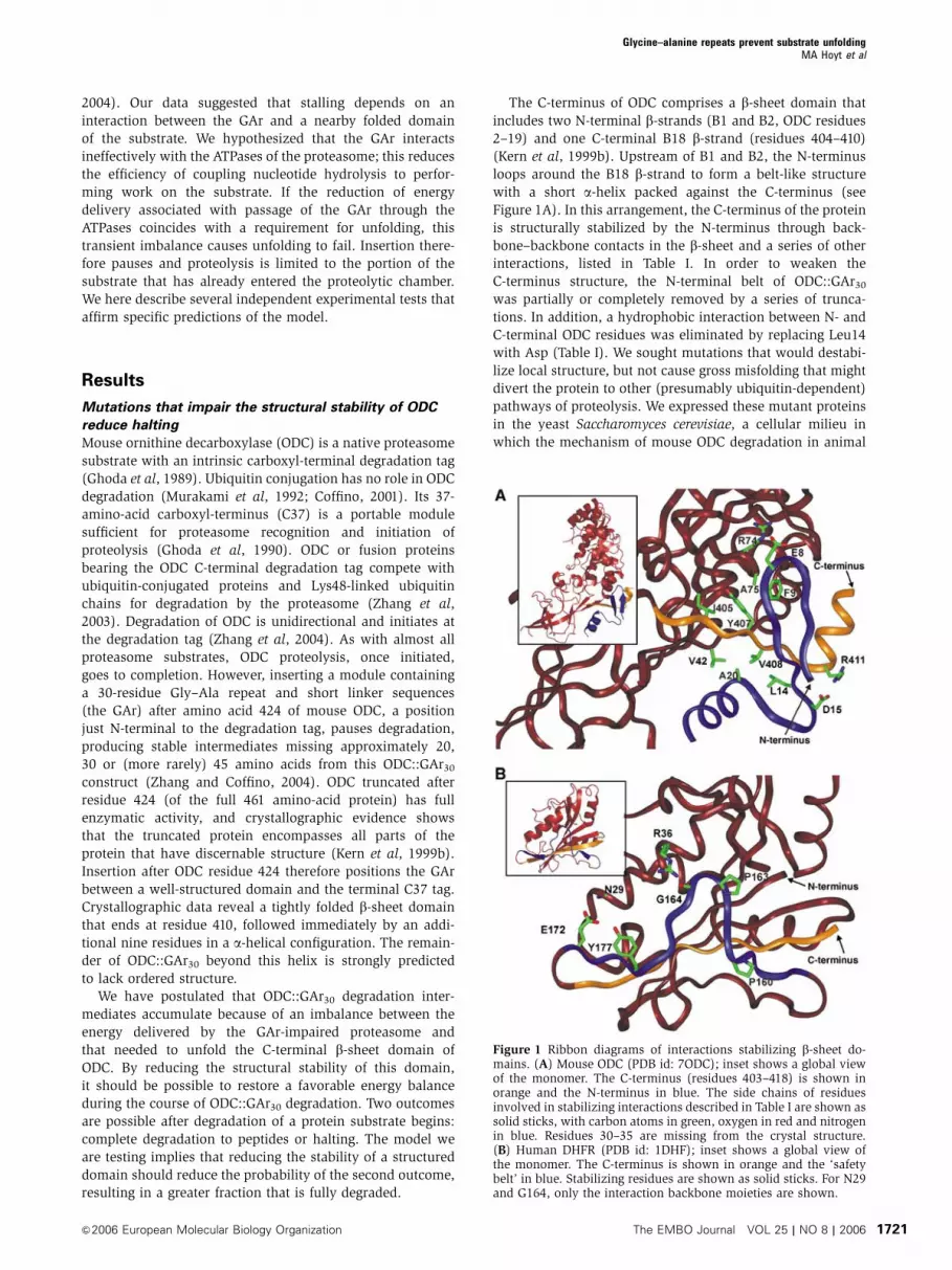
As measured by pulse-chase experiments, essentially all
the degradation products of ODC::GAr30 were intermediates
(Figure 2A). At each time point in the chase period, the
sum of the intensity of the parent (full-length) protein and
the intermediates is approximately constant. Quantitation
of the results of three independent pulse-chase experiments
of transformants expressing ODC::GAr30 (and two informa-
tive mutants described below) verified this assessment
(Figure 2B). An 11-amino-acid N-terminal truncation
(D11ODC::GAr30) did not reproducibly alter degradation
intermediate accumulation (data not shown). However,
the further removal of a stabilizing hydrophobic cluster
by the L14D mutation (in D11ODCL14D::GAr30) reduced
the accumulation of degradation intermediates. With
D11ODCL14D::GAr30, the sum of the parent and intermediate
bands declined during the chase. A significant fraction of
processing events therefore resulted in complete degradation
(Figure 2A and B). Furthermore, a more extensive truncation
of 17 amino acids (D17ODC::GAr30) reduced the fraction of
products that appeared as intermediates similarly to the
D11ODCL14D::GAr30 fusion protein. In summary, mutations
predicted to destabilize the ODC C-terminal b-sheet domain
reduced the ability of the GAr element to impede degradation.
GAr function thus depends on the stability of the domain
proximal to this element. Unfolding has been described as the
rate-limiting step in degradation of proteasome substrates
(Thrower et al, 2000). Consistent with these previous find-
ings, the half-life of ODC::GAr30 is about twice that of
D11ODCL14D::GAr30 or D17ODC::GAr30, the two mutants bear-
ing the destabilized b-sheet domain (Figure 2B).
The ratio of intermediates can be summarized by the value
PI, the fraction of substrates initiating degradation that end up
as intermediates. PI has a value between zero (no inter-
mediates, parent is completely degraded) and one (only
intermediates). This value facilitates comparisons of diverse
GAr-containing proteins (Table I). PI was approximately 1.0
for ODC::GAr30, and was not significantly changed in
D11ODC::GAr30. However, the further introduction of the
L14D mutation in the D11ODCL14D::GAr30 construct reduced
the value of PI to 0.63. Furthermore, the more extensive
truncation of 17 amino acids (D17ODC::GAr30) similarly
reduced PI to 0.66.
This analysis requires that the mutations do not stimulate
a turnover pathway independent of the ODC degradation
signal. To confirm that the proteins were solely degraded in
this way, we mutated an essential residue in the degrada-
tion tag, corresponding to Cys441 of mouse ODC (Miyazaki
et al, 1993). A C441A mutation was introduced into the fusion
proteins, and in each case prevented degradation (Figure 2C).
We conclude that these mutated fusion proteins are degraded
by an ODC-specific (and thus proteasome-dependent and
ubiquitin-independent) degradation pathway.
Perturbations that alter the structural stability of DHFR
also alter halting
Appending the C-terminal degradation signal of ODC to
diverse proteins, including dihydrofolate reductase (DHFR),
converts them to proteasome substrates (Loetscher et al,
1991; Li et al, 1998; Zhang et al, 2003). To investigate whether
the b-sheet domain of DHFR has the potential to interrupt
substrate processing by collaborating with a GAr, we con-
structed a fusion protein containing the entire human DHFR
open reading frame followed by the GAr30 module and the
37-residue ODC degradation tag, forming DHFR-GAr30-C37.
Pulse-chase analysis showed that DHFR-GAr30-C37 gene-
rated a single stable degradation product shorter than the
parent molecule (Figure 3A), in which approximately 20
residues were removed from its C-terminus (data not
shown). In contrast to ODC::GAr30, which is almost always
completely processed to intermediates (PIB1.0), DHFR-
GAr30-C37 degradation gave rise to intermediates only
about one-third of the time (PIB0.3). Like ODC, the
C-terminus of DHFR is a b-strand contained in a b-sheet
domain that involves three C-terminal b-strands and one
N-terminal b-strand (Prendergast et al, 1988). Here, the
stability of the C-terminal strand is enhanced by a ‘safety-
belt’ structure, where residues 157–168 loop around the last
b-strand. This segment is packed against the strand and
anchored to the rest of the molecule through specific inter-
actions (see Figure 1B and Table I). The conformation of this
loop is critically dependent on the presence of Pro 160 and
Pro 163. To determine whether destabilizing the C-terminal
b-sheet domain of the DHFR moiety influenced intermediate
accumulation, we followed a strategy similar to that used
with ODC. Three mutations anticipated to destabilize the
C-terminal b-sheet of DHFR were tested (Figure 1B, Table I).
Mutants with a substitution of Arg36 to Ser (DHFRR36S-GAr30-
C37), Glu172 to Ser (DHFRE172S-GAr30-C37) or Pro 160 and
163 to Ala (DHFRP160A/P163A-GAr30-C37) were constructed and
their degradation in yeast cells was examined by pulse-chase
analysis. The R36S mutation had little effect on intermediate
production and accumulation. However, both the E172S and
Table I Effects of perturbations predicted to alter stability of ODC and DHFR C-terminal b-sheet domains on intermediate accumulation
Altered residue Relevant mutant Affected interaction PIa
None (wild-type ODC) — None 1.0ODC-Glu8 D11ODC::GAr30 Glu8-Arg74 (salt bridge) 1.0ODC-Phe9 D11ODC::GAr30 Phe9-Ala75-Tyr407-Ile405 (hydrophobic cluster) 1.0ODC-Leu14 D11ODCL14D::GAr30 Leu14-Ala20-Val42-Val408 (hydrophobic cluster) 0.63ODC-Asp15 D17ODC::GAr30 All of the above, and Asp15-Arg411 (salt bridge) 0.66
None (wild-type DHFR) — None 0.31DHFR-Arg36 DHFRR36S-GAr30-C37 Arg36-Gly164 (polar interaction) 0.36DHFR-Glu172 DHFRE172S-GAr30-C37 Asn29-Glu172-Tyr177 (polar interaction) 0.10DHFR-Pro160 and Pro163 DHFRP160A/P163A-GAr30-C37 None (form structural loop) 0.07None (wild-type DHFR) Methotrexate binding to active site 0.90
aThe fraction of substrate degradation events that result in stable intermediates (e.g. for PI¼ 1.0, all degradation events result in intermediates).
Glycine–alanine repeats prevent substrate unfoldingMA Hoyt et al
The EMBO Journal VOL 25 | NO 8 | 2006 &2006 European Molecular Biology Organization1722

P160A/P163A substitutions had marked effects (Figure 3A).
The PI value for DHFR-GAr30-C37, 0.31, was reduced by the
E172S and P160A/P163A mutations, respectively, to 0.10 and
0.07 (Table I). Pulse-chase analysis demonstrated that all
three proteins containing a C441A substitution of the critical
cysteine were stable (Figure 3B).
Because processing of DHFR-GAr30-C37 is poised to pro-
duce either outcome with substantial probability, this con-
struct provides the opportunity to test the consequences of
both destabilization and stabilization of the DHFR moiety.
Methotrexate binds tightly to the DHFR active site and has
been used extensively to impair DHFR unfolding, for example
(Eilers and Schatz, 1986). We have previously shown that
methotrexate can block degradation of an ODC::DHFR fusion
protein (Zhang et al, 2004). Yeast cells expressing DHFR-
GAr30-C37 were treated with methotrexate or were left
untreated and subjected to pulse-chase analysis of turnover
and intermediate accumulation (Figure 3C). Treatment with
the stabilizing ligand increased PI three-fold, to 0.90 (Table I).
Together, the DHFR-destabilizing mutations and ligand-
mediated stabilization provided a bidirectional test of our
hypothesis and demonstrated that GAr function is strongly
influenced by the stability of an adjacent C-terminal domain.
Position of cutting sites that generate stable
intermediates not altered by extending the degradation
tag
The model being tested postulates that a structured domain
stops insertion of a GAr-containing substrate and thus halts
its proteolysis. This implies that the sites at which proteolysis
terminates must be positioned by reference to the folded
domain, not determined by the configuration of the C-term-
inal degradation tag. We tested this hypothesis by duplicating
the C37 degradation tag of an ODC::GAr30 fusion protein,
thus adding an additional 38 residues to the C-terminus.
We expressed ODC::GAr30 fusions with either the normal or
extended C-termini in yeast transformants and performed
pulse-chase experiments to compare the size of the inter-
0
20
40
60
80
100
120
140
0
20
40
60
80
100
120
0
20
40
60
80
100
0 15 30 45 60Chase time (min)
Total
Parent
Intermediates
% o
f tot
al a
t t =
0
ODC::GAr30
∆11ODCL14D::GAr30
∆17ODC::GAr30
ODCC441A::GAr30
∆11ODCC441A::GAr30
∆17ODCC441A::GAr30
0Chase time (min)
A
C
B
603015
0Chase time (min)
603015
Figure 2 Mutations that disrupt ODC-stabilizing interactions reduce the accumulation of degradation intermediates. (A) Pulse-chase analysisof yeast cells expressing wild-type or mutant ODC::GAr30 fusion proteins. The position of full-length ODC::GAr30 (solid arrow) or degradationintermediates (dashed arrows) is indicated. (B) Quantitation of ODC::GAr30 turnover data from (A), presented as the mean of threeindependent experiments. Error bars represent the standard deviation. The values for wild-type ODC::GAr30 (K), D17ODC::GAr30 (&) andD11ODCL14D::GAr30 (m) are expressed as a percentage of the total of the full-length parent fusion protein and its degradation intermediatesat time zero. The changes in the wild-type and mutant proteins are compared for the parent molecule (middle panel), its degradationintermediates (bottom panel) and the sum total of these (top panel) at each time point. (C) Pulse-chase analysis of ODC::GAr30 fusion proteinscontaining the C441A mutation in the ODC degradation tag. Arrows indicate bands as in (A).
Glycine–alanine repeats prevent substrate unfoldingMA Hoyt et al
&2006 European Molecular Biology Organization The EMBO Journal VOL 25 | NO 8 | 2006 1723

mediates they produce (Figure 4). As expected, the fractiona-
tion of the two full-length fusion proteins by SDS–PAGE was
consistent with the expected differences in their molecular
mass. The extension of the C-terminus had no apparent
effect on the degradation rate of the fusion protein when
compared to the unextended molecule. However, the mobility
of the intermediates derived from the two proteins during the
chase period was not distinguishably different. This implies
that cutting is both limited and positioned by structures that
lie on the N-terminal side of the sites of terminal proteolysis.
Spacing between GAr and folded domain is critical
for halting
Because the functional interaction between the GAr and
a folded domain is postulated to require that they arrive
simultaneously at their respective sites of action, altering
the spacing between these two elements should alter the
synchrony of their arrival and thus modify halting. A series of
homologous substrates were constructed and expressed in
yeast transformants to test this idea (Figure 5A). In the first of
these, 0, 7, 13, 19 or 25 residues were inserted between ODC
position 424 and the GAr30 tract. The constructs with spacer
lengths of 0, 7 or 13 residues differed little from ODC::GAr30
in the extent of halting (Figure 5B and C); for each of these,
most processing events produced intermediates (PI range
0.68–0.88). However, extending the spacer length to 19 or
25 residues sharply reduced the efficiency of intermediate
production (PI¼ 0.21 and 0.20). Because the folded b-sheet
domain of ODC extends to residue 410 (Kern et al, 1999a),
ODC::GAr30 (C37)
Chase time (min)
ODC::GAr30 (C75)
0 15 30 60 0 15 30 60
Figure 4 The positions of cutting sites that generate stable inter-mediates are not altered by extending the degradation tag. Pulse-chase analysis of yeast cells expressing ODC::GAr30 fusion proteinswith wild-type (C37) or extended (C75) degradation tags. Theparent bands and common intermediates are indicated by arrowsas in Figure 2.
DHFR-GAr30-C37 DHFR-GAr30-C37C441A
DHFRR36S-GAr30-C37
DHFRE172S-GAr30-C37
DHFRP160A/P163A-GAr30-C37
A B
DHFRE172S-GAr30-C37C441A
DHFRP160A/P163A-GAr30-C37C441A
C
Chase time (min)
DHFR-GAr30-C37
–MTX
+MTX
6030150
Chase time (min)6030150
Figure 3 The accumulation of DHFR-GAr30-C37 degradation inter-mediates is influenced by the stability of the DHFR domain. (A)Pulse-chase analysis of wild-type and mutant DHFR-GAr30-C37proteins. (B) Pulse-chase analysis of fusion proteins containingthe C441A mutation in the ODC degradation tag. (C) Pulse-chaseanalysis of DHFR-GAr30-C37 in yeast cells either with (þMTX) orwithout (�MTX) the addition of 20mM methotrexate. Arrowsindicate bands as in Figure 2.
A
0
20
40
60
80
100
120
0
20
40
60
80
100
120
0
20
40
60
80
100
0 15 30 45 60
Total
Parent
Intermediates
% o
f to
tal r
emai
nin
g
Chase time (min)
C
0
7
13
19
25
Spacerlength
0 15 30 60Chase time (min)
B
Spacer
GAr30
tagDegradationC37
7
13
19
2514
ODC β-sheetdomain
ODC, 411–424
Figure 5 The effects of spacing between GAr and b-sheet domainon production of intermediates. (A) Schematic of ODC::GAr30
constructs containing spacers of varying length between the ODCC-terminal b-sheet and the GAr30 domains. The number of spacerresidues is indicated below each construct. (B) Pulse-chase analysisof cells expressing constructs in (A). (C) Quantitation of turnoverdata for ODC[n]GAr30 fusions in (B) with spacer lengths of 0 (K),7 (&), 13 (D), 19 (&) or 25 (J) residues were analyzed as inFigure 2B.
Glycine–alanine repeats prevent substrate unfoldingMA Hoyt et al
The EMBO Journal VOL 25 | NO 8 | 2006 &2006 European Molecular Biology Organization1724
14 residues lie between the end of the b-sheet domain and
ODC residue 424. Therefore, the presence of 33 or 39 residues
between the b-sheet domain and the GAr30 tract reduced
its function, whereas 27 or fewer did not. (As reference,
in ODC::GAr30 used in the experiments described above, the
b-sheet domain-GAr distance is 26 residues.)
In the above series of constructs, all structural elements
were held constant except for the distance between the
b-sheet domain and the GAr30. Consequently, the distance
between ODC residue 424 and the C37 degradation tag, which
in the native protein immediately follows position 424,
ranged from 30 to 55 residues. To determine whether this
variable rather than the distance between the b-sheet domain
and the GAr is the critical parameter determining intermedi-
ate production, we compared three constructs in which
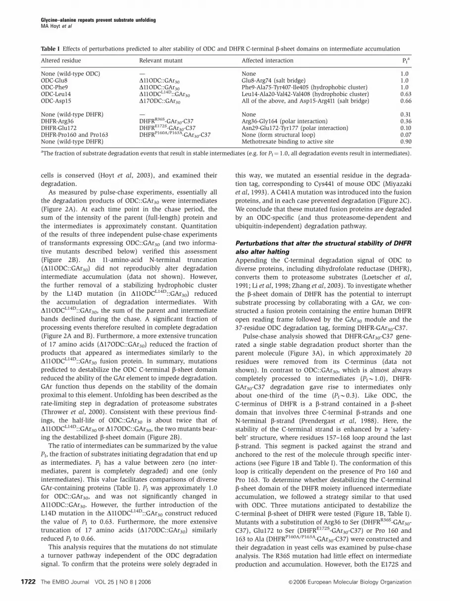
the spacing between ODC position 424 and C37 (Figure 6)
was identical. Moving all 25 flanking residues to the GAr
C-terminal side produced abundant intermediates, similar to
the standard ODC::GAr30 fusion protein. In contrast, when
the 25 residue flanking sequence was placed on the
N-terminal side of the GAr, no intermediates were detected.
This result supports the inference from the experiments
above that the critical variable is the spacing between the
b-sheet domain and the GAr30.
Intermediates are not trapped by proteasome
For the substrates described here to undergo partial proteo-
lysis, a part of their C-terminal end must have reached the
proteolytic chamber of the proteasome 20S core particle,
while a folded region of the same protein remains positioned
elsewhere, likely within or contiguous to the 19S regulatory
complex. We asked whether, when insertion ceases, the
substrate remains in an inserted mode, persistently asso-
ciated with the proteasome, or escapes instead. To address
these questions, we expressed ODC::GAr30 in a yeast strain in
which proteasomes had been equipped with an affinity tag to
facilitate their rapid isolation. Proteasomes were fractionated
from substrates and their degradation intermediates in buffer
containing ATP. We first affinity purified proteasomes with
a protein A tag fused to the b4 (Pre1) subunit of the 20S
proteolytic core. Western blotting for 20S proteins and for
the 19S component Rpt5 demonstrated that both 20S and
19S complexes were recovered with similar efficiencies in
the matrix-bound fraction—absolute recoveries were 5–10%
(Figure 7). This shows that the buffer composition and
fractionation conditions used effectively maintained an asso-
ciation between the 20S core and 19S regulatory complex.
Neither 20S proteins nor Rpt5 were associated with the
affinity matrix in a control strain lacking the affinity tag. To
increase the sensitivity of detection for trapped intermediates,
a 20-fold excess of the affinity pulldown fractions was
analyzed compared to total and supernatant fractions.
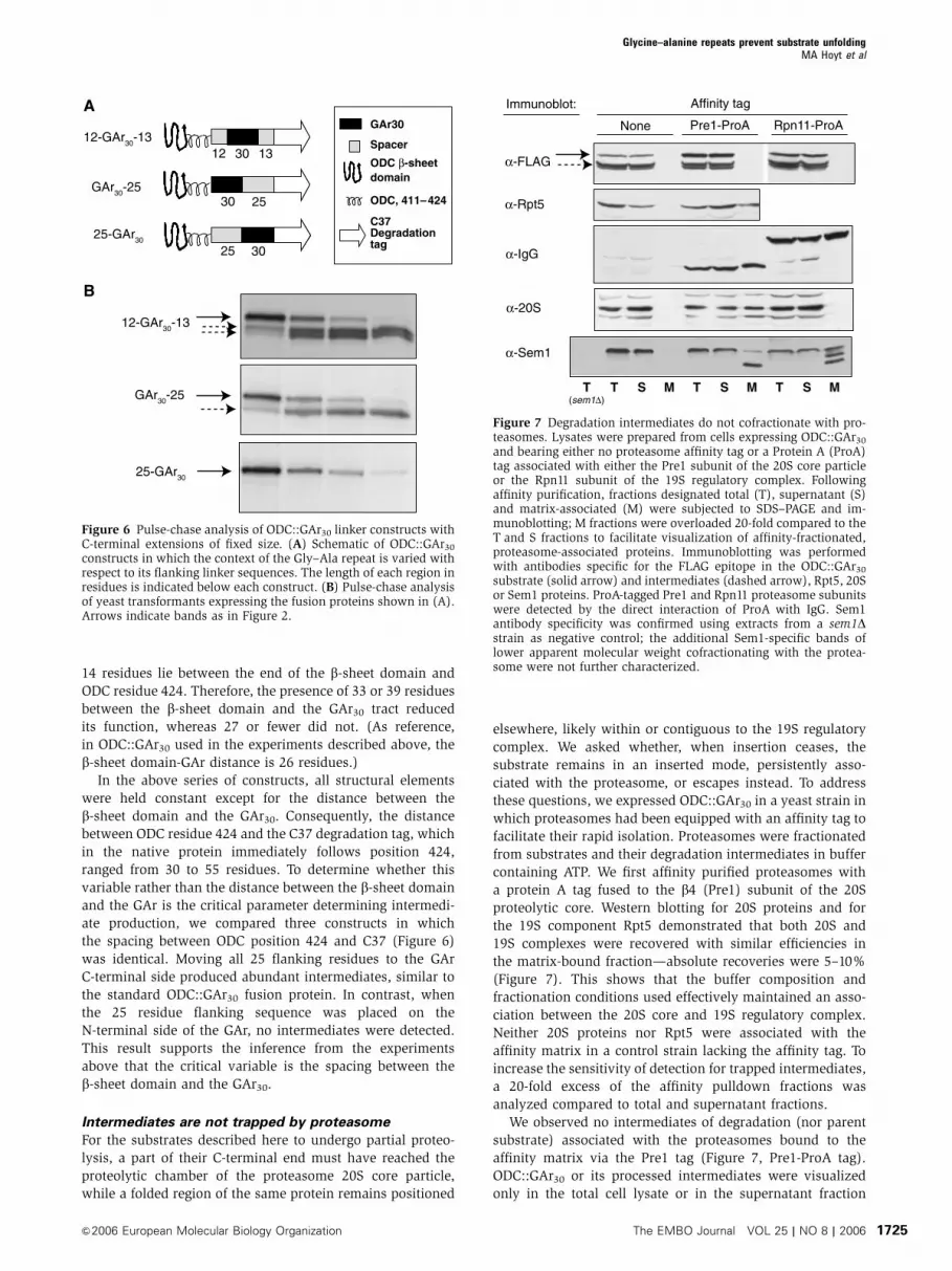
We observed no intermediates of degradation (nor parent
substrate) associated with the proteasomes bound to the
affinity matrix via the Pre1 tag (Figure 7, Pre1-ProA tag).
ODC::GAr30 or its processed intermediates were visualized
only in the total cell lysate or in the supernatant fraction
(sem1∆)
None Pre1-ProA Rpn11-ProA
Affinity tagImmunoblot:
α-FLAG
α-Rpt5
α-IgG
α-20S
α-Sem1
MMM SSS TTTT
Figure 7 Degradation intermediates do not cofractionate with pro-teasomes. Lysates were prepared from cells expressing ODC::GAr30
and bearing either no proteasome affinity tag or a Protein A (ProA)tag associated with either the Pre1 subunit of the 20S core particleor the Rpn11 subunit of the 19S regulatory complex. Followingaffinity purification, fractions designated total (T), supernatant (S)and matrix-associated (M) were subjected to SDS–PAGE and im-munoblotting; M fractions were overloaded 20-fold compared to theT and S fractions to facilitate visualization of affinity-fractionated,proteasome-associated proteins. Immunoblotting was performedwith antibodies specific for the FLAG epitope in the ODC::GAr30
substrate (solid arrow) and intermediates (dashed arrow), Rpt5, 20Sor Sem1 proteins. ProA-tagged Pre1 and Rpn11 proteasome subunitswere detected by the direct interaction of ProA with IgG. Sem1antibody specificity was confirmed using extracts from a sem1Dstrain as negative control; the additional Sem1-specific bands oflower apparent molecular weight cofractionating with the protea-some were not further characterized.
12 30 13
GAr30
Spacer
domain
ODC, 411–424
C37Degradationtag
12-GAr30-13
GAr30-25
25-GAr30
30 25
25 30
A
B
12-GAr30-13
GAr30-25
25-GAr30
ODC β-sheet
Figure 6 Pulse-chase analysis of ODC::GAr30 linker constructs withC-terminal extensions of fixed size. (A) Schematic of ODC::GAr30
constructs in which the context of the Gly–Ala repeat is varied withrespect to its flanking linker sequences. The length of each region inresidues is indicated below each construct. (B) Pulse-chase analysisof yeast transformants expressing the fusion proteins shown in (A).Arrows indicate bands as in Figure 2.
Glycine–alanine repeats prevent substrate unfoldingMA Hoyt et al
&2006 European Molecular Biology Organization The EMBO Journal VOL 25 | NO 8 | 2006 1725

following the pulldown or in the control nontagged pull-
down. We also performed similar experiments using a protein
A tag fused to the Rpn11 (Figure 7, Rpn11-ProA tag) or Rpn1
(data not shown) subunits of the 19S complex, with similar
results. Sem1 is a protein that has been recently identified
as a component of the proteasome 19S complex (Funakoshi
et al, 2004; Sone et al, 2004). However, the bulk of Sem1 is
found in low molecular weight fractions (Funakoshi et al,
2004), and recombinant Sem1 readily becomes incorporated
into proteasomes from Sem1-deficient cells (Sone et al, 2004).
These properties suggest Sem1 exists in exchangeable free
and proteasome-bound pools. We therefore tested Sem1
recovery as a positive control for proteasome association,
using the identical pulldown and processing conditions that
are used for ODC::GAr30. Sem1, in contrast to ODC::GAr30
and its derivatives, was readily visualized in pulldown frac-
tions from Pre1- or Rpn11-ProA-tagged strains. We conclude,
therefore, that ODC::GAr30 intermediates are not persistently
trapped in the 20S proteasome. Gel filtration fractionation of
ODC::GAr30 and its intermediates from untagged proteasomes
supported the same conclusion (data not shown). Together,
these results exclude persistent intermediate trapping by 19S,
20S or 26S forms of the proteasome. We also performed the
converse experiment, using an internal FLAG epitope tag
within ODC::GAr30 to perform the pulldown and Western
blots to evaluate the recovery of proteasome proteins in
association with ODC::GAr30 or its derivatives. None were
detected (data not shown).
Complete degradation of a mouse ODC molecule expressed
in S. cerevisiae takes about 10 min (Hoyt et al, 2003). In the
pulldown experiments, approximately 45 min elapsed bet-
ween cell lysis and completion of fractionation. We therefore
cannot infer whether exit takes place at a rate comparable to
the normal process of complete degradation or is more rapid.
Discussion
We have previously proposed (Zhang and Coffino, 2004), and
here test, the following simple model: Two elements of the
19S regulatory particle are postulated to function coordi-
nately. The first is a site impelling translocation, the ATPase
ring, which binds the substrate and advances it stepwise
toward the 20S core particle. The second is a site of constric-
tion within the regulatory particle, possibly the entrance to
a translocation channel through the ATPase ring. This
postulated constriction impairs transit of a folded domain.
The ATPase ring produces energy that performs work on the
substrate, some of which is used to unravel the folded
domain. The mode of energy transmission may be as simple
as a lever arm motion that applies a friction drive to the
polypeptide chain (Hinnerwisch et al, 2005). In the normal
case, energy supply continuously exceeds need, and the
substrate polypeptide continuously advances. If this balance
transiently swings in the other direction, with energy need
exceeding supply, insertion halts. If the substrate has pene-
trated sufficiently far into the core particle, proteolysis is
limited by the failure to advance further. The position at
which the substrate is cut reflects the furthest extent of
penetration into the proteolytic chamber. The remaining
substrate is released, and, if proteolysis has removed the
degradation tag, partial proteolytic products persist as stable
degradation intermediates.
Insertion can be stopped by a modestly stable folded
structure if the capacity of the ATPase ring to transmit energy
becomes impaired. The arrival of a GAr at the ATPase site is
postulated to produce such impairment. This may happen
because the tract of glycines and alanines (the two smallest
amino acids) presents a relatively featureless face to an
ATPase-associated friction drive apparatus that normally
advances the chain. This conjecture is consistent with the
known substrate interaction properties of bacterial ATPases.
ClpB, in collaboration with the DnaK chaperone system,
solubilizes and refolds aggregated proteins. Its binding site
(near the axial pore of the ATPase hexamer) prefers sub-
strates with aromatic and basic residues (Schlieker et al,
2004). The HslU ATPase of the HslUV bacterial protease has
a similar preference for aromatic and positively charged
amino acids at substrate residue positions important for
recognition (Burton et al, 2005).
Here, we tested this model in several ways. First,
we altered the stability of an ODC or DHFR folded domain
by introducing mutations expected to cause destabilization
or (for DHFR) by adding a stabilizing ligand. Within
ODC::GAr30, deletions were designed to destroy inter-
actions that stabilize the b-sheet domain proximal to the
GAr and the C37 degradation tag. Using DHFR constructs,
we pursued a similar strategy and obtained a similar result:
mutations directed toward reducing stability reduced inter-
mediate production. DHFR also provided the opportunity
to examine the effect of domain stabilization on inter-
mediate production. The stabilizing ligand methotrexate
markedly increased the appearance of intermediates. These
findings are consistent with a model of protease function
described by Matouschek and co-workers (Lee et al, 2001;
Prakash et al, 2004) who showed that the proteasome unfolds
and degrades substrates sequentially from an unstructured
initiation site.
Alternative mechanisms through which a GAr might pro-
duce degradation intermediates are less tenable. A GAr
may enhance endoproteolytic cleavage by the proteasome
(Liu et al, 2002a) and thus leave the remainder of the GAr-
containing fusion protein unprocessed. Such a model seems
unlikely, however, given that GAr function depends on the
stability of structural domains spatially distinct from the GAr
itself. Our data also exclude mechanisms whereby substrate
processing initiates, but ceases when components of the
proteasome encounter a GAr and become immobilized.
Such a sabotage mechanism implies the stable association
of intermediate with proteasome, contrary to our data show-
ing that their association is transient.
In addition to the anticipated quantitative effect of chan-
ging domain stability, an unanticipated change in the pattern
of intermediates was also seen. ODC::GAr30 produces three
intermediates, missing about 20, 30 and (more rarely) 45
residues from its C-terminus. This suggests that the insertion
process overcomes a series of energy barriers, a process in
which progressive clipping marks progressive subdomain
unfolding events. The mutations we have investigated uni-
formly reduced the accumulation of the intermediate missing
B20 residues, but had little effect on the others (Figure 2A).
This is consistent with selective reduction of the most proxi-
mal energy barrier, while more distal ones are little disturbed,
a result that might have been expected from the strategy of
mutant design.
Glycine–alanine repeats prevent substrate unfoldingMA Hoyt et al
The EMBO Journal VOL 25 | NO 8 | 2006 &2006 European Molecular Biology Organization1726

In a further independent test of the model, we varied
the spacing between the folded domain of ODC (assumed
here to end at the terminal b18 strand (Kern et al, 1999a)) and
the proximal boundary of a GAr of length 30. A distance of
14, 21 or 27 amino acids between these elements led to
efficient stalling of insertion. Increasing this distance to 33
amino acids strongly reduced stalling and an increase to 39
further reduced it. The distance between successive a-carbon
atoms of an extended polypeptide chain is B3 A, and so these
spacings of 14, 21 or 27 residues are equivalent to about 40,
60 and 80 A. Our data imply that the minimum separation
that supports functional interaction is 40 A or less. The result
is consistent with the possibility that the two functional sites
in fact colocalize. This is credible: if the substrate threads
through an axial pore of an ATPase annulus, the pore could
be the site of restriction. Separating the two elements by
100 A (33 residues) or more strongly impairs their capacity to
collaborate. The full 19S regulatory complex seen by electron
microscopy can be encompassed within a sphere about 150 A
in diameter (Walz et al, 1998). Failure of interaction at
distances approaching the dimensions of the 19S complex is
consistent with the requirement that both sites lie within the
complex.
By using pulse-chase data, we calculated the fraction of
processed substrates that partition to intermediates, PI, and
used this as the primary analytic tool for examining halting of
proteolysis. PI has a simple physical interpretation: it corre-
sponds to the probability that degradation, once initiated, will
cease. This can also be expressed as a ratio of rate constants,
whereby a substrate that is undergoing degradation will
alternatively undergo further degradation at a rate kD or
stop degradation (and likely exit the proteasome) at a rate
kI. Then, PI¼ kI/(kIþ kD). The proteasome is generally
assumed to operate on most ordinary substrates with a PI
that is close to zero. This is based on the perception that
degradation appears to be completed once it is begun. The
conclusion, however, rests largely on our failure to observe
intermediates. Two considerations limit our capacity for
detecting the presence or production of intermediates. The
first of these is technical: SDS–PAGE, the most commonly
used means to follow proteolysis, readily detects abundant
protein species of discrete size, but not lower abundance
populations of heterogeneous size. Therefore, only intermedi-
ates consisting of discrete molecular species, like those
described here, will be observed readily. Second, degradation
intermediates may include misfolded elements; these pro-
teins may be subjected to a second round of elimination by
the ubiquitin–proteasome system, thereby escaping detec-
tion. The value of PI for typical substrates therefore remains
an open question.
Understanding the import machinery of the proteasome
can benefit from considering better-understood models of
protein translocation, such as membrane transport systems
or bacterial ATP-dependent proteases. In principle, two me-
chanisms can be utilized, either a power stroke or Brownian
ratchet. By analogy with other systems in which ATPases are
used (Singleton et al, 2000; Kenniston et al, 2003; DeLaBarre
and Brunger, 2005; Hinnerwisch et al, 2005), a power stroke
model is the more likely. Consideration of the power stroke
mechanism may help to illuminate why the native GAr is so
long: a long GAr assures probabilistic iterative failure. If the
bacterial ClpXP protease is presented with a multidomain
substrate, it releases the substrate at an appreciable fre-
quency, yielding stable intermediates that are cut between
domain boundaries. A titin domain interrupts processing by
ClpXP about one time in three (Kenniston et al, 2005). This
significant failure rate (PIB0.3) does not require mediation
by a recognizable GAr-like sequence. The proteasome may be
a more powerful machine, so that a folded domain must be
supplemented by a slippery sequence to cause a comparable
rate of failure. Alternatively, the titin domain may have more
stringent unfolding requirements than those we have tested
in the proteasome. As discussed above, it is also possible that
proteasomes have a PI that significantly exceeds zero for
native folded proteins.
The Epstein–Barr virus has evolved a tract of 60–300
residues (in different isolates), which are exclusively glycines
and alanines. A sequence of such length did not likely evolve
to confer a global folding property, nor is it easy to imagine
a specific proteasome site that requires for interaction
(or impairment of interaction) the full span of a sequence
of such length and invariant composition. Much more likely
is a requirement for iterative impairment of many such
interactions, performed as the polypeptide chain advances
through the proteasome. Each completed power stroke thrust
would, in this view, displace the next such interaction to
a new position along the chain, a displacement equal to
the power stroke length. The estimated power stroke length
of other AAA ATPase motors, such as bacteriophage T7
helicase, is about 20 A (Singleton et al, 2000), which corre-
sponds to an extended peptide of about seven residues.
Suppose that failure of a single iteration of displacement
causes substrate release. A GAr of 300 residues would suffer
that risk more than 40 times, converting, for example, a 90%
success rate per iteration to a success rate of 1% for traver-
sing the full sequence (PIB0.99). When suitably juxtaposed
to a folded ODC domain, a GAr as short as seven residues can
cause failure of processive degradation about one time in
three (PIB0.3). A long GAr would require neither exquisite
positioning nor unidirectional chain processing, and would
assure that processivity is interrupted, even when the failure
rate per individual iteration is low.
Our data support the conclusion that degradation inter-
mediates are released from the proteasome, a finding also
true of ClpXP (Kenniston et al, 2005). Intermediate release
has several implications. First, it assures that the proteasome
system will not be functionally depleted by persistent futile
interactions. Second, it is consistent with a few exceptional
cases in which proteasomes are used for processing rather
than complete degradation (Rape and Jentsch, 2002).
Converting certain precursor proteins to mature transcription
factors requires them to be partially proteolyzed by protea-
somes and then liberated. One such transcription factor is
NFkB, the p50 subunit of which is generated from a p105
precursor by proteasomal truncation of its C-terminal end. It
is of interest that producing the p50 intermediate requires
both a structured domain within the N-terminal end of the
p105 precursor and a glycine-rich tract near the processing
junction. Removing the glycine-rich region (Orian et al, 1999)
or impairing the stability of the folded domain (Lee et al,
2001) or increasing spacing between the two (Tian et al,
2005) reduces halting, biasing degradation toward comple-
tion rather than production of the p50 protein. Although the
functional interactions of these elements obviously resemble
Glycine–alanine repeats prevent substrate unfoldingMA Hoyt et al
&2006 European Molecular Biology Organization The EMBO Journal VOL 25 | NO 8 | 2006 1727

those within the more artificial substrates we have studied,
it remains to be determined whether the glycine-rich region
of the p105 precursor confers functional properties similar
to a GAr.
Materials and methods
Yeast strainsAll GAr-containing fusion proteins were expressed from p414ADH-based vectors (Mumberg et al, 1995) in S. cerevisiae strain MHY501(MATa his3-D200 leu2-3,112lys2-801 trp1-1 ura3-52) (Chen et al,2002), unless otherwise noted. All yeast strains were maintainedand manipulated using standard procedures (Guthrie and Fink,1991). Strains MHY85 and MHY94, bearing C-terminal Staphy-lococcus aureus protein A tags on proteasome subunits Pre1 andRpn11, respectively, were generated by the integration of the tagsequence and the Kluyveromyces lactis TRP1 gene at theirchromosomal loci in strain MHY501 using described methods(Knop et al, 1999). For this transformation, a DNA fragmentencoding the TEV protease cleavage site, two protein A IgG-bindingdomains, and the K. lactis TRP1 gene, and flanked by 50 bp PRE1 orRPN11 chromosomal sequences to direct integration, was amplifiedfrom the TAP tag vector pBS1479 (Puig et al, 2001) by PCR.
Plasmid constructionDetails of the construction of plasmids used in this study areprovided in supplementary data.
Metabolic labeling, immunoprecipitations andsubstrate-trapping assayPulse-chase analysis was performed as described previously(Hoyt et al, 2003). GAr30-containing fusion proteins were immuno-precipitated using FLAG epitope tags located either within the GAr30
module itself, or at the N-terminus of the fusion protein, using anti-FLAG M2 affinity gel (Sigma).
Yeast strains bearing a Protein A affinity tag appended to theC-terminus of the Pre1 (strain MHY85) or Rpn11 (MHY94) protea-some subunits were transformed with a URA3-marked low-copyvector (p416ADH, (Mumberg et al, 1995)) expressing ODC::GAr30
(pJ67). Strain MHY501 was used as a negative control. Forcofractionation experiments, 40 ml cultures of the transformantswere harvested in late exponential growth phase (OD600¼B1.5),
washed with water, and resuspended in 300 ml lysis buffer (50 mMTris–HCl pH 7.5, 150 mM NaCl, 10% glycerol, 2 mM ATP and 5 mMMgCl2). The cells were mechanically disrupted in 2-ml screw-topmicrocentrifuge tubes with 200ml glass beads in a Mini-beadbeater(Biospec Products) for four pulses of 30 s duration with 30 s coolingon ice between pulses. The lysates were cleared by centrifugation(18 000 g, 15 min) and 750 mg of total protein in 0.5 ml total volumewas incubated with 5ml of either rabbit IgG agarose or anti-FlagM2 agarose at 41C for 20 min. The affinity-purified proteasomecomplexes were washed four times with 500 ml lysis buffer,resuspended in Laemmli SDS–PAGE loading buffer, and fractionatedby SDS–PAGE. The fractioned proteins were transferred tonitrocellulose filters, and the filters were blocked in Tris-bufferedsaline with 1% Triton X-100 (TBS-T) including 5% powderedlow-fat milk, and rinsed briefly with TBS-T. Proteins were detectedwith mouse anti-FLAG M2 antibody (Sigma, 1:2000 dilution)and TrueBlot anti-mouse Ig-horseradish peroxidase conjugates(eBioscience, 1:4000), or rabbit anti-Rpt5 (BioMol, 1:25000), anti-20S proteasome antibodies (gift of T Tamura, 1:2000) or anti-yeastSem1 antibodies (gift of M Funakoshi, 1:5000) and TrueBlot anti-rabbit Ig-horseradish peroxidase conjugates (1:4000). Immunoblotswere developed using the Amersham ECLplus detection kit andprotocol.
Data quantificationSignals from autoradiographic films were quantified using TotalLabsoftware (Nonlinear Dynamics). Data for the calculation of PI (seeDiscussion) was obtained from pulse-chase experiments with60 min chase periods. The term kI was calculated as the accumula-tion of intermediates between 0 and 60 min (Int60–Int0) divided bythe parent molecules lost during the same interval (P0–P60).Similarly, the term kD was calculated as (P0–P60)þ (Int0–Int60)/(P0þ Int0). PI¼ kI/(kIþ kD).
Supplementary dataSupplementary data are available at The EMBO Journal Online.
Acknowledgements
We thank Tomohiro Tamura, Daniel Finley, Kiran Madura andMinoru Funakoshi for reagents, and Fred Cohen and George Osterfor helpful comments on the manuscript. This work was supportedby NIH grants GM45335 and GM074760 to PC.
References
Burton RE, Baker TA, Sauer RT (2005) Nucleotide-dependentsubstrate recognition by the AAA+ HslUV protease. Nat StructMol Biol 12: 245–251
Chen H, MacDonald A, Coffino P (2002) Structural elements ofantizymes 1 and 2 required for proteasomal degradation ofornithine decarboxylase. J Biol Chem 277: 45957–45961
Coffino P (2001) Regulation of cellular polyamines by antizyme. NatRev Mol Cell Biol 2: 188–194
DeLaBarre B, Brunger AT (2005) Nucleotide dependent motion andmechanism of action of p97/VCP. J Mol Biol 347: 437–452
Eilers M, Schatz G (1986) Binding of a specific ligand inhibitsimport of a purified precursor protein into mitochondria.Nature 322: 228–232
Funakoshi M, Li X, Velichutina I, Hochstrasser M, Kobayashi H(2004) Sem1, the yeast ortholog of a human BRCA2-bindingprotein, is a component of the proteasome regulatory particlethat enhances proteasome stability. J Cell Sci 117: 6447–6454
Ghoda L, Phillips MA, Bass KE, Wang CC, Coffino P (1990)Trypanosome ornithine decarboxylase is stable because it lackssequences found in the carboxyl terminus of the mouse enzymewhich target the latter for intracellular degradation. J Biol Chem265: 11823–11826
Ghoda L, van Daalen Wetters T, Macrae M, Ascherman D, Coffino P(1989) Prevention of rapid intracellular degradation of ODC bya carboxyl-terminal truncation. Science 243: 1493–1495
Glickman MH, Rubin DM, Fried VA, Finley D (1998) The regulatoryparticle of the Saccharomyces cerevisiae proteasome. Mol Cell Biol18: 3149–3162
Groll M, Ditzel L, Lowe J, Stock D, Bochtler M, Bartunik HD, HuberR (1997) Structure of 20S proteasome from yeast at 2.4A resolu-tion. Nature 386: 463–471
Guthrie C, Fink GR (eds) (1991) Guide to Yeast Genetics andMolecular Biology. San Diego: Academic Press
Hinnerwisch J, Fenton WA, Furtak KJ, Farr GW, Horwich AL (2005)Loops in the central channel of ClpA chaperone mediate Proteinbinding, unfolding, and translocation. Cell 121: 1029–1041
Hoyt MA, Zhang M, Coffino P (2003) Ubiquitin-independentmechanisms of mouse ornithine decarboxylase degradation areconserved between mammalian and fungal cells. J Biol Chem278: 12135–12143
Kenniston JA, Baker TA, Fernandez JM, Sauer RT (2003) Linkagebetween ATP consumption and mechanical unfolding during theprotein processing reactions of an AAA+ degradation machine.Cell 114: 511–520
Kenniston JA, Baker TA, Sauer RT (2005) Partitioning betweenunfolding and release of native domains during ClpXP degrada-tion determines substrate selectivity and partial processing. ProcNatl Acad Sci USA 102: 1390–1395
Kern AD, Oliveira MA, Coffino P, Hackert ML (1999a) Structure ofmammalian ornithine decarboxylase at 1.6 # resolution: stereo-chemical implications of PLP-dependent amino acid decarboxy-lases. Structure 7: 567–581
Kern AD, Oliveira MA, Coffino P, Hackert ML (1999b) Structure ofmammalian ornithine decarboxylase at 1.6 A resolution: stereo-chemical implications of PLP-dependent amino acid decarboxy-lases. Struct Fold Des 7: 567–581
Glycine–alanine repeats prevent substrate unfoldingMA Hoyt et al
The EMBO Journal VOL 25 | NO 8 | 2006 &2006 European Molecular Biology Organization1728

Knop M, Siegers K, Pereira G, Zachariae W, Winsor B, Nasmyth K,Schiebel E (1999) Epitope tagging of yeast genes using a PCR-based strategy: more tags and improved practical routines. Yeast15: 963–972
Lee C, Schwartz MP, Prakash S, Iwakura M, Matouschek A (2001)ATP-dependent proteases degrade their substrates by processivelyunraveling them from the degradation signal. Mol Cell 7: 627–637
Levitskaya J, Coram M, Levitsky V, Imreh S, Steigerwald-MullenPM, Klein G, Kurilla MG, Masucci MG (1995) Inhibition ofantigen processing by the internal repeat region of the Epstein–Barr virus nuclear antigen-1. Nature 375: 685–688
Levitskaya J, Sharipo A, Leonchiks A, Ciechanover A, Masucci MG(1997) Inhibition of ubiquitin/proteasome-dependent protein de-gradation by the Gly-Ala repeat domain of the Epstein–Barr virusnuclear antigen 1. Proc Natl Acad Sci USA 94: 12616–12621
Li X, Zhao X, Fang Y, Jiang X, Duong T, Fan C, Huang CC, Kain SR(1998) Generation of destabilized green fluorescent protein asa transcription reporter. J Biol Chem 273: 34970–34975
Liu CW, Corboy MJ, DeMartino GN, Thomas PJ (2002a)Endoproteolytic activity of the proteasome. Science 12: 12
Liu CW, Millen L, Roman TB, Xiong H, Gilbert HF, Noiva R,DeMartino GN, Thomas PJ (2002b) Conformational remodelingof proteasomal substrates by PA700, the 19 S regulatory complexof the 26 S proteasome. J Biol Chem 277: 26815–26820
Loetscher P, Pratt G, Rechsteiner M (1991) The C terminus of mouseornithine decarboxylase confers rapid degradation on dihydro-folate reductase. J Biol Chem 266: 11213–11220
Lowe J, Stock D, Jap B, Zwickl P, Baumeister W, Huber R (1995)Crystal structure of the 20S proteasome from the archaeon T.acidophilum at 3.4 A resolution. Science 268: 533–539
Miyazaki Y, Matsufuji S, Murakami Y, Hayashi S (1993) Single amino-acid replacement is responsible for the stabilization of ornithinedecarboxylase in HMOA cells. Eur J Biochem 214: 837–844
Mumberg D, Muller R, Funk M (1995) Yeast vectors for thecontrolled expression of heterologous proteins in different geneticbackgrounds. Gene 156: 119–122
Murakami Y, Matsufuji S, Kameji T, Hayashi S, Igarashi K, TamuraT, Tanaka K, Ichihara A (1992) Ornithine decarboxylase isdegraded by the 26S proteasome without ubiquitination. Nature360: 597–599
Orian A, Schwartz AL, Israel A, Whiteside S, Kahana C,Ciechanover A (1999) Structural motifs involved in ubiquitin-mediated processing of the NF-kappaB precursor p105: roles ofthe glycine-rich region and a downstream ubiquitination domain[In Process Citation]. Mol Cell Biol 19: 3664–3673
Pickart CM, Cohen RE (2004) Proteasomes and their kin: proteasesin the machine age. Nat Rev Mol Cell Biol 5: 177–187
Prakash S, Tian L, Ratliff KS, Lehotzky RE, Matouschek A (2004) Anunstructured initiation site is required for efficient proteasome-mediated degradation. Nat Struct Mol Biol 11: 830–837
Prendergast NJ, Delcamp TJ, Smith PL, Freisheim JH (1988)Expression and site-directed mutagenesis of human dihydrofolatereductase. Biochemistry 27: 3664–3671
Puig O, Caspary F, Rigaut G, Rutz B, Bouveret E, Bragado-Nilsson E,Wilm M, Seraphin B (2001) The tandem affinity purification(TAP) method: a general procedure of protein complex purifica-tion. Methods 24: 218–229
Rape M, Jentsch S (2002) Taking a bite: proteasomal proteinprocessing. Nat Cell Biol 4: E113–E116
Rubin DM, Glickman MH, Larsen CN, Dhruvakumar S, Finley D(1998) Active site mutants in the six regulatory particle ATPasesreveal multiple roles for ATP in the proteasome. EMBO J 17:4909–4919
Schlieker C, Weibezahn J, Patzelt H, Tessarz P, Strub C, Zeth K,Erbse A, Schneider-Mergener J, Chin JW, Schultz PG, Bukau B,Mogk A (2004) Substrate recognition by the AAA+ chaperoneClpB. Nat Struct Mol Biol 11: 607–615
Singleton MR, Sawaya MR, Ellenberger T, Wigley DB (2000) Crystalstructure of T7 gene 4 ring helicase indicates a mechanism forsequential hydrolysis of nucleotides. Cell 101: 589–600
Sone T, Saeki Y, Toh-e A, Yokosawa H (2004) Sem1p is a novelsubunit of the 26 S proteasome from Saccharomyces cerevisiae.J Biol Chem 279: 28807–28816
Thrower JS, Hoffman L, Rechsteiner M, Pickart CM (2000)Recognition of the polyubiquitin proteolytic signal. EMBO J 19:94–102
Tian L, Holmgren RA, Matouschek A (2005) A conserved proces-sing mechanism regulates the activity of transcription factorsCubitus interruptus and NF-kappaB. Nat Struct Mol Biol 12:1045–1053
Vale RD (2000) AAA proteins. Lords of the ring. J Cell Biol 150:F13–F19
Walz J, Erdmann A, Kania M, Typke D, Koster AJ, Baumeister W(1998) 26S proteasome structure revealed by three-dimensionalelectron microscopy. J Struct Biol 121: 19–29
Zhang M, Coffino P (2004) Repeat sequence of Epstein–Barr VirusEBNA1 protein interrupts proteasome substrate processing. J BiolChem 279: 8635–8641
Zhang M, MacDonald AI, Hoyt MA, Coffino P (2004) Proteasomesbegin ornithine decarboxylase digestion at the carboxy terminus.J Biol Chem 279: 20959–20965
Zhang M, Pickart CM, Coffino P (2003) Determinants of proteasomerecognition of ornithine decarboxylase, a ubiquitin-independentsubstrate. EMBO J 22: 1488–1496
Glycine–alanine repeats prevent substrate unfoldingMA Hoyt et al
&2006 European Molecular Biology Organization The EMBO Journal VOL 25 | NO 8 | 2006 1729