Embed Size (px)
Citation preview
Statins Impair Antitumor Effects of Rituximab byInducing Conformational Changes of CD20Magdalena Winiarska
1,2, Jacek Bil
1, Ewa Wilczek
3, Grzegorz M. Wilczynski
4, Malgorzata Lekka
5, Patrick J. Engelberts
6,
Wendy J. M. Mackus6
, Elzbieta Gorska2
, Lukasz Bojarski7
, Tomasz Stoklosa1
, Dominika Nowis1
, Zuzanna Kurzaj1
,
Marcin Makowski1
, Eliza Glodkowska1
, Tadeusz Issat1
, Piotr Mrowka1
, Witold Lasek1
, Anna Dabrowska-Iwanicka8
,
Grzegorz W. Basak9
, Maria Wasik2
, Krzysztof Warzocha10
, Maciej Sinski11
, Zbigniew Gaciong11
, Marek Jakobisiak1
,
Paul W. H. I. Parren6
, Jakub Golab1*
1 Department of Immunology, Center of Biostructure Research, the Medical University of Warsaw, Warsaw, Poland, 2 Department of Laboratory Diagnostics and Clinical
Immunology, the Medical University of Warsaw, Warsaw, Poland, 3 Department of Pathology, Center of Biostructure Research, the Medical University of Warsaw, Warsaw,
Poland, 4 Laboratory of Molecular and Systemic Neuromorphology, Nencki Institute of Experimental Biology, Warsaw, Poland, 5 Department of Applied Spectroscopy, The
Henryk Niewodniczanski Institute of Nuclear Physics, Krakow, Poland, 6 Genmab, Utrecht, The Netherlands, 7 Laboratory of Neurodegeneration, International Institute of
Molecular and Cell Biology, Warsaw, Poland, 8 Department of Lymphoproliferative Disease, Maria Sklodowska-Curie Memorial Cancer Center, Institute of Oncology, Warsaw,
Poland, 9 Department of Hematology, Oncology and Internal Diseases, the Medical University of Warsaw, Warsaw, Poland, 10 Institute of Hematology and Transfusion
Medicine, Warsaw, Poland, 11 Department of Internal Diseases, Hypertension and Vascular Disease, the Medical University of Warsaw, Warsaw, Poland
Funding: This work was supported bygrants 1M19/M2 and 1M19/W1 fromthe Medical University of Warsaw and2 P05A 15529 from Polish Ministry ofScience. MM and LB are recipients ofthe Foundation for Polish ScienceAward. None of these organizationshad any influence on neither thecourse of the studies nor on thepreparation of the manuscript.
Competing Interests: The authorshave declared that no competinginterests exist.
Academic Editor: Helen Heslop,Baylor College of Medicine, UnitedStates of America
Citation: Winiarska M, Bil J, Wilczek E,Wilczynski GM, Lekka M, et al. (2008)Statins impair antitumor effects ofrituximab by inducing conformationalchanges of CD20. PLoS Med 5(3): e64.doi:10.1371/journal.pmed.0050064
Received: March 5, 2007Accepted: January 28, 2008Published: March 25, 2008
Copyright: � 2008 Winiarska et al.This is an open-access articledistributed under the terms of theCreative Commons AttributionLicense, which permits unrestricteduse, distribution, and reproduction inany medium, provided the originalauthor and source are credited.
Abbreviations: ADCC, antibody-dependent cellular cytotoxicity; AFM,atomic force microscopy; CDC,complement-dependent cytotoxicity;ChT, chymotrypsin; FTI,farnesyltransferase; GGTI,geranylgeranyltransferase; HMG-CoAR,3-hydroxy-3-methylglutaryl-coenzymeA reductase; MbCD, methyl-b-cyclodextrin; mAb, monoclonalantibody; MFI, mean fluorescenceintensity; MTT, 3-[4,5-dimethylthiazol-2-yl]-2,5-diphenyltetrazolium bromide;pN, piconewton; T, trypsin
* To whom correspondence should beaddressed. E-mail: [email protected]
A B S T R A C T
Background
Rituximab is used in the treatment of CD20þ B cell lymphomas and other B celllymphoproliferative disorders. Its clinical efficacy might be further improved by combinationswith other drugs such as statins that inhibit cholesterol synthesis and show promisingantilymphoma effects. The objective of this study was to evaluate the influence of statins onrituximab-induced killing of B cell lymphomas.
Methods and Findings
Complement-dependent cytotoxicity (CDC) was assessed by MTT and Alamar blue assays aswell as trypan blue staining, and antibody-dependent cellular cytotoxicity (ADCC) was assessedby a 51Cr release assay. Statins were found to significantly decrease rituximab-mediated CDCand ADCC of B cell lymphoma cells. Incubation of B cell lymphoma cells with statins decreasedCD20 immunostaining in flow cytometry studies but did not affect total cellular levels of CD20as measured with RT-PCR and Western blotting. Similar effects are exerted by other cholesterol-depleting agents (methyl-b-cyclodextrin and berberine), but not filipin III, indicating that thepresence of plasma membrane cholesterol and not lipid rafts is required for rituximab-mediated CDC. Immunofluorescence microscopy using double staining with monoclonalantibodies (mAbs) directed against a conformational epitope and a linear cytoplasmic epitoperevealed that CD20 is present in the plasma membrane in comparable amounts in control andstatin-treated cells. Atomic force microscopy and limited proteolysis indicated that statins,through cholesterol depletion, induce conformational changes in CD20 that result in impairedbinding of anti-CD20 mAb. An in vivo reduction of cholesterol induced by short-term treatmentof five patients with hypercholesterolemia with atorvastatin resulted in reduced anti-CD20binding to freshly isolated B cells.
Conclusions
Statins were shown to interfere with both detection of CD20 and antilymphoma activity ofrituximab. These studies have significant clinical implications, as impaired binding of mAbs toconformational epitopes of CD20 elicited by statins could delay diagnosis, postpone effectivetreatment, or impair anti-lymphoma activity of rituximab.
The Editors’ Summary of this article follows the references.
PLoS Medicine | www.plosmedicine.org March 2008 | Volume 5 | Issue 3 | e640502
PLoSMEDICINE
Introduction
Rituximab, a chimeric IgG1 monoclonal antibody (mAb)that binds the CD20 antigen, is approved for first-linetreatment of follicular and diffuse large B cell lymphoma incombination with chemotherapy, and for the treatment ofrheumatoid arthritis [1–3]. Anti-CD20 mAbs have also shownpromising therapeutic activity in other cancer and auto-immune indications such as chronic lymphocytic leukemia,Waldenstrom’s macroglobulinemia, systemic lupus erythema-tosus, and idiopathic thrombocytopenic purpura [3–5].Although rituximab has revolutionized the treatment ofvarious forms of B cell lymphomas, virtually all patientsexperience a relapse after single-agent treatment [6]. Noveltreatment regimens in which rituximab is combined withother anticancer agents are therefore studied intensively toincrease antitumor activity. Combination treatments thatbenefit from the good safety profile of rituximab are neededespecially for elderly patients who poorly tolerate moreintensive therapies.
One group of therapeutics for potential use in thecombination with rituximab includes competitive inhibitorsof 3-hydroxy-3-methylglutaryl-coenzyme A reductase (HMG-CoAR), referred to as statins. Due to their ability to lowerblood cholesterol levels, statins have been commonly used inelderly patients to prevent and to treat atherosclerosis of thecoronary vessels [7]. HMG-CoAR is the rate-limiting enzymeof the mevalonate pathway essential for the synthesis ofisoprenoid compounds including cholesterol, dolichol, andubiquinone. Isoprenoid prenyl groups (farnesyl- and gera-nylgeranyl pyrophosphate) are essential for posttranslationalmodification of numerous cellular proteins, including Rasand Rho family members [8]. Prenylation of these proteins isrequired for correct membrane localization and theirparticipation in various signaling pathways. By impairingprotein prenylation, statins exert significant cytostatic andcytotoxic effects against many solid tumor cell lines as well ashematological malignancies [9]. Some of the antitumor effectsof statins might also result from the ability of these drugs tointerfere with the formation of cholesterol-rich microdo-mains within the plasma membrane, commonly referred to aslipid rafts [10,11]. Importantly, statins were shown to exertantilymphoma effects in vitro [12,13] and in vivo [10,14]. Arecent European multicenter case-control study EPILYMPHrevealed that statin use is associated with a reduction inlymphoma risk in humans [15], although these results are atodds with studies carried out in Japan [16]. High-dosesimvastatin was safe and tolerable when combined withchemotherapy in patients with relapsed or refractorymyeloma or lymphoma [17], and several statins are nowundergoing clinical evaluation in patients with various typesof lymphomas. CD20, the target for rituximab, is a non-glycosylated integral membrane protein that is expressed onthe surface of virtually all normal and malignant B cells. CD20represents an attractive therapeutic antibody target, as itdoes not undergo shedding or internalization and neither acirculating isoform nor an endogenous ligand exist thatmight interfere with antibody binding. The antitumor effectsof rituximab include complement-mediated lysis, antibody-dependent cellular cytotoxicity (ADCC), induction of apop-tosis, and potential delayed ‘‘vaccine’’ effects resulting fromthe targeting of tumor-derived antigens from damaged tumor
cells to antigen-presenting cells [18,19]. Rituximab has beenshown to redistribute CD20 to lipid rafts, which are liquid-ordered domains of the plasma membrane composed of lipidaggregates with increased cholesterol content [20]. Rituxi-mab-mediated segregation of CD20 into lipid rafts correlateswith complement-mediated lysis of target cells [21,22]. Theintegrity of lipid rafts seems to play a crucial role for a CD20-induced calcium influx and induction of apoptosis [23].Cholesterol depletion with methyl-b-cyclodextrin (MbCD)profoundly reduces apoptosis [23] as well as src family kinase-dependent calcium mobilization [24] induced by rituximab-mediated crosslinking of CD20. Based on the potentialconvergence of lipid-raft–dependent antitumor effects ofrituximab with the lipid-modulating antitumor activity ofstatins, we decided to evaluate the influence of statins onrituximab-mediated cytotoxicity.
Methods
Cell CultureThe Burkitt’s lymphoma cell lines Raji, Ramos, and Daudi,
obtained from the American Tissue Culture Collection, werecultured in RPMI 1640 (Sigma Aldrich) supplemented with10% heat-inactivated FBS, 100 lg/ml streptomycin, and 250ng/ml amphoterycin B (Gibco Invitrogen). The cells werecultured at 37 8C in 5% CO2 in a humidified atmosphere andpassaged approximately every other day.
Leukocyte Isolation from Blood and Bone MarrowPeripheral blood (15 ml) of five patients was taken before
(day 0) or 3 d after atorvastatin (80 mg/d) treatment. Bonemarrow (10 ml) of four patients with B lymphoma wasaspirated from the hip to the heparinized tube. Peripheralblood or bone marrow was diluted twice with PBS (finalvolume, 20 ml). Next, 3 ml of Histopaque-1077 (SigmaAldrich) was pipetted into two conical centrifuge tubes. Atotal of 10 ml of diluted peripheral blood or bone marrowwas slowly layered on the top of Histopaque layer (bonemarrow floated on top of Histopaque layer). Probes werecentrifuged (400g for 15 minutes at 25 8C) without brake. Thewhite blood cell ring and plasma were isolated and washedtwice with PBS. The leukocyte pellet was resuspeneded in 5ml of medium (RPMI or OptiMEM) and counted in a Burkerchamber using Turk dye. Peripheral blood leukocytes wereused for the determination of CD20/CD21 ratio (describedbelow), and bone marrow cells were used for experimentswith MbCD (as described below for Raji cells) or frozen infreezing medium (500 ll medium, 500 ll of FBS, and 100 ll ofDMSO) at �80 8C.The five patients recruited for the in vivo study had
hypercholesterolemia, but were otherwise healthy volunteerswho were not statin users. Approval for the study wasobtained from the Institutional Review Board of the MedicalUniversity of Warsaw and was conducted according to theDeclaration of Helsinki. Each patient signed a writteninformed consent for the procedures.
ReagentsThe following statins were used: atorvastatin (Pfizer
Pharmaceuticals), cerivastatin (Bayer), mevastatin (ICN Bio-chemicals), simvastatin and lovastatin (both from Merck,Sharp & Dohme Research Laboratory), pravastatin (Bristol-
PLoS Medicine | www.plosmedicine.org March 2008 | Volume 5 | Issue 3 | e640503
Statins Impair Antitumor Effects of Rituximab
Myers Squibb), and fluvastatin (Novartis Pharma). Mevalonicacid, berberine chloride, filipin III, and MbCD were pur-chased from Sigma Aldrich. Farnesyltransferase-277 (FTI-277) and geranylgeranyltransferase-298 (GGTI-298) werefrom Calbiochem. Water-soluble cholesterol was purchasedfrom ICN Biochemicals. Rituximab, chimeric IgG,1 waspurchased from Roche. Ofatumumab (2F2; HuMax-CD20) isa fully human IgG1. B1, mouse IgG2a, was purchased fromCoulter.
Cell Viability AssaysAn assay with 3-[4,5-dimethylthiazol-2-yl]-2,5-diphenylte-
trazolium bromide (MTT) reduction was performed asdescribed [25]. Cells (control or drug-treated), rinsed twicewith PBS, were seeded into a 96-well flat bottom plate in FBS-free medium (1 3 105/well in 50 ll). Rituximab (10 lg/ml) andhuman AB serum (10% final concentration; as a complementsource) were added to a final volume of 100 ll/well. After a 1-h incubation, MTT solution (5 mg/ml) was added to each well.A 4-h incubation at 37 8C was stopped by the addition of 100ll of 20% SDS in a mixture of N,N-dimethylformamide anddistilled water (1:2, v/v). The absorbances of the samples weremeasured on a microplate reader (Bio-Rad Model 680 XR) at570 nm after overnight incubation at 37 8C. Cell viability wasexpressed as a relative viability of tumor cells (% of controlcultures incubated with medium only) and was calculated asfollows: relative viability¼ [(Ae� Ab) / (Ac� Ab)] 3 100, whereAb is the background absorbance, Ae is the experimentalabsorbance, and Ac is the absorbance of untreated controls.
For the trypan blue exclusion test, control and lovastatinpretreated cells were collected, and viable cells were countedin a Burker chamber using Turk dye. The percentage oftreated viable (unstained) cells as compared to viable cells wasdetermined as a percentage of viability.
Alamar blue (Biosource International) absorbance wasmeasured on a fluorescent microplate reader (FLUOStarOptima; BMG Labtech). The change in the color of theculture medium was monitored by measuring A540 and A620.
This measurement provided an absorbance value indicatingthe pretreatment metabolic activity and was used to normal-ize the post-treatment metabolic activity. Control andlovastatin-pretreated cells were seeded into 96-well plates,treated with rituximab (10 lg/ml) in the presence of 10%human serum (as a complement source). After 1 h, Alamarblue was added to a final dilution of 1:10. The color wasdeveloped after 24 h of incubation. Cell viability wascalculated as the ratio of absorbance of lovastatin-treatedcells to the absorbance of control cells.
For ADCC assays, Raji cells were labeled with 100 lCi 51Cr(Amersham Biosciences) for 1 h. After extensive washing inPBS, the cells were adjusted to 1 3 105 cells/ml and added toround-bottom microtiter plates (Greiner Bio-One). Then,rituximab (0.0005 to 2.5 lg/ml) or a control medium wereadded for 15 minutes followed by addition of peripheralblood mononuclear cells used as effector cells at an effector-to-target (E:T) cell ratio of 100:1. After incubation (4 h, 37 8C),assays were stopped by centrifugation, and 51Cr release fromtriplicate samples was measured as counts per minute (cpm)in a scintillation counter. Percentage of cellular cytotoxicitywas calculated using the formula: % specific lysis ¼(experimental cpm � basal cpm) / (maximal cpm � basalcpm) 3 100, where maximal 51Cr release was determined by
adding Triton X-100 (1.5% final concentration) to targetcells, and basal release was measured in the absence ofsensitizing antibodies and effector cells.
FACS AnalysisCells at a density of 1 3 106/ml were incubated with
saturating amounts of FITC-conjugated mAb against CD20(clone B9E9 [HRC20]; Beckman Coulter), and HLA-DR (cloneImmu357; Beckman Coulter ), CD45 RA (clone HI100; BectonDickinson) or PE-conjugated mAb against CD19 (clone J4.119;Beckman Coulter) and CD22 (clone SJ10.1H11; BeckmanCoulter) and FITC- or PE-conjugated IgG1 (isotypic control,Beckman Coulter; 1:5 dilution) for 30 min at room temper-ature in the dark. Prior to analysis cells were fixed for 20 minin OptilLyse C Lysing Solution (Beckman Coulter), washedtwice with PBS, and resuspended in PBS at the density 1 3
105. Cells were analyzed on an EPICS/XL-MCL flow cytometerusing System II software version 3.0. For rituximab binding,Raji, Ramos, or Daudi cells were incubated at room temper-ature for 30 min. After washing in PBS (three times), cellswere resuspended in 100 ll PBS and stained in the dark for 30min with polyclonal FITC-conjugated antibody (DAKOCyto-mation).Freshly isolated leukocytes of patients with hypercholester-
olemia were stained with FITC-conjugated CD20 mAb(ofatumumab, or B1) and PE-conjugated CD21 mAb (B-ly4;BD Pharmingen). Cells were incubated for 30 min at roomtemperature in the dark. After washing with FACS buffer,cells were analyzed on a FACS Calibur (Becton Dickinson)using Cell Quest Pro software version 5.2. CD20 mAb bindingintensity was identified by gating on viable cells in thelymphogate (FSC versus SSC) and CD21þ cells. The meanfluorescence intensity (MFI) serves as a measure for mAbbinding on a per-cell basis. The ratio of MFI of CD20/CD21mAb staining was calculated and served as a measure forCD20 expression.
Western BlottingControl cells or cells incubated with 10 lM lovastatin for 48
h were washed twice with PBS, pelleted, and lysed withradioimmunoprecipitation assay buffer containing Tris base50 mM, NaCl 150 mM, NP-40 1%, sodium deoxycholate0.25%, and EDTA 1 mM supplemented with Completeprotease inhibitor cocktail tablets (Roche Diagnostics).Protein concentration was measured using Bio-Rad ProteinAssay. Equal amounts of whole-cell proteins were separatedon 12.5% SDS-polyacrylamide gel, transferred onto Protrannitrocellulose membranes (Schleicher and Schuell Bio-Science), blocked with TBST (Tris-buffered saline [pH 7.4]and 0.05% Tween 20) supplemented with 5% nonfat milk and5% FBS. The following mAbs (at 1:1000 dilution) were usedfor the overnight incubation: anti-CD20 (NCL-CD20-L26;Novocastra Laboratories), and anti–ICAM-1 (Santa CruzBiotechnology). After extensive washing with TBST, themembranes were incubated for 45 min with peroxidase-conjugated ImmunoPure Goat Anti-Mouse IgG [F(ab9)2](Jackson ImmunoResearch Laboratories). The chemilumines-cence reaction for horseradish peroxidase was developedusing the SuperSignal WestPico Trail Kit (Pierce) on astandard x-ray film. The blots were stripped in 0.1 M glycine(pH 2.6) and reprobed with anti-tubulin mouse mAb(Calbiochem).
PLoS Medicine | www.plosmedicine.org March 2008 | Volume 5 | Issue 3 | e640504
Statins Impair Antitumor Effects of Rituximab
RT-PCRCells treated for indicated times with 10 lM lovastatin
were washed twice with PBS, pelleted, and treated with 1ml TRIzol Reagent (Invitrogen) to extract total RNAaccording to the manufacturer protocol. RNA concentra-tion was measured with an Eppendorf Biophotometer. Thefirst-strand cDNA synthesis containing 1 lg total RNA wasprimed with oligo(dT) using Omniscript RT Kit (Qiagen).Primers used for CD20 PCR amplification were forward: 59
TGAATGGGCTCTTCCACATTGCC39 and reverse: 59
CCTGGAAGAAGGCAAAGATCAGC39. The cycling condi-tions in the Mastercycler personal (Eppendorf) consisted ofa first step of 94 8C denaturation for 10 min, followed by 35cycles of annealing at 54 8C for 60 s, extension at 75 8C for90 s, and denaturation at 94 8C for 30 s, with a finalelongation step at 75 8C for 10 min using HotStar Taq DNAPolymerase (Qiagen). Amplification products were ana-lyzed by 1.5% agarose gel electrophoresis.
Transient siRNA TransfectionAt 24 h before transfection, Raji cells were seeded from
single-cell suspension at 2 3 105 cells/well in a 24-well plate.After overnight culture, cells were transfected with siRNAagainst HMG-CoAR (sequences provided by Qiagen) usingHiPerFect Transfection Reagent (Qiagen) according to themanufacturer’s protocol.
ImmunofluorescenceControl and lovastatin-pretreated cells were stained in
suspension at a density of 5 3 105/ml with anti-CD20 FITC-conjugated mAb (1:10 in PBS; Immunotech Coulter) for 30min at room temperature. After washing in PBS (three times),200 ll of the cell suspension was spun onto a cytospin slide.The slides were air-dried, acetone-fixed for 15 min at roomtemperature, washed 3 times with PBS, and incubated withanti-CD20 mAb (Novocastra; 1:100 in PBS with 5% normaldonkey serum [Jackson]) for 60 min at room temperature.The slides were washed three times in PBS and incubated withdonkey anti-mouse Alexa555-conjugated antibody (MolecularProbes; 1:200 for 30 min at room temperature). The slideswere washed, mounted in Vectashield (Vector Laboratories),and examined by fluorescence microscopy (Leica TCS SP2).
Surface Protein BiotinylationControl and lovastatin-treated cells, washed three times
with ice-cold PBS (pH 8.0) and resuspended at a density of 253 106 cells/ml, were surface-labeled with 2 mM (finalconcentration) EZ-link sulfo-NHS-biotin (Pierce) for 30 minat room temperature. Cells were washed three times (in PBSwith 100 mM glycine) and lysed with radioimmunoprecipita-tion assay lysis buffer containing protease inhibitors (asdescribed earlier). Next, samples were mixed for 1 h at roomtemperature with immobilized NeutrAvidin Protein (Pierce)to separate the biotinylated surface protein from non-biotinylated surface protein. After five washings, gel-boundcomplexes were boiled in 23 Laemmli sample buffer andanalyzed for CD20 by Western blotting using anti-CD20 mAb(Novocastra) as described above.
Atomic Force MicroscopyThe measurements were performed using a custom-made
atomic force microscope described in more detail elsewhere[26]. The device used for the experiment was equipped with a
‘‘liquid cell’’ setup to assure native conditions. Commerciallyavailable silicon nitride cantilevers with the spring constantof 0.01 N/m (MLCT-AUHW; Veeco Probes) and a nominal tipradius of about 20 nm were used as probes. All measurementswere performed in PBS buffer at room temperature. Twotypes of experiments were performed: (i) control measure-ments where the antigen-antibody interaction was studiedbetween rituximab attached to the atomic force microscopy(AFM) probe and CD20 present on a surface of intact Rajicells; and (ii) measurements with rituximab attached to theAFM probe and CD20 present on a surface of Raji cells,previously incubated for 48 h with 10 lM lovastatin. Forcecurves, reflecting the relationships between the cantileverdeflection and the relative sample position, were recorded atthe constant retraction velocity of 4.6 lm/s. The determinedsystem spring constant was about 0.00032 N/m. Thus, theloading rate defined as a product of these two values resultedin 1,472 piconewton (pN)/s. The total number of the curveswas about 1,500 for each measurement type. The measure-ments were repeated three times with newly immobilizedantibody and a new sample (Raji cells attached to the poly-L-lysine–coated glass coverslips). In order to assure that theforce measured by AFM originated from the specificinteraction between the antibody (rituximab) and its antigenCD20 present on the cell plasma membrane, the antigenswere blocked during the course of 1-h incubation oflovastatin-treated cells with PBS buffer containing 10 lg/mlof the antibody. Next, cells were washed and immediatelymeasured with AFM using the antibody functionalized AFMcantilever. The functionalization protocol was describedearlier [27]. Briefly, standard silicon nitride cantilevers werecleaned in acetone for 15 minutes and then irradiated withUV light for 30 minutes. After overnight silanization with 3-aminopropyltriethoxysilane (APTES; Sigma Aldrich) in adesiccator, cantilevers were immersed in glutaraldehyde(Fluka) dissolved in PBS (2.5%) for 30 minutes. Afterwards,they were washed with PBS buffer and functionalized bysinking in the antibody solution (10 lg/ml) for 1 h.
Digestion of Surface Proteins with Trypsin andChymotrypsinControl and lovastatin-treated Raji cells (2 3 106) were
rinsed with PBS, centrifuged under mild conditions (60g for 5min), and suspended in 100 ll PBS (pH 7.4). Cells wereincubated for 10 min at 37 8C with 0, 0.5, 1.5, 2, and 2.5 lg ofbovine pancreas trypsin (T; Sigma Aldrich) or chymotrypsin(ChT; Sigma Aldrich). After enzyme inactivation with 20 lgsoybean trypsin inhibitor (Sigma Aldrich), cells were centri-fuged (60g for 5 min), lysed by boiling in Laemmli samplebuffer, and subjected to SDS-PAGE. Separated proteins weretransferred to a nitrocellulose membrane and probed withanti-CD20 (Novocastra) and anti-tubulin (Calbiochem) mAbs.
Statistical AnalysisData from cytotoxicity experiments were calculated using
Microsoft Excel 98 and represent mean 6 standard deviation.Differences in cytotoxicity assays were analyzed for signifi-cance by Student’s t test. Significance was defined as a two-sided p , 0.05. All experiments using cell lines wereperformed at least three independent times, the AFMexperiment was performed twice, and the experiments using
PLoS Medicine | www.plosmedicine.org March 2008 | Volume 5 | Issue 3 | e640505
Statins Impair Antitumor Effects of Rituximab
freshly isolated (patient) cell material were performed oncebut always in duplicate.
Results
Statins Abrogate Antitumor Activity of Rituximab AgainstRaji B Cell Lymphoma Cells
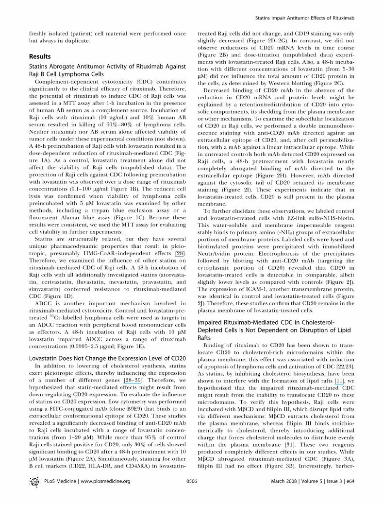
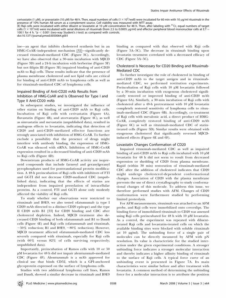
Complement-dependent cytotoxicity (CDC) contributessignificantly to the clinical efficacy of rituximab. Therefore,the potential of rituximab to induce CDC of Raji cells wasassessed in a MTT assay after 1-h incubation in the presenceof human AB serum as a complement source. Incubation ofRaji cells with rituximab (10 lg/mL) and 10% human ABserum resulted in killing of 60%–80% of lymphoma cells.Neither rituximab nor AB serum alone affected viability oftumor cells under these experimental conditions (not shown).A 48-h preincubation of Raji cells with lovastatin resulted in adose-dependent reduction of rituximab-mediated CDC (Fig-ure 1A). As a control, lovastatin treatment alone did notaffect the viability of Raji cells (unpublished data). Theprotection of Raji cells against CDC following preincubationwith lovastatin was observed over a dose range of rituximabconcentrations (0.1–100 lg/ml; Figure 1B). The reduced celllysis was confirmed when viability of lymphoma cellspreincubated with 5 lM lovastatin was examined by othermethods, including a trypan blue exclusion assay or afluorescent Alamar blue assay (Figure 1C). Because theseresults were consistent, we used the MTT assay for evaluatingcell viability in further experiments.
Statins are structurally related, but they have severalunique pharmacodynamic properties that result in pleio-tropic, presumably HMG-CoAR–independent effects [28].Therefore, we examined the influence of other statins onrituximab-mediated CDC of Raji cells. A 48-h incubation ofRaji cells with all additionally investigated statins (atorvasta-tin, cerivastatin, fluvastatin, mevastatin, pravastatin, andsimvastatin) conferred resistance to rituximab-mediatedCDC (Figure 1D).
ADCC is another important mechanism involved inrituximab-mediated cytotoxicity. Control and lovastatin-pre-treated 51Cr-labelled lymphoma cells were used as targets inan ADCC reaction with peripheral blood mononuclear cellsas effectors. A 48-h incubation of Raji cells with 10 lMlovastatin impaired ADCC across a range of rituximabconcentrations (0.0005–2.5 lg/ml; Figure 1E).
Lovastatin Does Not Change the Expression Level of CD20In addition to lowering of cholesterol synthesis, statins
exert pleiotropic effects, thereby influencing the expressionof a number of different genes [28–30]. Therefore, wehypothesized that statin-mediated effects might result fromdown-regulating CD20 expression. To evaluate the influenceof statins on CD20 expression, flow cytometry was performedusing a FITC-conjugated mAb (clone B9E9) that binds to anextracellular conformational epitope of CD20. These studiesrevealed a significantly decreased binding of anti-CD20 mAbto Raji cells incubated with a range of lovastatin concen-trations (from 1–20 lM). While more than 95% of controlRaji cells stained positive for CD20, only 30% of cells showedsignificant binding to CD20 after a 48-h pretreatment with 10lM lovastatin (Figure 2A). Simultaneously, staining for otherB cell markers (CD22, HLA-DR, and CD45RA) in lovastatin-
treated Raji cells did not change, and CD19 staining was onlyslightly decreased (Figure 2D–2G). In contrast, we did notobserve reductions of CD20 mRNA levels in time course(Figure 2B) and dose-titration (unpublished data) experi-ments with lovastatin-treated Raji cells. Also, a 48-h incuba-tion with different concentrations of lovastatin (from 5–30lM) did not influence the total amount of CD20 protein inthe cells, as determined by Western blotting (Figure 2C).Decreased binding of CD20 mAb in the absence of the
reduction in CD20 mRNA and protein levels might beexplained by a retention/redistribution of CD20 into cyto-solic compartments, its shedding from the plasma membraneor other mechanisms. To examine the subcellular localizationof CD20 in Raji cells, we performed a double immunofluor-escence staining with anti-CD20 mAb directed against anextracellular epitope of CD20, and, after cell permeabiliza-tion, with a mAb against a linear intracellular epitope. Whilein untreated controls both mAb detected CD20 expressed onRaji cells, a 48-h pretreatment with lovastatin nearlycompletely abrogated binding of mAb directed to theextracellular epitope (Figure 2H). However, mAb directedagainst the cytosolic tail of CD20 retained its membranestaining (Figure 2I). These experiments indicate that inlovastatin-treated cells, CD20 is still present in the plasmamembrane.To further elucidate these observations, we labeled control
and lovastatin-treated cells with EZ-link sulfo-NHS-biotin.This water-soluble and membrane impermeable reagentstably binds to primary amino (-NH2) groups of extracellularportions of membrane proteins. Labeled cells were lysed andbiotinylated proteins were precipitated with immobilizedNeutrAvidin protein. Electrophoresis of the precipitatesfollowed by blotting with anti-CD20 mAb (targeting thecytoplasmic portion of CD20) revealed that CD20 inlovastatin-treated cells is detectable in comparable, albeitslightly lower levels as compared with controls (Figure 2J).The expression of ICAM-1, another transmembrane protein,was identical in control and lovastatin-treated cells (Figure2J). Therefore, these studies confirm that CD20 remains in theplasma membrane of lovastatin-treated cells.
Impaired Rituximab-Mediated CDC in Cholesterol-Depleted Cells Is Not Dependent on Disruption of LipidRaftsBinding of rituximab to CD20 has been shown to trans-
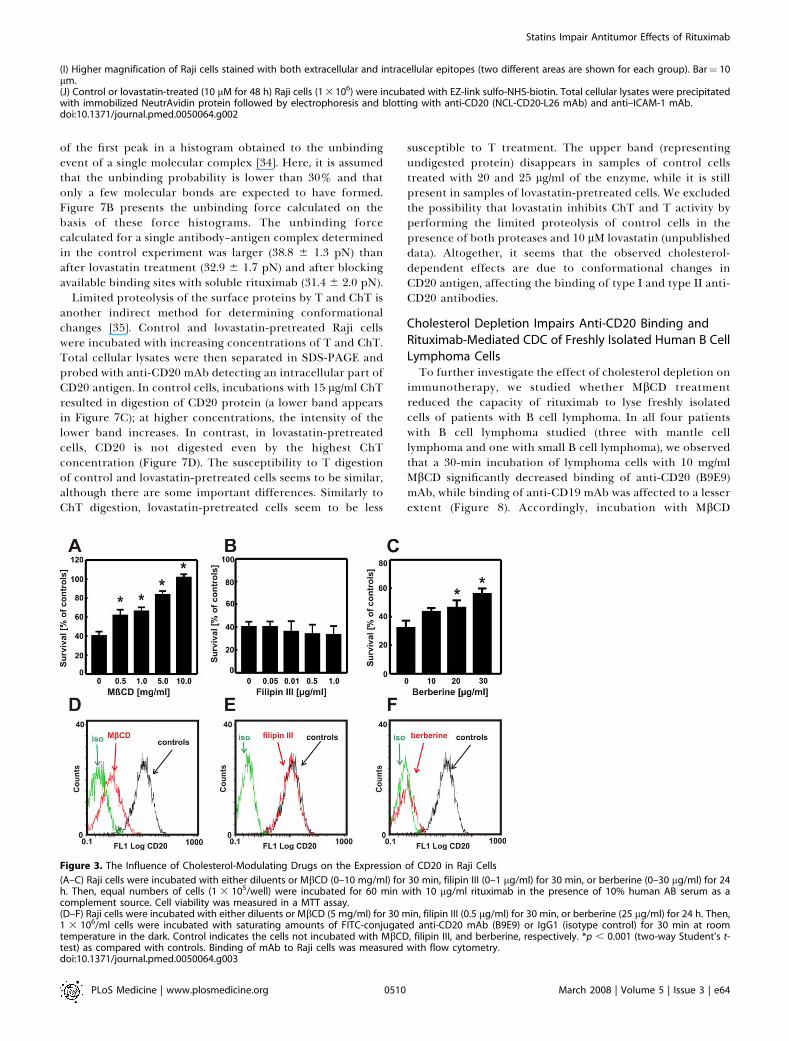
locate CD20 to cholesterol-rich microdomains within theplasma membrane; this effect was associated with inductionof apoptosis of lymphoma cells and activation of CDC [22,23].As statins, by inhibiting cholesterol biosynthesis, have beenshown to interfere with the formation of lipid rafts [11], wehypothesized that the impaired rituximab-mediated CDCmight result from the inability to translocate CD20 to thesemicrodomains. To verify this hypothesis, Raji cells wereincubated with MbCD and filipin III, which disrupt lipid raftsvia different mechanisms: MbCD extracts cholesterol fromthe plasma membrane, whereas filipin III binds stoichio-metrically to cholesterol, thereby introducing additionalcharge that forces cholesterol molecules to distribute evenlywithin the plasma membrane [31]. These two reagentsproduced completely different effects in our studies. WhileMbCD abrogated rituximab-mediated CDC (Figure 3A),filipin III had no effect (Figure 3B). Interestingly, berber-
PLoS Medicine | www.plosmedicine.org March 2008 | Volume 5 | Issue 3 | e640506
Statins Impair Antitumor Effects of Rituximab
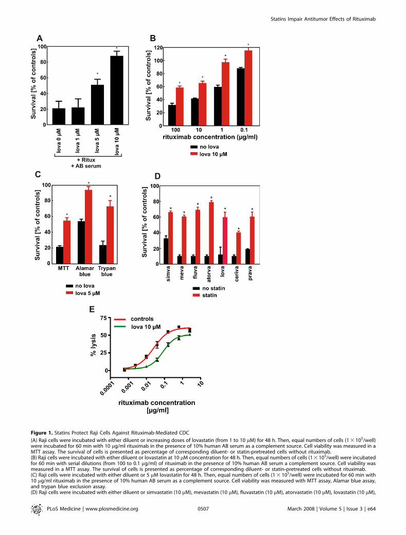
Figure 1. Statins Protect Raji Cells Against Rituximab-Mediated CDC
(A) Raji cells were incubated with either diluent or increasing doses of lovastatin (from 1 to 10 lM) for 48 h. Then, equal numbers of cells (1 3 105/well)were incubated for 60 min with 10 lg/ml rituximab in the presence of 10% human AB serum as a complement source. Cell viability was measured in aMTT assay. The survival of cells is presented as percentage of corresponding diluent- or statin-pretreated cells without rituximab.(B) Raji cells were incubated with either diluent or lovastatin at 10 lM concentration for 48 h. Then, equal numbers of cells (1 3 105/well) were incubatedfor 60 min with serial dilutions (from 100 to 0.1 lg/ml) of rituximab in the presence of 10% human AB serum a complement source. Cell viability wasmeasured in a MTT assay. The survival of cells is presented as percentage of corresponding diluent- or statin-pretreated cells without rituximab.(C) Raji cells were incubated with either diluent or 5 lM lovastatin for 48 h. Then, equal numbers of cells (1 3 105/well) were incubated for 60 min with10 lg/ml rituximab in the presence of 10% human AB serum as a complement source. Cell viability was measured with MTT assay, Alamar blue assay,and trypan blue exclusion assay.(D) Raji cells were incubated with either diluent or simvastatin (10 lM), mevastatin (10 lM), fluvastatin (10 lM), atorvastatin (10 lM), lovastatin (10 lM),
PLoS Medicine | www.plosmedicine.org March 2008 | Volume 5 | Issue 3 | e640507
Statins Impair Antitumor Effects of Rituximab
ine—an agent that inhibits cholesterol synthesis but in anHMG-CoAR–independent mechanism [32]—significantly de-creased rituximab-mediated CDC (Figure 3C). Accordingly,we have also observed that a 30-min incubation with MbCD(Figure 3D) and a 24-h incubation with berberine (Figure 3F)but not filipin III (Figure 3E) impaired binding of anti-CD20mAb to Raji cells. These results indicate that the presence ofplasma membrane cholesterol and not lipid rafts are criticalfor binding of anti-CD20 mAb to lymphoma cells as well asfor rituximab-mediated CDC of lymphoma cells.
Impaired Binding of Anti-CD20 mAb Results fromInhibition of HMG-CoAR and Is Observed for Type I andType II Anti-CD20 mAb
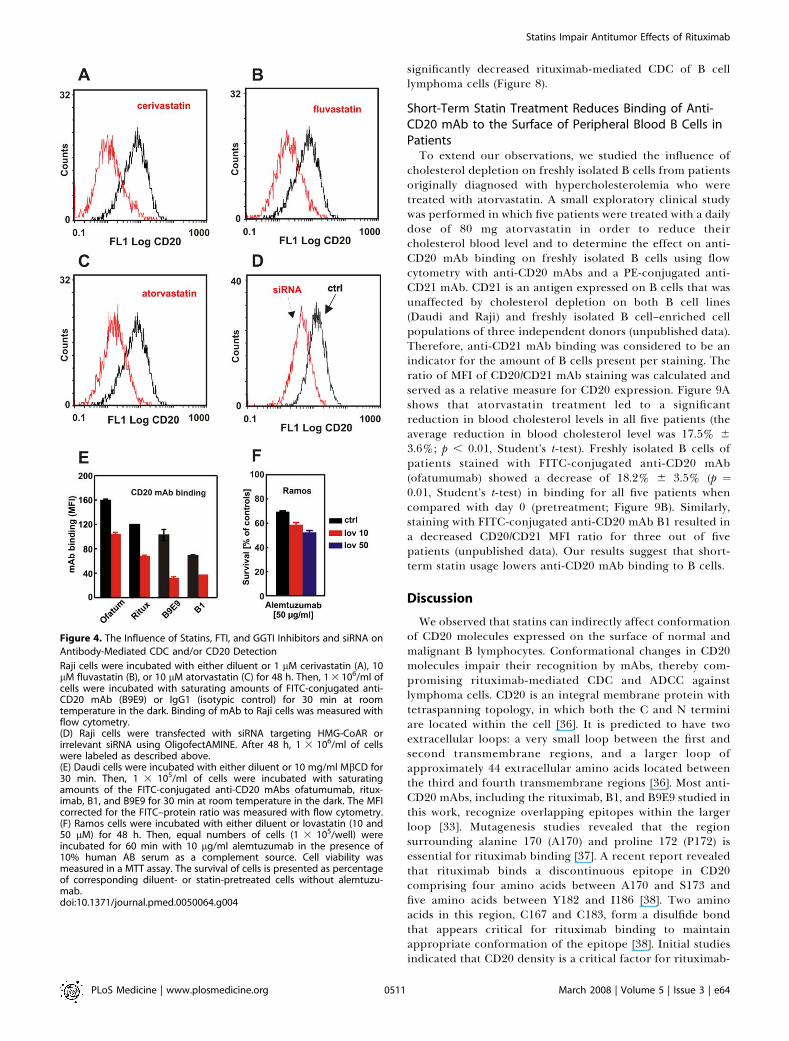
In subsequent studies, we investigated the influence ofother statins on binding of anti-CD20 mAb to Raji cells.Incubation of Raji cells with cerivastatin (Figure 4A),fluvastatin (Figure 4B), and atorvastatin (Figure 4C), as wellas simvastatin and mevastatin (unpublished data), resulted inanalogous effects to lovastatin, indicating that detection ofCD20 and anti-CD20–mediated effector functions arestrongly associated with inhibition of HMG-CoAR. To furtherexclude a possibility that the presence of drugs mightinterfere with antibody binding, the expression of HMG-CoAR was silenced with siRNA. Inhibition of HMG-CoARexpression resulted in a decreased binding of anti-CD20 mAbto Raji cells (Figure 4D).
Downstream products of HMG-CoAR activity are isopre-noid compounds that include farnesyl and geranylgeranylpyrophosphates used for posttranslational protein modifica-tion. A 48-h preincubation of Raji cells with inhibitors of FTIand GGTI did not decrease CD20-mediated CDC (unpub-lished data), indicating that the effects of statins areindependent from impaired prenylation of intracellularproteins. As a control, FTI and GGTI alone only modestlyaffected the viability of Raji cells.
To study whether our observations were restricted torituximab and B9E9, we also tested ofatumumab (a type ICD20 mAb directed to a distinct CD20 epitope) and the typeII CD20 mAb B1 [33] for CD20 binding and CDC aftercholesterol depletion. Indeed, MbCD treatment also de-creased CD20 binding of both ofatumumab and B1 to Daudicells (Figure 4E) and Raji cells (ofatumumab and rituximab,;50% reduction; B1 and B9E9, ;80% reduction). However,MbCD treatment affected ofatumumab-mediated CDC lessseverely compared with rituximab, especially for Raji cells(with 60% versus 82% of cells surviving respectively;unpublished data).
Importantly, preincubation of Ramos cells with 10 or 50lM lovastatin for 48 h did not impair alemtuzumab-mediatedCDC (Figure 4F). Alemtuzumab is a mAb approved forclinical use that binds CD52, which is a GPI-anchoredglycoprotein expressed on the surface of lymphoma cells.
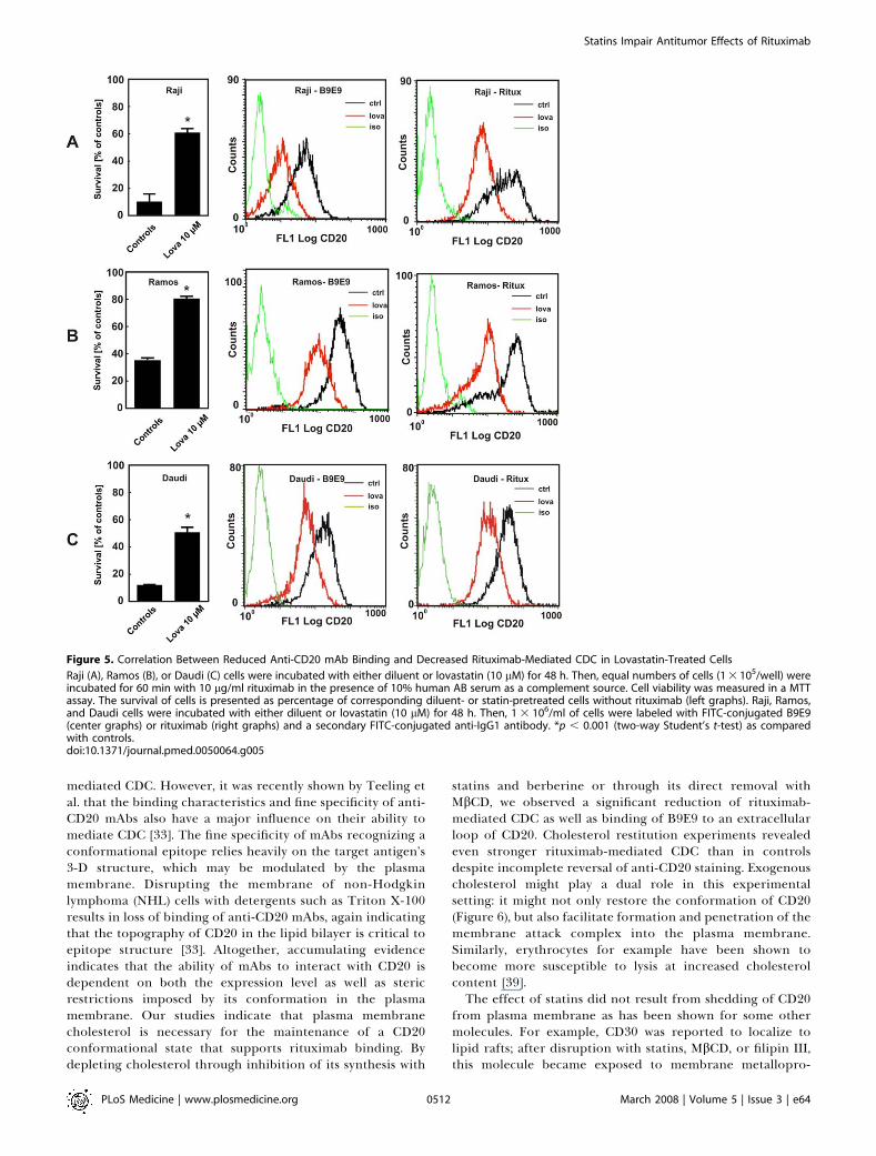
Studies with two additional lymphoma cell lines, Ramosand Daudi, showed a similar decrease in rituximab and B9E9
binding as compared with that observed with Raji cells(Figure 5A–5C). The decrease in rituximab binding uponlovastatin treatment correlated with a decreased efficacy ofCDC (Figure 5A–5C).
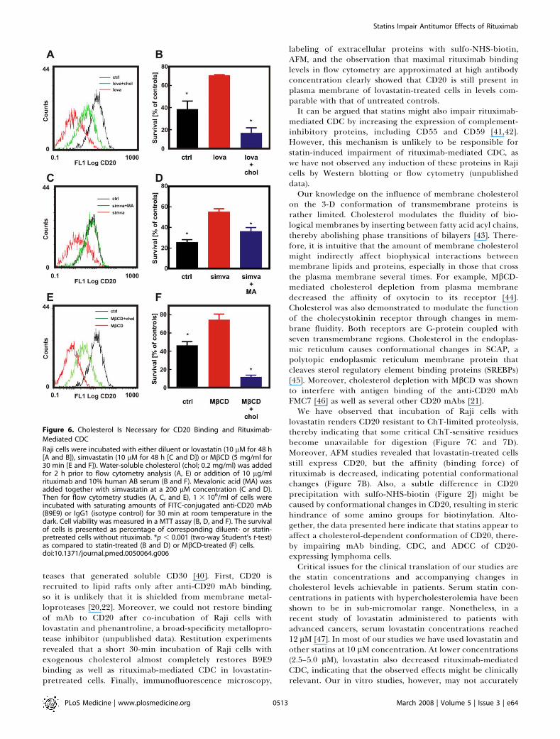
Cholesterol Is Necessary for CD20 Binding and Rituximab-Mediated CDCTo further investigate the role of cholesterol in binding of
anti-CD20 mAb to the target antigen and in rituximab-mediated CDC, we performed restitution experiments.Preincubation of Raji cells with 10 lM lovastatin followedby a 30-min incubation with exogenous cholesterol signifi-cantly restored or improved binding of anti-CD20 mAb(Figure 6A). Similarly, a 30-min incubation of Raji cells withcholesterol after a 48-h pretreatment with 10 lM lovastatincompletely restored sensitivity of lymphoma cells to ritux-imab-mediated CDC (Figure 6B). Accordingly, co-treatmentof Raji cells with mevalonic acid, a direct product of HMG-CoAR, completely restored binding of anti-CD20 mAb(Figure 6C) as well as rituximab-mediated CDC of statin-treated cells (Figure 5D). Similar results were obtained withexogenous cholesterol that significantly reversed MbCD-induced effects (Figure 6E and 6F).
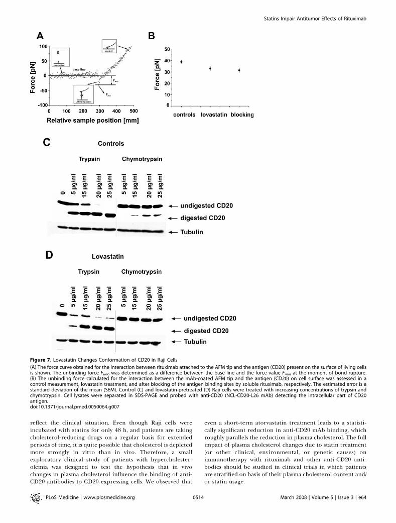
Lovastatin Changes Conformation of CD20Impaired rituximab-mediated CDC as well as impaired
binding of anti-CD20 mAb to Raji cells incubated with 10 lMlovastatin for 48 h did not seem to result from decreasedexpression or shedding of CD20 from plasma membrane.Rapid (within 30 min) restoration of rituximab-mediatedCDC after the addition of cholesterol indicates that CD20might undergo cholesterol-dependent conformationalchanges. Association of CD20 with the plasma membraneprevents the use of direct crystallography to study conforma-tional changes of this molecule. To address this issue, wetherefore performed studies with AFM. Changes of CD20conformation were furthermore studied by performinglimited proteolysis.For AFM measurements, rituximab was attached to an AFM
probe, and Raji cells were immobilized onto coverslips. Thebinding force of immobilized rituximab to CD20 was assessedusing Raji cells preincubated for 48 h with 10 lM lovastatin.As a control, the experiment was repeated with diluent-treated Raji cells and lovastatin-treated cells on which theavailable binding sites were blocked with soluble rituximab(at 10 lg/ml). The unbinding force of a single pair ofmolecules can be directly measured by AFM with pNresolution. Its value is characteristic for the studied inter-action under the given experimental conditions. A strongerunbinding force indicates a stronger molecular interactionand thereby indicates a higher affinity binding of rituximabto the surface of Raji cells. A typical force curve of anunbinding event is presented in Figure 7A. Its maincharacteristics were similar before and after treatment withlovastatin. A common method of determining the unbindingforce for a molecular interaction is to attribute the position
cerivastatin (1 lM), or pravastatin (10 lM) for 48 h. Then, equal numbers of cells (1 3 105/well) were incubated for 60 min with 10 lg/ml rituximab in thepresence of 10% human AB serum as a complement source. Cell viability was measured with MTT assay.(E) Raji cells were incubated with either diluent or lovastatin at 10 lM concentration for 48 h. Then, after labeling with 51Cr, equal numbers of targetcells (1 3 105/ml) were incubated with serial dilutions of rituximab (from 2.5 to 0.0005 lg/ml) and effector peripheral blood mononuclear cells at E:T¼100:1 for 4 h. *p , 0.001 (two-way Student’s t-test) as compared with controls.doi:10.1371/journal.pmed.0050064.g001
PLoS Medicine | www.plosmedicine.org March 2008 | Volume 5 | Issue 3 | e640508
Statins Impair Antitumor Effects of Rituximab
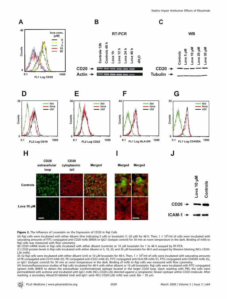
Figure 2. The Influence of Lovastatin on the Expression of CD20 in Raji Cells
(A) Raji cells were incubated with either diluent (line indicating 0 lM) or lovastatin (1–20 lM) for 48 h. Then, 1 3 106/ml of cells were incubated withsaturating amounts of FITC-conjugated anti-CD20 mAb (B9E9) or IgG1 (isotype control) for 30 min at room temperature in the dark. Binding of mAb toRaji cells was measured with flow cytometry.(B) CD20 mRNA levels in Raji cells incubated with either diluent (controls) or 10 lM lovastatin for 1 to 48 h assayed by RT-PCR.(C) CD20 protein levels in Raji cells incubated with either diluent or 5, 10, 20, and 30 lM lovastatin for 48 h and assayed by Western blotting (NCL-CD20-L26 mAb).(D–G) Raji cells were incubated with either diluent (ctrl) or 10 lM lovastatin for 48 h. Then, 1 3 106/ml of cells were incubated with saturating amountsof PE-conjugated anti-CD19 mAb (D), PE-conjugated anti-CD22 mAb (E), FITC-conjugated anti-HLA-DR mAb (F), FITC-conjugated anti-CD45RA mAb (G),or IgG1 (isotypic control) for 30 min at room temperature in the dark. Binding of mAb to Raji cells was measured with flow cytometry.(H) Immunofluorescence studies of Raji cells incubated for 48 h with either diluent or 10 lM lovastatin. Raji cells were incubated with FITC-conjugated(green) mAb (B9E9) to detect the extracellular (conformational) epitope located in the larger CD20 loop. Upon washing with PBS, the cells werepermeablized with acetone and incubated with IgG1 mAb (NCL-CD20-L26) directed against a cytoplasmic (linear) epitope within CD20 molecule. Afterwashing, a secondary Alexa555-labeled (red) anti-IgG1 (anti–NCL-CD20-L26) mAb was used. Bar ¼ 50 lm.
PLoS Medicine | www.plosmedicine.org March 2008 | Volume 5 | Issue 3 | e640509
Statins Impair Antitumor Effects of Rituximab
of the first peak in a histogram obtained to the unbindingevent of a single molecular complex [34]. Here, it is assumedthat the unbinding probability is lower than 30% and thatonly a few molecular bonds are expected to have formed.Figure 7B presents the unbinding force calculated on thebasis of these force histograms. The unbinding forcecalculated for a single antibody–antigen complex determinedin the control experiment was larger (38.8 6 1.3 pN) thanafter lovastatin treatment (32.9 6 1.7 pN) and after blockingavailable binding sites with soluble rituximab (31.4 6 2.0 pN).
Limited proteolysis of the surface proteins by T and ChT isanother indirect method for determining conformationalchanges [35]. Control and lovastatin-pretreated Raji cellswere incubated with increasing concentrations of T and ChT.Total cellular lysates were then separated in SDS-PAGE andprobed with anti-CD20 mAb detecting an intracellular part ofCD20 antigen. In control cells, incubations with 15 lg/ml ChTresulted in digestion of CD20 protein (a lower band appearsin Figure 7C); at higher concentrations, the intensity of thelower band increases. In contrast, in lovastatin-pretreatedcells, CD20 is not digested even by the highest ChTconcentration (Figure 7D). The susceptibility to T digestionof control and lovastatin-pretreated cells seems to be similar,although there are some important differences. Similarly toChT digestion, lovastatin-pretreated cells seem to be less
susceptible to T treatment. The upper band (representingundigested protein) disappears in samples of control cellstreated with 20 and 25 lg/ml of the enzyme, while it is stillpresent in samples of lovastatin-pretreated cells. We excludedthe possibility that lovastatin inhibits ChT and T activity byperforming the limited proteolysis of control cells in thepresence of both proteases and 10 lM lovastatin (unpublisheddata). Altogether, it seems that the observed cholesterol-dependent effects are due to conformational changes inCD20 antigen, affecting the binding of type I and type II anti-CD20 antibodies.
Cholesterol Depletion Impairs Anti-CD20 Binding and
Rituximab-Mediated CDC of Freshly Isolated Human B Cell
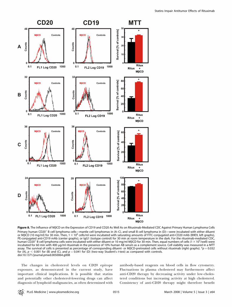
Lymphoma CellsTo further investigate the effect of cholesterol depletion on
immunotherapy, we studied whether MbCD treatmentreduced the capacity of rituximab to lyse freshly isolatedcells of patients with B cell lymphoma. In all four patientswith B cell lymphoma studied (three with mantle celllymphoma and one with small B cell lymphoma), we observedthat a 30-min incubation of lymphoma cells with 10 mg/mlMbCD significantly decreased binding of anti-CD20 (B9E9)mAb, while binding of anti-CD19 mAb was affected to a lesserextent (Figure 8). Accordingly, incubation with MbCD
(I) Higher magnification of Raji cells stained with both extracellular and intracellular epitopes (two different areas are shown for each group). Bar¼ 10lm.(J) Control or lovastatin-treated (10 lM for 48 h) Raji cells (1 3 106) were incubated with EZ-link sulfo-NHS-biotin. Total cellular lysates were precipitatedwith immobilized NeutrAvidin protein followed by electrophoresis and blotting with anti-CD20 (NCL-CD20-L26 mAb) and anti–ICAM-1 mAb.doi:10.1371/journal.pmed.0050064.g002
Figure 3. The Influence of Cholesterol-Modulating Drugs on the Expression of CD20 in Raji Cells
(A–C) Raji cells were incubated with either diluents or MbCD (0–10 mg/ml) for 30 min, filipin III (0–1 lg/ml) for 30 min, or berberine (0–30 lg/ml) for 24h. Then, equal numbers of cells (1 3 105/well) were incubated for 60 min with 10 lg/ml rituximab in the presence of 10% human AB serum as acomplement source. Cell viability was measured in a MTT assay.(D–F) Raji cells were incubated with either diluents or MbCD (5 mg/ml) for 30 min, filipin III (0.5 lg/ml) for 30 min, or berberine (25 lg/ml) for 24 h. Then,1 3 106/ml cells were incubated with saturating amounts of FITC-conjugated anti-CD20 mAb (B9E9) or IgG1 (isotype control) for 30 min at roomtemperature in the dark. Control indicates the cells not incubated with MbCD, filipin III, and berberine, respectively. *p , 0.001 (two-way Student’s t-test) as compared with controls. Binding of mAb to Raji cells was measured with flow cytometry.doi:10.1371/journal.pmed.0050064.g003
PLoS Medicine | www.plosmedicine.org March 2008 | Volume 5 | Issue 3 | e640510
Statins Impair Antitumor Effects of Rituximab
significantly decreased rituximab-mediated CDC of B celllymphoma cells (Figure 8).
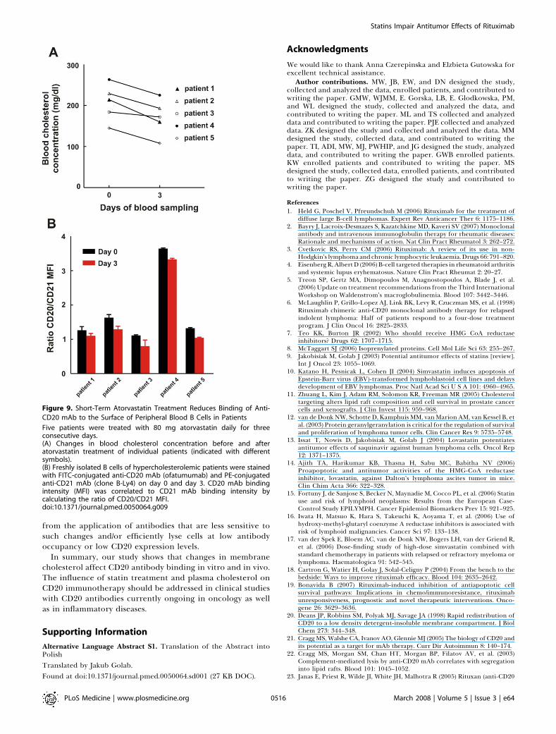
Short-Term Statin Treatment Reduces Binding of Anti-CD20 mAb to the Surface of Peripheral Blood B Cells inPatientsTo extend our observations, we studied the influence of
cholesterol depletion on freshly isolated B cells from patientsoriginally diagnosed with hypercholesterolemia who weretreated with atorvastatin. A small exploratory clinical studywas performed in which five patients were treated with a dailydose of 80 mg atorvastatin in order to reduce theircholesterol blood level and to determine the effect on anti-CD20 mAb binding on freshly isolated B cells using flowcytometry with anti-CD20 mAbs and a PE-conjugated anti-CD21 mAb. CD21 is an antigen expressed on B cells that wasunaffected by cholesterol depletion on both B cell lines(Daudi and Raji) and freshly isolated B cell–enriched cellpopulations of three independent donors (unpublished data).Therefore, anti-CD21 mAb binding was considered to be anindicator for the amount of B cells present per staining. Theratio of MFI of CD20/CD21 mAb staining was calculated andserved as a relative measure for CD20 expression. Figure 9Ashows that atorvastatin treatment led to a significantreduction in blood cholesterol levels in all five patients (theaverage reduction in blood cholesterol level was 17.5% 6
3.6%; p , 0.01, Student’s t-test). Freshly isolated B cells ofpatients stained with FITC-conjugated anti-CD20 mAb(ofatumumab) showed a decrease of 18.2% 6 3.5% (p ¼0.01, Student’s t-test) in binding for all five patients whencompared with day 0 (pretreatment; Figure 9B). Similarly,staining with FITC-conjugated anti-CD20 mAb B1 resulted ina decreased CD20/CD21 MFI ratio for three out of fivepatients (unpublished data). Our results suggest that short-term statin usage lowers anti-CD20 mAb binding to B cells.
Discussion
We observed that statins can indirectly affect conformationof CD20 molecules expressed on the surface of normal andmalignant B lymphocytes. Conformational changes in CD20molecules impair their recognition by mAbs, thereby com-promising rituximab-mediated CDC and ADCC againstlymphoma cells. CD20 is an integral membrane protein withtetraspanning topology, in which both the C and N terminiare located within the cell [36]. It is predicted to have twoextracellular loops: a very small loop between the first andsecond transmembrane regions, and a larger loop ofapproximately 44 extracellular amino acids located betweenthe third and fourth transmembrane regions [36]. Most anti-CD20 mAbs, including the rituximab, B1, and B9E9 studied inthis work, recognize overlapping epitopes within the largerloop [33]. Mutagenesis studies revealed that the regionsurrounding alanine 170 (A170) and proline 172 (P172) isessential for rituximab binding [37]. A recent report revealedthat rituximab binds a discontinuous epitope in CD20comprising four amino acids between A170 and S173 andfive amino acids between Y182 and I186 [38]. Two aminoacids in this region, C167 and C183, form a disulfide bondthat appears critical for rituximab binding to maintainappropriate conformation of the epitope [38]. Initial studiesindicated that CD20 density is a critical factor for rituximab-
Figure 4. The Influence of Statins, FTI, and GGTI Inhibitors and siRNA on
Antibody-Mediated CDC and/or CD20 Detection
Raji cells were incubated with either diluent or 1 lM cerivastatin (A), 10lM fluvastatin (B), or 10 lM atorvastatin (C) for 48 h. Then, 1 3 106/ml ofcells were incubated with saturating amounts of FITC-conjugated anti-CD20 mAb (B9E9) or IgG1 (isotypic control) for 30 min at roomtemperature in the dark. Binding of mAb to Raji cells was measured withflow cytometry.(D) Raji cells were transfected with siRNA targeting HMG-CoAR orirrelevant siRNA using OligofectAMINE. After 48 h, 1 3 106/ml of cellswere labeled as described above.(E) Daudi cells were incubated with either diluent or 10 mg/ml MbCD for30 min. Then, 1 3 105/ml of cells were incubated with saturatingamounts of the FITC-conjugated anti-CD20 mAbs ofatumumab, ritux-imab, B1, and B9E9 for 30 min at room temperature in the dark. The MFIcorrected for the FITC–protein ratio was measured with flow cytometry.(F) Ramos cells were incubated with either diluent or lovastatin (10 and50 lM) for 48 h. Then, equal numbers of cells (1 3 105/well) wereincubated for 60 min with 10 lg/ml alemtuzumab in the presence of10% human AB serum as a complement source. Cell viability wasmeasured in a MTT assay. The survival of cells is presented as percentageof corresponding diluent- or statin-pretreated cells without alemtuzu-mab.doi:10.1371/journal.pmed.0050064.g004
PLoS Medicine | www.plosmedicine.org March 2008 | Volume 5 | Issue 3 | e640511
Statins Impair Antitumor Effects of Rituximab
mediated CDC. However, it was recently shown by Teeling etal. that the binding characteristics and fine specificity of anti-CD20 mAbs also have a major influence on their ability tomediate CDC [33]. The fine specificity of mAbs recognizing aconformational epitope relies heavily on the target antigen’s3-D structure, which may be modulated by the plasmamembrane. Disrupting the membrane of non-Hodgkinlymphoma (NHL) cells with detergents such as Triton X-100results in loss of binding of anti-CD20 mAbs, again indicatingthat the topography of CD20 in the lipid bilayer is critical toepitope structure [33]. Altogether, accumulating evidenceindicates that the ability of mAbs to interact with CD20 isdependent on both the expression level as well as stericrestrictions imposed by its conformation in the plasmamembrane. Our studies indicate that plasma membranecholesterol is necessary for the maintenance of a CD20conformational state that supports rituximab binding. Bydepleting cholesterol through inhibition of its synthesis with
statins and berberine or through its direct removal withMbCD, we observed a significant reduction of rituximab-mediated CDC as well as binding of B9E9 to an extracellularloop of CD20. Cholesterol restitution experiments revealedeven stronger rituximab-mediated CDC than in controlsdespite incomplete reversal of anti-CD20 staining. Exogenouscholesterol might play a dual role in this experimentalsetting: it might not only restore the conformation of CD20(Figure 6), but also facilitate formation and penetration of themembrane attack complex into the plasma membrane.Similarly, erythrocytes for example have been shown tobecome more susceptible to lysis at increased cholesterolcontent [39].The effect of statins did not result from shedding of CD20
from plasma membrane as has been shown for some othermolecules. For example, CD30 was reported to localize tolipid rafts; after disruption with statins, MbCD, or filipin III,this molecule became exposed to membrane metallopro-
Figure 5. Correlation Between Reduced Anti-CD20 mAb Binding and Decreased Rituximab-Mediated CDC in Lovastatin-Treated Cells
Raji (A), Ramos (B), or Daudi (C) cells were incubated with either diluent or lovastatin (10 lM) for 48 h. Then, equal numbers of cells (1 3 105/well) wereincubated for 60 min with 10 lg/ml rituximab in the presence of 10% human AB serum as a complement source. Cell viability was measured in a MTTassay. The survival of cells is presented as percentage of corresponding diluent- or statin-pretreated cells without rituximab (left graphs). Raji, Ramos,and Daudi cells were incubated with either diluent or lovastatin (10 lM) for 48 h. Then, 1 3 106/ml of cells were labeled with FITC-conjugated B9E9(center graphs) or rituximab (right graphs) and a secondary FITC-conjugated anti-IgG1 antibody. *p , 0.001 (two-way Student’s t-test) as comparedwith controls.doi:10.1371/journal.pmed.0050064.g005
PLoS Medicine | www.plosmedicine.org March 2008 | Volume 5 | Issue 3 | e640512
Statins Impair Antitumor Effects of Rituximab
teases that generated soluble CD30 [40]. First, CD20 isrecruited to lipid rafts only after anti-CD20 mAb binding,so it is unlikely that it is shielded from membrane metal-loproteases [20,22]. Moreover, we could not restore bindingof mAb to CD20 after co-incubation of Raji cells withlovastatin and phenantroline, a broad-specificity metallopro-tease inhibitor (unpublished data). Restitution experimentsrevealed that a short 30-min incubation of Raji cells withexogenous cholesterol almost completely restores B9E9binding as well as rituximab-mediated CDC in lovastatin-pretreated cells. Finally, immunofluorescence microscopy,
labeling of extracellular proteins with sulfo-NHS-biotin,AFM, and the observation that maximal rituximab bindinglevels in flow cytometry are approximated at high antibodyconcentration clearly showed that CD20 is still present inplasma membrane of lovastatin-treated cells in levels com-parable with that of untreated controls.It can be argued that statins might also impair rituximab-
mediated CDC by increasing the expression of complement-inhibitory proteins, including CD55 and CD59 [41,42].However, this mechanism is unlikely to be responsible forstatin-induced impairment of rituximab-mediated CDC, aswe have not observed any induction of these proteins in Rajicells by Western blotting or flow cytometry (unpublisheddata).Our knowledge on the influence of membrane cholesterol
on the 3-D conformation of transmembrane proteins israther limited. Cholesterol modulates the fluidity of bio-logical membranes by inserting between fatty acid acyl chains,thereby abolishing phase transitions of bilayers [43]. There-fore, it is intuitive that the amount of membrane cholesterolmight indirectly affect biophysical interactions betweenmembrane lipids and proteins, especially in those that crossthe plasma membrane several times. For example, MbCD-mediated cholesterol depletion from plasma membranedecreased the affinity of oxytocin to its receptor [44].Cholesterol was also demonstrated to modulate the functionof the cholecystokinin receptor through changes in mem-brane fluidity. Both receptors are G-protein coupled withseven transmembrane regions. Cholesterol in the endoplas-mic reticulum causes conformational changes in SCAP, apolytopic endoplasmic reticulum membrane protein thatcleaves sterol regulatory element binding proteins (SREBPs)[45]. Moreover, cholesterol depletion with MbCD was shownto interfere with antigen binding of the anti-CD20 mAbFMC7 [46] as well as several other CD20 mAbs [21].We have observed that incubation of Raji cells with
lovastatin renders CD20 resistant to ChT-limited proteolysis,thereby indicating that some critical ChT-sensitive residuesbecome unavailable for digestion (Figure 7C and 7D).Moreover, AFM studies revealed that lovastatin-treated cellsstill express CD20, but the affinity (binding force) ofrituximab is decreased, indicating potential conformationalchanges (Figure 7B). Also, a subtle difference in CD20precipitation with sulfo-NHS-biotin (Figure 2J) might becaused by conformational changes in CD20, resulting in sterichindrance of some amino groups for biotinylation. Alto-gether, the data presented here indicate that statins appear toaffect a cholesterol-dependent conformation of CD20, there-by impairing mAb binding, CDC, and ADCC of CD20-expressing lymphoma cells.Critical issues for the clinical translation of our studies are
the statin concentrations and accompanying changes incholesterol levels achievable in patients. Serum statin con-centrations in patients with hypercholesterolemia have beenshown to be in sub-micromolar range. Nonetheless, in arecent study of lovastatin administered to patients withadvanced cancers, serum lovastatin concentrations reached12 lM [47]. In most of our studies we have used lovastatin andother statins at 10 lM concentration. At lower concentrations(2.5–5.0 lM), lovastatin also decreased rituximab-mediatedCDC, indicating that the observed effects might be clinicallyrelevant. Our in vitro studies, however, may not accurately
Figure 6. Cholesterol Is Necessary for CD20 Binding and Rituximab-
Mediated CDC
Raji cells were incubated with either diluent or lovastatin (10 lM for 48 h[A and B]), simvastatin (10 lM for 48 h [C and D]) or MbCD (5 mg/ml for30 min [E and F]). Water-soluble cholesterol (chol; 0.2 mg/ml) was addedfor 2 h prior to flow cytometry analysis (A, E) or addition of 10 lg/mlrituximab and 10% human AB serum (B and F). Mevalonic acid (MA) wasadded together with simvastatin at a 200 lM concentration (C and D).Then for flow cytometry studies (A, C, and E), 1 3 106/ml of cells wereincubated with saturating amounts of FITC-conjugated anti-CD20 mAb(B9E9) or IgG1 (isotype control) for 30 min at room temperature in thedark. Cell viability was measured in a MTT assay (B, D, and F). The survivalof cells is presented as percentage of corresponding diluent- or statin-pretreated cells without rituximab. *p , 0.001 (two-way Student’s t-test)as compared to statin-treated (B and D) or MbCD-treated (F) cells.doi:10.1371/journal.pmed.0050064.g006
PLoS Medicine | www.plosmedicine.org March 2008 | Volume 5 | Issue 3 | e640513
Statins Impair Antitumor Effects of Rituximab
reflect the clinical situation. Even though Raji cells wereincubated with statins for only 48 h, and patients are takingcholesterol-reducing drugs on a regular basis for extendedperiods of time, it is quite possible that cholesterol is depletedmore strongly in vitro than in vivo. Therefore, a smallexploratory clinical study of patients with hypercholester-olemia was designed to test the hypothesis that in vivochanges in plasma cholesterol influence the binding of anti-CD20 antibodies to CD20-expressing cells. We observed that
even a short-term atorvastatin treatment leads to a statisti-cally significant reduction in anti-CD20 mAb binding, whichroughly parallels the reduction in plasma cholesterol. The fullimpact of plasma cholesterol changes due to statin treatment(or other clinical, environmental, or genetic causes) onimmunotherapy with rituximab and other anti-CD20 anti-bodies should be studied in clinical trials in which patientsare stratified on basis of their plasma cholesterol content and/or statin usage.
Figure 7. Lovastatin Changes Conformation of CD20 in Raji Cells
(A) The force curve obtained for the interaction between rituximab attached to the AFM tip and the antigen (CD20) present on the surface of living cellsis shown. The unbinding force Funb was determined as a difference between the base line and the force value Fmin at the moment of bond rupture.(B) The unbinding force calculated for the interaction between the mAb-coated AFM tip and the antigen (CD20) on cell surface was assessed in acontrol measurement, lovastatin treatment, and after blocking of the antigen binding sites by soluble rituximab, respectively. The estimated error is astandard deviation of the mean (SEM). Control (C) and lovastatin-pretreated (D) Raji cells were treated with increasing concentrations of trypsin andchymotrypsin. Cell lysates were separated in SDS-PAGE and probed with anti-CD20 (NCL-CD20-L26 mAb) detecting the intracellular part of CD20antigen.doi:10.1371/journal.pmed.0050064.g007
PLoS Medicine | www.plosmedicine.org March 2008 | Volume 5 | Issue 3 | e640514
Statins Impair Antitumor Effects of Rituximab
The changes in cholesterol levels on CD20 epitopeexposure, as demonstrated in the current study, haveimportant clinical implications. It is possible that statinsand potentially other cholesterol-lowering drugs can affectdiagnosis of lymphoid malignancies, as often determined with
antibody-based reagents on blood cells in flow cytometry.Fluctuations in plasma cholesterol may furthermore affectanti-CD20 therapy by decreasing activity under low-choles-terol conditions but increasing activity at high cholesterol.Consistency of anti-CD20 therapy might therefore benefit
Figure 8. The Influence of MbCD on the Expression of CD19 and CD20 As Well As on Rituximab-Mediated CDC Against Primary Human Lymphoma Cells
Primary human CD20þ B cell lymphoma cells—mantle cell lymphomas in (A–C), and small B cell lymphoma in (D)—were incubated with either diluentor MbCD (10 mg/ml) for 30 min. Then, 1 3 106 cells/ml were incubated with saturating amounts of FITC-conjugated anti-CD20 mAb (B9E9, left graphs),PE-conjugated anti-CD19 mAb (center graphs), or IgG1 (isotype control) for 30 min at room temperature in the dark. For the rituximab-mediated CDC,human CD20þ B cell lymphoma cells were incubated with either diluent or 10 mg/ml MbCD for 30 min. Then, equal numbers of cells (1 3 105/well) wereincubated for 60 min with 400 lg/ml rituximab in the presence of 10% human AB serum as a complement source. Cell viability was measured in a MTTassay. The survival of cells is presented as percentage of corresponding diluent- or MbCD-pretreated cells without rituximab (right graphs). *p¼ 0.032for (A), p , 0.001 for (B) and (C), and p¼ 0.041 for (D) (two-way Student’s t-test) as compared with controls.doi:10.1371/journal.pmed.0050064.g008
PLoS Medicine | www.plosmedicine.org March 2008 | Volume 5 | Issue 3 | e640515
Statins Impair Antitumor Effects of Rituximab
from the application of antibodies that are less sensitive tosuch changes and/or efficiently lyse cells at low antibodyoccupancy or low CD20 expression levels.
In summary, our study shows that changes in membranecholesterol affect CD20 antibody binding in vitro and in vivo.The influence of statin treatment and plasma cholesterol onCD20 immunotherapy should be addressed in clinical studieswith CD20 antibodies currently ongoing in oncology as wellas in inflammatory diseases.
Supporting Information
Alternative Language Abstract S1. Translation of the Abstract intoPolish
Translated by Jakub Golab.
Found at doi:10.1371/journal.pmed.0050064.sd001 (27 KB DOC).
Acknowledgments
We would like to thank Anna Czerepinska and Elzbieta Gutowska forexcellent technical assistance.
Author contributions. MW, JB, EW, and DN designed the study,collected and analyzed the data, enrolled patients, and contributed towriting the paper. GMW, WJMM, E. Gorska, LB, E. Glodkowska, PM,and WL designed the study, collected and analyzed the data, andcontributed to writing the paper. ML and TS collected and analyzeddata and contributed to writing the paper. PJE collected and analyzeddata. ZK designed the study and collected and analyzed the data. MMdesigned the study, collected data, and contributed to writing thepaper. TI, ADI, MW, MJ, PWHIP, and JG designed the study, analyzeddata, and contributed to writing the paper. GWB enrolled patients.KW enrolled patients and contributed to writing the paper. MSdesigned the study, collected data, enrolled patients, and contributedto writing the paper. ZG designed the study and contributed towriting the paper.
References1. Held G, Poschel V, Pfreundschuh M (2006) Rituximab for the treatment of
diffuse large B-cell lymphomas. Expert Rev Anticancer Ther 6: 1175–1186.2. Bayry J, Lacroix-Desmazes S, Kazatchkine MD, Kaveri SV (2007) Monoclonal
antibody and intravenous immunoglobulin therapy for rheumatic diseases:Rationale and mechanisms of action. Nat Clin Pract Rheumatol 3: 262–272.
3. Cvetkovic RS, Perry CM (2006) Rituximab: A review of its use in non-Hodgkin’s lymphomaand chronic lymphocytic leukaemia.Drugs 66: 791–820.
4. EisenbergR, AlbertD (2006) B-cell targeted therapies in rheumatoid arthritisand systemic lupus eryhematosus. Nature Clin Pract Rheumat 2: 20–27.
5. Treon SP, Gertz MA, Dimopoulos M, Anagnostopoulos A, Blade J, et al.(2006) Update on treatment recommendations from the Third InternationalWorkshop on Waldenstrom’s macroglobulinemia. Blood 107: 3442–3446.
6. McLaughlin P, Grillo-Lopez AJ, Link BK, Levy R, Czuczman MS, et al. (1998)Rituximab chimeric anti-CD20 monoclonal antibody therapy for relapsedindolent lymphoma: Half of patients respond to a four-dose treatmentprogram. J Clin Oncol 16: 2825–2833.
7. Teo KK, Burton JR (2002) Who should receive HMG CoA reductaseinhibitors? Drugs 62: 1707–1715.
8. McTaggart SJ (2006) Isoprenylated proteins. Cell Mol Life Sci 63: 255–267.9. Jakobisiak M, Golab J (2003) Potential antitumor effects of statins [review].
Int J Oncol 23: 1055–1069.10. Katano H, Pesnicak L, Cohen JI (2004) Simvastatin induces apoptosis of
Epstein-Barr virus (EBV)-transformed lymphoblastoid cell lines and delaysdevelopment of EBV lymphomas. Proc Natl Acad Sci U S A 101: 4960–4965.
11. Zhuang L, Kim J, Adam RM, Solomon KR, Freeman MR (2005) Cholesteroltargeting alters lipid raft composition and cell survival in prostate cancercells and xenografts. J Clin Invest 115: 959–968.
12. van de Donk NW, Schotte D, Kamphuis MM, vanMarion AM, van Kessel B, etal. (2003) Protein geranylgeranylation is critical for the regulation of survivaland proliferation of lymphoma tumor cells. Clin Cancer Res 9: 5735–5748.
13. Issat T, Nowis D, Jakobisiak M, Golab J (2004) Lovastatin potentiatesantitumor effects of saquinavir against human lymphoma cells. Oncol Rep12: 1371–1375.
14. Ajith TA, Harikumar KB, Thasna H, Sabu MC, Babitha NV (2006)Proapoptotic and antitumor activities of the HMG-CoA reductaseinhibitor, lovastatin, against Dalton’s lymphoma ascites tumor in mice.Clin Chim Acta 366: 322–328.
15. Fortuny J, de Sanjose S, Becker N, Maynadie M, Cocco PL, et al. (2006) Statinuse and risk of lymphoid neoplasms: Results from the European Case-Control Study EPILYMPH. Cancer Epidemiol Biomarkers Prev 15: 921–925.
16. Iwata H, Matsuo K, Hara S, Takeuchi K, Aoyama T, et al. (2006) Use ofhydroxy-methyl-glutaryl coenzyme A reductase inhibitors is associated withrisk of lymphoid malignancies. Cancer Sci 97: 133–138.
17. van der Spek E, Bloem AC, van de Donk NW, Bogers LH, van der Griend R,et al. (2006) Dose-finding study of high-dose simvastatin combined withstandard chemotherapy in patients with relapsed or refractory myeloma orlymphoma. Haematologica 91: 542–545.
18. Cartron G, Watier H, Golay J, Solal-Celigny P (2004) From the bench to thebedside: Ways to improve rituximab efficacy. Blood 104: 2635–2642.
19. Bonavida B (2007) Rituximab-induced inhibition of antiapoptotic cellsurvival pathways: Implications in chemo/immunoresistance, rituximabunresponsiveness, prognostic and novel therapeutic interventions. Onco-gene 26: 3629–3636.
20. Deans JP, Robbins SM, Polyak MJ, Savage JA (1998) Rapid redistribution ofCD20 to a low density detergent-insoluble membrane compartment. J BiolChem 273: 344–348.
21. CraggMS,Walshe CA, Ivanov AO, GlennieMJ (2005) The biology of CD20 andits potential as a target for mAb therapy. Curr Dir Autoimmun 8: 140–174.
22. Cragg MS, Morgan SM, Chan HT, Morgan BP, Filatov AV, et al. (2003)Complement-mediated lysis by anti-CD20 mAb correlates with segregationinto lipid rafts. Blood 101: 1045–1052.
23. Janas E, Priest R, Wilde JI, White JH, Malhotra R (2005) Rituxan (anti-CD20
Figure 9. Short-Term Atorvastatin Treatment Reduces Binding of Anti-
CD20 mAb to the Surface of Peripheral Blood B Cells in Patients
Five patients were treated with 80 mg atorvastatin daily for threeconsecutive days.(A) Changes in blood cholesterol concentration before and afteratorvastatin treatment of individual patients (indicated with differentsymbols).(B) Freshly isolated B cells of hypercholesterolemic patients were stainedwith FITC-conjugated anti-CD20 mAb (ofatumumab) and PE-conjugatedanti-CD21 mAb (clone B-Ly4) on day 0 and day 3. CD20 mAb bindingintensity (MFI) was correlated to CD21 mAb binding intensity bycalculating the ratio of CD20/CD21 MFI.doi:10.1371/journal.pmed.0050064.g009
PLoS Medicine | www.plosmedicine.org March 2008 | Volume 5 | Issue 3 | e640516
Statins Impair Antitumor Effects of Rituximab
antibody)-induced translocation of CD20 into lipid rafts is crucial forcalcium influx and apoptosis. Clin Exp Immunol 139: 439–446.
24. Unruh TL, Li H, Mutch CM, Shariat N, Grigoriou L, et al. (2005) Cholesteroldepletion inhibits src family kinase-dependent calcium mobilization andapoptosis induced by rituximab crosslinking. Immunology 116: 223–232.
25. Golab J, Nowis D, Skrzycki M, Czeczot H, Baranczyk-Kuzma A, et al. (2003)Antitumor effects of photodynamic therapy are potentiated by 2-methoxyestradiol. A superoxide dismutase inhibitor. J Biol Chem 278:407–414.
26. Lekka M, Lekki J, Marszalek M, Golonka P, Stachura Z, et al. (1999) Localelastic properties of cells studied by SFM. Appl Surf Sci 141: 345–349.
27. Laidler P, Dulinska J, Lekka M, Lekki J (2005) Expression of prostatespecific membrane antigen in androgen-independent prostate cancer cellline PC-3. Arch Biochem Biophys 435: 1–14.
28. Liao JK, Laufs U (2005) Pleiotropic effects of statins. Annu Rev PharmacolToxicol 45: 89–118.
29. Alegret M, Silvestre JS (2006) Pleiotropic effects of statins and relatedpharmacological experimental approaches. Methods Find Exp ClinPharmacol 28: 627–656.
30. Ito MK, Talbert RL, Tsimikas S (2006) Statin-associated pleiotropy: Possibleeffects beyond cholesterol reduction. Pharmacotherapy 26: 85S–97S.
31. Smart EJ, Anderson RG (2002) Alterations in membrane cholesterol thataffect structure and function of caveolae. Methods Enzymol 353: 131–139.
32. Kong W, Wei J, Abidi P, Lin M, Inaba S, et al. (2004) Berberine is a novelcholesterol-lowering drug working through a unique mechanism distinctfrom statins. Nat Med 10: 1344–1351.
33. Teeling JL, Mackus WJ, Wiegman LJ, van den Brakel JH, Beers SA, et al.(2006) The biological activity of human CD20 monoclonal antibodies islinked to unique epitopes on CD20. J Immunol 177: 362–371.
34. Tees DF, Waugh RE, Hammer DA (2001) A microcantilever device to assessthe effect of force on the lifetime of selectin-carbohydrate bonds. Biophys J80: 668–682.
35. Feramisco JD, Radhakrishnan A, Ikeda Y, Reitz J, Brown MS, et al. (2005)Intramembrane aspartic acid in SCAP protein governs cholesterol-inducedconformational change. Proc Natl Acad Sci U S A 102: 3242–3247.
36. Polyak MJ, Tailor SH, Deans JP (1998) Identification of a cytoplasmic regionof CD20 required for its redistribution to a detergent-insoluble membranecompartment. J Immunol 161: 3242–3248.
37. Polyak MJ, Deans JP (2002) Alanine-170 and proline-172 are criticaldeterminants for extracellular CD20 epitopes; heterogeneity in the finespecificity of CD20 monoclonal antibodies is defined by additionalrequirements imposed by both amino acid sequence and quaternarystructure. Blood 99: 3256–3262.
38. Binder M, Otto F, Mertelsmann R, Veelken H, Trepel M (2006) The epitoperecognized by rituximab. Blood 108: 1975–1978.
39. Cohen AM, Shnitzky M (1982) Modulation of complement lysis of humanerythrocytes by the membrane lipid viscosity. Vox Sang 43: 23–27.
40. von Tresckow B, Kallen KJ, von Strandmann EP, Borchmann P, Lange H, etal. (2004) Depletion of cellular cholesterol and lipid rafts increasesshedding of CD30. J Immunol 172: 4324–4331.
41. Mason JC, Ahmed Z, Mankoff R, Lidington EA, Ahmad S, et al. (2002)Statin-induced expression of decay-accelerating factor protects vascularendothelium against complement-mediated injury. Circ Res 91: 696–703.
42. Kinderlerer AR, Steinberg R, Johns M, Harten SK, Lidington EA, et al.(2006) Statin-induced expression of CD59 on vascular endothelium inhypoxia: a potential mechanism for the anti-inflammatory actions of statinsin rheumatoid arthritis. Arthritis Res Ther 8: R130.
43. Burger K, Gimpl G, Fahrenholz F (2000) Regulation of receptor function bycholesterol. Cell Mol Life Sci 57: 1577–1592.
44. Klein U, Gimpl G, Fahrenholz F (1995) Alteration of the myometrial plasmamembrane cholesterol content with beta-cyclodextrin modulates thebinding affinity of the oxytocin receptor. Biochemistry 34: 13784–13793.
45. Brown AJ, Sun L, Feramisco JD, Brown MS, Goldstein JL (2002) Cholesteroladdition to ER membranes alters conformation of SCAP, the SREBP escortprotein that regulates cholesterol metabolism. Mol Cell 10: 237–245.
46. Polyak MJ, Ayer LM, Szczepek AJ, Deans JP (2003) A cholesterol-dependentCD20 epitope detected by the FMC7 antibody. Leukemia 17: 1384–1389.
47. Holstein SA, Knapp HR, Clamon GH, Murry DJ, Hohl RJ (2006)Pharmacodynamic effects of high dose lovastatin in subjects with advancedmalignancies. Cancer Chemother Pharmacol 57: 155–164.
Editors’ Summary
Background. Lymphomas are common cancers of the lymphatic system,the tissues and organs that produce and store the white blood cells(lymphocytes) that fight infections. In healthy people, the cells in thelymph nodes (collections of lymphocytes in the armpit, groin, and neck)and other lymphatic organs divide to form new cells only when the bodyneeds them. Lymphomas form when a T or B lymphocyte starts to divideuncontrollably. The first sign of lymphoma is often a painless swelling inthe armpit, groin, or neck caused by lymphocyte overgrowth in a lymphnode. Eventually, the abnormal (malignant) lymphocytes, which provideno protection against infectious diseases, spread throughout the body.Treatments for lymphoma include chemotherapy (drugs that kill rapidlydividing cells) and radiotherapy. In addition, a drug called rituximab wasrecently developed for the treatment of some types of B cell lymphoma.Rituximab is a monoclonal antibody, a laboratory-produced protein. Itbinds to a protein called CD20 that is present on the surface of bothnormal and malignant B lymphocytes and induces cell killing throughprocesses called ‘‘complement-dependent cytotoxity’’ (CDC) and ‘‘anti-body-dependent cellular cytotoxity’’ (ADCC).
Why Was This Study Done? Although rituximab lengthens the lives ofpatients with some types of B cell lymphoma, it is not a cure—thelymphoma usually recurs. Researchers are trying to increase theeffectiveness of rituximab by combining it with other anticancer agents.One group of drugs that might be combined with rituximab is the‘‘statins,’’ drugs that reduce the risk of heart disease by lowering thelevel of cholesterol (a type of fat) in the blood. In laboratory experiments,statins kill some cancer cells, in part by altering the fat composition oftheir outer (plasma) membrane. In addition, some population-basedstudies suggest that statin treatment might slightly decrease the risk ofdeveloping some kinds of cancer, including lymphoma. Statins arealready undergoing clinical evaluation in combination with chemo-therapy for the treatment of lymphoma, but in this study, the researchersinvestigate the influence of statins on rituximab-induced killing of B celllymphomas.
What Did the Researchers Do and Find? When the researchers testedthe ability of rituximab and statin combinations to kill B cell lymphomacells growing in dishes, they found that statins decreased rituximab-dependent CDC and ADCC of these cells. Statin treatment, they report,did not alter the total amount of CD20 made by the lymphoma cells orthe amount of CD20 in their plasma membranes, but it did reduce thebinding of another anti-CDC20 monoclonal antibody to the cells.
Because both this antibody and rituximab bind to a specific three-dimensional structure in CD20 (a ‘‘conformational epitope’’), theresearchers hypothesized that statins might alter rituximab-inducedkilling by affecting the shape of the CD20 molecule on the lymphomacell surface. To test this idea, they used two techniques—atomic forcemicroscopy and limited proteolysis. The data obtained using bothapproaches confirmed that statins induce shape changes in CD20.Finally, the researchers took B cells from five patients who had takenstatins for a short time and showed that this treatment had reduced theamount of anti-CD20 monoclonal antibody able to bind to these cells.
What Do These Findings Mean? These findings indicate that statinschange the shape of the CD20 molecules on the surface of normal andmalignant B lymphocytes, probably by changing the amount ofcholesterol in the cell membrane. This effect of statins has severalclinical implications, which means that cancer specialists should checkwhether patients with known or suspected B cell lymphoma are takingstatins to treat high cholesterol. First, the impaired binding ofmonoclonal antibodies to conformational epitopes of CD20 in patientsbeing treated with statins might delay the diagnosis of B cell lymphomas(CD20 binding to lymphocytes is used during the diagnosis oflymphomas). Second, some patients with B cell lymphoma may receivean incorrect diagnosis and may not be offered rituximab. Finally, becausestatins impair the anti-lymphoma activity of rituximab, a possibility thatneeds to be investigated in clinical studies, cancer specialists shouldcheck that patients with B cell lymphoma are not taking statins beforeprescribing rituximab.
Additional Information. Please access these Web sites via the onlineversion of this summary at http://dx.doi.org/10.1371/journal.pmed.0050064.
� The MedlinePlus has an encyclopedia page on lymphoma and a list oflinks to other sources of information on lymphoma (in English andSpanish)� The US National Cancer Institute provides information about
lymphoma and about statins and cancer prevention (in English andSpanish)� The UK charity Cancerbackup provides information for patients and
caregivers on different types of B-cell lymphoma and on rituximab� The US Leukemia and Lymphoma Society also provides information for
patients and caregivers about lymphoma
PLoS Medicine | www.plosmedicine.org March 2008 | Volume 5 | Issue 3 | e640517
Statins Impair Antitumor Effects of Rituximab


