Embed Size (px)
Citation preview

MOLECULAR AND CELLULAR BIOLOGY, Sept. 2003, p. 6159–6173 Vol. 23, No. 170270-7306/03/$08.00�0 DOI: 10.1128/MCB.23.17.6159–6173.2003Copyright © 2003, American Society for Microbiology. All Rights Reserved.
Cyclin D1 Repression of Peroxisome Proliferator-Activated Receptor �Expression and Transactivation
Chenguang Wang,1 Nagarajan Pattabiraman,1 Jian Nian Zhou,2 Maofu Fu,1 Toshiyuki Sakamaki,1
Chris Albanese,1 Zhiping Li,1 Kongming Wu,1 James Hulit,2 Peter Neumeister,2 Phyllis M. Novikoff,3
Michael Brownlee,3 Philipp E. Scherer,4 Joan G. Jones,5 Kathleen D. Whitney,5
Lawrence A. Donehower,6 Emily L. Harris,7 Thomas Rohan,8
David C. Johns,9 and Richard G. Pestell1*Department of Oncology, Lombardi Cancer Center, Georgetown University, Washington, D.C. 200071; Departments of
Developmental and Molecular Biology,2 Medicine,3 Epidemiology and Population Health,8 Pathology,5 and Cell Biology,4
Albert Einstein College of Medicine, Bronx, New York 10461; Division of Molecular Virology, Baylor Collegeof Medicine, Houston. Texas 770306; Center for Health Research, Kaiser Permanente, Portland,
Oregon 972277; and Department of Neurosurgery, The Johns HopkinsHospital, Baltimore, Maryland 212319
Received 21 November 2002/Returned for modification 10 January 2003/Accepted 9 May 2003
The cyclin D1 gene is overexpressed in human breast cancers and is required for oncogene-induced tumor-igenesis. Peroxisome proliferator-activated receptor � (PPAR�) is a nuclear receptor selectively activated byligands of the thiazolidinedione class. PPAR� induces hepatic steatosis, and liganded PPAR� promotesadipocyte differentiation. Herein, cyclin D1 inhibited ligand-induced PPAR� function, transactivation, expres-sion, and promoter activity. PPAR� transactivation induced by the ligand BRL49653 was inhibited by cyclinD1 through a pRB- and cdk-independent mechanism, requiring a region predicted to form an helix-loop-helix(HLH) structure. The cyclin D1 HLH region was also required for repression of the PPAR� ligand-bindingdomain linked to a heterologous DNA binding domain. Adipocyte differentiation by PPAR�-specific ligands(BRL49653, troglitazone) was enhanced in cyclin D1�/� fibroblasts and reversed by retroviral expression ofcyclin D1. Homozygous deletion of the cyclin D1 gene, enhanced expression by PPAR� ligands of PPAR� andPPAR�-responsive genes, and cyclin D1�/� mice exhibit hepatic steatosis. Finally, reduction of cyclin D1abundance in vivo using ponasterone-inducible cyclin D1 antisense transgenic mice, increased expression ofPPAR� in vivo. The inhibition of PPAR� function by cyclin D1 is a new mechanism of signal transduction crosstalk between PPAR� ligands and mitogenic signals that induce cyclin D1.
The cyclin-dependent kinase holoenzymes are a family ofserine/threonine kinases that play a pivotal role in controllingprogression through the cell cycle (38, 47). Dysregulation ofthe cell cycle control apparatus is an almost uniform aberrationin tumorigenesis (48). The cyclins encode regulatory subunitsof the kinases which phosphorylate specific proteins, includingthe retinoblastoma (pRB) protein, to promote transitionthrough specific cell cycle checkpoints (47, 57). Cyclin D1 playsa pivotal role in G1/S phase cell cycle progression in fibroblastsand is rate limiting in growth factor- or estrogen-induced mam-mary epithelial cell proliferation (29, 67). Cyclin D1 overex-pression is found in �30% of human breast cancers, correlat-ing with poor prognosis (23). Several different oncogenicsignals induce cyclin D1 expression, including mutations of theRas and Wnt/APC/�-catenin pathway (2, 49). Mammary-tar-geted expression of cyclin D1 is sufficient for the induction ofmammary adenocarcinoma, and cyclin D1�/� mice are resis-tant to ErbB2-induced tumorigenesis (53, 64).
In addition to binding cyclin-dependent kinases 4 and 6
(cdk4 and cdk6) and pRB, cyclin D1 forms physical associa-tions with P/CAF (p300/CBP-associated factor), Myb, MyoD,and the cyclin D1 myb-like binding protein (DMP1) (16, 20, 31,39). Binding of cyclin D1 to the estrogen receptor alpha (ER�)enhances ligand-independent reporter gene activity, and ligand-ed androgen receptor reporter gene activity is inhibited bycyclin D1 (33, 39, 68). The in vivo or genetic evidence indicat-ing a requirement for cyclin D1 in nuclear receptor functionremained to be determined.
The peroxisome proliferator-activator receptors, includingPPAR�, PPAR�, and PPAR�, are ligand-activated nuclearreceptors (42). Their modular structure resembles those ofother nuclear hormone receptors with N-terminal AF-1, aDNA binding domain, and a carboxyl-terminal ligand-bindingdomain (LBD). PPAR� was cloned as a transcription factorinvolved in fat cell differentiation and is required for the in-duction of adipocyte differentiation (41, 51). Adenoviral deliv-ery of PPAR� to the livers of mice induces hepatic steatosis,consistent with an important role for PPAR� in hepatocellularlipid biosynthesis (65). The PPAR� ligands include eico-sanoids, such as 15-deoxy-�12,14-prostaglandin J2 (15d-PGJ2),and synthetic ligands of the thiazolidinedione (TZD) class.PPAR� agonists inhibit the growth of human colorectal cancercells (45) and promote fibroblast and breast epithelial celldifferentiation (14, 32).
* Corresponding author. Mailing address: Department of Oncology,Lombardi Cancer Center, Research Building, Room E501, 3970 Res-ervoir Rd. NW, Box 571468, Georgetown University, Washington, DC20007. Phone: (202) 687-2110. Fax: (202) 687-6402. E-mail: [email protected].
6159

Since at least 1.6 million patients take TZDs as antidiabeticagents (42), it is important to understand PPAR� function,including its possible role in cancer. The possibility of inhibit-ing tumor cellular proliferation using PPAR� ligands as non-toxic therapeutics has provided the impetus to assess theirefficacy in animal models. Mutation within the adenomatouspolyposis coli (APC) pathway occurs frequently in human co-lon cancer and is sufficient for the induction of gastrointestinalpolyposis in the Min mouse. PPAR� ligands inhibited thegrowth of implanted colonic tumors with APC mutations inone study (44); however, treatment of Min mice with PPAR�ligands increased polyposis in other studies (27, 43). Togetherthese findings raise the possibility that PPAR� ligand effects invivo may be governed by specific genetic determinants. Iden-tifying the molecular genetic determinants governing PPAR�regulation of cellular proliferation and tumorigenesis in vivo istherefore of considerable importance. We examined the role ofcyclin D1 as a genetic determinant of PPAR� function, sincecyclin D1 has been implicated in the genesis of breast andcolon cancer.
MATERIALS AND METHODS
Reporter genes, expression vectors, DNA transfection, and luciferase assays.The acyl-coenzyme A oxidase triple PPAR� response element (PPRE) luciferasereporter gene [(AOX)3LUC], PPAR�-GAL4, pCMX-PPAR�, and UAS5E1B-TATALUC, the adenoviral vectors for ecdysone receptors (Ad-DB-Ecr-IRES-GFP and DB-Ecr), and retroviral vectors pBPSTR1 cyclin D1 sense and MSCV-IRES-GFP (9) were previously described (19, 39, 56). Cyclin D1 mutants weregenerated by PCR and subcloned into p3xFLAG-CMV-10 (Sigma). Cells weretransfected by Superfect Transfection reagent (Qiagen, Valencia, Calif.). TheHeLa, cyclin D1�/� mouse embryonic fibroblasts (MEFs,) and 3T3 cells (cyclinD1�/� and cyclin D1�/�) were previously described (1, 3a, 52). The medium waschanged after 5 h, cells were treated with ligand or vehicle as indicated (seefigure legends), and luciferase activity was determined after 24 h. Luciferaseactivity was normalized for transfection efficiency with �-galactosidase reportersas an internal control. Luciferase assays were performed at room temperaturewith an Autolumat LB 953 (EG&G Berthold) (55). The fold effect was deter-mined by comparison to the empty expression vector cassette, and statisticalanalyses were performed using the Mann Whitney U test.
Viral infection. The construction and preparation of the pBPSTR1-cyclin D1,ectopic, replication-defective helper virus was previously described (25). Retro-viruses were prepared by transient cotransfection with helper virus into 293Tcells using calcium phosphate precipitation (http://www.stanford.edu/group/nolan). cyclin D1�/� mouse embryonic fibroblasts (MEFs) were centrifuged for90 min at 400 g rpm at room temperature in DMEM medium with 10% fetalbovine serum, half volume of fresh retroviral supernatants, and 8 g of poly-brene/ml and then incubated overnight at 37°C in 5% CO2. The following day themedia of the cyclin D1�/� MEFs was changed to DMEM with 10% fetal bovineserum and cultured for differentiation assay. A retroviral expression plasmidencoding green fluorescent protein (GFP) was included for monitoring infectionefficiency (9).
Western blots and semiquantitative RT-PCR. The antibodies were polyclonalcyclin D1 antibody Ab3 for Western blot analysis, anti-PPAR� polyclonal anti-body H100, and monoclonal E8 (Santa Cruz Biotechnology), anti-S3/12 (46), andanti-guanine dissociation inhibitor (GDI) (25) as a protein loading control. Themembrane was incubated with horseradish peroxidase-conjugated secondary an-tibody (Santa Cruz Biotechnology) and washed three times with 0.05% Tween20–phosphate-buffered saline (PBS). Immunoreactive proteins were visualizedby the enhanced chemiluminescence system (Amersham, Arlington Heights, Ill.),and their abundance was quantified by phosphorimaging (computing densitom-eter [Image Quant, version 1.11]; Molecular Dynamics, Sunnyvale, Calif.). Semi-quantitative reverse transcription (RT)-PCR was conducted using 4 g of totalliver RNA from three age-matched pairs of wild-type (wt) and cyclin D1�/� micewhich was reverse transcribed with the SuperScript II kit (Invitrogen) usingOligo(dT)12-18. The cDNA was diluted threefold with water sequentially threetimes; 1 l of each of the dilutions was used in a PCR. Taq polymerase (TaKaRa)was used to amplify PPAR� and �-actin using 35 cycles and 20 cycles of 94°C for45 s, 58°C for 45 s, and 72°C for 45 s, respectively. The primers were the
following: PPAR�, GTTGACACAGAGATGCCATTC (5� primer) and GGTTCTTCATGAGGCCTG (3� primer); �-actin, TGTTACCAACTGGGACGACA(5� primer) and AAGGAAGGCTGGAAAAGAGC (3� primer).
Induction of differentiation. Primary MEFs were isolated from 14-day-post-coitus mouse embryos (1). 3T3-L1 and MEF cells were maintained at confluencefor 1 day before being switched to basal differentiation medium (DMEM sup-plemented with 10% charcoal-stripped serum and 10 mg of insulin/liter). Dif-ferentiation was induced by serum supplemented with 0.2 mM methylisobutylx-anthine (Sigma), 5 M dexamethasone (Sigma), and insulin (Sigma) for 3 days.Subsequently, cells were maintained in basal differentiation medium supple-mented with BRL49653 (0.2 M) (P. G. Treagust [Smithkline Beecham, WestSussex, United Kingdom]) or troglitazone (5 M) (Sanky Co., Ltd., Tokyo,Japan) or vehicle as indicated. Retroviral infection was conducted as previouslydescribed (25) (http://www.stanford.edu/group/nolan). For Oil Red-O staining,cells were fixed in 10% formaldehyde in PBS for 1 h and rinsed with water andethanol. Cells were stained with Oil Red-O solution (six parts saturated OilRed-O dye in isopropanol plus four parts water) at 37°C for 15 min, washed with70% ethanol, and then rinsed with ddH2O. Cells were inspected by microscopy,and staining was quantified after incubation with 4% NP-40 in isopropanol andmeasuring of absorbance at 520 nm.
Ponasterone-inducible cyclin D1 antisense-IRES-GFP transgenic mice. ThecDNA for the mouse cyclin D1 gene (a gift from V. Fantl) was subjected tointernal deletion of bases 294 to 640 by PstI/StuI digestion followed by blunt-endligation. An EcoRI fragment containing the modified murine cyclin D1 cDNAwas inserted in the antisense orientation into the transgene shuttle vector,EGRE3�MMTV-BGH PolyA (3), modified to contain the IRES-GFP compo-nent from IRES2-EGFP (Clontech). The purified DNA was microinjected at theAECOM transgenic facility.
Immunostaining of human breast tumor samples. The studies were approvedby the Albert Einstein College of Medicine Institutional Review Board. Materialfrom 36 benign breast disease specimens was selected randomly from the KaiserPermanent Northwest’s Benign Breast Disease Registry using breast tissue ob-tained over 24 years. The tissues were evaluated by two pathologists using astandard template to identify histological features of benign breast disease,including proliferative and nonproliferative fibrocystic change, epithelial hyper-plasia with and without atypia, and papillomas. Materials from 33 consecutivepatients with infiltrating ductal or lobular carcinoma were obtained from the filesof the Einstein Division of the Montefiore Medical Center. The benign andmalignant material was formalin fixed and paraffin embedded. The primaryantibodies were to cyclin D1 (monoclonal antibody [MAb] DCS-6, and poly-clonal (Ab-3,), and to PPAR� (MAb E-8, Santa Cruz). The secondary antibodieswere goat anti-mouse immunoglobulin G conjugated to horseradish peroxidase.For immunoperoxidase staining of paraffin-embedded tissue sections, the SantaCruz Biotechnology ABC Staining Systems were used. Briefly, specimens wereincubated for 1 h in 2% normal blocking serum derived from the same species inwhich the secondary antibody was raised in PBS. Sections were incubated withthe primary antibody for 30 min at room temperature, washed with three changesof PBS for 5 min each, incubated for 30 min at 1 g/ml diluted in PBS with 2%normal blocking serum, and then washed with three changes of PBS for 5 mineach. Incubation was for 30 min with avidin biotin enzyme reagent. More than300 cells were counted and scored for percent immunopositivity as previouslydescribed (26).
Morphological analysis of liver tissues. The genotyping of cyclin D1�/� micewas conducted as previously described (1). Morphological analysis of hepaticcells was determined using the methyl-green pyronin method for overall histo-logic study, which detects DNA and RNA in the nucleus and cytoplasm (28, 36);Oil Red-O method for staining of neutral lipids normally seen in Ito cells (28,35); an acid phosphatase lead enzyme method using CMP as the substrate forKupffer cells and lysosomes in hepatic cells (e.g., hepatocytes) (36); and adiaminobenzidine method at pH 9.7 for the presence of catalase in peroxisomesand microperoxisomes (34, 35). Livers from wt and knockout mice were fixed ina fixative containing cold 4% paraformaldehyde, 2% glutaraldehyde, cacodylatebuffer (pH 7.4) for 3 to 5 h (22). Sections of livers were prepared on a freezingmicrotome after cryoprotection in increasing concentrations of sucrose. Thesections were then processed by the above-described methods, after which theywere viewed with the light microscope.
Structural modeling of cyclin D1/PPAR�. The coordinates for the three-dimension model of cyclin D1 were generated based on the sequence alignmentbetween cyclin D1 and cyclin A and the published crystal structures of cyclin A(6). Modeling was performed using the homology-modeling program PROMOD(37). In the homology model of cyclin D1, the region required for PPAR� re-pression (from 142 to 178) formed a helix-loop-helix. Within this region a clusterof hydrophobic residues (amino acids [aa] 137 to 148, LLXXXLLLVXXL) was
6160 WANG ET AL. MOL. CELL. BIOL.

identified. Since this sequence forms a helix, some of the hydrophobic residueswere exposed to solvent. We therefore modeled this helix binding to the sameregion (residues 280 to 318 in PPAR�) that corepressors and coactivators bindto. By using the published X-ray crystal structures of PPAR�-corepressor (62)and PPAR�-coactivator (61) complexes and the homology model of cyclin D1, athree-dimensional model of the complex between PPAR� and cyclin D1 wasgenerated using the molecular modeling program INSIGHT (Accelrys Inc., SanDiego, Calif.). The coordinates for the whole complex were subjected to energyminimization using the AMBER force field (8).
RESULTS
Cyclin D1 repression of PPAR� transactivation. We exam-ined the role of cyclin D1 in regulating PPAR�-dependentgene activity. Cyclin D1-deficient (cyclin D1�/�) 3T3 cells,generated from cyclin D1�/� mice (3a), were transfected withthe wt PPAR� and a reporter gene encoding multimericsequences of a PPAR�-response element from the acyl co-enzyme A oxidase gene promoter, (AOX)3LUC. In the pres-ence of a PPAR� expression plasmid, the specific ligands,BRL49653 and Troglitazone, induced (AOX)3LUC three- tofivefold. Coexpression of cyclin D1 wt in cyclin D1�/� 3T3 cellsrepressed ligand-induced (AOX)3LUC reporter activity by40% (Fig. 1B). In order to identify the mechanisms by whichcyclin D1 repressed liganded PPAR�-dependent gene tran-scription, point mutations of cyclin D1 were assessed (Fig. 1Bto D). Each of the mutants was expressed as well as the wt incultured cells (Fig. 1D). The cyclin D1 KE mutant is defectivein binding cdks, the GH mutant is defective in pRB binding,and the 254/255 mutant is defective in binding the p160 coac-tivators (66). Each of these cyclin D1 mutants inhibited ligan-ded PPAR� reporter activity to the same level as cyclin D1 wt(Fig. 1B). The T286A mutant, which evades GSK3-� phos-phorylation in vitro and remains nuclear throughout the cellcycle in cultured cells (4), repressed PPAR� activity to a levelsimilar to that with cyclin D1 wt. Sequential C-terminal dele-tion of cyclin D1 to N178 or amino-terminal deletion of thecdk- and pRB-binding regions maintained repression. N-ter-minal deletion from 143 to 179 (C4) abolished repression,indicating a requirement for residues between 143 and 179 forfull repression (Fig. 1C). Together these studies suggest thatcyclin D1 inhibits liganded PPAR� activity through a cdk-independent mechanism that requires the region of cyclin D1between residues 143 and 179. To assess the effect of cyclin D1on expression levels of PPAR� from the viral expression plas-mid, Western blotting was conducted with 293 cells eithertransfected with pCMV-PPAR� alone or in the presence ofcotransfected cyclin D1 expression vector. The relative abun-dance of PPAR� from the expression plasmid, assessed by theFlag epitope of PPAR�, was not affected by cyclin D1 (Fig.1E).
We examined further the role of cyclin D1 in regulatingPPAR�-dependent gene activity using HeLa cells as previouslydescribed (54), transfecting cells with the wt PPAR� and areporter gene (AOX)3LUC (Fig. 2A). Addition of 15d-PGJ2,which is also capable of activating PPAR�, induced reporteractivity 3.5-fold. Coexpression of cyclin D1 inhibited 15d-PGJ2-induced PPAR�-dependent reporter activity by 60% (Fig. 2A).Cyclin D1 repressed ligand-induced (AOX)3LUC reporter ac-tivity at several different concentrations of vector (Fig. 2A) andligand (1, 2, 5, 5, and 10 m) (data not shown). Transfection of
cells with a tetracycline-regulated expression vector for cyclinD1 repressed 15d-PGJ2-induced (AOX)3LUC reporter activityby 60% (Fig. 2B). 15d-PGJ2-induced (AOX)3LUC reporteractivity was unaffected by either cyclin D2 or cyclin D3. Theactivities of the viral promoters (CMV-LUC and RSV-LUC)and the cyclin E, c-Jun, and JunB luciferase reporter vectorswere not regulated by cyclin D1 (39; also data not shown). Wenext assessed the PPAR�1 promoter, since it is a PPAR�-responsive reporter gene (15). PPAR�1 promoter activity wastwofold more active in cyclin D1�/� 3T3 compared with wt 3T3cells, normalized for transfection efficiency. The PPAR�1 pro-moter was repressed two- to threefold by cyclin D1 overexpres-sion in cyclin D1�/� 3T3 cells (Fig. 2C). The relative abun-dance of PPAR�1 mRNA was assessed by semiquantitativeRT-PCR using liver cell extracts from either cyclin D1 wt orcyclin D1�/� mice. The relative abundance of PPAR�1 mRNAwas increased twofold in the cyclin D1�/� mice (Fig. 2D). Todetermine whether cyclin D1 was capable of inhibiting thetransactivation domain of PPAR�, a heterologous reportersystem was used. To assess DNA-binding independent PPAR�activity, the PPAR� LBD linked to the Gal4 DNA bindingdomain was used with a reporter construct consisting of amultimeric DNA binding site for the Gal4 DNA binding do-main linked to the luciferase reporter gene. The heterologousreporter system was introduced into cyclin D1-deficient (cyclinD1�/�) 3T3 cells, and luciferase activity was normalized to thatof a cotransfected Renilla luciferase reporter gene. Cotrans-fection of the expression vector for cyclin D1 into cyclin D1�/�
3T3 cells inhibited PPAR�-Gal4 activity 50% (Fig. 2E). Aswith repression of liganded (AOX)3LUC reporter activity, theregion from 143 to 179 was required for repression of PPAR�-Gal4 activity.
PPAR� expression is reduced in human benign breast dis-ease and cancers correlating with increased cyclin D1 abun-dance. We determined PPAR� abundance in murine mam-mary tumors, reasoning that expression of cyclin D1 andPPAR� may be reciprocal. In mammary tumors induced bymammary tissue-targeted oncogenes (ErbB2, Ras, and Src),the relative abundance of cyclin D1 was increased and thatof PPAR� was decreased compared with results for normalmammary epithelium (Fig. 3A and B). Cyclin D1 abundancewas also increased in the mammary tumor compared withthat in the adjacent mammary epithelium in the same ani-mal, with reciprocal changes in PPAR� expression (Fig. 3Aand B).
Immunohistochemical assessment was made of cyclin D1and PPAR� in normal breast epithelium, benign breast lesions,and infiltrating ER�� breast cancers (Fig. 3C). PPAR� wasdetectable in normal mammary epithelium as previously re-ported (32). Preimmune sera showed no nuclear staining ofbreast epithelial cells (not shown). The majority of humanbreast cancers were cyclin D1 immunopositive and PPAR�negative. The samples of human benign disease included aspectrum of histopathological subtypes (nonproliferative, pro-liferative [epithelial cell hyperplasia with or without atypia,sclerosing adenosis, papilloma, and fibroadenoma]). The be-nign breast lesions displayed increased cyclin D1 immunopos-itivity and reduced PPAR� staining compared with normalmammary epithelium (Fig. 3D). Thus, in human ER�-positive
VOL. 23, 2003 CYCLIN D1 INHIBITION OF PPAR� FUNCTION 6161

breast tumors, reduced PPAR� abundance is found in con-junction with increased cyclin D1 levels.
Inhibition of PPAR� ligand-induced differentiation by cy-clin D1. Although cyclin D1 inhibited PPAR�-responsive re-
porter gene activity, it was important to determine whethercyclin D1 inhibited the in vivo function of liganded PPAR�.Liganded PPAR� is both necessary and sufficient for the in-duction of Oil Red-O positive staining as a marker of adipocyte
FIG. 1. The region of cyclin D1 required for repression of PPAR� activity. (A) Cyclin D1�/� 3T3 cells were transfected with reporter plasmid(AOX)3TK LUC carrying response elements specific for PPAR� (1 g). prl-TK LUC was cotransfected as an internal control. Cells were cotransfectedwith the expression vector encoding human PPAR� in either the presence or absence of cyclin D1 (pCMV-cyclin D1) or (B and C) expression vectorsfor cyclin D1 mutants. The effect of the coexpressed cyclin D1 construction on ligand-induced luciferase reporter activity is shown as mean � standarderror of the mean. Activity of the parental prl-TK Luc reporter, which lacks the (AOX)3 element, was not affected by cyclin D1. (D) Western blottingfor the Flag epitope of the cyclin D1 constructs. (E). Western blot of cells transfected with the pCMV-cyclin D1 and PPAR� expression vector.
FIG. 2. Cyclin D1 inhibits liganded PPAR� transactivation function. (A) The (AOX)3 luciferase reporter (1 g) was transfected into HeLa cellswith the expression vector encoding the human PPAR� in either the presence or absence of cyclin D1 (pCMV-cyclin D1). Comparison was made withthe effect of the expression of equal amounts of empty expression vector cassette (pRC/CMV). 15d-PGJ2 (10 M) was added as indicated.The results are shown as mean � standard error of the mean throughout. (B) (AOX)3LUC reporter activity in HeLa cells transfected with tetracycline-inducible vector pcz-cyclin D1. (C). PPAR�1 promoter activity in cyclin D1�/� or cyclin D1�/� 3T3 cells cotransfected with pCMV-cyclin D1 or controlvector as indicated. (E) Semiquantitative RT-PCR for PPAR�1 from mRNA of livers of cyclin D1�/� or cyclin D1�/� mice. The PPAR� LBD constructlinked to the Gal4 DNA binding domain was assessed for activity using the heterologous reporter (UAS)5E1BTATA LUC in the presence or absenceof the expression vectors for cyclin D1 in cyclin D1�/� 3T3 cells. The relative transactivation level was shown as luciferase activity represented bylight units measured in cells cotransfected with a specific receptor expression plasmid. Reporter gene activity was normalized to prl-TK LUC activity.
6162 WANG ET AL. MOL. CELL. BIOL.

VOL. 23, 2003 CYCLIN D1 INHIBITION OF PPAR� FUNCTION 6163
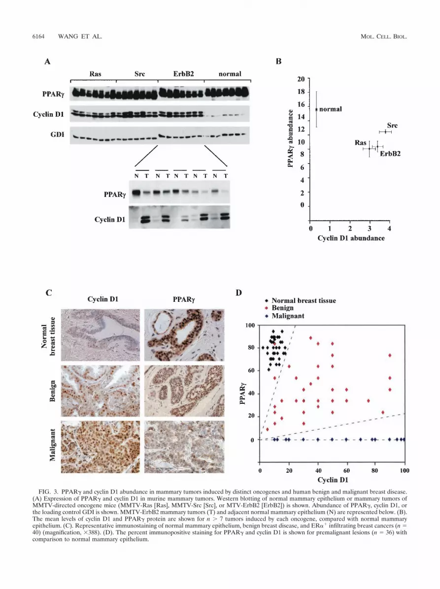
FIG. 3. PPAR� and cyclin D1 abundance in mammary tumors induced by distinct oncogenes and human benign and malignant breast disease.(A) Expression of PPAR� and cyclin D1 in murine mammary tumors. Western blotting of normal mammary epithelium or mammary tumors ofMMTV-directed oncogene mice (MMTV-Ras [Ras], MMTV-Src [Src], or MTV-ErbB2 [ErbB2]) is shown. Abundance of PPAR�, cyclin D1, orthe loading control GDI is shown. MMTV-ErbB2 mammary tumors (T) and adjacent normal mammary epithelium (N) are represented below. (B).The mean levels of cyclin D1 and PPAR� protein are shown for n � 7 tumors induced by each oncogene, compared with normal mammaryepithelium. (C). Representative immunostaining of normal mammary epithelium, benign breast disease, and ER�� infiltrating breast cancers (n 40) (magnification, 388). (D). The percent immunopositive staining for PPAR� and cyclin D1 is shown for premalignant lesions (n 36) withcomparison to normal mammary epithelium.
6164 WANG ET AL. MOL. CELL. BIOL.

phenotype in immortalized 3T3 cells (40). We comparedPPAR� ligand-regulated adipogenesis in cyclin D1�/� versuscyclin D1�/� MEFs prior to immortalization. 3T3-L1 adipo-genesis medium was not capable of inducing MEF differenti-ation into adipocytes. Addition of the PPAR� agonists rosigli-tazone (0.2 M) or troglitazone (5 M) induced a modest (8%positive) but significant lipid droplet accumulation in cyclinD1�/� wt MEFs. In contrast, more than 35% of the cyclinD1-deficient MEFs showed Oil Red-O positive staining (Fig.4A and C).
These findings suggest that the deficiency of cyclin D1 orfactors regulated by cyclin D1 contributes to the adipocytephenotype induced by the specific PPAR� ligands. To deter-mine whether cyclin D1 deficiency of the cyclin D1�/� MEFswas the key regulator of the PPAR� ligand-induced adipocytephenotype, cyclin D1�/� MEFs were infected with a retrovi-ruses encoding cyclin D1 prior to the differentiation protocol.Expression of cyclin D1 inhibited rosiglitazone (0.2 M)-in-duced or troglitazone (5 M)-induced lipid accumulation inthe cyclin D1�/� MEFs (Fig. 4B and D). The transductionefficiency of the MEFs was �90%, assessed by transductionwith the viral vector encoding GFP (Fig. 4E). These studiesprovide genetic evidence that cyclin D1 is necessary and suffi-cient to regulate the adipogenic differentiation function ofPPAR�.
Reduction of cyclin D1 by ponasterone-regulated cyclin D1antisense transgenic mice induces PPAR� abundance. Thecorrelative reciprocal expression profile of cyclin D1 andPPAR� in mammary epithelium was assessed further to deter-mine whether cyclin D1 inhibited PPAR� expression. PPAR�levels were increased in untreated cyclin D1�/� 3T3 cells (Fig.5A) and cyclin D1�/� MEFs (Fig. 5B), suggesting that cyclinD1 may inhibit PPAR� expression, consistent with our findingthat cyclin D1 repressed the PPAR�1 promoter. To examinefurther whether cyclin D1 inhibited PPAR� abundance in vivo,we generated transgenic mice in which the murine cyclin D1antisense cDNA linked to a GFP transgene was regulatedunder control of the ecdysone enhancer (Fig. 5Ca). We hadpreviously used ponasterone-inducible cyclin D1 antisense toreduce cyclin D1 abundance in cultured cells (63). Transgenetransmission was confirmed by genomic Southern blotting (Fig.5Cb). To induce expression of the transgene, an ecdysonereceptor (Bbyx), adenoviruses were used that expresses GFPfrom a second cistron (DB-Ecr-IRES-GFP, Fig. 5Cc). Theentire experiment was conducted on two separate occasions,and representative results are shown. Comparison was madewith equal titer of an adenovirus for the ecdysone receptor thatdoes not express GFP.
The adenoviral receptors encoding the ecdysone receptorwere introduced into transgenic cyclin D1 antisense mice orcontrol strain-matched mice, and activity of the receptors wasinduced using ponasterone pellets (Fig. 5Cd). Serum samplesdemonstrated the induction of ecdysone enhancer-drivenSEAP or luciferase activity in parallel experiments (data notshown). Analysis was performed on the livers of transgenic andantisense transgenic mice. PPAR� is expressed in hepatocytes,and the expression of PPAR� is induced by TZDs in vivo (13).It has been hypothesized that the insulin-sensitizing effects ofTZDs may result in part from effects on PPAR� expressionand function in muscle and liver (12, 13). The induction of the
GFP from the second cistron of the cyclin D1 antisense trans-gene was observed by Western blotting (Fig. 5Ce). Analysis ofhepatic tissue evidenced the presence of GFP by immunoflu-orescence and by Western blotting with mice transduced withthe DB-Ecr-IRES-GFP adenovirus, used as a positive controlfor GFP in parallel experiments (data not shown). The abun-dance of cyclin D1 protein was reduced by 90% for the cyclinD1 antisense transgenic mice compared with results for controlmice (n 2). The reduction in cyclin D1 protein levels corre-lated with the induction in PPAR� abundance in vivo (Fig.5Ce), providing further evidence that cyclin D1 inhibitsPPAR� expression in vivo.
Cyclin D1 inhibits PPAR� ligand-induced gene expressionwithout affecting C/EBP� or C/EBP�. The expression of genesknown to be either upstream (C/EBP�) (60) or downstream(adipocyte complement related protein of 30 kDa [ACRP30],C3/12) of PPAR� in the adipocyte differentiation program wasnext assessed. C/EBP�-mediated adipocyte differentiation re-quires PPAR�; however, PPAR�-mediated differentiation isindependent of CEBP�, placing PPAR� as a downstream ef-fector of CEBP� (40). Analysis was performed during differ-entiation of cyclin D1�/� and cyclin D1�/� MEFs. Normaliza-tion of protein loading was performed using GDI (25). Cellswere harvested every day for 7 days and lysed, and the celllysates were subjected to Western blotting for ACRP30 andC3/12. Prior to the induction of differentiation by the PPAR�ligands BRL49653 (Fig. 6) and troglitazone (data not shown),cyclin D1 was detectable in the cyclin D1�/� but not the cyclinD1�/� MEFs. PPAR� levels were increased two- to threefoldin the cyclin D1�/� MEFs compared with results in the cyclinD1 �/� MEFs (Fig. 6), consistent with the finding that cyclinD1 inhibits both PPAR� expression and transactivation. In theundifferentiated state, cyclin D1�/� and cyclin D1�/� MEFsexpressed similar levels of C/EBP� and C/EBP�.
ACRP30 (adiponectin/apM1/AdipoQ/GBP28) is an adipo-cyte-secreted protein induced during fat cell differentiation (5)induced by the PPAR� ligand TZD (13). At day 6, the levels ofthe adipocyte differentiation markers downstream of PPAR�(ACRP30 and S3/12) were substantially higher in the cyclinD1�/� MEFs than in the cyclin D1�/� MEFs. The levels ofACRP30 was �100-fold higher in the cyclin D1�/� MEFs thanin the wt MEFs. In both wt and cyclin D1�/� MEFs, the levelsof ACRP30 were increased 10-fold by BRL49653 or troglita-zone. Upon induction of differentiation, both cyclin D1�/� andcyclin D1�/� MEFs showed a similar pattern of C/EBP� andC/EBP� expression. These studies suggest that inhibition ofadipocyte differentiation by cyclin D1 occurs distal to C/EBPsand involves inhibition of PPAR� function. Transduction ofcyclin D1�/� MEFs with a cyclin D1 retrovirus inhibited theinduction of differentiation by BRL49653 and reduced thetime-dependent induction of ACRP30 (Fig. 6B).
Adipogenic steatosis in cyclin D1�/� mouse liver. In previousstudies, adenoviral delivery of PPAR�1 overexpression in themurine liver induced hepatic steatosis (65). If repression ofPPAR�1 activity by cyclin D1 was functionally significant invivo, it would be predicted that the cyclin D1�/� mice woulddemonstrate features of hepatic steatosis. Detailed morpho-metric and histological analysis was therefore conducted of thelivers from cyclin D1�/� and wt littermate controls. Acid phos-phatase cytochemistry revealed that the Kupffer cells in the
VOL. 23, 2003 CYCLIN D1 INHIBITION OF PPAR� FUNCTION 6165

FIG. 4. Cyclin D1 deficiency enhances PPAR� function. (A) MEFs derived from either cyclin D1�/� or cyclin D1�/� mice were treated withvehicle, BRL-49653 (1 M), and troglitazone (5 M), and lipid accumulation was assessed. cyclin D1�/� MEFs exhibit lipid accumulation by Oil
6166 WANG ET AL. MOL. CELL. BIOL.
cyclin D1�/� mice were enlarged, appeared increased in num-bers (Fig. 7), and developed an extensive lysosomal systemfilled with lipid deposits (Fig. 7). In addition, acid phosphatasecytochemistry revealed that the lysosomes of hepatocytes ap-peared larger and appeared to be distributed more widely inthe cytoplasm in contrast to the wt hepatocytes lysosomes,which were more concentrated near the bile canaliculus. Neu-tral lipid staining using Oil Red-O showed a substantial in-crease in neutral lipid droplets in the cyclin D1�/�. Ito cellsfrom cyclin D1�/� mice were increased in numbers and en-larged, with more numerous and larger lipid spheres, com-pared with Ito cells of wt littermate controls. Changes werealso found in hepatocytes and included increased numbers ofmicroperoxisomes, and cytoplasmic lipid spheres were noted(Fig. 7). Thus, the livers of cyclin D1�/� mice display thefeatures of hepatic steatosis consistent with increased PPAR�activity.
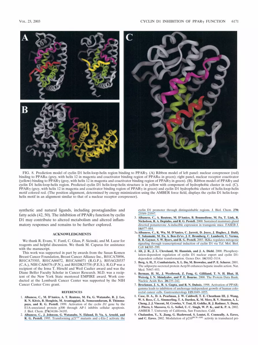
Three-dimensional modeling of the cyclin D1 143-179 re-gion. In view of the importance of the region of cyclin D1 from143 to 179 in repression of PPAR� transactivation, we deter-mined the predicted structure of this domain using the homol-ogy modeling program PROMOD (37) (see Materials andMethods). The coordinates for the three-dimensional model ofcyclin D1 were generated based on the sequence alignmentbetween cyclin D1 and cyclin A and the published crystalstructures of cyclin A (6). We generated models from thepublished crystal structure of PPAR� corepressor (62) (Fig.8A, left) and PPAR�-coactivator (61) (Fig. 8A, right). Thecoactivator is shown in yellow. PPAR� is shown in grey withthe region of PPAR� contacting the coactivator shown ingreen. In the homology model of cyclin D1, the region from142 to 179 forms a helix-loop-helix, shown in Fig. 8B in yellowand red. The cluster of hydrophobic residues (aa 137 to 148,LLXXXLLLVXXL) was then modeled binding to the sameregion where corepressors and coactivators bind to PPAR�(61). Using these two PPAR structures and the homologymodel of cyclin D1, a three-dimensional model of the complexbetween PPAR� and cyclin D1 was generated (Fig. 8C) (IN-SIGHT; Accelrys Inc., San Diego, Calif.). The hydrophobiccluster region of the cyclin D1 HLH is shown in red in Fig. 8C.
DISCUSSION
The present studies demonstrate for the first time in vivoantagonism between a collaborative oncogene, cyclin D1, anda nuclear receptor, PPAR�. Cyclin D1 inhibited PPAR�-de-pendent reporter activity and repressed the trans activity ofPPAR� when linked to a heterologous DNA binding domain.Consistent with previous findings that PPAR� expression isinduced by PPAR� (12), cyclin D1 also inhibited PPAR� ex-pression and promoter activity. The reduction in cyclin D1abundance in cyclin D1�/� 3T3 cells, cyclin D1�/� MEFs, andcyclin D1 antisense transgenic mice correlated with the induc-tion of PPAR� expression (Fig. 5). The induction of adipocyte
differentiation by PPAR�-specific ligands was substantially en-hanced in cyclin D1-deficient cells. The reintroduction of cyclinD1 into cyclin D1-deficient cells abolished the adipogenic phe-notype, consistent with a key role for cyclin D1 as an inhibitorof PPAR�-specific functional activity. Since cyclin D1 abun-dance is regulated by diverse oncogenic and mitogenic stimuli,the inhibition of PPAR� transactivation by cyclin D1 may haveimportant implications for signal transduction and tumorigen-esis.
Repression of PPAR� transactivation by cyclin D1 was in-dependent of the cdk and pRb binding functions and requireda C-terminal region of cyclin D1 that is predicted to form ahelix-loop-helix structure. The mechanism by which cyclin D1regulated PPAR� activity is thus distinct from that regulatingthe ER� through recruiting a p160 coactivator SRC-1 in vitro(66), as an SRC-1 binding point mutation of cyclin D1 main-tained wt repression of liganded PPAR� activity. Herein, ge-netic deletion of cyclin D1 enhanced PPAR� ligand-mediateddifferentiation of MEFs into adipocytes. Although interactionsbetween cyclin D1 and nuclear receptors have been previouslydescribed, the present studies provide strong genetic evidencefor a functional interaction between cyclin D1 and a nuclearreceptor. Ectopic expression of PPAR� induced differentiationof NIH-3T3 fibroblast cells into fat-laden adipocyte cells (51).In the present studies, the cyclin D1�/� MEF adipogenic phe-notype induced by PPAR� ligands was reversed by cyclin D1overexpression. ACRP30 was increased 50-fold in the cyclinD1�/� MEFs, consistent with the enhanced induction ofthe adipogenic phenotype. The relatively modest increase inPPAR� abundance, together with the dramatic enhancementof PPAR� activity in the cyclin D1�/� MEFs, is consistent withthe reporter gene studies in which cyclin D1 inhibited PPAR�trans activity.
These studies raise the possibility that reduced PPAR� ex-pression, together with increased cyclin D1, may be a geneticfeature of the transition from normal breast epithelium, tobenign breast disease and adenocarcinoma. PPAR� immuno-positivity was decreased in benign breast disease comparedwith normal mammary epithelium and was reduced further inadenocarcinomas. Cyclin D1 immunopositivity increased fromnormal epithelium to benign disease and adenocarcinomas.The reduction in PPAR� expression in the cyclin D1-infectedMEFs, together with the finding of increased levels of PPAR�mRNA and protein in cyclin D1�/� livers by microarray (notshown) and Western blotting, suggest that cyclin D1 inhibitsPPAR� expression. The overexpression of cyclin D1 with ER�reflects poor prognosis in human breast cancer. Given therepression of PPAR� function and expression by cyclin D1 andthe cytoinhibitory role of PPAR� in breast epithelium, thesestudies raise the question of whether reduced PPAR� maycontribute to poor prognosis in a subset of patients. The re-duction in PPAR� staining in proliferative breast disease sug-gests that further studies of PPAR� as a prognostic indicator
Red-O staining in the presence of BRL-49653 or troglitazone, shown as mean � standard error of the mean for three experiments in panel C. (B)cyclin D1�/� MEFs were infected with either a parental retrovirus or retroviruses encoding cyclin D1. Two days postconfluence, cells were treatedwith differentiation media and troglitazone (5 M). Seven days later, cells were stained with Oil Red-O to visualize the degree of lipidaccumulation. The results were quantitated for two separate experiments (D). Representative infection of cyclin D1�/� MEFs with viral GFPexpression construct is shown. Tro, troglitazone; DMSO, dimethyl sulfoxide.
VOL. 23, 2003 CYCLIN D1 INHIBITION OF PPAR� FUNCTION 6167

FIG. 5. Transgenic inducible cyclin D1 antisense regulates PPAR� abundance in vivo. Western blot of cyclin D1�/� 3T3 (A) and cyclin D1�/�
MEFs (B) with antibodies as indicated. (Ca) An ecdysone enhancer-driven cyclin D1 antisense transgene (schematic shown), was used to generatetransgenic mice (Tg) confirmed by genomic Southern blot (Cb). (Cc) Adenoviral vectors encoding receptors for the Bombyx LBD either with orwithout a second cistron for GFP were used to infect the cyclin D1 antisense transgenic mice (Cd). (Ce) Transgenic mice were treated withponasterone to induce transgene expression, and Western blotting of hepatic extracts was performed at 5 days for the loading control (GDI), cyclinD1 abundance and GFP from the second cistron of the cyclin D1 antisense, and PPAR�.
6168 WANG ET AL. MOL. CELL. BIOL.

and candidate target for prevention or therapy of humanbreast cancer warrants consideration.
The present studies are important in demonstrating a func-tional antagonism between a collaborative oncogene, cyclinD1, and a candidate tumor suppressor, PPAR�. Several linesof evidence suggest that PPAR� may function as a tumorsuppressor (42). Consistent with a role for PPAR� as an in-hibitor of tumorigenesis, heterozygous mutations of PPAR�were detected in 4 of 55 patients with colon cancer (45). Infollicular thyroid cancer, a fusion oncoprotein has been de-scribed, formed by a chromosomal translocation betweenPPAR�1 and PAX8 with a deletion in its C-terminal activationdomain. The PPAR� fusion protein functioned as a powerfuldominant-negative of wt PPAR� and was not observed in be-nign follicular adenomas (24). The addition of PPAR� ligands(TZD or 15d-PGJ2) inhibited breast and colonic cellular pro-liferation (7, 14, 32, 52). In contrast, cyclin D1 abundance is
induced by diverse oncogenic and mitogenic signals in breastand colonic epithelial cells and functions as a collaborativeoncogene (18, 38). Cyclin D1 antisense inhibits the growth ofmurine mammary tumors derived from MMTV-ErbB2 mice(25), and cyclin D1�/� mice are resistant to the induction oftumor formation by ErbB2 (64). Since PPAR� inhibits theexpression of several genes promoting tumor invasion (thoseencoding iNOS, gelatinase B, matrix metalloproteinases [10,21, 30], and UPA) (data not shown), cyclin D1 antagonism ofPPAR� function may enhance expression of tumor invasiongenes.
The functional antagonism between PPAR� and cyclin D1may also have implications for signal transduction cross talk.Cyclin D1 is induced by diverse mitogenic signaling pathways,including those of Src, Rac mutants, Dbl proteins, and �-cate-nin, and the NF-�B signaling pathway (1, 2, 17, 26, 49, 55, 56,58, 59). PPAR� activity is also induced by a large number of
FIG. 6. Enhanced PPAR�-responsive gene expression in cyclin D1�/� cells. (A) MEFs derived from either cyclin D1�/� or cyclin D1�/� micewere treated with BRL-49653 (1 M). Western blotting was conducted for PPAR� and the PPAR�-responsive genes, ACRP30 (11) and S3/12.Levels of cyclin D1, C/EBP�, C/EBP�, and the loading control (GDI) are shown. (B) Western blot analysis of cyclin D1�/� MEFs or cyclin D1�/�
MEFs infected with a cyclin D1 retrovirus. MEFs were treated for the time points indicated.
VOL. 23, 2003 CYCLIN D1 INHIBITION OF PPAR� FUNCTION 6169
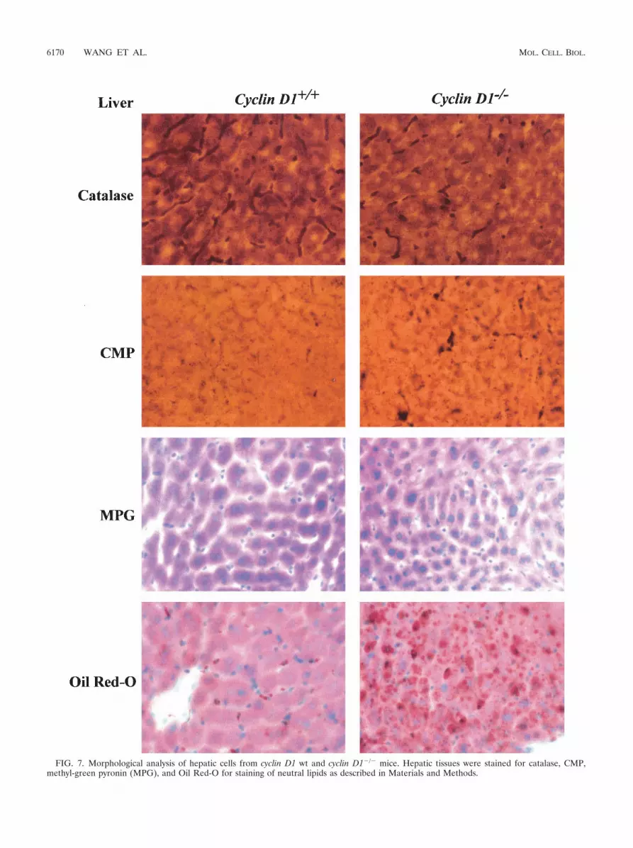
FIG. 7. Morphological analysis of hepatic cells from cyclin D1 wt and cyclin D1�/� mice. Hepatic tissues were stained for catalase, CMP,methyl-green pyronin (MPG), and Oil Red-O for staining of neutral lipids as described in Materials and Methods.
6170 WANG ET AL. MOL. CELL. BIOL.
synthetic and natural ligands, including prostaglandins andfatty acids (42, 50). The inhibition of PPAR� function by cyclinD1 may contribute to altered metabolism and altered inflam-matory responses and remains to be further explored.
ACKNOWLEDGMENTS
We thank R. Evans, V. Fantl, C. Glass, P. Sicinski, and M. Lazar forreagents and helpful discussion. We thank M. Caparas for assistancewith the manuscript.
This work was supported in part by awards from the Susan KomenBreast Cancer Foundation, Breast Cancer Alliance Inc., R01CA70896,R01CA75503, R01CA86072, R01CA86071 (R.G.P.), R03AG20337(C.A.), NIH CA06576 (P.N.), and R01DK55758 (P.E.S.). R.G.P was arecipient of the Irma T. Hirschl and Weil Caulier award and was theDiane Belfer Faculty Scholar in Cancer Research. M.D. was a recip-ient of the New York State mentored EMPIRE award. Work con-ducted at the Lombardi Cancer Center was supported by the NIHCancer Center Core grant.
REFERENCES
1. Albanese, C., M. D’Amico, A. T. Reutens, M. Fu, G. Watanabe, R. J. Lee,R. N. Kitsis, B. Henglein, M. Avantaggiati, K. Somasundaram, B. Thimma-paya, and R. G. Pestell. 1999. Activation of the cyclin D1 gene by theE1A-associated protein p300 through AP-1 inhibits cellular apoptosis.J. Biol. Chem. 274:34186–34195.
2. Albanese, C., J. Johnson, G. Watanabe, N. Eklund, D. Vu, A. Arnold, andR. G. Pestell. 1995. Transforming p21ras mutants and c-Ets-2 activate the
cyclin D1 promoter through distinguishable regions. J. Biol. Chem. 270:23589–23597.
3. Albanese, C., A. Reutens, M. D’Amico, B. Boumediene, M. Fu, T. Link, R.Nicholson, R. A. Depinho, and R. G. Pestell. 2000. Sustained mammary glanddirected ponasterone A-inducible expression in transgenic mice. FASEB J.14:877–884.
3a.Albanese, C., K. Wu, M. D’Amico, C. Jarrett, D. Joyce, J. Hughes, J. Hulit,T. Sakamaki, M. Fu, A. Ben-Ze’ev, J. F. Bromberg, C. Lamberti, U. Verma,R. B. Gaynor, S. W. Byers, and R. G. Pestell. 2003. IKK� regulates mitogenicsignaling through transcriptional induction of cyclin D1 via Tcf. Mol. Biol.Cell 14:585–599.
4. Alt, J. R., J. L. Cleveland, M. Hannink, and J. A. Diehl. 2000. Phosphory-lation-dependent regulation of cyclin D1 nuclear export and cyclin D1-dependent cellular transformation. Genes Dev. 14:3102–3114.
5. Berg, A. H., T. Combatsiaris, X. L. Du, M. Brownlee, and P. E. Scherer. 2001.The adipocyte-secreted protein Acrp30 enhances hepatic insulin action. Nat.Med. 7:947–953.
6. Berman, H. M., J. Westbrook, Z. Feng, G. Gilliland, T. N. H. Bhat, H.Weissig, I. N. Shindyalov, and P. E. Bourne. 2000. The Protein Data Bank.Nucleic Acids Res. 28:235–242.
7. Brockman, J. A., R. A. Gupta, and R. N. Dubois. 1998. Activation of PPAR-gamma leads to inhibition of anchorage-independent growth of human colo-rectal cancer cells. Gastroenterology 115:1049–1055.
8. Case, D. A., D. A. Pearlman, J. W. Caldwell, T. E. Cheatham III, J. Wang,W. S. Ross, C. L. Simmerling, T. A. Darden, K. M. Merz, R. V. Stanton, A. L.Cheng, J. J. Vincent, M. Crowley, V. Tsui, H. Gohlke, R. J. Radmer, Y. Duan,J. Pitera, I. Massova, G. L. Seibel, U. C. Singh, W. P. K., and K. P. A. 2002.AMBER 7. University of California, San Francisco, Calif.
9. Chalandon, Y., X. Jiang, G. Hazlewood, S. Loutet, E. Conneally, A. Eaves,and C. Eaves. 2002. Modulation of p210BCR-ABL activity in transduced pri-
FIG. 8. Prediction model of cyclin D1 helix-loop-helix region binding to PPAR�. (A) Ribbon model of left panel: nuclear corepressor (red)binding to PPAR� (grey, with helix 12 in magenta and coactivator binding region of PPAR� in green); right panel, nuclear receptor coactivator(yellow) binding to PPAR� (grey, with helix 12 in magenta and coactivator binding region of PPAR� in green). (B). Ribbon model of PPAR� andcyclin D1 helix-loop-helix region. Predicted cyclin D1 helix-loop-helix structure is in yellow with component of hydrophobic cluster in red. (C).PPAR� (grey, with helix 12 in magenta and coactivator binding region of PPAR� in green) and cyclin D1 hydrophobic cluster of helix-loop-helixmotif colored red. (The position alignment, determined by energy minimization using the AMBER force field, displays the cyclin D1 helix-loop-helix motif in an alignment similar to that of a nuclear receptor corepressor).
VOL. 23, 2003 CYCLIN D1 INHIBITION OF PPAR� FUNCTION 6171

mary human hematopoietic cells controls lineage programming. Blood 99:3197–3204.
10. Chawla, A., Y. Barak, L. Nagy, D. Liao, P. Tontonoz, and R. M. Evans. 2001.PPAR-gamma dependent and independent effects on macrophage-gene ex-pression in lipid metabolism and inflammation. Nat. Med. 7:48–52.
11. Combs, T. P., J. A. Wagner, J. Berger, T. Doebber, W. J. Wang, B. B. Zhang,M. Tanen, A. H. Berg, S. O’Rahilly, D. B. Savage, K. Chatterjee, S. Weiss,P. J. Larson, K. M. Gottesdiener, B. J. Gertz, M. J. Charron, P. E. Scherer,and D. E. Moller. 2002. Induction of adipocyte complement-related proteinof 30 kilodaltons by PPARgamma agonists: a potential mechanism of insulinsensitization. Endocrinology 143:998–1007.
12. Davies, G. F., R. L. Khandelwahl, and W. J. Roesler. 1999. Troglitazoneinduces expression of PPAR� in liver. Mol. Cell. Biol. Res. Commun. 2:202–208.
13. Davies, G. F., P. J. McFie, R. L. Khandelwahl, and W. J. Roesler. 2002.Unique ability of troglitazone to upregulate peroxisome proliferator-acti-vated receptor-g expression in hepatocytes. J. Pharmacol. Exp. Ther. 300:72–77.
14. Elstner, E., C. Muller, K. Koshizuka, E. A. Williamson, D. Park, H. Asou, P.Shintaku, J. W. Said, D. Heber, and H. P. Koeffler. 1998. Ligands forperoxisome proliferator-activated receptor� and retinoic acid receptor in-hibit growth and induce apoptosis of human breast cancer cells in vitro andin BNX mice. Proc. Natl. Acad. Sci. USA 95:8806–8811.
15. Fajas, L., K. Schoonjans, L. Gelman, J. B. Kim, J. Najib, G. Martin, J. C.Fruchart, M. Briggs, B. M. Spiegelman, and J. Auwerx. 1999. Regulation ofperoxisome proliferator-activated receptor gamma expression by adipocytedifferentiation and determination factor 1/sterol regulatory element bindingprotein 1: implications for adipocyte differentiation and metabolism. Mol.Cell. Biol. 19:5495–5503.
16. Ganter, B., S.-L. Fu, and J. S. Lipsick. 1998. D-type cyclins repress tran-scriptional activation by the v-Myb but not the c-Myb DNA-binding domain.EMBO J. 17:255–268.
17. Guttridge, D. C., C. Albanese, J. Y. Reuther, R. G. Pestell, and A. S. Baldwin.1999. NF-�B controls cell growth and differentiation through the transcrip-tional regulation of cyclin D1. Mol. Cell. Biol. 19:5785–5799.
18. Hanahan, D., and R. Weinberg. 2000. The hallmarks of cancer. Cell 100:57–70.
19. Hoppe, U. C., E. Marban, and D. C. Johns. 1999. Adenovirus-mediatedinducible expression in vivo by a hybrid ecdysone receptor. Mol. Ther.1:159–164.
20. Inoue, K., and C. J. Sherr. 1998. Gene expression and cell cycle arrestmediated by transcription factor DMP1 is antagonized by D-type cyclinsthrough a cyclin-dependent-kinase-independent mechanism. Mol. Cell. Biol.18:1590–1600.
21. Jiang, C., A. T. Ting, and B. Seed. 1998. PPAR-� agonists inhibit productionof monocyte inflammatory cytokines. Nature 391:82–86.
22. Karnovsky, M. J. 1965. A formaldehyde-glutaraldehyde fixative of high os-molarity for use in electron microscopy. J. Cell Biol. 27:137A-138A.
23. Kenny, F. S., R. Hui, E. A. Musgrove, J. M. Gee, R. W. Blamey, R. I.Nicholson, R. L. Sutherland, and J. F. Robertson. 1999. Overexpression ofcyclin D1 messenger RNA predicts for poor prognosis in estrogen receptor-positive breast cancer. Clin. Cancer Res. 5:2069–2076.
24. Kroll, T. G., P. Sarraf, L. Pecciarini, C. J. Chen, E. Mueller, B. M.Spiegelman, and J. A. Fletcher. 2000. PAX8-PPARgamma1 fusion oncogenein human thyroid carcinoma. Science 289:1357–1360.
25. Lee, R. J., C. Albanese, M. Fu, M. D’Amico, B. Lin, G. Watanabe, G. K. I.Haines, P. M. Siegel, M. C. Hung, Y. Yarden, J. M. Horowitz, W. J. Muller,and R. G. Pestell. 2000. Cyclin D1 is required for transformation by activatedNeu and is induced through an E2F-dependent signaling pathway. Mol. Cell.Biol. 20:672–683.
26. Lee, R. J., C. Albanese, R. J. Stenger, G. Watanabe, G. Inghirami, G. K. I.Haines, M. Webster, W. J. Muller, J. S. Brugge, R. J. Davis, and R. G.Pestell. 1999. pp60v-src induction of cyclin D1 requires collaborative interac-tions between the extracellular signal-regulated kinase, p38, and Jun kinasepathways: a role for cAMP response element-binding protein and activatingtranscription factor-2 in pp60v-src signaling in breast cancer cells. J. Biol.Chem. 274:7341–7350.
27. Lefebvre, A. M., I. Chen, P. Desreumaux, J. Najib, J. C. Fruchart, K. Geboes,M. Briggs, R. Heyman, and J. Auwerx. 1998. Activation of the peroxisomeproliferator-activated receptor gamma promotes the development of colontumors in C57BL/6J-APCMin/� mice. Nat. Med. 4:1053–1057.
28. Lillie, R. D. 1965. Histopathologic technic and practical histochemistry, 3rded. McGraw-Hill Book Co., Inc., New York, N.Y.
29. Lukas, J., J. Bartkova, and J. Bartek. 1996. Convergence of mitogenicsignalling cascades from diverse classes of receptors at the cyclin D-cyclindependent kinase-pRb-controlled G1 checkpoint. Mol. Cell. Biol. 16:6917–6925.
30. Marx, N., F. Mach, A. Sauty, J. H. Leung, M. N. Sarafi, R. M. Ransohoff, P.Libby, J. Plutzky, and A. D. Luster. 2000. Peroxisome proliferator-activatedreceptor-gamma activators inhibit IFN-gamma-induced expression of the Tcell-active CXC chemokines IP-10, Mig, and I-TAC in human endothelialcells. J. Immunol. 164:6503–6508.
31. McMahon, C., T. Suthiphongchai, J. DiRenzo, and M. E. Ewen. 1999. P/CAFassociates with cyclin D1 and potentiates its activation of the estrogen re-ceptor. Proc. Natl. Acad. Sci. USA 96:5382–5387.
32. Mueller, E., P. Sarraf, P. Tontonoz, R. M. Evans, K. J. Martin, M. Zhang,C. Fletcher, S. Singer, and B. M. Spiegelman. 1998. Terminal differentiationof human breast cancer through PPAR�. Mol. Cell 1:465–470.
33. Neuman, E., M. H. Ladha, N. Lin, T. M. Upton, S. J. Miller, J. DiRenzo,R. G. Pestell, P. W. Hinds, S. F. Dowdy, M. Brown, and M. E. Ewen. 1997.Cyclin D1 stimulation of estrogen receptor transcriptional activity indepen-dent of cdk4. Mol. Cell. Biol. 17:5338–5347.
34. Novikoff, A. B., P. M. Novikoff, C. Davis, and N. Quintana. 1972. Studies onMicroperoxisomes. II. A cytochemical method for light and electron micros-copy. J. Histochem. Cytochem. 2:1006–1022.
35. Novikoff, P. M., and A. B. Novikoff. 1972. Peroxisomes in absorptive cells ofmammalian small intestine. J. Cell Biol. 53:532–560.
36. Novikoff, P. M., and A. Yam. 1978. Sites of lipoprotein particles in normal rathepatocytes. J. Cell Biol. 76:1–11.
37. Peitsch, M. C. 1996. PROMOD and SWISS-MODEL: internet-based toolsfor automated comparative protein modeling. Biochem. Soc. Trans. 24:274–279.
38. Pestell, R. G., C. Albanese, A. T. Reutens, J. E. Segall, R. J. Lee, and A.Arnold. 1999. The cyclins and cyclin-dependent kinase inhibitors in hor-monal regulation of proliferation and differentiation. Endocrine Rev. 20:501–534.
39. Reutens, A. T., M. Fu, G. Watanabe, C. Albanese, M. J. McPhaul, S. P. Balk,O. A. Janne, J. J. Palvimo, and R. G. Pestell. 2001. Cyclin D1 binds theandrogen receptor and regulates hormone-dependent signaling in a p300/CBP-associated factor (P/CAF)-dependent manner. Mol. Endocrinol. 15:797–811.
40. Rosen, E. D., C.-H. Hsu, X. Wang, S. Sakai, M. W. Freeman, F. J. Gonzalez,and B. M. Spiegelman. 2002. C/EBP induces adipogenesis through PPAR�:a unified pathway. Genes Dev. 16:22–26.
41. Rosen, E. D., P. Sarraf, A. E. Troy, G. Bradwin, K. Moore, D. S. Milstone,B. M. Spiegelman, and R. M. Mortensen. 1999. PPAR gamma is required forthe differentiation of adipose tissue in vivo and in vitro. Mol. Cell 4:611–617.
42. Rosen, E. D., and B. M. Spiegelman. 2001. PPAR�: a nuclear regulator ofmetabolism, differentiation, and cell growth. J. Biol. Chem. 276:37731–37734.
43. Saez, E., P. Tontonoz, M. C. Nelson, J. G. Alvarez, U. T. Ming, S. M. Baird,V. A. Thomazy, and R. M. Evans. 1998. Activators of the nuclear receptorPPARgamma enhance colon polyp formation. Nat. Med. 4:1058–1061.
44. Sarraf, P., E. Mueller, D. Jones, F. J. King, D. J. DeAngelo, J. B. Partridge,S. A. Holden, L. B. Chen, S. Singer, C. Fletcher, and B. M. Spiegelman. 1998.Differentiation and reversal of malignant changes in colon cancer throughPPARgamma. Nat. Med. 4:1046–1052.
45. Sarraf, P., E. Mueller, W. M. Smith, H. M. Wright, J. B. Kum, L. A.Aaltonen, A. de la Chapelle, B. M. Spiegelman, and C. Eng. 1999. Loss-of-function mutations in PPAR gamma associated with human colon cancer.Mol. Cell 3:799–804.
46. Scherer, P. E., P. E. Bickel, M. Kotler, and H. F. Lodish. 1998. Cloning ofcell-specific secreted and surface proteins by subtractive antibody screening.Nat. Biotechnol. 16:581–586.
47. Sherr, C. J. 1996. Cancer cell cycles. Science 274:1672–1677.48. Sherr, C. J., and J. M. Roberts. 1999. CDK inhibitors: positive and negative
regulators of G1-phase progression. Genes Dev. 13:1501–1512.49. Shtutman, M., J. Zhurinsky, I. Simcha, C. Albanese, M. D’Amico, R. Pestell,
and A. Ben-Ze’ev. 1999. The cyclin D1 gene is a target of the �-catenin/LEF-1 pathway. Proc. Natl. Acad. Sci. USA 96:5522–5527.
50. Spiegelman, B. M. 1998. PPAR-gamma: adipogenic regulator and thiazo-lidinedione receptor. Diabetes 47:507–514.
51. Tontonoz, P., E. Hu, and B. M. Spiegelman. 1994. Stimulation of adipogen-esis in fibroblasts by PPAR�2, a lipid-activated transcription factor. Cell79:1147–1156.
52. Wang, C., M. Fu, M. D’Amico, C. Albanese, J. N. Zhou, M. Brownlee, M. P.Lisanti, V. K. Chatterjee, M. A. Lazar, and R. G. Pestell. 2001. Inhibition ofcellular proliferation through IkappaB kinase-independent and peroxisomeproliferator-activated receptor gamma-dependent repression of cyclin D1.Mol. Cell. Biol. 21:3057–3070.
53. Wang, T. C., R. D. Cardiff, L. Zukerberg, E. Lees, A. Arnold, and E. V.Schmidt. 1994. Mammary hyperplasia and carcinoma in MMTV-cyclin D1transgenic mice. Nature 369:669–671.
54. Wang, X. L., J. Oosterhof, and N. Duarte. 1999. Peroxisome proliferator-activated receptor gamma C1613T polymorphism and coronary artery dis-ease. Cardiovasc. Res. 44:588–594.
55. Watanabe, G., C. Albanese, R. J. Lee, A. Reutens, G. Vairo, B. Henglein, andR. G. Pestell. 1998. Inhibition of cyclin D1 kinase activity is associated withE2F-mediated inhibition of cyclin D1 promoter activity through E2F andSp1. Mol. Cell. Biol. 18:3212–3222.
56. Watanabe, G., A. Howe, R. J. Lee, C. Albanese, I.-W. Shu, A. N. Karnezis, L.Zon, J. Kyriakis, K. Rundell, and R. G. Pestell. 1996. Induction of cyclin D1by simian virus 40 small tumor antigen. Proc. Natl. Acad. Sci. USA 93:12861–12866.
6172 WANG ET AL. MOL. CELL. BIOL.

57. Weinberg, R. A. 1995. The retinoblastoma protein and cell cycle control. Cell81:323–330.
58. Westwick, J. K., Q. T. Lambert, G. J. Clark, M. Symons, L. Van Aelst, R. G.Pestell, and C. J. Der. 1997. Rac regulation of transformation, gene expres-sion and actin organisation by multiple, PAK-independent pathways. Mol.Cell. Biol. 17:1324–1335.
59. Westwick, J. K., R. J. Lee, Q. T. Lambert, M. Symons, R. G. Pestell, C. J.Der, and I. P. Whitehead. 1998. Transforming potential of Dbl family pro-teins correlates with transcription from the cyclin D1 promoter but not withactivation of Jun NH2-terminal kinase, p38/Mpk2, serum response factor, orc-Jun. J. Biol. Chem. 273:16739–16747.
60. Wu, Z., Y. Xie, N. L. Bucher, and S. R. Farmer. 1995. Conditional ectopicexpression of C/EBP beta in NIH-3T3 cells induces PPAR gamma andstimulates adipogenesis. Genes Dev. 9:2350–2363.
61. Xu, H. E., M. H. Lambert, V. G. Montana, K. D. Plunket, L. B. Moore, J. L.Collins, J. A. Oplinger, S. A. Kliewer, R. T. Gampe, Jr., D. D. Mckee, J. T.Moore, and T. M. Willson. 2001. Structural determinants of ligand bindingselectivity between the peroxisome proliferator-activated receptors. Proc.Natl. Acad. Sci. USA 98:13919–13924.
62. Xu, H. E., T. B. Stanley, V. G. Montana, M. H. Lambert, B. G. Shearer, J. E.Cobb, D. D. Mckee, C. M. Galardi, K. D. Plunket, R. T. Nolte, D. J. Parks,J. T. Moore, S. A. Kliewer, T. M. Willson, and J. B. Stimmel. 2002. Structural
basis for antagonist-mediated recruitment of nuclear co-repressors by PPARalpha. Nature 415:813–817.
63. Yu, B., M. E. Lane, R. G. Pestell, C. Albanese, and S. Wadler. 2000. Down-regulation of cyclin D1 alters cdk4 and cd2 specific phosphorylation ofretinoblastoma protein. Mol. Cell. Biol. Res. Commun. 3:352–359.
64. Yu, Q., Y. Geng, and P. Sicinski. 2001. Specific protection against breastcancers by cyclin D1 ablation. Nature 411:1017–1021.
65. Yu, S., K. Matsusue, P. Kashireddy, W.-Q. Cao, V. Yeldandi, A. V. Yeldandi,M. S. Rao, F. J. Gonzalez, and J. K. Reddy 2003. Adipocyte-specific geneexpression and adipogenic steatosis in the mouse liver due to peroxisomeproliferator-activated receptor �1 (PPAR�1) overexpression. J. Biol. Chem.278:498–505.
66. Zwijsen, R. M. L., R. S. Buckle, E. M. Hijmans, C. J. M. Loomans, and R.Bernards. 1998. Ligand-independent recruitment of steroid receptor coac-tivators to estrogen receptor by cyclin D1. Genes Dev. 12:3488–3498.
67. Zwijsen, R. M. L., R. Klompmaker, E. B. H. G. M. Wientjens, P. M. P.Kristel, B. van der Burg, and R. J. A. M. Michalides. 1996. Cyclin D1 triggersautonomous growth of breast cancer cells by governing cell cycle exit. Mol.Cell. Biol. 16:2554–2560.
68. Zwijsen, R. M. L., E. Wientjens, R. Klompmaker, J. van der Sman, R.Bernards, and R. J. A. M. Michalides. 1997. CDK-independent activation ofestrogen receptor by cyclin D1. Cell 88:405–415.
VOL. 23, 2003 CYCLIN D1 INHIBITION OF PPAR� FUNCTION 6173


