Embed Size (px)
Citation preview
Journal of Molecular and Cellular Cardiology 43 (2007) 73–84www.elsevier.com/locate/yjmcc
Original article
Adiponectin and its receptors are expressed in adult ventricularcardiomyocytes and upregulated by activation of peroxisome
proliferator-activated receptor γ
Guoliang Ding a, Qianhong Qin a, Nu He b, Sharon C. Francis-David a,Jie Hou a, Jian Liu a, Ernest Ricks a, Qinglin Yang a,c,⁎
a Cardiovascular Research Institute, Morehouse School of Medicine, 720 Westview Dr. SW, Atlanta, GA 30310, USAb Department of Pharmacology, Morehouse School of Medicine, 720 Westview Dr. SW, Atlanta, GA 30310, USA
c Department of Microbiology, Biochemistry and Immunology, Morehouse School of Medicine, 720 Westview Dr. SW, Atlanta, GA 30310, USA
Received 18 March 2007; accepted 16 April 2007Available online 27 April 2007
Abstract
Adiponectin is a protein hormone involved in maintaining energy homeostasis in metabolically active tissues. It enhances glucose and lipidmetabolism via activation of AMP-dependent kinase (AMPK) in skeletal muscle and liver. Energy homeostasis is vital for the heart to work as apump. In this study, we investigated whether adiponectin and its receptors are expressed in adult ventricular cardiomyocytes. We observedadiponectin transcript and protein in cultured ventricular cardiomyocytes isolated from adult rat, by quantitative real-time PCR, ELISA assays,Western blots, and immunofluorescent staining. In addition, we detected adiponectin receptor (AdipoR1 and AdipoR2) expression in the heart.AdipoR1 was expressed in rat myocardium at a level of ∼50% of that in skeletal muscle; whereas adipoR2 was expressed at a similar level to thatin liver. Rosiglitazone, a Peroxisome proliferator activated receptor γ (PPARγ) activator, substantially elevated expression of adiponectin incultured cardiomyocytes and its secretion into cultured media. Rosiglitazone also increased adipoR1 and adipoR2 expression in cardiomyocytes.Treatment of recombinant globular adiponectin in cultured cardiomyocytes increased fatty acid oxidation and glucose uptake via activation ofAMPK, suggesting a role for adiponectin in cardiac energy metabolism. Together, these data establish the existence of a local cardiac-specificadiponectin system that is regulated by PPARγ. Moreover, these findings indicate a role for adiponectin on normal myocardial energyhomeostasis, in part, through the activation of AMPK.© 2007 Elsevier Inc. All rights reserved.
Keywords: Adiponectin; AdipoR1; AdipoR2; Rosiglitazone; AMPK; Fatty acid oxidation; Glucose uptake; Cardiomyocytes
1. Introduction
Adiponectin is a protein hormone secreted predominantly bydifferentiated adipocytes and involved in energy homeostasis. Itmodulates glucose and lipid metabolism via activating AMP-activated protein kinase (AMPK) activities [1,2]. Adiponectincontains an N-terminal collagenous domain and a C-terminalglobular domain. The globular domain as a proteolytic productis the most potent form that activates AMPK activity, enhances
⁎ Corresponding author. Cardiovascular Research Institute, MorehouseSchool of Medicine, 720 Westview Dr. SW, Atlanta, GA 30310, USA.Fax: +1 404 752 1042.
E-mail address: [email protected] (Q. Yang).
0022-2828/$ - see front matter © 2007 Elsevier Inc. All rights reserved.doi:10.1016/j.yjmcc.2007.04.014
fatty acid oxidation (FAO) and glucose uptake in skeletalmuscle and liver [2]. The adiponectin receptors, AdipoR1 andAdipoR2, have been characterized [3]. AdipoR1 is abundantlyexpressed in skeletal muscle, whereas AdipoR2 is predomi-nantly expressed in the liver [3]. Adiponectin and its receptorcan be detected in cultured atrial cardiomyocytes [4]. Moreover,left-ventricular pressure-overload induced by transverse aorticbanding in adiponectin-deficient mice results in exacerbatedconcentric cardiac hypertrophy and increased mortality withincreased extracellular signal-regulated kinase (ERK) anddiminished AMPK signaling in mouse heart [5]. However, itis uncertain whether adiponectin and/or its receptors areexpressed in adult ventricular cardiomyocytes and how theyare regulated.
74 G. Ding et al. / Journal of Molecular and Cellular Cardiology 43 (2007) 73–84
Fatty acids and glucose are essential fuel sources utilized bycardiac muscle to meet its energy needs. Peroxisome prolif-erator activated receptors (PPARs: α, β/δ and γ), members ofthe nuclear hormone receptor superfamily of ligand-dependenttranscription factors, transcriptionally regulate energy metabo-lism and homeostasis; whereas AMPK post-transcriptionallyactivates glucose and fatty acid metabolism during hypoxicstress or extreme metabolic demand [6–8]. However, little isknown about the upstream regulator(s) that modulate AMPKactivities in cardiomyocytes. As important determinants ofenergy metabolism; PPARγ, adiponectin and AMPK may playimportant roles in maintaining energy homeostasis in cardio-myocytes. Thus, understanding their role in cardiomyocytefunction is crucial.
In the present study, we demonstrate that adiponectin and itsreceptors are expressed and relatively abundant in adultventricular cardiomyocytes. PPARγ activation enhances theexpressions of adiponectin and its receptors. Moreover,adiponectin treatment activates AMPK and elevates FAO andglucose uptake in cultured ventricular cardiomyocytes. Theseresults establish the importance of the adiponectin system inadult ventricles and implicate an important role for adiponectinin maintaining myocardial energy homeostasis.
2. Materials and methods
2.1. Culture of adult rat cardiomyocytes
Adult rat (Sprague–Dawley, Taconic, Petersburgh, NY)ventricular cardiomyocytes were isolated and cultured asdescribed previously [9] with minor modifications. Adult rats(2–3-month-old) were anesthetized with ketamine (4 mg/kgIM) and xylazine (50 mg/kg IM), as previously described [10].The heart was quickly removed and retrogradely perfused underconstant pressure (60 mm Hg) at 37 °C for ∼4 min with a Ca2+-free buffer containing (in mM) (130)NaCl, (5)KCl, (5)MgSO4,(21)glucose, (1)NaH2PO4, (20)taurine, (5)creatine, (5)Napyruvate, (23)HEPES. Enzymatic digestion was initiated byadding collagenase II (1 mg/mL, Worthington) and CaCl2(50 μM) to the perfusion solution. After ∼10 min of digestion,the ventricle was quickly removed and gently teased into smallpieces with fine forceps in the perfusion solution with 50 μMCaCl2. The ventricle tissues were further dissociated usingplastic transfer pipettes with different-sized openings (2-mm,1.5-mm, and then 1-mm diameters) until all the large pieceswere dispersed into cell suspension. The solution was filter with70 μm nylon cell strainer (Fisher Scientific, Waltham, MA) toexclude un-digested tissues. After the cardiomyocytes werepelleted by gravity for ∼10 min, the supernatant was aspiratedand the cardiomyocytes were resuspended in the perfusionsolution containing serum free and calcium (50 μM). The abovesteps were repeated 3 times to eliminate other cell types such asadipocytes. Increased calcium concentrations were achievedby gradually adding CaCl2 from 50 mM to 1 mM. Thecardiomyocytes pooled from three hearts were then plated in sixculture dishes, which were pre-coated for 1 h with 10 μg/mLmouse laminin (Invitrogen, Carlsbad, CA) in PBS at 37 °C.
Cardiomyocytes were cultured for 1 h to allow attachment to theculture dish and carefully washed with serum free M199 media3 times. The un-attached cells were discarded. The abovecardiomyocytes were cultured for 12 h in M199 media with 5%fetal bovine serum, penicillin (100 U/mL) and streptomycin(100 μg/mL). After 24 h of culturing in serum-free conditions,the cells either collected for transcript and protein analyses orfor further experiment with rosiglitazone (10 μM) treatments.Potential contamination of adipocytes was further excluded bynegative Oil-red O staining on the above cultured cardiomyo-cytes. The culture protocol yielded an average of 80% rod-shaped myocytes at a plating density of 50 cells/mm2 that wereviable at pH 7.2 for 48 h. Experiments were performed the dayfollowing isolation and culture.
All experiments were conducted in accordance with theprinciples and procedures described by NIH Guidelines forthe Care and Use of Experimental Animals and were approvedby Institutional Animal Care and Use committee of AtlantaUniversity Center.
2.2. Culture of 3T3-L1 preadipocytes and differentiatedadipocyte
3T3-L1 preadipocytes were cultured in DMEM containing10% calf serum with penicillin (100 U/mL) and streptomycin(100 μg/mL) [11]. For MDI (methylisobutylxanthine, dexa-methasone, and insulin) induced differentiation, confluentpreadipocyte monolayers were incubated for 48 h in DMEMcontaining 10% fetal bovine serum and a differentiation cocktailconsisting of methylisobutylxanthine (115 μg/mL), dexametha-sone (390 ng/mL), and insulin (10 μM). After 48 h, the cellswere maintained in DMEM containing 10% fetal bovine serum,antibiotics, and insulin. The medium was changed every 2 days.The cell morphology was monitored daily for the appearance ofcytoplasmic lipid droplets, using a phase-contrast microscopeand oil red O staining.
2.3. Transcript analyses
2.3.1. RNA isolationThe left ventricle, liver and skeletal muscle (tibialis anterior
muscle) from anesthetized rats were harvested and frozen inliquid nitrogen. After homogenization, total RNA from theabove tissues was isolated using the RNA isolation kit(Qiagen, Valencia, CA) per the manufacturer's instructions.Total RNA was also extracted from cell lysates from cardio-myocytes and adipocytes using the same method and reversetranscribed into cDNA using the Superscript first strand cDNAsynthesis kit (Invitrogen, Carlsbad, CA) per manufacturer'sinstructions.
2.3.2. Quantitative real-time PCRThe mRNA levels were quantitated by using quantitative
real-time PCR (QPCR). Specific primers were designed foreach gene of interest followed by standard PCR reactionchemistry with the addition of the fluorescent DNA-bindingdye, SYBR Green I (Roche, Indianapolis, IN). Serial dilutions
75G. Ding et al. / Journal of Molecular and Cellular Cardiology 43 (2007) 73–84
of an external standard with a predefined known concentrationwere used to create a standard curve. QPCR results for eachgene were analyzed and reported by LightCycler softwareversion 3.5 (Roche, Indianapolis, IN). The determination ofunknown sample concentration involved determination of thecrossing point value, which correlated inversely with the log ofthe initial template concentration. Values were controlled forreverse transcription efficiency and cDNA loading by normal-izing with an endogenous control (β-actin).
Primer sequences (all for rat): adipocyte fatty acid–bindingprotein (aP2, GenBank accession: NM_053365), forward:TTGTGGGGACCTGGAAACT, reverse: TGTCATCTGGGGT-GATTTCA (222 bp); Adiponectin (GenBank Accession No.NM_144744), forward: 5′-TCCTGGTCACAATGGGATACC-3′, reverse: 5′-ATCTCCTGGGTCACCCTTAGG-3′ (109 bp) [12];
AdipoR1 (GenBank accession: DQ148391), forward: 5′-GCTGGCCTTTATGCTGCTCG-3′, reverse: 5′-TCTAGGCCG-TAACGGAATTC-3′ (158 bp); AdipoR2 (GenBank accession:DQ148392), forward: 5′-CCACAACCTTGCTTCATCTA-3′,reverse: 5′-GATACTGAGGGGTGGCAAAC-3′ (100 bp). ThePCR amplification reaction was done in 20 μl containing 4 mMMgCl2, 0.5 μM each primer pair, 2 μl FastStart SYBRGreen mix(Roche, Indianapolis, IN), and 2 μl template cDNA. Efficiencyfor each primer pair was assessed by using serial dilutions of RTproduct.
2.4. Protein analyses
2.4.1. Western blotsFor the detection of adiponectin, total protein samples were
extracted and subjected to SDS-PAGE electrophoresis. For thedetection of adipoR1 and adipoR2, protein samples from bothplasma membrane (PM) and light microsome (LM) fractionswere extracted according to the method of Mitsumoto and Klip[13]. Briefly, the heart, liver and skeletal muscle (∼30 mg) werehomogenized in homogenization buffer A (250 mmol/Lsucrose, 5 mmol/L NaN3, 2 mmol/L EGTA, 200 μmol/Lphenylmethylsulfonyl fluoride [PMSF], 1 μmol/L pepstatin A,1 μmol/L aprotinin, and 20 mmol/L HEPES [pH 7.4]). Thehomogenate was centrifuged at 760×g for 5 min to removenuclei and unbroken cells. The supernatant was centrifuged at31,000×g for 60 min to pellet the crude plasma membrane(PM). The light microsomes (LM) were collected from the31,000×g supernatant by centrifugation at 190,000×g for60 min (Optima TLX Utracentrifuge, Beckman, Fullerton,CA). Both PM and LM pellets were suspended in thehomogenization buffer and frozen at −80 °C until use. Forthe detection of adiponectin, protein samples were extractedfrom isolated adult rat cardiomyocytes. Approximately 30–50 mg isolated adult rat cardiomyocyte was homogenized inbuffer B (50 mM Tris–HCl, containing 105 mM NaCl, 1% NP-40, 1% sodium deoxycholate, 0.1% SDS and 2 mM EDTA,5 mmol/L NaN3, 2 mmol/L EGTA, 200 μmol/L phenylmethyl-sulfonyl fluoride [PMSF], 1 μmol/L pepstatin A, 1 μmol/Laprotinin (pH=7.5) and centrifuged (14,000 rpm) for 15 min.The supernatant was collected as a cytosol fraction. Forimmunoblotting, 50 μg of denatured protein for detecting
adiponectin and 30 μg of denatured protein for detectingadipoR1, adipoR2 were subjected to sodium dodecyl sulfate-polyacrylamide gel electrophoresis (SDS-PAGE) on a 12.5%polyacrylamide gel (Bio-Rad, Hercules, CA), and separatedproteins were electrophoretically transferred onto polyvinyli-dene difluoride (PVDF) membranes (Bio-Rad, Hercules, CA).Nonspecific binding was blocked by incubation with 5% nonfatdry milk for 1 h. The PVDF membranes were then incubatedwith the following antibodies overnight at 4 °C. Rabbitantibodies for adipoR1 (Alpha Diagnostic, San Antonio, TX)as well as adipoR2 (Phoenix Pharmaceuticals, Burlingame, CA)were used. Both of these antibodies have been used in previousstudies for the specific detection of adiponectin receptors[14,15]. Polyclonal antibody for adiponectin was obtained fromSanta Cruz biotechnology (Santa Cruz, CA, cat#: sc-17044-R,lot #: I1304). A blocking peptide (Santa Cruz Biotechnology,Santa Cruz, CA, cat#: sc-17044-P, lot #: H142) was alsoobtained and applied as a negative control. The concentration ofanti-adiponectin antibody was 1:1000. After washing in Tween-20 buffer, the membranes were incubated with goat anti-rabbithorseradish peroxidase (1:1000) (Santa Cruz Biotechnology,Santa Cruz, CA) for 1 h at room temperature. Antibodies foracetyl CoA carboxylase II (ACC) and phosphorylated ACC(pACC) were purchased from Santa Cruz Biotechnology. TheTyphoon phosphoImager scanner (Amersham, Piscataway, NJ)was used for quantitative measurement of protein signals.Sample loadings were normalized by immunoblotting with ananti-actin polyclonal antibody (SIGMA-Aldrich, St. Louis,MO).
2.4.2. ELISA assessment of adiponectin concentration in mediaof cultured cardiomyocytes
Media from adult cardiomyocyte cultures were analyzedusing a Rat Adiponectin ELISA kit from B-Bridge (Sunnyvale,CA). The assay was performed according to the manufacturer'sprotocol. Briefly, the wells of a microtiter plate coated with apretitered amount of adiponectin antibody were loaded with100 μl volumes of duplicate samples and adiponectin standardsin the order of ascending concentration. After a secondbiotinylated anti-rat polyclonal antibody was added andwashed, 100 μl of enzyme solution (streptavidin–horseradishperoxidase) and 100 μl of substrate solution were added. Theenzyme activity was measured spectrophotometrically by theincreased absorbance at 450 nm. The amount of capturedadiponectin in the samples was calculated from a referencecurve generated in the same assay with reference standards ofknown concentrations of adiponectin with SOFTmax PROsoftware (Molecular Devices Corporation, Sunnyvale, CA).The minimum detectable limit is 15.6 pg/mL of adiponectin.
2.5. Indirect immunofluorescent staining
Expression and localization of adiponectin, adipoR1 andadipoR2 in cardiomyocytes were examined using indirectfluorescent staining. Cultured rat adult cardiomyocytes inchamber slides were fixed in 4% formalin buffer for 20 min.After serial washing with PBS and blocking in 10% horse serum
76 G. Ding et al. / Journal of Molecular and Cellular Cardiology 43 (2007) 73–84
in PBS for 1 h, the same primary antibodies for adiponectin,adipoR1 and adipoR2 used for immunoblotting were diluted in10% FBS/PBS and applied to the slides and incubated for 12 hat 4 °C. Secondary anti-IgG antibody of the specific species ofthe primary antibodies and conjugate with Alexa Fluor dyes(Molecular Probe, Carlsbad, CA) were used. Cardiomyocytestreated with normal rabbit IgG served as negative control. Theimmunofluorescent staining cardiomyocytes were then exam-ined using a confocal microscope.
2.6. 14C Palmitate oxidation assay
Rates of FAO in cultured cardiomyocytes on laminin-coatedplates were evaluated by 14C Palmitate oxidation assays.Measurements of cellular palmitate oxidation rates wereperformed as described [16,17]. Cultured cardiomyocytesfrom 2-month-old rats treated with adiponectin (2 μg/mL)were pre-incubated in assay buffer containing [(in mMol/L),HEPES (20), NaH2PO4 (1), MgSO4 (0.4), CaCl2 (1), NaCl(120), KCl (5), glucose (5), palmitic acid (0.2), oleic acid (0.2),pH 7.4] for 30 min at 37 °C. Subsequently, a trace amount of[1-14C]palmitic acid (1 μCi/mL, PerkinElmer life Sciences,Waltham, MA) was added and the pre-incubation was continuedfor an additional 30 min to reach steady state. Thereafter, a vialcontaining 500 μl of CO2-trapping medium (NaOH, 0.1 Mol/L)was inserted and the flasks were subsequently sealed airtight.Oxidation was terminated immediately after 30 min by injectionof 500 μl HClO4 (5 Mol/L) through the seal on the lid of theflask. Flasks were then stored at 4 °C overnight and the trappingmedium was assessed for 14C activity by liquid scintillationcounting.
2.7. Glucose uptakes in isolated cardiomyocytes
Glucose transport assays were performed in triplicate in 12-well (22 mm diameter) laminin-coated tissue culture plates asdescribed before [17]. Laminin-plated cardiomyocytes from 2-month-old rats were washed 3 times with PBS and cultured in1 mL of glucose- and serum-free DMEM. Then, globularadiponectin (2 μg/mL) and insulin (10 nMol/L) were added.After 30 min, 1 μCi/μl [3H] 2-deoxyglucose (PerkinElmer lifeSciences, Waltham, MA) was added to the culture dish. After30 min, the cultured cardiomyocytes were washed 3 times withcold PBS and lysed in 500 μl of NaOH 1N for 20 min at 37 °C.A 400-μl aliquot of lysed cells was counted to determine thespecific activity of [3H] 2-deoxyglucose normalized to proteinconcentration.
2.8. Myocardial malonyl CoA content
Malonyl CoA content was measured by HPLC in cardio-myocytes with and without treatment of globular adiponectin(2 mg/mL) for 30 min. Malonyl CoA was extracted fromcultured cardiomyocytes with 5% sulfosalicylic acid containing50 μM of dithioerythritol in 1:9 w/v (mg/μl) proportion andmeasured with HPLC separation using previously describedmethodology [18,19].
2.9. Malonyl CoA decarboxylase (MCD) activity
The enzyme activity was assayed according to a publishedmethod [20] with slight modification. MCD activity wereestimated by measuring the amount of 14CO2 generated from[2-14C] malonyl CoA in cardiomyocytes with and withouttreatment of globular adiponectin (2 mg/mL) for 30 min. Thecell lysis buffer containing 75 mMKCl, 20 mM sucrose, 10 mMHepes, 1 mM EGTA, 50 mM NaF, 5 mM NaPPi, 1 mMdithiothreitol, and a protease inhibitor cocktail was used.Samples were subjected to sonication on ice for ∼5 s andwhole cell lysates were used. Protein concentrations of the celllysates were determined to normalize the final results. Thereaction mixture containing 10 μmol of the Tris–HCl buffer (pH8.0), 0.01 μmol of DTE, 0.02 μmol of [2-14C] malonyl CoA(Amersham, Piscataway, NJ), and the enzyme in a total volumeof 0.1 ml was incubated for 10 min at 30 °C. The 14CO2
generated was assessed as described in palmitate oxidationmeasurement.
2.10. Statistical analysis
Comparisons were analyzed by Student's t-test (for twogroups; Pb0.05) or one factor or mixed, two-factor analysis ofvariance (ANOVA) followed by Student–Newman–Keuls andBonferroni tests (for three or more groups). Results arepresented as mean±S.E.M.
3. Results
3.1. Adiponectin is expressed in cardiomyocytes isolated fromadult left ventricles from rats
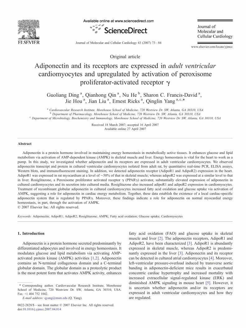
Adiponectin is an important regulator of energy metabolismas indicated by its potent effects on FAO and glucose utilizationin skeletal muscle and in liver [2]. QPCR revealed thatadiponectin mRNA was expressed in RNA samples isolatedfrom adult rat cardiomyocytes, albeit at modest levels comparedto differentiated adipocytes (Fig. 1a). Adiponectin transcriptwas also detectable in cultured neonatal cardiomyocytes but at amuch lower concentration than that of adult cardiomyocytes(data not shown). On the other hand, we did not detect anyadiponectin expression in other cell types such as HeLa cells,predifferentiated 3T3-L1 cells and human embryonic kidney-293 (HEK-293) cells (data not shown). To assess whether thecardiomyocyte cultures were contaminated with adipocytes, weexamined the expression of adipocyte fatty acid-bindingprotein, aP2, an adipocyte marker, in cultured cardiomyocytesand differentiated adipocytes with RT-PCR. While aP2 was notdetected in RNA samples from adult cardiomyocyte, theexpression of aP2 was abundant in RNA samples from thedifferentiated adipocytes (Fig. 1b). Therefore, the adiponectintranscript detected from cultured cardiomyocytes is not likelyfrom adipocyte contamination. Western blots using adiponectin-specific antibody for adiponectin revealed that adiponectinprotein is expressed in rat adult cardiomyocytes, but largelyundetectable in neonatal rat cardiomyocytes (Fig. 1c). Pre-

Fig. 1. Adiponectin expression in cardiomyocytes. (a) Quantitative real time RT-PCR measurement of adiponectin relative to β-actin in cultured cardiomyocytesisolated from adult rats and in cultured differentiated adipocytes. Data are expressed as mean±S.E.M., n=6, *Pb0.01. (b) Quantitative real time RT-PCRmeasurement of aP2 relative to β-actin with equal loading of cDNA samples from cultured adult rat cardiomyocytes and differentiated adipocytes. Data are expressedas mean±S.E.M., n=4, *Pb0.01. (c) Western blot analyses of adiponectin protein in protein samples extracted from cultured adipocytes, adult and neonatal ratcardiomyocytes (ARC and NRC, respectively). Preincubation of adiponectin blocking peptide serves as negative control. The relative levels of adiponectin werenormalized to actin. Data are expressed as mean±S.E.M., n=4, *Pb0.01. (d) Immunofluorescent staining with anti-adiponectin on cultured cardiomyocytes. Thenegative controls were stained with IgG only. Experiments were performed three times with similar results.
77G. Ding et al. / Journal of Molecular and Cellular Cardiology 43 (2007) 73–84
incubation of the protein samples with a blocking epitopepeptide largely eliminated the specific band of adiponectin inprotein samples from both cardiomyocytes and adipocytes (Fig.1c). Analyses with LTQ mass spectrometer further confirmedthat the peptide mapping patterns of the particular band matchedwith adiponectin (data not shown). In addition, immunofluor-escent staining with anti-adiponectin antibody of culturedcardiomyocytes revealed that adiponectin is expressed incardiomyocytes (Fig. 1d). No staining was observed in cardio-
myocytes incubated with normal rabbit IgG (Fig. 1d). Thesedata provide definitive evidence that adiponectin is indeedexpressed in adult ventricular cardiomyocytes.
3.2. Both adiponectin receptors, adipoR1 and adipoR2, areexpressed in the heart
AdipoR1 is expressed abundantly in skeletal muscle, andadipoR2 is predominantly expressed in liver [3]. However, little
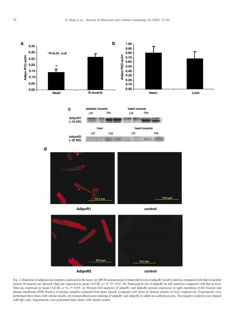
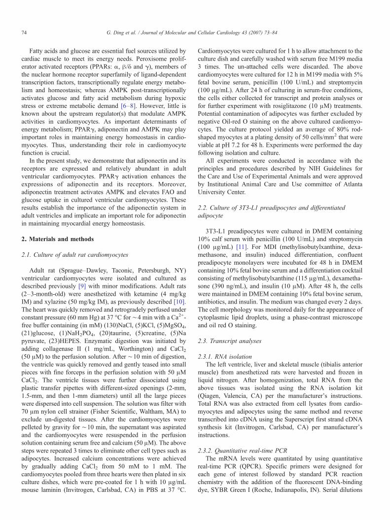
Fig. 2. Detection of adiponectin receptors expressed in the heart. (a) QPCRmeasurement of transcript levels of adipoR1 in left ventricles compared with that in skeletalmuscle (S-muscle) are showed. Data are expressed as mean±S.E.M., n=6, *Pb0.01. (b) Transcript levels of adipoR2 in left ventricles compared with that in liver.Data are expressed as mean±S.E.M., n=6, PN0.05. (c) Western blot analyses of adipoR1 and adipoR2 protein expression in light membrane (LM) fraction andplasma membrane (PM) fraction of protein samples extracted from heart muscle compared with those in skeletal muscle or liver, respectively. Experiments wereperformed three times with similar results. (d) Immunofluorescent staining of adipoR1 and adipoR2 in adult rat cardiomyocytes. The negative controls were stainedwith IgG only. Experiments were performed three times with similar results.
78 G. Ding et al. / Journal of Molecular and Cellular Cardiology 43 (2007) 73–84
79G. Ding et al. / Journal of Molecular and Cellular Cardiology 43 (2007) 73–84
is known about the expression of adipoR1 and adipoR2 inventricular muscle. By QPCR, we found that transcripts of bothadipoR1 and adipoR2 are expressed in rat ventricle tissues(Figs. 2a and b). AdipoR1 mRNA expression was approxi-mately 50% of that in skeletal muscle. On the other hand,adipoR2 mRNAwas expressed at a level similar to that found inliver. Next, we examined adipoR1 and adipoR2 proteinexpression in cytosolic and membrane fractions from adultleft ventricles. Our results show that both adipoR1 and adipoR2are expressed in the plasma membrane (PM) fraction at similarlevels to that observed in skeletal muscle and liver (Fig. 2c).Similarly, immunofluorescent staining confirmed that adipoR1and adipoR2 proteins are expressed in adult cardiomyocytesisolated from left ventricles (Fig. 2d). Together, the data indicatethat both adipoR1 and adipoR2 are localized to the membraneof adult ventricular cardiomyocytes.
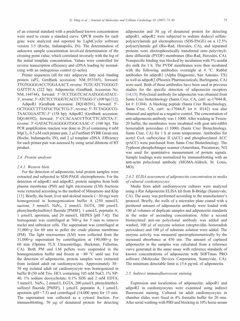
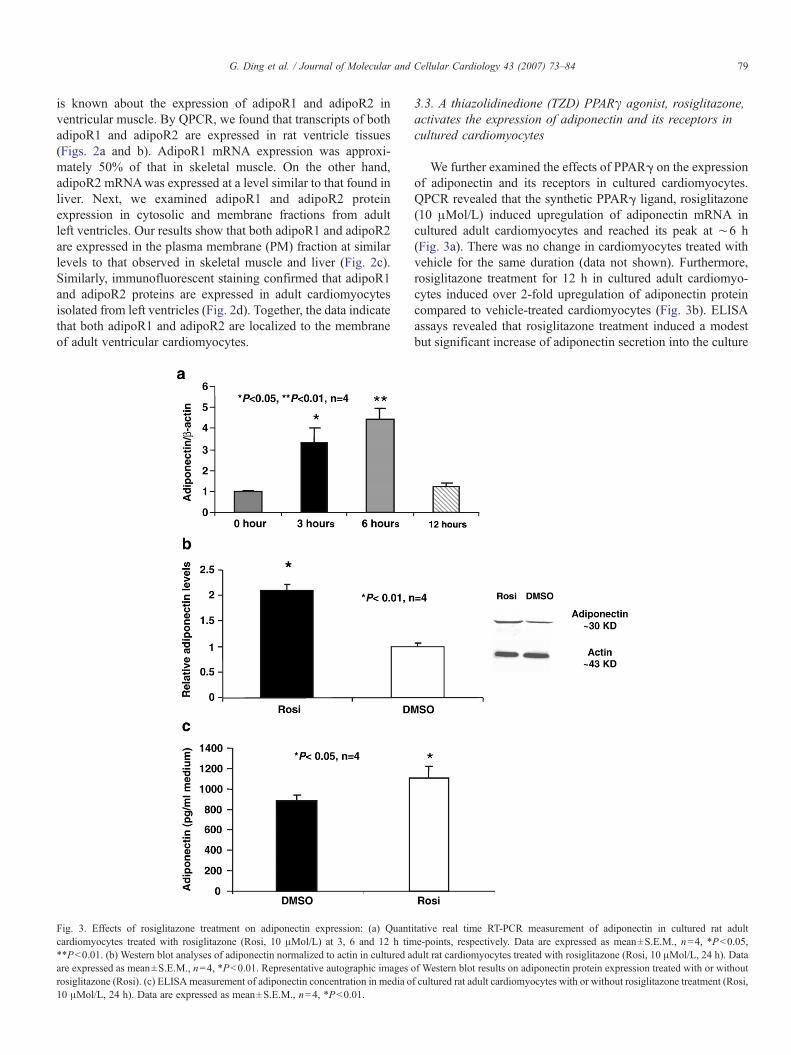
Fig. 3. Effects of rosiglitazone treatment on adiponectin expression: (a) Quantcardiomyocytes treated with rosiglitazone (Rosi, 10 μMol/L) at 3, 6 and 12 h tim**Pb0.01. (b) Western blot analyses of adiponectin normalized to actin in cultured aare expressed as mean±S.E.M., n=4, *Pb0.01. Representative autographic images orosiglitazone (Rosi). (c) ELISA measurement of adiponectin concentration in media o10 μMol/L, 24 h). Data are expressed as mean±S.E.M., n=4, *Pb0.01.
3.3. A thiazolidinedione (TZD) PPARγ agonist, rosiglitazone,activates the expression of adiponectin and its receptors incultured cardiomyocytes
We further examined the effects of PPARγ on the expressionof adiponectin and its receptors in cultured cardiomyocytes.QPCR revealed that the synthetic PPARγ ligand, rosiglitazone(10 μMol/L) induced upregulation of adiponectin mRNA incultured adult cardiomyocytes and reached its peak at ∼6 h(Fig. 3a). There was no change in cardiomyocytes treated withvehicle for the same duration (data not shown). Furthermore,rosiglitazone treatment for 12 h in cultured adult cardiomyo-cytes induced over 2-fold upregulation of adiponectin proteincompared to vehicle-treated cardiomyocytes (Fig. 3b). ELISAassays revealed that rosiglitazone treatment induced a modestbut significant increase of adiponectin secretion into the culture
itative real time RT-PCR measurement of adiponectin in cultured rat adulte-points, respectively. Data are expressed as mean±S.E.M., n=4, *Pb0.05,dult rat cardiomyocytes treated with rosiglitazone (Rosi, 10 μMol/L, 24 h). Dataf Western blot results on adiponectin protein expression treated with or withoutf cultured rat adult cardiomyocytes with or without rosiglitazone treatment (Rosi,
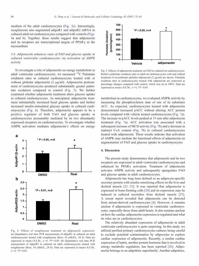
Fig. 5. Effects of adiponectin treatment on FAO in cultured rat cardiomyocytes.Relative palmitate oxidation rates in adult rat cardiomyocytes with and withouttreatment of recombinant globular adiponectin (2 μg/ml) are shown. Palmitateoxidation rates in cardiomyocytes treated with adiponectin are expressed aspercentage changes compared with control, which was set at 100%. Data areexpressed as mean±S.E.M., n=4, *Pb0.05.
80 G. Ding et al. / Journal of Molecular and Cellular Cardiology 43 (2007) 73–84
medium of the adult cardiomyocytes (Fig. 3c). Interestingly,rosiglitazone also augmented adipoR1 and adipoR2 mRNA incultured adult rat cardiomyocytes compared with controls (Figs.4a and b). Together, these results suggest that adiponectinand its receptors are transcriptional targets of PPARγ in themyocardium.
3.4. Adiponectin enhances rates of FAO and glucose uptake incultured ventricular cardiomyocytes via activation of AMPKactivity
To investigate a role of adiponectin on energy metabolism inadult ventricular cardiomyocytes, we measured 14C Palmitateoxidation rates in cultured cardiomyocytes treated with orwithout globular adiponectin (2 μg/ml). Adiponectin pretreat-ment of cardiomyocytes produced substantially greater palmi-tate oxidation compared to control (Fig. 5). We furtherexamined whether adiponectin treatment alters glucose uptakein cultured cardiomyocytes. As anticipated, adiponectin treat-ment substantially increased basal glucose uptake and furtherincreased insulin-stimulated glucose uptake in cultured cardi-omyocytes (Fig. 6). Therefore, adiponectin appears to be apositive regulator of both FAO and glucose uptake incardiomyocytes presumably mediated by its two abundantlyexpressed receptors on cardiomyocytes. To investigate whetherAMPK activation mediates adiponectin's effects on energy
Fig. 4. Effects of rosiglitazone treatment on adiponectin expression:(a) Quantitative real time PCR measurement of adipoR1 in cultured rat adultcardiomyocytes treated with rosiglitazone (Rosi, 10 μMol/L, 24 h). Data areexpressed as mean±S.E.M., n=6, *Pb0.05. (b) Quantitative real time PCRmeasurement of adipoR2 in cultured rat adult cardiomyocytes treated withrosiglitazone (Rosi, 10 μMol/L, 24 h). Data are expressed as mean±S.E.M.,n=6, *Pb0.01.
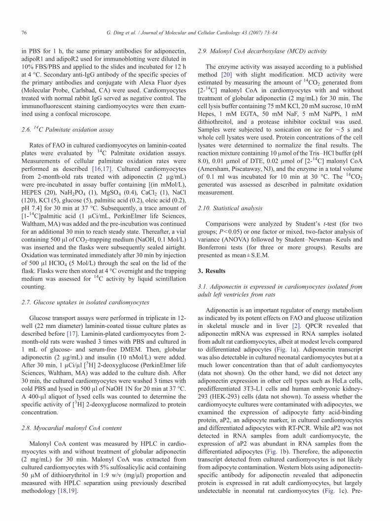
metabolism in cardiomyocytes, we evaluated AMPK activity bymeasuring the phosphorylation state of one of its substratesACC. As expected, cardiomyocytes treated with adiponectindemonstrated increased pACC without altering ACC proteinlevels compared with vehicle treated cardiomyocytes (Fig. 7a).The increase in pACC levels peaked at 15 min after adiponectintreatment (Fig. 7a). ACC activation was associated with asubsequent increase of MCD activity (Fig. 7b) and a decrease inmalonyl CoA content (Fig. 7b) in cultured cardiomyocytestreated with adiponectin. These results indicate that activationof AMPK may mediate the functional effects of adiponectin onaugmentation of FAO and glucose uptake in cardiomyocytes.
4. Discussion
The present study demonstrates that adiponectin and its tworeceptors are expressed in adult ventricular cardiomyocytes andenhanced by PPARγ activation. Treatment of adiponectinactivates AMPK activity and subsequently upregulates FAOand glucose uptake in adult cardiomyocytes.
Adiponectin has long been defined as an adipocyte-specificsecretary protein with insulin-sensitizing effects on the liver andskeletal muscle [21–23]. It was reported that adiponectin isexpressed in bone-forming cells [24] and its expression may beinduced in cultured myotubes from skeletal muscle [25].A recent report revealed that adiponectin can be detectedfrom atrium-derived cardiomyocytes [4]. However, it remainsunclear if adiponectin is expressed in ventricular cardiomyo-cytes, especially those from adult hearts. It also remains unclearon how the cardiac adiponectin expression is regulated and whatits roles are in cardiomyocytes.
The relatively abundant expression of adiponectin in adultventricular cardiomyocytes is quite surprising. In this study, weutilized purified primary cardiomyocytes cultures being carefulto exclude potential contamination by adipocytes to explorecardiac expression of adiponectin. Recently, a similar cardiacexpression of leptin, another protein hormone that is involved inenergy metabolic regulation, has been reported [26]. Adipo-nectin belongs to an adipokine superfamily. Another adipokine,
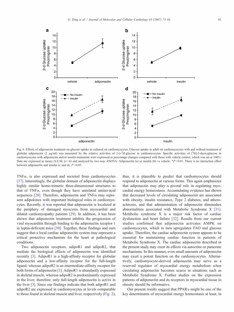
Fig. 6. Effects of adiponectin treatment on glucose uptake in cultured rat cardiomyocytes. Glucose uptake in adult rat cardiomyocytes with and without treatment ofglobular adiponectin (2 μg/ml) was measured by the relative activities of 2-D-3H-glucose in cardiomyocytes. Specific activities of [3H]-2-deoxyglucose incardiomyocytes with adiponectin and/or insulin treatments were expressed as percentage changes compared with those with vehicle control, which was set at 100%.Data are expressed as mean±S.E.M. (n=6) and analyzed by two-way ANOVA. Adiponectin (a) or insulin (b) vs vehicle: *Pb0.01. There is no interaction effectbetween adiponectin and insulin (c and d): PN0.05.
81G. Ding et al. / Journal of Molecular and Cellular Cardiology 43 (2007) 73–84
TNFα, is also expressed and secreted from cardiomyocytes[27]. Interestingly, the globular domain of adiponectin displayshighly similar homo-trimeric three-dimensional structures tothat of TNFα, even though they have unrelated amino-acidsequences [28]. Therefore, adiponectin and TNFα may repre-sent adipokines with important biological roles in cardiomyo-cytes. Recently, it was reported that adiponectin is localized atthe periphery of damaged myocytes from myocardial anddilated cardiomyopathy patients [29]. In addition, it has beenshown that adiponectin treatment inhibits the progression ofviral myocarditis through binding to the adiponectin receptor 1in leptin-deficient mice [30]. Together, these findings and ourssuggest that a local cardiac adiponectin system may represent acritical protective mechanism for the heart at pathologicalconditions.
Two adiponectin receptors, adipoR1 and adipoR2, thatmediate the biological effects of adiponectin was identifiedrecently [3]. AdipoR1 is a high-affinity receptor for globularadiponectin and a low-affinity receptor for the full-lengthligand, whereas adipoR2 is an intermediate-affinity receptor forboth forms of adiponectin [3]. AdipoR1 is abundantly expressedin skeletal muscle, whereas adipoR2 is predominantly expressedin the liver; therefore, only full-length adiponectin is active inthe liver [3]. Since our findings indicate that both adipoR1 andadipoR2 are expressed in cardiomyocytes at levels comparableto those found in skeletal muscle and liver, respectively (Fig. 2);
thus, it is plausible to predict that cardiomyocytes shouldrespond to adiponectin at various forms. This again emphasizesthat adiponectin may play a pivotal role in regulating myo-cardial energy homeostasis. Accumulating evidence has shownthat decreased levels of circulating adiponectin are associatedwith obesity, insulin resistance, Type 2 diabetes, and athero-sclerosis, and that administration of adiponectin diminishesabnormalities associated with Metabolic Syndrome X [31].Metabolic syndrome X is a major risk factor of cardiacdysfunction and heart failure [32]. Results from our currentstudies confirmed that adiponectin activates AMPK oncardiomyocytes, which in turn upregulates FAO and glucoseuptake. Therefore, the cardiac adiponectin system appears to beessential for maintaining cardiac function in patients ofMetabolic Syndrome X. The cardiac adiponectin described inthe present study may exert its effects via autocrine or paracrinemechanisms. In this manner, even small amounts of adiponectinmay exert a potent function on the cardiomyocytes. Alterna-tively, cardiomyocyte-derived adiponectin may serve as areserved regulator of myocardial energy metabolism whencirculating adiponectin becomes scarce in situations such asMetabolic Syndrome X. Further studies on the expressionpatterns of adiponectin and its receptors in myocardial tissue inobesity should be informative.
Our present results suggest that PPARγ might be one of thekey determinants of myocardial energy homeostasis at least, in
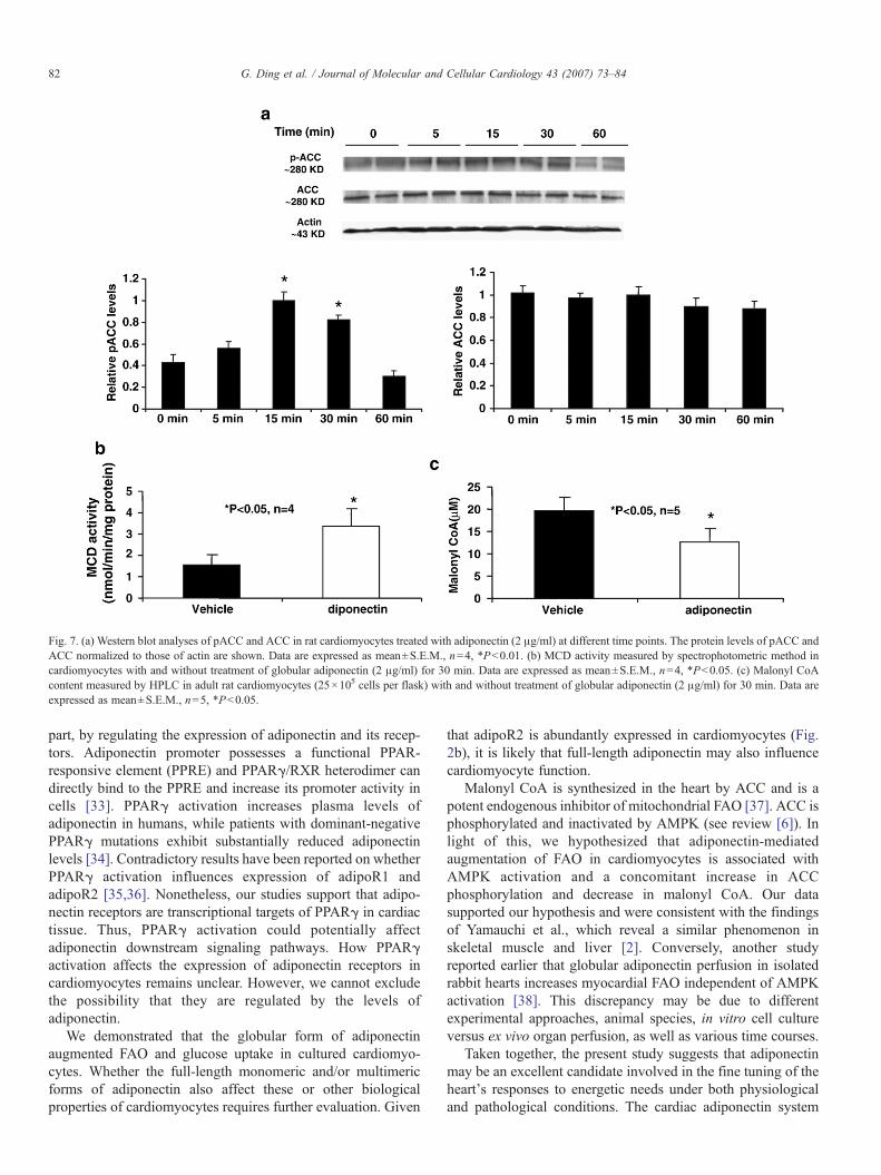
Fig. 7. (a) Western blot analyses of pACC and ACC in rat cardiomyocytes treated with adiponectin (2 μg/ml) at different time points. The protein levels of pACC andACC normalized to those of actin are shown. Data are expressed as mean±S.E.M., n=4, *Pb0.01. (b) MCD activity measured by spectrophotometric method incardiomyocytes with and without treatment of globular adiponectin (2 μg/ml) for 30 min. Data are expressed as mean±S.E.M., n=4, *Pb0.05. (c) Malonyl CoAcontent measured by HPLC in adult rat cardiomyocytes (25×105 cells per flask) with and without treatment of globular adiponectin (2 μg/ml) for 30 min. Data areexpressed as mean±S.E.M., n=5, *Pb0.05.
82 G. Ding et al. / Journal of Molecular and Cellular Cardiology 43 (2007) 73–84
part, by regulating the expression of adiponectin and its recep-tors. Adiponectin promoter possesses a functional PPAR-responsive element (PPRE) and PPARγ/RXR heterodimer candirectly bind to the PPRE and increase its promoter activity incells [33]. PPARγ activation increases plasma levels ofadiponectin in humans, while patients with dominant-negativePPARγ mutations exhibit substantially reduced adiponectinlevels [34]. Contradictory results have been reported on whetherPPARγ activation influences expression of adipoR1 andadipoR2 [35,36]. Nonetheless, our studies support that adipo-nectin receptors are transcriptional targets of PPARγ in cardiactissue. Thus, PPARγ activation could potentially affectadiponectin downstream signaling pathways. How PPARγactivation affects the expression of adiponectin receptors incardiomyocytes remains unclear. However, we cannot excludethe possibility that they are regulated by the levels ofadiponectin.
We demonstrated that the globular form of adiponectinaugmented FAO and glucose uptake in cultured cardiomyo-cytes. Whether the full-length monomeric and/or multimericforms of adiponectin also affect these or other biologicalproperties of cardiomyocytes requires further evaluation. Given
that adipoR2 is abundantly expressed in cardiomyocytes (Fig.2b), it is likely that full-length adiponectin may also influencecardiomyocyte function.
Malonyl CoA is synthesized in the heart by ACC and is apotent endogenous inhibitor of mitochondrial FAO [37]. ACC isphosphorylated and inactivated by AMPK (see review [6]). Inlight of this, we hypothesized that adiponectin-mediatedaugmentation of FAO in cardiomyocytes is associated withAMPK activation and a concomitant increase in ACCphosphorylation and decrease in malonyl CoA. Our datasupported our hypothesis and were consistent with the findingsof Yamauchi et al., which reveal a similar phenomenon inskeletal muscle and liver [2]. Conversely, another studyreported earlier that globular adiponectin perfusion in isolatedrabbit hearts increases myocardial FAO independent of AMPKactivation [38]. This discrepancy may be due to differentexperimental approaches, animal species, in vitro cell cultureversus ex vivo organ perfusion, as well as various time courses.
Taken together, the present study suggests that adiponectinmay be an excellent candidate involved in the fine tuning of theheart's responses to energetic needs under both physiologicaland pathological conditions. The cardiac adiponectin system
83G. Ding et al. / Journal of Molecular and Cellular Cardiology 43 (2007) 73–84
may coordinate regulation of myocardial energy balance inresponse to systemic and local needs under physiologic andpathologic conditions. Therefore, adiponectin may be apotential therapeutic alternative in treating cardiac energeticanomalies.
Acknowledgments
This work was partially supported by grants from NIH(MBRS S06GM08248, 1R01HL085499 and 1R01HL084456)and a scientist development award from the American HeartAssociation national center. The investigation was conducted ina facility constructed with support from Research FacilitiesImprovement Grant #C06 RR-1 C06 RR07571. We thank Dr.Bryan Slinker and Mr. Yong Liu for technical help on statisticalanalyses.
References
[1] Scherer PE, Williams S, Fogliano M, Baldini G, Lodish HF. A novel serumprotein similar to C1q, produced exclusively in adipocytes. J Biol Chem1995 (Nov. 10);270(45):26746–9.
[2] Yamauchi T, Kamon J, Minokoshi Y, Ito Y, Waki H, Uchida S, et al.Adiponectin stimulates glucose utilization and fatty-acid oxidation byactivating AMP-activated protein kinase. Nat Med 2002 (Nov.);8(11):1288–95.
[3] Yamauchi T, Kamon J, Ito Y, Tsuchida A, Yokomizo T, Kita S, et al.Cloning of adiponectin receptors that mediate antidiabetic metaboliceffects. Nature 2003 (Jun. 12);423(6941):762–9.
[4] Pineiro R, Iglesias MJ, Gallego R, Raghay K, Eiras S, Rubio J, et al.Adiponectin is synthesized and secreted by human and murine cardio-myocytes. FEBS Lett 2005 (Sep. 26);579(23):5163–9.
[5] Shibata R, Ouchi N, Ito M, Kihara S, Shiojima I, Pimentel DR, et al.Adiponectin-mediated modulation of hypertrophic signals in the heart. NatMed 2004 (Dec.);10(12):1384–9.
[6] Hardie DG, Carling D. The AMP-activated protein kinase–fuel gauge ofthe mammalian cell? Eur J Biochem 1997 (Jun. 1);246(2):259–73.
[7] Kemp BE, Mitchelhill KI, Stapleton D, Michell BJ, Chen ZP, Witters LA.Dealing with energy demand: the AMP-activated protein kinase. TrendsBiochem Sci 1999 (Jan.);24(1):22–5.
[8] Tian R, Musi N, D'Agostino J, Hirshman MF, Goodyear LJ. Increasedadenosine monophosphate-activated protein kinase activity in rat heartswith pressure-overload hypertrophy. Circulation 2001 (Oct. 2);104(14):1664–9.
[9] Mitcheson JS, Hancox JC, Levi AJ. Cultured adult cardiac myocytes:future applications, culture methods, morphological and electrophysiolo-gical properties. Cardiovasc Res 1998 (Aug.);39(2):280–300.
[10] Shimoyama M, Hayashi D, Takimoto E, Zou Y, Oka T, Uozumi H, et al.Calcineurin plays a critical role in pressure overload-induced cardiachypertrophy. Circulation 1999 (Dec. 14);100(24):2449–54.
[11] Kim YC, Gomez FE, Fox BG, Ntambi JM. Differential regulation of thestearoyl-CoA desaturase genes by thiazolidinediones in 3T3-L1 adipo-cytes. J Lipid Res 2000 (Aug.);41(8):1310–6.
[12] Nagao K, Inoue N, Wang YM, Yanagita T. Conjugated linoleic acidenhances plasma adiponectin level and alleviates hyperinsulinemia andhypertension in Zucker diabetic fatty (fa/fa) rats. Biochem Biophys ResCommun 2003 (Oct. 17);310(2):562–6.
[13] Mitsumoto Y, Klip A. Development regulation of the subcellulardistribution and glycosylation of GLUT1 and GLUT4 glucose transportersduring myogenesis of L6 muscle cells. J Biol Chem 1992 (Mar. 5);267(7):4957–62.
[14] Inukai K, Nakashima Y, Watanabe M, Takata N, Sawa T, Kurihara S, et al.Regulation of adiponectin receptor gene expression in diabetic mice. Am JPhysiol: Endocrinol Metab 2005 (May);288(5):E876–82.
[15] Caminos JE, Nogueiras R, Gallego R, Bravo S, Tovar S, Garcia-CaballeroT, et al. Expression and regulation of adiponectin and receptor in humanand rat placenta. J Clin Endocrinol Metab 2005 (Jul.);90(7):4276–86.
[16] Cheng L, Ding G, Qin Q, Xiao Y, Woods D, Chen YE, et al. Peroxisomeproliferator-activated receptor [delta] activates fatty acid oxidation incultured neonatal and adult cardiomyocytes. Biochem Biophys ResCommun 2004 (2004/1/9);313(2):277–86.
[17] Cheng L, Ding G, Qin Q, Huang Y, Lewis W, He N, et al. Cardiomyocyte-restricted peroxisome proliferator-activated receptor-delta deletion per-turbs myocardial fatty acid oxidation and leads to cardiomyopathy. NatMed 2004 (Nov.);10(11):1245–50.
[18] King MT, Reiss PD. Separation and measurement of short-chaincoenzyme-A compounds in rat liver by reversed-phase high-performanceliquid chromatography. Anal Biochem 1985 (Apr.);146(1):173–9.
[19] Saddik M, Gamble J, Witters LA, Lopaschuk GD. Acetyl-CoAcarboxylase regulation of fatty acid oxidation in the heart. J Biol Chem1993 (Dec. 5);268(34):25836–45.
[20] Kim YS, Kolattukudy PE. Malonyl-CoA decarboxylase from the uropygialgland of waterfowl: purification, properties, immunological comparison,and role in regulating the synthesis of multimethyl-branched fatty acids.Arch Biochem Biophys 1978 (Oct.);190(2):585–97.
[21] Yamamoto K, Ohki R, Lee RT, Ikeda U, Shimada K. Peroxisomeproliferator-activated receptor gamma activators inhibit cardiac hypertro-phy in cardiac myocytes. Circulation 2001;104(14):1670–5.
[22] Yamauchi T, Kamon J, Waki H, Terauchi Y, Kubota N, Hara K, et al. Thefat-derived hormone adiponectin reverses insulin resistance associatedwith both lipoatrophy and obesity. Nat Med 2001 (Aug.);7(8):941–6.
[23] Berg AH, Combs TP, Du X, Brownlee M, Scherer PE. The adipocyte-secreted protein Acrp30 enhances hepatic insulin action. Nat Med 2001(Aug.);7(8):947–53.
[24] Berner HS, Lyngstadaas SP, Spahr A, Monjo M, Thommesen L, DrevonCA, et al. Adiponectin and its receptors are expressed in bone-formingcells. Bone 2004 (Oct.);35(4):842–9.
[25] Delaigle AM, Jonas JC, Bauche IB, Cornu O, Brichard SM. Induction ofadiponectin in skeletal muscle by inflammatory cytokines: in vivo and invitro studies. Endocrinology 2004 (Dec);145(12):5589–97.
[26] Purdham DM, Zou MX, Rajapurohitam V, Karmazyn M. Rat heart is a siteof leptin production and action. Am J Physiol Heart Circ Physiol 2004(Dec.);287(6):H2877–84.
[27] Meldrum DR. Tumor necrosis factor in the heart. Am J Physiol 1998(Mar.);274(3 Pt 2):R577–95.
[28] Shapiro L, Scherer PE. The crystal structure of a complement-1q familyprotein suggests an evolutionary link to tumor necrosis factor. Curr Biol1998 (Mar. 12);8(6):335–8.
[29] Takahashi T, Saegusa S, Sumino H, Nakahashi T, Iwai K, Morimoto S,et al. Adiponectin, T-cadherin and tumour necrosis factor-alpha indamaged cardiomyocytes from autopsy specimens. J Int Med Res 2005(Mar.–Apr.);33(2):236–44.
[30] Takahashi T, Saegusa S, SuminoH, Nakahashi T, Iwai K,Morimoto S, et al.Adiponectin replacement therapy attenuates myocardial damage in leptin-deficient mice with viral myocarditis. J Int Med Res 2005 (Mar.–Apr.);33(2):207–14.
[31] Matsuzawa Y, Funahashi T, Kihara S, Shimomura I. Adiponectin andmetabolic syndrome. Arterioscler, Thromb, Vasc Biol 2004 (Jan.);24(1):29–33.
[32] Kenchaiah S, Evans JC, Levy D, Wilson PW, Benjamin EJ, Larson MG,et al. Obesity and the risk of heart failure. N Engl J Med 2002 (Aug.1);347(5):305–13.
[33] Iwaki M, Matsuda M, Maeda N, Funahashi T, Matsuzawa Y, MakishimaM, et al. Induction of adiponectin, a fat-derived antidiabetic andantiatherogenic factor, by nuclear receptors. Diabetes 2003 (Jul.);52(7):1655–63.
[34] Combs TP, Wagner JA, Berger J, Doebber T, Wang WJ, Zhang BB, et al.Induction of adipocyte complement-related protein of 30 kilodaltons byPPARgamma agonists: a potential mechanism of insulin sensitization.Endocrinology 2002 (Mar.);143(3):998–1007.
[35] Chinetti G, Zawadski C, Fruchart JC, Staels B. Expression of adiponectinreceptors in human macrophages and regulation by agonists of the nuclear

84 G. Ding et al. / Journal of Molecular and Cellular Cardiology 43 (2007) 73–84
receptors PPARalpha, PPARgamma, and LXR. Biochem Biophys ResCommun 2004 (Jan. 30);314(1):151–8.
[36] Kaltenbach S, Staiger H, Weisser M, Haas C, Stumvoll M, Machicao F, etal. Adiponectin receptor gene expression in human skeletal muscle cells isnot regulated by fibrates and thiazolidinediones. Int J Obes (Lond) 2005(Jul.);29(7):760–5.
[37] Lopaschuk GD, Witters LA, Itoi T, Barr R, Barr A. Acetyl-CoAcarboxylase involvement in the rapid maturation of fatty acid oxidationin the newborn rabbit heart. J Biol Chem 1994 (Oct. 14);269(41):25871–8.
[38] Onay-Besikci A, Altarejos JY, Lopaschuk GD. gAd-globular head domainof adiponectin increases fatty acid oxidation in newborn rabbit hearts.J Biol Chem 2004 (Oct. 22);279(43):44320–6.


