Embed Size (px)
Citation preview

Eric Metzger, Judith M.MuÈ ller,Stefano Ferrari1, Reinhard Buettner2 andRoland SchuÈ le3
UniversitaÈts-Frauenklinik und Zentrum fuÈr Klinische Forschung,Klinikum der UniversitaÈt Freiburg, Breisacherstrasse 66,D-79106 Freiburg, 2Institut fuÈr Pathologie, UniversitaÈtsklinikum Bonn,Sigmund-Freud-Strasse 25, D-53127 Bonn, Germany and 1Institute ofMedical Radiobiology, University of ZuÈrich, August-Forel-Strasse 7,CH-8008 ZuÈrich, Switzerland
3Corresponding authore-mail: [email protected]
In addition to the classical activation by ligands,nuclear receptor activity is also regulated by ligand-independent signalling. Here, we unravel a novel sig-nal transduction pathway that links the RhoA effectorprotein kinase C-related kinase PRK1 to the tran-scriptional activation of the androgen receptor (AR).Stimulation of the PRK signalling cascade results in aligand-dependent superactivation of AR. We showthat AR and PRK1 interact both in vivo and in vitro.The transactivation unit 5 (TAU-5) located in theN-terminus of AR suf®ces for activation by PRK1.Thus, TAU-5 de®nes a novel, signal-inducible trans-activation domain. Furthermore, PRK1 promotes afunctional complex of AR with the co-activator TIF-2.Importantly, PRK signalling also stimulates ARactivity in the presence of adrenal androgens, whichare still present in prostate tumour patients subjectedto testicular androgen ablation therapy. Moreover,PRK1 activates AR even in the presence of the ARantagonist cyproterone acetate that is used in the clin-ical management of prostate cancer. Since prostatetumours strongly overexpress PRK1, our data supporta model in which AR activity is controlled by PRKsignalling.Keywords: androgen receptor/nuclear hormone receptor/prostate cancer/protein kinase C-related kinase/transcriptional co-activator
Introduction
The androgen receptor (AR) is a member of the steroidhormone receptor family of ligand-activated transcriptionfactors that regulate diverse biological functions includingcell growth and differentiation, development, homeostasisand various organ functions in the adult (Mangelsdorfet al., 1995; Cato and Peterzierl, 1998). The AR shares acommon modular structure with other nuclear receptorsand is composed of several domains that mediate DNAbinding, dimerization, ligand binding and transcriptionalactivity (Mangelsdorf et al., 1995). The ligand bindingdomain (LBD) performs a number of functions including
transcriptional repression or activation by generating theproper interaction surface for multiple partners, includingco-repressors and co-activators (Renaud and Moras,2000). Like other steroid receptors the AR contains aC-terminal ligand-dependent activation function 2 (AF-2)and an additional N-terminal ligand-independent activ-ation function 1 (AF-1). The AR also harbours a less well-de®ned transcription activation unit termed TAU-5(Jenster et al., 1995; Jenster, 1999). The TAU-5 is locatedbetween amino acids 360 and 528 and is required for fullactivation of the AR (Jenster et al., 1995; Bevan et al.,1999). However, signals that control TAU-5, and therebyAR activity, are unknown. Recent data show agonist-induced interaction between the N-terminal domain(NTD) and the LBD of the AR, supporting the view thatboth the AF-1 and the AF-2 are required for transcriptionalactivation (Ikonen et al., 1997; He et al., 2000). It has beensuggested that the ligand-induced conformational changesof the LBD in concert with physical interactions with theNTD render the AR fully active (Ikonen et al., 1997;Bevan et al., 1999).
Androgens and the AR are essential for the differenti-ation, development and maintenance of male reproductivefunctions and non-reproductive organs (Trapman andBrinkmann, 1996; Jenster, 1999). In the prostate, the ARis expressed in secretory epithelial cells that respond toandrogens. According to the current model, the AR playsan important role in the development of prostate cancerthat mainly originates from epithelial cells. Growth andsurvival of primary prostate cancer cells is criticallydependent on androgens (Gregory et al., 1998). Conse-quently, androgen ablation therapy is the standard clinicalprocedure used to inhibit prostate tumour growth.However, despite reduced circulating androgen levelsand even in the presence of AR antagonists, most prostatecancers recur and progress to a terminal stage. Themolecular mechanisms responsible for tumour recurrenceare not well de®ned, but one possible scenario might be thestimulation of AR activity by cross-talk with othersignalling pathways (Culig et al., 1994).
In addition to the classical ligand-induced activationmechanisms, nuclear receptors use alternative strategies totransmit extracellular signals into altered gene expressionprograms. Growth factors or stimulation of proteinkinases A (PKA) or C (PKC) activate the AR even in theabsence of ligand, or show a synergistic effect with ligand(Trapman and Brinkmann, 1996). Growth factor-regulatedsignalling pathways also control the activity of nuclearreceptor co-activators. Recent data show that co-activatorsof the p160 family such as SRC-1 or AIB1 are direct targetsof mitogen activated protein (MAP) kinases, whichmodulate their transcriptional activity (Font de Mora andBrown, 2000; Rowan et al., 2000). An alternative strategyto regulate co-factor function is employed by Rho
A novel inducible transactivation domain inthe androgen receptor: implications for PRKin prostate cancer
The EMBO Journal Vol. 22 No. 2 pp. 270±280, 2003
270 ã European Molecular Biology Organization

GTPases. RhoA induces translocation of the co-activatorFHL2 from the cell membrane to the nucleus, leading totranscriptional activation of FHL2- and AR-dependentgenes (MuÈller et al., 2002).
By affecting multiple signalling pathways Rho familymembers regulate cell cycle progression, cellular trans-formation, metastasis and have been implicated in tran-scriptional regulation (Bar-Sagi and Hall, 2000). The Rhofamily GTPases work as molecular switches, beinginactive when bound to GDP and active when bound toGTP. The exchange of GTP for GDP induces aconformational change that allows the GTPases to bindeffector molecules such as the protein kinase C-relatedkinases (PRKs), which initiate downstream signallingevents (Hall, 1998; Bishop and Hall, 2000). The PRKsconstitute a subfamily of serine/threonine kinases thatcomprises PRK1 (also called PKN), PRK2 and PKNb(Mukai et al., 1995; Palmer et al., 1995; Oishi et al., 1999).PRKs contain three highly-conserved regions: (i) anN-terminal regulatory domain spanning three repeats ofcharged amino acids with leucine zipper-like sequences(designated CZ region); (ii) a C-terminal catalytic domain;and (iii) a so-called D region located between the CZregion and the catalytic domain (Mukai et al., 1995).RhoA binds to the CZ region through the RhoA effectormotif class I in a GTP-dependent manner therebyenhancing PRK kinase activity. The physical interactionof PRK1 with transcription factors such as NeuroD2 orPCD17, and the nuclear localization suggest a potentialrole for PRKs in transcriptional regulation (Takanagaet al., 1998; Shibata et al., 1999).
Recently, we demonstrated that stimulation of the Rhosignalling pathway induces translocation of the co-activator FHL2 to the nucleus leading to activation ofthe AR by FHL2 (MuÈller et al., 2002). We now identify anovel pathway independent of FHL2 that links the RhoAeffectors PRK1 and PRK2 to the transcriptional activationof AR. Here we show that stimulation of PRK1 signallingby extracellular stimuli or co-expression of constitutivelyactive RhoA or PRK1 mutants results in ligand-dependentsuperactivation of AR-regulated genes. AR and PRK1interact both in vivo and in vitro. Structure±functionanalyses demonstrate that the TAU-5 in the AR-NTDsuf®ces for activation by PRK1, thus de®ning the TAU-5as a novel, signal-inducible transactivation domain. Inaddition, PRK1 signalling also induces the transcriptionalactivity of mineralocorticoid receptor (MR), progesteronereceptor (PR) and p160 co-activators, further supportingthe importance of this signalling pathway for the tran-scriptional regulation of nuclear receptors. Our datashow that prostate tumours highly overexpress PRK1.Importantly, PRK1 activates AR not only in the presenceof agonists, but also when adrenal androgens or even theantagonist cyproterone acetate (CPA) are present. Takentogether, our results demonstrate that the transcriptionalactivity of AR is controlled by PRK signalling in additionto ligands, providing an explanation for prostate cancergrowth even during androgen ablation therapy.
Results
The current dilemma in androgen ablation therapy is theaberrant regulation of AR activity. This clearly points out
the relevance of other yet unde®ned signalling pathwaysleading to AR-dependent tumour growth. Recently, weshowed that Rho family members are overexpressed inhuman prostate tumours and that stimulation of the Rhosignalling pathway leads to an agonist-dependent co-activation of AR-regulated target genes (MuÈller et al.,2002). To further delineate the mechanisms of Rhosignalling to AR, we tested the in¯uence of Rho effectorson AR transcriptional activity. Therefore, we stimulatedAR-dependent gene expression with physiological con-centrations of the bioactive lipid sphingosine-1-phosphate(SPP) in the presence of AR and an MMTV luciferasereporter (MMTV-LUC). The serum component SPPactivates small GTPases of the Rho family via G protein-coupled receptors (Pyne and Pyne, 2000). SPP stimulationof endogenous Rho GTPases increases the agonist-dependent activation of the MMTV-LUC reporter(Figure 1A; MuÈller et al., 2002). Both the agonist andSPP-induced reporter activity is blocked by either the Rho-speci®c inhibitor Clostridium botulinum C3 exoenzyme(Busch and Aktories, 2000) or the dominant-negativeRhoA mutant RhoA N19 (Figure 1A). In addition, thedominant-negative mutants of the Rho effectors PRK1(dnPRK1) and PRK2 (dnPRK2) potently inhibit agonistand SPP-induced AR activity (Figure 1A). Wild-typePRK1 (PRK1wt) does not in¯uence AR transcriptionalactivity (Figure 1A). In the absence of either AR or ligandthe MMTV-LUC reporter is not in¯uenced (our unpub-lished data). The dominant-negative PRK mutants alsoblock ligand-dependent stimulation of AR by the con-stitutively active RhoA mutant RhoA V14 (Figure 1B),whereas the dominant-negative mutants Rac1 N17, Cdc42N17 or Ras N17 fail to show any inhibitory effect(Figure 1B). All dominant-negative mutants are functionalin our assay system and down-regulate the v-Src-inducedactivity of a SRE-LUC reporter (Chiariello et al., 2001;our unpublished data). Furthermore, neither the varioussignal transduction factors nor the chemical inhibitorsused in this study alter AR protein levels (seeSupplementary ®gure 1, available at The EMBO JournalOnline). Taken together, these data show for the ®rst timean involvement of PRKs in RhoA-mediated activation ofAR.
To examine the Rho®PRK signalling pathway leadingto AR activation in more detail, we tested whetherexpression of GDP±GTP exchange factors (GEFs) isable to promote AR-dependent gene expression. GEFsbind small GTPases and are involved in promotingnucleotide exchange (Cerione and Zheng, 1996). Theactivated form of the Rho-speci®c GEF NET1 (NET1 DN)stimulates the AR- and agonist-dependent activity ofMMTV-LUC in 293 cells to a similar extent as theconstitutively active RhoA V14 (Figure 1C). We then usedthe N-terminus of PRK1 (PRK1.N), which acts as adominant-negative mutant in RhoA- and NET1 signallingby binding to RhoA±GTP (Sahai et al., 1998). When testedin our assay system PRK1.N blocks NET1 DN andRhoA V14 stimulated agonist-dependent AR activation(Figure 1C), further indicating the importance of PRKfor AR activation. Accordingly, the PRK inhibitorsRo31-8220 or HA 1077 (Davies et al., 2000) abolishedagonist-dependent AR activation by RhoA V14(Figure 1D). In contrast, blocking the MAP kinase
PRK signalling and AR
271

pathway by the MEK1 inhibitor PD 98059, inhibiting S6kinase/TOR with rapamycin, or using the p38 MAP kinaseinhibitor SB 203580 does not interfere with the transcrip-
tional activation by RhoA V14 (Figure 1D). The effect-iveness of the inhibitors (our unpublished data) wascon®rmed according to MuÈller et al. (2002). Next, we
Fig. 1. The RhoA effectors PRK1 and PRK2 mediate transcriptional superactivation of the AR. 293 (A±E and G) and PC3-AR cells (F) were trans-fected with MMTV-LUC reporter and AR expression plasmids with or without 10±10 M R1881. (A) Stimulation of the endogenous Rho signallingpathway by SPP leads to the activation of AR. Activation is blocked either by C3 exoenzyme, or by dominant-negative forms of RhoA (RhoA N19),PRK1 (dnPRK1), PRK2 (dnPRK2), but not wild-type PRK1 (PRK1wt). Cells were treated with either vehicle or 1.2 mM SPP. (B) Dominant-negativePRK1 (dnPRK1) and PRK2 (dnPRK2), but not the dominant-negative forms of Rac1 (Rac1 N17), Cdc42 (Cdc42 N17) or Ras (Ras N17) block ARactivation by constitutively active RhoA (RhoA V14). (C) The dominant-negative acting N-terminus of PRK1 (PRK1.N) blocks AR activation byRhoA V14 or by the RhoA-speci®c constitutively active GEF NET1 (NET1 DN). (D) Ligand-dependent activation of AR by RhoA V14 is blocked bythe PRK inhibitors Ro31-8220 or HA 1077 but not by the MEK1 inhibitor PD 98059, the S6 kinase/TOR inhibitor rapamycin or the p38 MAP kinaseinhibitor SB 203580. As indicated, cells were transfected with RhoA V14 expression plasmids and treated with either 5 3 10±7 M Ro31-8220,3 3 10±5 M HA 1077, 50 ng/ml rapamycin, 10±5 M PD 98059 or 10±5 M SB 203580. (E) AR is blocked by the PRK inhibitors Ro31-8220 orHA 1077. Cells co-transfected with the constitutively active forms of PRK1 (PRK1*) or PRK2 (PRK2*) were cultured in the absence or presence ofthe PRK inhibitors Ro31-8220 (5 3 10±7 M) or HA 1077 (3 3 10±5 M). (F) The PRK inhibitor Ro31-8220 blocks endogenous AR activity in PC3-ARprostate tumour cells. Cells were treated with 7 3 10±7 M Ro31-8220. (G) In¯uence of Rho effector signalling molecules on AR activity. Wild-typeLIMK1 and LIMK2, constitutively active Rho-kinase a (ROCK*), MKK3b [MKK3E(b)], or MKK6b [MKK6E(b)] and dominant-negative p38g(dnp38g) or protein kinase Ca (dnPKCa) were analysed for their ability to modulate AR activity. Constitutively active PRK1 (PRK1*) wasco-expressed as indicated.
E.Metzger et al.
272

analysed constitutively active mutants of PRK1 (PRK1*)and PRK2 (PRK2*). As demonstrated in Figure 1E, AR isstrongly activated by both constitutively active mutants ofPRK in an agonist-dependent manner and both PRKinhibitors Ro31-8220 or HA 1077 block this activation.Furthermore, Ro31-8220 also blocks endogenous ARtranscriptional activity in PC3-AR prostate tumour cells(Figure 1F). As a control, we analysed thyroid hormonereceptor a-dependent reporter activity, which is neitherin¯uenced by co-expression of PRKs nor by treatmentwith Ro31-8220 (our unpublished data). Since Ro31-8220is known to also inhibit the kinases PKCa, RSK, MSK,S6K and GSK3b (Davies et al., 2000), we veri®ed thatthese kinases do not in¯uence AR activity (Figure 1G; ourunpublished data). To further validate PRK as thedownstream effector of Rho, we assayed several mol-ecules involved in Rho signalling (Bishop and Hall, 2000).Neither wild-type LIM kinase1 (LIMK1) or LIM kinase2(LIMK2), nor constitutively active Rho±kinase a(ROCK*), MKK3b [MKK3E(b)], or MKK6b [MKK6E(b)]are able to induce AR-dependent reporter expression(Figure 1G). In addition, neither dominant-negativemutants of p38g (dnp38g) or PKCa (dnPKCa)(Figure 1G), nor DMPK, PAK, CRIK or MRCKa (ourunpublished data), show any effect. Taken together, theseresults demonstrate that blocking the endogenousRho®PRK signalling pathway by PRK inhibitors ordominant-negative PRK mutants severely impairs agonist-dependent AR transactivation. In addition, our data showthat constitutively active mutants of the Rho effector PRKstimulate agonist-dependent transcriptional activity of AR.
To examine the potential regulation of other steroidhormone receptors by PRK1 signalling the MMTVpromoter was chosen because the receptors for gluco-corticoids (GR), PR, MR and AR are known to regulate itsexpression in a ligand-dependent fashion (SchuÈle et al.,1990). Co-expression of constitutively active PRK1 andMR or PR results in a ligand-dependent superactivation(Figure 2A), whereas the ligand-dependent transcriptionalactivity of GR is only marginally in¯uenced. The sameresults were obtained when the activation assay wasperformed with different agonist concentrations for thevarious receptors (our unpublished data). When tested onthe prostate-speci®c probasin promoter or on MMTV-LUC, PRK signalling stimulates agonist-dependent ARactivity in 293 or DU145 human prostate tumour cells(Figure 2B). These results show that stimulation of AR byPRKs is functional in different cell types and not restrictedto a special promoter context.
Using immuno¯uorescence analyses we investigatedthe subcellular distribution of AR and PRK proteins. ARand the constitutively active mutant PRK1* are present inthe cytoplasm in the human 293 cell line in the absence ofligand, whereas addition of the AR agonist R1881 resultsin nuclear AR and triggers PRK1* nuclear localization(Table I). Equally, CPA promotes nuclear localizationof both proteins. In contrast, the agonist-loaded mutantAR (ARDNLS) that is defective in nuclear transloca-tion (Poukka et al., 2000) does not support nuclearredistribution of PRK1* (Table I). In agreement with thetransactivation data, agonist-loaded MR but not GR leadsto enhanced nuclear translocation of PRK1* (Table I).
To demonstrate interaction between AR and PRK1,extracts from 293 cells transfected with expressionplasmids for PRK1 and Flag epitope-tagged AR wereimmunoprecipitated using an a-Flag antibody (Figure 3A).Western blot analysis shows that AR and PRK1 interactweakly in the absence of ligand, whereas the Flag-AR±PRK1 complex is ef®ciently precipitated in the presence ofthe AR agonist R1881. No PRK1 was found in immuno-precipitated complexes using Flag transfected controlextracts (Figure 3A), thus demonstrating speci®city.Similar results were obtained with constitutively activePRK1 (our unpublished data). More importantly, AR andPRK1 endogenously expressed in primary human prostatetumours associate in vivo (Figure 3B). Extracts fromprimary human prostate tumours treated with R1881 wereimmunoprecipitated using an a-PRK1 antibody. As shownin Figure 3B, the endogenous AR is ef®ciently co-immunoprecipitated with the a-PRK1 antibody, thusdemonstrating the relevance of this interaction in vivo.To further analyse the interaction, we performed in vivoGST pull-down experiments (Figure 3C). The fusionproteins GST±AR-NTD, GST±AR-DBD and GST±AR-LBD were co-expressed with either myc epitope-taggedconstitutively active PRK1 (PRK1*) or wild-type PRK1
Fig. 2. Analysis of the PRK signalling. (A) PRK1 signalling mediatesactivation of AR, MR and PR, but not of GR. 293 cells were co-trans-fected with MMTV-LUC reporter and expression plasmids coding foreither AR, MR, PR or GR in combination with or without PRK1*.Cells were treated with the corresponding agonists (10±10 M R1881,10±9 M aldosterone, 10±9 M R5020 or 10±10 M DEX). (B) AR super-activation by PRK is independent of a particular promoter context orcell type. 293 and DU145 cells were stimulated with 10±10 M R1881 asindicated. Cells were co-transfected with AR, constitutively activeRhoA V14, PRK1* or PRK2*, and either MMTV-LUC or PB-LUC.
PRK signalling and AR
273

(PRK1wt) in 293 cells. Protein complexes were af®nitypuri®ed from whole-cell extracts on glutathione±Sepharose and subsequently detected using either a-mycor a-PRK1 antibodies. As illustrated in Figure 3C, bothPRK1wt and PRK1* speci®cally interact with theAR-NTD. In contrast, the AR-DBD or AR-LBD fails todo so. As expected, no interaction is observed with thecontrol proteins GST or GST±GCNF. The AR-NTD alsophysically interacts with PRK1wt and PRK1* in vitro(Figure 3D). Pull-down experiments with bacteriallyexpressed GST±AR-NTD fusion protein and [35S]methio-nine-labelled in vitro translated PRK1wt and PRK1*show interaction with the AR-NTD or the AR-TAU-5respectively, but not with the control proteins GST orGST±RORb. In conclusion, this set of experimentsdemonstrates association of AR and PRK1 both in vivoand in vitro.
To delineate the domains in the AR that suf®ce forPRK1 signalling, we tested AR deletion mutants intransactivation assays. Deletion of the entire LBD createsthe ligand-independent, constitutively active mutantAR1±627 (Jenster et al., 1995). This mutant (MUT1) isstrongly activated by PRK1 signalling (Figure 4A) thusdemonstrating that a yet undescribed PRK-inducibletransactivation function is located in the AR-NTD.Surprisingly, further analyses revealed that not the AF-1located between residues 150±350, but the TAU-5spanning residues 360±528 is responsible for the stimu-lation by PRK1* (Figure 4A). Conversely, when fused to aheterologous Gal4 DNA binding domain the resultingmutant Gal±TAU-5 suf®ces for potent transcriptionalactivation by constitutively active PRK1 (Figure 4B).Taken together, these results show that the AR-TAU-5 isthe ®rst PRK-inducible transactivation function described.
Recent data demonstrate that ligand-induced conforma-tional changes of the AR-LBD in concert with physicalinteractions with the AR-NTD result in transcriptional
activation of the AR whereby p160 co-activators promotethese functional interactions (Ikonen et al., 1997; Bevanet al., 1999). To further analyse the functional conse-quences of PRK1 signalling, we performed mammaliantwo-hybrid assays with Gal±AR-LBD and VP16±AR-NTD fusion proteins. In agreement with previous data(Ikonen et al., 1997) the LBD and the NTD interact in aligand-dependent manner, which is re¯ected in agonist-induced reporter activity (Figure 4C). In the presence ofconstitutively active PRK1 the functional interaction of
Table I. Subcellular localization of PRK1
Transfection Ligand PRK1 staining (%)
C C + N and N
PRK1* ± 79 21PRK1* R1881 78 22PRK1* + AR ± 77 23PRK1* + AR R1881 45 55PRK1* + AR CPA 52 48PRK1wt + AR ± 100 0PRK1wt + AR R1881 97 3PRK1* + AR DNLS ± 67 33PRK1* + AR DNLS R1881 72 28PRK1* + GFP-MR ± 77 23PRK1* + GFP-MR aldosterone 56 44PRK1* + GR-GFP ± 86 14PRK1* + GR-GFP DEX 79 21
293 cells were transfected with the indicated expression plasmids.Immunostained cells were analysed for exclusively cytoplasmiclocalization (C) or the appearance of cytoplasmic and nuclear (C + N)or nuclear (N) localization of PRK1* or PRK1wt in double-labelledcells (n > 100; SD < 5%). Ligands (10±9 M R1881, 10±5 M CPA,10±7 M aldosterone, 10±7 M DEX) were applied for 2 h beforeharvesting.
Fig. 3. PRK1 interacts with the AR in vivo and in vitro. (A) PRK1 co-immunoprecipitates with the AR. 293 cells transfected with eitherexpression plasmids for wild-type PRK1 (PRK1wt), Flag-AR or Flagalone in the absence or presence of 10±10 M R1881. Extracts wereimmunoprecipitated with an a-Flag antibody. Five per cent of theextract used for immunoprecipitation was loaded as input. Westernblots were decorated with an a-PRK1 antibody. (B) AR co-immunopre-cipitates with PRK1. Extracts from primary human prostate tumourswere immunoprecipitated with an a-PRK1 antibody or mouse IgG(control) in the presence of 10±10 M R1881. Five per cent of the extractused for immunoprecipitation was loaded as input. Western blots weredecorated with a-AR and PRK1 antibodies. (C) PRK1 associates withthe NTD of AR. In vivo GST pull-down assays were performed usinglysates from 293 cells transfected with expression plasmids for eitherPRK1wt or myc-tagged PRK1* and GST fusion proteins of the indi-cated AR domains. Five per cent of the extract used for immuno-precipitation was loaded as input. Western blots were decorated witheither a-PRK1 or a-myc antibodies. GST or GST±GCNF proteins wereused as controls. (D) PRK1 interacts with the AR-NTD or the TAU-5in vitro. GST pull-down assays were performed with [35S]methionine-labelled PRK1wt or PRK1* with the corresponding bacteriallyexpressed GST±AR fusion proteins. GST and GST±RORb proteinswere used as controls.
E.Metzger et al.
274

the AR-LBD with the NTD is greatly enhanced, thusproviding a mechanistic explanation for the stimulation ofAR activity (Figure 4C). Since PRK1 neither contains anautonomous transactivation domain nor phosphorylatesthe AR, nor enhances the physical interaction betweenthe AR-LBD and NTD in GST pull-down assays (ourunpublished data), we tested whether PRK1 signalling
might stimulate AR by modulating the transcriptionalactivity of AR-associated co-factors (Figure 4D). The co-activator FHL2 does not respond to PRK1*, clearlydemonstrating that PRK1 signalling is independent ofFHL2 (Figure 4D). In contrast, the transcriptional activityof the p160 co-activators TIF-2, AIB1 and SRC-1is signi®cantly stimulated by PRK1* (Figure 4D).
Fig. 4. Activation mode of PRK1. (A) The AR N-terminal TAU-5 domain suf®ces for transcriptional activation by PRK1. The schematically depictedAR deletion mutants were analysed on the MMTV-LUC reporter in 293 cells with or without co-expression of PRK1*. Deletion mutants containingthe LBD of AR were analysed with or without 10±10 M R1881. (B) The activity of Gal±TAU-5 was analysed on the Gal-dependent reporter G5E1b-LUC in the absence or presence of PRK1*. Relative Gal activity was set to 1. (C) PRK1 signalling enhances the N- and C-terminal interaction of ARin an agonist-dependent manner. 293 cells co-transfected with the G5E1b-LUC reporter and Gal±AR-LBD expression plasmids were stimulated with10±10 M R1881. As indicated, PRK1*, VP16 and VP16±AR-NTD were co-expressed. (D) Co-factor activation by PRK1. Gal fusion proteins of the co-activators FHL2, TIF-2, AIB1 and SRC-1 were tested on G5E1b-LUC in 293 cells with PRK1* as indicated. Relative Gal activity was set to 1.(E) PRK1 associates with the transcriptional co-activator TIF-2 in vivo. 293 cells were transfected with expression plasmids for myc-tagged constitu-tively active PRK1 (PRK1*). Extracts were immunoprecipitated with a-TIF-2 or control antibodies. Five per cent of the extract was loaded as input.Western blots were decorated with a-TIF-2 or a-myc antibodies. (F) PRK1 signalling enhances the interaction of TIF-2 and AR-NTD. 293 cells weretransfected with the G5E1b-LUC reporter. As indicated, Gal, Gal±TIF-2, PRK1*, VP16 and VP16±AR-NTD were co-expressed. (G) AR and PRK1co-immunoprecipitate with TIF-2. 293 cells were transfected with expression plasmids for myc-tagged constitutively active PRK1 (PRK1*) and AR(Flag-AR). Extracts were treated with a-TIF-2 or control antibodies to immunoprecipitate endogenous TIF-2. Five per cent of the extract was loadedas input. Western blots were decorated with a-TIF-2, a-Flag or a-myc antibodies.
PRK signalling and AR
275

Co-immunoprecipitation experiments performed with ana-TIF-2 antibody and cell extracts from 293 cellstransfected with PRK1* show that endogenously ex-pressed TIF-2 interacts ef®ciently with PRK1* in vivo(Figure 4E). Since PRK1* interacts with both AR andTIF-2, we then asked whether PRK1* would be able topromote a functional interaction between the two proteins.Mammalian two-hybrid assays with Gal±TIF-2 andVP16±AR-NTD fusion proteins show low reporter activityre¯ecting weak interaction (Figure 4F). In the presence oflimited amounts of PRK1* Gal±TIF-2 activity is onlyweakly stimulated, whereas the functional interactionbetween Gal±TIF-2 and VP16±AR-NTD is signi®cantlyenhanced (Figure 4F). In addition, immunoprecipitation ofendogenously expressed TIF-2 with an a-TIF-2 antibodyresults in the co-precipitation of AR and PRK1*(Figure 4G). This data further suggest the formation of afunctional complex consisting of TIF-2, AR and PRK1*.
Since p160 co-activators and Rho family members areoverexpressed in human prostate tumours (Gregory et al.,2001; MuÈller et al., 2002), we analysed the expression ofPRK1 in human prostate in normal and pathologicsituations. Immunohistochemical analyses of PRK1expression were performed in tissue sections obtainedfrom radical prostatectomies, which contained areas ofbenign prostate as well as prostate carcinoma. Neither inthe secretory epithelium of normal prostate tissue nor inthe cells of the normal basal compartment was signi®cantPRK1 immunoreactivity detected (Figure 5A, arrow-heads). All cancer specimens obtained from ten differentradical prostatectomies revealed a robust increase in PRK1immunoreactivity (Figure 5B, arrowheads). We alsocompared AR expression in normal prostate versusprostate tumours in the same cancer specimens usedabove. As indicated for the majority of prostate tumours(Leav et al., 2001), AR expression is not signi®cantlyaltered (Figure 5C and D, arrowheads). In addition,western blot analysis from resected human prostate tissueshows that PRK1 is overexpressed in prostate tumourscompared with normal prostate (Figure 5E). Takentogether, our data demonstrate strong overexpression ofPRK1 in prostate carcinoma and co-expression of AR andPRK1 in vivo.
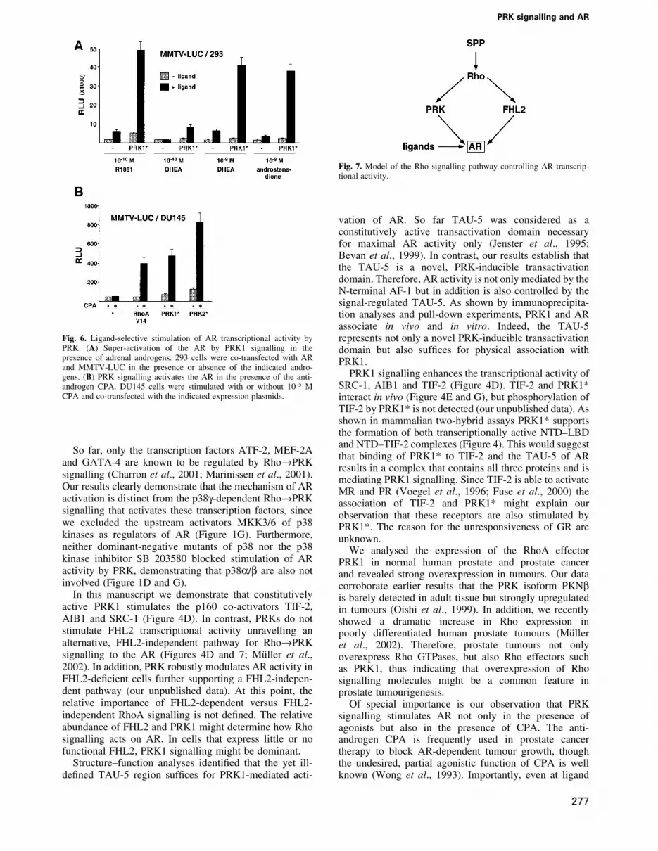
Finally, we analysed whether stimulation of AR tran-scriptional activity by Rho®PRK signalling is regulatedby clinically relevant AR ligands. During testicularandrogen ablation therapy, adrenal androgens present inthe peripheral blood play an important role in AR-dependent prostate cancer progression, since adrenalglands continue to supply the adrenal androgens dehydro-epiandrosterone (DHEA) and androstenedione. Theseandrogen metabolites exhibit weak agonist activity, butare metabolized into potent agonists such as dihydro-testosterone in the prostate (Straub et al., 1998; Gregoryet al., 2001). Thus, we tested whether PRK1 signallingwould be able to stimulate AR activity at physiologicalconcentrations of the adrenal androgens DHEA andandrostenedione in 293 cells that do not metabolizeandrogens. At concentrations of 10±10 M DHEA or10±8 M androstenedione, respectively, AR is barelyactivated by these weak androgens (Figure 6A).Importantly, however, PRK1 signalling strongly inducesAR activity in the presence of the adrenal androgens
(Figure 6A). We then tested the anti-androgen CPA, whichis commonly used during androgen ablation therapy toblock the effects of androgens (Leewansangtong andCrawford, 1998). As expected, CPA at 10±5 M onlymarginally activates the AR (Figure 6B). However, PRKsignalling potently stimulates AR activity in the presenceof the antagonist CPA. Combined with the observedoverexpression of PRK in prostate tumours (Figure 5)these results might, at least in part, provide an explanationfor the clinically well-documented observation (Jenster,1999) that during prostate cancer progression AR-dependent gene expression is upregulated in the absenceof testicular androgens and even in the presence ofantagonists.
Discussion
Here we have identi®ed a novel signalling pathway thatlinks the RhoA effectors PRK1 and PRK2 to thetranscriptional activation of the AR (Figure 7). Thecombined use of either the PRK inhibitors Ro31-8220 orHA 1077, or dominant-negative and constitutively activemutants of PRK1 show that PRK is the Rho effectorleading to AR-dependent activation. Since the PRKinhibitor Ro31-8220 also blocks GSK3b, S6K, RSK,MSK and PKCa activity (Davies et al., 2000), weexcluded that these kinases mediate the regulation of ARby PRK signalling. Furthermore, by ruling out other majorsignalling pathways we demonstrate speci®city for PRKs(Figure 1D, E and G).
Fig. 5. Overexpression of PRK1 in human prostate cancer. Little PRK1immunoreactivity is detected in the secretory epithelium of normalprostate (A, arrowheads), but strong PRK1 immunoreactivity is detec-ted in prostate tumours (B, arrowheads). AR immunostaining revealsthe presence of AR in benign secretory cells of the gland (C, arrow-heads), as well as in carcinoma cells (D, arrowheads). All sectionswere taken from the same radical prostatectomy specimen.Magni®cation: 3250. (E) Normal human prostate and prostate tumourextracts were analysed in western blots using an a-PRK1 antibody.
E.Metzger et al.
276
So far, only the transcription factors ATF-2, MEF-2Aand GATA-4 are known to be regulated by Rho®PRKsignalling (Charron et al., 2001; Marinissen et al., 2001).Our results clearly demonstrate that the mechanism of ARactivation is distinct from the p38g-dependent Rho®PRKsignalling that activates these transcription factors, sincewe excluded the upstream activators MKK3/6 of p38kinases as regulators of AR (Figure 1G). Furthermore,neither dominant-negative mutants of p38 nor the p38kinase inhibitor SB 203580 blocked stimulation of ARactivity by PRK, demonstrating that p38a/b are also notinvolved (Figure 1D and G).
In this manuscript we demonstrate that constitutivelyactive PRK1 stimulates the p160 co-activators TIF-2,AIB1 and SRC-1 (Figure 4D). In contrast, PRKs do notstimulate FHL2 transcriptional activity unravelling analternative, FHL2-independent pathway for Rho®PRKsignalling to the AR (Figures 4D and 7; MuÈller et al.,2002). In addition, PRK robustly modulates AR activity inFHL2-de®cient cells further supporting a FHL2-indepen-dent pathway (our unpublished data). At this point, therelative importance of FHL2-dependent versus FHL2-independent RhoA signalling is not de®ned. The relativeabundance of FHL2 and PRK1 might determine how Rhosignalling acts on AR. In cells that express little or nofunctional FHL2, PRK1 signalling might be dominant.
Structure±function analyses identi®ed that the yet ill-de®ned TAU-5 region suf®ces for PRK1-mediated acti-
vation of AR. So far TAU-5 was considered as aconstitutively active transactivation domain necessaryfor maximal AR activity only (Jenster et al., 1995;Bevan et al., 1999). In contrast, our results establish thatthe TAU-5 is a novel, PRK-inducible transactivationdomain. Therefore, AR activity is not only mediated by theN-terminal AF-1 but in addition is also controlled by thesignal-regulated TAU-5. As shown by immunoprecipita-tion analyses and pull-down experiments, PRK1 and ARassociate in vivo and in vitro. Indeed, the TAU-5represents not only a novel PRK-inducible transactivationdomain but also suf®ces for physical association withPRK1.
PRK1 signalling enhances the transcriptional activity ofSRC-1, AIB1 and TIF-2 (Figure 4D). TIF-2 and PRK1*interact in vivo (Figure 4E and G), but phosphorylation ofTIF-2 by PRK1* is not detected (our unpublished data). Asshown in mammalian two-hybrid assays PRK1* supportsthe formation of both transcriptionally active NTD±LBDand NTD±TIF-2 complexes (Figure 4). This would suggestthat binding of PRK1* to TIF-2 and the TAU-5 of ARresults in a complex that contains all three proteins and ismediating PRK1 signalling. Since TIF-2 is able to activateMR and PR (Voegel et al., 1996; Fuse et al., 2000) theassociation of TIF-2 and PRK1* might explain ourobservation that these receptors are also stimulated byPRK1*. The reason for the unresponsiveness of GR areunknown.
We analysed the expression of the RhoA effectorPRK1 in normal human prostate and prostate cancerand revealed strong overexpression in tumours. Our datacorroborate earlier results that the PRK isoform PKNbis barely detected in adult tissue but strongly upregulatedin tumours (Oishi et al., 1999). In addition, we recentlyshowed a dramatic increase in Rho expression inpoorly differentiated human prostate tumours (MuÈlleret al., 2002). Therefore, prostate tumours not onlyoverexpress Rho GTPases, but also Rho effectors suchas PRK1, thus indicating that overexpression of Rhosignalling molecules might be a common feature inprostate tumourigenesis.
Of special importance is our observation that PRKsignalling stimulates AR not only in the presence ofagonists but also in the presence of CPA. The anti-androgen CPA is frequently used in prostate cancertherapy to block AR-dependent tumour growth, thoughthe undesired, partial agonistic function of CPA is wellknown (Wong et al., 1993). Importantly, even at ligand
Fig. 7. Model of the Rho signalling pathway controlling AR transcrip-tional activity.
Fig. 6. Ligand-selective stimulation of AR transcriptional activity byPRK. (A) Super-activation of the AR by PRK1 signalling in thepresence of adrenal androgens. 293 cells were co-transfected with ARand MMTV-LUC in the presence or absence of the indicated andro-gens. (B) PRK signalling activates the AR in the presence of the anti-androgen CPA. DU145 cells were stimulated with or without 10±5 MCPA and co-transfected with the indicated expression plasmids.
PRK signalling and AR
277

concentrations where the antagonist CPA exhibits nopartial agonistic function PRK signalling potently stimu-lates AR transcriptional activity (Figure 6B). Duringtesticular androgen ablation therapy the concentrations ofadrenal androgens such as DHEA and androstenedione inthe peripheral blood of human males is of importance tocontrol the growth of prostate tumours. Our data now showthat AR is strongly stimulated by PRK1 signalling in thepresence of physiological concentrations of DHEA andandrostenedione. Prostate tumours not only overexpressco-activators such as TIF-2 and FHL2, and Rho GTPases,but also show a dramatic increase in PRK expression(Figure 5; Gregory et al., 2001; MuÈller et al., 2002).Therefore, our results provide, at least in part, anexplanation for the clinically well documented observationthat during prostate cancer progression AR-dependentgene expression is upregulated in the absence of testicularandrogens or even in the presence of antagonists such asCPA. Consequently, PRK might be a promising thera-peutic target and inhibitors of PRK signalling may turn outto be bene®cial in the treatment of prostate cancer.
Materials and methods
PlasmidsThe following plasmids were described previously: AR, PR, MR, GR,Gal-TIF-2, Gal-SRC-1, Gal-FHL2, CMX-Flag, CMX-GST, MMTV-LUC, PB-LUC, TK-LUC and G5E1b-LUC (MuÈller et al., 2000); RhoAV14, Rac1 N17, C3, Cdc42 N17, Ras N17, LIMK1, LIMK2, RhoA N19,PRK2* (tr.PRK2wt), NET1 DN and Gal-AIB1 (MuÈller et al., 2002);PRK1.N (PKN.N) (Sahai et al., 1998); PRK1* (mycDNPRK1) andPRK1wt (Flynn et al., 1998); dnPRK1(PRK1 K644E) (Takahashi et al.,1998); dnPKCa (PKCaK368R) (UÈ berall et al., 1997); dnp38g[p38g(AF)], MKK3E(b) and MKK6E(b) (Wang et al., 2000); ROCK*(ROKa1±543) (Leung et al., 1996); GST-AR-NTD (Alen et al., 1999);MUT1 to MUT5 (pAR5, 106, 126, 99 and 113), Gal-TAU-5 (G106)(Jenster et al., 1995); VP16-AR-NTD, Gal-AR-LBD and pFlag-AR(Ikonen et al., 1997). dnPRK2 (PRK2 KD1) was kindly provided byB.L.Quillian, Indianapolis; GST-TAU-5 (GST±AR 360±546), GFP-MR,GR-GFP and ARDNLS (pSG5-ARD612±633) by A.C.Cato, Karlsruhe;GST-RORb by E.Greiner, Heidelberg; GST-GCNF by H.Greschik,Strasbourg. To construct CMX-GST-AR-DBD, CMX-GST-AR-LBD,and pGEX4T1-AR-LBD the corresponding fragments (AR-DBD: aminoacids 539±623; AR-LBD: amino acids 624±919) were PCR ampli®ed andinserted at the BamHI site of CMX-GST and pGEX4T1 (Pharmacia). Allplasmids were veri®ed by double-stranded sequencing.
TransfectionsDU145 and 293 cells were cultured in DMEM and PC3-AR in Ham'sF-12 supplemented with 10% double-stripped fetal calf serum (dsFCS).Transient transfection assays were carried out in 12-well plates (1 3 105
cells per well) as described previously (MuÈller et al., 2002). Aftertransfection, cells were cultured in DMEM or Ham's F-12 supplementedwith 0.5% dsFCS. The total amount of transfected DNA was keptconstant (4 mg) by adding the corresponding amounts of empty expressionplasmids and pUC18. The following amounts per well were used: 500 ngreporter plasmids MMTV-LUC, PB-LUC or G5E1b-LUC; 25 ngexpression plasmids for AR, PR, ARDNLS, GFP-MR, GR-GFP, GR,MR, dnPKCa, PRK1*, PRK2*, PRK1wt, VP16-AR-NTD or Gal-AR-LBD; 10 ng expression plasmids for Gal-AIB1, Gal-SRC-1, Gal-TIF-2 or Gal-FHL2; 12.5 ng expression plasmids for MKK3E(b),MKK6E(b), or ROCK*. One hundred nanogrammes of all otherexpression plasmids were transfected per well. Chemicals were obtainedas indicated: PD 98059, SB 203580, SPP (BioMol); Ro31-8220 (AlexisBiochemicals); HA 1077 (Calbiochem); R5020, aldosterone, dexametha-sone, DHEA and androstenedione (Sigma); R1881 and CPA (ScheringAG, Berlin); rapamycin (ICN Biomedicals Inc.). All chemicals wereapplied for a total of 20 h except PC3-AR cells, which were treated for 8 h.Luciferase activity was assayed as described previously (MuÈller et al.,2002). All experiments were repeated at least ®ve times.
Immuno¯uorescenceTransfected cells were analysed essentially as described previously(MuÈller et al., 2002). Cells were seeded on coverslips coated with®bronectin and gelatin. Primary antibody staining was performed with theindicated dilutions: a-AR (N-20) (1:4000; Santa Cruz), a-myc (1:10 000;Santa Cruz) and a-PRK1 (1:2000; Transduction Lab). Subcellularlocalization was visualized using secondary Alexa Fluor 488- and 546-labelled antibodies (1:4000; Molecular Probes). Nuclei were stained with1 mg/ml DAPI (Roche).
Co-immunoprecipitation assays and western blot analyses293 cells were transfected in 15 cm dishes with 10 mg of PRK1wt,PRK1*, Flag-AR or CMX-Flag and cultured in the presence of 10±10 MR1881 as indicated. Total cell extract was prepared in the presence orabsence of 10±10 M R1881 in IP buffer (50 mM Tris±HCl pH 8.0, 170 mMNaCl, 0.1% NP-40, 20% glycerol, 50 mM NaF, 2 mM NaV, 0.2 mMDTT, 1 mg/ml BSA, 0.1 mM Pefabloc). Extracts from normal humanprostate and prostate tumour tissue were prepared in the presence of10±10 M R1881 in IP buffer. Following centrifugation, pre-clearedsupernatants were incubated for 2 h with M2 a-Flag antibody (Sigma),a-TIF-2 (Transduction Lab), or a-PRK1 antibody (Transduction Lab)and GammaBindÔ±Sepharose 4B (Pharmacia) in IP buffer. Precipitatedprotein complexes were subsequently washed four times either in thepresence or absence of 10±10 M R1881 and subsequently analysed on 10%SDS polyacrylamide gels. Western blots were decorated with M2, a-AR441 (Santa Cruz), a-TIF-2, a-PRK1 or a-cyclinA (control) antibodies.Secondary antibody and chemoluminescence procedure was performedaccording to the manufacturer's instructions (Amersham).
In vivo GST pull-down assaysFor in vivo GST pull-down experiments, 293 cells grown in 15 cm disheswere co-transfected with 10 mg of expression vectors for GST, GST-AR-NTD, GST-AR-DBD, GST-AR-LBD, GST-GCNF, myc-taggedPRK1* or PRK1wt. Cells were grown for 18 h in DMEM containing0.5% dsFCS with or without 10±10 M R1881. Whole-cell extracts wereprepared by repeated freeze and thaw cycles either in NENT75-Mo buffer(20 mM Tris pH 6.8, 75 mM NaCl, 1 mM EDTA, 0.1% NP-40, 25%glycerol, 20 mM Na-molybdate) for cells transfected with myc-DNPRK1or in buffer B1 (20 mM HEPES±KOH pH 7.7, 500 mM KCl, 25 mMMgCl2, 100 mM EDTA pH 8.0, 10 mM DTT, 0.15% NP-40) for cellstransfected with PRK1wt. GST fusion proteins were immobilized onglutathione±Sepharose (Pharmacia) for 1 h at 4°C. After washing, boundproteins were analysed by SDS±PAGE, followed by immunodetectionwith a-myc or a-PRK1 antibodies. Equal expression of GST fusionproteins was controlled in western blots using an a-GST antibody. Allantibodies were obtained from Santa Cruz.
In vitro GST pull-down assaysExpression of GST fusion proteins (Pharmacia) and the coupled in vitrotranscription±translation reaction (Promega) were performed accordingto the manufacturer's instructions. GST pull-down assays were performedas previously described (MuÈller et al., 2000) using buffer containing250 mM KCl. Ten per cent of the in vitro translated proteins were loadedas input.
ImmunohistochemistryStainings were performed using a protocol for antigen retrieval andindirect immunoperoxidase as described previously (MuÈller et al., 2000).a-PRK1 antibody (Transduction Lab) was used at a dilution of 1:50, anti-mouse IgG (1:500; Dako) was used as secondary antibody andimmunoreactions were visualized with the ABC-complex diluted 1:50in PBS (Vectastain, Vector).
Supplementary dataSupplementary data are available at The EMBO Journal Online.
Acknowledgements
This work was supported by grants from the DeutscheForschungsgemeinschaft (SFB 388) and the Schering AG, Berlin to RS.We thank G.Baier, A.O.Brinkmann, M.Brown, A.C.Cato, F.Claessens,J.Han, K.S.Erdmann, O.A.Janne, L.Lim, Y.Ono, J.Palvimo, P.J.Parker,B.L.Quilliam, A.Soler, R.Treisman and U.Wetterauer for generouslyproviding reagents and support, the members of the SchuÈle lab especiallyPhilip Hublitz and Thomas GuÈnther for fruitful discussion. Special thanksto Ellen Paggen for excellent technical assistance.
E.Metzger et al.
278

References
Alen,P., Claessens,F., Verhoeven,G., Rombauts,W. and Peeters,B.(1999) The androgen receptor amino-terminal domain plays a keyrole in p160 coactivator-stimulated gene transcription. Mol. Cell.Biol., 19, 6085±6097.
Bar-Sagi,D. and Hall,A. (2000) Ras and Rho GTPases: a family reunion.Cell, 103, 227±238.
Bevan,C.L., Hoare,S., Claessens,F., Heery,D.M. and Parker,M.G. (1999)The AF1 and AF2 domains of the androgen receptor interact withdistinct regions of SRC1. Mol. Cell. Biol., 19, 8383±8392.
Bishop,A.L. and Hall,A. (2000) Rho GTPases and their effector proteins.Biochem. J., 348, 241±255.
Busch,C. and Aktories,K. (2000) Microbial toxins and the glycosylationof rho family GTPases. Curr. Opin. Struct. Biol., 10, 528±535.
Cato,A.C. and Peterzierl,H. (1998) The androgen receptor as mediator ofgene expression and signal transduction pathways. Trends Endocrinol.Metab., 9, 150±154.
Cerione,R.A. and Zheng,Y. (1996) The Dbl family of oncogenes. Curr.Opin. Cell Biol., 8, 216±222.
Charron,F., Tsimiklis,G., Arcand,M., Robitaille,L., Liang,Q., Molkentin,J.D., Meloche,S. and Nemer,M. (2001) Tissue-speci®c GATA factorsare transcriptional effectors of the small GTPase RhoA. Genes Dev.,15, 2702±2719.
Chiariello,M., Marinissen,M.J. and Gutkind,J.S. (2001) Regulation ofc-myc expression by PDGF through Rho GTPases. Nat. Cell Biol., 3,580±586.
Culig,Z., Hobisch,A., Cronauer,M.V., Radmayr,C., Trapman,J.,Hittmair,A., Bartsch,G. and Klocker,H. (1994) Androgen receptoractivation in prostatic tumor cell lines by insulin-like growth factor-I,keratinocyte growth factor, and epidermal growth factor. Cancer Res.,54, 5474±5478.
Davies,S.P., Reddy,H., Caivano,M. and Cohen,P. (2000) Speci®city andmechanism of action of some commonly used protein kinaseinhibitors. Biochem. J., 351, 95±105.
Flynn,P., Mellor,H., Palmer,R., Panayotou,G. and Parker,P.J. (1998)Multiple interactions of PRK1 with RhoA. Functional assignment ofthe Hr1 repeat motif. J. Biol. Chem., 273, 2698±2705.
Font de Mora,J. and Brown,M. (2000) AIB1 is a conduit for kinase-mediated growth factor signaling to the estrogen receptor. Mol. Cell.Biol., 20, 5041±5047.
Fuse,H., Kitagawa,H. and Kato,S. (2000) Characterization oftransactivational property and coactivator mediation of ratmineralocorticoid receptor activation function-1 (AF-1). Mol.Endocrinol., 14, 889±899.
Gregory,C.W., Hamil,K.G., Kim,D., Hall,S.H., Pretlow,T.G., Mohler,J.L. and French,F.S. (1998) Androgen receptor expression inandrogen-independent prostate cancer is associated with increasedexpression of androgen-regulated genes. Cancer Res., 58, 5718±5724.
Gregory,C.W., He,B., Johnson,R.T., Ford,O.H., Mohler,J.L., French,F.S.and Wilson,E.M. (2001) A mechanism for androgen receptor-mediated prostate cancer recurrence after androgen deprivationtherapy. Cancer Res., 61, 4315±4319.
Hall,A. (1998) Rho GTPases and the actin cytoskeleton. Science, 279,509±514.
He,B., Kemppainen,J.A. and Wilson,E.M. (2000) FXXLF and WXXLFsequences mediate the NH2-terminal interaction with the ligandbinding domain of the androgen receptor. J. Biol. Chem., 275,22986±22994.
Ikonen,T., Palvimo,J.J. and Janne,O.A. (1997) Interaction between theamino- and carboxyl-terminal regions of the rat androgen receptormodulates transcriptional activity and is in¯uenced by nuclearreceptor coactivators. J. Biol. Chem., 272, 29821±29828.
Jenster,G. (1999) The role of the androgen receptor in the developmentand progression of prostate cancer. Semin. Oncol., 26, 407±421.
Jenster,G., van der Korput,H.A., Trapman,J. and Brinkmann,A.O. (1995)Identi®cation of two transcription activation units in the N-terminaldomain of the human androgen receptor. J. Biol. Chem., 270,7341±7346.
Leav,I., Lau,K.M., Adams,J.Y., McNea,J.E., Taplin,M.E., Wang,J.,Singh,H. and Ho,S.M. (2001) Comparative studies of the estrogenreceptors b and a and the androgen receptor in normal human prostateglands, dysplasia, and in primary and metastatic carcinoma. Am. J.Pathol., 159, 79±92.
Leewansangtong,S. and Crawford,E.D. (1998) Maximal androgen
withdrawal for prostate cancer therapy: current status and futurepotential. Endocr.-Related Cancer, 5, 325±339.
Leung,T., Chen,X.Q., Manser,E. and Lim,L. (1996) The p160 RhoA-binding kinase ROK a is a member of a kinase family and is involvedin the reorganization of the cytoskeleton. Mol. Cell. Biol., 16,5313±5327.
Mangelsdorf,D.J. et al. (1995) The nuclear receptor superfamily: thesecond decade. Cell, 83, 835±839.
Marinissen,M.J., Chiariello,M. and Gutkind,J.S. (2001) Regulation ofgene expression by the small GTPase Rho through the ERK6 (p38g)MAP kinase pathway. Genes Dev., 15, 535±553.
Mukai,H., Mori,K., Takanaga,H., Kitagawa,M., Shibata,H.,Shimakawa,M., Miyahara,M. and Ono,Y. (1995) Xenopus PKN:cloning and sequencing of the cDNA and identi®cation of conserveddomains. Biochim. Biophys. Acta, 1261, 296±300.
MuÈller,J.M. et al. (2000) FHL2, a novel tissue-speci®c coactivator of theandrogen receptor. EMBO J., 19, 359±369.
MuÈller,J.M., Metzger,E., Greschik,H., Bosserhoff,A.K., Mercep,L.,Buettner,R. and SchuÈle,R. (2002) The transcriptional coactivatorFHL2 transmits Rho signals from the cell membrane into the nucleus.EMBO J., 21, 736±748.
Oishi,K., Mukai,H., Shibata,H., Takahashi,M. and Ona,Y. (1999)Identi®cation and characterization of PKNb, a novel isoform ofprotein kinase PKN: expression and arachidonic acid dependency aredifferent from those of PKNa. Biochem. Biophys. Res. Commun., 261,808±814.
Palmer,R.H., Ridden,J. and Parker,P.J. (1995) Cloning and expressionpatterns of two members of a novel protein-kinase-C-related kinasefamily. Eur. J. Biochem., 227, 344±351.
Poukka,H., Karvonen,U., Yoshikawa,N., Tanaka,H., Palvimo,J.J. andJanne,O.A. (2000) The RING ®nger protein SNURF modulatesnuclear traf®cking of the androgen receptor. J. Cell Sci., 113,2991±3001.
Pyne,S. and Pyne,N. (2000) Sphingosine 1-phosphate signalling via theendothelial differentiation gene family of G-protein-coupled receptors.Pharmacol. Ther., 88, 115±131.
Renaud,J.P. and Moras,D. (2000) Structural studies on nuclear receptors.Cell. Mol. Life Sci., 57, 1748±1769.
Rowan,B.G., Garrison,N., Weigel,N.L. and O`Malley,B.W. (2000)8-Bromo-cyclic AMP induces phosphorylation of two sites inSRC-1 that facilitate ligand-independent activation of the chickenprogesterone receptor and are critical for functional cooperationbetween SRC-1 and CREB binding protein. Mol. Cell. Biol., 20,8720±8730.
Sahai,E., Alberts,A.S. and Treisman,R. (1998) RhoA effector mutantsreveal distinct effector pathways for cytoskeletal reorganization, SRFactivation and transformation. EMBO J., 17, 1350±1361.
SchuÈle,R., Rangarajan,P., Kliewer,S., Ransone,L.J., Bolado,J., Yang,N.,Verma,I.M. and Evans,R.M. (1990) Functional antagonism betweenoncoprotein c-Jun and the glucocorticoid receptor. Cell, 62,1217±1226.
Shibata,H., Oda,H., Mukai,H., Oishi,K., Misaki,K., Ohkubo,H. andOno,Y. (1999) Interaction of PKN with a neuron-speci®c basichelix±loop±helix transcription factor, NDRF/NeuroD2. Brain Res.Mol. Brain Res., 74, 126±134.
Straub,R.H., Konecna,L., Hrach,S., Rothe,G., Kreutz,M., Scholmerich,J.,Falk,W. and Lang,B. (1998) Serum dehydroepiandrosterone (DHEA)and DHEA sulfate are negatively correlated with serum interleukin-6(IL-6), and DHEA inhibits IL-6 secretion from mononuclear cells inman in vitro: possible link between endocrinosenescence andimmunosenescence. J. Clin. Endocrinol. Metab., 83, 2012±2017.
Takahashi,M., Mukai,H., Toshimori,M., Miyamoto,M. and Ono,Y.(1998) Proteolytic activation of PKN by caspase-3 or relatedprotease during apoptosis. Proc. Natl Acad. Sci. USA, 95,11566±11571.
Takanaga,H., Mukai,H., Shibata,H., Toshimori,M. and Ono,Y. (1998)PKN interacts with a paraneoplastic cerebellar degeneration-associated antigen, which is a potential transcription factor. Exp.Cell Res., 241, 363±372.
Trapman,J. and Brinkmann,A.O. (1996) The androgen receptor inprostate cancer. Pathol. Res. Pract., 192, 752±760.
UÈ berall,F., Giselbrecht,S., Hellbert,K., Fresser,F., Bauer,B.,Gschwendt,M., Grunicke,H.H. and Baier,G. (1997) ConventionalPKC-a, novel PKC-e and PKC-t, but not atypical PKC-l areMARCKS kinases in intact NIH 3T3 ®broblasts. J. Biol. Chem.,272, 4072±4078.
Voegel,J.J., Heine,M.J., Zechel,C., Chambon,P. and Gronemeyer,H.
PRK signalling and AR
279

(1996) TIF2, a 160 kDa transcriptional mediator for the ligand-dependent activation function AF-2 of nuclear receptors. EMBO J.,15, 3667±3675.
Wang,X., McGowan,C.H., Zhao,M., He,L., Downey,J.S., Fearns,C.,Wang,Y., Huang,S. and Han,J. (2000) Involvement of the MKK6-p38gcascade in g-radiation-induced cell cycle arrest. Mol. Cell. Biol., 20,4543±4552.
Wong,C.I., Zhou,Z.X., Sar,M. and Wilson,E.M. (1993) Steroidrequirement for androgen receptor dimerization and DNA binding.Modulation by intramolecular interactions between the NH2-terminaland steroid-binding domains. J. Biol. Chem., 268, 19004±19012.
Received June 19, 2002; revised November 6, 2002;accepted November 14, 2002
E.Metzger et al.
280


