Embed Size (px)
Citation preview
Transactivation of Schizosaccharomyces pombecdt2þ stimulates a Pcu4–Ddb1–CSN ubiquitinligase
Cong Liu1, Marius Poitelea1,Adam Watson1, Shu-hei Yoshida2,Chikashi Shimoda2, Christian Holmberg3,Olaf Nielsen3 and Antony M Carr1,*1Genome Damage and Stability Centre, University of Sussex, Brighton,UK, 2Department of Biology, Graduate School of Science, Osaka CityUniversity, Osaka, Japan and 3Department of Genetics, University ofCopenhagen, Copenhagen K, Denmark
Cullin-4 forms a scaffold for multiple ubiquitin ligases.
In Schizosaccharomyces pombe, the Cullin-4 homologue
(Pcu4) physically associates with Ddb1 and the COP9
signalosome (CSN). One target of this complex is Spd1.
Spd1 regulates ribonucleotide reductase (RNR) activity.
Spd1 degradation during S phase, or following DNA damage
of G2 cells, results in the nuclear export of the small RNR
subunit. We demonstrate that Cdt2, an unstable WD40
protein, is a regulatory subunit of Pcu4–Ddb1–CSN ubi-
quitin ligase. cdt2 deletion stabilises Spd1 and prevents
relocalisation of the small RNR subunit from the nucleus
to the cytoplasm. cdt2þ is periodically transcribed by the
Cdc10/DSC1 transcription factor during S phase and tran-
siently transcribed following DNA damage of G2 cells,
corresponding to Spd1 degradation profiles. Cdt2 co-pre-
cipitates with Spd1, and Cdt2 overexpression results in
constitutive Spd1 degradation. We propose that Cdt2
incorporation into the Pcu4–Ddb1–CSN complex prompts
Spd1 targeting and subsequent degradation and that
Cdt2 is a WD40 repeat adaptor protein for Cullin-
4-based ubiquitin ligase.
The EMBO Journal (2005) 24, 3940–3951. doi:10.1038/
sj.emboj.7600854; Published online 27 October 2005
Subject Categories: cell cycle; genome stability & dynamics
Keywords: Cct complex; Cullin; damage checkpoint;
ribonucleotide reductase; signalosome
Introduction
Genes required for DNA synthesis are periodically tran-
scribed during the mitotic cell cycle, peaking at G1–S. The
Schizosaccharomyces pombe G1/S transition is coordinated by
the DSC1 (DNA Synthesis Control or MBF factor) transcrip-
tion complex. DSC1 contains the cdc10, rep1, rep2, res1 and
res2 gene products and recognises the MCB (MluI cell cycle
box) DNA sequence (White et al, 2001). cdc10 loss-of-function
mutants have profound effects on MCB-dependent transcrip-
tion (Hofmann and Beach, 1994). Most known Cdc10-depen-
dent target genes are directly involved in DNA replication.
These include genes encoding the replication initiation fac-
tors Cdc18 and Cdt1 and enzymatic functions required for
DNA synthesis such as Cdc22, the large subunit of ribo-
nucleotide reductase (RNR).
RNR catalyses the rate-limiting step for dNTP production
and is required for replicative DNA synthesis and DNA repair
(Elledge et al, 1992). RNR is a heterotetramer formed by two
large subunits (R1 subunit; encoded by cdc22R1 in S. pombe)
and two small subunits (R2 subunit; encoded by suc22R2).
RNR activity is tightly regulated to maintain the balanced
dNTP pools necessary for high-fidelity DNA synthesis. In
Saccharomyces cerevisiae, RNR gene expression is under
tight cell cycle control and separate genes encoding stress-
specific subunits are induced after DNA damage via the
MEC1/RAD53 pathway (Elledge et al, 1993). An additional
level of control over RNR activity in S. cerevisiae is exerted
by Sml1, which binds to and inhibits the function of the R1
subunit (Chabes et al, 1999). In order to allow replication,
Sml1 is degraded in S phase in a MEC1/RAD53-dependent
manner (Zhao et al, 1998). MEC1/RAD53-dependent Sml1
degradation accounts for the essential function of the
S. cerevisiae checkpoint pathway. In fission yeast, the MEC1
homologue rad3þ is not essential and there is currently no
evidence for an R1 subunit inhibitor.
A novel mode of RNR regulation by subcellular compart-
mentalisation was recently uncovered and is conserved
between S. pombe (Liu et al, 2003) and S. cerevisiae (Yao
et al, 2003). In S. pombe, when RNR activity is not required
(e.g. G2 cells), the small subunit (Suc22R2) is found in the
nucleus whereas the large subunit (Cdc22R1) is largely cyto-
plasmic. When RNR activity is required (e.g. DNA replication
or DNA repair synthesis), Suc22R2 translocates to the cyto-
plasm where it is presumed to engage with Cdc22R1 to form
active RNR (Liu et al, 2003). Suc22R2 localisation to the
nucleus is dependent on the presence of the 124 aa protein
Spd1, a potential Suc22R2 nuclear anchor. At the start of each
S phase, Spd1 is degraded by the ubiquitin–proteasome
system (UPS). Periodic degradation of Spd1 in S phase does
not require rad3þ (MEC1 homologue) activity and is check-
point pathway independent. In contrast, Spd1 degradation in
G2 after DNA damage requires the rad3/chk1 DNA damage
checkpoint (Liu et al, 2003). Both cell cycle and damage-
induced Spd1 degradation are mediated by Spd1 ubiquityla-
tion, which requires components of the COP9 signalosome
complex (CSN), the S. pombe Cullin-4 homologue (Pcu4) and
the Ddb1 protein (Liu et al, 2003; Holmberg et al, 2005).
The UPS is the primary mechanism for regulated degrada-
tion of cellular proteins (Hershko and Ciechanover, 1998).
Targeting of specific substrates to UPS is mediated by E3Received: 11 March 2005; accepted: 7 October 2005; publishedonline: 27 October 2005
*Corresponding author. Genome Damage and Stability Centre,University of Sussex, Falmer, Brighton BN1 9RQ, UK.Tel.: þ 44 1273 678122; Fax þ 44 1273 678121;E-mail: [email protected]
The EMBO Journal (2005) 24, 3940–3951 | & 2005 European Molecular Biology Organization | All Rights Reserved 0261-4189/05
www.embojournal.org
The EMBO Journal VOL 24 | NO 22 | 2005 &2005 European Molecular Biology Organization
EMBO
THE
EMBOJOURNAL
THE
EMBOJOURNAL
3940
ubiquitin ligases. Multiple ubiquitin ligase families exist that
can be broadly categorised into three classes: HECT domain
E3’s, single subunit RING E3’s and multi-subunit RING E3’s
(Pickart, 2001). Multi-subunit RING E3’s are often exempli-
fied by SCF (Skp1–Cullin–F box) complexes, a collection of
RING ubiquitin ligases assembled on a common cullin-1
scaffold. CUL1-based SCF complexes have a modular design;
one end of cullin binds Skp1, which in turn recruits one of
many different F-box proteins to act as a substrate adaptor.
The other end of cullin binds Rbx1, a RING finger protein that
recruits an E2 enzyme (Deshaies, 1999). Unlike CUL1, the
CUL4A and B proteins do not have Skp1-binding motifs.
However, CUL4A associates with DDB1 (Damaged DNA-
binding protein) as well as WD40 repeat proteins and has
been proposed to scaffold a subfamily of SCF-like E3 com-
plexes (Groisman et al, 2003; Wertz et al, 2004).
CUL4A–DDB1 ubiquitin ligases play roles in gene expres-
sion and DNA replication by selectively degrading target
proteins (including c-JUN and CDT1; Higa et al, 2003;
Wertz et al, 2004) and were recently found tightly associated
with CSN (Groisman et al, 2003; Liu et al, 2003). CSN consists
of eight subunits (Csn1–8) that share one-to-one similarity to
the 19S regulatory lid complex of the 26S proteasome (Wei
and Deng, 2003). CSN functions are pleiotropic, which can be
attributed to interactions with multiple cullins and thus
multiple cullin-based ubiquitin ligases. CSN recruits several
enzymatic activities to cullin-based E3’s including Csn5/
Jab1-dependent deneddylase and the Ubp12 deubiquitinase
(Cope et al, 2002; Zhou et al, 2003). The phenotypic con-
sequences of CSN loss reflect this pleiotropism and include
cellular and developmental defects (Chamovitz et al, 1996;
Schwechheimer et al, 2001) as well as DNA damage response
defects (Mundt et al, 1999; Groisman et al, 2003).
We previously identified S. pombe csn1 and csn2 null
mutants as DNA damage sensitive and delayed in DNA
replication (Mundt et al, 1999). Null mutants corresponding
to other CSN subunits do not share these phenotypes, in-
dicating a novel function for these two components (Mundt
et al, 2002). Purification of fission yeast CSN identified Ddb1
and Pcu4, but not other cullins, as strongly associated
proteins (Liu et al, 2003). pcu4 and ddb1 null mutants
share the DNA damage sensitivity and S-phase defects of
csn1 and csn2 mutants. These phenotypes correlate with
defective Spd1 degradation (Liu et al, 2003; Holmberg et al,
2005). A Pcu4–Ddb1–CSN ubiquitin ligase thus catalyses
Spd1 polyubiquitylation in S phase and in response to DNA
damage. Here we report that the WD40 repeat protein Cdt2
(Cdc10-dependent transcript 2) functions as the adaptor
protein and key activator to target Spd1 to the Pcu4–Ddb1–
CSN ubiquitin ligase. We show that Pcu4–Ddb1–CSN activity
against Spd1 is largely dependent on the regulation of cdt2þ
gene expression.
Results
Cdt2 copurified with Pcu4–CSN protein complex
Csn2-TAP affinity purification identified six CSN subunits
and Pcu4–Ddb1 at Bstoichiometric concentrations (Liu
et al, 2003). Analysis of several repeat mass spectrometry
data sets revealed one minor band generated p12 peptides
with masses matching the predicted fragmentation of Cdt2
(a WD repeat protein of 55.058 kDa) (32.4% coverage). These
data suggested Cdt2 as a potential Csn2-TAP interactor.
Concomitantly, a cdt2 mutant was found to display DNA
replication and repair defects reminiscent of phenotypes
associated with csn1, csn2, ddb1 and pcu4 deletion
(Yoshida et al, 2003).
As judged from Spd1 degradation profiles, the Pcu4–Ddb1–
CSN ubiquitin ligase is activated during unperturbed S phase
in a checkpoint-independent manner and can be activated
by the DNA damage checkpoint when G2 cells suffer DNA
damage. No link has been established between the Pcu4–
Ddb1–CSN complex and potential activating signals. Because
the cdt2 transcript and gene product accumulate specifically
in S phase (Yoshida et al, 2003), we investigated if Cdt2
regulates Spd1 degradation by activating Pcu4–Ddb1–CSN.
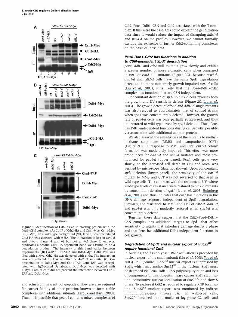
First, we confirmed the Cdt2 interaction with Pcu4–Ddb1–
CSN using immunoprecipitation (IP). cdt2-HA csn1-Myc cells
were arrested in S phase (4 h, 20 mM hydroxyurea (HU))
to induce Cdt2 and extracts were prepared. Cdt2-HA co-
immunoprecipitated (co-IPed) specifically with Csn1-Myc
(Figure 1A, lane 3). This interaction was csn2 and ddb1
dependent (lanes 4 and 6, respectively). The csn5 null mutant
does not share the replication and repair phenotypes asso-
ciated with loss of csn1 and csn2 (Mundt et al, 2002) and,
consistent with this, the interaction was not csn5 dependent
(lane 5). We next examined the interaction between Cdt2-HA
and Ddb1-Myc (Figure 1B). Cdt2-HA co-IPed specifically with
Ddb1-Myc. This was not dependent on any CSN subunits or
pcu4. Thus, Pcu4 does not bridge the Cdt2-HA–Ddb1-Myc
interaction. The data suggest either that Ddb1 and Cdt2 bind
directly to each other or that their interaction is mediated by
unidentified bridging factor(s). We also find that the Ddb1-
Myc Csn1-TAP interaction is independent of Cdt2 (Figure 1C).
Together, these results are compatible with Cdt2 interacting
with Ddb1 and assembling with Pcu4 and CSN to form a
multi-subunit ubiquitin ligase.
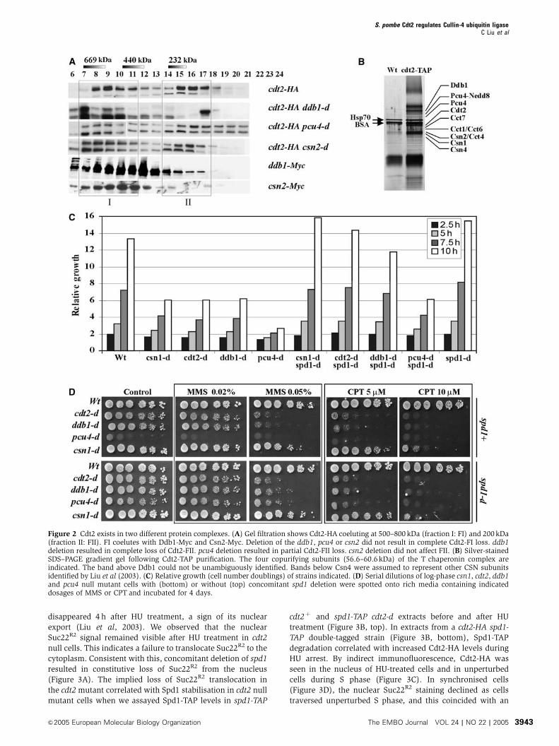
Cdt2 co-precipitates with the T complex
Size-exclusion chromatography (Figure 2A) indicated that
Cdt2-HA fractionates as two peaks: B400–800 kDa (I) and
B200 kDa (II). Peak I overlapped with Ddb1 and Csn2
proteins, consistent with a Cdt2–Ddb1–CSN complex. Peak
II overlapped with Ddb1, but did not coelute with Csn2.
Deletion of ddb1 disrupted peak II and deletion of pcu4
reduced its intensity. Loss of csn2 did not affect peak II
integrity. This may indicate the presence of Cdt2 complex
containing Ddb1 and possibly Pcu4, but not containing CSN.
In all the deletion backgrounds tested, peak I, although
sometimes reduced in intensity, was not lost. This could
be explained if Cdt2 associates with additional protein
complexes.
To identify further Cdt2-interacting proteins, we purified
Cdt2-TAP. HU-arrested (4 h, 20 mM) cells were used to in-
crease Cdt2 levels in the extract. Following TAP purification,
Coomassie staining bands from SDS–PAGE (Figure 2B) were
identified by MALDI-TOF mass spectrometry. Cdt2-TAP
copurified with Pcu4–Ddb1–CSN components, as expected.
No other cullin or cullin-associated proteins were identified.
However, four T complex subunits (Cct proteins: Chaperonin
containing TCP-1) were identified. The Tcomplex is a group II
eukaryotic cytosolic chaperonin composed of eight homolo-
gous subunits of similar size (Spiess et al, 2004). Cct proteins
are essential because they function to correctly fold tubulin
S. pombe Cdt2 regulates Cullin-4 ubiquitin ligaseC Liu et al
&2005 European Molecular Biology Organization The EMBO Journal VOL 24 | NO 22 | 2005 3941
and actin from nascent polypeptides. They are also required
for correct folding of other proteins known to form stable
complexes with additional subunits (Leroux and Hartl, 2000).
Thus, it is possible that peak I contains mixed complexes of
Cdt2–Pcu4–Ddb1–CSN and Cdt2 associated with the T com-
plex. If this were the case, this could explain the gel filtration
data since it would reduce the impact of disrupting ddb1-d
and pcu4-d on the profiles. However, we cannot formally
exclude the existence of further Cdt2-containing complexes
on the basis of these data.
Pcu4–Ddb1–Cdt2 has functions in addition
to CSN-dependent Spd1 degradation
pcu4, ddb1 and cdt2 null mutants grow slowly and exhibit
a greater number of more elongated cells when compared
to csn1 or csn2 null mutants (Figure 2C). Because pcu4-d,
ddb1-d and cdt2-d cells have the same Spd1 degradation
defect as the more moderately growth-impaired csn1-d cells
(Liu et al, 2003), it is likely that the Pcu4–Ddb1–Cdt2
complex has functions that are CSN independent.
Concomitant deletion of spd1 in csn1-d cells reverses both
the growth and UV sensitivity defects (Figure 2C; Liu et al,
2003). The growth defect of cdt2-d and ddb1-d single mutants
was also rescued to approximately that of control strains
when spd1 was concomitantly deleted. However, the growth
rate of pcu4-d cells was only partially suppressed, and thus
not restored to wild-type levels by spd1 deletion. Thus, Pcu4
has Ddb1-independent functions during cell growth, possibly
via association with additional adaptor proteins.
We also assayed the sensitivities of the mutants to methyl-
methane sulphonate (MMS) and camptothecin (CPT)
(Figure 2D). In response to MMS and CPT, csn1-d colony
formation was moderately impaired. This effect was more
pronounced for ddb1-d and cdt2-d mutants and most pro-
nounced for pcu4-d (upper panel). Pcu4 cells grow very
slowly, so the increased cell death in CPT and MMS was
verified by microscopy (data not shown). Upon concomitant
spd1 deletion (lower panel), the sensitivity of the csn1-d
mutant to MMS and CPT was not reversed to that seen in
wild-type cells. This contrasts with the response to UV, where
wild-type levels of resistance were restored to csn1-d mutants
by concomitant deletion of spd1 (Liu et al, 2003; Holmberg
et al, 2005) and thus indicates that csn1 has functions in the
DNA damage response independent of Spd1 degradation.
Similarly, the resistance to MMS and CPT of cdt2-d, ddb1-d
and pcu4-d was only modestly restored when spd1-d was
concomitantly deleted.
Together, these data suggest that the Cdt2–Pcu4–Ddb1–
CSN complex has additional targets to Spd1 that affect
sensitivity to agents that introduce damage during S phase
and that Pcu4 has additional Ddb1-independent functions in
cell growth.
Degradation of Spd1 and nuclear export of Suc22R2
require functional Cdt2
In budding and fission yeast, RNR activation is preceded by
nuclear export of the small subunit (Liu et al, 2003; Yao et al,
2003). In S. pombe, Suc22R2 nuclear export is suppressed by
Spd1, which may anchor Suc22R2 in the nucleus. Spd1 must
be degraded via Pcu4–Ddb1–CSN polyubiquitylation and loss
of components of this ubiquitin ligase causes Spd1 stabilisa-
tion, constitutive nuclear localisation of Suc22R2 and slow S
phase. To explore if Cdt2 is required to regulate RNR localisa-
tion, Suc22R2 nuclear export was monitored by indirect
immunofluorescence (Figure 3A). In wild-type cells,
Suc22R2 localised in the nuclei of log-phase G2 cells and
Figure 1 Identification of Cdt2 as an interacting protein with thePcu4–CSN complex. (A) Co-IP of Cdt2-HA and Csn1-Myc. Csn1-MycIP (a-Myc): In a wild-type background (Wt, lane 3), co-precipitatedCdt2-HA was detected with a-HA. The interaction is lost in csn2-dand ddb1-d (lanes 4 and 6) but not csn5-d (lane 5) extracts.*Indicates a second Cdt2-HA-dependent band we assume to be adegradation product. The intensity of this band varies betweenexperiments. (B) Co-IP of Cdt2-HA and Ddb1-Myc. Ddb1-Myc wasIPed with a-Myc. Cdt2-HA was detected with a-HA. The interactionwas not affected by loss of other Pcu4–CSN subunits. (C) Co-precipitation of Ddb1-Myc and Csn1-TAP. Csn1-TAP was precipi-tated with IgG-coated Dynabeads. Ddb1-Myc was detected witha-Myc. Loss of cdt2 did not prevent the interaction between Csn1-TAP and Ddb1-Myc.
S. pombe Cdt2 regulates Cullin-4 ubiquitin ligaseC Liu et al
The EMBO Journal VOL 24 | NO 22 | 2005 &2005 European Molecular Biology Organization3942
disappeared 4 h after HU treatment, a sign of its nuclear
export (Liu et al, 2003). We observed that the nuclear
Suc22R2 signal remained visible after HU treatment in cdt2
null cells. This indicates a failure to translocate Suc22R2 to the
cytoplasm. Consistent with this, concomitant deletion of spd1
resulted in constitutive loss of Suc22R2 from the nucleus
(Figure 3A). The implied loss of Suc22R2 translocation in
the cdt2 mutant correlated with Spd1 stabilisation in cdt2 null
mutant cells when we assayed Spd1-TAP levels in spd1-TAP
cdt2þ and spd1-TAP cdt2-d extracts before and after HU
treatment (Figure 3B, top). In extracts from a cdt2-HA spd1-
TAP double-tagged strain (Figure 3B, bottom), Spd1-TAP
degradation correlated with increased Cdt2-HA levels during
HU arrest. By indirect immunofluorescence, Cdt2-HA was
seen in the nucleus of HU-treated cells and in unperturbed
cells during S phase (Figure 3C). In synchronised cells
(Figure 3D), the nuclear Suc22R2 staining declined as cells
traversed unperturbed S phase, and this coincided with an
Figure 2 Cdt2 exists in two different protein complexes. (A) Gel filtration shows Cdt2-HA coeluting at 500–800 kDa (fraction I: FI) and 200 kDa(fraction II: FII). FI coelutes with Ddb1-Myc and Csn2-Myc. Deletion of the ddb1, pcu4 or csn2 did not result in complete Cdt2-FI loss. ddb1deletion resulted in complete loss of Cdt2-FII. pcu4 deletion resulted in partial Cdt2-FII loss. csn2 deletion did not affect FII. (B) Silver-stainedSDS–PAGE gradient gel following Cdt2-TAP purification. The four copurifying subunits (56.6–60.6 kDa) of the T chaperonin complex areindicated. The band above Ddb1 could not be unambiguously identified. Bands below Csn4 were assumed to represent other CSN subunitsidentified by Liu et al (2003). (C) Relative growth (cell number doublings) of strains indicated. (D) Serial dilutions of log-phase csn1, cdt2, ddb1and pcu4 null mutant cells with (bottom) or without (top) concomitant spd1 deletion were spotted onto rich media containing indicateddosages of MMS or CPT and incubated for 4 days.
S. pombe Cdt2 regulates Cullin-4 ubiquitin ligaseC Liu et al
&2005 European Molecular Biology Organization The EMBO Journal VOL 24 | NO 22 | 2005 3943
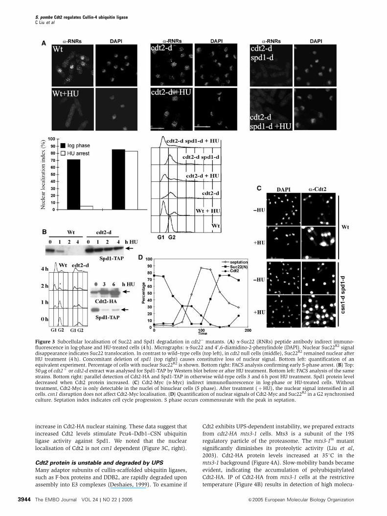
increase in Cdt2-HA nuclear staining. These data suggest that
increased Cdt2 levels stimulate Pcu4–Ddb1–CSN ubiquitin
ligase activity against Spd1. We noted that the nuclear
localisation of Cdt2 is not csn1 dependent (Figure 3C, right).
Cdt2 protein is unstable and degraded by UPS
Many adaptor subunits of cullin-scaffolded ubiquitin ligases,
such as F-box proteins and DDB2, are rapidly degraded upon
assembly into E3 complexes (Deshaies, 1999). To examine if
Cdt2 exhibits UPS-dependent instability, we prepared extracts
from cdt2-HA mts3-1 cells. Mts3 is a subunit of the 19S
regulatory particle of the proteasome. The mts3-1ts mutant
significantly diminishes its proteolytic activity (Liu et al,
2003). Cdt2-HA protein levels increased at 351C in the
mts3-1 background (Figure 4A). Slow-mobility bands became
evident, indicating the accumulation of polyubiquitylated
Cdt2-HA. IP of Cdt2-HA from mts3-1 cells at the restrictive
temperature (Figure 4B) results in detection of high molecu-
Figure 3 Subcellular localisation of Suc22 and Spd1 degradation in cdt2� mutants. (A) a-Suc22 (RNRs) peptide antibody indirect immuno-fluorescence in log-phase and HU-treated cells (4 h). Micrographs: a-Suc22 and 40,6-diamidino-2-phenylindole (DAPI). Nuclear Suc22R2 signaldisappearance indicates Suc22 translocation. In contrast to wild–type cells (top left), in cdt2 null cells (middle), Suc22R2 remained nuclear afterHU treatment (4 h). Concomitant deletion of spd1 (top right) causes constitutive loss of nuclear signal. Bottom left: quantification of anequivalent experiment. Percentage of cells with nuclear Suc22R2 is shown. Bottom right: FACS analysis confirming early S-phase arrest. (B) Top:50mg of cdt2þ or cdt2-d extract was analysed for Spd1-TAP by Western blot before or after HU treatment. Bottom left: FACS analysis of the samestrains. Bottom right: parallel detection of Cdt2-HA and Spd1-TAP in otherwise wild-type cells 3 and 6 h post HU treatment. Spd1 protein leveldecreased when Cdt2 protein increased. (C) Cdt2-Myc (a-Myc) indirect immunofluorescence in log-phase or HU-treated cells. Withouttreatment, Cdt2-Myc is only detectable in the nuclei of binuclear cells (S phase). After treatment (þHU), the nuclear signal intensified in allcells. csn1 disruption does not affect Cdt2-Myc localisation. (D) Quantification of nuclear signals of Cdt2-Myc and Suc22R2 in a G2 synchronisedculture. Septation index indicates cell cycle progression. S phase occurs commensurate with the peak in septation.
S. pombe Cdt2 regulates Cullin-4 ubiquitin ligaseC Liu et al
The EMBO Journal VOL 24 | NO 22 | 2005 &2005 European Molecular Biology Organization3944
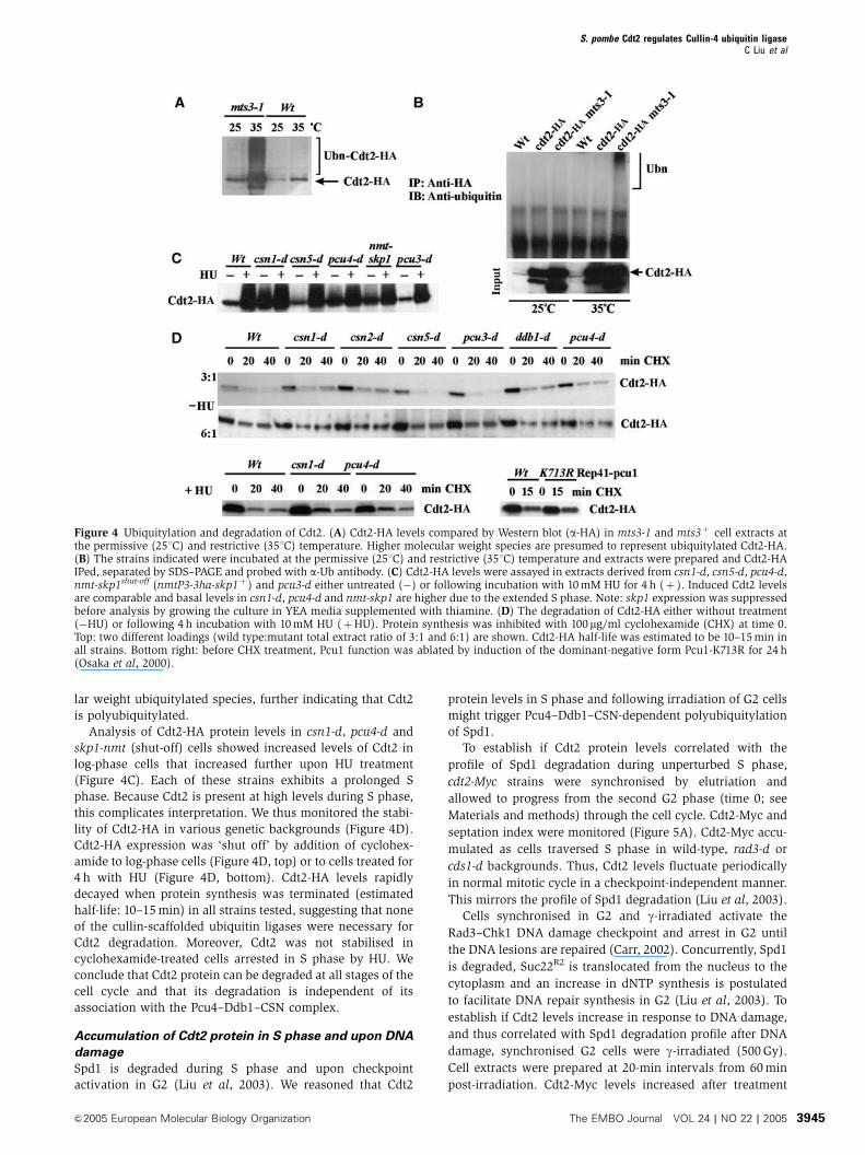
lar weight ubiquitylated species, further indicating that Cdt2
is polyubiquitylated.
Analysis of Cdt2-HA protein levels in csn1-d, pcu4-d and
skp1-nmt (shut-off) cells showed increased levels of Cdt2 in
log-phase cells that increased further upon HU treatment
(Figure 4C). Each of these strains exhibits a prolonged S
phase. Because Cdt2 is present at high levels during S phase,
this complicates interpretation. We thus monitored the stabi-
lity of Cdt2-HA in various genetic backgrounds (Figure 4D).
Cdt2-HA expression was ‘shut off’ by addition of cyclohex-
amide to log-phase cells (Figure 4D, top) or to cells treated for
4 h with HU (Figure 4D, bottom). Cdt2-HA levels rapidly
decayed when protein synthesis was terminated (estimated
half-life: 10–15 min) in all strains tested, suggesting that none
of the cullin-scaffolded ubiquitin ligases were necessary for
Cdt2 degradation. Moreover, Cdt2 was not stabilised in
cyclohexamide-treated cells arrested in S phase by HU. We
conclude that Cdt2 protein can be degraded at all stages of the
cell cycle and that its degradation is independent of its
association with the Pcu4–Ddb1–CSN complex.
Accumulation of Cdt2 protein in S phase and upon DNA
damage
Spd1 is degraded during S phase and upon checkpoint
activation in G2 (Liu et al, 2003). We reasoned that Cdt2
protein levels in S phase and following irradiation of G2 cells
might trigger Pcu4–Ddb1–CSN-dependent polyubiquitylation
of Spd1.
To establish if Cdt2 protein levels correlated with the
profile of Spd1 degradation during unperturbed S phase,
cdt2-Myc strains were synchronised by elutriation and
allowed to progress from the second G2 phase (time 0; see
Materials and methods) through the cell cycle. Cdt2-Myc and
septation index were monitored (Figure 5A). Cdt2-Myc accu-
mulated as cells traversed S phase in wild-type, rad3-d or
cds1-d backgrounds. Thus, Cdt2 levels fluctuate periodically
in normal mitotic cycle in a checkpoint-independent manner.
This mirrors the profile of Spd1 degradation (Liu et al, 2003).
Cells synchronised in G2 and g-irradiated activate the
Rad3–Chk1 DNA damage checkpoint and arrest in G2 until
the DNA lesions are repaired (Carr, 2002). Concurrently, Spd1
is degraded, Suc22R2 is translocated from the nucleus to the
cytoplasm and an increase in dNTP synthesis is postulated
to facilitate DNA repair synthesis in G2 (Liu et al, 2003). To
establish if Cdt2 levels increase in response to DNA damage,
and thus correlated with Spd1 degradation profile after DNA
damage, synchronised G2 cells were g-irradiated (500 Gy).
Cell extracts were prepared at 20-min intervals from 60 min
post-irradiation. Cdt2-Myc levels increased after treatment
Figure 4 Ubiquitylation and degradation of Cdt2. (A) Cdt2-HA levels compared by Western blot (a-HA) in mts3-1 and mts3þ cell extracts atthe permissive (251C) and restrictive (351C) temperature. Higher molecular weight species are presumed to represent ubiquitylated Cdt2-HA.(B) The strains indicated were incubated at the permissive (251C) and restrictive (351C) temperature and extracts were prepared and Cdt2-HAIPed, separated by SDS–PAGE and probed with a-Ub antibody. (C) Cdt2-HA levels were assayed in extracts derived from csn1-d, csn5-d, pcu4-d,nmt-skp1shut-off (nmtP3-3ha-skp1þ ) and pcu3-d either untreated (�) or following incubation with 10 mM HU for 4 h (þ ). Induced Cdt2 levelsare comparable and basal levels in csn1-d, pcu4-d and nmt-skp1 are higher due to the extended S phase. Note: skp1 expression was suppressedbefore analysis by growing the culture in YEA media supplemented with thiamine. (D) The degradation of Cdt2-HA either without treatment(�HU) or following 4 h incubation with 10 mM HU (þHU). Protein synthesis was inhibited with 100 mg/ml cyclohexamide (CHX) at time 0.Top: two different loadings (wild type:mutant total extract ratio of 3:1 and 6:1) are shown. Cdt2-HA half-life was estimated to be 10–15 min inall strains. Bottom right: before CHX treatment, Pcu1 function was ablated by induction of the dominant-negative form Pcu1-K713R for 24 h(Osaka et al, 2000).
S. pombe Cdt2 regulates Cullin-4 ubiquitin ligaseC Liu et al
&2005 European Molecular Biology Organization The EMBO Journal VOL 24 | NO 22 | 2005 3945
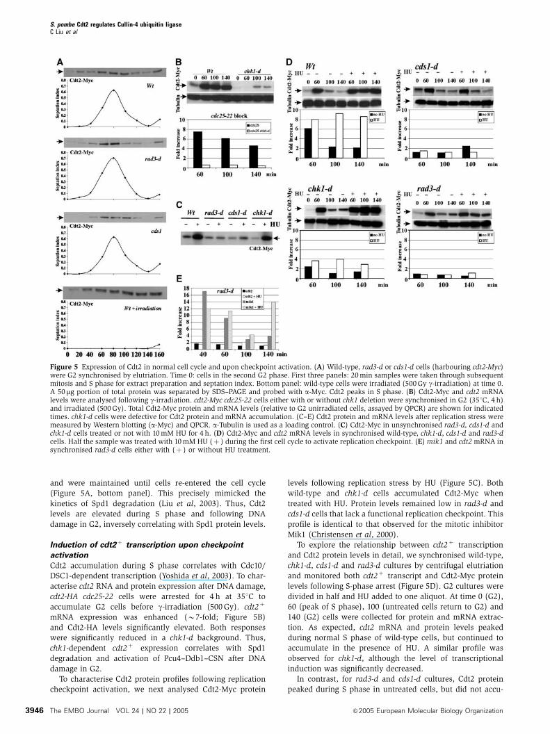
and were maintained until cells re-entered the cell cycle
(Figure 5A, bottom panel). This precisely mimicked the
kinetics of Spd1 degradation (Liu et al, 2003). Thus, Cdt2
levels are elevated during S phase and following DNA
damage in G2, inversely correlating with Spd1 protein levels.
Induction of cdt2þ transcription upon checkpoint
activation
Cdt2 accumulation during S phase correlates with Cdc10/
DSC1-dependent transcription (Yoshida et al, 2003). To char-
acterise cdt2 RNA and protein expression after DNA damage,
cdt2-HA cdc25-22 cells were arrested for 4 h at 351C to
accumulate G2 cells before g-irradiation (500 Gy). cdt2þ
mRNA expression was enhanced (B7-fold; Figure 5B)
and Cdt2-HA levels significantly elevated. Both responses
were significantly reduced in a chk1-d background. Thus,
chk1-dependent cdt2þ expression correlates with Spd1
degradation and activation of Pcu4–Ddb1–CSN after DNA
damage in G2.
To characterise Cdt2 protein profiles following replication
checkpoint activation, we next analysed Cdt2-Myc protein
levels following replication stress by HU (Figure 5C). Both
wild-type and chk1-d cells accumulated Cdt2-Myc when
treated with HU. Protein levels remained low in rad3-d and
cds1-d cells that lack a functional replication checkpoint. This
profile is identical to that observed for the mitotic inhibitor
Mik1 (Christensen et al, 2000).
To explore the relationship between cdt2þ transcription
and Cdt2 protein levels in detail, we synchronised wild-type,
chk1-d, cds1-d and rad3-d cultures by centrifugal elutriation
and monitored both cdt2þ transcript and Cdt2-Myc protein
levels following S-phase arrest (Figure 5D). G2 cultures were
divided in half and HU added to one aliquot. At time 0 (G2),
60 (peak of S phase), 100 (untreated cells return to G2) and
140 (G2) cells were collected for protein and mRNA extrac-
tion. As expected, cdt2 mRNA and protein levels peaked
during normal S phase of wild-type cells, but continued to
accumulate in the presence of HU. A similar profile was
observed for chk1-d, although the level of transcriptional
induction was significantly decreased.
In contrast, for rad3-d and cds1-d cultures, Cdt2 protein
peaked during S phase in untreated cells, but did not accu-
Figure 5 Expression of Cdt2 in normal cell cycle and upon checkpoint activation. (A) Wild-type, rad3-d or cds1-d cells (harbouring cdt2-Myc)were G2 synchronised by elutriation. Time 0: cells in the second G2 phase. First three panels: 20 min samples were taken through subsequentmitosis and S phase for extract preparation and septation index. Bottom panel: wild-type cells were irradiated (500 Gy g-irradiation) at time 0.A 50 mg portion of total protein was separated by SDS–PAGE and probed with a-Myc. Cdt2 peaks in S phase. (B) Cdt2-Myc and cdt2 mRNAlevels were analysed following g-irradiation. cdt2-Myc cdc25-22 cells either with or without chk1 deletion were synchronised in G2 (351C, 4 h)and irradiated (500 Gy). Total Cdt2-Myc protein and mRNA levels (relative to G2 unirradiated cells, assayed by QPCR) are shown for indicatedtimes. chk1-d cells were defective for Cdt2 protein and mRNA accumulation. (C–E) Cdt2 protein and mRNA levels after replication stress weremeasured by Western blotting (a-Myc) and QPCR. a-Tubulin is used as a loading control. (C) Cdt2-Myc in unsynchronised rad3-d, cds1-d andchk1-d cells treated or not with 10 mM HU for 4 h. (D) Cdt2-Myc and cdt2 mRNA levels in synchronised wild-type, chk1-d, cds1-d and rad3-dcells. Half the sample was treated with 10 mM HU (þ ) during the first cell cycle to activate replication checkpoint. (E) mik1 and cdt2 mRNA insynchronised rad3-d cells either with (þ ) or without HU treatment.
S. pombe Cdt2 regulates Cullin-4 ubiquitin ligaseC Liu et al
The EMBO Journal VOL 24 | NO 22 | 2005 &2005 European Molecular Biology Organization3946

mulate following HU treatment (Figure 5D). This behaviour is
similar to the profile we previously reported for Mik1 protein
levels through the cell cycle in similar experiments
(Christensen et al, 2000). However, we observed a significant
difference between the cdt2þ transcript profile and the
mik1þ transcript profile: previous analysis of mik1þ tran-
scription (Christensen et al, 2000) demonstrated that cell
cycle-dependent transcription in unperturbed cells was in-
dependent of the rad3 and cds1 replication checkpoint genes.
In contrast, these data (Figure 5D) and several repeats (not
shown) demonstrate that cdt2þ transcription in unperturbed
S phase is rad3 and cds1 dependent.
Figure 6 Cdt2 is an adaptor protein for Spd1. (A) Top: Rep41(HA-Cdt2) plasmid or vector control was induced by the absence of thiamine(22 h) in spd1-TAP strains shown. Half of each culture was treated (20 mM HU for 4 h). Spd1 levels were monitored by SDS–PAGE and IgGbinding to TAP. a-PSTAIR (Cdc2 peptide antibody) was used as a loading control. Overexpressed Cdt2-HA (a-HA, top) caused Spd1degradation. Overproduction of Cdt2 did not rescue the csn1-d defect for Spd1 degradation (lanes 5 and 6). Bottom left: nuclear signal ofSuc22R2 was quantified in the presence of overexpressed Cdt2 (Rep1 plasmid, 22 h without thiamine). Bottom right: Cdt2-HA overexpressionrescues the slow S-phase phenotype of cdt2-d cells but does not affect cell cycle profiles of wild-type cells. (B) Co-precipitation from extracts ofindicated strains of Cdt2-Myc with Spd1-TAP. Strains contained the csn1-d mutation to prevent Spd1-TAP degradation. We estimated o5% ofCdt2-Myc co-precipitates. (C) Truncated HA-tagged Cdt2 constructs containing different WD40 repeat domains expressed in S. pombe fromRep41-HA. Cdt2 constructs were pulled down from soluble extracts using purified Spd1-GST (B38 kDa) or GST alone (25 kDa). Only Cdt2-Mand Cdt2-C fragments were efficiently pulled down by Spd1-GST. (D) A model of Pcu4–CSN complex containing Cdt2. Cdt2 protein is regulatedby a combination of constant UPS-dependent degradation, cell cycle and checkpoint-driven transcription and an uncharacterised cell cycle-dependent post-transcriptional mechanism. Together with Ddb1, Cdt2 adapts Spd1 to the Pcu4-scaffolded RING-E2 enzyme for ubiquitylation.CSN is obligatory for Pcu4–Ddb1–Cdt2 function, possibly via regulation of neddylation and E2 loading.
S. pombe Cdt2 regulates Cullin-4 ubiquitin ligaseC Liu et al
&2005 European Molecular Biology Organization The EMBO Journal VOL 24 | NO 22 | 2005 3947
To verify this difference, we compared the transcription
profiles of cdt2þ and mik1þ in the same synchronous rad3-d
culture (Figure 5E). In contrast with the steady expression
level of cdt2þ , mik1þ mRNA level displayed a dramatic
increase upon S-phase entry in rad3-d cells (Figure 5E).
These results suggest that the rad3þ -dependent checkpoint
pathway plays an additional role in controlling the expression
of cdt2þ during unperturbed cell cycle progression as well as
during checkpoint activation. Because the S-phase profile of
cdt2þ transcription and Cdt2 protein levels are uncoupled
in unperturbed cell cycles in the rad3 mutant background
(cf. Figure 5A and E), we also infer that an unknown post-
transcriptional mechanism coexists with the transcriptional
control of cdt2þ to ensure that the Cdt2 protein levels
oscillate during the normal cell cycle.
Cdt2 is the adaptor protein for Spd1
cdt2 is required for Spd1 degradation and Cdt2 protein
accumulation correlates with Spd1 degradation. However,
the role of Cdt2 as a Pcu4–Ddb1–CSN activator remains
elusive. To determine if the simple presence of Cdt2 results
in Spd1 degradation, cdt2 was transcriptionally induced
from the nmt promoter. Rep41(HA-Cdt2) was introduced
into spd1-TAP cdt2-d and spd1-TAP csn1-d cells and cdt2
transcript was induced by removal of thiamine from the
media, both with and without HU treatment. Spd1-TAP
protein levels were monitored by Western blot. The presence
of the cdt2-expresing plasmid resulted in lower levels of
Spd1-TAP protein, irrespective of HU treatment (Figure 6A,
lanes 3 and 4). Even basal level of Cdt2 expression from
the plasmid (in the presence of thiamine) was sufficient
to promote Spd1 degradation (data not shown). Over-
production of Cdt2 mimicked the situation seen in HU-treated
cells (cf. lanes 2 and 3) but did not significantly affect cell
cycle progression profiles (Figure 6A, bottom right): Spd1-
TAP levels were already low and were not further decreased
by HU treatment (lane 4). However, Cdt2 expression did
not overcome the Spd1 degradation defect of csn1-d cells
(lanes 5 and 6). As expected, overexpression of Cdt2 reduced
nuclear localisation of Suc22R2 (Figure 6A, bottom left).
Thus, the abundance of Cdt2 protein determines, at least in
part, the activation of Pcu4–Ddb1–CSN ubiquitin ligase
against Spd1.
To demonstrate that Cdt2 can interact with Spd1, Cdt2-Myc
was tested for co-precipitation with Spd1-TAP. This experi-
ment was performed in csn1-d background to ensure that
Spd1 remained stable in the presence of HU-induced Cdt2. A
significant amount of Cdt2-Myc protein co-precipitated with
Spd1-TAP (Figure 6B, lane 6). No signal was detected in
control strains (lanes 4 and 5). To define an interaction
domain of Cdt2 for Spd1, extracts from cells expressing
truncated HA-Cdt2 fragments were probed with recombinant
GST-Spd1 or GST protein as control. Only HA-Cdt2 middle
and C-terminal fragments containing the third and fourth
WD40 repeats specifically bound to GST-Spd1 beads
(Figure 6C).
Discussion
In this work, we have identified the WD repeat protein Cdt2
as an adaptor protein for a Pcu4–Ddb1 ubiquitin ligase that
directs the degradation of the RNR inhibitor Spd1. We provide
evidence that Cdt2 protein level determines (at least in part)
Spd1 degradation. Cdt2 protein levels fluctuate during un-
perturbed S phase in a manner independent of the DNA
structure checkpoint pathway. In contrast, DNA damage
induces Cdt2 levels in G2 cells in a manner that is dependent
on the DNA damage checkpoint.
The Pcu4–Ddb1–CSN–Cdt2 protein complex
Pcu4 is the closest S. pombe protein homologous to human
CUL4A. All characterised CUL4A-scaffolded ubiquitin ligases
are associated with DDB1 and one or more WD40 repeat
proteins such as DDB2, CSA or COP1 (Groisman et al, 2003;
Liu et al, 2003; Wertz et al, 2004). The best-characterised
CUL4A-scaffolded ubiquitin ligase is the DCXDET1�COP1 com-
plex (DDB1–CUL4A–X-box). Its proposed architecture
conforms to the SCF paradigm, with an unknown motif
in DET1 (termed the ‘X-box’) permitting association of
a dimeric substrate adaptor (DET1–COP1) with DDB1. The
C-terminal WD40 repeats of COP1 then function to recruit
substrates including c-JUN and other bZIP transcription
factors (Wertz et al, 2004). The architecture of the Spd1-
specific Pcu4-scaffolded E3 is consistent with the DCX model:
Ddb1 is required for the interaction between the Cdt2
‘substrate adaptor’ and the cullin (Pcu4). Cdt2 recruits
the target protein Spd1 for ubiquitylation, possibly via
WD40 repeats (Figure 6C). However, no S. pombe DET1
analogue copurified with Cdt2 and we believe it unlikely
(but it cannot be formally excluded) that Cdt2 associates
with a secondary adaptor (i.e. a DET1-like protein). This is
consistent with characterisation of the human DCXCSA and
DCXDDB2 complexes (Groisman et al, 2003), each of which
contained DDB1, CUL4A, a putative WD repeat substrate
adaptor and CSN, but not DET1. Possibly, the ‘X-box’ is
contained within CSA and DDB2 proteins or the WD40
repeats may bind both substrates and DDB1. Indeed, multiple
mechanisms of adaptor protein recruitment may be available
to DDB1.
The role of Pcu4–Ddb1–CSN ubiquitin ligase
The CSN complex interacts with several cullins in human
cells and is required to regulate their deneddylation.
However, CSN plays a novel role in association with cullin-
4-scaffolded E3’s. In S. pombe, Pcu4 and CSN specifically
copurify and CSN is essential for Pcu4-dependent ubiquityla-
tion of Spd1, implying a positive regulatory function.
Conversely, biochemical data suggest a negative regulatory
role for human CSN in CUL4A-dependent ubiquitylation
(Groisman et al, 2003). Resolution of this conundrum awaits
further experimentation.
Current models, based on the interaction with other cullins
(Wu et al, 2002), suggest that CSN attaches to the Pcu4
C-terminus and that this is important for regulating
Pcu4 neddylation status, which in turn facilitates E2 loading
onto Rbx1/Pip1 within the Pcu4 complex. This suggests
that the substrate recognition by the Pcu4–Ddb1–CSN
complex is mediated by Cdt2 protein association with
Ddb1. We can exclude that CSN regulates the association
of Cdt2 with Pcu4–Ddb1–CSN complex because Cdt2 levels,
subcellular localisation and association with Ddb1 do not
require csn1 or csn2.
Substrate degradation by CUL1-scaffolded ubiquitin ligases
(SCF complexes) is regulated by substrate phosphorylation,
S. pombe Cdt2 regulates Cullin-4 ubiquitin ligaseC Liu et al
The EMBO Journal VOL 24 | NO 22 | 2005 &2005 European Molecular Biology Organization3948
which promotes substrate binding to the F-box substrate
adaptor subunits. Despite extensive analysis (unpublished
data), we have found no evidence for Spd1 phosphorylation
and since simple overexpression of Cdt2 can mimic HU-
induced Spd1 degradation (Figure 6A), we conclude that
Cdt2 protein abundance is the key step in activating Pcu4–
Ddb1–CSN ubiquitin ligase against Spd1.
Cdt2 protein abundance is regulated, in part, through
cdt2þ transcription and in part through an unknown post-
transcriptional mechanism. This mechanism may dictate the
translation efficiency of the cdt2 message. The Cdt2 protein
itself is unstable and this instability is not cell cycle regulated.
Uncovering what factors are required for Cdt2 degradation
(we have shown here that it does not require any cullin-based
ubiquitin ligases or CSN) and what mechanism underlies the
increase in Cdt2 levels during S phase and after DNA damage
will be informative.
The interaction with T complex
Besides actin and tubulin folding, the Cct chaperonin has
been implicated in the correct assembly of several multi-
subunit E3 ligases. The CDC20 and CDH1 subunits of the
anaphase-promoting complex (APC) require Cct proteins for
binding and activation of APC (Camasses et al, 2003). Cct
proteins also mediate the formation of the VHL-elongin BC
ubiquitin ligases and unassembled VHL protein remains
associated with the T complex (Feldman et al, 1999;
Melville et al, 2003). Some tumour-causing mutations inter-
fere with chaperonin association of VHL and thus its incor-
poration into the complex (Feldman et al, 2003).
Furthermore, it has been demonstrated that a group of
WD40 beta-propeller proteins transiently interact with Cct
and require the chaperonin to reach their native folded state
(Siegers et al, 2003). Thus, it is perhaps not surprising that
this chaperonin associates with Cdt2 in S. pombe. Most likely,
Cct proteins facilitate Cdt2 folding and/or assembly with the
Pcu4–Ddb1–CSN scaffold. This additional association of Cdt2
protein with Cct proteins may explain why, during gel filtra-
tion, a large Cdt2-containing protein complex remains
(Figure 2, peak I) in the absence of ddb1 or pcu4. However,
we cannot formally exclude the presence of other Cdt2-
interacting proteins in peak I.
rad3þ -dependent checkpoints and cdt2 regulation
We demonstrate that increased Cdt2 abundance in S phase
and after DNA damage drives Spd1 degradation, subsequent
Suc22R2 cytoplasmic relocalisation and RNR activation.
cdt2þ is a Cdc10/DSC target during S phase and transcription
is also under the control of the chk1þ -dependent checkpoint
pathways following DNA damage in G2. The rad3þ–chk1þ
pathway is absolutely required for the cdt2þ messenger and
protein accumulation after DNA damage of G2 cells. The
simplest explanation is that Chk1 phosphorylates and acti-
vates a transcription factor that promotes G2 transcription of
cdt2þ and potentially of other genes. Previously, we identi-
fied several genes that are transcriptionally induced after
DNA damage of G2 cells (Watson et al, 2004). A subset of
these were DNA damage checkpoint dependent. Intriguingly,
this subset included genes previously identified as DSC/
Cdc10 transcription targets at the G1–S transition, including
Cdc22R1, Cdc18 and Cdt2 itself. It is thus possible that Chk1
specifically activates Cdc10/DSC-dependent transcription
when DNA damage is detected outside of S phase in order
to ensure a supply of enzymes and dNTPs for DNA repair
synthesis.
In response to replication arrest by HU, we observed that
the rad3–cds1-dependent replication checkpoint was required
to maintain cdt2þ transcript and Cdt2 protein levels. We have
previously observed that the activation of the Rad3–Cds1-
dependent replication checkpoint resulted in similar accumu-
lation of mik1þ transcript and Mik1 protein (Christensen
et al, 2000). However, we did not find evidence that check-
point directly targeted mik1 expression or Mik1 protein
stability. Instead, we proposed that the replication checkpoint
indirectly led to mik1þ transcript and Mik1 protein accumu-
lation because cells are blocked within S phase, where mik1þ
is transcribed and Mik1 protein is predetermined to be
resistant to degradation. Thus, our observations concerning
Cdt2 do not require an interpretation in which the replication
checkpoint directly controls cdt2þ transcription or Cdt2
protein abundance.
Our analysis of Cdt2 protein stability in S- and G2-phase-
arrested cells suggests that degradation rates do not drama-
tically vary through the cells cycle. We thus propose that
transcription and/or translation determine Cdt2 protein le-
vels. It is possible that Cdt2 accumulates in S-phase-arrested
cells because the S-phase checkpoint directly targets cdt2þ
transcription and/or translation factors. However, the most
parsimonious interpretation of the data is that checkpoint
activity maintains cells within the S-phase compartment of
the cell cycle where cdt2þ is predetermined to be transcribed
and translated.
Unlike our previous findings concerning the lack of de-
pendence of mik1þ transcription on the rad3 and checkpoint
pathway in unperturbed S phase, we identified a requirement
for both rad3þ and cds1þ function for periodic cdt2þ
transcription in unperturbed S phase. However, loss of per-
iodic S-phase transcription did not prevent periodic Cdt2
accumulation. Thus, while transcriptional profiles clearly
contribute to Cdt2 protein profiles, mechanisms additional
to transcription must act to regulate Cdt2 protein level. Why
cdt2þ transcription and mik1þ transcription differ with
respect to rad3–cds1 dependence remains to be established.
Both are Cdc10/DSC dependent, so it must reflect a subtle
aspect of transcriptional control that has not yet been
addressed in S. pombe.
Conclusion
RNR regulation by checkpoint pathways is conserved through
evolution, reflecting the importance of controlling dNTP
pools. Disrupting relative and total dNTP concentrations is
mutagenic (Chabes et al, 2003) and destabilises the genome.
In addition, other metabolic products depend on the correct
nucleotide pool balance. A recent example is the identifica-
tion in S. pombe of interactions between RNR and phthalate
metabolism (Stolz et al, 2004). In both S. pombe and
S. cerevisiae, subcellular compartmentalisation of RNR
subunits is one of several mechanisms contributing to RNR
regulation. Understanding how this conserved pathway
operates and is regulated will inform our understanding of
an aspect of DNA metabolism potentially important in main-
taining genomic stability and which is a target of medically
important pharmaceuticals.
S. pombe Cdt2 regulates Cullin-4 ubiquitin ligaseC Liu et al
&2005 European Molecular Biology Organization The EMBO Journal VOL 24 | NO 22 | 2005 3949
Materials and methods
Plasmid construction and protein expressionspd1 ORF was amplified by PCR from an S. pombe cDNA library andcloned into pGEX-KG. A sequenced clone was transformed intoBL21 (Escherichia coli) and induced with 1 mM IPTG (OD600¼ 0.5;4 h, 371C). GST-Spd1 was extracted, enriched and purified at 41C inNETN buffer (0.5% NP-40; 20 mM Tris, pH 8.0; 100 mM NaCl; 1 mMEDTA; 1 mM PMSF) on glutathione beads.
Genetics, cell biology and centrifugal elutriationStrain nmtP3-3ha-skp1þ was a gift from Dr T Toda. Double mutantswere isolated by genetics, except cdt2 ddb1 as the two genes are9.736 kb apart. Sequential tagging/deletion was used in these cases.Elutriation (JE-5.0, Beckman Coulter): Mid-log cultures were loadedand small G2 cells were collected, harvested and resuspendedin rich medium. Cell cycle progression and septation were followedby staining cells with DAPI and Calcofluor, respectively. In allelutriation experiments, time zero (time 0) was defined as cells thathad been through one complete cycle and entered the second G2after elutriation.
Protein extracts, gel filtration and Western blotsCells were disrupted with glass beads in either HB buffer (25 mMTris–HCl, pH 7.5; 15 mM EGTA, pH 7.5; 15 mM MgCl2; 0.1% NP-40;1 mM DTT) or IP buffer (20 mM Tris–HCl, pH 7.4; 150 mM NaCl;0.5% Triton X-100; 1 mM DTT), supplemented with proteaseinhibitor (Complete EDTA-free, Roche) and 1 mM PMSF. Extractswere precleared (14 000 r.p.m., microfuge). Protein concentrationwas quantified by Bradford assay. For size exclusion, 1 mg ofprotein was passed through a Sephadex 200HR 10/30 column(Pharmacia) and 500ml fractions were collected. To ensuredetectable levels of Cdt2 protein, cells were first incubated in10 mM HU for 4 h.
ImmunoprecipitationPrecleared supernatants were incubated with anti-HA/Myc anti-bodies (1 h, 41C). Immunocomplexes were absorbed onto 20ml
protein G beads (1 h). Beads were pelleted, washed 3� (HB or IPbuffer) and 50ml SDS sample buffer was added before SDS–PAGE.For TAP purification, the protocol was as described (Liu et al, 2003).Elutes were concentrated and resolved in 4–20% gradient SDS–PAGE. Coomassie-stained bands were identified by MALDI-TOF(Yates, 1998).
Immunofluorescent microscopyBriefly, cells were fixed in 3.75% paraformaldehyde in PEM(100 mM PIPES; 1 mM EGTA, pH 6.9; 1 mM MgCl2), permeabilisedwith 1.25 mg/ml zymolase T20 (ICN Biomedicals, Cleveland, OH)and 1% Triton X-100 in PEMþ 1.2 M sorbitol. Proteins werevisualised using TRITC or FITC-conjugated secondary antibodiesin PEMþ 1% BSA, 0.1% NaN3 and 100 mM lysine hydrochloride.
Quantitative PCRTotal RNA (B20 mg) was RQ1 RNAse free DNAse (Promega,Madison, WI) treated and reverse transcribed (Stratascript fromStratagene). Quantitative PCR (QPCR) used BrilQuantiTect SYBRGreen PCR mix (Qiagen). Duplicate samples were run on anMX4000 (Stratagene). Relative transcript quantities were deter-mined using the 2�DDCt formula: Ct¼ cycle at which fluorescence isstatistically above background; DCt¼difference in Ct of the gene ofinterest and Ct of the normaliser gene (cdc2); DDCt¼difference inDCt at time of sampling after synchronisation (time¼ t) and DCt attime¼ 0. Reaction efficiencies for the primers were determined asequivalent using serial S. pombe genomic DNA dilutions.
Acknowledgements
We thank Dr Stead (COGEME proteomics facility, Aberdeen) forinvaluable help with mass spectrometry and Dr Kanji Furuya and DrJo Murray for critical reading of the manuscript. CL was supportedby Cancer Research UK grant C5514. AW and MP were supported byMRC grant G0001129.
References
Camasses A, Bogdanova A, Shevchenko A, Zachariae W (2003) TheCCT chaperonin promotes activation of the anaphase-promotingcomplex through the generation of functional Cdc20. Mol Cell 12:87–100
Carr AM (2002) DNA structure dependent checkpoints as regulatorsof DNA repair. DNA Repair 1: 983–994
Chabes A, Domkin V, Thelander L (1999) Yeast Sml1, a proteininhibitor of ribonucleotide reductase. J Biol Chem 274:36679–36683
Chabes A, Georgieva B, Domkin V, Zhao X, Rothstein R, ThelanderL (2003) Survival of DNA damage in yeast directly depends onincreased dNTP levels allowed by relaxed feedback inhibition ofribonucleotide reductase. Cell 112: 391–401
Chamovitz DA, Wei N, Osterlund MT, von Arnim AG, Staub JM,Matsui M, Deng XW (1996) The COP9 complex, a novel multi-subunit nuclear regulator involved in light control of a plantdevelopmental switch. Cell 86: 115–121
Christensen PU, Bentley NJ, Martinho RG, Nielsen O, Carr AM(2000) Mik1 levels accumulate in S phase and may mediate anintrinsic link between S phase and mitosis. Proc Natl Acad SciUSA 97: 2579–2584
Cope GA, Suh GS, Aravind L, Schwarz SE, Zipursky SL, Koonin EV,Deshaies RJ (2002) Role of predicted metalloprotease motif ofJab1/Csn5 in cleavage of Nedd8 from Cul1. Science 298: 608–611
Deshaies RJ (1999) SCF and Cullin/Ring H2-based ubiquitin ligases.Annu Rev Cell Dev Biol 15: 435–467
Elledge SJ, Zhou Z, Allen JB (1992) Ribonucleotide reductase:regulation, regulation, regulation. Trends Biochem Sci 17: 119–123
Elledge SJ, Zhou Z, Allen JB, Navas TA (1993) DNA damage and cellcycle regulation of ribonucleotide reductase. BioEssays 15: 333–339
Feldman DE, Spiess C, Howard DE, Frydman J (2003) Tumorigenicmutations in VHL disrupt folding in vivo by interfering withchaperonin binding. Mol Cell 12: 1213–1224
Feldman DE, Thulasiraman V, Ferreyra RG, Frydman J (1999)Formation of the VHL–elongin BC tumor suppressor complex ismediated by the chaperonin TRiC. Mol Cell 4: 1051–1061
Groisman R, Polanowska J, Kuraoka I, Sawada J, Saijo M, DrapkinR, Kisselev AF, Tanaka K, Nakatani Y (2003) The ubiquitin ligaseactivity in the DDB2 and CSA complexes is differentially regulatedby the COP9 signalosome in response to DNA damage. Cell 113:357–367
Hershko A, Ciechanover A (1998) The ubiquitin system. Annu RevBiochem 67: 425–479
Higa LA, Mihaylov IS, Banks DP, Zheng J, Zhang H (2003)Radiation-mediated proteolysis of CDT1 by CUL4-ROC1 andCSN complexes constitutes a new checkpoint. Nat Cell Biol 5:1008–1015
Hofmann JF, Beach D (1994) cdt1 is an essential target of the Cdc10/Sct1 transcription factor: requirement for DNA replication andinhibition of mitosis. EMBO J 13: 425–434
Holmberg C, Fleck O, Hansen HA, Liu C, Slaaby R, Carr AM, NielsenO (2005) Ddb1 controls genome stability and meiosis in fissionyeast. Genes Dev 19: 853–862
Leroux MR, Hartl FU (2000) Protein folding: versatility of thecytosolic chaperonin TRiC/CCT. Curr Biol 10: R260–R264
Liu C, Powell KA, Mundt K, Wu L, Carr AM, Caspari T (2003) Cop9/signalosome subunits and Pcu4 regulate ribonucleotide reductaseby both checkpoint-dependent and -independent mechanisms.Genes Dev 17: 1130–1140
Melville MW, McClellan AJ, Meyer AS, Darveau A, Frydman J(2003) The Hsp70 and TRiC/CCT chaperone systems cooperatein vivo to assemble the von Hippel–Lindau tumor suppressorcomplex. Mol Cell Biol 23: 3141–3151
Mundt KE, Liu C, Carr AM (2002) Deletion mutants in COP9/signalosome subunits in fission yeast Schizosaccharomycespombe display distinct phenotypes. Mol Biol Cell 13: 493–502
S. pombe Cdt2 regulates Cullin-4 ubiquitin ligaseC Liu et al
The EMBO Journal VOL 24 | NO 22 | 2005 &2005 European Molecular Biology Organization3950
Mundt KE, Porte J, Murray JM, Brikos C, Christensen PU, Caspari T,Hagan IM, Millar JBA, Simanis V, Hofmann K, Carr AM (1999)The COP9/signalosome is conserved in fission yeast and has arole in S-phase. Curr Biol 9: 1427–1430
Osaka F, Saeki M, Katayama S, Aida N, Toh EA, Kominami K, TodaT, Suzuki T, Chiba T, Tanaka K, Kato S (2000) Covalent modifierNEDD8 is essential for SCF ubiquitin-ligase in fission yeast.EMBO J 19: 3475–3484
Pickart CM (2001) Mechanisms underlying ubiquitination. AnnuRev Biochem 70: 503–533
Schwechheimer C, Serino G, Callis J, Crosby WL, Lyapina S,Deshaies RJ, Gray WM, Estelle M, Deng XW (2001) Interactionsof the COP9 signalosome with the E3 ubiquitin ligase SCFTIRI inmediating auxin response. Science 292: 1379–1382
Siegers K, Bolter B, Schwarz JP, Bottcher UM, Guha S, Hartl FU(2003) TRiC/CCT cooperates with different upstream chaperonesin the folding of distinct protein classes. EMBO J 22: 5230–5240
Spiess C, Meyer AS, Reissmann S, Frydman J (2004) Mechanism ofthe eukaryotic chaperonin: protein folding in the chamber ofsecrets. Trends Cell Biol 14: 598–604
Stolz J, Caspari T, Carr AM, Sauer N (2004) Cell division defects ofSchizosaccharomyces pombe liz1� mutants are caused by defectsin pantothenate uptake. Eukaryot Cell 3: 406–412
Watson A, Mata J, Bahler J, Carr A, Humphrey T (2004) Global geneexpression responses of fission yeast to ionizing radiation. MolBiol Cell 15: 851–860
Wei N, Deng XW (2003) The COP9 signalosome. Annu Rev Cell DevBiol 19: 261–286
Wertz IE, O’Rourke KM, Zhang Z, Dornan D, Arnott D, Deshaies RJ,Dixit VM (2004) Human De-etiolated-1 regulates c-Jun by assem-bling a CUL4A ubiquitin ligase. Science 303: 1371–1374
White S, Khaliq F, Sotiriou S, McInerny CJ (2001) The role of DSC1components cdc10+, rep1+ and rep2+ in MCB gene transcrip-tion at the mitotic G1–S boundary in fission yeast. Curr Genet 40:251–259
Wu K, Chen A, Tan P, Pan ZQ (2002) The Nedd8-conjugated ROC1-CUL1 core ubiquitin ligase utilizes Nedd8 charged surface resi-dues for efficient polyubiquitin chain assembly catalyzed byCdc34. J Biol Chem 277: 516–527
Yao R, Zhang Z, An X, Bucci B, Perlstein DL, Stubbe J, Huang M(2003) Subcellular localization of yeast ribonucleotide reductaseregulated by the DNA replication and damage checkpoint path-ways. Proc Natl Acad Sci USA 100: 6628–6633
Yates III JR (1998) Mass spectrometry and the age of the proteome.J Mass Spectrom 33: 1–19
Yoshida SH, Al-Amodi H, Nakamura T, McInerny CJ, Shimoda C(2003) The Schizosaccharomyces pombe cdt2(+) gene, a target ofG1–S phase-specific transcription factor complex DSC1, is requiredfor mitotic and premeiotic DNA replication. Genetics 164: 881–893
Zhao X, Muller EG, Rothstein R (1998) A suppressor of two essentialcheckpoint genes identifies a novel protein that negatively affectsdNTP pools. Mol Cell 2: 329–340
Zhou C, Wee S, Rhee E, Naumann M, Dubiel W, Wolf DA (2003)Fission yeast COP9/signalosome suppresses cullin activitythrough recruitment of the deubiquitylating enzyme Ubp12p.Mol Cell 11: 927–938
S. pombe Cdt2 regulates Cullin-4 ubiquitin ligaseC Liu et al
&2005 European Molecular Biology Organization The EMBO Journal VOL 24 | NO 22 | 2005 3951


