Embed Size (px)
Citation preview
Molecular Biology of the CellVol. 16, 2003–2017, April 2005
Ace2p Controls the Expression of Genes Required for CellSeparation in Schizosaccharomyces pombe□D
Maria Luisa Alonso-Nunez,* Hanbing An,† Ana Belen Martın-Cuadrado,*Sapna Mehta,† Claudia Petit,† Matthias Sipiczki,‡ Francisco del Rey,*Katheleen L. Gould,† and Carlos R. Vazquez de Aldana*
*Departamento de Microbiologıa y Genetica, Instituto de Microbiologıa Bioquımica, CSIC/Universidad deSalamanca, Campus Miguel de Unamuno, 37007 Salamanca, Spain; †Howard Hughes Medical Institute andDepartment of Cell and Developmental Biology, Vanderbilt University School of Medicine, Nashville, TN37232; and ‡Department of Genetics, University of Debrecen, 4010 Debrecen, Hungary
Submitted June 2, 2004; Revised January 18, 2005; Accepted January 20, 2005Monitoring Editor: Randy Schekman
Schizosaccharomyces pombe cells divide by medial fission through contraction of an actomyosin ring and deposition ofa multilayered division septum that must be cleaved to release the two daughter cells. Here we describe the identificationof seven genes (adg1�, adg2�, adg3�, cfh4�, agn1�, eng1�, and mid2�) whose expression is induced by the transcriptionfactor Ace2p. The expression of all of these genes varied during the cell cycle, maximum transcription being observedduring septation. At least three of these proteins (Eng1p, Agn1p, and Cfh4p) localize to a ring-like structure that surroundsthe septum region during cell separation. Deletion of the previously uncharacterized genes was not lethal to the cells, butproduced defects or delays in cell separation to different extents. Electron microscopic observation of mutant cellsindicated that the most severe defect is found in eng1� agn1� cells, lacking the Eng1p endo-�-1,3-glucanase and the Agn1pendo-�-glucanase. The phenotype of this mutant closely resembled that of ace2� mutants, forming branched chains ofcells. This suggests that these two proteins are the main activities required for cell separation to be completed.
INTRODUCTION
Cytokinesis is the final stage of the cell cycle during whichthe daughter cells separate physically and become two in-dependent entities. In a variety of organisms, the force nec-essary for cell cleavage is provided by an actomyosin-basedcontractile ring, coupled to the synthesis of new membrane,which is inserted at the division site (reviewed by Hales etal., 1999; Robinson and Spudich, 2000; Guertin et al., 2002). Inyeast and fungi, the presence of a rigid cell wall that com-pletely surrounds the cell gives rise to an additional stepduring cytokinesis, because a separation septum needs to beassembled to avoid cell lysis during cytokinesis.
Schizosaccharomyces pombe is an excellent organism tostudy cytokinesis because it divides by medial fission afterassembly and contraction of an actomyosin ring, followedby cleavage of the septum (for a review, see Feierbach andChang, 2001). Cytokinesis and cell division are broughtabout by the action of the actomyosin ring, whose constric-tion is perfectly coordinated with the synthesis of the pri-mary septum. The isolation and characterization of cytoki-nesis mutants has allowed the identification of the manyproteins involved in the different steps of this process. Es-
tablishment of the division site at the center of the cell beginsearly on the cell cycle—during the onset of mitosis—withthe assembly of the contractile ring at the cell cortex adjacentto the nucleus (for a review, see Chang, 2001). The mid1�,plo1�, and pom1� genes are required for the division plane tobe established and for correct positioning of the actomyosinring (Chang and Nurse, 1996; Sohrmann et al., 1996; Bahlerand Pringle, 1998; Bahler et al., 1998a; Mulvihill et al., 1999;Bahler and Nurse, 2001). Coordination of ring contractionand the nuclear cycle requires a network of regulatory pro-teins that are collectively referred to as the Septation Initia-tion Network (SIN). These proteins also control the forma-tion of the primary septum during constriction of theactomyosin ring. Genetic studies have indicated that activa-tion of the SIN pathway might regulate Cps1p, a �-1,3-glucan synthase subunit essential for the assembly of thedivision septum (Le Goff et al., 1999; Liu et al., 2000). Thisseptum has a three-layer structure (Johnson et al., 1982), witha central primary septum (mainly composed of linear �-1,3-glucan) surrounded on both sides by two secondary septa(composed of �-1,6-branched �-1,3-glucan, and �-1,6-glu-can; Humbel et al., 2001).
Cell separation requires the dissolution of the primaryseptum for the daughter cells to become two independententities. On completion of mitosis, the primary septum un-dergoes rapid degradation, accompanied by local erosion ofthe adjacent regions of the cell wall. During the last fewyears, the isolation of mutants affected to different extents incell separation has provided some insight into the mecha-nistic details of this process. Mutants with a cell-cell sepa-ration phenotype include mutations in components of theexocyst complex (sec6�, sec8�, sec10�, and exo70�), an anillin
This article was published online ahead of print in MBC in Press(http://www.molbiolcell.org/cgi/doi/10.1091/mbc.E04–06–0442)on February 2, 2005.□D The online version of this article contains supplemental materialat MBC Online (http://www.molbiolcell.org).
Address correspondence to: Carlos R. Vazquez de Aldana([email protected]).
© 2005 by The American Society for Cell Biology 2003
homologue (mid2�), septins (spn3� and spn4�), an endo-�-1,3-glucanase (agn1�), an endo-�-1,3-glucanase (eng1�), cal-cineurin (ppb1�), a MAPK (pmk1�), a MAPK phosphatase(pmp1�), two transcription factors (sep1� and ace2�), andsubunits of the mediator complex (sep10�, sep11� andsep15�; Sipiczki et al., 1993; Yoshida et al., 1994; Toda et al.,1996; Ribar et al., 1997; Sugiura et al., 1998; Zilahi et al., 2000a;Szilagyi et al., 2002; Wang et al., 2002; Berlin et al., 2003;Martın-Cuadrado et al., 2003; Tasto et al., 2003; Dekker et al.,2004). The exocyst is an octameric protein complex presentin many organisms and is involved in tethering vesicles tospecific sites on the plasma membrane (Wang et al., 2002).Based on the fact that mutants in different subunits show adefect in cell separation, it was proposed that this complexmight be involved in the delivery of the hydrolytic proteinsthat are important for cell cleavage to the septum. One suchenzyme could be the product of the eng1� gene, which codesfor a protein with endo-�-1,3-glucanase activity, which hasbeen shown to be the major enzymatic activity involved inthe dissolution of the primary septum (Martın-Cuadrado etal., 2003). In addition, it has recently been reported that theendo-�-1,3-glucanase Agn1p is also required for cell separa-tion (Dekker et al., 2004). The defect in cell separation pro-duced by eng1� deletion is not very severe but results in theformation of groups of four connected cells. A similar phe-notype has also been reported for mutants lacking mid2�, ananillin homologue that is required for the assembly andstabilization of the septin ring during cytokinesis and forseveral septin mutants (Berlin et al., 2003; Tasto et al., 2003).It has been suggested that Mid2p and septins may be re-quired for proper exocytosis of the enzymes involved inseptum cleavage. Mutation in two transcription factors alsointerferes with cell separation. sep1� encodes a transcriptionfactor highly homologous to the HNF-3/forkhead familypresent in higher eukaryotic cells and also in other micro-organisms (Ribar et al., 1997). ace2�, a homologue of theSaccharomyces cerevisiae Ace2p, codes for a transcription fac-tor of the C2H2 zinc-finger family (Martın-Cuadrado et al.,2003). Deletion of each of these genes results in a severe cellseparation defect, hyphal growth, and branching being ob-served. It has been shown that sep1� controls the expressionof mid2�, whereas the transcription of eng1� is dependent onace2� (Martın-Cuadrado et al., 2003; Tasto et al., 2003). Thefact that the phenotype of sep1� or ace2� mutants is moresevere than that of cells lacking eng1� or mid2� suggests thatthese transcription factors also control the expression ofother genes involved in cell-cell separation.
Here we have identified by microarray analysis a group ofgenes whose expression is dependent on the transcriptionfactor Ace2p and hence is named AceII-dependent genes(adg). Northern analyses revealed that the expression of theadg genes is dependent on Ace2p and that ace2� transcrip-tion requires Sep1p. Mutants lacking the identified genesshowed modest cell separation defects, but a double mutantdevoid of the Eng1p endo-�-1,3-glucanase and the Agn1pendo-1,3-�-glucanase showed a phenotype very similar tothat of ace2� mutants. According to these observations, thesetwo enzymes are likely to be the major enzymatic activitiesinvolved in the dissolution of the septum during cell sepa-ration.
MATERIALS AND METHODS
Strains, Growth Conditions, and Genetic ManipulationsThe S. pombe strains used in this study are listed in Table 1. Yeast cells weregrown on YES medium or minimal media (EMM) with appropriate supple-ments (Moreno et al., 1991). Yeast transformations were performed with the
lithium acetate method (Ito et al., 1983). For overexpression experiments usingthe nmt1� promoter, cells were grown in EMM containing 15 �M thiamine upto the logarithmic phase. Then, the cells were harvested, washed three timeswith EMM, and inoculated in fresh medium (without thiamine) at an OD595� 0.05.
Synchronization of strains carrying the thermosensitive cdc25-22 mutationwas achieved by growing the cells at the permissive temperature (25°C) toearly log phase (OD595 � 0.5) and then shifting the cultures to 37°C for 4 h.Cells were released from arrest by transfer to 25°C, and samples were takenevery 20 min. Cells were also synchronized by centrifugal elutriation in aBeckman JE 5.0 elutriator rotor (Fullerton, CA). Cells synchronized in early G2were collected and inoculated into YE medium at 32°C. Synchrony wasmonitored by DAPI staining and estimation of the percentage of binucleatecells.
Construction of Null Mutants and PlasmidsThe entire coding sequences of adg1� (SPAPJ760.03c), adg2�
(SPAC19G12.16c), and adg3� (SPCC74.07c) were deleted by replacing thecoding sequences with the kanMX4 cassette or the ura4� gene. The deletioncassettes were constructed using the recombinant PCR approach described byWach et al. (1996). For this purpose, DNA fragments of 300–500 base pairscorresponding to the 5� and 3� flanking regions of each gene were PCR-amplified using specific oligonucleotide pairs. The resulting fragments werethen fused by recombinant PCR to the kanMX4 cassette (which confers resis-tance to the antibiotic G418) or to the ura4� gene. The oligonucleotide se-quences used are available upon request.
Plasmid pA16, containing the ace2� ORF without promoter, was con-structed by PCR amplification of the coding sequence with oligonucleotidesthat generated XhoI and SacI sites at the ends and then cloning the amplifiedfragment into the corresponding sites of vector pAU-KS. The wild-type ace2�
promoter was amplified with oligonucleotides that generated KpnI and XhoIsites at the ends and was cloned into the corresponding sites of plasmid pA16,yielding pB1. Deletion of the five copies of the TGTTTAC sequence wasachieved by successive rounds of PCR, cloning the final product between theKpnI and XhoI sites of plasmid pA16, to generate plasmid pC4. The nmt1�
terminator was introduced at the KpnI site of the three plasmids.
Epitope TaggingThe eng1�, cfh4�, agn1�, adg1�, adg2�, and adg3� genes were tagged at theirchromosomal loci at their 3� ends with sequences encoding the green fluo-rescent protein (GFP) by a PCR-mediated strategy as described previously(Bahler et al., 1998b). Proper integration of these epitope cassettes was con-firmed by PCR and immunoblotting.
Microscopy TechniquesMicroscopy was performed with a Leica DMRXA microscope (Deerfield, IL)equipped for Nomarski optics and epifluorescence and photographed with aPhotometrics Sensys CCD camera (Tucson, AZ). For the imaging of theGFP-tagged strains, a Carl Zeiss MicroImaging, Axiovert II inverted micro-scope (Thornwood, NY) equipped with an UltraView LCI real-time scanninghead confocal (PerkinElmer, Norwalk, CT) and a 488-nm argon ion laser (forGFP excitation) was used. Images were captured on an Orca-ER CCD camera(Hamamatsu, Bridgewater, NJ) using Ultra-View software (PerkinElmer) andwere then processed using Volocity 2.0 software (Improvision, Lexington,MA). Visualization of nuclei was accomplished by staining cells fixed in 70%ethanol, washed two times in phosphate-buffered saline (PBS), and resus-pended in PBS with DAPI (4�,6�-diamidino-2-phenylindole).
For transmission electron microscopy (TEM), the cells were stained withpotassium permanganate according to the protocol described by Johnson et al.(1982). Electron photomicrographs were taken with a Jeol Jem-1010 electronmicroscope (Peabody, MA).
Protein MethodsTotal cell extracts of S. pombe were prepared in SDS-lysis buffer (Gould et al.,1991) and immunoprecipitations were carried out using rabbit polyclonalanti-GFP serum. For immunoblotting, proteins were resolved by SDS-PAGEon 4–12% gradient gels. Protein transfer, blotting, and ECL detection wereperformed using standard procedures.
RNA Isolation, Northern Blot, and Microarray AnalysisCells (1.3 � 109) were collected at different time intervals after release fromthe restrictive temperature (37°C) or from different mutant strains, and totalRNA was prepared using the method described by Percival-Smith and Segall(1984). For Northern blot analysis, 12.5 �g of RNA was used. The DNA probesused to detect the different transcripts were DNA fragments obtained by PCRwith specific oligonucleotides. To analyze gene expression in the cdc7 mutant,RNA was isolated by hot acid phenol extraction, as described previously(Burns et al., 1999). RNA, 20 �g, were transferred to a Duralon-UV membraneand probed with 32P-labeled probes corresponding to coding regions ofeng1�, cdc22�, and his3�. The blots were exposed to Phosphor-Imager screens
M. L. Alonso-Nunez et al.
Molecular Biology of the Cell2004
and visualized with ImageQuant 5.2 on an Amersham Biosciences Typhoon9200 scanner (Piscataway, NJ).
For microarray analysis, mRNA was purified from wild-type or ace2�mutants during exponential growth. Hybridization to glass slides containing4976 S. pombe ORFs was performed by Eurogentec (Serain, Belgium).
RESULTS
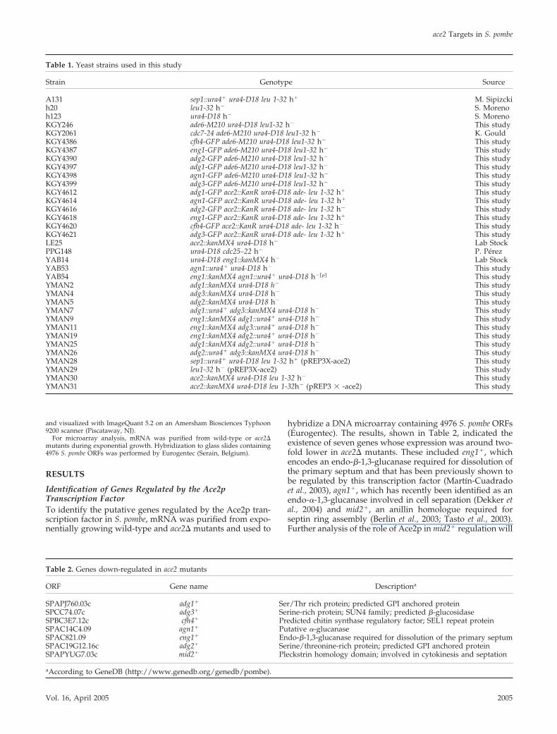
Identification of Genes Regulated by the Ace2pTranscription FactorTo identify the putative genes regulated by the Ace2p tran-scription factor in S. pombe, mRNA was purified from expo-nentially growing wild-type and ace2� mutants and used to
hybridize a DNA microarray containing 4976 S. pombe ORFs(Eurogentec). The results, shown in Table 2, indicated theexistence of seven genes whose expression was around two-fold lower in ace2� mutants. These included eng1�, whichencodes an endo-�-1,3-glucanase required for dissolution ofthe primary septum and that has been previously shown tobe regulated by this transcription factor (Martın-Cuadradoet al., 2003), agn1�, which has recently been identified as anendo-�-1,3-glucanase involved in cell separation (Dekker etal., 2004) and mid2�, an anillin homologue required forseptin ring assembly (Berlin et al., 2003; Tasto et al., 2003).Further analysis of the role of Ace2p in mid2� regulation will
Table 1. Yeast strains used in this study
Strain Genotype Source
A131 sep1::ura4� ura4-D18 leu 1-32 h� M. Sipizckih20 leu1-32 h� S. Morenoh123 ura4-D18 h� S. MorenoKGY246 ade6-M210 ura4-D18 leu1-32 h� This studyKGY2061 cdc7-24 ade6-M210 ura4-D18 leu1-32 h� K. GouldKGY4386 cfh4-GFP ade6-M210 ura4-D18 leu1-32 h� This studyKGY4387 eng1-GFP ade6-M210 ura4-D18 leu1-32 h� This studyKGY4390 adg2-GFP ade6-M210 ura4-D18 leu1-32 h� This studyKGY4397 adg1-GFP ade6-M210 ura4-D18 leu1-32 h� This studyKGY4398 agn1-GFP ade6-M210 ura4-D18 leu1-32 h� This studyKGY4399 adg3-GFP ade6-M210 ura4-D18 leu1-32 h� This studyKGY4612 adg1-GFP ace2::KanR ura4-D18 ade- leu 1-32 h� This studyKGY4614 agn1-GFP ace2::KanR ura4-D18 ade- leu 1-32 h� This studyKGY4616 adg2-GFP ace2::KanR ura4-D18 ade- leu 1-32 h� This studyKGY4618 eng1-GFP ace2::KanR ura4-D18 ade- leu 1-32 h� This studyKGY4620 cfh4-GFP ace2::KanR ura4-D18 ade- leu 1-32 h� This studyKGY4621 adg3-GFP ace2::KanR ura4-D18 ade- leu 1-32 h� This studyLE25 ace2::kanMX4 ura4-D18 h� Lab StockPPG148 ura4-D18 cdc25–22 h� P. PérezYAB14 ura4-D18 eng1::kanMX4 h� Lab StockYAB53 agn1::ura4� ura4-D18 h� This studyYAB54 eng1::kanMX4 agn1::ura4� ura4-D18 h���� This studyYMAN2 adg1::kanMX4 ura4-D18 h� This studyYMAN4 adg3::kanMX4 ura4-D18 h� This studyYMAN5 adg2::kanMX4 ura4-D18 h� This studyYMAN7 adg1::ura4� adg3::kanMX4 ura4-D18 h� This studyYMAN9 eng1::kanMX4 adg1::ura4� ura4-D18 h� This studyYMAN11 eng1::kanMX4 adg3::ura4� ura4-D18 h� This studyYMAN19 eng1::kanMX4 adg2::ura4� ura4-D18 h� This studyYMAN25 adg1::kanMX4 adg2::ura4� ura4-D18 h� This studyYMAN26 adg2::ura4� adg3::kanMX4 ura4-D18 h� This studyYMAN28 sep1::ura4� ura4-D18 leu 1-32 h� (pREP3X-ace2) This studyYMAN29 leu1-32 h� (pREP3X-ace2) This studyYMAN30 ace2::kanMX4 ura4-D18 leu 1-32 h� This studyYMAN31 ace2::kanMX4 ura4-D18 leu 1-32h� (pREP3 � -ace2) This study
Table 2. Genes down-regulated in ace2 mutants
ORF Gene name Descriptiona
SPAPJ760.03c adg1� Ser/Thr rich protein; predicted GPI anchored proteinSPCC74.07c adg3� Serine-rich protein; SUN4 family; predicted �-glucosidaseSPBC3E7.12c cfh4� Predicted chitin synthase regulatory factor; SEL1 repeat proteinSPAC14C4.09 agn1� Putative �-glucanaseSPAC821.09 eng1� Endo-�-1,3-glucanase required for dissolution of the primary septumSPAC19G12.16c adg2� Serine/threonine-rich protein; predicted GPI anchored proteinSPAPYUG7.03c mid2� Pleckstrin homology domain; involved in cytokinesis and septation
aAccording to GeneDB (http://www.genedb.org/genedb/pombe).
ace2 Targets in S. pombe
Vol. 16, April 2005 2005
be described elsewhere (Petit and Gould, unpublished re-sults). The other four genes have not been characterizedpreviously, although one of them (SPBC3E7.12c, cfh4�)shows sequence similarity to the S. cerevisiae CHS4 gene(which encodes a regulatory subunit of the chitin synthaseIII). The other three genes (SPAPJ760.03c, SPAC19G12.16c,and SPCC74.07c) code for unknown proteins, although allthree have the characteristic of secreted proteins, becausethey contain a putative signal sequence at the N-terminus,they are Ser-Thr rich proteins, and two of them(SPAPJ760.03c and SPAC19G12.16c) contain a predictedGPI-modification site (De Groot et al., 2003). For a completedescription of the structure of the proteins and alignments toclosely related proteins, see Supplementary Materials. Inter-estingly, analysis of the promoter region of these genesshowed that they contain two or three copies of the hex-anucleotide CCAGCC (separated by 100–200 nucleotides) inthe promoter region. This sequence has been described asthe binding site for S. cerevisiae Ace2p (Dohrmann et al., 1992;McBride et al., 1999), which suggests that they could also betargets for this transcription factor in S. pombe. Accordingly,the three unknown genes were named adg1�
(SPAPJ760.03c), adg2� (SPAC19G12.16c), and adg3�
(SPCC74.07c), which stands for Ace2-dependent genes.
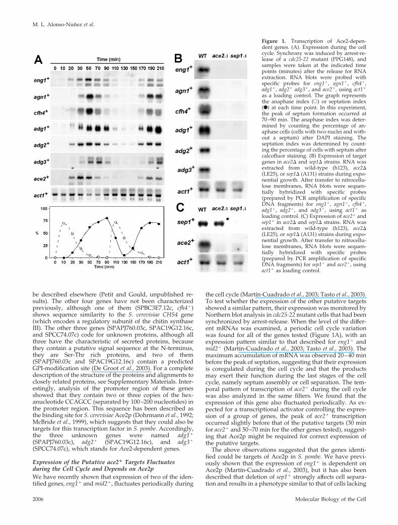
Expression of the Putative ace2� Targets Fluctuatesduring the Cell Cycle and Depends on Ace2pWe have recently shown that expression of two of the iden-tified genes, eng1� and mid2�, fluctuates periodically during
the cell cycle (Martın-Cuadrado et al., 2003; Tasto et al., 2003).To test whether the expression of the other putative targetsshowed a similar pattern, their expression was monitored byNorthern blot analysis in cdc25-22 mutant cells that had beensynchronized by arrest-release. When the level of the differ-ent mRNAs was examined, a periodic cell cycle variationwas found for all of the genes tested (Figure 1A), with anexpression pattern similar to that described for eng1� andmid2� (Martın-Cuadrado et al., 2003; Tasto et al., 2003). Themaximum accumulation of mRNA was observed 20–40 minbefore the peak of septation, suggesting that their expressionis coregulated during the cell cycle and that the productsmay exert their function during the last stages of the cellcycle, namely septum assembly or cell separation. The tem-poral pattern of transcription of ace2� during the cell cyclewas also analyzed in the same filters. We found that theexpression of this gene also fluctuated periodically. As ex-pected for a transcriptional activator controlling the expres-sion of a group of genes, the peak of ace2� transcriptionoccurred slightly before that of the putative targets (30 minfor ace2� and 50–70 min for the other genes tested), suggest-ing that Ace2p might be required for correct expression ofthe putative targets.
The above observations suggested that the genes identi-fied could be targets of Ace2p in S. pombe. We have previ-ously shown that the expression of eng1� is dependent onAce2p (Martın-Cuadrado et al., 2003), but it has also beendescribed that deletion of sep1� strongly affects cell separa-tion and results in a phenotype similar to that of cells lacking
Figure 1. Transcription of Ace2-depen-dent genes. (A). Expression during the cellcycle. Synchrony was induced by arrest-re-lease of a cdc25-22 mutant (PPG148), andsamples were taken at the indicated timepoints (minutes) after the release for RNAextraction. RNA blots were probed withspecific probes for eng1�, agn1�, cfh4�,adg1�, adg2� adg3�, and ace2�, using act1�
as a loading control. The graph representsthe anaphase index (E) or septation index(F) at each time point. In this experiment,the peak of septum formation occurred at70–90 min. The anaphase index was deter-mined by counting the percentage of an-aphase cells (cells with two nuclei and with-out a septum) after DAPI staining. Theseptation index was determined by count-ing the percentage of cells with septum aftercalcofluor staining. (B) Expression of targetgenes in ace2� and sep1� strains. RNA wasextracted from wild-type (h123), ace2�(LE25), or sep1� (A131) strains during expo-nential growth. After transfer to nitrocellu-lose membranes, RNA blots were sequen-tially hybridized with specific probes(prepared by PCR amplification of specificDNA fragments) for eng1�, agn1�, cfh4�,adg1�, adg2�, and adg3�, using act1� asloading control. (C) Expression of ace2� andsep1� in ace2� and sep1� strains. RNA wasextracted from wild-type (h123), ace2�(LE25), or sep1� (A131) strains during expo-nential growth. After transfer to nitrocellu-lose membranes, RNA blots were sequen-tially hybridized with specific probes(prepared by PCR amplification of specificDNA fragments) for sep1� and ace2�, usingact1� as loading control.
M. L. Alonso-Nunez et al.
Molecular Biology of the Cell2006
ace2� (Sipiczki et al., 1993; Sipiczki and Bozsik, 2000; Martın-Cuadrado et al., 2003). To study the relationship betweenthese two transcription factors and the putative target genes,Northern analyses were performed to compare their expres-sion in wild-type, ace2�, and sep1� mutants. These analysesrevealed that the expression of the seven genes identified inour microarray analysis was clearly reduced in both theace2� and sep1� mutants (Figure 1B and unpublished data),suggesting that—directly or indirectly—both transcriptionfactors are required for their expression.
A Transcriptional Cascade Controls the Last Steps of theCell CycleIn S. cerevisiae, a transcriptional cascade controls gene ex-pression during the last stages of the cell cycle. Thus, theforkhead transcription factors Fkh1p and Fkh2p activate theexpression of Ace2p, which in turn regulates the transcrip-tion of a group of genes that are specifically expressed in thedaughter cell and are involved in cell separation (Zhu et al.,2000; Colman-Lerner et al., 2001; Simon et al., 2001; Baladronet al., 2002). Because the expression of eng1� and the othergenes requires both the forkhead-like factor Sep1p andAce2p, we decided to test whether a similar situation occursin S. pombe cells. To this end, the expression of the twotranscription factors was analyzed in wild-type and mutantcells lacking either of the two genes. Interestingly, sep1� wastranscribed at normal levels in ace2� mutants (Figure 1C),but ace2� was not expressed in the sep1� deletion strain,suggesting that in fission yeast there is also a transcriptionalcascade controlling gene expression during the last stages ofthe cell cycle, ace2� transcription being dependent on Sep1p.
This result suggests that Ace2p is the most direct activatorof gene expression for these seven genes and that the defectin expression observed in sep1� mutants could be indirectand due to a failure to activate the transcription of ace2�.One prediction of this hypothesis is that expression of ace2�
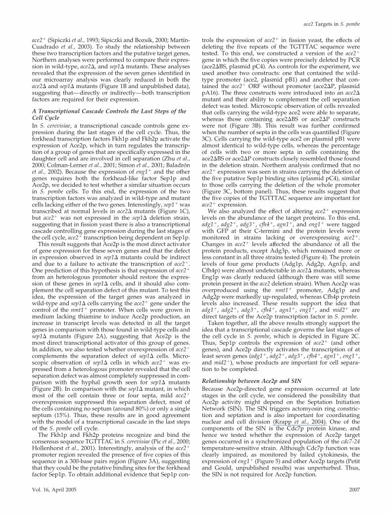
from an heterologous promoter should restore the expres-sion of these genes in sep1� cells, and it should also com-plement the cell separation defect of this mutant. To test thisidea, the expression of the target genes was analyzed inwild-type and sep1� cells carrying the ace2� gene under thecontrol of the nmt1� promoter. When cells were grown inmedium lacking thiamine to induce Ace2p production, anincrease in transcript levels was detected in all the targetgenes in comparison with those found in wild-type cells andsep1� mutants (Figure 2A), suggesting that Ace2p is themost direct transcriptional activator of this group of genes.In addition, we also tested whether overexpression of ace2�
complements the separation defect of sep1� cells. Micro-scopic observation of sep1� cells in which ace2� was ex-pressed from a heterologous promoter revealed that the cellseparation defect was almost completely suppressed in com-parison with the hyphal growth seen for sep1� mutants(Figure 2B). In comparison with the sep1� mutant, in whichmost of the cell contain three or four septa, mild ace2�
overexpression suppressed this separation defect, most ofthe cells containing no septum (around 80%) or only a singleseptum (15%). Thus, these results are in good agreementwith the model of a transcriptional cascade in the last stepsof the S. pombe cell cycle.
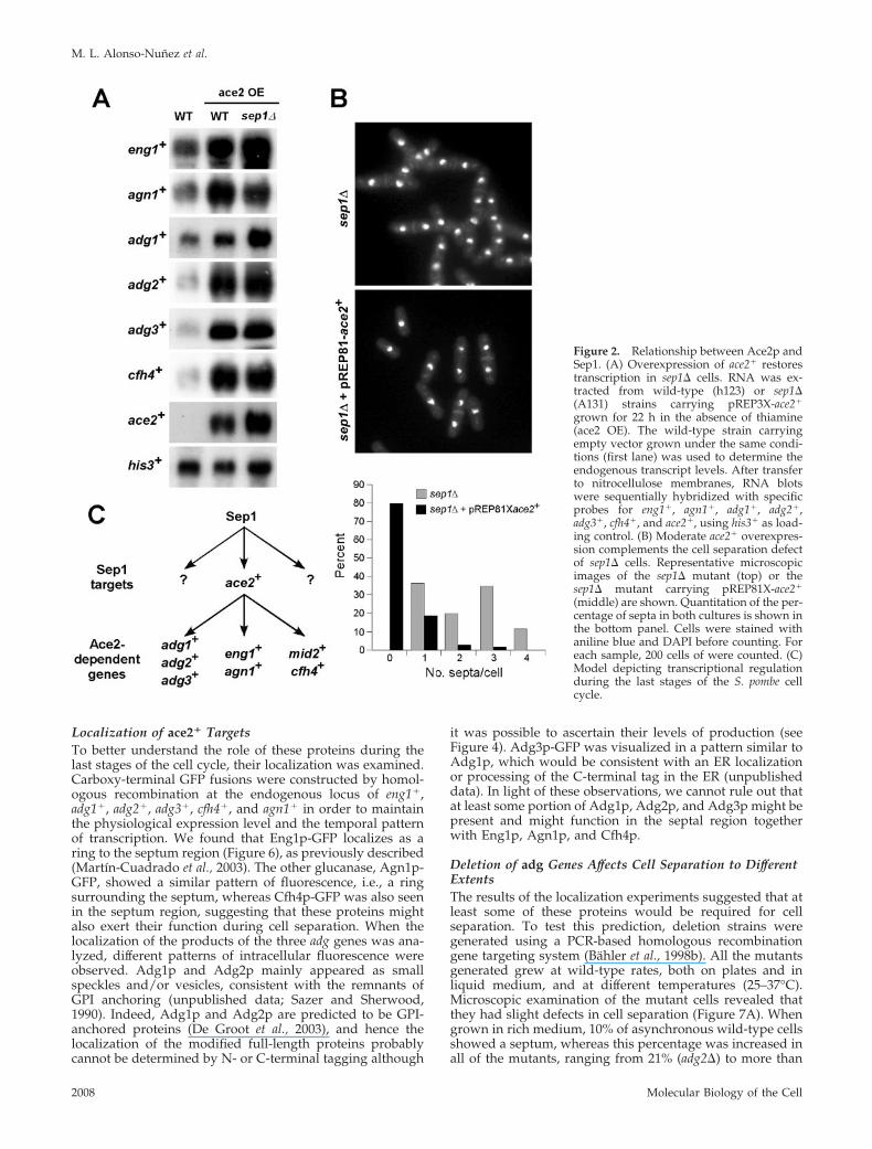
The Fkh1p and Fkh2p proteins recognize and bind theconsensus sequence TGTTTAC in S. cerevisiae (Pic et al., 2000;Hollenhorst et al., 2001). Interestingly, analysis of the ace2�
promoter region revealed the presence of five copies of thissequence in a 300-base pairs region (Figure 3A), suggestingthat they could be the putative binding sites for the forkheadfactor Sep1p. To obtain additional evidence that Sep1p con-
trols the expression of ace2� in fission yeast, the effects ofdeleting the five repeats of the TGTTTAC sequence weretested. To this end, we constructed a version of the ace2�
gene in which the five copies were precisely deleted by PCR(ace2�BS, plasmid pC4). As controls for the experiment, weused another two constructs: one that contained the wild-type promoter (ace2, plasmid pB1) and another that con-tained the ace2� ORF without promoter (ace2�P, plasmidpA16). The three constructs were introduced into an ace2�mutant and their ability to complement the cell separationdefect was tested. Microscopic observation of cells revealedthat cells carrying the wild-type ace2 were able to separate,whereas those containing ace2�BS or ace2�P constructswere not (Figure 3B). This result was further confirmedwhen the number of septa in the cells was quantified (Figure3C). Cells carrying the wild-type ace2 on plasmid pB1 werealmost identical to wild-type cells, whereas the percentageof cells with two or more septa in cells containing theace2�BS or ace2�P constructs closely resembled those foundin the deletion strain. Northern analysis confirmed that noace2� expression was seen in strains carrying the deletion ofthe five putative Sep1p binding sites (plasmid pC4), similarto those cells carrying the deletion of the whole promoter(Figure 3C, bottom panel). Thus, these results suggest thatthe five copies of the TGTTTAC sequence are important forace2� expression.
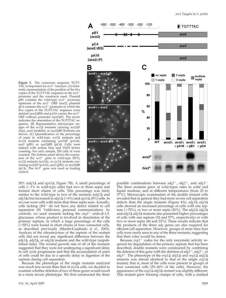
We also analyzed the effect of altering ace2� expressionlevels on the abundance of the target proteins. To this end,adg1�, adg2�, adg3�, cfh4�, agn1�, and eng1� were taggedwith GFP at their C-termini and the protein levels weremonitored in strains lacking or overexpressing ace2�.Changes in ace2� levels affected the abundance of all theprotein products, except Adg3p, which remained more orless constant in all three strains tested (Figure 4). The proteinlevels of four gene products (Adg1p, Adg2p, Agn1p, andCfh4p) were almost undetectable in ace2� mutants, whereasEng1p was clearly reduced (although there was still someprotein present in the ace2 deletion strain). When Ace2p wasoverproduced using the nmt1� promoter, Adg1p andAdg2p were markedly up-regulated, whereas Cfh4p proteinlevels also increased. These results support the idea thatadg1�, adg2�, adg3�, cfh4�, agn1�, eng1�, and mid2� aredirect targets of the Ace2p transcription factor in S. pombe.
Taken together, all the above results strongly support theidea that a transcriptional cascade governs the last stages ofthe cell cycle in S. pombe, which is depicted in Figure 2C.Thus, Sep1p controls the expression of ace2� (and othergenes), and Ace2p directly activates the transcription of atleast seven genes (adg1�, adg2�, adg3�, cfh4�, agn1�, eng1�,and mid2�), whose products are important for cell separa-tion to be completed.
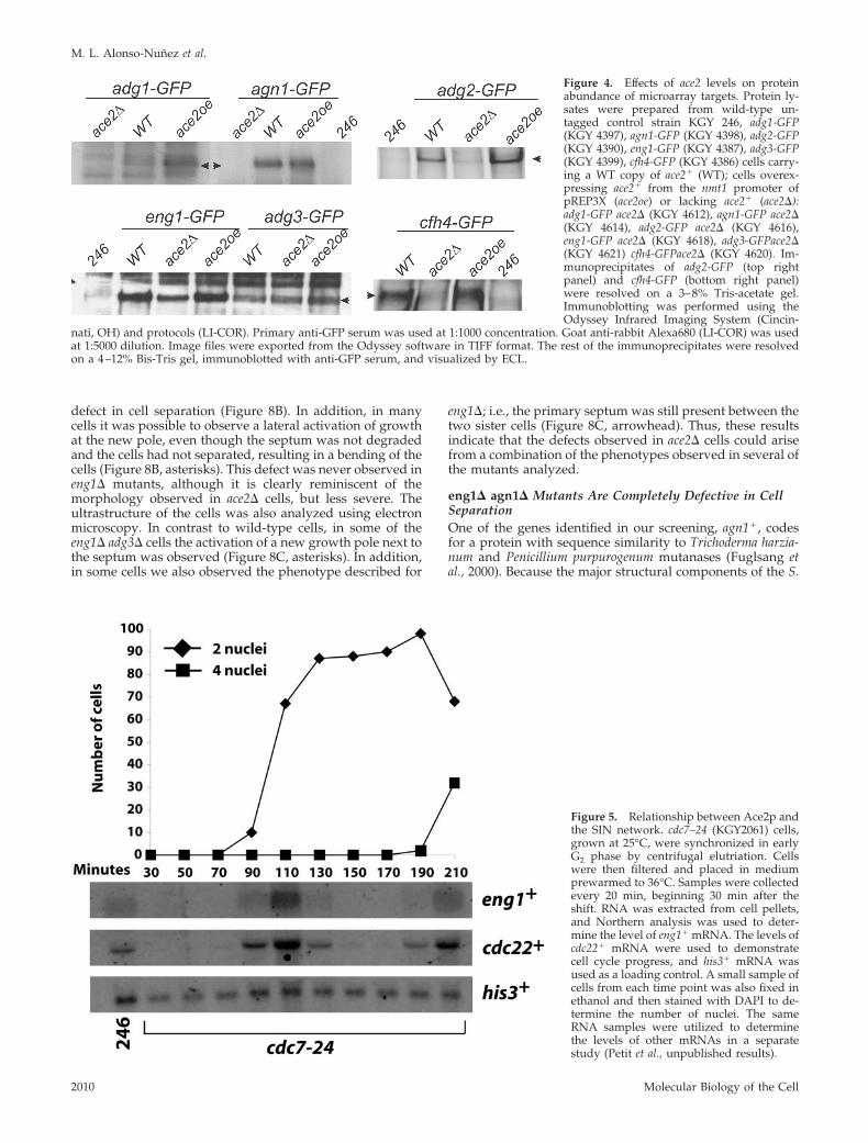
Relationship between Ace2p and SINBecause Ace2p-directed gene expression occurred at latestages in the cell cycle, we considered the possibility thatAce2p activity might depend on the Septation InitiationNetwork (SIN). The SIN triggers actomyosin ring constric-tion and septation and is also important for coordinatingnuclear and cell division (Krapp et al., 2004). One of thecomponents of the SIN is the Cdc7p protein kinase, andhence we tested whether the expression of Ace2p targetgenes occurred in a synchronized population of the cdc7-24temperature-sensitive strain. Although Cdc7p function wasclearly impaired, as monitored by failed cytokinesis, theexpression of eng1� (Figure 5) and other Ace2p targets (Petitand Gould, unpublished results) was unperturbed. Thus,the SIN is not required for Ace2p function.
ace2 Targets in S. pombe
Vol. 16, April 2005 2007
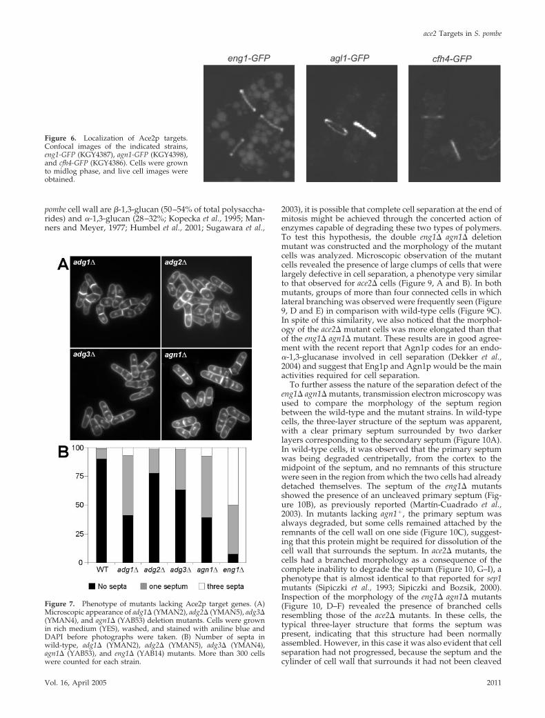
Localization of ace2� TargetsTo better understand the role of these proteins during thelast stages of the cell cycle, their localization was examined.Carboxy-terminal GFP fusions were constructed by homol-ogous recombination at the endogenous locus of eng1�,adg1�, adg2�, adg3�, cfh4�, and agn1� in order to maintainthe physiological expression level and the temporal patternof transcription. We found that Eng1p-GFP localizes as aring to the septum region (Figure 6), as previously described(Martın-Cuadrado et al., 2003). The other glucanase, Agn1p-GFP, showed a similar pattern of fluorescence, i.e., a ringsurrounding the septum, whereas Cfh4p-GFP was also seenin the septum region, suggesting that these proteins mightalso exert their function during cell separation. When thelocalization of the products of the three adg genes was ana-lyzed, different patterns of intracellular fluorescence wereobserved. Adg1p and Adg2p mainly appeared as smallspeckles and/or vesicles, consistent with the remnants ofGPI anchoring (unpublished data; Sazer and Sherwood,1990). Indeed, Adg1p and Adg2p are predicted to be GPI-anchored proteins (De Groot et al., 2003), and hence thelocalization of the modified full-length proteins probablycannot be determined by N- or C-terminal tagging although
it was possible to ascertain their levels of production (seeFigure 4). Adg3p-GFP was visualized in a pattern similar toAdg1p, which would be consistent with an ER localizationor processing of the C-terminal tag in the ER (unpublisheddata). In light of these observations, we cannot rule out thatat least some portion of Adg1p, Adg2p, and Adg3p might bepresent and might function in the septal region togetherwith Eng1p, Agn1p, and Cfh4p.
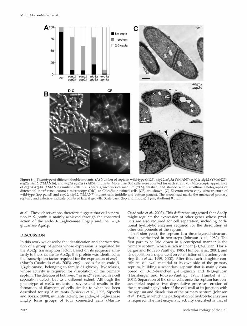
Deletion of adg Genes Affects Cell Separation to DifferentExtentsThe results of the localization experiments suggested that atleast some of these proteins would be required for cellseparation. To test this prediction, deletion strains weregenerated using a PCR-based homologous recombinationgene targeting system (Bahler et al., 1998b). All the mutantsgenerated grew at wild-type rates, both on plates and inliquid medium, and at different temperatures (25–37°C).Microscopic examination of the mutant cells revealed thatthey had slight defects in cell separation (Figure 7A). Whengrown in rich medium, 10% of asynchronous wild-type cellsshowed a septum, whereas this percentage was increased inall of the mutants, ranging from 21% (adg2�) to more than
Figure 2. Relationship between Ace2p andSep1. (A) Overexpression of ace2� restorestranscription in sep1� cells. RNA was ex-tracted from wild-type (h123) or sep1�(A131) strains carrying pREP3X-ace2�
grown for 22 h in the absence of thiamine(ace2 OE). The wild-type strain carryingempty vector grown under the same condi-tions (first lane) was used to determine theendogenous transcript levels. After transferto nitrocellulose membranes, RNA blotswere sequentially hybridized with specificprobes for eng1�, agn1�, adg1�, adg2�,adg3�, cfh4�, and ace2�, using his3� as load-ing control. (B) Moderate ace2� overexpres-sion complements the cell separation defectof sep1� cells. Representative microscopicimages of the sep1� mutant (top) or thesep1� mutant carrying pREP81X-ace2�
(middle) are shown. Quantitation of the per-centage of septa in both cultures is shown inthe bottom panel. Cells were stained withaniline blue and DAPI before counting. Foreach sample, 200 cells of were counted. (C)Model depicting transcriptional regulationduring the last stages of the S. pombe cellcycle.
M. L. Alonso-Nunez et al.
Molecular Biology of the Cell2008
50% (adg1� and agn1�; Figure 7B). A small percentage ofcells (1% in wild-type cells) had two or three septa andformed short chains of cells. This percentage was fairlysimilar to the wild-type in two of the mutants (adg2� andadg3�) but increased in adg1� (6%) and agn1� (8%) cells. Inno case were cells with more than three septa seen. Actually,cells lacking cfh4� do not have any defect related to cellseparation (H. Valdivieso, personal communication). Ascontrols, we used mutants lacking the eng1� endo-�-1,3-glucanase, whose product is involved in dissolution of theprimary septum, in which a large percentage of the cells(50%) were found in short chains of four connected cells,as described previously (Martın-Cuadrado et al., 2003).Analysis of the ultrastructure of the septum of the mutantcells did not reveal any significant difference between theadg1�, adg2�, or adg3� mutants and the wild-type (unpub-lished data). The normal growth rate of all of the mutantssuggested that they were not undergoing a significant delayin cell cycle progression and that the accumulation of pairsof cells could be due to a specific delay in digestion of theseptum during cell separation.
Because the phenotype of the single mutants analyzedwas much less severe than that of ace2� cells, we decided toexamine whether deletion of two of these genes would resultin a more severe phenotype. We first constructed the three
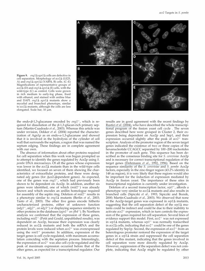
possible combinations between adg1�, adg2�, and adg3�.The three mutants grew at wild-types rates in solid andliquid medium, and at different temperatures (from 25 to37°C). Microscopic examination of the double mutant cellsrevealed that in general they had more severe cell separationdefects than the single mutants (Figure 8A). adg1� adg3�cells showed an increased percentage of cells with one sep-tum (70%), or two or more septa (20%). The adg1� adg2�and adg2� adg3� mutants also presented higher percentagesof cells with one septum (52 and 57%, respectively) or withtwo or more septa (40 and 22%). These results indicate thatthe products of the three adg genes are also required forefficient cell separation. However, groups of more than fourcells were rarely seen in any of the three mutants, suggestingthat their roles would be minor.
Because eng1� codes for the only enzymatic activity re-quired for degradation of the primary septum that has beendescribed, double mutants were constructed by combiningthe deletion of this gene with the deletion of adg1�, adg2�, oradg3�. The phenotype of the eng1� adg1� and eng1� adg2�mutants was almost identical to that of the single eng1�mutant; that is, most of the cells were present in groups offour connected cells (55–60% of the cells). However, theappearance of the eng1� adg3� mutant was slightly different.This mutant grew forming clumps of cells, with a marked
Figure 3. The consensus sequence TGTT-TAC is important for ace2� function. (A) Sche-matic representation of the position of the fivecopies of the TGTTTAC sequence in the ace2�
promoter and the constructs used. PlasmidpB1 contains the wild-type ace2� promoterupstream of the ace2� ORF (ace2), plasmidpC4 contains the ace2� promoter in which thefive copies of the TGTTTAC sequence weredeleted (ace2�BS) and pA16 carries the ace2�
ORF without promoter (ace2�P). The arrowindicates the orientation of the TGTTTAC se-quence. (B) Representative microscopic im-ages of the ace2� mutants carrying ace2�P(top), ace2 (middle), or ace2�BS (bottom) areshown. (C) Quantification of the percentageof septa in wild-type, ace2� mutants andace2� mutants containing ace2�P (pA16),ace2 (pB1) or ace2�BS (pC4). Cells werestained with aniline blue and DAPI beforecounting. For each sample, 200 cells of werecounted. The bottom panel shows the expres-sion of the ace2� gene in wild-type (WT),ace2� mutants (ace2�), or ace2� mutants con-taining ace2�P (pA16), ace2 (pB1), or ace2�BS(pC4). The his3� gene was used as loadingcontrol.
ace2 Targets in S. pombe
Vol. 16, April 2005 2009
defect in cell separation (Figure 8B). In addition, in manycells it was possible to observe a lateral activation of growthat the new pole, even though the septum was not degradedand the cells had not separated, resulting in a bending of thecells (Figure 8B, asterisks). This defect was never observed ineng1� mutants, although it is clearly reminiscent of themorphology observed in ace2� cells, but less severe. Theultrastructure of the cells was also analyzed using electronmicroscopy. In contrast to wild-type cells, in some of theeng1� adg3� cells the activation of a new growth pole next tothe septum was observed (Figure 8C, asterisks). In addition,in some cells we also observed the phenotype described for
eng1�; i.e., the primary septum was still present between thetwo sister cells (Figure 8C, arrowhead). Thus, these resultsindicate that the defects observed in ace2� cells could arisefrom a combination of the phenotypes observed in several ofthe mutants analyzed.
eng1� agn1� Mutants Are Completely Defective in CellSeparationOne of the genes identified in our screening, agn1�, codesfor a protein with sequence similarity to Trichoderma harzia-num and Penicillium purpurogenum mutanases (Fuglsang etal., 2000). Because the major structural components of the S.
Figure 4. Effects of ace2 levels on proteinabundance of microarray targets. Protein ly-sates were prepared from wild-type un-tagged control strain KGY 246, adg1-GFP(KGY 4397), agn1-GFP (KGY 4398), adg2-GFP(KGY 4390), eng1-GFP (KGY 4387), adg3-GFP(KGY 4399), cfh4-GFP (KGY 4386) cells carry-ing a WT copy of ace2� (WT); cells overex-pressing ace2� from the nmt1 promoter ofpREP3X (ace2oe) or lacking ace2� (ace2�):adg1-GFP ace2� (KGY 4612), agn1-GFP ace2�(KGY 4614), adg2-GFP ace2� (KGY 4616),eng1-GFP ace2� (KGY 4618), adg3-GFPace2�(KGY 4621) cfh4-GFPace2� (KGY 4620). Im-munoprecipitates of adg2-GFP (top rightpanel) and cfh4-GFP (bottom right panel)were resolved on a 3–8% Tris-acetate gel.Immunoblotting was performed using theOdyssey Infrared Imaging System (Cincin-
nati, OH) and protocols (LI-COR). Primary anti-GFP serum was used at 1:1000 concentration. Goat anti-rabbit Alexa680 (LI-COR) was usedat 1:5000 dilution. Image files were exported from the Odyssey software in TIFF format. The rest of the immunoprecipitates were resolvedon a 4–12% Bis-Tris gel, immunoblotted with anti-GFP serum, and visualized by ECL.
Figure 5. Relationship between Ace2p andthe SIN network. cdc7–24 (KGY2061) cells,grown at 25°C, were synchronized in earlyG2 phase by centrifugal elutriation. Cellswere then filtered and placed in mediumprewarmed to 36°C. Samples were collectedevery 20 min, beginning 30 min after theshift. RNA was extracted from cell pellets,and Northern analysis was used to deter-mine the level of eng1� mRNA. The levels ofcdc22� mRNA were used to demonstratecell cycle progress, and his3� mRNA wasused as a loading control. A small sample ofcells from each time point was also fixed inethanol and then stained with DAPI to de-termine the number of nuclei. The sameRNA samples were utilized to determinethe levels of other mRNAs in a separatestudy (Petit et al., unpublished results).
M. L. Alonso-Nunez et al.
Molecular Biology of the Cell2010
pombe cell wall are �-1,3-glucan (50–54% of total polysaccha-rides) and �-1,3-glucan (28–32%; Kopecka et al., 1995; Man-ners and Meyer, 1977; Humbel et al., 2001; Sugawara et al.,
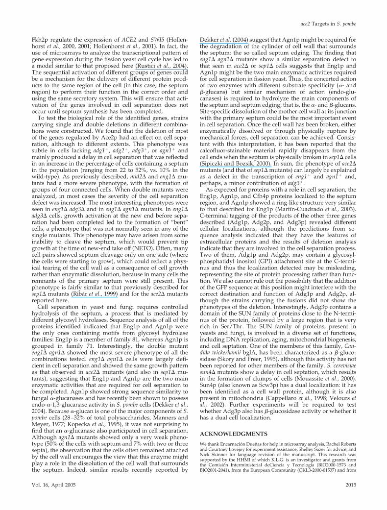
2003), it is possible that complete cell separation at the end ofmitosis might be achieved through the concerted action ofenzymes capable of degrading these two types of polymers.To test this hypothesis, the double eng1� agn1� deletionmutant was constructed and the morphology of the mutantcells was analyzed. Microscopic observation of the mutantcells revealed the presence of large clumps of cells that werelargely defective in cell separation, a phenotype very similarto that observed for ace2� cells (Figure 9, A and B). In bothmutants, groups of more than four connected cells in whichlateral branching was observed were frequently seen (Figure9, D and E) in comparison with wild-type cells (Figure 9C).In spite of this similarity, we also noticed that the morphol-ogy of the ace2� mutant cells was more elongated than thatof the eng1� agn1� mutant. These results are in good agree-ment with the recent report that Agn1p codes for an endo-�-1,3-glucanase involved in cell separation (Dekker et al.,2004) and suggest that Eng1p and Agn1p would be the mainactivities required for cell separation.
To further assess the nature of the separation defect of theeng1� agn1� mutants, transmission electron microscopy wasused to compare the morphology of the septum regionbetween the wild-type and the mutant strains. In wild-typecells, the three-layer structure of the septum was apparent,with a clear primary septum surrounded by two darkerlayers corresponding to the secondary septum (Figure 10A).In wild-type cells, it was observed that the primary septumwas being degraded centripetally, from the cortex to themidpoint of the septum, and no remnants of this structurewere seen in the region from which the two cells had alreadydetached themselves. The septum of the eng1� mutantsshowed the presence of an uncleaved primary septum (Fig-ure 10B), as previously reported (Martın-Cuadrado et al.,2003). In mutants lacking agn1�, the primary septum wasalways degraded, but some cells remained attached by theremnants of the cell wall on one side (Figure 10C), suggest-ing that this protein might be required for dissolution of thecell wall that surrounds the septum. In ace2� mutants, thecells had a branched morphology as a consequence of thecomplete inability to degrade the septum (Figure 10, G–I), aphenotype that is almost identical to that reported for sep1mutants (Sipiczki et al., 1993; Sipiczki and Bozsik, 2000).Inspection of the morphology of the eng1� agn1� mutants(Figure 10, D–F) revealed the presence of branched cellsresembling those of the ace2� mutants. In these cells, thetypical three-layer structure that forms the septum waspresent, indicating that this structure had been normallyassembled. However, in this case it was also evident that cellseparation had not progressed, because the septum and thecylinder of cell wall that surrounds it had not been cleaved
Figure 6. Localization of Ace2p targets.Confocal images of the indicated strains,eng1-GFP (KGY4387), agn1-GFP (KGY4398),and cfh4-GFP (KGY4386). Cells were grownto midlog phase, and live cell images wereobtained.
Figure 7. Phenotype of mutants lacking Ace2p target genes. (A)Microscopic appearance of adg1� (YMAN2), adg2� (YMAN5), adg3�(YMAN4), and agn1� (YAB53) deletion mutants. Cells were grownin rich medium (YES), washed, and stained with aniline blue andDAPI before photographs were taken. (B) Number of septa inwild-type, adg1� (YMAN2), adg2� (YMAN5), adg3� (YMAN4),agn1� (YAB53), and eng1� (YAB14) mutants. More than 300 cellswere counted for each strain.
ace2 Targets in S. pombe
Vol. 16, April 2005 2011
at all. These observations therefore suggest that cell separa-tion in S. pombe is mainly achieved through the concertedaction of the endo-�-1,3-glucanase Eng1p and the �-1,3-glucanase Agn1p.
DISCUSSION
In this work we describe the identification and characteriza-tion of a group of genes whose expression is regulated bythe Ace2p transcription factor. Based on its sequence simi-larity to the S. cerevisiae Ace2p, this protein was identified asthe transcription factor required for the expression of eng1�
(Martın-Cuadrado et al., 2003). eng1� codes for an endo-�-1,3-glucanase, belonging to family 81 glycosyl hydrolases,whose activity is required for dissolution of the primaryseptum. The deletion of both eng1� or ace2� resulted in a cellseparation defect, but to a different extent. Although thephenotype of ace2� mutants is severe and results in theformation of filaments of cells similar to what has beendescribed for sep1� mutants (Sipiczki et al., 1993; Sipiczkiand Bozsik, 2000), mutants lacking the endo-�-1,3-glucanaseEng1p form groups of four connected cells (Martın-
Cuadrado et al., 2003). This difference suggested that Ace2pmight regulate the expression of other genes whose prod-ucts are also required for cell separation, including addi-tional hydrolytic enzymes required for the dissolution ofother components of the septum.
In fission yeast, the septum is a three-layered structurethat is synthesized in two steps (Johnson et al., 1982). Thefirst part to be laid down in a centripetal manner is theprimary septum, which is rich in linear �-1,3-glucan (Horis-berger and Rouver-Vauthey, 1985; Humbel et al., 2001), andits deposition is dependent on constriction of the actomyosinring (Liu et al., 1999, 2000). After this, each daughter con-tributes cell wall material to its own side of the primaryseptum, building a secondary septum that is mainly com-posed of �-1,6-branched �-1,3-glucan and �-1,6-glucan(Horisberger and Rouver-Vauthey, 1985; Humbel et al.,2001). Separation of the sister cells once the septum has beenassembled requires two degradative processes: erosion ofthe surrounding cylinder of the cell wall at its junction withthe septum and dissolution of the primary septum (Johnsonet al., 1982), in which the participation of hydrolytic enzymesis required. The first enzymatic activity described is that of
Figure 8. Phenotype of different double mutants. (A) Number of septa in wild-type (h123), adg1� adg3� (YMAN7), adg1� adg2� (YMAN25),adg2� adg3� (YMAN26), and eng1� agn1� (YAB54) mutants. More than 300 cells were counted for each strain. (B) Microscopic appearanceof eng1� adg3� (YMAN11) mutant cells. Cells were grown in rich medium (YES), washed, and stained with Calcofluor. Photographs ofdifferential interference contrast microscopy (DIC) or Calcofluor-stained cells (CF) are shown. (C) Electron microscopy ultrastructure ofwild-type (top panel) and eng1� adg3� (YMAN7) mutant cells (middle and bottom panels). The arrowhead marks the uncleaved primaryseptum, and asterisks indicate points of lateral growth. Scale bars, (top and middle) 1 �m; (bottom) 0.5 �m .
M. L. Alonso-Nunez et al.
Molecular Biology of the Cell2012
the endo-�-1,3-glucanase encoded by eng1�, which is re-quired for dissolution of the �-1,3-glucan-rich primary sep-tum (Martın-Cuadrado et al., 2003). Whereas this article wasunder revision, Dekker et al. (2004) reported the character-ization of Agn1p as en endo-�-1,3-glucanase and showedthat it is involved in the hydrolysis of the cylinder of cellwall that surrounds the septum, a region that was named theseptum edging. These findings are in complete agreementwith our own.
The absence of information about other proteins requiredfor cell separation when this work was begun prompted usto attempt to identify the genes regulated by Ace2p using S.pombe DNA microarrays. Of all the genes whose expressionwas lower in the ace2� mutants than in the wild-type cellsidentified, we focused on seven of them showing the char-acteristics of extracellular proteins, and these were desig-nated adg genes (for Ace2-dependent genes). As expected,one of the genes was eng1�, which had previously beenshown to be dependent on Ace2p. In addition, another sixgenes were identified, one of which (mid2�) was alreadyknown and which encodes an anillin homologue requiredfor assembly of the septin ring. Deletion of mid2� results ina phenotype similar to eng1� mutants (Berlin et al., 2003;Tasto et al., 2003). The other five genes encode hithertouncharacterized proteins, either of unknown function(adg1�, adg2�, or adg3�) or displaying sequence similarity toother proteins in the databases (agn1� or cfh4�). By Northernanalysis we confirmed that the expression of these genes,including mid2� (Petit and Gould, unpublished results), wasdependent on Ace2p, because their expression was clearlyreduced in ace2� mutants and both the mRNA and theprotein levels were induced when ace2� was overexpressedusing the nmt1� promoter. In addition, expression of thetarget genes is cell cycle-regulated, with maximum accumu-lation coinciding with the septation process. Interestingly,the expression of ace2� was also cell cycle-regulated and thepeak of maximum expression occurred before that of theother genes, as expected for a transcriptional regulator. Our
results are in good agreement with the recent findings byRustici et al. (2004), who have described the whole transcrip-tional program of the fission yeast cell cycle . The sevengenes described here were grouped in Cluster 2, their ex-pression being dependent on Ace2p and Sep1, and theirexpression occurred slightly after the peak of ace2� tran-scription. Analysis of the promoter region of the seven targetgenes indicated the existence of two or three copies of thehexanucleotide CCAGCC separated by 100–200 nucleotidesin the promoter of each gene. This sequence has been de-scribed as the consensus binding site for S. cerevisiae Ace2pand is necessary for correct transcriptional regulation of thetarget genes (Dohrmann et al., 1992, 1996). Based on thesequence similarity of the S. cerevisiae and S. pombe Ace2pfactors, especially in the zinc-finger region (43.2% identity in148 aa region), it is very likely that these regions would alsobe important for the induction of expression mediated byAce2p in fission yeast. The importance of these sites intranscriptional regulation is currently under investigation.
Deletion of a second transcription factor, sep1�, affords aphenotype very similar to ace2� mutants and also results inbranched cells (Sipiczki et al., 1993; Sipiczki and Bozsik,2000; Martın-Cuadrado et al., 2003). We observed that noneof the Ace2p-target genes was expressed in sep1� mutants,suggesting that the cell separation defect of the sep1� mu-tants could be indirect and could be due to their inability toactivate ace2� expression, which in turn controls the expres-sion of the genes required for cell separation. Several lines ofevidence support this model. First, ace2� was not expressedin sep1� mutants, whereas sep1� was normally transcribedin ace2� cells, indicating that ace2� could be one of the genesregulated by Sep1p. Second, the expression of ace2� from anheterologous promoter restored the expression of the targetgenes in a sep1� strain and suppressed the cell separationdefect of this mutant, indicating that the genes involved incell separation were more directly regulated by Ace2p.However, suppression of the separation defect was not com-plete, indicating that Ace2p might be regulated by other
Figure 9. eng1� agn1� cells are defective incell separation. Morphology of ace2� (LE25,A) and eng1� agn1� (YAB54, B) cells. (C–E)Magnifications of representative groups oface2� (D) and eng1� agn1� (E) cells, with thewild-type (C) as control. Cells were grownin rich medium to early-log phase, fixedwith ethanol, and stained with aniline blueand DAPI. eng1� agn1� mutants show amycelial and branched phenotype, similarto ace2� mutants, although the cells are lesselongated. Scale bar, 10 �m.
ace2 Targets in S. pombe
Vol. 16, April 2005 2013

mechanisms. Although we have ruled out the contributionof the SIN in controlling Ace2p function, it is possible that,by analogy with S. cerevisiae Ace2p, Cdk1p, or Orb6p (sim-ilar to S. cerevisiae Cbk1p) phosphorylation might be in-volved in its regulation (O’Conallain et al., 1999; Weiss et al.,2002; Nelson et al., 2003; Ufano et al., 2004). It is also possiblethat Sep1p not only regulates ace2� transcription, but alsothe activity or localization of Ace2p. In fact, many interac-tions between Sep1p and cell cycle regulators, such as wee�,cdc25�, or cdc2�, have been described (Sipiczki et al., 2000;Zilahi et al., 2000b). Finally, the promoter of ace2� containedfive copies of the sequence TGTTTAC in a 300-nt region. InS. cerevisiae, this sequence is the binding site for the forkheadtranscription factors Fkh1p and Fkh2p (Pic et al., 2000; Hol-lenhorst et al., 2001), which suggested that they could also bethe binding sites for the forkhead-like factor Sep1p. We haveshown that deletion of the five repeats eliminated ace2�
transcription and function, indicating that they are impor-tant for ace2� expression. Interestingly, two of these sites (at
�375 base pairs and �220 base pairs) are located 8 and 15 ntdownstream of the GCAACG/A sequence (the PCB ele-ment), which is the binding site for the MADS box proteinMbx1p (Buck et al., 2004). These authors have proposed thatMbx1p might form a complex with Sep1p and a secondforkhead-like factor, Fkh2p, to control periodic gene tran-scription in M phase. Surprisingly, they found that deletionof sep1� or fkh2� abolished periodic expression of ace2� andother genes, but deletion of mbx1� only reduced the ampli-tude of the expression, suggesting that Sep1p and Fkh2pmay control periodic gene transcription in the absence of theassociated MADS box protein. These results, together withour data, indicate that correct temporal regulation of ace2�
expression requires both the TGTTTAC sequences, possiblyas binding sites for Sep1p and/or Fkh2p, and the PCBelement (for Mbx1p binding). The transcriptional cascade inthe M/G1 transition is not a unique feature of S. pombe cells,because a similar situation has been described for S. cerevi-siae, in which the fork-head transcription factors Fkh1p and
Figure 10. Electron microscopy ultrastruc-ture of wild-type and mutant cells. Wild-type (A), eng1� (B), agn1� (C), eng1� agn1�(D–F), and ace2� (G–I) cells were grown tomidlog phase before preparation for elec-tron microscopy. The septum region ofeng1� mutants (B) shows the presence ofuncleaved primary septum (arrowheads). Inagn1� mutants, the primary septum is de-graded, but some cells remain attached bythe cell wall at one of the ends (C, arrow-heads). Double eng1� agn1� mutants grewas branched filaments, in which the septumwas not cleaved (D–F). ace2� shows similardefects in cell separation, forming branch-ing filaments of cells (G–I). The rectangles inD and G indicate the region magnified in Eand H, respectively. The seemingly abnor-mal shape of cells (F and I) is the conse-quence of their different spatial positions inthe branched filaments. Scale bars, 0.5 �m(A, B, E, and H) or 1 �m (C, D, F, G, and I).
M. L. Alonso-Nunez et al.
Molecular Biology of the Cell2014
Fkh2p regulate the expression of ACE2 and SWI5 (Hollen-horst et al., 2000, 2001; Hollenhorst et al., 2001). In fact, theuse of microarrays to analyze the transcriptional pattern ofgene expression during the fission yeast cell cycle has led toa model similar to that proposed here (Rustici et al., 2004).The sequential activation of different groups of genes couldbe a mechanism for the delivery of different protein prod-ucts to the same region of the cell (in this case, the septumregion) to perform their function in the correct order andusing the same secretory system. This will ensure that acti-vation of the genes involved in cell separation does notoccur until septum synthesis has been completed.
To test the biological role of the identified genes, strainscarrying single and double deletions in different combina-tions were constructed. We found that the deletion of mostof the genes regulated by Ace2p had an effect on cell sepa-ration, although to different extents. This phenotype wassubtle in cells lacking adg1�, adg2�, adg3�, or agn1� andmainly produced a delay in cell separation that was reflectedin an increase in the percentage of cells containing a septumin the population (ranging from 22 to 52%, vs. 10% in thewild-type). As previously described, mid2� and eng1� mu-tants had a more severe phenotype, with the formation ofgroups of four connected cells. When double mutants wereanalyzed, in most cases the severity of the cell separationdefect was increased. The most interesting phenotypes wereseen in eng1� adg3� and in eng1� agn1� mutants. In eng1�adg3� cells, growth activation at the new end before sepa-ration had been completed led to the formation of “bent”cells, a phenotype that was not normally seen in any of thesingle mutants. This phenotype may have arisen from someinability to cleave the septum, which would prevent tipgrowth at the time of new-end take off (NETO). Often, manycell pairs showed septum cleavage only on one side (wherethe cells were starting to grow), which could reflect a phys-ical tearing of the cell wall as a consequence of cell growthrather than enzymatic dissolution, because in many cells theremnants of the primary septum were still present. Thisphenotype is fairly similar to that previously described forsep1� mutants (Ribar et al., 1999) and for the ace2� mutantsreported here.
Cell separation in yeast and fungi requires controlledhydrolysis of the septum, a process that is mediated bydifferent glycosyl hydrolases. Sequence analysis of all of theproteins identified indicated that Eng1p and Agn1p werethe only ones containing motifs from glycosyl hydrolasefamilies: Eng1p is a member of family 81, whereas Agn1p isgrouped in family 71. Interestingly, the double mutanteng1� agn1� showed the most severe phenotype of all thecombinations tested. eng1� agn1� cells were largely defi-cient in cell separation and showed the same growth patternas that observed in ace2� mutants (and also in sep1� mu-tants), suggesting that Eng1p and Agn1p are the two mainenzymatic activities that are required for cell separation tobe completed. Agn1p showed strong sequence similarity tofungal �-glucanases and has recently been shown to possessendo-�-1,3-glucanase activity in S. pombe cells (Dekker et al.,2004). Because �-glucan is one of the major components of S.pombe cells (28–32% of total polysaccharides, Manners andMeyer, 1977; Kopecka et al., 1995), it was not surprising tofind that an �-glucanase also participated in cell separation.Although agn1� mutants showed only a very weak pheno-type (50% of the cells with septum and 7% with two or threesepta), the observation that the cells often remained attachedby the cell wall encourages the view that this enzyme mightplay a role in the dissolution of the cell wall that surroundsthe septum. Indeed, similar results recently reported by
Dekker et al. (2004) suggest that Agn1p might be required forthe degradation of the cylinder of cell wall that surroundsthe septum: the so called septum edging. The finding thateng1� agn1� mutants show a similar separation defect tothat seen in ace2� or sep1� cells suggests that Eng1p andAgn1p might be the two main enzymatic activities requiredfor cell separation in fission yeast. Thus, the concerted actionof two enzymes with different substrate specificity (�- and�-glucans) but similar mechanism of action (endo-glu-canases) is required to hydrolyze the main components ofthe septum and septum edging, that is, the �- and �-glucans.Site-specific dissolution of the mother cell wall at its junctionwith the primary septum could be the most important eventin cell separation. Once the cell wall has been broken, eitherenzymatically dissolved or through physically rupture bymechanical forces, cell separation can be achieved. Consis-tent with this interpretation, it has been reported that thecalcofluor-stainable material rapidly disappears from thecell ends when the septum is physically broken in sep1� cells(Sipiczki and Bozsik, 2000). In sum, the phenotype of ace2�mutants (and that of sep1� mutants) can largely be explainedas a defect in the transcription of eng1� and agn1� and,perhaps, a minor contribution of adg3�.
As expected for proteins with a role in cell separation, theEng1p, Agn1p, and Cfh4p proteins localized to the septumregion, and Agn1p showed a ring-like structure very similarto that described for Eng1p (Martın-Cuadrado et al., 2003).C-terminal tagging of the products of the other three genesdescribed (Adg1p, Adg2p, and Adg3p) revealed differentcellular localizations, although the predictions from se-quence analysis indicated that they have the features ofextracellular proteins and the results of deletion analysisindicate that they are involved in the cell separation process.Two of them, Adg1p and Adg2p, may contain a glycosyl-phosphatidyl inositol (GPI) attachment site at the C-termi-nus and thus the localization detected may be misleading,representing the site of protein processing rather than func-tion. We also cannot rule out the possibility that the additionof the GFP sequence at this position might interfere with thecorrect destination and function of Adg1p and Adg2p, al-though the strains carrying the fusions did not show thephenotypes of the deletion. Interestingly, Adg3p contains adomain of the SUN family of proteins close to the N-termi-nus of the protein, followed by a large region that is veryrich in Ser/Thr. The SUN family of proteins, present inyeasts and fungi, is involved in a diverse set of functions,including DNA replication, aging, mitochondrial biogenesis,and cell septation. One of the members of this family, Can-dida wickerhamii bglA, has been characterized as a �-gluco-sidase (Skory and Freer, 1995), although this activity has notbeen reported for other members of the family. S. cerevisiaesun4� mutants show a delay in cell septation, which resultsin the formation of clumps of cells (Mouassite et al., 2000).Sun4p (also known as Scw3p) has a dual localization: it hasbeen identified as a cell wall protein, although it is alsopresent in mitochondria (Cappellaro et al., 1998; Velours etal., 2002). Further experiments will be required to testwhether Adg3p also has �-glucosidase activity or whether ithas a dual cell localization.
ACKNOWLEDGMENTS
We thank Encarnacion Duenas for help in microarray analysis, Rachel Robertsand Courtney Lovejoy for experiment assistance, Shelley Sazer for advice, andNick Skinner for language revision of the manuscript. This research wassupported by the HHMI of which K.L.G. is an investigator and grants fromthe Comision Interministerial deCiencia y Tecnologıa (BIO2000-1573 andBIO2001-2041), from the European Community (QKL3-2000-01537) and from
ace2 Targets in S. pombe
Vol. 16, April 2005 2015
the Hungarian grant agency OTKA (T 042694). M.L.A.-N. is recipient of afellowship from the Ministerio deEducacion y Ciencia (Spain).
REFERENCES
Bahler, J., and Nurse, P. (2001). Fission yeast Pom1p kinase activity is cellcycle regulated and essential for cellular symmetry during growth and divi-sion. EMBO J. 20, 1064–1073.
Bahler, J., and Pringle, J. R. (1998). Pom1p, a fission yeast protein kinase thatprovides positional information for both polarized growth and cytokinesis.Genes Dev. 12, 1356–1370.
Bahler, J., Steever, A. B., Wheatley, S., Wang, Y., Pringle, J. R., Gould, K. L.,and McCollum, D. (1998a). Role of polo kinase and Mid1p in determining thesite of cell division in fission yeast. J. Cell Biol. 143, 1603–1616.
Bahler, J., Wu, J. Q., Longtine, M. S., Shah, N. G., McKenzie, A., Steever, A. B.,Wach, A., Philippsen, P., and Pringle, J. R. (1998b). Heterologous modules forefficient and versatile PCR-based gene targeting in Schizosaccharomyces pombe.Yeast 14, 943–951.
Baladron, V., Ufano, S., Duenas, E., Martın-Cuadrado, A. B., Rey, F.D., andVazquez de Aldana, C. R. (2002). Eng1p, an endo-1,3-�-glucanase localized atthe daughter side of the septum, is involved in cell separation in Saccharomy-ces cerevisiae. Eukaryot. Cell 1, 774–786.
Berlin, A., Paoletti, A., and Chang, F. (2003). Mid2p stabilizes septin ringsduring cytokinesis in fission yeast. J. Cell Biol. 160, 1083–1092.
Buck, V., Ng, S., Ruiz-Garcia, A. B., Papadopoulou, K., Bhatti, S., Samuel,J. M., Anderson, M., Millar, J. B., and McInerny, C. J. (2004). Fkh2p and Sep1pregulate mitotic gene transcription in fission yeast. J. Cell Sci. 117, 5623–5632.
Burns, C. G., Ohi, R., Krainer, A. R., and Gould, K. L. (1999). Evidence thatMyb-related CDC5 proteins are required for pre-mRNA splicing. Proc. Natl.Acad. Sci. USA 96, 13789–13794.
Cappellaro, C., Mrsa, V., and Tanner, W. (1998). New potential cell wallglucanases of Saccharomyces cerevisiae and their involvement in mating. J.Bacteriol. 180, 5030–5037.
Chang, F. (2001). Studies in fission yeast on mechanisms of cell division siteplacement. Cell Struct. Funct. 26, 539–544.
Chang, F., and Nurse, P. (1996). How fission yeast fission in the middle. Cell84, 191–194.
Colman-Lerner, A., Chin, T. E., and Brent, R. (2001). Yeast Cbk1 and Mob2activate daughter-specific genetic programs to induce asymmetric cell fates.Cell 107, 739–750.
De Groot, P. W., Hellingwerf, K. J., and Klis, F. M. (2003). Genome-wideidentification of fungal GPI proteins. Yeast 20, 781–796.
Dekker, N., Speijer, D., Grun, C. H., van den Berg, M., de Haan, A., andHochstenbach, F. (2004). Role of the �-glucanase Agn1p in fission-yeast cellseparation. Mol. Biol. Cell 15, 3903–3914.
Dohrmann, P. R., Butler, G., Tamai, K., Dorland, S., Greene, J. R., Thiele, D. J.,and Stillman, D. J. (1992). Parallel pathways of gene regulation: homologousregulators SWI5 and ACE2 differentially control transcription of HO andchitinase. Genes Dev. 6, 93–104.
Dohrmann, P. R., Voth, W. P., and Stillman, D. J. (1996). Role of negativeregulation in promoter specificity of the homologous transcriptional activa-tors Ace2p and Swi5p. Mol. Cell. Biol. 16, 1746–1758.
Feierbach, B., and Chang, F. (2001). Cytokinesis and the contractile ring infission yeast. Curr. Opin. Microbiol. 4, 713–719.
Fuglsang, C. C., Berka, R. M., Wahleithner, J. A., Kauppinen, S., Shuster, J. R.,Rasmussen, G., Halkier, T., Dalboge, H., and Henrissat, B. (2000). Biochemicalanalysis of recombinant fungal mutanases. A new family of �1,3-glucanaseswith novel carbohydrate-binding domains. J. Biol. Chem. 275, 2009–2018.
Gould, K. L., Moreno, S., Owen, D. J., Sazer, S., and Nurse, P. (1991). Phos-phorylation at Thr167 is required for Schizosaccharomyces pombe p34cdc2 func-tion. EMBO J. 10, 3297–3309.
Guertin, D.A., Trautmann, S., and McCollum, D. (2002). Cytokinesis in eu-karyotes. Microbiol. Mol. Biol. Rev. 66, 155–178.
Hales, K. G., Bi, E., Wu, J.-Q., Adam, J. C., Yu, I.-C., and Pringle, J. R. (1999).Cytokinesis: an emerging unified theory for eukaryotes? Curr. Opin. Cell Biol.11, 717–725.
Hollenhorst, P. C., Bose, M. E., Mielke, M. R., Muller, U., and Fox, C. A. (2000).Forkhead genes in transcriptional silencing, cell morphology and the cellcycle. Overlapping and distinct functions for FKH1 and FKH2 in Saccharomy-ces cerevisiae. Genetics 154, 1533–1548.
Hollenhorst, P. C., Pietz, G., and Fox, C. A. (2001). Mechanisms controllingdifferential promoter-occupancy by the yeast forkhead proteins Fkh1p andFkh2p: implications for regulating the cell cycle and differentiation. GenesDev. 15, 2445–2456.
Horisberger, M., and Rouver-Vauthey, M. (1985). Cell wall architecture of thefission yeast Schizosaccharomyces pombe. Experientia 41, 748–750.
Humbel, B. M., Konomi, M., Takagi, T., Kamasawa, N., Ishijima, S. A., andOsumi, M. (2001). In situ localization of �-glucans in the cell wall of Schizo-saccharomyces pombe. Yeast 18, 433–444.
Ito, H., Fukuda, K., Murata, K., and Kimura, A. (1983). Transformation ofintact yeast cells treated with alkali cation. J. Bacteriol. 153, 163–168.
Johnson, B. F., Calleja, G. B., Zuker, M., and McDonald, T. J. (1982). Celldivision: key to cellular morphogenesis in the fission yeast Schizosaccharomy-ces pombe. Int. Rev. Cytol. 75, 167–208.
Kopecka, M., Fleet, G. H., and Phaff, H. J. (1995). Ultrastructure of the cell wallof Schizosaccharomyces pombe following treatment with various glucanases. J.Struct. Biol. 114, 140–152.
Krapp, A., Gulli, M. P., and Simanis, V. (2004). SIN and the art of splitting thefission yeast cell. Curr. Biol. 14, R722–R730.
Le Goff, X., Woollard, A., and Simanis, V. (1999). Analysis of the cps1 geneprovides evidence for a septation checkpoint in Schizosaccharomyces pombe.Mol. Gen. Genet. 262, 163–172.
Liu, J., Wang, H., and Balasubramanian, M. K. (2000). A checkpoint thatmonitors cytokinesis in Schizosaccharomyces pombe. J. Cell Sci. 113, 1223–1230.
Liu, J., Wang, H., McCollum, D., and Balasubramanian, M. K. (1999). Drc1p/Cps1p, a 1,3-�-glucan synthase subunit, is essential for division septumassembly in Schizosaccharomyces pombe. Genetics 153, 1193–1203.
Manners, D. J., and Meyer, M. T. (1977). The molecular structures of someglucans from the cell wall of Schizosaccharomyces pombe. Carbohydr. Res. 57,189–203.
Martın-Cuadrado, A. B., Duenas, E., Sipiczki, M., Vazquez de Aldana, C. R.,and Rey, F. d. (2003). The endo-�-1,3-glucanase Eng1p is required for disso-lution of the primary septum during cell separation in Schizosaccharomycespombe. J. Cell Sci. 116, 1689–1698.
McBride, H. J., Yu, Y., and Stillman, D. J. (1999). Distinct regions of the Swi5and Ace2 transcription factors are required for specific gene activation. J. Biol.Chem. 274, 21029–21036.
Moreno, S., Klar, A., and Nurse, P. (1991). Molecular genetics analysis offission yeast Schizosaccharomyces pombe. Methods Enzymol. 194, 795–823.
Mouassite, M., Camougrand, N., Schwob, E., Demaison, G., Laclau, M., andGuerin, M. (2000). The ’SUN� family: yeast SUN4/SCW3 is involved in cellseptation. Yeast 16, 905–919.
Mulvihill, D. P., Petersen, J., Ohkura, H., Glover, D. M., and Hagan, I. M.(1999). Plo1 kinase recruitment to the spindle pole body and its role in celldivision in Schizosaccharomyces pombe. Mol. Biol. Cell 10, 2771–2785.
Nelson, B. et al. (2003). RAM: a conserved signaling network that regulatesAce2p transcriptional activity and polarized morphogenesis. Mol. Biol. Cell14, 3782–3803.
O’Conallain, C., Doolin, M. T., Taggart, C., Thornton, F., and Butler, G. (1999).Regulated nuclear localisation of the yeast transcription factor Ace2p controlsexpression of chitinase (CTS1) in Saccharomyces cerevisiae. Mol. Gen. Genet.262, 275–282.
Percival-Smith, A., and Segall, J. (1984). Isolation of DNA sequences prefer-entially expressed during sporulation in Saccharomyces cerevisiae. Mol. Cell.Biol. 4, 142–150.
Pic, A., Lim, F. L., Ross, S. J., Veal, E. A., Johnson, A. L., Sultan, M. R., West,A. G., Johnston, L. H., Sharrocks, A. D., and Morgan, B. A. (2000). Theforkhead protein Fkh2 is a component of the yeast cell cycle transcriptionfactor SFF. EMBO J. 19, 3750–3761.
Ribar, B., Banrevi, A., and Sipiczki, M. (1997). sep1� encodes a transcription-factor homologue of the HNF-3/forkhead DNA-binding-domain family inSchizosaccharomyces pombe. Gene 202, 1–5.
Ribar, B., Grallert, A., Olah, E., and Szallasi, Z. (1999). Deletion of the sep1�
forkhead transcription factor homologue is not lethal but causes hyphalgrowth in Schizosaccharomyces pombe. Biochem. Biophys. Res. Commun. 263,465–474.
Robinson, D. N., and Spudich, J. A. (2000). Towards a molecular understand-ing of cytokinesis. Trends Cell. Biol. 10, 228–237.
Rustici, G., Mata, J., Kivinen, K., Lio, P., Penkett, C. J., Burns, G., Hayles, J.,Brazma, A., Nurse, P., and Bahler, J. (2004). Periodic gene expression programof the fission yeast cell cycle. Nat. Genet. 36, 809–817.
M. L. Alonso-Nunez et al.
Molecular Biology of the Cell2016
Sazer, S., and Sherwood, S. W. (1990). Mitochondrial growth and DNAsynthesis occur in the absence of nuclear DNA replication in fission yeast.J. Cell Sci. 97, 509–516.
Simon, I. et al. (2001). Serial regulation of transcriptional regulators in theyeast cell cycle. Cell 106, 697–708.
Sipiczki, M., and Bozsik, A. (2000). The use of morphomutants to investigateseptum formation and cell separation in Schizosaccharomyces pombe. Arch.Microbiol. 174, 386–392.
Sipiczki, M., Grallert, B., and Miklos, I. (1993). Mycelial and syncytial growthin Schizosaccharomyces pombe induced by novel septation mutations. J. Cell Sci.104, 485–493.
Sipiczki, M., Yamaguchi, M., Grallert, A., Takeo, K., Zilahi, E., Bozsik, A., andMiklos, I. (2000). Role of cell shape in determination of the division plane inSchizosaccharomyces pombe: random orientation of septa in spherical cells. J.Bacteriol. 182, 1693–1701.
Skory, C. D., and Freer, S. N. (1995). Cloning and characterization of a geneencoding a cell-bound, extracellular �-glucosidase in the yeast Candida wick-erhamii. Appl. Environ. Microbiol. 61, 518–525.
Sohrmann, M., Fankhauser, C., Brodbeck, C., and Simanis, V. (1996). Thedmf1/mid1 gene is essential for correct positioning of the division septum infission yeast. Genes Dev. 10, 2707–2719.
Sugawara, T., Sato, M., Takagi, T., Kamasaki, T., Ohno, N., and Osumi, M.(2003). In situ localization of cell wall �-1,3-glucan in the fission yeast Schizo-saccharomyces pombe. J. Electron. Microsc. 52, 237–242.
Sugiura, R., Toda, T., Shuntoh, H., Yanagida, M., and Kuno, T. (1998). pmp1�,a suppressor of calcineurin deficiency, encodes a novel MAP kinase phospha-tase in fission yeast. EMBO J. 17, 140–148.
Szilagyi, Z., Grallert, A., Nemeth, N., and Sipiczki, M. (2002). The Schizosac-charomyces pombe genes sep10 and sep11 encode putative general transcriptionalregulators involved in multiple cellular processes. Mol. Genet. Genom. 268,553–562.
Tasto, J. J., Morrell, J. L., and Gould, K. L. (2003). An anillin homologue,Mid2p, acts during fission yeast cytokinesis to organize the septin ring andpromote cell separation. J. Cell Biol. 160, 1093–1103.
Toda, T., Dhut, S., Superti-Furga, G., Gotoh, Y., Nishida, E., Sugiura, R., andKuno, T. (1996). The fission yeast pmk1� gene encodes a novel mitogenactivated protein kinase homolog which regulates cell integrity and functions
coordinately with the protein kinase C pathway. Mol. Cell. Biol. 16, 6752–6764.
Ufano, S., Pablo, M. E., Calzada, A., Rey, F. d., and Vazquez de Aldana, C. R.(2004). The Swm1p subunit of the APC/Cyclosome is required for activationof the daughter-specific gene expression program mediated by Ace2p duringgrowth at high temperature in Saccharomyces cerevisiae. J. Cell Sci. 117, 545–557.
Velours, G., Boucheron, C., Manon, S., and Camougrand, N. (2002). Dual cellwall/mitochondria localization of the ’SUN� family proteins. FEMS Micro-biol. Lett. 207, 165–172.
Wach, A. (1996). PCR-synthesis of marker cassettes with long flanking ho-mology regions for gene disruptions in Saccharomyces cerevisiae. Yeast 12,259–265.
Wang, H., Tang, X., Liu, J., Trautmann, S., Balasundaram, D., McCollum, D.,and Balasubramanian, M. K. (2002). The multiprotein exocyst complex isessential for cell separation in Schizosaccharomyces pombe. Mol. Biol. Cell 13,515–529.
Weiss, E. L., Kurischko, C., Zhang, C., Shokat, K., Drubin, D. G., and Luca,F. C. (2002). The Saccharomyces cerevisiae Mob2p-Cbk1p kinase complex pro-motes polarized growth and acts with the mitotic exit network to facilitatedaughter cell-specific localization of Ace2p transcription factor. J. Cell Biol.158, 885–900.
Yoshida, T., Toda, T., and Yanagida, M. (1994). A calcineurin-like gene ppb1�
in fission yeast: mutant defects in cytokinesis, cell polarity, mating andspindle pole body positioning. J. Cell Sci. 107, 1725–1735.
Zhu, G., Spellman, P. T., Volpe, T., Brown, P. O., Botstein, D., Davis, T. N., andFutcher, B. (2000). Two yeast forkhead genes regulate the cell cycle andpseudohyphal growth. Nature 406, 90–94.
Zilahi, E., Miklos, I., and Sipiczki, M. (2000a). The Schizosaccharomyces pombesep15� gene encodes a protein homologous to the Med8 subunit of theSaccharomyces cerevisiae transcriptional mediator complex. Curr. Genet. 38,227–232.
Zilahi, E., Salimova, E., Simanis, V., and Sipiczki, M. (2000b). The S. pombe sep1gene encodes a nuclear protein that is required for periodic expression of thecdc15 gene. FEBS Lett. 481, 105–108.
ace2 Targets in S. pombe
Vol. 16, April 2005 2017


