Embed Size (px)
Citation preview

1635Research Article
IntroductionTropomyosin (Tm) is an evolutionarily conserved linear !-helical coiled-coil actin binding protein that is required for thestabilisation and maintenance of actin filaments withineukaryotic cells (Perry, 2001). Tm polymerises to form strandsthat associate with actin filaments and provide structuralstability as well as providing a strategy for modulating actinfilament formation and function in muscle cells. These Tm-stabilised actin filaments facilitate the execution of a plethoraof actin-cytoskeleton-dependent cellular processes includingcytokinesis, organelle transport, endocytosis and musclecontraction. The best characterised role of Tm is in theregulation of muscle contraction, where the protein modulatesthe association between actin and myosin; however, non-muscle Tms play an important role in stabilising andmodulating the function of the F-actin component of cellularmicrofilaments (Gunning et al., 2005). Immunochemicalstaining localises Tm within continuous dynamicmicrofilaments in stress fibres, microfilament meshworks,polygonal networks and contractile rings. Acetylation of the N-terminal methionine of mammalian Tm is required for theseproteins to associate with actin (Monteiro et al., 1994;Urbancikova and Hitchcock-DeGregori, 1994). Structuralstudies suggest that this amino acetylation brings about aconformational change, stabilising the amino coiled-coilstructure thus promoting polymerisation through head-to-tail
interactions (Brown et al., 2001; Palm et al., 2003). Bycontrast, fibroblast Tm variants do not require N-terminalacetylation because these splice variants have an N-terminalextension that may stabilise the protein amino coiled-coilstructure and thus promote filament formation (Pittenger andHelfman, 1992).
Simple unicellular eukaryotes such as yeast have provided aunique model system to study the function of Tm. The genomeof the budding yeast Saccharomyces cerevisiae contains twotropomyosin genes TPM1 and TPM2 encoding proteins withnon-overlapping functions (Drees et al., 1995; Liu andBretscher, 1989). These are shorter than metazoantropomyosins, which associate with either six or seven actinmonomers, in contrast to the five and four actin monomerspredicted for Tpm1 and Tpm2, respectively. Although deletingthe TPM2 gene from the genome has no obvious consequence,TPM1 is an essential gene required for the function of the typeV myosin, Myo2p (Drees et al., 1995; Liu and Bretscher, 1992;Schott et al., 1999). Consistent with studies from otherorganisms, budding yeast Tpm1 has also been shown to beacetylated at its N-terminus (Polevoda et al., 2003; Singer andShaw, 2003).
By contrast, the genome of the fission yeastSchizosaccharomyces pombe contains a single tropomyosingene, cdc8+, which encodes a protein of 161 amino acids(Balasubramanian et al., 1992). The Cdc8 protein is required
Tropomyosin is an evolutionarily conserved !-helicalcoiled-coil protein that promotes and maintains actinfilaments. In yeast, Tropomyosin-stabilised filaments areused by molecular motors to transport cargoes or togenerate motile forces by altering the dynamics of filamentgrowth and shrinkage. The Schizosaccharomyces pombetropomyosin Cdc8 localises to the cytokinetic actomyosinring during mitosis and is absolutely required for itsformation and function. We show that Cdc8 associates withactin filaments throughout the cell cycle and is subjected topost-translational modification that does not vary with cellcycle progression. At any given point in the cell cycle 80%of Cdc8 molecules are acetylated, which significantlyenhances their affinity for actin. Reconstructions ofelectron microscopic images of actin-Cdc8 filamentsestablish that the majority of Cdc8 strands sit in the
‘closed’ position on actin filaments, suggesting a role in theregulation of myosin binding. We show that Cdc8 regulatesthe equilibrium binding of myosin to actin withoutaffecting the rate of myosin binding. Unacetylated Cdc8isoforms bind actin, but have a reduced ability to regulatemyosin binding to actin. We conclude that althoughacetylation of Cdc8 is not essential, it provides a regulatorymechanism for modulating actin filament integrity andmyosin function.
Supplementary material available online athttp://jcs.biologists.org/cgi/content/full/120/9/1635/DC1
Key words: Acetylation, Actin, Tropomyosin, Cdc8,Schizosaccharomyces pombe, Fission yeast
Summary
Acetylation regulates tropomyosin function in thefission yeast Schizosaccharomyces pombeKalomoira Skoumpla1,*, Arthur T. Coulton2,*, William Lehman3, Michael A. Geeves2 and Daniel P. Mulvihill1,‡
1Cell and Developmental Biology Group and 2Protein Science Group, Department of Biosciences, University of Kent at Canterbury, Canterbury,CT2 7NJ, UK3Department of Physiology and Biophysics, Boston University School of Medicine, Boston, MA 02118, USA*These authors contributed equally to this work‡Author for correspondence (e-mail: [email protected])
Accepted 5 March 2007Journal of Cell Science 120, 1635-1645 Published by The Company of Biologists 2007doi:10.1242/jcs.001115
Jour
nal o
f Cel
l Sci
ence

1636
for the formation and maintenance of actin filaments duringboth mitotic and meiotic lifecycles (Balasubramanian et al.,1992; Kurahashi et al., 2002; Pelham and Chang, 2001). Itlocalises to the cytokinetic actomyosin ring (CAR) duringmitosis, where it plays an essential role during cytokinesis(Balasubramanian et al., 1992). Strains lacking functionalCdc8 fail to septate yet continue to undergo nuclear division,resulting in the accumulation of long multinucleate aseptatecells.
Here we report a comprehensive cross-disciplinary studyencompassing the fields of cell and molecular biology,biochemistry and structural biology to examine the propertiesof the fission yeast tropomyosin, Cdc8. We localised Cdc8 atboth the cellular and structural level. We show that it associateswith actin filaments with a specific pattern of localisationthrough the entire cell cycle. Biochemical analyses indicatethat Cdc8 levels remain constant throughout the cell cycle, and80% of the protein is consistently acetylated. We go on to showthat this acetylation alters the ability of the protein to associatewith actin and regulate myosin.
ResultsCharacterisation of anti-Cdc8 antiseraAntibodies were generated against full-length heterologouslyexpressed untagged Cdc8 protein. Western blot analysisconfirmed the specificity of the anti-Cdc8 antisera (Fig. 1A).A single band was detected on 10% SDS-PAGE gelscorresponding to ~27 kDa in extracts from wild-type cells (Fig.1A, lane 1). Reduced mobility of Cdc8 is likely to be aconsequence of its coiled-coil nature. The intensity of this bandwas markedly increased in extracts prepared from cells inwhich Cdc8 was additionally expressed from the multicopyplasmid pREP41cdc8+ (Fig. 1A, lane 2). An additional bandof equal intensity to the endogenous protein migrated at ~52kDa in extracts prepared from DPM703 cells expressing N-terminally tagged GFP-Cdc8 from an integrant copy of the gfp-
cdc8 gene under the control of the nmt41 promoter (Fig. 1A,lane 3). An identical GFP-Cdc8 band pattern was revealedwhen the membrane was probed with anti-GFP antibody (Fig.1B, lane 3). These results demonstrate the anti-Cdc8 seraspecifically recognises the Cdc8 protein and also that proteinexpression is equivalent from the integrated INTnmt41 vectorand the endogenous cdc8+ promoter.
Cdc8 protein levels do not change throughout the celldivision cycleCdc8 has been reported to localise to the CAR during mitosis(Balasubramanian et al., 1992), we therefore explored whetherCdc8 levels fluctuate during the cell cycle. cdc25-22 myo2-gccells were synchronised and allowed to grow for two full celldivision cycles (Fig. 1D), and samples taken for fluorescencemicroscopy and biochemical analysis. Western blot analysis ofthe extracts using the anti-Cdc8 sera revealed that the proteinmigrated as a doublet, with ~80% of the protein always presentin the faster migrating band. Fluctuations in Cdc8 levels didnot accompany cell cycle progression through the cell divisioncycle (Fig. 1E). Measurements comparing the intensity of theCdc8 doublets to the !-tubulin (Fig. 1F) revealed that the ratioof Cdc8 to !-tubulin levels did not fluctuate detectably.
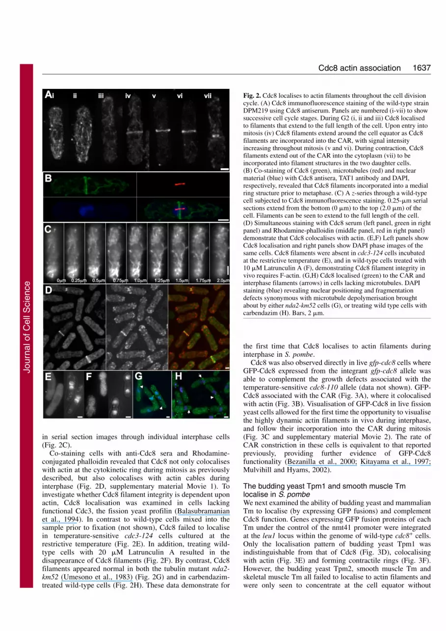
Cdc8 localises to actin filaments during interphaseImmunofluorescence revealed that Cdc8 localised to filaments,which extended throughout the length of the cell duringinterphase. The filaments were seen in cells throughout G2(Fig. 2Ai-iii), until the onset of mitosis (Fig. 2Aiv), when theCdc8 filaments extended around the cell equator (Fig. 2Aiv-vii). Simultaneous staining with anti-Cdc8 and TAT1antibodies revealed Cdc8 filaments incorporated into thenascent CAR in cells possessing a short pro-metaphase spindle(Fig. 2B). Upon completion of mitosis, Cdc8 filamentsextended from the edges of the CAR (Fig. 2Avii) into thesubsequent daughter cells. Cdc8 filaments can clearly be seen
Journal of Cell Science 120 (9)
Fig. 1. Cdc8 protein levels do not fluctuate during thecell division cycle. (A) A novel anti-Cdc8 antibodywas characterised by western blot of protein extractsfrom wild-type strains containing either the multicopyplasmid pREP41 (empty vector) or pREP41cdc8, orfrom a wild-type strain in which the plasmidpINT41gfp-cdc8 had been integrated at the leu1+
locus. All cells were grown in de-repressed conditions(– thiamine). An extract of wild-type cells containingthe empty vector, pREP41, shows a Cdc8 band thatmigrates at 27 kDa. The intensity of the bandincreased in wild-type cells in which cdc8+ wasadditionally expressed from a plasmid under thecontrol of the nmt41 promoter (pREP41cdc8+).Extracts from cells expressing chromosome integratedgfp-cdc8 showed an additional GFP-tagged Cdc8 at~54kDa, the intensity of which was comparable to theendogenous Cdc8 band. The same GFP-Cdc8 band was seen when the membrane was probed with anti-GFP antibody (B). (C) The samemembrane probed with TAT1 to demonstrate equal loading of protein. A population of DPM126 cells were synchronised using transient blockand release of the cdc25-22 allele. Two cell cycles were followed and samples were taken every 20 minutes for western blot analysis and forscoring the appearance of actin rings and septation index at each time point. Positions of molecular mass markers (in kDa) are indicated.(D) The proportion of cells possessing cytokinetic actomyosin rings (O) or a septum (+) at each time point over the two cell cycles. A westernblot of whole cell extracts from each time point was probed with both Cdc8 anti-sera (E) and TAT1 antibody (F). Cdc8 migrates as a constantdoublet throughout the cell cycle. Quantification of the ratio of TAT1 to Cdc8 doublet signals were analysed from three independentexperiments and revealed no fluctuation in Cdc8 levels as cells progress through the cell cycle.
Jour
nal o
f Cel
l Sci
ence
1637Cdc8 actin association
in serial section images through individual interphase cells(Fig. 2C).
Co-staining cells with anti-Cdc8 sera and Rhodamine-conjugated phalloidin revealed that Cdc8 not only colocaliseswith actin at the cytokinetic ring during mitosis as previouslydescribed, but also colocalises with actin cables duringinterphase (Fig. 2D, supplementary material Movie 1). Toinvestigate whether Cdc8 filament integrity is dependent uponactin, Cdc8 localisation was examined in cells lackingfunctional Cdc3, the fission yeast profilin (Balasubramanianet al., 1994). In contrast to wild-type cells mixed into thesample prior to fixation (not shown), Cdc8 failed to localisein temperature-sensitive cdc3-124 cells cultured at therestrictive temperature (Fig. 2E). In addition, treating wild-type cells with 20 "M Latrunculin A resulted in thedisappearance of Cdc8 filaments (Fig. 2F). By contrast, Cdc8filaments appeared normal in both the tubulin mutant nda2-km52 (Umesono et al., 1983) (Fig. 2G) and in carbendazim-treated wild-type cells (Fig. 2H). These data demonstrate for
the first time that Cdc8 localises to actin filaments duringinterphase in S. pombe.
Cdc8 was also observed directly in live gfp-cdc8 cells whereGFP-Cdc8 expressed from the integrant gfp-cdc8 allele wasable to complement the growth defects associated with thetemperature-sensitive cdc8-110 allele (data not shown). GFP-Cdc8 associated with the CAR (Fig. 3A), where it colocalisedwith actin (Fig. 3B). Visualisation of GFP-Cdc8 in live fissionyeast cells allowed for the first time the opportunity to visualisethe highly dynamic actin filaments in vivo during interphase,and follow their incorporation into the CAR during mitosis(Fig. 3C and supplementary material Movie 2). The rate ofCAR constriction in these cells is equivalent to that reportedpreviously, providing further evidence of GFP-Cdc8functionality (Bezanilla et al., 2000; Kitayama et al., 1997;Mulvihill and Hyams, 2002).
The budding yeast Tpm1 and smooth muscle Tmlocalise in S. pombeWe next examined the ability of budding yeast and mammalianTm to localise (by expressing GFP fusions) and complementCdc8 function. Genes expressing GFP fusion proteins of eachTm under the control of the nmt41 promoter were integratedat the leu1 locus within the genome of wild-type cdc8+ cells.Only the localisation pattern of budding yeast Tpm1 wasindistinguishable from that of Cdc8 (Fig. 3D), colocalisingwith actin (Fig. 3E) and forming contractile rings (Fig. 3F).However, the budding yeast Tpm2, smooth muscle Tm andskeletal muscle Tm all failed to localise to actin filaments andwere only seen to concentrate at the cell equator without
Fig. 2. Cdc8 localises to actin filaments throughout the cell divisioncycle. (A) Cdc8 immunofluorescence staining of the wild-type strainDPM219 using Cdc8 antiserum. Panels are numbered (i-vii) to showsuccessive cell cycle stages. During G2 (i, ii and iii) Cdc8 localisedto filaments that extend to the full length of the cell. Upon entry intomitosis (iv) Cdc8 filaments extend around the cell equator as Cdc8filaments are incorporated into the CAR, with signal intensityincreasing throughout mitosis (v and vi). During contraction, Cdc8filaments extend out of the CAR into the cytoplasm (vii) to beincorporated into filament structures in the two daughter cells.(B) Co-staining of Cdc8 (green), microtubules (red) and nuclearmaterial (blue) with Cdc8 antisera, TAT1 antibody and DAPI,respectively, revealed that Cdc8 filaments incorporated into a medialring structure prior to metaphase. (C) A z-series through a wild-typecell subjected to Cdc8 immunofluorescence staining. 0.25-"m serialsections extend from the bottom (0 "m) to the top (2.0 "m) of thecell. Filaments can be seen to extend to the full length of the cell.(D) Simultaneous staining with Cdc8 serum (left panel, green in rightpanel) and Rhodamine-phalloidin (middle panel, red in right panel)demonstrate that Cdc8 colocalises with actin. (E,F) Left panels showCdc8 localisation and right panels show DAPI phase images of thesame cells. Cdc8 filaments were absent in cdc3-124 cells incubatedat the restrictive temperature (E), and in wild-type cells treated with10 "M Latrunculin A (F), demonstrating Cdc8 filament integrity invivo requires F-actin. (G,H) Cdc8 localised (green) to the CAR andinterphase filaments (arrows) in cells lacking microtubules. DAPIstaining (blue) revealing nuclear positioning and fragmentationdefects synonymous with microtubule depolymerisation broughtabout by either nda2-km52 cells (G), or treating wild type cells withcarbendazim (H). Bars, 2 "m.
Jour
nal o
f Cel
l Sci
ence

1638
recruiting to the CAR during mitosis (Fig. 3G). In contrast toCdc8, when Tpm1, Tpm2 or either of the metazoan GFP-Tmswere overexpressed, the fusion proteins concentrated to asingle large bright aggregate within the cytoplasm (Fig. 3I),which was not associated with actin (not shown). An additionalconsequence of overexpressing Tm is that elongateduninucleate cells accumulated within the culture – a phenotypesynonymous with a cell cycle arrest.
Next, the ability of Tpm1, Tpm2, smooth Tm and skeletalTm to complement the temperature-sensitive cdc8-110 allelewas examined (Fig. 4A,B) in cdc8-110 cells in which genesencoding each Tm had been integrated into the genome andwere under the control of the nmt41 promoter. Cultures wereincubated at 36°C for 4 hours and the number of nuclei in 200cells was examined. Only cultures of cells expressing Cdc8 orsmooth muscle Tm had the normal percentage of binucleateaseptate cells (12%), whereas cells expressing Tpm1, Tpm2 orskeletal Tm accumulated as bi-nucleate and tetra-nucleatecells, demonstrating an inability to complement Cdc8 function.
Growth curves were generated for each strain at 36°C (Fig.4B). Only smooth muscle Tm was able to fully complementthe temperature-sensitive cdc8-110 allele (Fig. 4B, orangeline), producing a growth curve indistinguishable from cellsexpressing Cdc8 (Fig. 4B, black line). Budding yeast TPM1and TPM2 were only able to partially complement the cdc8-110 mutation (Fig. 4B, blue and green lines, respectively);however, the cells grew significantly slower than strainsexpressing either cdc8 or the smooth muscle Tm. Skeletalmuscle tropomyosin (Fig. 4B, dark blue line), was the only Tmexamined that failed to form any colonies on plates incubatedat 36°C (data not shown). These data indicate that only Cdc8and smooth muscle Tm were capable of fully complementingCdc8 function when they were stably expressed at levelsequivalent to the endogenous Cdc8 protein.
We were surprised that smooth muscle Tm was able tocomplement Cdc8 function, yet failed to localise to actinfilaments. One possibility was that its failure to localise was aresult of it being unable to compete with Cdc8 for actinbinding. We therefore examined GFP–smooth-Tm localisationin a cdc8-110 strain, and found that in this background itlocalised to actin filaments (Fig. 3H), which indicates thatalthough smooth muscle Tm is able to complement Cdc8function, it is unable to compete with the endogenous proteinfor actin binding in vivo.
Cdc8 is acetylated in vivoTo establish the significance of Cdc8 migration as a doublet onSDS-PAGE gels, Cdc8 was purified from fission yeast cells.Silver staining revealed the purified protein migrated on 12.5%SDS-PAGE gels as a doublet (Fig. 4C) – both bands wererecognised by the Cdc8 antisera (Fig. 4D). As in cell extracts,~80% of the purified Cdc8 migrated in the faster migratingband. The purity and mass of the protein was determined byelectron-spray mass spectroscopy, and revealed two peaks of18,964.7 Da and 19,005.5 Da (Fig. 4E) mass. The masses ofthe peaks are consistent for a mixed population of unacetylatedand acetylated Cdc8 (predicted masses are 18,964 and 19,006respectively), and the inability to sequence the purifiedendogenous protein indicates that this occurred at the N-terminus of the protein. The relative size of the peak of thelarger polypeptide was approximately four times that of the
Journal of Cell Science 120 (9)
Fig. 3. Localisation of GFP-Tm in S. pombe. (A) GFPautofluorescence of live DPM751 cells, show Cdc8 localises to theCAR during mitosis (left panel) and cytoplasmic filaments ininterphase cells (right panel). (B) GFP-Cdc8 (top panel and green inbottom panel) colocalisation with actin (middle panel and red inbottom panel) was confirmed in merged images (bottom panel) ofDPM751 cells stained with Rhodamine-conjugated phalloidin.(C) Real-time imaging of GFP-Cdc8 in live DPM809 cellsrevealed the dynamic nature of actin filaments in vivo.(D) Autofluorescence of DPM837 reveals the budding yeasttropomyosin TPM1 localises to the CAR in fission yeast cells.Rhodamine-phalloidin staining (Red) shows that GFP-Tpm1 (green)colocalises with actin (E). (F) Time-lapse imaging of DPM837reveals GFP-Tpm1 recruits to a functional CAR. (G) Imaging of liveDPM841 cells indicates smooth muscle Tm concentrate to the cellequator but fails to localise to the CAR in the presence of wild typeCdc8; but localises to the CAR in cells bearing the cdc8-110 allele(DPM924) when incubated at 36°C (H). (I) OverexpressedGFP–smooth-muscle-Tm concentrates to a single fluorescentamorphous cytoplasmic aggregate and brings about anaccumulation of uninucleate elongated cells (GFP, green; phase, red).Bars, 2 "m.
Jour
nal o
f Cel
l Sci
ence

1639Cdc8 actin association
smaller one, suggesting that the faster migrating Cdc8 band onSDS-PAGE gels is an acetylated form of the protein.
Affinity of endogenous and recombinant Cdc8tropomyosin for actinIt has been suggested that acetylation stabilises Tm’s coiled-coil structure at the amino terminus (Palm et al., 2003),allowing Tm dimers to interact to form filaments capable ofassociating with F-actin cables (Cammarato et al., 2004). Wewished to explore whether acetylation could play a similar rolein modifying the affinity of Cdc8 for actin and therefore the invivo function in S. pombe.
When recombinant tropomyosins are expressed in E. coli,they fail to become acetylated on their N-terminal Met residue,allowing the opportunity to examine the actin-bindingproperties of unacetylated Cdc8. A modified Cdc8 isoformwith an Ala-Ser di-peptide amino extension (Cdc8-AS) wasalso heterologously produced. This modification mimicsacetylation in studies of other Tms (Greenfield et al., 1994;Maytum et al., 2000; Monteiro et al., 1994). Massspectroscopic analysis of recombinant protein confirmed theisolation of unacetylated proteins of masses 18964.3 Da and
19122.4 Da respectively. The actin affinity of eachrecombinant protein was determined, and compared with thatof the endogenous Cdc8 dimer purified from S. pombe.
An example of an SDS-PAGE gel used for determiningbinding affinities is illustrated in Fig. 5A. The two gels shownrepresent a co-sedimentation experiment involvingrecombinant Cdc8. The top gel shows the pellet fractions, andthe bottom gel illustrates the supernatant fractions. All samplescontain 10 "M actin incubated with increasing concentrationsof Cdc8 dimer from left (lane 1) to right (lane 14). In both gels,actin is the top band, the density of which remains constant inall the samples. The lower, faster migrating band is Cdc8, thedensity of which increases from the lowest to the highest Cdc8dimer concentrations. Data for the three Cdc8 dimer isoformswere plotted and resulted in binding curves of sigmoidal shape,consistent for a Tm-binding curve (Fig. 5B). The bindingcoefficients (K50%) were determined after fitting thecooperative binding curves using the Hill equation (Table 1).
Co-sedimentation assay gels of endogenous Cdc8 purifiedfrom S. pombe (Fig. 5C) revealed the faster migratingcomponent of the doublet has a higher affinity for actin thanthe slower band. The majority of the slower migrating
Fig. 4. Cdc8 is acetylated in vivo. (A,B) cdc8-110 cellsexpressing additional integrant Tm genes under the controlof the nmt41 promoter were grown at 36°C in EMM2lacking thiamine. (A) The percentage of cells with two(black bars) or more than two (grey bars) nuclei werescored for each strain after 4 hours. (B) Growth curvesgenerated for each strain over a 6 hour growth at 36°C.Only Cdc8 and smooth muscle Tm were capable of fullycomplementing the cdc8-110 mutation. Endogenous Cdc8was purified from mid-log phase S. pombe cells andanalysed alongside total protein extracts by silver stainingof SDS-PAGE gels (C) and western blot analysis usingCdc8 antisera (D). A single doublet migrating at ~30kDawas revealed by silver staining (C), while Cdc8 antiserarecognised identical doublets in each lane. (E) Massspectroscopy analysis of the purified endogenous Cdc8revealed ~20% of this protein had a mass of 18964.1(predicted mass of Cdc8: 18,964.7 Da), while theremaining 80% had a mass pf 19005.5 Da, whichcorresponds to the predicted mass of acetylated Cdc8.
Jour
nal o
f Cel
l Sci
ence

1640
endogenous Cdc8 remains in the supernatant in co-sedimentation assays. The two isoforms of endogenous Cdc8were purified from the supernatant and pellet assay fractions(Fig. 5D), and mass spectroscopy revealed the faster migratingCdc8 band is acetylated, whereas the slower form isunacetylated. Therefore we can conclude that 80% of the Cdc8is constantly acetylated throughout the cell cycle (Fig. 1B).
In contrast to the budding yeast, skeletal, and smooth muscletropomyosins, the presence of the AS insertion at the N-
terminus of Cdc8 had little affect on its actin affinity. Unlikemany tropomyosins, unacetylated Cdc8 binds cooperatively toactin. The acetylated endogenous Cdc8 has a ~tenfold tighteraffinity for actin than either recombinant protein. Sequenceanalysis revealed there is a significant divergence in the N-terminal region adjacent to the predicted amino overlap region(McLachlan and Stewart, 1975) of Cdc8 when compared withother Tms (Fig. 6A), suggesting this region is important inregulating the filament formation.
Cdc8 regulates myosin binding to actinWe wished to explore whether Cdc8 played a role in regulatingthe interaction of myosin with actin. Pyrene-labelled actin wastitrated with myosin motor domains under equilibrium bindingconditions to assess whether the different Cdc8 proteins couldregulate the binding of myosin motor domains (now referredto as myosin) to actin. Each titration was repeated, andrepresentative data is presented in Fig. 6B, which show plotsof the raw data (grey lines) in which myosin had been titratedagainst phalloidin-stabilised pyrene actin or pyrene actinsaturated with Cdc8, Cdc8-AS or endogenous acetylated Cdc8dimers. The plot for actin alone is shown on each graph forcomparison. The sigmoidal shapes of the curves obtained forall three isoforms indicate that Cdc8 has an innate ability toregulate myosin binding by causing an initial inhibition ofmyosin binding (compare to actin alone), however thisinhibition is most dramatic in the presence of acetylated Cdc8.Curves were fitted to a two-state model (Maytum et al., 2000;McKillop and Geeves, 1993), to take into account the absenceof control proteins such as troponin in this system. The bestfits for each Cdc8 isoform (black lines) were superimposedonto the same axes as the corresponding raw data. Table 2shows the K1 (association constant for actin binding tomyosin), KT (the equilibrium between the on and off states ofactin, also called the C and M states) and n (cooperative unitsize), values that were calculated for each Cdc8 isoform. Theaffinity of myosin for actin (K1) did not vary dramaticallybetween the three Cdc8 proteins (Table 2). Owing to the lowsigmoidity of the Cdc8 and Cdc8-AS curves, the cooperativeunit size (n) cannot be clearly defined using this system. Inconclusion, the ability to regulate myosin binding to actin is anintrinsic property of Cdc8, and this inhibitory affect is in turnenhanced upon Tm acetylation.
Cdc8 filaments regulate myosin by occupying the closedposition on actinMyosin binding is regulated by changes in the position the Tmstrand sits upon the actin filament. Structural and biochemicalstudies have demonstrated that skeletal tropomyosin can bindto actin filaments in a blocked, closed or open position, whichhave decreasing inhibitory effects on myosin binding to actin
Journal of Cell Science 120 (9)
Fig. 5. Cdc8 requires N-terminal acetylation to associate with actinfilaments. (A) 10 "M actin incubated with increasing concentrationsof heterologous Cdc8 (1-20 "M in experiment shown) at 20°C for 30minutes in 20 mM MOPS, 30 mM KCl, 5 mM MgCl2, pH 7.0. Theactin was pelleted at 100,000 g and the equivalent samples of thepellet (upper panel) and supernatant (lower panel) were run on anSDS-PAGE gel, which was stained with Coomassie Blue.(B) Binding constants (K50%) were measured as the free Tm dimerconcentration at which the actin filament is half saturated by Cdc8dimer, and was determined as the ratio of density of actin against thefree concentrations of Cdc8 (circles), Cdc8-AS (+) or endogenousCdc8 (!) dimers, as measured by quantitative analysis ofsupernatant and pellet co-sedimentation gels (A), and calculatedusing the Hill equation (see Table 1). (C) Supernatant and pellet co-sedimentation gels of unacetylated and endogenous Cdc8. The fastermigrating endogenous Cdc8 band associated with actin in the pellet,while the slower migrating band was the prominent form in thesupernatant. (D) Endogenous Cdc8 purified from actin co-sedimentation pellets from C migrated more slowly thanunacetylated Cdc8 purified from E. coli.
Table 1. Actin-binding properties of yeast tropomyosinsProtein (Dimer) Origin K50 ("M) Reference
Cdc8 E. coli 4.9 This studyCdc8-AS E. coli 5.5 This studyCdc8 S. pombe 0.6 This studyTpm1/Tpm2 E. coli – (Maytum et al., 2000)Tpm1-AS E. coli 0.3-0.8 (Maytum et al., 2000)Tpm2-AS E. coli ~0.5 (Maytum et al., 2001)
Jour
nal o
f Cel
l Sci
ence

1641Cdc8 actin association
(McKillop and Geeves, 1993; Vibert et al., 1997). One furtherway to elucidate the role of Cdc8 in myosin regulation is toexamine whether acetylated Cdc8 affects the rate at whichmyosin binds to actin. Fast reaction kinetics experimentsrevealed that the presence of acetylated Cdc8 had no affect onthe rate of initial myosin binding (not shown). This isconsistent with Cdc8 filaments occupying the closed and notthe blocked position on actin filaments, as the latter wouldinhibit binding and reduce the rate of myosin binding to actin.These data predict that Cdc8 filaments modulate myosinfunction by sitting predominantly in the closed position on
actin filaments, thus inhibiting its initial binding until thetropomyosin moves cooperatively into the open position.
To confirm this, image reconstruction of electronmicrographs of actin-Cdc8 filaments was carried out to revealwhere Cdc8 strands sit on actin. In contrast to control F-actinsamples lacking tropomyosin (Fig. 7A), yeast tropomyosinstrands could be detected directly in electron micrographs ofnegatively stained F-actin–Cdc8 filaments (Fig. 7B) and wasfound coiling around the actin filaments with a characteristicpattern (Lehman et al., 1994) (Fig. 7B, arrows). In addition,excess unbound Cdc8 could be seen to form narrow elongatedand continuous filaments, free of actin, in the background ofF-actin and Cdc8 tropomyosin samples (not shown),suggesting that end-to-end bonded Cdc8 forms filaments arestable even in isolation (Flicker et al., 1982).
Helical reconstruction of single actin-Cdc8 filaments alwaysshowed tropomyosin strands in the closed position (n=15).Averages of eight filament reconstructions with the best phaseagreement were constructed from micrographs of actin-Cdc8filaments as described previously (Lehman et al., 1994; Lehmanet al., 2000; Vibert et al., 1997). Surface views of F-actin-Cdc8reconstructions revealed Cdc8 exclusively occupies the ‘closed’position on actin (Fig. 8B,C), i.e. it localises on the outer edgeof actin subdomains 3 and 4 next to the cleft separating the inner(subdomains 3 and 4) and outer (subdomains 1 and 2) domainsof actin, which is the same position previously described fortropomyosin alone or troponin-tropomyosin-regulated filamentsin the presence of Ca2+ (the closed or Ca2+-induced position)(Lehman et al., 1994; Lehman et al., 2000; Pirani et al., 2005).Helical projections (Fig. 8D,E) and transverse sections (F andG) of maps of the 3D reconstructions confirmed the positionalspecificity of the tropomyosin strands in the closed position.Reconstructions of actin-Cdc8 filaments were displayed at 5 and10 sigma above the mean density (Fig. 8B,C respectively), andcorresponded to a resolution of 26 Å. The Cdc8 density is seento form a continuous strand at a five times higher threshold thannormally considered the industry standard. The density cross-section is consistent with Cdc8 existing as a dimer. The abilityto observe continuous tropomyosin density at such highthreshold levels in reconstructions (sigma=10) suggests a levelof precision greater than that for the binding of othertropomyosins, such as for skeletal muscle !-tropomyosin in theabsence of troponin, in comparable actin-tropomyosincomplexes (Lehman et al., 2000; Pirani et al., 2005).
DiscussionThe tropomyosins play a key role in regulating the dynamicsof actin filaments in cells as diverse as muscle fibres and yeast.Their function requires that the protein forms parallel dimersthat polymerise head to tail to generate strands which coil
Fig. 6. Acetylated Cdc8 regulates myosin function in vitro.(A) Bioinformatic analysis reveals divergence within the N-terminusof S. pombe Cdc8. (B) Titration curves for myosin binding to either50 nM phalloidin-stabilised pyrene-labelled actin alone (light greyline) or actin saturated with unacetylated Cdc8, Cdc8-AS orendogenous Cdc8 (black line). Curves of best fit data aresuperimposed on each Cdc8 graph (dark grey). The actin curve ispresent for reference in all graphs. Titration curves for each of theCdc8 isoforms were sigmoidal in shape, demonstrating that all threehave an inhibitory affect on myosin binding, although the inhibitionis significantly stronger for the endogenous acetylated Cdc8.
Table 2. Parameters of fits for titration curvesProtein (Dimer) K1 (#104 M–1) KT K2 n Reference
Cdc8 3.36 0.47 200 7 This studyCdc8-AS 2.39 0.99 200 ND* This studyAcetylated Cdc8 5.37 0.02 200 7 This studyTpm1-AS 4.43 0.37 200 9 (Maytum et al., 2000)Tpm2-AS 4.22 0.33 200 7 (Maytum et al., 2001)
*ND, not determined.
Jour
nal o
f Cel
l Sci
ence

1642
around actin filaments providing stability to the polymer. Theposition the Tm strand sits upon the actin cable cannot onlyaffect the stability of the thin filament, but also regulates theinteraction of myosin with actin. The two budding yeast Tmsboth have crucial but non-overlapping roles in stabilising actinfilaments, upon which myosin motors transport cargoes (Dreeset al., 1995; Liu and Bretscher, 1992). By contrast, the fissionyeast S. pombe possesses a single tropomyosin, which isessential for formation and contraction of the CAR(Balasubramanian et al., 1992). It is also required for themaintenance of polarised cell growth as demonstrated by thedumbbell-shaped morphology of cells lacking functional Cdc8(Chang et al., 1996; Pelham, Jr and Chang, 2001). Here wereport that in addition to stabilising actin filaments throughoutthe cell cycle, Cdc8 can also regulate myosin function.
In this study, we have carried out a detailed analysis of thebiochemical, molecular and cell biological properties of Cdc8,which has facilitated the further elucidation of its cellularfunction. Parallel localisation studies using either an anti-Cdc8
antibody or a functional GFP-Cdc8 fusion protein confirmedCdc8 localisation at the CAR. In addition, Cdc8 also localiseswith actin to a complex lattice of filaments, which extendthroughout the cytoplasm of interphase cells. In addition, thenovel gfp-cdc8 strain described here has proven to be aninvaluable tool to further investigate the dynamic nature ofthese filaments in vivo. The dynamic filaments were shown tocoincide with actin, because their integrity was dependent uponpolymerised actin. These findings are consistent with Cdc8having a role both in maintaining CAR integrity during celldivision and in stabilising actin filaments during interphasethus maintaining polarised cell growth (Balasubramanian et al.,1992; Ishiguro and Kobayashi, 1996; Pelham and Chang,2001).
Using an anti-Cdc8 antibody in conjunction with massspectroscopy it was possible to determine that Cdc8 proteinlevels did not vary and that 80% of the protein was N-terminally acetylated throughout the cell division cycle.Acetylation plays an important role in modulating theefficiency with which many tropomyosins interact with actinby modulating the structure of the N-terminus that facilitateshead-to-tail interactions of Tm dimers (Hitchcock-DeGregoriand Heald, 1987; Palm et al., 2003; Urbancikova andHitchcock-DeGregori, 1994). This is certainly the case inbudding yeast where recombinant Tpm1 and Tpm2 require theaddition of an Ala-Ser (AS) dipeptide, which is a good mimicof N-terminal acetylation for metazoan Tms (Monteiro et al.,1994), to see any detectable association with actin (Maytum etal., 2000; Maytum et al., 2001).
Expression of GFP-Tm fusion proteins in fission yeast cellsallowed the opportunity to compare the ability of each Tm tolocalise with its ability to function in S. pombe. Of the Tmsexamined, only Cdc8 and smooth muscle Tm were able tolocalise and function in vivo, however the smooth muscle Tmcould not compete with the fission yeast Tm for actin bindingin vivo. Budding yeast Tpm1 was able to localise but onlypartially complement Cdc8 function. However, Tpm2 failed tolocalise, which may be explained by the fact that Tpm2 lacksan internal region conserved in Tpm1 and Cdc8, which may beessential for Tm function in S. pombe. Biochemical analysisrevealed that unmodified recombinant Cdc8 is capable ofassociating with actin, albeit with a low affinity, which is incontrast to either budding yeast Tm, and may in part explainwhy both Tpm1 and Tpm2 were unable to complement Cdc8function in vivo. Tpm2 and skeletal Tm fusion proteins bothconcentrate near the CAR during mitosis, yet actin filamentassociation was barely detectable. In addition, overexpressionof Tpm1, Tpm2, smooth or skeletal muscle Tm resulted in anaccumulation of elongated uninucleate cells. As it is onlypossible for two Tm filaments to simultaneously associate withthe same single actin polymer, overexpressing Tm will notmake more Tm associate with actin filaments. The arrest weobserve is therefore probably brought about by eitherdisrupting the dynamic properties of fission yeast actinfilaments or by triggering a stress response. Consistent with thelatter, actin is not seen to associate with the aggregate, and weobserve no defects when we overexpress Cdc8 in S. pombecells. However, the precise nature of the arrest is currentlyunder investigation.
Purifying endogenous Cdc8 from fission yeast cells inmilligram quantities allowed the opportunity to define the
Journal of Cell Science 120 (9)
Fig. 7. Electron micrographs of F-actin-Cdc8 complexes. Negativestaining of (A) F-actin alone (no tropomyosin), (B) F-actin-Cdc8;note the obliquely oriented strands that are characteristic oftropomyosin (Lehman et al., 1994) (several are indicated witharrows, and are best seen by viewing the figure at a glancing angle).When compared with control F-actin, the actin subunit structure ofthe decorated filaments appeared less well defined, owing to thebinding of additional protein. Occasionally, unbound Cdc8 formedvery narrow but elongated and continuous filaments, visible insamples of F-actin and Cdc8 tropomyosin (not shown). Bar, 50 nm.
Jour
nal o
f Cel
l Sci
ence

1643Cdc8 actin association
actin-binding constant of an endogenous yeast Tm for the firsttime. This has revealed that N-terminal acetylation of Cdc8results in its binding to actin with an affinity similar to that ofboth Tpm1-AS and Tpm2-AS from budding yeast, but atenfold tighter affinity than for unmodified Cdc8. Similarly, inco-sedimentation assays of endogenous Cdc8, acetylated Cdc8was exclusively seen in the actin pellet, whereas thesupernatant predominantly contained the unacetylated isoform.It is unclear at this stage why a significant sub-population ofthe cellular Cdc8 is unmodified, but this is currently underinvestigation. However, analysis of Cdc8 from cells lacking F-actin suggests that Cdc8 acetylation is not dependent uponother components of the actin cytoskeleton (data not shown).
Surprisingly, addition of an Ala-Ser dipeptide at the N-terminus did not mimic Cdc8 N-terminal acetylation, becausethe K50% for Cdc8-AS was not significantly different fromunacetylated Cdc8. Why Cdc8 should behave so differentlyfrom other Tms is unclear; however, there is significantdivergence in the N-terminal Cdc8 sequence adjacent to thepredicted 8-10 amino acid head-to-tail overlap region for thisTm (McLachlan and Stewart, 1975), which could have asignificant affect upon Tm-Tm contacts.
We have not yet determined whether any of the non-endogenous Tms are acetylated in fission yeast, but the factthat only smooth Tm can bind to actin in the absence of N-terminal modification whereas Tpm1, Tpm2 and skeletal Tmdo not (Coulton et al., 2006; Maytum et al., 2001) may explainwhy only smooth muscle Tm was able to function in yeast (Fig.4A,B). In addition, skeletal Tm requires the interaction of thetroponin complex in order to bind actin with a high affinity,which may explain why this protein failed to either localise orfunction in S. pombe. Although endogenous S. cerevisiaeTpm1 has been purified and shown to associate with actinpreviously, neither the K50% or KT equilibrium constants haveyet been defined for endogenous acetylated forms of either ofthe budding yeast Tms. Study of acetylated Tpm1 and Tpm2may reveal whether the AS insertion mimics the true nature ofacetylation for any of the fungal Tms.
The fact that Cdc8 binds to actin raises the possibility thatit may also have a role in regulating myosin function. Cdc8confers cooperativity (sigmoidal titration curves) on themyosin motor domain binding to actin, which indicates apotential to regulate myosin binding. The low value of KT (theequilibrium constant between the closed and open forms of thefilament) indicates that in the absence of myosin the Cdc8spends 98% of its time in the C or ‘off’ state. This value of KTis much smaller than for most tropomyosins studied to date(KT=0.02) and suggests that the C-state is stabilised relative tothe O-state (Lehman et al., 2000; Maytum et al., 1999; Piraniet al., 2005).
A number of recent studies suggest that movement of Tmon actin between these different conformations is related to theflexibility of the protein (Bacchiocchi et al., 2004; Chen andLehrer, 2004; Heller et al., 2003; Lehrer et al., 1997; Singh andHitchcock-DeGregori, 2003), with more rapid transitions beingseen with flexible Tms. For Cdc8, removal of the acetylationgroup results in KT being reduced from 0.02 to 0.47, i.e. theopen state occupancy rises from 2% to 32%. Thus the increasedstability of head-to-tail interactions facilitated by acetylationincreases the stability of the Cdc8 strand in the closedconformation on actin. Polymerised acetylated Cdc8 may
Fig. 8. Three-dimensional reconstructions of negatively stained thinfilaments. Surface views of reconstructions of (A) F-actin controlfilaments (actin subdomains 1 to 4 marked on one actin monomer) and(B,C) F-actin-Cdc8. Additional density corresponding to tropomyosin(arrows) is observed for the filaments containing Cdc8. Cdc8 occupiesthe ‘closed’ position on actin, i.e. it localises on the outer edge of actinsubdomains 3 and 4 next to the cleft separating the inner (subdomains 3and 4) and outer (subdomains 1 and 2) domains of actin, which is thesame position previously described for troponin-tropomyosin-regulatedfilaments in the absence of Ca2+ (Lehman et al., 1994; Vibert et al.,1997). Reconstructions were displayed at 5 sigma above the meandensity in A and B and at 10 sigma in C; note the Cdc8 density forms acontinuous strand even at very high threshold values. Helical projections(D,E) and transverse sections (F,G) of maps of the 3D reconstructions inA and B. (D,F) F-actin (E,G), F-actin-Cdc8. Actin subdomainsnumbered in (F). Helical projections (i.e. densities in the reconstructionsprojected onto a plane perpendicular to the axis of the filament), whichshow the average location of Cdc8 (arrows) on the inner domain of actin(E). Transverse sections show that the densities associated with Cdc8 areadjacent to subdomains 2 and 4 of actin (G). Also note that Cdc8 is notobserved on pure actin filaments (no Cdc8) (A,D,F).
Jour
nal o
f Cel
l Sci
ence

1644
therefore have a reduced flexibility or higher affinity for actin inthe C-state when compared with other Tms, with acetylationmodulating the stability or interactions of the Cdc8 strand. Thesedata are consistent with the finding that only Tms with a low KT(i.e. smooth muscle Tm and fibroblast Tm) are capable ofcomplementing Cdc8 function in vivo, providing strongevidence that unlike the budding yeast Tms, Cdc8 has essentialroles in regulating myosin function in addition to stabilising actinfilaments (Balasubramanian et al., 1992; Coulton et al., 2006;Maytum et al., 2001).
These data are consistent with the high quality of the EMreconstructions for the actin-Cdc8 filaments. These filamentsshow less variability than other actin-Tm filaments studied todate. This is consistent with the Cdc8 being predominantly in asingle (i.e. closed) conformation. Unfortunately, because of itslow actin affinity, it is not experimentally feasible to examine theposition of unacetylated Cdc8 upon actin filaments by electronmicroscopy at this time.
EM studies of actin-Cdc8 filaments suggest that Cdc8 doesnot require actin to form a number of stable end-to-endinteractions. Preliminary EM studies of rotary-shadowed Cdc8reveals strands in the absence of actin – a property not reportedfor other tropomyosins – again showing increased strength ofTm-Tm contacts. Yeast mutant analysis may provide a means bywhich to explore the structure and stability of Tm filaments. Itis intriguing to speculate that the rigid conformation of theacetylated Cdc8 may affect the actin polymer, thus increasingthe energy requirement to move Cdc8 into the open position.Overlay comparison of the structure of actin filaments in theabsence and presence of acetylated Cdc8 suggests a reproducibleand consistent difference in the position of the actin densities(not shown). However the small sample size in this study doesnot provide conclusive evidence for this hypothesis at present.
The cellular requirement for actin filaments with tropomyosinstrand formation remains unclear. We show here that Cdc8 canform stable complexes with actin and can have the potential toregulate the interaction of myosin motors with actin. How sucha regulation would work in the cell remains undefined, and iscurrently being explored within our laboratory. However, aprevious study provides in vivo evidence for a role of Tm inregulating unconventional myosin function in non-muscle cells(Tang and Ostap, 2001). Using a genetically tractable system,such as yeast, will allow the opportunity to rapidly defineregulator proteins and regions within the Tm dimer required forit to regulate myosin-actin association in non-muscle cells.
Materials and MethodsYeast cell culture and strainsThe strains used in this study are listed in supplementary material Table S1. Cellculture and maintenance were carried out as described (Moreno et al., 1991). Cellswere grown in minimal medium (EMM2) supplemented with the appropriate aminoacids. Synchronous cultures were generated by transient arrest using the cdc25-22mutation in a strain possessing the myo2-gfp allele (Mulvihill and Hyams, 2002).
Molecular biologyS. pombe cdc8+ cDNA was isolated from fission yeast genomic DNA using ExpandHigh fidelity polymerase with the primers oDM111 (5$-CATATGGATAAGCTT -AGAGAGAAAATTAATGCCGCTCGT-3$) and oDM112 (5$-GGGCCCCTACAA -ATCCTCAAGAGCTT-3$), cloned into pTOPO2.1 (Invitrogen) and sequenced(creating pTOPOcdc8+). The cdc8-AS allele was created by PCR using primersoDM109 (5$-CATATGGCTAGCATGG-ATAAGCTTAGAGAGAAAAT-3$) andoDM112, cloned into pTOPO2.1 and sequenced (creating pTOPOcdc8-AS). Thecdc8+ and cdc8-AS alleles were subsequently cloned into the pJC20 bacterial T7expression vector (Konrad, 1993), and the S. pombe expression plasmids pREP41,pINT41 and pINT41NGFP (Craven et al., 1998). cDNA encoding for smTM, skTM,
TPM1 and TPM2 were isolated from lab stocks, sequenced and each cloned into theNdeI-BamHI sites of pINT41 and pINT41NGFP.
Immunological techniquesStandard immunological methods were used as described (Harlow and Lane, 1988).Anti-Cdc8 antibodies were created by immunising an SPF rabbit with purified full-length recombinant Cdc8 protein with Titre Max Gold adjuvant. Sera were collectedat 1-month intervals 1 week after each boost.
Western blotting5#107 cells were harvested from mid-log phase cultures of S. pombe cells, washedin ice-cold STOP buffer (Simanis and Nurse, 1986), and frozen in liquid nitrogen priorto storage at –80°C. Protein was extracted by resuspending cells in EXPY buffer [HEbuffer (Simanis and Nurse, 1986) supplemented with 1 "g/ml AEBSF, 1 "g/mlantipain, 1 "g/ml aprotinin, 10 "M benzamidine, 1 "g/ml chymostatin, 1 "g/ml E64,1 "g/ml leupeptin, 1 "g/ml pepstatin, 5 "M phenanthroline, and 1 mM PMSF] anddisrupting them with 500 "l of acid-washed glass beads in a ThermoSavant Fastprep120 (MP Biochemicals, Irvine, CA). Samples were then boiled with SDS-PAGEloading buffer. Western blotting detection was carried out using alkaline phosphatasereagents. Cdc8 levels were determined by measuring band intensities on the scannedblot using Scion image software. Cdc8 serum was used at a concentration of 1:1000,anti-GFP antibodies (generous gift from W. Gullick, University of Kent, UK) wereused at 1:100 and TAT1 anti-!-tubulin antibodies were used at 1:100 (Woods et al.,1989).
Fluorescence microscopyImmunofluorescence microscopy was performed as described (Hagan and Hyams,1988) except glutaraldehyde was omitted. Anti-Cdc8 sera was used at a dilution of1:100. Cells were visualised using an Olympus IX71 microscope withPLAPO100XO/TIRFM-SP/1.45NA lens mounted on a PIFOC® Z-axis focus drive(Physik Instruments, Karlsruhe, Germany) and illuminated with an automated 300WXenon light source (Sutter, Novato, CA) with appropriate filters. Micrographs werecaptured with a Coolsnap HQ digital camera (Roper Scientific, Tucson, AZ)controlled with Metamorph software (Molecular Devices, Downington, PA). Livecells were grown, visualised, captured and images were processed as describedpreviously (Grallert et al., 2004).
Protein purificationRecombinant Cdc8, and Cdc8-AS proteins were expressed (from pJC20cdc8+ andpJC20cdc8-AS) in BL21 DE3 cells. Mid-log cultures were grown for 3 hours with100 mg/l IPTG. Cells were harvested, resuspended in 30 ml lysis buffer (20 mM Tris-HCl pH 7.5, 100 mM NaCl, 2 mM EGTA and 5 mM MgCl2), lysed by sonication andheated at 80°C for 10 minutes. Denatured proteins were removed by centrifugation.The soluble Cdc8 was isolated by isoelectric precipitation at pH 4.55, resuspended in20 ml FPLC running buffer (5 mM PO4
- pH 7.0, 100 mM NaCl) with 10 mg/l DNaseand 10 mg/l RNase, and incubated at 4°C for 2 hours. The Cdc8 was purified twiceby FPLC using 2 # 5 ml Pharmacia HiTrap-Q columns in tandem, by elution with a100-900 mM NaCl gradient. Cdc8 was isolated from appropriate fractions byisolectric precipitation at pH 4.55 and resuspended in 5 mM Tris-HCl pH 7.0. Thepurity and mass of the proteins were determined by mass spectroscopy, while parallelBradford, gel densitometry and spectroscopic analyses determined proteinconcentrations. Endogenous Cdc8 was isolated from cells from 20 litres of mid-logfission yeast cells. Harvested cells were resuspended in EXPY buffer, cell walls weredisrupted, and incubated at 80°C for 10 minutes. Endogenous Cdc8 was purified byisoelectric precipitation and FPLC as for recombinant Cdc8. Rabbit actin was purifiedas described previously (Spudich and Watt, 1971).
Co-sedimentation assays and fluorescence titrationsThese were undertaken as described previously (Coulton et al., 2006) using a modifiedversion of the McKillop and Geeves model (Maytum et al., 1999).
Electron microscopy and helical reconstructionsCdc8 purified from S. pombe (endogenous) (120 "M) was mixed with F-actin (24"M), to allow Cdc8-actin filament formation, in buffer containing 20 mM MOPS, pH7.0, 30 mM KCl and 5 mM MgCl2. The actin-Cdc8 filaments were diluted 20-fold inthe same buffer immediately prior to applying to carbon-coated electron microscopygrids. Samples were negatively stained, electron microscopy images captured at45,000# under low-dose conditions (~12e–/Å), and helical reconstructions werecarried out as described previously (Lehman et al., 2000; Moody et al., 1990; Owenet al., 1996).
We gratefully acknowledge Roger Craig (University ofMassachusetts School of Medicine) for use of Electron Microscopyfacilities, supported by NIH Shared Instrument grant RR08426 (toRoger Craig). We thank Agnes Grallert and Steve Bagley from thePatterson Cancer Research Institute for their time, advice and use ofmicroscopes to capture Cdc8 filaments time-lapse sequences and
Journal of Cell Science 120 (9)
Jour
nal o
f Cel
l Sci
ence
1645Cdc8 actin association
Victoria Hatch for technical assistance. We also thank members of theGeeves and Mulvihill labs for helpful discussions and comments on themanuscript. This study was funded by a BBSRC David PhillipsFellowship to D.P.M. and NIH grants HL36153 (to W.L.) andAR041637 (to M.A.G.).
ReferencesBacchiocchi, C., Graceffa, P. and Lehrer, S. S. (2004). Myosin-induced movement of
alphaalpha, alphabeta, and betabeta smooth muscle tropomyosin on actin observed bymultisite FRET. Biophys. J. 86, 2295-2307.
Balasubramanian, M. K., Helfman, D. M. and Hemmingsen, S. M. (1992). A newtropomyosin essential for cytokinesis in the fission yeast S. pombe. Nature 360, 84-87.
Balasubramanian, M. K., Hirani, B. R., Burke, J. D. and Gould, K. L. (1994). TheSchizosaccharomyces pombe cdc3+ gene encodes a profilin essential for cytokinesis. J.Cell Biol. 125, 1289-1301.
Bezanilla, M., Wilson, J. M. and Pollard, T. D. (2000). Fission yeast myosin-II isoformsassemble into contractile rings at distinct times during mitosis. Curr. Biol. 10, 397-400.
Brown, J. H., Kim, K. H., Jun, G., Greenfield, N. J., Dominguez, R., Volkmann, N.,Hitchcock-DeGregori, S. E. and Cohen, C. (2001). Deciphering the design of thetropomyosin molecule. Proc. Natl. Acad. Sci. USA 98, 8496-8501.
Cammarato, A., Hatch, V., Saide, J., Craig, R., Sparrow, J. C., Tobacman, L. S. andLehman, W. (2004). Drosophila muscle regulation characterized by electron microscopyand three-dimensional reconstruction of thin filament mutants. Biophys. J. 86, 1618-1624.
Chang, F., Woollard, A. and Nurse, P. (1996). Isolation and characterization of fissionyeast mutants defective in the assembly and placement of the contractile actin ring. J.Cell Sci. 109, 131-142.
Chen, Y. and Lehrer, S. S. (2004). Distances between tropomyosin sites across the musclethin filament using luminescence resonance energy transfer: evidence for tropomyosinflexibility. Biochemistry 43, 11491-11499.
Coulton, A., Lehrer, S. S. and Geeves, M. A. (2006). Functional homodimers andheterodimers of recombinant smooth muscle tropomyosin. Biochemistry 45, 12853-12858.
Craven, R. A., Griffiths, D. J., Sheldrick, K. S., Randall, R. E., Hagan, I. M. and Carr,A. M. (1998). Vectors for the expression of tagged proteins in Schizosaccharomycespombe. Gene 221, 59-68.
Drees, B., Brown, C., Barrell, B. G. and Bretscher, A. (1995). Tropomyosin is essentialin yeast, yet the TPM1 and TPM2 products perform distinct functions. J. Cell Biol. 128,383-392.
Flicker, P. F., Phillips, G. N., Jr and Cohen, C. (1982). Troponin and its interactions withtropomyosin. An electron microscope study. J. Mol. Biol. 162, 495-501.
Grallert, A., Krapp, A., Bagley, S., Simanis, V. and Hagan, I. M. (2004). Recruitmentof NIMA kinase shows that maturation of the S. pombe spindle-pole body occurs overconsecutive cell cycles and reveals a role for NIMA in modulating SIN activity. GenesDev. 18, 1007-1021.
Greenfield, N. J., Stafford, W. F. and Hitchcock-DeGregori, S. E. (1994). The effect ofN-terminal acetylation on the structure of an N-terminal tropomyosin peptide and alphaalpha-tropomyosin. Protein Sci. 3, 402-410.
Gunning, P. W., Schevzov, G., Kee, A. J. and Hardeman, E. C. (2005). Tropomyosinisoforms: divining rods for actin cytoskeleton function. Trends Cell Biol. 15, 333-341.
Hagan, I. M. and Hyams, J. S. (1988). The use of cell division cycle mutants to investigatethe control of microtubule distribution in the fission yeast Schizosaccharomyces pombe.J. Cell Sci. 89, 343-357.
Harlow, E. and Lane, D. (1988). Antibodies: A Laboratory Manual. Cold Spring Harbor:Cold Spring Harbor Laboratory Press.
Heller, M. J., Nili, M., Homsher, E. and Tobacman, L. S. (2003). Cardiomyopathictropomyosin mutations that increase thin filament Ca2+ sensitivity and tropomyosin N-domain flexibility. J. Biol. Chem. 278, 41742-41748.
Hitchcock-DeGregori, S. E. and Heald, R. W. (1987). Altered actin and troponin bindingof amino-terminal variants of chicken striated muscle alpha-tropomyosin expressed inEscherichia coli. J. Biol. Chem. 262, 9730-9735.
Ishiguro, J. and Kobayashi, W. (1996). An actin point-mutation neighboring the‘hydrophobic plug’ causes defects in the maintenance of cell polarity and septumorganization in the fission yeast Schizosaccharomyces pombe. FEBS Lett. 392, 237-241.
Kitayama, C., Sugimoto, A. and Yamamoto, M. (1997). Type II myosin heavy chainencoded by the myo2 gene composes the contractile ring during cytokinesis inSchizosaccharomyces pombe. J. Cell Biol. 137, 1309-1319.
Konrad, M. (1993). Molecular analysis of the essential gene for adenylate kinase from thefission yeast Schizosaccharomyces pombe. J. Biol. Chem. 268, 11326-11334.
Kurahashi, H., Imai, Y. and Yamamoto, M. (2002). Tropomyosin is required for the cellfusion process during conjugation in fission yeast. Genes Cells 7, 375-384.
Lehman, W., Craig, R. and Vibert, P. (1994). Ca(2+)-induced tropomyosin movement inLimulus thin filaments revealed by three-dimensional reconstruction. Nature 368, 65-67.
Lehman, W., Hatch, V., Korman, V., Rosol, M., Thomas, L., Maytum, R., Geeves, M.A., Van Eyk, J. E., Tobacman, L. S. and Craig, R. (2000). Tropomyosin and actinisoforms modulate the localization of tropomyosin strands on actin filaments. J. Mol.Biol. 302, 593-606.
Lehrer, S. S., Golitsina, N. L. and Geeves, M. A. (1997). Actin-tropomyosin activation ofmyosin subfragment 1 ATPase and thin filament cooperativity. The role of tropomyosinflexibility and end-to-end interactions. Biochemistry 36, 13449-13454.
Liu, H. P. and Bretscher, A. (1989). Disruption of the single tropomyosin gene in yeastresults in the disappearance of actin cables from the cytoskeleton. Cell 57, 233-242.
Liu, H. and Bretscher, A. (1992). Characterization of TPM1 disrupted yeast cellsindicates an involvement of tropomyosin in directed vesicular transport. J. Cell Biol.118, 285-299.
Maytum, R., Lehrer, S. S. and Geeves, M. A. (1999). Cooperativity and switching withinthe three-state model of muscle regulation. Biochemistry 38, 1102-1110.
Maytum, R., Geeves, M. A. and Konrad, M. (2000). Actomyosin regulatory propertiesof yeast tropomyosin are dependent upon N-terminal modification. Biochemistry 39,11913-11920.
Maytum, R., Konrad, M., Lehrer, S. S. and Geeves, M. A. (2001). Regulatory propertiesof tropomyosin effects of length, isoform, and N-terminal sequence. Biochemistry 40,7334-7341.
McKillop, D. F. and Geeves, M. A. (1993). Regulation of the interaction between actinand myosin subfragment 1, evidence for three states of the thin filament. Biophys. J. 65,693-701.
McLachlan, A. D. and Stewart, M. (1975). Tropomyosin coiled-coil interactions: evidencefor an unstaggered structure. J. Mol. Biol. 98, 293-304.
Monteiro, P. B., Lataro, R. C., Ferro, J. A. and Reinach, F. de C. (1994). Functionalalpha-tropomyosin produced in Escherichia coli. A dipeptide extension can substitute theamino-terminal acetyl group. J. Biol. Chem. 269, 10461-10466.
Moody, C., Lehman, W. and Craig, R. (1990). Caldesmon and the structure of smoothmuscle thin filaments: electron microscopy of isolated thin filaments. J. Muscle Res. CellMotil. 11, 176-185.
Moreno, S., Klar, A. and Nurse, P. (1991). Molecular genetic analysis of fission yeastSchizosaccharomyces pombe. Meth. Enzymol. 194, 795-823.
Mulvihill, D. P. and Hyams, J. S. (2002). Cytokinetic actomyosin ring formation andseptation in fission yeast are dependent on the full recruitment of the polo-like kinasePlo1 to the spindle pole body and a functional spindle assembly checkpoint. J. Cell Sci.115, 3575-3586.
Nurse, P., Thuriaux, P. and Nasmyth, K. (1976). Genetic control of the cell division cyclein the fission yeast Schizosaccharomyces pombe. Mol. Gen. Genet. 146, 167-178.
Owen, C. H., Morgan, D. G. and DeRosier, D. J. (1996). Image analysis of helical objects:the Brandeis Helical Package. J. Struct. Biol. 116, 167-175.
Palm, T., Greenfield, N. J. and Hitchcock-DeGregori, S. E. (2003). Tropomyosin endsdetermine the stability and functionality of overlap and troponin T complexes. Biophys.J. 84, 3181-3189.
Pelham, R. J., Jr and Chang, F. (2001). Role of actin polymerization and actin cables inactin-patch movement in Schizosaccharomyces pombe. Nat. Cell Biol. 3, 235-244.
Perry, S. V. (2001). Vertebrate tropomyosin: distribution, properties and function. J. MuscleRes. Cell Motil. 22, 5-49.
Pirani, A., Xu, C., Hatch, V., Craig, R., Tobacman, L. S. and Lehman, W. (2005). Singleparticle analysis of relaxed and activated muscle thin filaments. J. Mol. Biol. 346, 761-772.
Pittenger, M. F. and Helfman, D. M. (1992). In vitro and in vivo characterization of fourfibroblast tropomyosins produced in bacteria: TM-2, TM-3, TM-5a, and TM-5b are co-localized in interphase fibroblasts. J. Cell Biol. 118, 841-858.
Polevoda, B., Cardillo, T. S., Doyle, T. C., Bedi, G. S. and Sherman, F. (2003). Nat3pand Mdm20p are required for function of yeast NatB Nalpha-terminal acetyltransferaseand of actin and tropomyosin. J. Biol. Chem. 278, 30686-30697.
Schott, D., Ho, J., Pruyne, D. and Bretscher, A. (1999). The COOH-terminal domain ofMyo2p, a yeast myosin V, has a direct role in secretory vesicle targeting. J. Cell Biol.147, 791-808.
Simanis, V. and Nurse, P. (1986). The cell cycle control gene cdc2+ of fission yeast encodesa protein kinase potentially regulated by phosphorylation. Cell 45, 261-268.
Singer, J. M. and Shaw, J. M. (2003). Mdm20 protein functions with Nat3 protein toacetylate Tpm1 protein and regulate tropomyosin-actin interactions in budding yeast.Proc. Natl. Acad. Sci. USA 100, 7644-7649.
Singer, J. M., Hermann, G. J. and Shaw, J. M. (2000). Suppressors of mdm20 in yeastidentify new alleles of ACT1 and TPM1 predicted to enhance actin-tropomyosininteractions. Genetics 156, 523-534.
Singh, A. and Hitchcock-DeGregori, S. E. (2003). Local destabilization of thetropomyosin coiled coil gives the molecular flexibility required for actin binding.Biochemistry 42, 14114-14121.
Spudich, J. A. and Watt, S. (1971). The regulation of rabbit skeletal muscle contraction.I. Biochemical studies of the interaction of the tropomyosin-troponin complex with actinand the proteolytic fragments of myosin. J. Biol. Chem. 246, 4866-4871.
Tang, N. and Ostap, E. M. (2001). Motor domain-dependent localization of myo1b (myr-1). Curr. Biol. 11, 1131-1135.
Umesono, K., Toda, T., Hayashi, S. and Yanagida, M. (1983). Cell division cycle genesnda2 and nda3 of the fission yeast Schizosaccharomyces pombe control microtubularorganization and sensitivity to anti-mitotic benzimidazole compounds. J. Mol. Biol. 168,271-284.
Urbancikova, M. and Hitchcock-DeGregori, S. E. (1994). Requirement of amino-terminal modification for striated muscle alpha-tropomyosin function. J. Biol. Chem. 269,24310-24315.
Vibert, P., Craig, R. and Lehman, W. (1997). Steric-model for activation of muscle thinfilaments. J. Mol. Biol. 266, 8-14.
Woods, A., Sherwin, T., Sasse, R., Macrae, T. H., Baines, A. J. and Gull, K. (1989)Definition of individual components within the cytoskeleton of Trypanosoma bruceiby a library of monoclonal-antibodies. J. Cell Sci., 93, 491-500.
Jour
nal o
f Cel
l Sci
ence


