Embed Size (px)
Citation preview
C6-Ceramide-Coated Catheters Promote Re-Endothelialization ofStretch-Injured Arteries
Sean M. O’Neill1, Dina K. Olympia1, Todd E. Fox1, J. Tony Brown1, Thomas C. Stover1, KristyL. Houck1, Ronald Wilson2, Peter Waybill3, Mark Kozak4, Steven W. Levison5, NorbertWeber6, Linda M. Karavodin7, and Mark Kester1,*1 Department of Pharmacology, Penn State College of Medicine, Hershey, PA2 Department of Comparative Medicine, Penn State College of Medicine, Hershey, PA3 Department of Radiology, Penn State College of Medicine, Hershey, PA4 Department of Medicine, Penn State College of Medicine, Hershey, PA5 Department of Neurosciences, New Jersey Medical School, Newark, NJ6 Department of Chemistry and Chemical Biology, Rutgers University, NJ7 REVA Medical, Inc., San Diego, CA, USA
AbstractObjective—Drug eluting stents have recently been associated with the increased risk of adversethrombogenic events and/or late luminal loss, which is highly associated with incomplete re-endothelialization. The increased risks behoove the design of alternative delivery modalities and/ordrugs that do not compromise the re-endotheliaization process. The objective of the present study isto elucidate the biological mechanism(s) by which non-stent-based delivery modalities for the anti-proliferative lipid metabolite, C6-ceramide, could lead to a reduction in arterial injury afterangioplasty.
Results—Immunohistochemical studies in rabbit and porcine models suggest that C6-ceramide-coated balloon catheters limit arterial stenosis without inhibiting endothelial wound healingresponses. Specifically, C6-ceramide-coated balloon catheters reduce internal elastica injury with acorresponding reduction in medial fracture length in a 28-day porcine coronary artery stretch model.In addition, C6-ceramide decreases the formation of the fibrin matrix to possibly augment thesubsequent wound healing response. We hypothesized that differential metabolism of exogenousceramide by coronary endothelial and smooth muscle cells could explain the apparent discrepancybetween the anti-proliferative actions of ceramide and the pro-wound healing responses of ceramide.Human coronary artery endothelial cells (HCAEC), in contrast to human coronary artery smoothmuscle cells (HCASMC), preferentially express ceramide kinase and form ceramide-1-phosphate,which promotes endothelial cell survival.
Conclusion—Differential metabolism of ceramide between HCASMC and HCAEC offers amechanism by which ceramide preferentially limits smooth muscle cell growth, in the presence ofactive wound healing. The combinatorial ability of ceramide to limit vascular smooth muscleproliferation and promote re-endothelialization, offers the potential for C6-ceramide-coated cathetersto serve as adjuncts to stent-based modalities or as a stand-alone treatment.
*Address correspondence to this author at the 500 University Dr. Hershey, PA 17033, USA; Tel: (717)531-8964; Fax: (717)531-5013;[email protected].
NIH Public AccessAuthor ManuscriptVasc Dis Prev. Author manuscript; available in PMC 2009 November 26.
Published in final edited form as:Vasc Dis Prev. 2008 August 1; 5(3): 200–210. doi:10.2174/156727008785133809.
NIH
-PA Author Manuscript
NIH
-PA Author Manuscript
NIH
-PA Author Manuscript
INTRODUCTIONDespite the overwhelming successes with drug-eluting stents, treatment of stenotic injury inpatients with complex, tortuous, or diffuse lesions, especially in diabetic patients, is still limited[1,2]. Moreover, drug-eluting stents have recently fallen back into controversy due to the riskof increased thrombotic events and/or late luminal loss [3,4]. In addition, the potentiallyproblematic use of long term clopidogrel treatment in certain populations as well as the costof stent-based therapies have led to re-evaluation of drug eluting stents [5]. As the most usefulpredictor of thrombotic events with drug-eluting stents is incomplete re-endothelialization[6], therapies that maintain active wound healing while limiting neointimal hyperplasia maybe a potentially useful therapeutic strategy. To this end, local delivery via a balloon catheterof a lipophilic drug that limits intimal hyperplasia while promoting endothelial coverage of theballoon-induced injury is an attractive option. Utilizing non-stent-based drug deliverymodalities, such as a drug-coated percutaneous transluminal coronary angioplasty ballooncatheter, may offer an alternative treatment option for patients with diffuse injury or in-stentrestenosis. In fact, a paclitaxel-coated therapeutic balloon has been shown to be a novel methodfor prevention of restenosis in a porcine overstretch stent model [7], as well as for treatmentof human coronary in-stent restenosis [8].
We previously demonstrated that cell-permeable ceramide, which inhibits growth factor-mediated signaling cascades, reduced neointimal hyperplasia without systemic complicationsin a rabbit carotid model of stretch injury [9]. Despite demonstration of the vascular anti-proliferative actions of ceramide-coated balloons, the effects on other components ofrestenosis, including re-endothelialization, remodeling, and inflammation, have not beendemonstrated. The present study suggests that C6-ceramide-coated balloons limit arterialstenosis without inhibiting endothelial wound healing responses, further implicating the useof ceramide-coated therapeutic balloons for the treatment of diffuse vascular stenosis as seenin diabetic patients. Moreover, the use of ceramide-coated therapeutic balloons has the potentialto promote active endothelial wound healing not only in coronary arteries, but also, in largerdiameter arteries.
Materials and MethodsAll animal studies were approved by the Pennsylvania State University College of MedicineInstitutional Animal Care and Use Committee and conform to the Guide for the Care and Useof Laboratory Animals published by the US National Institutes of Health (NIH Publication No.85-23, revised 1996).
Porcine Angioplasty SurgeryMixed breed domestic heparinized (100 mg/kg) male swine weighing between 45 – 56 kg wereused in the studies. Access to the arterial system was made through a femoral cut-down andballoon catheters were guided and deployed under fluoroscopy. Appropriate sizing of selectedarterials was calculated from x- ray film. 3.3, 5, and 8 Fr balloon dilation catheters were usedand sized in the coronary (left anterior descending or circumflex), renal, and iliac arteries,respectively. Sham-operated arteries were used in all experiments. Balloon catheters wereinflated three times to manufacturers specification (8–10 ATM) for 30 sec intervals. One monthafter angioplasty, swine were assessed radiologically and then euthanized. Arteries wereremoved, fixed, and processed for hematoxylin/eosin staining. Experiments were performedat LyChron, Inc. (Mountain View, CA), as well as Penn State College of Medicine. Similarresults were obtained at both facilities. Histochemical analyses for the experiments run atLyChron were contracted to the Armed Forces Institute of Pathology (Washington, DC).
O’Neill et al. Page 2
Vasc Dis Prev. Author manuscript; available in PMC 2009 November 26.
NIH
-PA Author Manuscript
NIH
-PA Author Manuscript
NIH
-PA Author Manuscript
Cholesterol Fed Rabbit Angioplasty SurgeryWe now extend our previous work in normal-fed rabbits [9], performing quantitativemorphometric analyses of carotid artery intimal hyperplasia and medial hypertrophy inangioplastied arteries from 2% cholesterol fed New Zealand white (NZW) rabbit (RSI,Mocksville, NC). The left carotid artery was used as a sham-treated control in all studies. Therewere no significant differences between sham control, vehicle control, or ceramide-treatedarteries in terms of either tissue wet weight or cellular protein content. Other controls includedligation of the external carotid artery without balloon angioplasty, which did not developneointimal or thrombolytic events in the common carotid.
Catheter Coating Preparation3.3, 5, and 8 Fr balloon dilation catheters were coated with lipid gels (C6-ceramide (0.5% C6-ceramide [Avanti Polar Lipids, Alabaster, Alabama] in 90:10 ethanol: DMSO) or dihydro-C6-ceramide (0.5% dihydro-C6-ceramide [BIOMOL International, Plymouth Meeting, PA] in90:10 ethanol: DMSO) or vehicle (90:10 ethanol: DMSO)), in double blinded protocol.
Histochemistry and ImmunohistochemistryHematoxylin and eosin staining was performed according to our previously published work.[9] For Sudan IV staining, fixed sections were immersed in a 1% Sudan IV in a 1:1 70% ethanol:acetone solution for 5 min. Immunohistochemical staining for PDGF-ββ staining utilized agoat polyclonal antibody for PDGF-ββ (R&D Systems, Minneapolis, MN) and streptavidinhorseradish peroxidase (Amersham, Chicago, IL) for visualization.
Proliferation and Apoptosis Assays3H-thymidine incorporation [10] and caspase 3/7 activities [11] were used as surrogate markersto assess proliferation and apoptosis, respectively, in human coronary artery smooth musclecells (HCASMC) (Cascade Biologics, Portland, Oregon) and human coronary arteryendothelial cells (HCAEC) (Cell Applications, Inc, San Diego, California). Ceramidemetabolites were delivered in 80–90 nm diameter liposomal vesicles as described [11]. Controlliposomes were made up without ceramide metabolites, but contained the same amount of totallipids.
Fibrinogen Adsorption AssayPoly(DTE carbonate), a well-characterized tyrosine-derived polymer [12], was mixed withpaclitaxel or C6-ceramide in a 90:10 ratio using tetrahydrofuran (THF) as a solvent. The spin-coating of the polymer mix on gold-coated quartz crystals and the detection of polymer-adsorbed fibrinogen were performed using a Quartz Crystal Microbalance with Dissipationmonitoring (QCM-D) as previously described [13]. Human fibrinogen at a concentration of 3mg/mL was used for all experiments.
Quantitative Real-Time RT-PCRTotal RNA was isolated using Qiagen RNeasy Mini Kit (Qiagen, Inc, Valencia, CA) accordingto manufacturer’s protocol. RNA quantity and quality were assessed using the Agilent 2100Bioanalyzer with the RNA 6000 Nano Assay (Agilent, Palo Alto, CA). cDNA synthesis wasperformed on total RNA using Superscript III Reverse Transcriptase (Invitrogen, Carlsbad,CA) and quantitative PCR on samples using previously described methods [14]. Detectionused the 7900HT Sequence Detection System (Applied Biosystems), 384-well optical plates,and Assay-On-Demand (Applied Biosystems, Foster City, CA) gene specific primers andprobes [15], maintained at Penn State College of Medicine Functional Genomics CoreFacilities. The relative quantities of neutral and alkaline ceramidase, sphingosine kinase-1,
O’Neill et al. Page 3
Vasc Dis Prev. Author manuscript; available in PMC 2009 November 26.
NIH
-PA Author Manuscript
NIH
-PA Author Manuscript
NIH
-PA Author Manuscript
ceramide kinase, galactosylceramide synthase, and glucosylceramide synthase mRNAexpression were calculated using ABI SDS 2.2.2 RQ software and the 2−ΔΔCt analysis method[16] with β-actin as the endogenous control. Final results are given as relative expressionnormalized to vehicle treated HCAEC samples.
Lipid Quantification by Mass SpectroscopySphingolipids from HCASMC and HCAEC cells were analyzed by electrospray ionization-tandem mass spectrometry (ESI-MS/MS) based on the method described by Merrill et al.[17] and modified by us [18]. The mass spectrometry data was collected using an ABI 4000 QTrap (Applied Biosystems, Foster City, CA) mass spectrometer equipped with a turbo ion spraysource. The peak areas for the different sphingolipid subspecies were compared with that ofthe internal standards.
Toxicology and Toxicokinetic StudiesToxicology studies were performed via contract services at Redford Laboratories, AR using6–10 week Sprague Dawley rats or 6-month Beagle dogs. The test article was a formulationof 1.5 mg/mL C6-ceramide in 2% ethanol, 3% PVP, 8% Chromophor EL and 87% water.Dosing solutions were prepared as specified by Pharmatek Laboratories, San Diego or AAIInternational, Charleston SC; containing GMP grade C6-ceramide obtained from Avanti PolarLipids (Alabaster AL). In the multiple dose dog studies, the only adverse finding noted was adose-dependent recovery period noted in both ceramide and vehicle control groups(prostration, increased saliva, reddened skin), which resolved rapidly, suggesting the transientadverse effects of the high dose ethanol vehicle, not related to the test article. Necropsy,standard histology, clinical chemistry, and coagulation findings were normal.
Statistical AnalysisThe results are expressed as mean ± standard error of at least three independent experiments.Probability (p) values ≤ 0.05 (Student’s t-test) were considered to indicate statisticallysignificant differences. Results of the quantitative real-time RT-PCR were compared usingone-way ANOVA.
RESULTSC6-Ceramide-Therapeutic Balloons Reduce Internal Elastica Injury with a CorrespondingReduction in Medial Fracture Length
C6-ceramide’s putative role in the prevention of injury-induced stenosis has been based on itswell-documented ability to induce cell cycle arrest in VSM cells [10,19] and to limit neointimalhyperplasia in a balloon injury rabbit carotid artery model [9]. We have extended thesepreviously reported data by investigating C6-ceramide therapeutic balloons in a one-monthporcine model of stretch injury. Initially, we evaluated coronary artery injury and resultantstenosis in a 1.3-1 overstretch porcine coronary artery mode. As shown in Fig. (1A), C6-ceramide therapeutic balloons reduced internal elastica injury with a corresponding reductionin fracture sites and in medial fracture length. Moreover, stenotic injury at the fracture siteswas significantly reduced. In these n = 6–9 separate, double-blinded, randomized porcineexperiments, all of the C6-ceramide-treated coronary vessels responded with less proliferationand less injury as compared to the corresponding vehicle balloon control arteries. Both medialfracture length and stenosis were reduced by over 50% with ceramide treatment (Fig. (1B)).As controls, pre- and post-artery diameter, injury ratio, and device diameter per compliancevalues did not change as a function of ceramide treatment (Fig. (1C)). These data support thecontention that local acute delivery of C6-ceramide to injured arteries limits smooth muscleproliferation as a possible consequence of diminishing proliferative signaling cascades [9],
O’Neill et al. Page 4
Vasc Dis Prev. Author manuscript; available in PMC 2009 November 26.
NIH
-PA Author Manuscript
NIH
-PA Author Manuscript
NIH
-PA Author Manuscript
diminishing arterial trauma/injury, or promoting wound healing (re-endothelialization)responses.
We next confirmed these porcine coronary experiments with additional studies investigatingthe effects of C6-therapeutic balloons on other porcine arterial beds. In Fig. (1D), we show thatC6-ceramide therapeutic balloons limit stenosis by greater than 50% in renal and iliac vessels.Again, in these n = 5 porcine two-week experiments, coronary vessel stenosis was also reducedby acute, direct C6-treatment. Histochemical (Fig. (1E)) and radiological (Fig. (1F))assessments show significant stenosis and occlusion in the control iliac tree after angioplastythat was limited by C6-ceramide treatment.
C6-Ceramide-Coated Balloon Limits Arterial Stenosis without Inhibiting Endothelial Wound-Healing Responses
It appears that overstretch with the C6-therapuetic balloon resulted in re-endothelialization ofthe traumatized artery (Fig. (1E)). To confirm augmented re-endothelialization of damaged,over-stretched arteries with C6-ceramide in vivo, we utilized the rabbit carotid arteryangioplasty model. Three days after angioplasty, we demonstrate that endothelial cells expresselevated levels of PDGF-ββ, a hallmark of a healing endothelial lining, after treatment withceramide-treated, but not control DMSO-treated, balloon catheters (Fig. (1G)). These datasupport a role for ceramide-enhanced wound healing to limit stenotic injury.
To begin to probe the mechanism(s) of diminished arterial stenosis after angioplasty with theC6-therapeutic balloon despite active wound healing, we utilized an in vitro approach. Wedirectly compared the effects of C6-ceramide upon HCASMC and HCAEC. As controls, otheranti-proliferative therapeutic agents for the vasculature such as rapamycin and paclitaxel wereinvestigated. The ED50 value for growth inhibition of HCASMC by a 24 hr treatment of C6-ceramide was approximately 100 nmol/L (Fig. (2A)). Thus, this concentration of ceramide wasutilized in further experimentation. 100 nmol/L C6-ceramide significantly inhibited HCASMCgrowth (Fig. (2B)), but not HCAEC growth (Fig. (2C)), after a 24 hr treatment. As controls,paclitaxel, at an equivalent 100 nmol/L concentration, inhibited both HCASMC and HCAECproliferation, while rapamycin inhibited HCAEC, but not HCASMC proliferation.
HCAEC and HCASMC Differentially Express mRNA for Ceramide Metabolizing EnzymesThe mechanistic explanation for the differential effect of exogenous C6-ceramide uponvascular smooth muscle and endothelial cells may involve ceramide metabolism. Specifically,we hypothesize that HCAEC preferentially metabolizes exogenous ceramide into ceramide-1-phopshate (C-1-P), a pro-mitogenic, anti-apoptotic second messenger (Fig. (3A)). Unlikeceramide, C-1-P has been shown to be pro-mitogenic [20]. Using quantitative real-timepolymerase chain reaction analysis (QRT-PCR) to measure gene expression of ceramidemetabolic enzymes, we now provide evidence for ceramide metabolism in the diametricresponses of vascular endothelial and smooth muscle cells to C6-ceramide treatment.HCASMC cultures had significantly lower mRNA levels of ceramide kinase, the enzyme thatforms ceramide-1-phosphate (C-1-P), relative to levels in HCAEC cell cultures (Fig. (3B)).The relative levels of ceramide kinase in HCAEC cultures were over twice that of HCASMCcultures. Therefore, HCAEC may have the enhanced capacity to metabolize ceramide intoceramide-1-phosphate (C-1-P). Interestingly, mRNA levels of ceramidases (neutral andalkaline) and sphingosine kinase, the enzymes responsible for the formation of pro-mitogenicsphingosine-1-phosphate (S-1-P), did not significantly differ between the HCAEC andHCASMC cultures (data not shown).
In addition, QRT-PCR analysis showed HCASMC cultures expressed significantly highermRNA levels of galactosylceramide synthase and glucosylceramide synthase, relative to levels
O’Neill et al. Page 5
Vasc Dis Prev. Author manuscript; available in PMC 2009 November 26.
NIH
-PA Author Manuscript
NIH
-PA Author Manuscript
NIH
-PA Author Manuscript
expressed in HCAEC cultures (Fig. (3C–3D)). In HCASMC cultures, relative mRNA levelsof these enzymes that are responsible for the generation of galactosyl- and glucosylceramidespecies were both over three times the respective mRNA levels in HCAEC cultures.Glycosylated ceramide metabolites are often less bioactive than native ceramide. Takentogether, HCAEC and HCASMC differentially express mRNAs for ceramide metabolizingenzymes, which may offer an explanation for the dichotomous actions of exogenous C6-ceramide upon vascular tissues.
Ceramide-1-Phosphate Accumulates in Human Coronary Artery Endothelial CellsUsing HPLC/MS/MS analysis, we next examined if the metabolism of exogenously deliveredC6-ceramide to HCASMC and HCAEC correlates with the differences between sphingolipidenzyme mRNA levels in the two cell lines. Consistent with the higher expression of ceramidekinase mRNA found in HCAEC compared to HCASMC, levels of C-1-P in HCAEC areapproximately twice the levels found in HCASMC (Table 1) after treatment with C6-ceramide.Furthermore, following treatment with C6-ceramide, galactosyl- and glucosylceramidespecies, which were quantified together as cerebroside mass, were 6-fold higher in HCASMC(Table 1). This correlates well with levels of galactosyl- and glucosylceramide synthase mRNAin HCASMC, which are, as previously mentioned, over 3 times the levels found in HCAEC.Although expression of sphingosine kinase mRNA did not differ significantly betweenHCAEC and HCASMC, mass levels of mitogenic S-1-P in HCAEC were 50% greater thanlevels in HCASMC (data not shown), further supporting the ability of C6-ceramide to inhibitSMC growth while promoting re-endothelialization of the wounded artery. No significant massdifferences between the cell types were observed for other sphingolipids, including totalceramides, sphingomyelin, and sphingosine (Table 1, data not shown).
C-1-P Enhances VEGF-Induced Endothelial Cell SurvivalOur studies suggest that HCAEC have the capacity and ability to generate endogenous C-1-Pand/or to metabolize exogenously applied ceramide into C-1-P. Although C-1-P has beenshown to be proliferative and/or pro-inflammatory in certain cell types, its effects on HCAEChave not been validated. Exogenously delivered C8–C-1-P in liposomal formulations, at aconcentration of 1 μmol/L, neither directly induced proliferation in HCAEC nor significantlyenhanced VEGF-induced HCAEC proliferation as determined via 3H-thymidine incorporation(Fig. (4A)). In contrast, exogenously delivered C8–C-1-P (1 μmol/L) promoted endothelial cellsurvival by attenuating serum deprivation-induced apoptosis in HCAEC, using caspase 3/7activities as a marker of apoptosis (Fig. (4B)). Moreover, under serum-deprived conditions,C8–C-1-P modestly enhanced VEGF induced endothelial cell survival (Fig. (4B)). Thus, thephysiological correlate of elevated ceramide kinase mRNA expression and elevated C-1-Pmass in HCAEC may be diminished apoptotic potential, consistent with enhanced woundhealing responses to exogenous ceramide.
C6-Ceramide Modifies the Formation of the Fibrin MatrixEnhanced fibrinogen deposition and persistent fibrin formation often is associated withthrombus formation as well as a lack of efficient endothelial wound healing responses.Interestingly, surface-adsorbed fibrinogen reportedly initiates the acute inflammatory responseto implanted polymers [21]. We, thus, evaluated the effects of C6-ceramide to modify theformation of the fibrin matrix and, in part, to regulate the early wound healing response. Weexamined fibrinogen adsorption to C6-ceramide coated films and demonstrated that fibrinogenbinding was significantly reduced by approximately 35–40% vs the bare film, while a paclitaxelcoating had little or no effect (Fig. (5)).
In the next series of experiments, we investigated in vivo this putative anti-thrombogenic,negative remodeling mechanism of action for ceramide. We utilized a cholesterol-fed rabbit
O’Neill et al. Page 6
Vasc Dis Prev. Author manuscript; available in PMC 2009 November 26.
NIH
-PA Author Manuscript
NIH
-PA Author Manuscript
NIH
-PA Author Manuscript
model of hyperlipidemia. In this model, carotid arteries responded to angioplasty with positiveremodeling, thrombosis, and neointimal hyperplasia. We demonstrated that this model hasincreased levels of cholesterol and LDL, with diminished concentrations of HDL (Fig. (6A–6C)). Moreover, Sudan 4 staining revealed enhanced lipid accumulation within the neointimaland thrombogenic core of the angioplastied carotid arteries (Fig. (6D)). C6-ceramidetherapeutic balloon treatment reduced the degree of positive remodeling and thrombosis at thesite of injury (Fig. (6E–6G)), which correlated with a significant reduction of stenosis (Fig.(6I)). Dihydro-C6-ceramide, an inactive ceramide analogue, did not reduce thrombogenesis orstenosis within the damaged artery (Fig. (6H)). Taken together, the ability of ceramide tomodify the formation of the fibrin matrix may regulate the subsequent early healing responseand diminish thrombotic events, suggesting an ancillary mechanism by which ceramidecontributes to normal healing.
C6-Ceramide is Non-Toxic in Animal ModelsNon-clinical toxicity and pharmacokinetic profiles have been analyzed for C6-ceramide in foursingle-dose studies (rat and dog), two multiple-dose sub-chronic studies (rat [28 days] and dog[14 days]), and two pharmacokinetic analyses (rat and dog).
At doses up to 15 mg/kg in rats and 7.5 mg/kg in dogs there were no drug-related adverseeffects based on clinical observations, body weights, feed consumption, hematologicalparameters, clinical chemistry, electrocardiograms, gross pathology, organ weights, andhistopathology (data not shown). There was also no evidence of mutagenicity, assayed bymicrobial cell mutagenesis, mammalian cell mutagenesis, and the mouse bone marrowmicronucleus test with C6-ceramide formulations up to 1000 mg/kg (BioReliance Rockville,MD).
In pharmacokinetic intravenous injection studies, the maximal plasma concentration of C6-ceramide (Tmax) was achieved within 2 min of injection. There was good dose proportionalityof the maximum plasma concentration of C6-ceramide (Cmax) and area under the curve(AUCall). Elimination from the plasma was biphasic with a short half-life in the distributionphase (around 3–5 min in rats and 4–7 min in dogs) and a longer half-life in the terminal phase(about 1.5 h in rats and between 1.5 – 1.83 h in dogs). Maximum tolerated and therapeuticdoses of C6-ceramide were determined for rat and dog; therapeutic ranges of doses weredetermined for pig and rabbit. We evaluated the non-clinical toxicity of C6-ceramide as itrelates to a potential human dose of approximately 150 μg given as a coating on anintracoronary device. Table 2 presents the maximum tolerated C6-ceramide concentrations intwo species, the conversion to the human equivalent dose, and the multiple of the expectedclinical dose as a measure of the safety of this compound. In all cases, the expected clinicaldose of ceramide used on the therapeutic balloon is three orders of magnitude below themaximum tolerated concentration of ceramide.
DISCUSSIONEven though ceramides are usually considered anti-proliferative or pro-apoptotic mediators,tissue specific metabolism can alter the pleiotropic responses to exogenous ceramideanalogues. For example, fibroblasts often respond to exogenous ceramide with a proliferativeresponse [22]. In the present study, human vascular endothelial cells, in contrast to humanvascular smooth muscle cells, responded to ceramide with minimal growth arrest in vitro andpro-wound healing responses in vivo. The reason for this dichotomy between VSM andendothelial cell responses could be at the level of differential metabolism of exogenouslyapplied C6-ceramide.
O’Neill et al. Page 7
Vasc Dis Prev. Author manuscript; available in PMC 2009 November 26.
NIH
-PA Author Manuscript
NIH
-PA Author Manuscript
NIH
-PA Author Manuscript
We have now shown that ceramide kinase mRNA expression and C-1-P mass (in C6-ceramidetreated cells) are elevated in HCAEC compared to HCASMC. As opposed to the general anti-proliferative effects of ceramide, C-1-P has been shown to be pro-mitogenic and promote cellsurvival in several cell types [20]. We have extended these studies to show that C-1-Paugmented VEGF induced cell survival in HCAEC. In this way, exogenous ceramide analoguescan be preferentially metabolized into ceramide-1-phosphate in endothelial cells, possiblypromoting a pro-wound healing response consistent with the lack of prothrombotic events invitro (Fig. (5)) and in vivo (Fig. (6)). It is of interest that the mRNA and protein levels of theS-1-P receptor (subtype 1) are also over-expressed in coronary artery endothelial cells vscoronary artery smooth muscle cells [23], and that ceramidases are actively released by murineendothelial cells [24], these findings are consistent with an increased endothelial wound healingresponse mediated by phosphorylated ceramide metabolites.
Another metabolic difference between HCASMC and HCAEC is at the level of cerebrosides.We have shown that HCASMC have increased expression of glucosyl- and galactosylceramidesynthases, correlating with an increased mass of cerebroside species. The role of cerebrosidesand/or cerebroside metabolites in VSM is somewhat controversial. Lactosylceramide has beenshown to be proliferative in VSM [25] while glucosyl-and galactosylceramide species havebeen shown to be somewhat less bioactive than ceramide species [26]. Regardless, basal humancoronary artery smooth muscle cells metabolized exogenous ceramide into theseglycosphinogolipid metabolites and not into the highly pro-mitogenic phosphorylated C-1-Pand S-1-P species.
The preferential metabolism of ceramide to C-1-P by HCAEC may explain how exogenousceramide is a mediator of VSM cell cycle arrest or senescence [9,10] while simultaneouslypromoting endothelial cell wound healing responses. Another mechanism that may beresponsible for pro-wound healing responses at the site of injury may involve a reduction offibrinogen absorption/deposition on injured arteries. The ability of ceramide to modify theformation of the fibrin matrix may contribute to diminished thrombus formation as well asenhanced growth factor- or C-1-P-induced re-endothelialization. In fact, C-1-P has recentlybeen shown to directly activate phospholipase A2-induced arachidonate release, an importantmediator in controlled inflammatory responses such as re-endothelialization or wound healing.Other reports have also commented upon the mechanisms by which ceramide can limit vascularinjury. For instance, intravenous or intracisternal delivery of cell permeable ceramideanalogues reduces infarct size in SHR rats via induction of tolerance to ischemia [27].
Even though our studies indicate that exogenous ceramide may reduce thrombogenic orfibrogenic events in hypercholesterolemic models, its role in developing atherosclerotic lesionsis still somewhat controversial. Ceramide has been shown to be a component of LDL particles[28], yet it is now believed that ceramide metabolites, including S-1-P, lead to foam celldevelopment, atherogenesis, and thrombogenesis [29,30]. In fact, ceramide, itself, may also bebeneficial in limiting atherosclerosis through ceramide-dependent eNOS expression [31] andthe inhibition of TNF-induced adhesion protein expression [32], subsequent to HDL bindingto endothelial scavenger receptors. Ceramide also inhibits gene transcription of sterolregulatory element binding proteins to mediate a physiological feedback mechanism to lowercholesterol biosynthesis [33]. Taken together, the promotion of negative remodeling and/orwound healing are events consistent with the limitation of proliferation, thrombogenesis andfibrogenesis observed in our ceramide-treated hypercholesterolemic rabbit model of arterialinjury.
Despite successes with drug eluting stents (DES), limiting restenosis from 20 to 9%, analysesof cost-effectiveness have raised some issues concerning populations and diseases suitable forthese devices as well as their ultimate effects upon patient mortality [34]. In fact, the Basel
O’Neill et al. Page 8
Vasc Dis Prev. Author manuscript; available in PMC 2009 November 26.
NIH
-PA Author Manuscript
NIH
-PA Author Manuscript
NIH
-PA Author Manuscript
Stent Cost Effectiveness Trial (BASKET) study has recently concluded that cost-effectivenessissues should limit DES to elderly populations (>65 years) with lesions greater than 20 mm orarteries thinner than 2.5 mm [35]. Moreover, the use of DES for tortuous, disperse, bifurcated,and multiple vascular lesions, especially in diabetes or peripheral arterial disease, may belimited by definition. The fact that ceramide-coated balloon catheters greater than 8 mm limitstenosis in porcine iliac and renal arteries argue for the use of this modality for diffuse injuryin large non-coronary beds. Our lipid-based coating process for balloon catheters requires nopolymer matrix or X-ray/contrast medium substances, which could augment secondarythrombus formation or inflammatory processes. In addition, the coating process offers ahomogeneous surface coating suitable for delivery of hydrophobic substances such as C6-ceramide. This may offer an advantage over stent coatings where these types of drugs mayaccumulate within or near stent struts, leading to undesirable toxicological side effects due tothe drug or the polymer. Advantages of coating conventional PCTA catheters go beyondeconomical considerations; PCTA catheters offer enhanced flexibility as well as the ability totreat lesions beyond the stent coverage area or to treat in-stent restenosis. These considerationshave recently been exploited by the Paclitaxel balloon coating system [7,8,36], where inhibitionof restenosis can occur independent of chronic delivery via DES implantation [8].
In conclusion, the use of a therapeutic balloon as an adjunct to traditional and drug-elutingstent therapy or as a stand-alone modality has direct clinical applicability and may meet unmetmedical conditions associated with pro-inflammatory, pro-mitogenic arterial or venous (SVG)lesions. Moreover, the delivery of an agent that limits neointimal hyperplasia while promotingendothelial wound healing responses directly addresses the major health concern with drugeluting stents.
AcknowledgmentsNIH Grants HL66371 and HL076789 to MK, and EB003057-02 and SBIR Grant HLO75925-01 to NW supportedthis work. Mark Kester, Ph.D. participated in a related but separate project sponsored in part by REVA Medical, Inc.We would like to acknowledge the Functional Genomics Core Facilities at the Penn State College of Medicine fortheir assistance in quantitative PCR analyses. We thank Bruce Stanley and Pingqi Dai of the Proteomics/MassSpectrometry Core Facility of the Section of Research Resources, Penn State College of Medicine for assistance withlipodomic experiments. We would also like to thank Joan Zeltinger, Ph.D. and Joachim Kohn, Ph.D. who providedinsights into the fibrinogen studies and Ray Rothstein for immunohistochemical analysis. As well, we would like tothank Robert Schultz, Ph.D. and Jessica Earley, who helped guide the porcine studies. Finally, we thank Annie Kozakfor her help with editing.
References1. Elezi S, Dibra A, Schömig A, Kastrati A. Current drug-eluting stents in complex patients and lesions.
Minerva Cardioangiol 2006;54:5–22. [PubMed: 16467738]2. Nielsen TT, Botker HE. Percutaneous coronary intervention in diabetic patients: a problem? Horm
Metab Res 2005;37 (Suppl 1):83–9. [PubMed: 15918116]3. Iijima R, Mehilli J, Schömig A, Kastrati A. Clinical evidence on polymer-based sirolimus and paclitaxel
eluting stents. Minerva Cardioangiol 2006;54:539–55. [PubMed: 17019392]4. Agostoni P, Valgimigli M, Abbate A, Cosgrave J, Pilati M, Biondi-Zoccai GG. Is late luminal loss an
accurate predictor of the clinical effectiveness of drug-eluting stents in the coronary arteries? Am JCardiol 2006;97:603–5. [PubMed: 16490421]
5. Pfisterer M, Brunner-La Rocca HP, Buser PT, et al. BASKET-LATE Investigators. Late clinical eventsafter clopidogrel discontinuation may limit the benefit of drug-eluting stents: an observational studyof drug-eluting vs bare-metal stents. J Am Coll Cardiol 2006;48:2584–91. [PubMed: 17174201]
6. Finn AV, Joner M, Nakazawa G, et al. Pathological correlates of late drug-eluting stent thrombosis:strut coverage as a marker of endothelialization. Circulation 2007;115:2435–41. [PubMed: 17438147]
O’Neill et al. Page 9
Vasc Dis Prev. Author manuscript; available in PMC 2009 November 26.
NIH
-PA Author Manuscript
NIH
-PA Author Manuscript
NIH
-PA Author Manuscript
7. Scheller B, Speck U, Abramjuk C, Bernhardt U, Böhm M, Nickenig G. Paclitaxel balloon coating, anovel method for prevention and therapy of restenosis. Circulation 2004;110:810–4. [PubMed:15302790]
8. Scheller B, Hehrlein C, Bocksch W, et al. Treatment of coronary in-stent restenosis with a paclitaxel-coated balloon catheter. N Engl J Med 2006;255:2113–24. [PubMed: 17101615]
9. Charles R, Sandirasegarane L, Yun J, et al. Ceramide-coated balloon catheters limit neointimalhyperplasia after stretch injury in carotid arteries. Circ Res 2000;87:282–8. [PubMed: 10948061]
10. Bourbon NA, Sandirasegarane L, Kester M. Ceramide-induced inhibition of Akt is mediated throughprotein kinase Czeta: implications for growth arrest. J Biol Chem 2002;277:3286–92. [PubMed:11723139]
11. Stover T, Kester M. Liposomal delivery enhances short-chain ceramide-induced apoptosis of breastcancer cells. J Pharmacol Exp Ther 2003;307:468–75. [PubMed: 12975495]
12. Bourke SL, Kohn J. Polymers derived from the amino acid L-tyrosine: polycarbonates, polyarylatesand copolymers with poly(ethylene glycol). Adv Drug Deliv Rev 2003;55:447–66. [PubMed:12706045]
13. Weber N, Wendel HP, Kohn J. Formation of viscoelastic protein layers on polymeric surfaces relevantto platelet adhesion. J Biomed Mater Res A 2005;72:420–7. [PubMed: 15678483]
14. Bowyer JF, Pogge AR, Delongchamp RR, et al. A threshold neurotoxic amphetamine exposureinhibits parietal cortex expression of synaptic plasticity-related genes. Neuroscience 2007;144:66–76. [PubMed: 17049170]
15. Maley D, Mei J, Lu H, Johnson DL, Ilyin SE. Multiplexed RT- PCR for high throughput screeningapplications. Comb Chem High Throughput Screen 2004;7:727–32. [PubMed: 15578934]
16. Livak KJ, Schmittgen TD. Analysis of relative gene expression data using real-time quantitative PCRand the 2(−Delta Delta C(T)) Method. Methods 2001;25:402–8. [PubMed: 11846609]
17. Merrill AH Jr, Sullards MC, Allegood JC, Kelly S, Wang E. Sphingolipidomics: high-throughput,structure-specific, and quantitative analysis of sphingolipids by liquid chromatography tandem massspectrometry. Methods 2005;36:207–24. [PubMed: 15894491]
18. Fox TE, Han X, Kelly S, et al. Diabetes alters sphingolipid metabolism in the retina: a potentialmechanism of cell death in diabetic retinopathy. Diabetes 2006;55:3573–80. [PubMed: 17130506]
19. Johns DG, Webb RC, Charpie JR. Impaired ceramide signalling in spontaneously hypertensive ratvascular smooth muscle: a possible mechanism for augmented cell proliferation. J Hypertens2001;19:63–70. [PubMed: 11204306]
20. Gomez-Munoz A. Ceramide 1-phosphate/ceramide, a switch between life and death. BiochimBiophys Acta 2006;1758:2049–56. [PubMed: 16808893]
21. Tang L, Eaton JW. Fibrin(ogen) mediates acute inflammatory responses to biomaterials. J Exp Med1993;178:2147–56. [PubMed: 8245787]
22. Olivera A, Buckley NE, Spiegel S. Sphingomyelinase and cell-permeable ceramide analogs stimulatecellular proliferation in quiescent Swiss 3T3 fibroblasts. J Biol Chem 1992;267:26121–7. [PubMed:1464623]
23. Alewijnse AE, Peters SL, Michel MC. Cardiovascular effects of sphingosine-1-phosphate and othersphingomyelin metabolites. Br J Pharmacol 2004;143:666–84. [PubMed: 15504747]
24. Romiti E, Meacci E, Tani M, et al. Neutral/alkaline and acid ceramidase activities are actively releasedby murine endothelial cells. Biochem Biophys Res Commun 2000;275:746–51. [PubMed: 10973793]
25. Rajesh M, Kolmakova A, Chatterjee S. Novel role of lactosylceramide in vascular endothelial growthfactor-mediated angiogenesis in human endothelial cells. Circ Res 2005;97:796–804. [PubMed:16151023]
26. Gouaze-Andersson V, Cabot MC. Glycosphingolipids and drug resistance. Biochim Biophys Acta2006;1758:2096–103. [PubMed: 17010304]
27. Furuya K, Ginis I, Takeda H, Chen Y, Hallenbeck JM. Cell permeable exogenous ceramide reducesinfarct size in spontaneously hypertensive rats supporting in vitro studies that have implicatedceramide in induction of tolerance to ischemia. J Cereb Blood Flow Metab 2001;21:226–32.[PubMed: 11295877]
O’Neill et al. Page 10
Vasc Dis Prev. Author manuscript; available in PMC 2009 November 26.
NIH
-PA Author Manuscript
NIH
-PA Author Manuscript
NIH
-PA Author Manuscript
28. Lightle S, Tosheva R, Lee A, et al. Elevation of ceramide in serum lipoproteins during acute phaseresponse in humans and mice: role of serine-palmitoyl transferase. Arch Biochem Biophys2003;419:120–8. [PubMed: 14592455]
29. Hammad SM, Taha TA, Nareika A, Johnson KR, Lopes-Virella MF, Obeid LM. Oxidized LDLimmune complexes induce release of sphingosine kinase in human U937 monocytic cells.Prostaglandins Other Lipid Mediat 2006;79:126–40. [PubMed: 16516816]
30. Siess W. Athero- and thrombogenic actions of lysophosphatidic acid and sphingosine-1-phosphate.Biochim Biophys Acta 2002;1582:204–15. [PubMed: 12069830]
31. Li XA, Titlow WB, Jackson BA, et al. High density lipoprotein binding to scavenger receptor, ClassB, type I activates endothelial nitric-oxide synthase in a ceramide-dependent manner. J Biol Chem2002;277:11058–63. [PubMed: 11792700]
32. Xia P, Vadas MA, Rye KA, Barter PJ, Gamble JR. High density lipoproteins (HDL) interrupt thesphingosine kinase signaling pathway: A possible mechanism for protection against atherosclerosisby HDL. J Biol Chem 1999;274:33143–7. [PubMed: 10551885]
33. Worgall TS, Johnson RA, Seo T, Gierens H, Deckelbaum RJ. Unsaturated fatty acid-mediateddecreases in sterol regulatory element-mediated gene transcription are linked to cellular sphingolipidmetabolism. J Biol Chem 2002;277:3878–85. [PubMed: 11707431]
34. Lemos PA, Serruys PW, Sousa JE. Drug-eluting stents: cost vs clinical benefit. Circulation2003;107:3003–7. [PubMed: 12821586]
35. Kaiser C, Brunner-La Rocca HP, Buser PT, et al. BASKET Investigators. Incremental cost-effectiveness of drug-eluting stents compared with a third-generation bare-metal stent in a real-worldsetting: randomised Basel Stent Kosten Effektivitats Trial (BASKET). Lancet 2005;366:921–9.[PubMed: 16154019]
36. Speck U, Scheller B, Abramjuk C, et al. Neointima inhibition: comparison of effectiveness of non-stent-based local drug delivery and a drug-eluting stent in porcine coronary arteries. Radiology2006;240:411–8. [PubMed: 16864669]
O’Neill et al. Page 11
Vasc Dis Prev. Author manuscript; available in PMC 2009 November 26.
NIH
-PA Author Manuscript
NIH
-PA Author Manuscript
NIH
-PA Author Manuscript
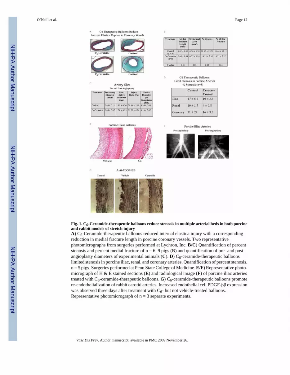
Fig. 1. C6-Ceramide-therapeutic balloons reduce stenosis in multiple arterial beds in both porcineand rabbit models of stretch injuryA) C6-Ceramide-therapeutic balloons reduced internal elastica injury with a correspondingreduction in medial fracture length in porcine coronary vessels. Two representativephotomicrographs from surgeries performed at Lychron, Inc. B/C) Quantification of percentstenosis and percent medial fracture of n = 6–9 pigs (B) and quantification of pre- and post-angioplasty diameters of experimental animals (C). D) C6-ceramide-therapeutic balloonslimited stenosis in porcine iliac, renal, and coronary arteries. Quantification of percent stenosis,n = 5 pigs. Surgeries performed at Penn State College of Medicine. E/F) Representative photo-micrograph of H & E stained sections (E) and radiological image (F) of porcine iliac arteriestreated with C6-ceramide-therapeutic balloons. G) C6-ceramide-therapeutic balloons promotere-endothelialization of rabbit carotid arteries. Increased endothelial cell PDGF-ββ expressionwas observed three days after treatment with C6- but not vehicle-treated balloons.Representative photomicrograph of n = 3 separate experiments.
O’Neill et al. Page 12
Vasc Dis Prev. Author manuscript; available in PMC 2009 November 26.
NIH
-PA Author Manuscript
NIH
-PA Author Manuscript
NIH
-PA Author Manuscript
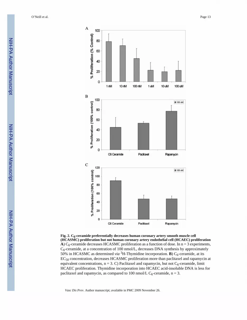
Fig. 2. C6-ceramide preferentially decreases human coronary artery smooth muscle cell(HCASMC) proliferation but not human coronary artery endothelial cell (HCAEC) proliferationA) C6-ceramide decreases HCASMC proliferation as a function of dose. In n = 3 experiments,C6-ceramide, at a concentration of 100 nmol/L, decreases DNA synthesis by approximately50% in HCASMC as determined via 3H-Thymidine incorporation. B) C6-ceramide, at itsEC50 concentration, decreases HCASMC proliferation more than paclitaxel and rapamycin atequivalent concentrations, n = 3. C) Paclitaxel and rapamycin, but not C6-ceramide, limitHCAEC proliferation. Thymidine incorporation into HCAEC acid-insoluble DNA is less forpaclitaxel and rapamycin, as compared to 100 nmol/L C6-ceramide, n = 3.
O’Neill et al. Page 13
Vasc Dis Prev. Author manuscript; available in PMC 2009 November 26.
NIH
-PA Author Manuscript
NIH
-PA Author Manuscript
NIH
-PA Author Manuscript
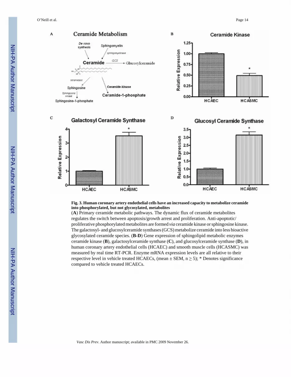
Fig. 3. Human coronary artery endothelial cells have an increased capacity to metabolize ceramideinto phosphorylated, but not glycosylated, metabolites(A) Primary ceramide metabolic pathways. The dynamic flux of ceramide metabolitesregulates the switch between apoptosis/growth arrest and proliferation. Anti-apoptotic/proliferative phosphorylated metabolites are formed via ceramide kinase or sphingosine kinase.The galactosyl- and glucosylceramide synthases (GCS) metabolize ceramide into less bioactiveglycosylated ceramide species. (B-D) Gene expression of sphingolipid metabolic enzymesceramide kinase (B), galactosylceramide synthase (C), and glucosylceramide synthase (D), inhuman coronary artery endothelial cells (HCAEC) and smooth muscle cells (HCASMC) wasmeasured by real time RT-PCR. Enzyme mRNA expression levels are all relative to theirrespective level in vehicle treated HCAECs, (mean ± SEM, n ≥ 5); * Denotes significancecompared to vehicle treated HCAECs.
O’Neill et al. Page 14
Vasc Dis Prev. Author manuscript; available in PMC 2009 November 26.
NIH
-PA Author Manuscript
NIH
-PA Author Manuscript
NIH
-PA Author Manuscript
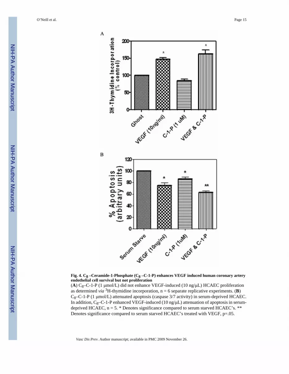
Fig. 4. C8 –Ceramide-1-Phosphate (C8 –C-1-P) enhances VEGF induced human coronary arteryendothelial cell survival but not proliferation(A) C8–C-1-P (1 μmol/L) did not enhance VEGF-induced (10 ng/μL) HCAEC proliferationas determined via 3H-thymidine incorporation, n = 6 separate replicative experiments. (B)C8–C-1-P (1 μmol/L) attenuated apoptosis (caspase 3/7 activity) in serum-deprived HCAEC.In addition, C8–C-1-P enhanced VEGF-induced (10 ng/μL) attenuation of apoptosis in serum-deprived HCAEC, n = 5. * Denotes significance compared to serum starved HCAEC’s. **Denotes significance compared to serum starved HCAEC’s treated with VEGF, p=.05.
O’Neill et al. Page 15
Vasc Dis Prev. Author manuscript; available in PMC 2009 November 26.
NIH
-PA Author Manuscript
NIH
-PA Author Manuscript
NIH
-PA Author Manuscript
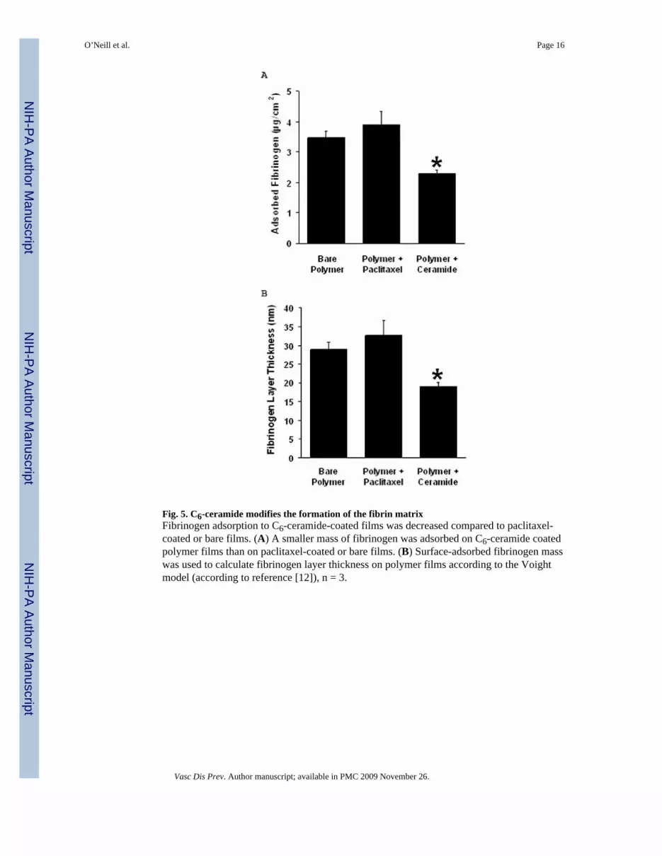
Fig. 5. C6-ceramide modifies the formation of the fibrin matrixFibrinogen adsorption to C6-ceramide-coated films was decreased compared to paclitaxel-coated or bare films. (A) A smaller mass of fibrinogen was adsorbed on C6-ceramide coatedpolymer films than on paclitaxel-coated or bare films. (B) Surface-adsorbed fibrinogen masswas used to calculate fibrinogen layer thickness on polymer films according to the Voightmodel (according to reference [12]), n = 3.
O’Neill et al. Page 16
Vasc Dis Prev. Author manuscript; available in PMC 2009 November 26.
NIH
-PA Author Manuscript
NIH
-PA Author Manuscript
NIH
-PA Author Manuscript
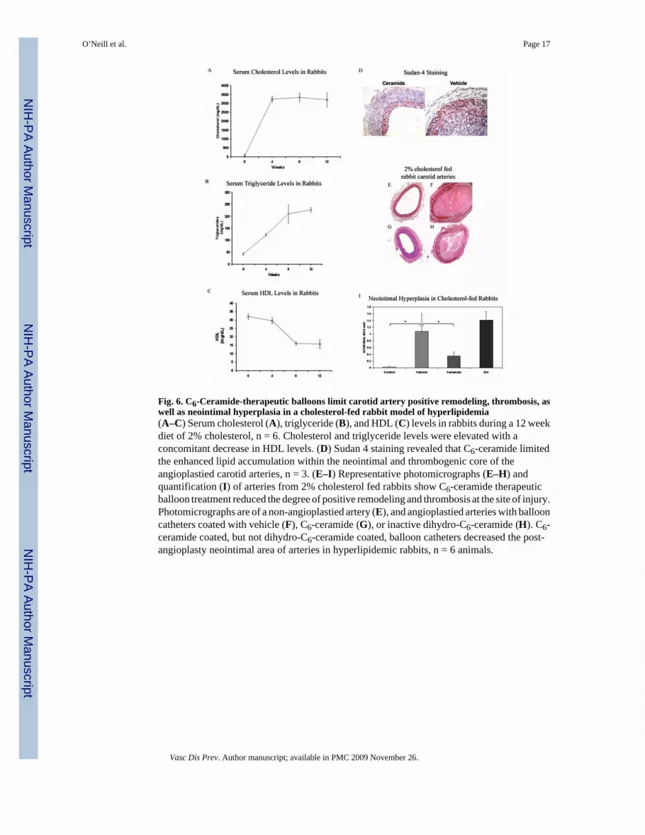
Fig. 6. C6-Ceramide-therapeutic balloons limit carotid artery positive remodeling, thrombosis, aswell as neointimal hyperplasia in a cholesterol-fed rabbit model of hyperlipidemia(A–C) Serum cholesterol (A), triglyceride (B), and HDL (C) levels in rabbits during a 12 weekdiet of 2% cholesterol, n = 6. Cholesterol and triglyceride levels were elevated with aconcomitant decrease in HDL levels. (D) Sudan 4 staining revealed that C6-ceramide limitedthe enhanced lipid accumulation within the neointimal and thrombogenic core of theangioplastied carotid arteries, n = 3. (E–I) Representative photomicrographs (E–H) andquantification (I) of arteries from 2% cholesterol fed rabbits show C6-ceramide therapeuticballoon treatment reduced the degree of positive remodeling and thrombosis at the site of injury.Photomicrographs are of a non-angioplastied artery (E), and angioplastied arteries with ballooncatheters coated with vehicle (F), C6-ceramide (G), or inactive dihydro-C6-ceramide (H). C6-ceramide coated, but not dihydro-C6-ceramide coated, balloon catheters decreased the post-angioplasty neointimal area of arteries in hyperlipidemic rabbits, n = 6 animals.
O’Neill et al. Page 17
Vasc Dis Prev. Author manuscript; available in PMC 2009 November 26.
NIH
-PA Author Manuscript
NIH
-PA Author Manuscript
NIH
-PA Author Manuscript
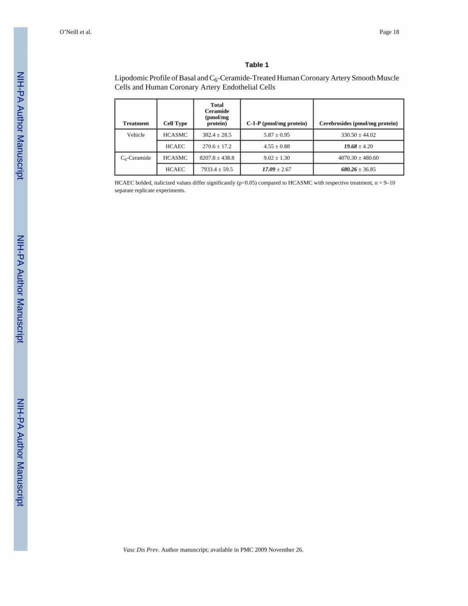
Table 1
Lipodomic Profile of Basal and C6-Ceramide-Treated Human Coronary Artery Smooth MuscleCells and Human Coronary Artery Endothelial Cells
Treatment Cell Type
TotalCeramide(pmol/mgprotein) C-1-P (pmol/mg protein) Cerebrosides (pmol/mg protein)
Vehicle HCASMC 382.4 ± 28.5 5.87 ± 0.95 330.50 ± 44.02
HCAEC 270.6 ± 17.2 4.55 ± 0.88 19.68 ± 4.20
C6-Ceramide HCASMC 8207.8 ± 438.8 9.02 ± 1.30 4070.30 ± 480.60
HCAEC 7933.4 ± 59.5 17.09 ± 2.67 680.26 ± 36.85
HCAEC bolded, italicized values differ significantly (p<0.05) compared to HCASMC with respective treatment, n = 9–10separate replicate experiments.
O’Neill et al. Page 18
Vasc Dis Prev. Author manuscript; available in PMC 2009 November 26.
NIH
-PA Author Manuscript
NIH
-PA Author Manuscript
NIH
-PA Author Manuscript
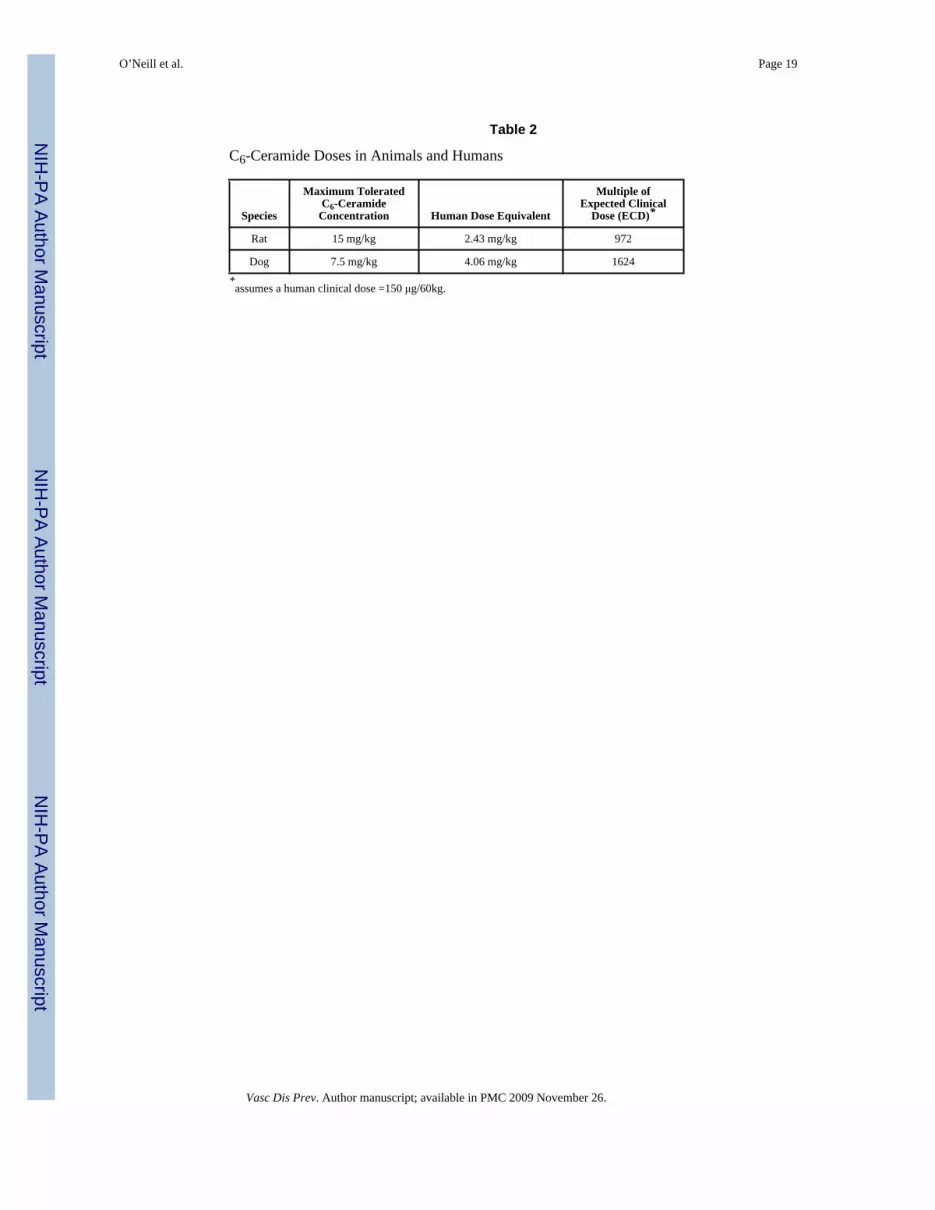
Table 2
C6-Ceramide Doses in Animals and Humans
Species
Maximum ToleratedC6-Ceramide
Concentration Human Dose Equivalent
Multiple ofExpected Clinical
Dose (ECD)*
Rat 15 mg/kg 2.43 mg/kg 972
Dog 7.5 mg/kg 4.06 mg/kg 1624
*assumes a human clinical dose =150 μg/60kg.
O’Neill et al. Page 19
Vasc Dis Prev. Author manuscript; available in PMC 2009 November 26.
NIH
-PA Author Manuscript
NIH
-PA Author Manuscript
NIH
-PA Author Manuscript


![[Reliability and survival of arterial catheters: optimal dynamic response]](https://img.dokumen.tips/doc/110x75/634de7d0d38be601b805f367/reliability-and-survival-of-arterial-catheters-optimal-dynamic-response.jpg)